© 2013 pearson education, inc. membrane transport: active processes two types of active processes...
TRANSCRIPT
© 2013 Pearson Education, Inc.
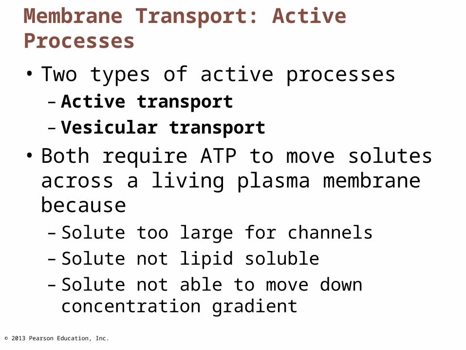
Membrane Transport: Active Processes
• Two types of active processes– Active transport– Vesicular transport
• Both require ATP to move solutes across a living plasma membrane because– Solute too large for channels– Solute not lipid soluble– Solute not able to move down concentration
gradient
© 2013 Pearson Education, Inc.
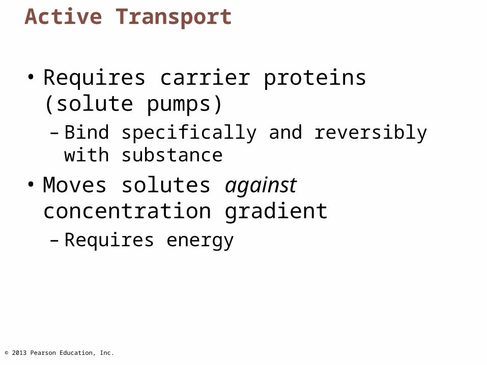
Active Transport
• Requires carrier proteins (solute pumps)– Bind specifically and reversibly with
substance
• Moves solutes against concentration gradient– Requires energy
© 2013 Pearson Education, Inc.
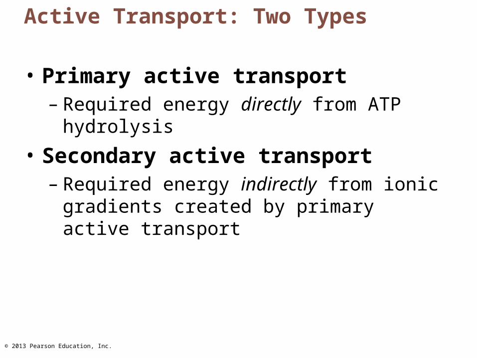
Active Transport: Two Types
• Primary active transport– Required energy directly from ATP hydrolysis
• Secondary active transport– Required energy indirectly from ionic
gradients created by primary active transport
© 2013 Pearson Education, Inc.
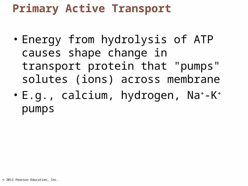
Primary Active Transport
• Energy from hydrolysis of ATP causes shape change in transport protein that "pumps" solutes (ions) across membrane
• E.g., calcium, hydrogen, Na+-K+ pumps
© 2013 Pearson Education, Inc.
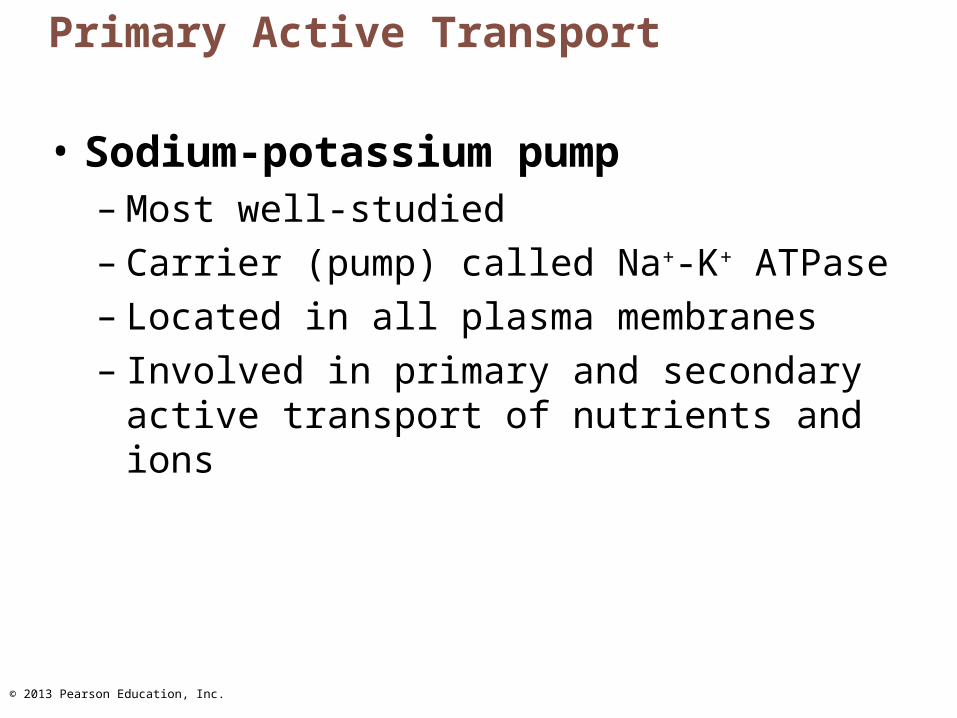
Primary Active Transport
• Sodium-potassium pump– Most well-studied– Carrier (pump) called Na+-K+ ATPase– Located in all plasma membranes– Involved in primary and secondary active
transport of nutrients and ions
© 2013 Pearson Education, Inc.
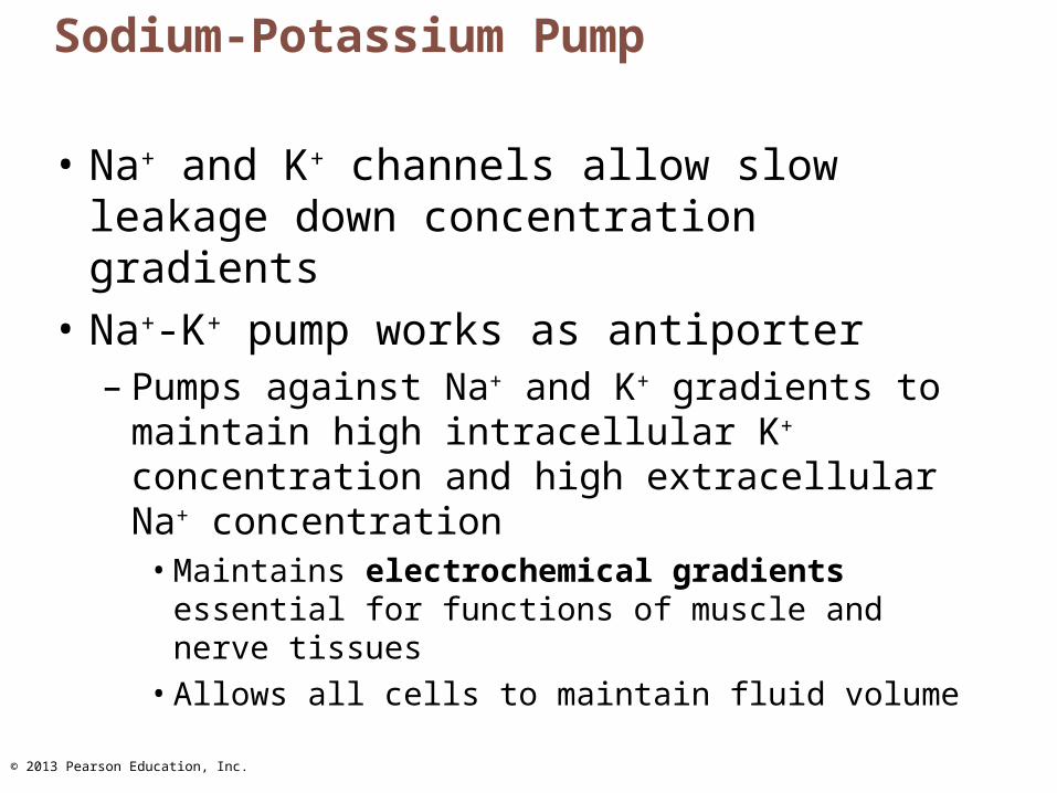
Sodium-Potassium Pump
• Na+ and K+ channels allow slow leakage down concentration gradients
• Na+-K+ pump works as antiporter– Pumps against Na+ and K+ gradients to
maintain high intracellular K+ concentration and high extracellular Na+ concentration
• Maintains electrochemical gradients essential for functions of muscle and nerve tissues
• Allows all cells to maintain fluid volume

© 2013 Pearson Education, Inc.
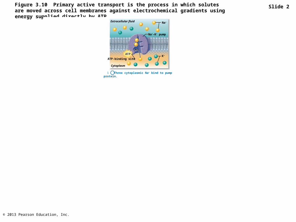
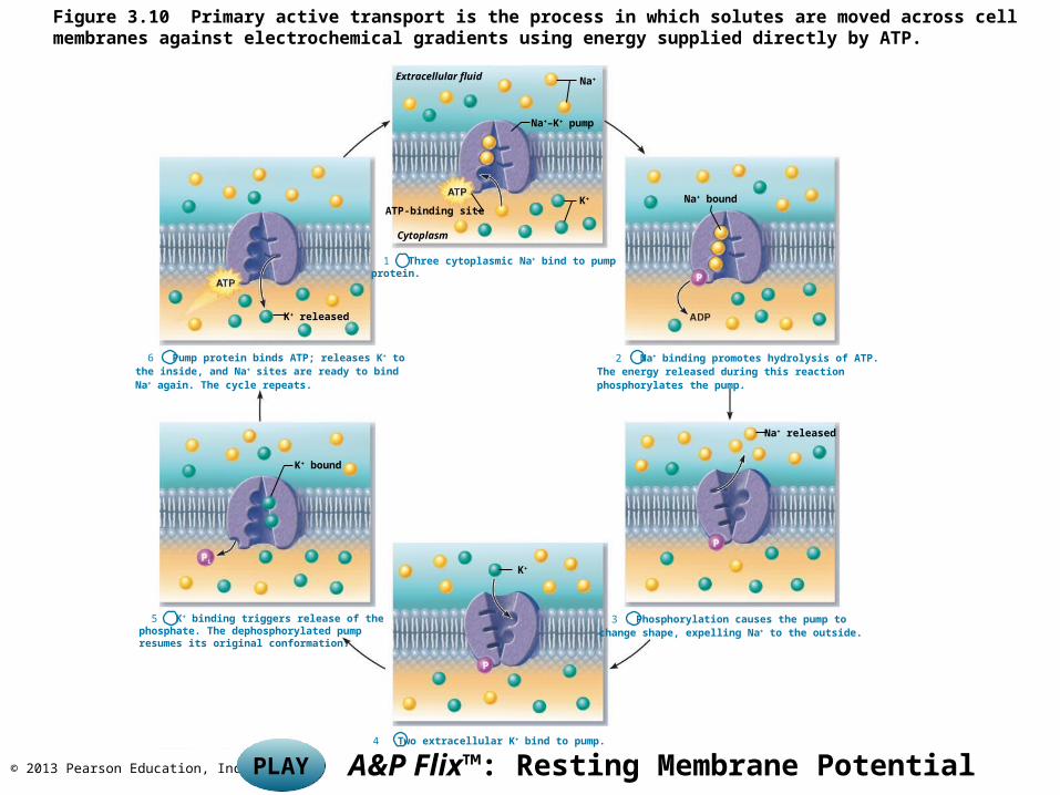
Figure 3.10 Primary active transport is the process in which solutes are moved across cell membranes against electrochemical gradients using energy supplied directly by ATP.
Slide 1
Extracellular fluid Na+
Na+–K+ pump
K+
ATP-binding site
Cytoplasm
1 Three cytoplasmic Na+ bind to pumpprotein.
K+ released
6 Pump protein binds ATP; releases K+ to the inside, and Na+ sites are ready to bind Na+ again. The cycle repeats.
2 Na+ binding promotes hydrolysis of ATP.The energy released during this reactionphosphorylates the pump.
K+ bound
5 K+ binding triggers release of thephosphate. The dephosphorylated pumpresumes its original conformation.
K+
4 Two extracellular K+ bind to pump.
3 Phosphorylation causes the pump tochange shape, expelling Na+ to the outside.
Na+ bound
Na+ released
P
P
P
Pi
© 2013 Pearson Education, Inc.
Figure 3.10 Primary active transport is the process in which solutes are moved across cell membranes against electrochemical gradients using energy supplied directly by ATP.
Slide 2
Extracellular fluid Na+
Na+–K+ pump
K+
Cytoplasm
1 Three cytoplasmic Na+ bind to pumpprotein.
ATP-binding site
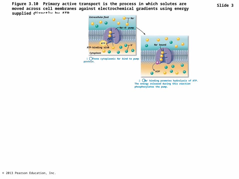
© 2013 Pearson Education, Inc.
Figure 3.10 Primary active transport is the process in which solutes are moved across cell membranes against electrochemical gradients using energy supplied directly by ATP.
Slide 3
Extracellular fluid Na+
Na+–K+ pump
K+
Cytoplasm
1 Three cytoplasmic Na+ bind to pumpprotein.
2 Na+ binding promotes hydrolysis of ATP.The energy released during this reactionphosphorylates the pump.
Na+ bound
P
ATP-binding site
© 2013 Pearson Education, Inc.
Figure 3.10 Primary active transport is the process in which solutes are moved across cell membranes against electrochemical gradients using energy supplied directly by ATP.
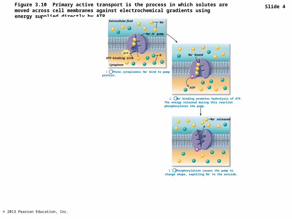
Slide 4
Extracellular fluid Na+
Na+–K+ pump
K+
Cytoplasm
1 Three cytoplasmic Na+ bind to pumpprotein.
2 Na+ binding promotes hydrolysis of ATP.The energy released during this reactionphosphorylates the pump.
3 Phosphorylation causes the pump tochange shape, expelling Na+ to the outside.
Na+ bound
Na+ released
P
P
ATP-binding site
© 2013 Pearson Education, Inc.
Figure 3.10 Primary active transport is the process in which solutes are moved across cell membranes against electrochemical gradients using energy supplied directly by ATP.
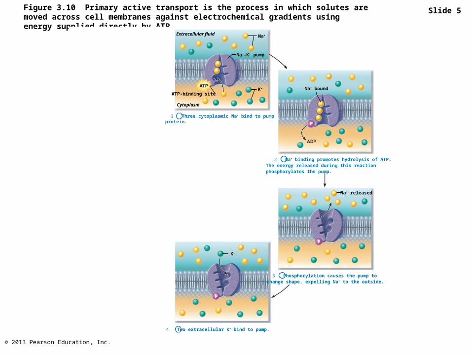
Slide 5
Extracellular fluid Na+
Na+–K+ pump
K+
Cytoplasm
1 Three cytoplasmic Na+ bind to pumpprotein.
2 Na+ binding promotes hydrolysis of ATP.The energy released during this reactionphosphorylates the pump.
K+
4 Two extracellular K+ bind to pump.
3 Phosphorylation causes the pump tochange shape, expelling Na+ to the outside.
Na+ bound
Na+ released
P
P
P
ATP-binding site
© 2013 Pearson Education, Inc.
Figure 3.10 Primary active transport is the process in which solutes are moved across cell membranes against electrochemical gradients using energy supplied directly by ATP.
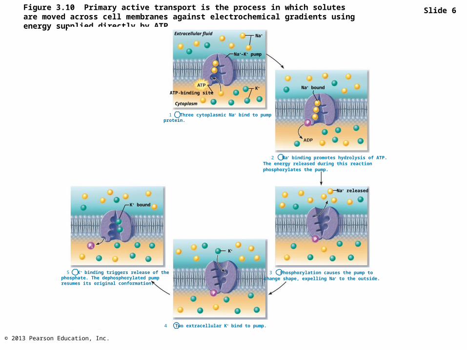
Slide 6
Extracellular fluid Na+
Na+–K+ pump
K+
Cytoplasm
1 Three cytoplasmic Na+ bind to pumpprotein.
2 Na+ binding promotes hydrolysis of ATP.The energy released during this reactionphosphorylates the pump.
K+ bound
5 K+ binding triggers release of thephosphate. The dephosphorylated pumpresumes its original conformation.
K+
4 Two extracellular K+ bind to pump.
3 Phosphorylation causes the pump tochange shape, expelling Na+ to the outside.
Na+ bound
Na+ released
P
P
P
Pi
ATP-binding site
© 2013 Pearson Education, Inc.
Figure 3.10 Primary active transport is the process in which solutes are moved across cell membranes against electrochemical gradients using energy supplied directly by ATP.
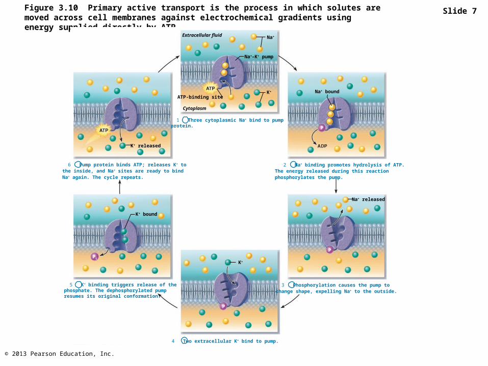
Slide 7
Extracellular fluid Na+
Na+–K+ pump
K+
Cytoplasm
1 Three cytoplasmic Na+ bind to pumpprotein.
K+ released
6 Pump protein binds ATP; releases K+ to the inside, and Na+ sites are ready to bind Na+ again. The cycle repeats.
2 Na+ binding promotes hydrolysis of ATP.The energy released during this reactionphosphorylates the pump.
K+ bound
5 K+ binding triggers release of thephosphate. The dephosphorylated pumpresumes its original conformation.
K+
4 Two extracellular K+ bind to pump.
3 Phosphorylation causes the pump tochange shape, expelling Na+ to the outside.
Na+ bound
Na+ released
P
P
P
Pi
ATP-binding site
© 2013 Pearson Education, Inc.
Figure 3.10 Primary active transport is the process in which solutes are moved across cellmembranes against electrochemical gradients using energy supplied directly by ATP.
PLAYPLAY A&P Flix™: Resting Membrane Potential
Extracellular fluid Na+
Na+–K+ pump
K+
Cytoplasm
1 Three cytoplasmic Na+ bind to pumpprotein.
K+ released
6 Pump protein binds ATP; releases K+ to the inside, and Na+ sites are ready to bind Na+ again. The cycle repeats.
2 Na+ binding promotes hydrolysis of ATP.The energy released during this reactionphosphorylates the pump.
K+ bound
5 K+ binding triggers release of thephosphate. The dephosphorylated pumpresumes its original conformation.
K+
4 Two extracellular K+ bind to pump.
3 Phosphorylation causes the pump tochange shape, expelling Na+ to the outside.
Na+ bound
Na+ released
P
P
P
Pi
ATP-binding site
© 2013 Pearson Education, Inc.
Secondary Active Transport
• Depends on ion gradient created by primary active transport
• Energy stored in ionic gradients used indirectly to drive transport of other solutes
© 2013 Pearson Education, Inc.
Secondary Active Transport
• Cotransport—always transports more than one substance at a time– Symport system: Substances transported in
same direction– Antiport system: Substances transported in
opposite directions
© 2013 Pearson Education, Inc.
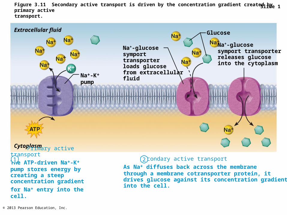
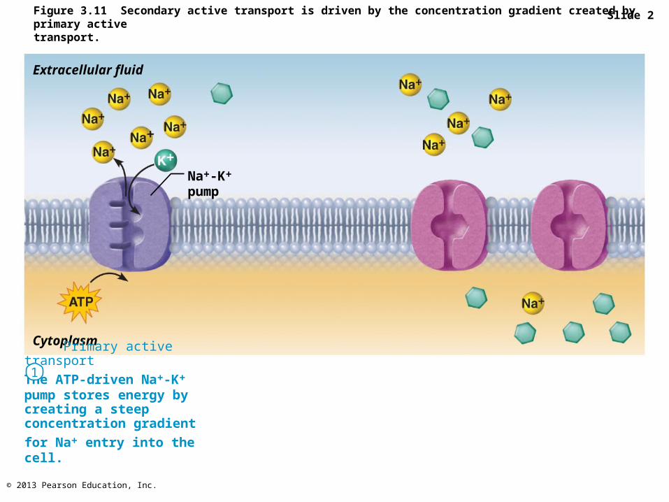
Figure 3.11 Secondary active transport is driven by the concentration gradient created by primary activetransport.
Extracellular fluid
Na+-glucosesymporttransporterloads glucosefrom extracellularfluid
Na+-glucosesymport transporterreleases glucoseinto the cytoplasm
Glucose
Na+-K+
pump
Cytoplasm
Primary active transport
The ATP-driven Na+-K+ pump stores energy by creating a steep concentration gradient
for Na+ entry into the cell.
Secondary active transport
As Na+ diffuses back across the membranethrough a membrane cotransporter protein, itdrives glucose against its concentration gradientinto the cell.
1 2
Slide 1
© 2013 Pearson Education, Inc.
Figure 3.11 Secondary active transport is driven by the concentration gradient created by primary activetransport.
Slide 2
Extracellular fluid
Na+-K+
pump
Cytoplasm
Primary active transport
The ATP-driven Na+-K+ pump stores energy by creating a steep concentration gradient
for Na+ entry into the cell.
1

© 2013 Pearson Education, Inc.
Figure 3.11 Secondary active transport is driven by the concentration gradient created by primary activetransport.
Slide 3
Extracellular fluid
Na+-glucosesymporttransporterloads glucosefrom extracellularfluid
Na+-glucosesymport transporterreleases glucoseinto the cytoplasm
Glucose
Na+-K+
pump
Cytoplasm
Primary active transport
The ATP-driven Na+-K+ pump stores energy by creating a steep concentration gradient
for Na+ entry into the cell.
Secondary active transport
As Na+ diffuses back across the membranethrough a membrane cotransporter protein, itdrives glucose against its concentration gradientinto the cell.
1 2
© 2013 Pearson Education, Inc.
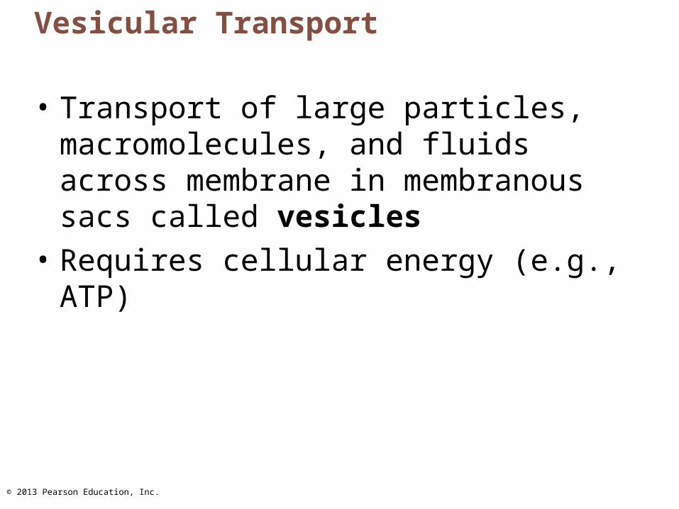
Vesicular Transport
• Transport of large particles, macromolecules, and fluids across membrane in membranous sacs called vesicles
• Requires cellular energy (e.g., ATP)
© 2013 Pearson Education, Inc.
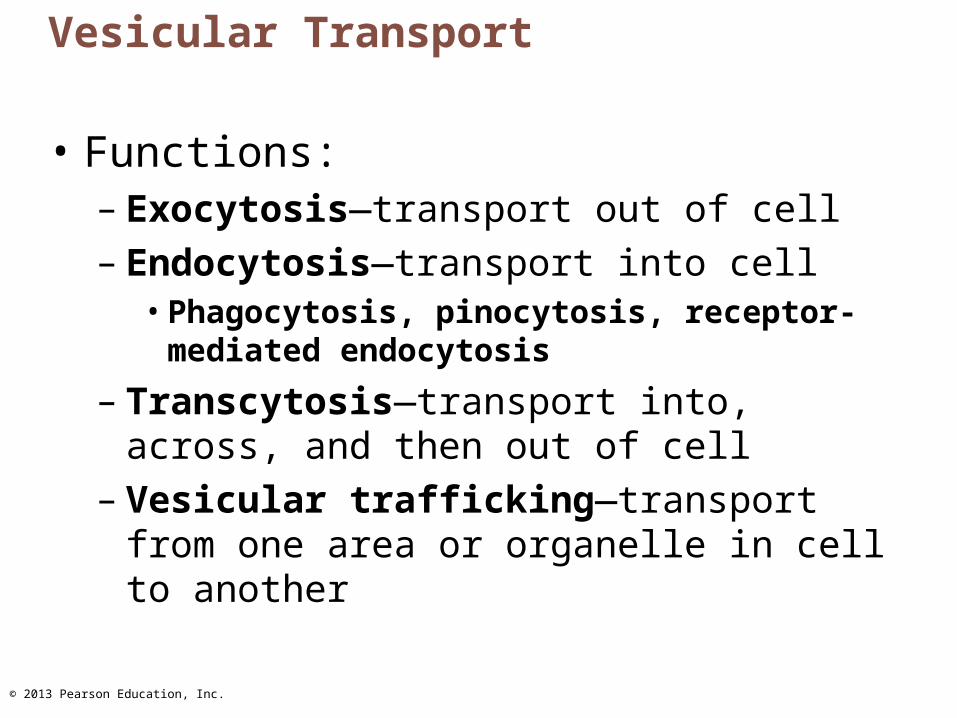
Vesicular Transport
• Functions:– Exocytosis—transport out of cell – Endocytosis—transport into cell
• Phagocytosis, pinocytosis, receptor-mediated endocytosis
– Transcytosis—transport into, across, and then out of cell
– Vesicular trafficking—transport from one area or organelle in cell to another
© 2013 Pearson Education, Inc.
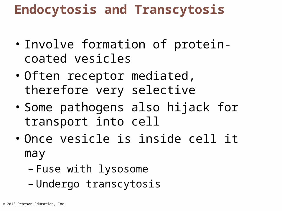
Endocytosis and Transcytosis
• Involve formation of protein-coated vesicles
• Often receptor mediated, therefore very selective
• Some pathogens also hijack for transport into cell
• Once vesicle is inside cell it may– Fuse with lysosome– Undergo transcytosis

© 2013 Pearson Education, Inc.
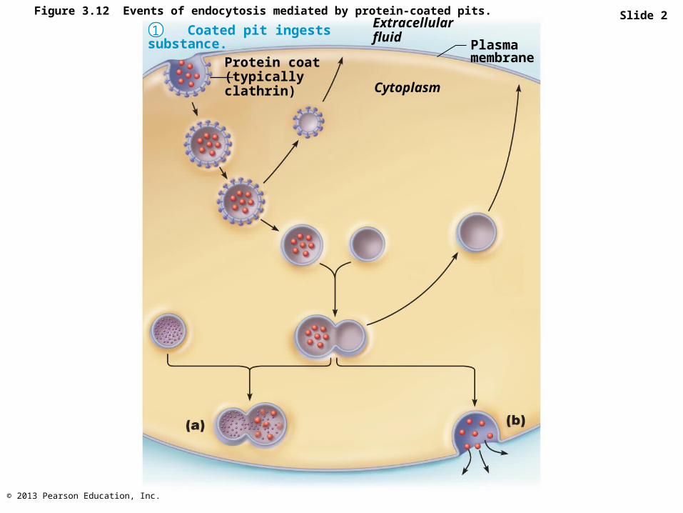
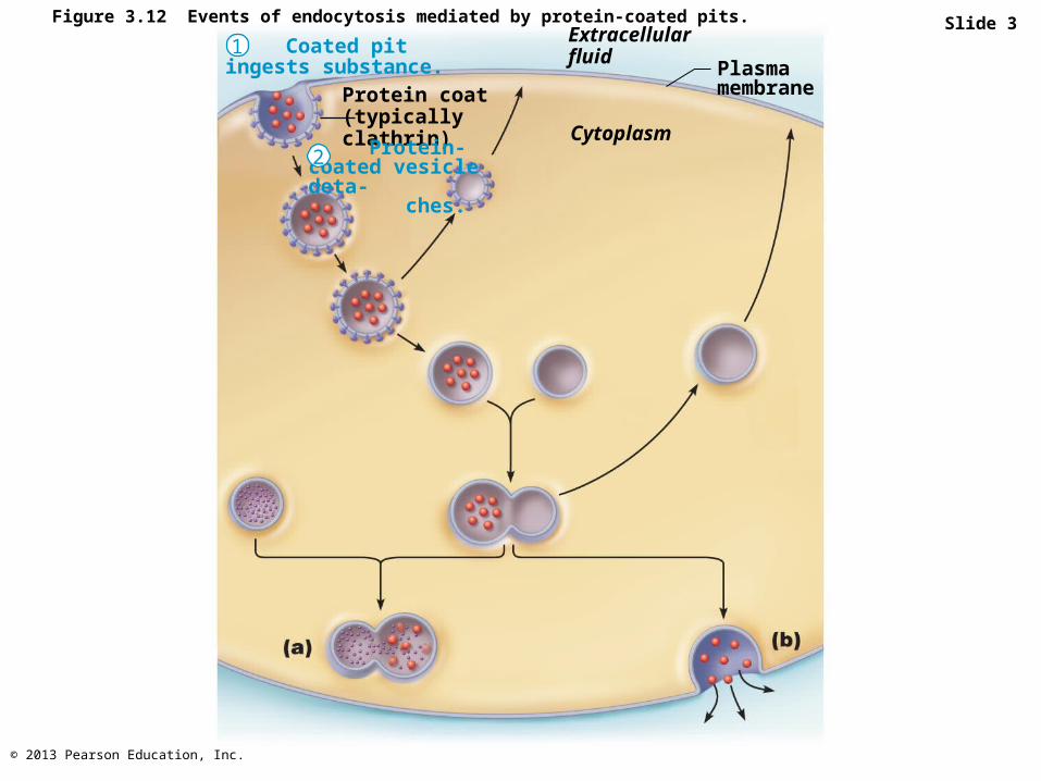
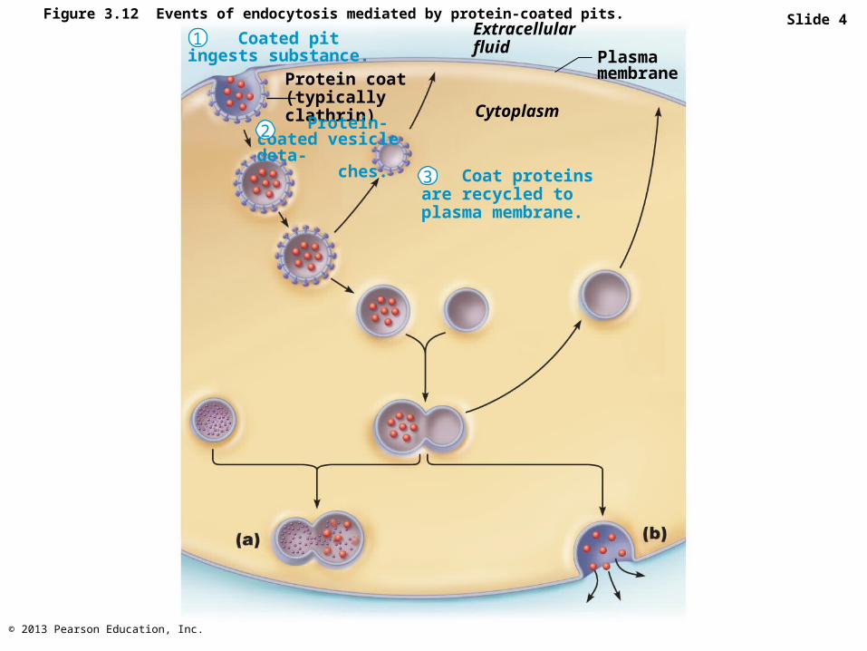
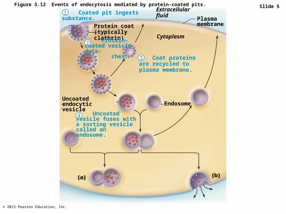
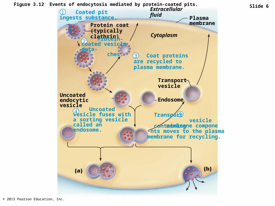
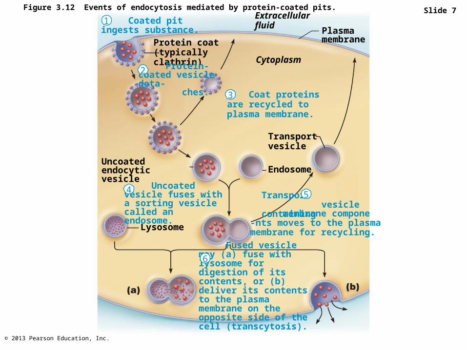
Figure 3.12 Events of endocytosis mediated by protein-coated pits. Slide 1 Coated pit ingests substance.
Coat proteins are recycled to plasma membrane.
1
Protein coat(typicallyclathrin)
Protein-coated vesicle deta- ches.
Transportvesicle
EndosomeUncoated endocyticvesicle
Transport vesicle containing
Uncoated vesicle fuses with a sorting vesicle called an endosome.
Fused vesicle may (a) fuse with lysosome for digestion of its contents, or (b) deliver its contents to the plasma membrane on the opposite side of the cell (transcytosis).
Extracellular fluid
Plasma membrane
Cytoplasm
Lysosome
2
membrane compone-nts moves to the plasmamembrane for recycling.
3
5 4
6
© 2013 Pearson Education, Inc.
Slide 2Figure 3.12 Events of endocytosis mediated by protein-coated pits.
Coated pit ingests substance. 1
Protein coat(typicallyclathrin)
Extracellular fluid
Plasma membrane
Cytoplasm
© 2013 Pearson Education, Inc.
Slide 3Figure 3.12 Events of endocytosis mediated by protein-coated pits.
Coated pit ingests substance. 1
Protein coat(typicallyclathrin)
Extracellular fluid
Plasma membrane
Protein-coated vesicle deta- ches.
2Cytoplasm
© 2013 Pearson Education, Inc.
Slide 4Figure 3.12 Events of endocytosis mediated by protein-coated pits.
Coated pit ingests substance. 1
Protein coat(typicallyclathrin)
Extracellular fluid
Plasma membrane
Protein-coated vesicle deta- ches.
2Cytoplasm
Coat proteins are recycled to plasma membrane.
3
© 2013 Pearson Education, Inc.
Slide 5Figure 3.12 Events of endocytosis mediated by protein-coated pits.
Coated pit ingests substance. 1
Protein coat(typicallyclathrin)
Extracellular fluid
Plasma membrane
Protein-coated vesicle deta- ches.
2Cytoplasm
Coat proteins are recycled to plasma membrane.
3
EndosomeUncoated endocyticvesicle
Uncoated vesicle fuses with a sorting vesicle called an endosome.
4
© 2013 Pearson Education, Inc.
Slide 6Figure 3.12 Events of endocytosis mediated by protein-coated pits.
Coated pit ingests substance. 1
Protein coat(typicallyclathrin)
Extracellular fluid
Plasma membrane
Protein-coated vesicle deta- ches.
2Cytoplasm
Coat proteins are recycled to plasma membrane.
3
EndosomeUncoated endocyticvesicle
Uncoated vesicle fuses with a sorting vesicle called an endosome.
4
Transportvesicle
membrane compone-nts moves to the plasmamembrane for recycling.
5 Transport vesicle containing
© 2013 Pearson Education, Inc.
Slide 7Figure 3.12 Events of endocytosis mediated by protein-coated pits.
Coated pit ingests substance.
Coat proteins are recycled to plasma membrane.
1
Protein coat(typicallyclathrin)
Protein-coated vesicle deta- ches.
Transportvesicle
EndosomeUncoated endocyticvesicle
Uncoated vesicle fuses with a sorting vesicle called an endosome.
Fused vesicle may (a) fuse with lysosome for digestion of its contents, or (b) deliver its contents to the plasma membrane on the opposite side of the cell (transcytosis).
Extracellular fluid
Plasma membrane
Cytoplasm
Lysosome
2
3
4
6
Transport vesicle containing membrane compone-nts moves to the plasmamembrane for recycling.
5
© 2013 Pearson Education, Inc.
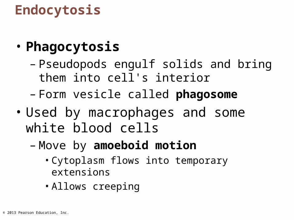
Endocytosis
• Phagocytosis– Pseudopods engulf solids and bring them into
cell's interior– Form vesicle called phagosome
• Used by macrophages and some white blood cells– Move by amoeboid motion
• Cytoplasm flows into temporary extensions• Allows creeping
© 2013 Pearson Education, Inc.
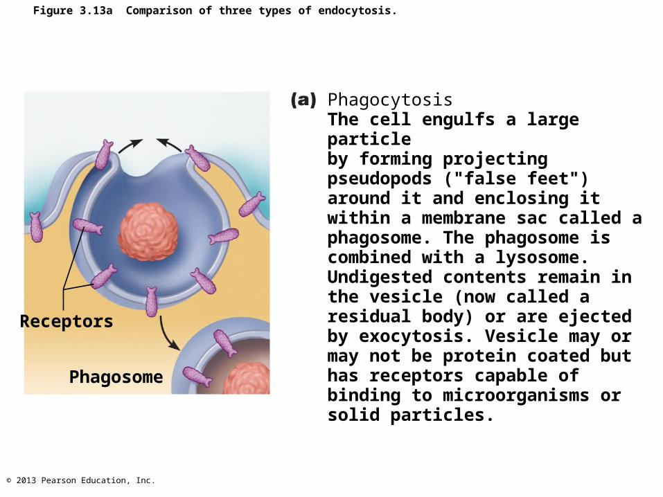
Figure 3.13a Comparison of three types of endocytosis.
Receptors
Phagosome
PhagocytosisThe cell engulfs a large particleby forming projecting pseudopods ("false feet") around it and enclosing it within a membrane sac called a phagosome. The phagosome is combined with a lysosome. Undigested contents remain inthe vesicle (now called a residual body) or are ejected by exocytosis. Vesicle may or may not be protein coated but has receptors capable of binding to microorganisms or solid particles.
© 2013 Pearson Education, Inc.
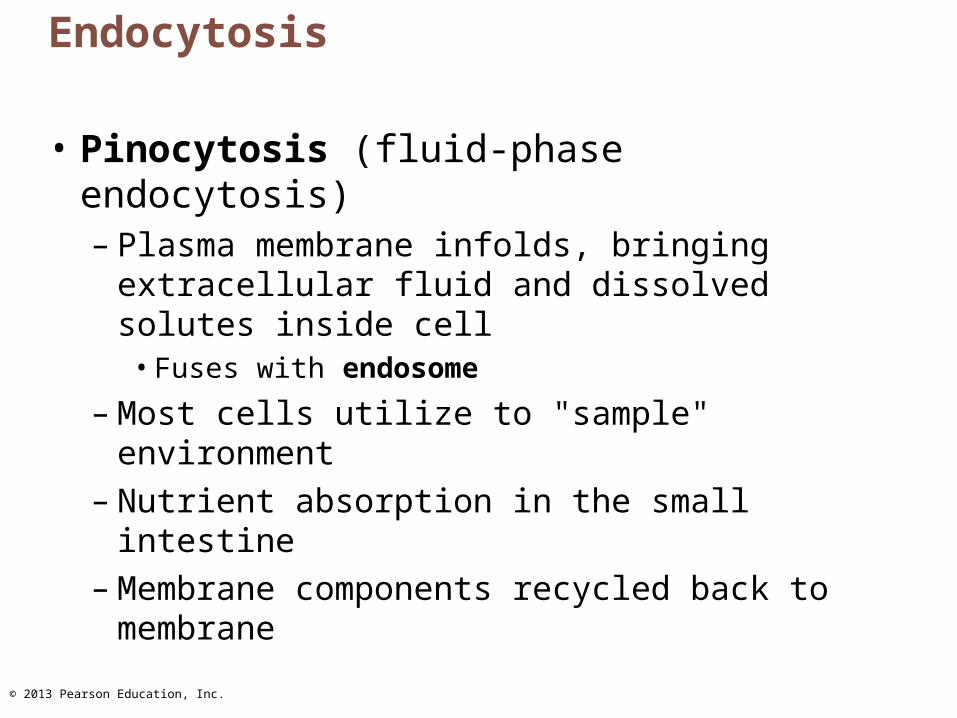
Endocytosis
• Pinocytosis (fluid-phase endocytosis)– Plasma membrane infolds, bringing
extracellular fluid and dissolved solutes inside cell
• Fuses with endosome
– Most cells utilize to "sample" environment – Nutrient absorption in the small intestine – Membrane components recycled back to
membrane
© 2013 Pearson Education, Inc.
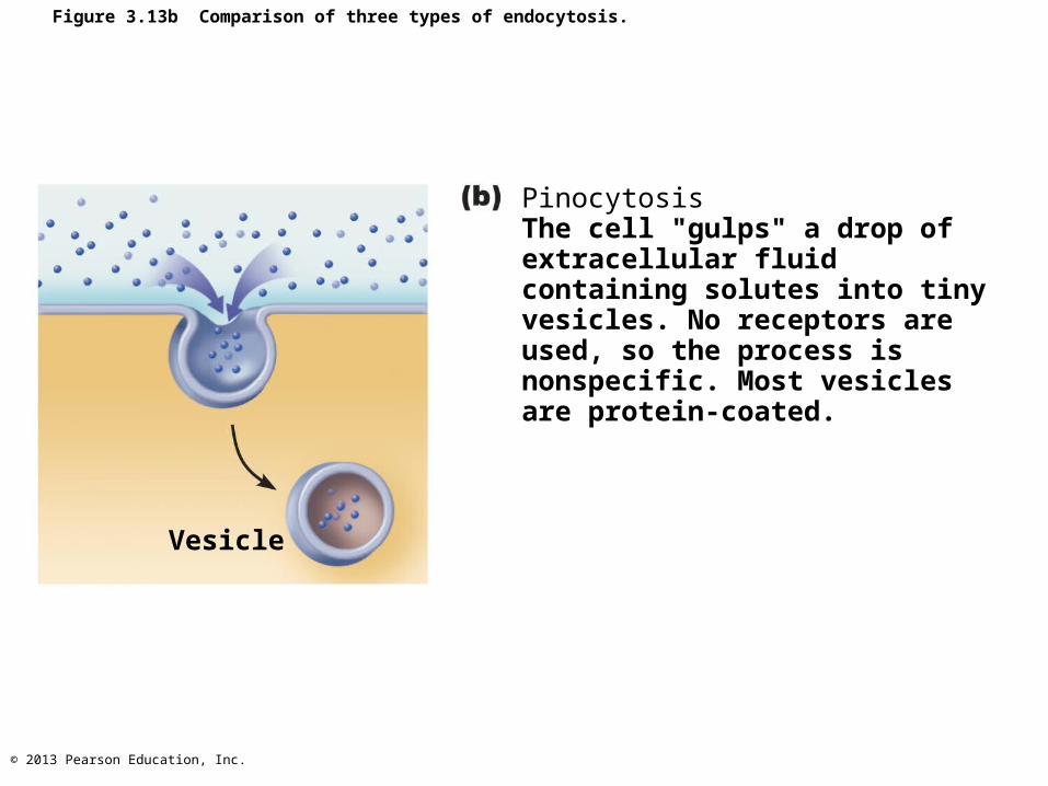
Figure 3.13b Comparison of three types of endocytosis.
Vesicle
PinocytosisThe cell "gulps" a drop of extracellular fluid containing solutes into tiny vesicles. No receptors are used, so the process is nonspecific. Most vesicles are protein-coated.
© 2013 Pearson Education, Inc.
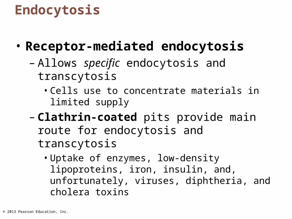
Endocytosis
• Receptor-mediated endocytosis– Allows specific endocytosis and transcytosis
• Cells use to concentrate materials in limited supply
– Clathrin-coated pits provide main route for endocytosis and transcytosis
• Uptake of enzymes, low-density lipoproteins, iron, insulin, and, unfortunately, viruses, diphtheria, and cholera toxins

© 2013 Pearson Education, Inc.
Receptor-Mediated Endocytosis
• Different coat proteins– Caveolae
• Capture specific molecules (folic acid, tetanus toxin) and use transcytosis
• Involved in cell signaling but exact function unknown
– Coatomer• Function in vesicular trafficking
© 2013 Pearson Education, Inc.
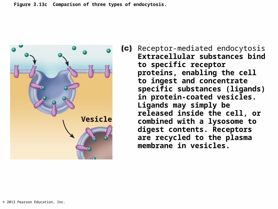
Figure 3.13c Comparison of three types of endocytosis.
Vesicle
Receptor-mediated endocytosisExtracellular substances bind to specific receptor proteins, enabling the cell to ingest and concentrate specific substances (ligands) in protein-coated vesicles. Ligands may simply be released inside the cell, or combined with a lysosome to digest contents. Receptors are recycled to the plasma membrane in vesicles.
© 2013 Pearson Education, Inc.
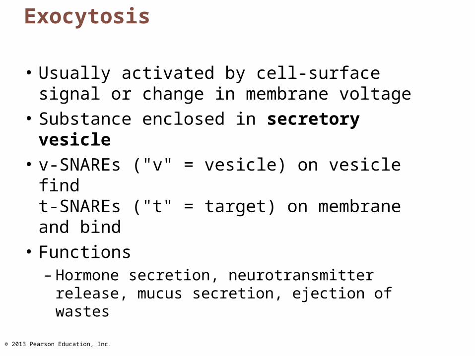
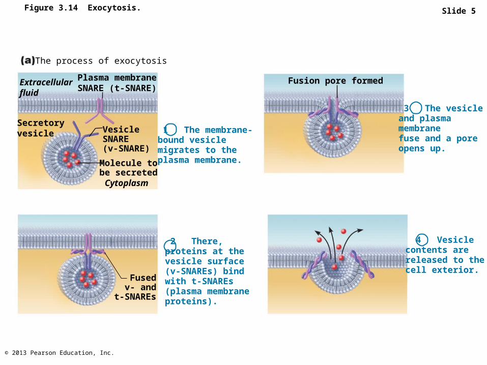
Exocytosis
• Usually activated by cell-surface signal or change in membrane voltage
• Substance enclosed in secretory vesicle
• v-SNAREs ("v" = vesicle) on vesicle findt-SNAREs ("t" = target) on membrane and bind
• Functions– Hormone secretion, neurotransmitter release,
mucus secretion, ejection of wastes

© 2013 Pearson Education, Inc.
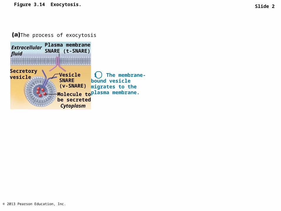
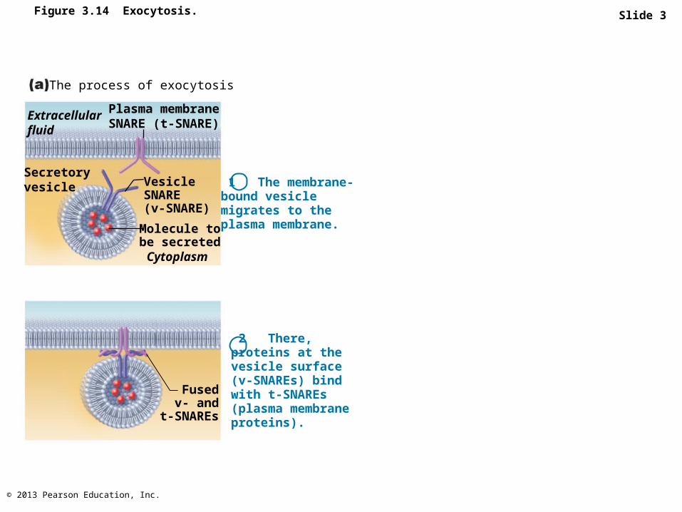
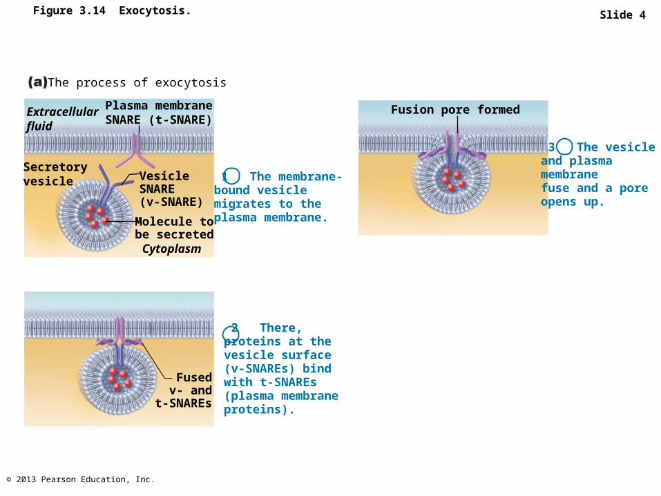
Figure 3.14 Exocytosis. Slide 1
Extracellularfluid
Plasma membraneSNARE (t-SNARE)
The process of exocytosis
Secretoryvesicle Vesicle
SNARE(v-SNARE)
Molecule tobe secretedCytoplasm
1 The membrane-bound vesiclemigrates to theplasma membrane.
2 There, proteins at the vesicle surface (v-SNAREs) bind with t-SNAREs (plasma membrane proteins).
Fusedv- and
t-SNAREs
3 The vesicle and plasma membranefuse and a poreopens up.
Fusion pore formed
4 Vesicle contents are released to thecell exterior.
© 2013 Pearson Education, Inc.
Slide 2Figure 3.14 Exocytosis.
Extracellularfluid
Plasma membraneSNARE (t-SNARE)
The process of exocytosis
Secretoryvesicle Vesicle
SNARE(v-SNARE)
Molecule tobe secretedCytoplasm
1 The membrane-bound vesiclemigrates to theplasma membrane.
© 2013 Pearson Education, Inc.
Slide 3Figure 3.14 Exocytosis.
Extracellularfluid
Plasma membraneSNARE (t-SNARE)
The process of exocytosis
Secretoryvesicle Vesicle
SNARE(v-SNARE)
Molecule tobe secretedCytoplasm
1 The membrane-bound vesiclemigrates to theplasma membrane.
2 There, proteins at the vesicle surface (v-SNAREs) bind with t-SNAREs (plasma membrane proteins).
Fusedv- and
t-SNAREs
© 2013 Pearson Education, Inc.
Slide 4Figure 3.14 Exocytosis.
Extracellularfluid
Plasma membraneSNARE (t-SNARE)
The process of exocytosis
Secretoryvesicle Vesicle
SNARE(v-SNARE)
Molecule tobe secretedCytoplasm
1 The membrane-bound vesiclemigrates to theplasma membrane.
2 There, proteins at the vesicle surface (v-SNAREs) bind with t-SNAREs (plasma membrane proteins).
Fusedv- and
t-SNAREs
3 The vesicle and plasma membranefuse and a poreopens up.
Fusion pore formed
© 2013 Pearson Education, Inc.
Slide 5Figure 3.14 Exocytosis.
Extracellularfluid
Plasma membraneSNARE (t-SNARE)
The process of exocytosis
Secretoryvesicle Vesicle
SNARE(v-SNARE)
Molecule tobe secretedCytoplasm
1 The membrane-bound vesiclemigrates to theplasma membrane.
2 There, proteins at the vesicle surface (v-SNAREs) bind with t-SNAREs (plasma membrane proteins).
Fusedv- and
t-SNAREs
3 The vesicle and plasma membranefuse and a poreopens up.
Fusion pore formed
4 Vesicle contents are released to thecell exterior.
© 2013 Pearson Education, Inc.
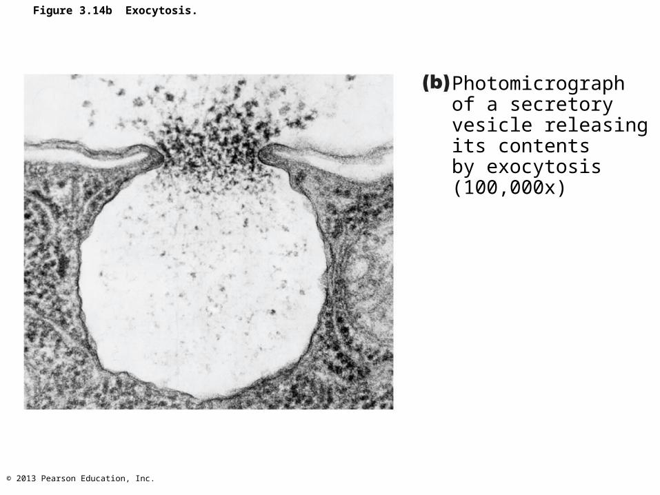
Figure 3.14b Exocytosis.
Photomicrograph of a secretoryvesicle releasingits contents by exocytosis (100,000x)
© 2013 Pearson Education, Inc.
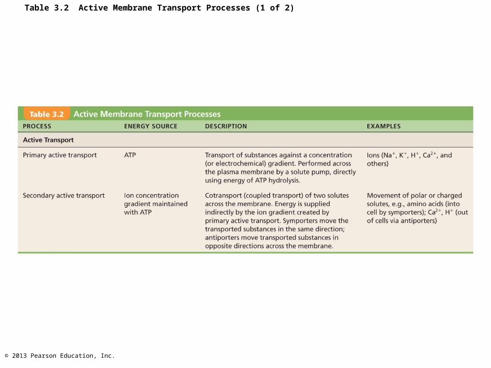
Table 3.2 Active Membrane Transport Processes (1 of 2)
© 2013 Pearson Education, Inc.
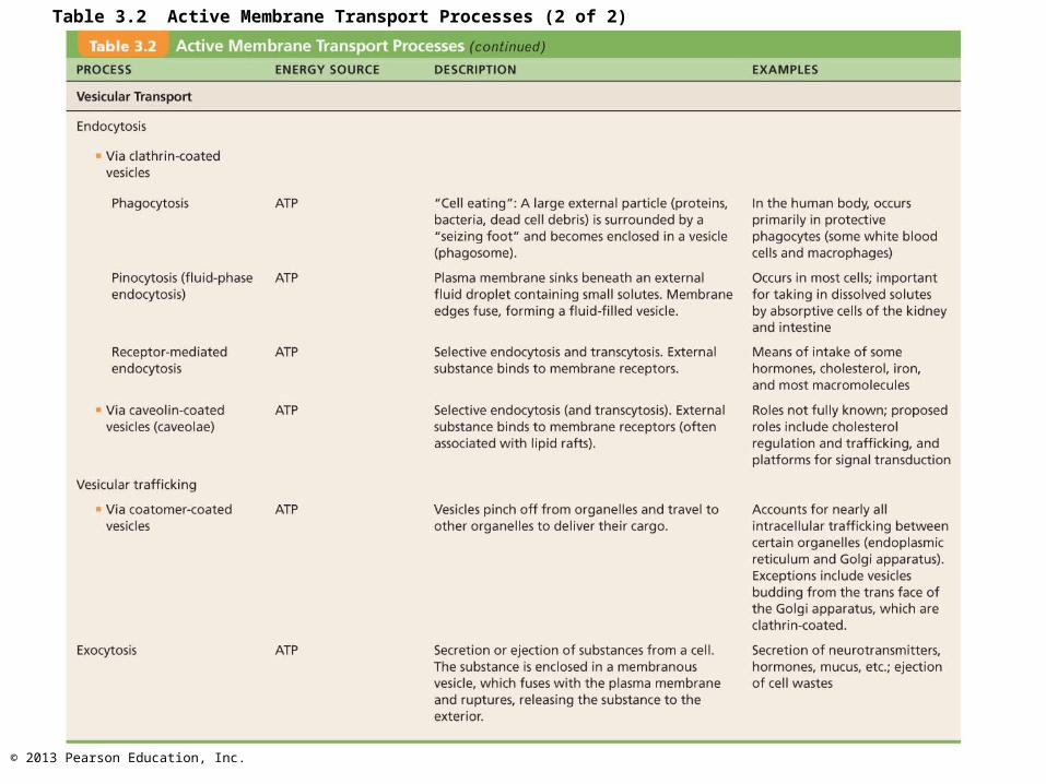
Table 3.2 Active Membrane Transport Processes (2 of 2)
© 2013 Pearson Education, Inc.
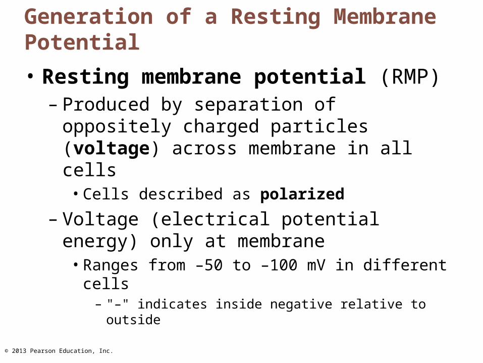
Generation of a Resting Membrane Potential
• Resting membrane potential (RMP)– Produced by separation of oppositely charged
particles (voltage) across membrane in all cells
• Cells described as polarized
– Voltage (electrical potential energy) only at membrane
• Ranges from –50 to –100 mV in different cells– "–" indicates inside negative relative to outside
© 2013 Pearson Education, Inc.
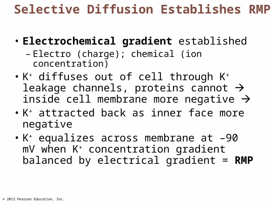
Selective Diffusion Establishes RMP
• Electrochemical gradient established– Electro (charge); chemical (ion concentration)
• K+ diffuses out of cell through K+ leakage channels, proteins cannot inside cell membrane more negative
• K+ attracted back as inner face more negative
• K+ equalizes across membrane at –90 mV when K+ concentration gradient balanced by electrical gradient = RMP
© 2013 Pearson Education, Inc.
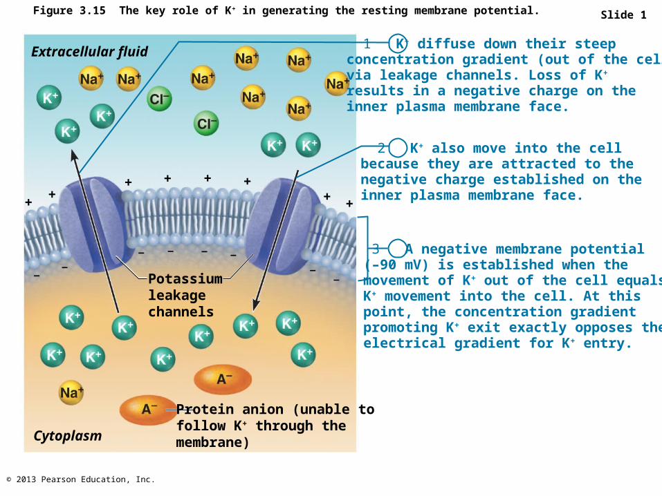
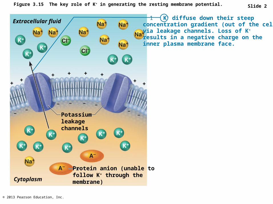
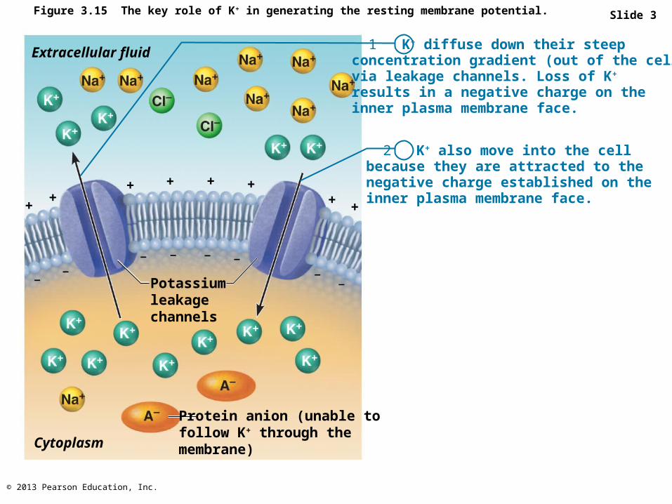
Figure 3.15 The key role of K+ in generating the resting membrane potential.
1 K+ diffuse down their steepconcentration gradient (out of the cell)via leakage channels. Loss of K+
results in a negative charge on theinner plasma membrane face.
2 K+ also move into the cellbecause they are attracted to thenegative charge established on theinner plasma membrane face.
3 A negative membrane potential(–90 mV) is established when themovement of K+ out of the cell equalsK+ movement into the cell. At thispoint, the concentration gradientpromoting K+ exit exactly opposes theelectrical gradient for K+ entry.
Extracellular fluid
Potassiumleakagechannels
Protein anion (unable tofollow K+ through themembrane)Cytoplasm
++
+ + + ++ +
––
–––––
–
Slide 1
© 2013 Pearson Education, Inc.
Figure 3.15 The key role of K+ in generating the resting membrane potential.
1 K+ diffuse down their steepconcentration gradient (out of the cell)via leakage channels. Loss of K+
results in a negative charge on theinner plasma membrane face.
Extracellular fluid
Potassiumleakagechannels
Protein anion (unable tofollow K+ through themembrane)Cytoplasm
++
+ + + ++ +
––
–––––
–
Slide 2
© 2013 Pearson Education, Inc.
Figure 3.15 The key role of K+ in generating the resting membrane potential.
1 K+ diffuse down their steepconcentration gradient (out of the cell)via leakage channels. Loss of K+
results in a negative charge on theinner plasma membrane face.
2 K+ also move into the cellbecause they are attracted to thenegative charge established on theinner plasma membrane face.
Extracellular fluid
Potassiumleakagechannels
Protein anion (unable tofollow K+ through themembrane)Cytoplasm
++
+ + + ++ +
––
–––––
–
Slide 3

© 2013 Pearson Education, Inc.
Figure 3.15 The key role of K+ in generating the resting membrane potential.
1 K+ diffuse down their steepconcentration gradient (out of the cell)via leakage channels. Loss of K+
results in a negative charge on theinner plasma membrane face.
2 K+ also move into the cellbecause they are attracted to thenegative charge established on theinner plasma membrane face.
3 A negative membrane potential(–90 mV) is established when themovement of K+ out of the cell equalsK+ movement into the cell. At thispoint, the concentration gradientpromoting K+ exit exactly opposes theelectrical gradient for K+ entry.
Extracellular fluid
Potassiumleakagechannels
Protein anion (unable tofollow K+ through themembrane)Cytoplasm
++
+ + + ++ +
––
–––––
–
Slide 4
© 2013 Pearson Education, Inc.
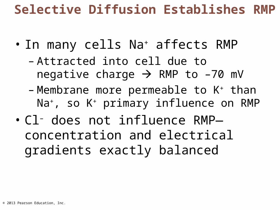
Selective Diffusion Establishes RMP
• In many cells Na+ affects RMP– Attracted into cell due to negative charge
RMP to –70 mV – Membrane more permeable to K+ than Na+, so
K+ primary influence on RMP
• Cl– does not influence RMP—concentration and electrical gradients exactly balanced
© 2013 Pearson Education, Inc.
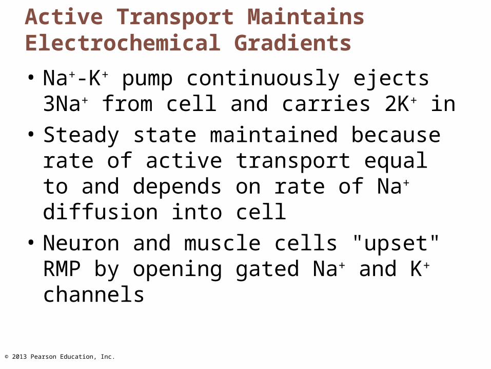
Active Transport Maintains Electrochemical Gradients
• Na+-K+ pump continuously ejects 3Na+ from cell and carries 2K+ in
• Steady state maintained because rate of active transport equal to and depends on rate of Na+ diffusion into cell
• Neuron and muscle cells "upset" RMP by opening gated Na+ and K+ channels
© 2013 Pearson Education, Inc.
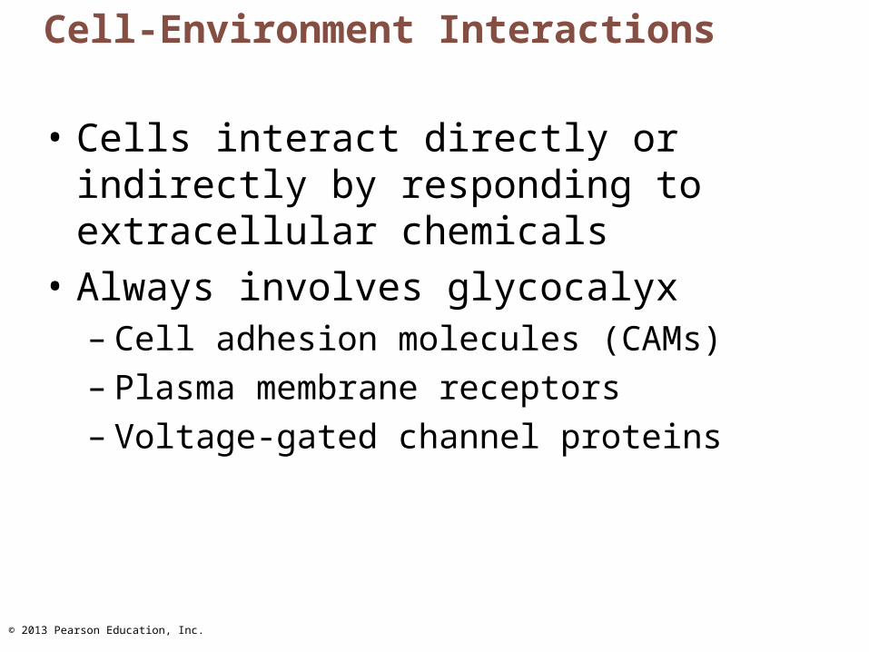
Cell-Environment Interactions
• Cells interact directly or indirectly by responding to extracellular chemicals
• Always involves glycocalyx– Cell adhesion molecules (CAMs)– Plasma membrane receptors– Voltage-gated channel proteins

© 2013 Pearson Education, Inc.
Roles of Cell Adhesion Molecules
• Thousands on approximately every cell in body• Anchor to extracellular matrix or each other• Assist in movement of cells past one another• Attract WBCs to injured or infected areas• Stimulate synthesis or degradation of adhesive
membrane junctions• Transmit intracellular signals to direct cell
migration, proliferation, and specialization
© 2013 Pearson Education, Inc.
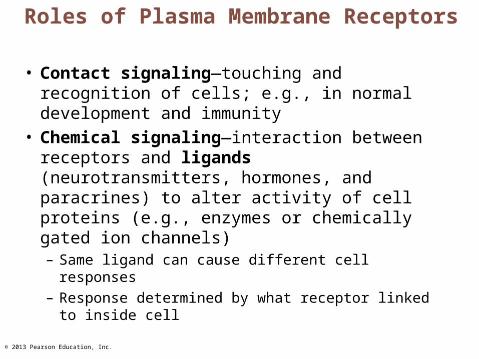
Roles of Plasma Membrane Receptors
• Contact signaling—touching and recognition of cells; e.g., in normal development and immunity
• Chemical signaling—interaction between receptors and ligands (neurotransmitters, hormones, and paracrines) to alter activity of cell proteins (e.g., enzymes or chemically gated ion channels)– Same ligand can cause different cell responses– Response determined by what receptor linked to
inside cell
© 2013 Pearson Education, Inc.
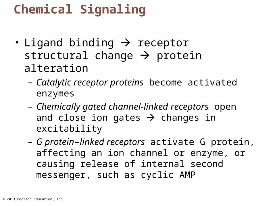
Chemical Signaling
• Ligand binding receptor structural change protein alteration– Catalytic receptor proteins become activated
enzymes– Chemically gated channel-linked receptors open and
close ion gates changes in excitability– G protein–linked receptors activate G protein,
affecting an ion channel or enzyme, or causing release of internal second messenger, such as cyclic AMP
© 2013 Pearson Education, Inc.
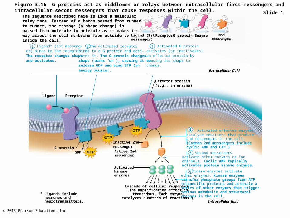
Figure 3.16 G proteins act as middlemen or relays between extracellular first messengers and intracellular second messengers that cause responses within the cell.
Ligand (1st messenger)
Receptor G protein Enzyme 2ndmessenger
Ligand* (1st messeng-er) binds to the receptor. The receptor changes shape and activates.
The activated receptorbinds to a G protein and acti-vates it. The G protein changesshape (turns “on”), causing it torelease GDP and bind GTP (an energy source).
Activated G proteinactivates (or inactivates) an effector protein by causing its shape tochange.
Effector protein(e.g., an enzyme)
Extracellular fluid
G proteinGDP
Intracellular fluid
Cascade of cellular responses (The amplification effect istremendous. Each enzyme
catalyzes hundreds of reactions.)
Activatedkinaseenzymes
Active 2ndmessenger
Inactive 2nd messenger
Activated effector enzymescatalyze reactions that produce2nd messengers in the cell.(Common 2nd messengers includecyclic AMP and Ca2+.)
Second messengersactivate other enzymes or ionchannels. Cyclic AMP typicallyactivates protein kinase enzymes.
Kinase enzymes activateother enzymes. Kinase enzymestransfer phosphate groups from ATPto specific proteins and activate aseries of other enzymes that triggervarious metabolic and structuralchanges in the cell.
* Ligands include hormones and neurotransmitters.
ReceptorLigand
1 2 3
4
5
6
Slide 1The sequence described here is like a molecularrelay race. Instead of a baton passed from runnerto runner, the message (a shape change) ispassed from molecule to molecule as it makes itsway across the cell membrane from outside toinside the cell.
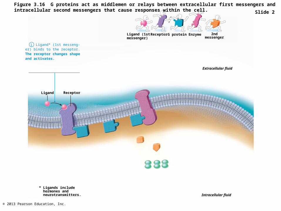
© 2013 Pearson Education, Inc.
Figure 3.16 G proteins act as middlemen or relays between extracellular first messengers and intracellular second messengers that cause responses within the cell.
Ligand (1st messenger)
Receptor G protein Enzyme 2ndmessenger
Ligand* (1st messeng-er) binds to the receptor. The receptor changes shape and activates.
Extracellular fluid
Intracellular fluid
* Ligands include hormones and neurotransmitters.
ReceptorLigand
1
Slide 2
© 2013 Pearson Education, Inc.
Figure 3.16 G proteins act as middlemen or relays between extracellular first messengers and intracellular second messengers that cause responses within the cell.
Ligand (1st messenger)
Receptor G protein Enzyme 2ndmessenger
Ligand* (1st messeng-er) binds to the receptor. The receptor changes shape and activates.
Extracellular fluid
Intracellular fluid
* Ligands include hormones and neurotransmitters.
ReceptorLigand
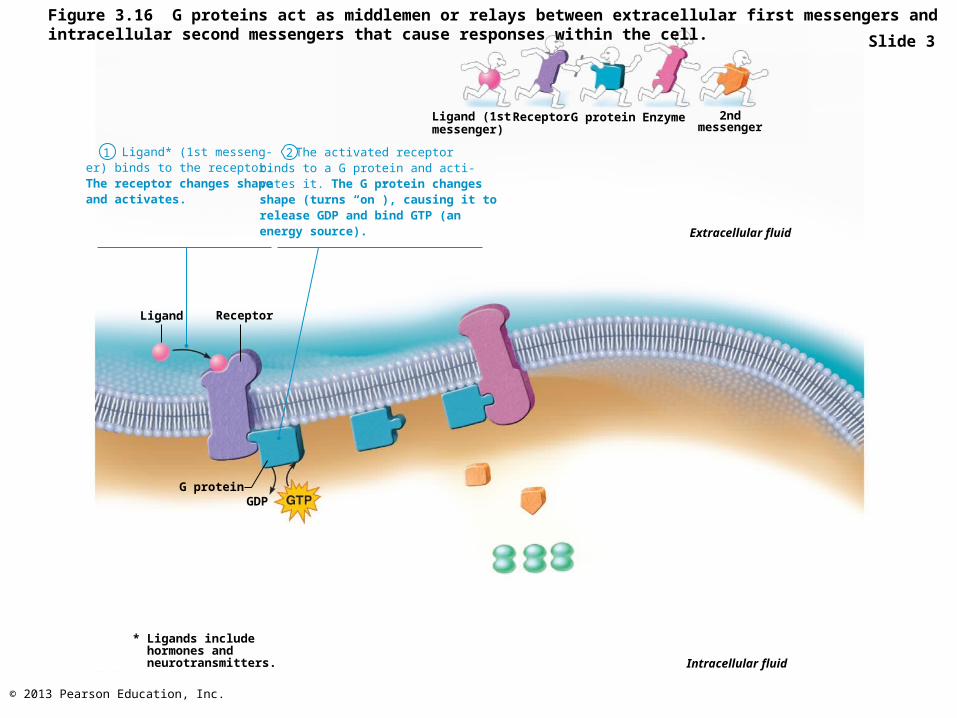
1 The activated receptorbinds to a G protein and acti-vates it. The G protein changesshape (turns “on”), causing it torelease GDP and bind GTP (an energy source).
G proteinGDP
2
Slide 3
© 2013 Pearson Education, Inc.
Figure 3.16 G proteins act as middlemen or relays between extracellular first messengers and intracellular second messengers that cause responses within the cell.
Ligand (1st messenger)
Receptor G protein Enzyme 2ndmessenger
Ligand* (1st messeng-er) binds to the receptor. The receptor changes shape and activates.
Extracellular fluid
Intracellular fluid
* Ligands include hormones and neurotransmitters.
ReceptorLigand
1 The activated receptorbinds to a G protein and acti-vates it. The G protein changesshape (turns “on”), causing it torelease GDP and bind GTP (an energy source).
G proteinGDP
Activated G proteinactivates (or inactivates) an effector protein by causing its shape tochange.
Effector protein(e.g., an enzyme)
2 3
Slide 4
© 2013 Pearson Education, Inc.
Figure 3.16 G proteins act as middlemen or relays between extracellular first messengers and intracellular second messengers that cause responses within the cell.
Ligand (1st messenger)
Receptor G protein Enzyme 2ndmessenger
Ligand* (1st messeng-er) binds to the receptor. The receptor changes shape and activates.
Extracellular fluid
Intracellular fluid
* Ligands include hormones and neurotransmitters.
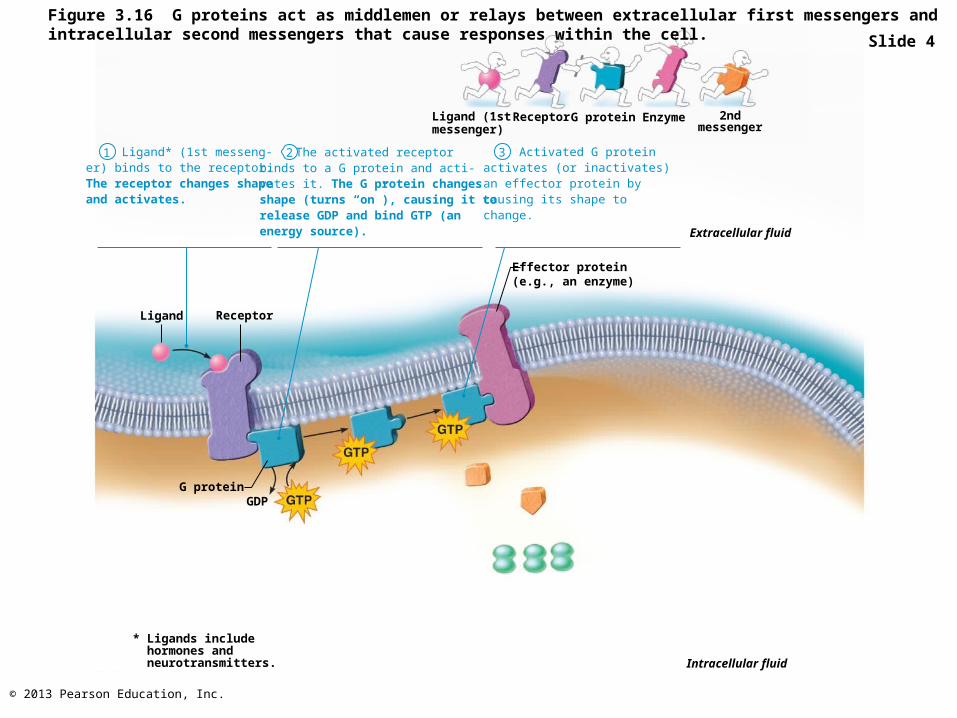
ReceptorLigand
1 The activated receptorbinds to a G protein and acti-vates it. The G protein changesshape (turns “on”), causing it torelease GDP and bind GTP (an energy source).
G proteinGDP
Activated G proteinactivates (or inactivates) an effector protein by causing its shape tochange.
Effector protein(e.g., an enzyme)
2 3
Active 2ndmessenger
Inactive 2nd messenger
Activated effector enzymescatalyze reactions that produce2nd messengers in the cell.(Common 2nd messengers includecyclic AMP and Ca2+.)
4
Slide 5
© 2013 Pearson Education, Inc.
Figure 3.16 G proteins act as middlemen or relays between extracellular first messengers and intracellular second messengers that cause responses within the cell.
Ligand (1st messenger)
Receptor G protein Enzyme 2ndmessenger
Ligand* (1st messeng-er) binds to the receptor. The receptor changes shape and activates.
Extracellular fluid
Intracellular fluid
* Ligands include hormones and neurotransmitters.
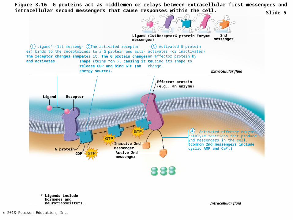
ReceptorLigand
1 The activated receptorbinds to a G protein and acti-vates it. The G protein changesshape (turns “on”), causing it torelease GDP and bind GTP (an energy source).
G proteinGDP
Activated G proteinactivates (or inactivates) an effector protein by causing its shape tochange.
Effector protein(e.g., an enzyme)
2 3
Active 2ndmessenger
Inactive 2nd messenger
Activated effector enzymescatalyze reactions that produce2nd messengers in the cell.(Common 2nd messengers includecyclic AMP and Ca2+.)
4
Activatedkinaseenzymes
Second messengersactivate other enzymes or ionchannels. Cyclic AMP typicallyactivates protein kinase enzymes.
5
Slide 6
© 2013 Pearson Education, Inc.
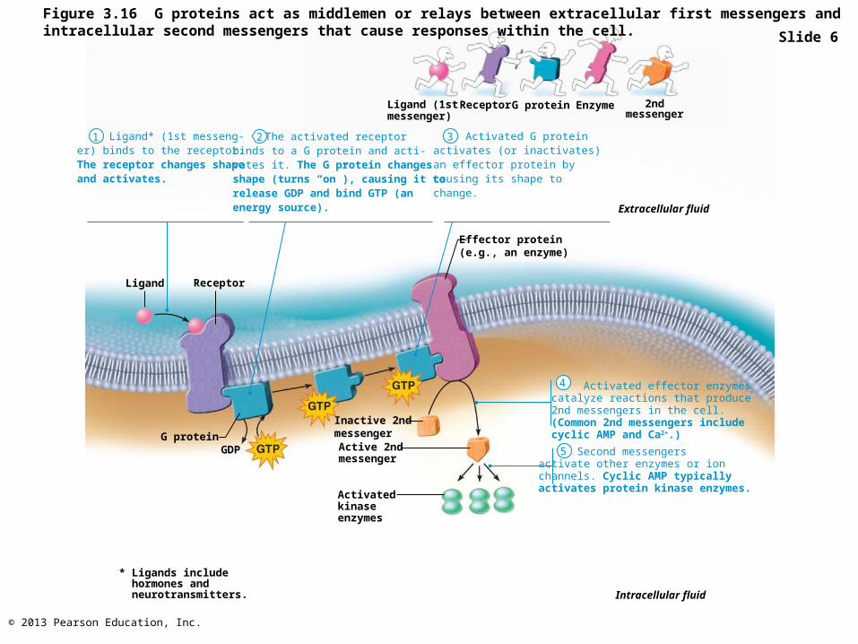
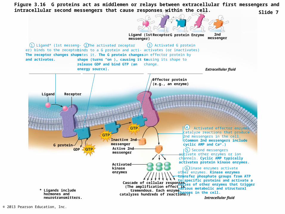
Figure 3.16 G proteins act as middlemen or relays between extracellular first messengers and intracellular second messengers that cause responses within the cell.
Ligand (1st messenger)
Receptor G protein Enzyme 2ndmessenger
Ligand* (1st messeng-er) binds to the receptor. The receptor changes shape and activates.
The activated receptorbinds to a G protein and acti-vates it. The G protein changesshape (turns “on”), causing it torelease GDP and bind GTP (an energy source).
Activated G proteinactivates (or inactivates) an effector protein by causing its shape tochange.
Effector protein(e.g., an enzyme)
Extracellular fluid
G proteinGDP
Intracellular fluid
Cascade of cellular responses (The amplification effect istremendous. Each enzyme
catalyzes hundreds of reactions.)
Activatedkinaseenzymes
Active 2ndmessenger
Inactive 2nd messenger
Activated effector enzymescatalyze reactions that produce2nd messengers in the cell.(Common 2nd messengers includecyclic AMP and Ca2+.)
Second messengersactivate other enzymes or ionchannels. Cyclic AMP typicallyactivates protein kinase enzymes.
Kinase enzymes activateother enzymes. Kinase enzymestransfer phosphate groups from ATPto specific proteins and activate aseries of other enzymes that triggervarious metabolic and structuralchanges in the cell.
* Ligands include hormones and neurotransmitters.
ReceptorLigand
1 2 3
4
5
6
Slide 7