˘ˇ ˆ ˙ ˆ - diva portaluu.diva-portal.org/smash/get/diva2:278318/fulltext02.pdf · me acerco...
TRANSCRIPT

������������������� ���������� �
����
���������� �������������� ������������������� ������� ����������������������������������������
�������� ����������������������������������������������
���� ��!�""�#
����$�%$��&$'�����()��$�%%'�(��)�( ��*�+�*��* *��,��$$-�$'

����������������������� ������� �������������������������������������������������� �� ��������!���������"�������#���#�#����#$%�&�'���(���������'������'�)(����(��*(�����������+�����������������,����(�
��������
-����.��/���#�#��,''����'�0��'����/�������(������1�����������2����������3��� ������������ ��������������������� �������������� ������������������� �������� ������������������������������������4$ ��56����� ��������20�/�$571$�1&&61544715�
�����������������������������������������������+��(��(������'����������(����������'�������������������������.���1����������(������������3����������������������(������� '� ������(������� ��������� ���'����� �(��� ��������� �(��� '��� �� �(�� ���������������������/������ ������� (��� ���� �����.��� ��� ���������� ����������� ��� ��� �� �������
���������'���(��'���������'�������������2��(����(��������������������+��������������������(������'������������������(�
�'��������� ��������0����'����������� ������������������������+��(� �+�������.��8�#�����##��� ����������9��������������������������������+�����������������+�����������:�����(������(������!(��� ���� +��� ��������� +��(� �(�� ������� ��������� ��� �(����'���� �(�� ����������
���'����� +���� ���������� �� ������ '� ������ ��� ��������� ��(���� ��� +���� ��� �������������������� *(�� '����� �(���� +��� ����.��� '�� ��������� ��������� �������� ��� ��������������������;��������������(������������������+���������������(�������������������� ����� ��(����� ����������� ���(���� ��� �������� '� ��1�'��������� ���;���� +�������������*(�� �������� ��������� �(��� ������ ������� +��(� �##� �� ����� ������� (��(��
�������������������(���������+��(��#��������2����������������������������������������+���'��������(��(�����������
�(������������������������'������������''�����������(�����)�)����������������������(��������������3� ������ ��''������ �� �����:�����(���� ��(���� ��� ��������� +��� '��� ���+��
�(���+�������.�������������������-�+�����(��(���������������������(��������(���##��������������������������������������������������:�����(�������(���#������'����*(��������'� �(���+�;����(���.��� �(����������(���������� ������������ �� �(�
(������������������������������� ����������� '� ��������� ���������� � �1������ +���� ���������� ����� �
����'������������������������������������������+��������������������������������'��������������'������������������
���� �������������������������(�����������������������������������������������(������+(���������'���������������
���������� � !����� ��������"������������#���������������� �!�Surface Biotechnology,%&�'((!�������������� ����! SE-751 23 UPPSALA, Sweden
<�/�������-����.��#�#
200/��4&�14��620�/�$571$�1&&61544715��%�%��%��%����1��#4�6�8(���%::���;����:������=��>��%�%��%��%����1��#4�69

“Ella está en el horizonte. Me acerco dos pasos, ella se aleja dos pasos. Camino diez pasos y el horizonte se corre diez pasos más allá. Por mucho que yo camine, nunca la alcanzaré. ¿Para qué sirve la utopía? Para eso sirve: para caminar.”
Eduardo Galeano


List of Papers
This thesis is based on the following papers, which are referred to in the text by their Roman numerals.
I Ferraz, N., Nilsson, B., Hong, J., Karlsson Ott, M. (2008)
Nanoporesize affects complement activation. Journal of Bio-medical Material Research, 87A: 575-581
II Ferraz, N., Carlsson, J., Hong, J., Karlsson Ott, M. (2008) Influence of nanoporesize on platelet adhesion and activation. Journal of Materials Science: Materials in Medicine, 19: 3115-3121
III Ferraz, N., Hong, J., Karlsson Ott, M. (2009) Procoagulant behavior and platelet microparticle generation on nanoporous alumina. Journal of Biomaterials Applications. In press, DOI: 10.1177/0885328209338639.
IV Ferraz, N., Karlsson Ott, M., Hong, J. (2009)
Time sequence of blood activation by nanoporous alumina: Studies on platelets and complement system. Submitted
V Ferraz, N., Hong, J., Santin, M., Karlsson Ott, M. (2009)
Nanoporosity of alumina surfaces induces different patterns of activation in adhering monocytes/macrophages. Submitted
Reprints were made with permission from the respective publishers.


Contents
Introduction ................................................................................................... 11 Biomaterials ............................................................................................. 11
Biomaterial surface topography ........................................................... 12 Host reactions to biomaterials .................................................................. 12
Protein adsorption on biomaterial surfaces .......................................... 14 Complement system ................................................................................. 14
Complement system and biomaterials ................................................. 16 Haemostasis .............................................................................................. 17
Blood coagulation cascade .................................................................. 17 Biomaterials and the coagulation pathways ......................................... 18
Platelets .................................................................................................... 18 Platelet derived microparticles ............................................................ 19 Platelets and biomaterials .................................................................... 20
Monocytes and Macrophages ................................................................... 21 Macrophages and biomaterials ............................................................ 22
Nanoporous alumina ................................................................................ 22
Present Investigation ..................................................................................... 24 Aims of investigation ............................................................................... 24 Experimental procedures .......................................................................... 25
Nanoporous alumina membranes ........................................................ 25 Heparin coating .................................................................................... 25 Blood contact activation experiments .................................................. 26 Monocyte/Macrophage studies ............................................................ 31 Statistics ............................................................................................... 33
Results and discussion .............................................................................. 34 Paper I .................................................................................................. 34 Papers II and III ................................................................................... 37 Paper IV ............................................................................................... 46 Paper V ................................................................................................ 53
Conclusions .............................................................................................. 58 Future perspectives ................................................................................... 59
Acknowledgements ....................................................................................... 60
Summary in Swedish .................................................................................... 63
References ..................................................................................................... 66


Abbreviations
ADP Adenosine phosphate Al2O3 Aluminium oxide AP Alternative pathway AT Antithrombin BCA Bicinchoninic acid BSA Bovine serum albumin C Complement component CD Cluster of differentiation CP Classical pathway EDTA Ethylenediamine tetraacetic acid ELISA Enzyme-linked immunosorbent assay F Coagulation factor FCS Foetal calf serum FITC Fluorescein isothiocyanate FSC Forward light scatter GP Glycoprotein HBSS Hank’s balanced salt solution HRP Horse radish peroxidase IgG Immunoglobulin G IgM Immunoglobulin M IL Interleukin LDH Lactate dehydrogenase LP Lectin pathway MAC Membrane attack complex MASP MBL-associated serine proteases MBL Mannan-binding lectin MM Monocytes/macrophages MP Microparticles NAD Nicotinamide adenine dinucleotide PAGE Polyacrylamide gel electrophoresis PBS Phosphate buffered saline PMMA Polymethylmethacrylate PMP Platelet microparticles pNA p-nitroaniline PRP Platelet rich plasma

sC5b-9 Soluble membrane attack complex SDS Sodium dodecyl sulfate SEM Scanning electron microscopy SSC Side light scatter ß-TG ß-Thromboglobulin TAT Thrombin-antithrombin TMX Thermanox TNF-α Tumor necrosis factor-alpha TSP-1 Thrombospondin-1 vWF von Willebrand factor

11
Introduction
Biomaterials Materials used in medical devices and intended to interact with biological systems are referred to as biomaterials. Although there is a focus on in vivo purposes, a broader definition includes materials for in vitro applications. Cell culturing, biotechnology process equipment and diagnostic gene arrays are some examples [1].
Biomaterials are normally classified into ceramics, metals, polymers and natural materials, with composites defined as a combination of two or more materials. Their distinct physical and chemical properties as well as their biological behaviour, determine their different fields of application. For blood contact-related applications, metals and polymers are the chosen mate-rials. Polymers can also be used to replace soft tissues, e.g. skin, tendon, cartilage, vessel walls, etc. Ceramics are widely used for the repair and re-construction of hard tissue, along with metallic implants [2].
The goal of biomaterial science is to develop “biocompatible” materials. Biocompatibility has been defined as “the ability of a material to perform with an appropriate host response in a specific application”[1]. “Appropri-ate host response” is a wide term that includes resistance to blood clotting, absence of chronic inflammation or other impartment of cellular functions. This term may have to be specifically defined for each biomaterial applica-tion since for example what is a highly biocompatible biomaterial for hard tissue implants might not be adequate for cardiovascular applications.
There is a long evolution of biomaterial science, since early times when it was characterized by a high degree of “trial and error” to nowadays when bioengineered materials are being developed [3, 4]. The new generation of biomaterials is being designed based on the knowledge of inflammation and wound healing processes and the inspiration that can be obtained from the study of biological systems. The goal is to design biologically inspired mate-rials that trigger and guide normal wound healing, show good tissue integra-tion and minimize non-desirable host reactions to the material. Some of the applied strategies are: develop biomaterial surfaces with a low degree of non-specific interactions with proteins and blood cells (e.g. surface modifi-cations with polymer hydrogels or immobilized heparin), enhance specific cell adhesion by modifying the material surface with biologically active mo-lecules (e.g. surface coating with peptides that mimic part of the extracellular

12
matrix) or design topographically patterned surfaces which resemble the surface properties of biological systems (e.g. implant modification with por-ous coatings consisting of polymers, ceramics or composites that promote bone cell attachment and proliferation) [4].
Biomaterial surface topography The surface topography of biomaterials has been proven to influence bio-compatibility, in terms of both inflammatory response and tissue integration.
Traditionally when designing new biomaterials the focus was on obtain-ing controlled microstructured surfaces. Influence of surface microtopogra-phy on cell behaviour has therefore been extensively studied [5-8].
In recent years the potential value of ‘nano’ in biomaterial science has been recognized. The development of easy and fast fabrication methods of nanostructured surfaces has made it possible to move forward in this field [7, 9, 10]. Components of the biological systems have properties governed by the nanometer scale. Protein, nucleic acid and lipid size, folding and pat-terns; extracellular matrix topography and bone surface grain sizes are just some examples [11-13]. Therefore, designing topographically patterned sur-faces that resemble those found in the biological extracellular environment of the tissues is a promising approach to improve the integration of medical implants and, as a consequence, their clinical performance.
Indeed, a broad range of cells (osteoblasts, fibroblasts, neutrophils, macrophages, endothelial, epithelial and smooth muscle cells) have been shown to react to nanoscale features in terms of cell adhesion, morphology, orientation and activity. These features included grooves, ridges, spikes, islands, wells, nodes and pores [7, 14-18]. However, the mechanisms of cel-lular response to biomaterial nanotopography have not been completely elu-cidated. Two possible mechanisms have been proposed: a) an indirect mechanism where material nanotopography affects protein adsorption in terms of amount, conformational changes, orientation and exposure of cell binding sites, b) a direct effect of surface nanotopography on cell response, where cells are capable of sensing nanometric structures and thereby res-ponding to these features [19-21]. For instance, surface topography may modulate the interfacial forces that guide cytoskeleton formation and mem-brane receptor organization which in turn will affect intracellular signaling [20].
Host reactions to biomaterials [22-24] When a biomaterial is implanted into living tissue, a cascade of host reac-tions occur at the tissue-material interface. Tissue and cellular host reactions to injury include inflammation, wound healing and foreign body response.
13
The early response to injury mainly involves the blood and the vascula-ture. An exudation process takes place, i.e. fluid and plasma proteins escape from the vascular system to the injured tissue together with the recruitment of leukocytes and platelets. Plasma proteins adsorb to the material surface and activation of the coagulation pathways, the complement system, the fibrinolytic system and platelets occur. Platelets and neutrophils are consid-ered the main cellular mediators of the acute inflammatory response. Plate-lets participate in clot formation and release chemical mediators. The main role of the neutrophils is to engulf and degrade the foreign material. Due to the size disparity between the inflammatory cells and the material, neutro-phils are generally not able to phagocytose the biomaterial. However, certain events in phagocytosis may still occur, such as secretion of granular content.
Following the above described events, monocytes arrive to the implant site and differentiate into macrophages. Macrophages also attempt to engulf the material and release a great number of biologically active products that influence both inflammation and wound healing. The continuous stimulation of the macrophages maintains the inflammatory activity, leading to a chronic inflammation. Foreign body giant cells are formed by fusion of mono-cytes/macrophages in an attempt to phagocytose the material and have the potential to actively participate in the inflammatory response through the production of cytokines.
In parallel to the inflammatory process, wound healing takes place. The normal wound healing process includes the formation of granulation tissue, characterized by proliferation of small new blood vessels and fibroblasts. The granulation tissue is subsequently transformed to regenerate normal tissue. In some cases complete restoration of tissue architecture is possible, however in most cases the granulation tissue is remodeled into scar tissue. When the inflammatory reaction can not be resolved, i.e. the body can not heal the “injury”, the foreign body reaction is observed. This reaction is characterized by the presence of foreign body giant cells, mildly active in-flammatory cells and a fibrous capsule that surrounds the implant.
Hence, the tissue response to an implant could be resolved in two differ-ent ways: a) resolution of the inflammatory exudate, leading to the formation of newly organized tissue at the interface or b) fibrous capsule formation with very little restitution of normal tissue structure. With some exceptions, the final outcome of a biomaterial implantation is generally the fibrous en-capsulation. Therefore, nowadays the main focus for biomaterial scientists is to modify the inflammatory response towards healing and implant integra-tion.
Other processes that may occur at the biomaterial-tissue interface and that will affect the performance of the biomaterial are: infection, tumorigenesis and thromboembolic complications [25].

14
Protein adsorption on biomaterial surfaces It is well established that the host response to a biomaterial is mediated by the layer of surface-adsorbed proteins [26]. The adsorption procedure occurs immediately after implantation and is described as a dynamic process, in-volving surface induced conformational changes and surface protein ex-change [26, 27]. The latest phenomenon is described as the Vroman effect, where initially adsorbed proteins are replaced by more surface active ones that are present in lower concentrations [28]. The exchange behaviour is influenced by the total protein concentration and by the material properties such as its surface energy (hydrophobic vs. hydrophilic), composition and surface structure.
The subsequent phenomena in the host response, i.e. activation of the cas-cade systems of blood and recruitment, adherence and activation of blood cells, are governed by the initially adsorbed proteins. Changes in terms of type, amount, conformation and orientation of these proteins will thus have a large effect on cell behaviour at the biomaterial-tissue interface [29, 30].
Figure 1. When cells encounter a biomaterial surface they “see” a layer of adsorbed proteins. The type, amount and conformation of these proteins will influence the cell behaviour on the biomaterial surface.
Complement system [31-33] The complement system is part of the innate immune system, with a primary function of removing microorganisms and other foreign substances. It has the ability to discriminate non-self structures from self components. The complement cascade consists of more than 30 soluble and membrane bound proteins that facilitate phagocytosis by opsonization of foreign substances, release inflammatory mediators and perform lysis of cells through pore for-

15
mation. The central event in complement activation is the proteolytic cleav-age of C3 into C3a and C3b which is achieved by two multi-subunit enzyme complexes, the C3 convertases. These enzymes can be assembled by three different activation pathways: the classical pathway, the alternative pathway and the mannan-binding lectin pathway.
The classical pathway is activated when C1 complex (C1qC1r2C1s2) rec-ognizes the Fc portions of IgM or IgG already bound to an antigen. Acti-vated C1 is then able to cleave C4 into C4a and C4b. C4b attaches to the target surface while C4a remains as a soluble anaphylatoxin. C2 binds to the attached C4b and is cleaved by C1 into C2a and C2b. The classical C3 con-vertase C4bC2a is then formed. When C4bC2a acts on C3, C3a is released and C3b binds to C4b, forming the classical C5 convertase (C4bC2aC3b). The classical pathway can also be triggered by bound C-reactive protein and self-structures released from damaged cells.
The alternative pathway is activated by covalent binding of C3b to for-eign surfaces that do not provide adequate down-regulation of the pathway. The initial C3b molecules for the AP activation are produced by the classical convertase or by spontaneous hydrolysis of C3 in the fluid phase. The C3 hydrolysis product (C3(H2O) or C3i) has the ability to bind factor B, forming the C3 convertase precursor (C3(H20)B). This complex is stabilized by the action of factor D, which cleaves factor B, releases Ba and as a consequence a stable soluble C3 convertase is formed (C3(H2O)Bb). This convertase pro-duces more C3b (both surface bound and in solution). Surface bound C3b and activated B, together form the alternative C3 convertase (C3bBb). Bound C3 convertase cleaves additional circulating C3 molecules, generat-ing more C3b molecules. Since C3b participates in the C3 convertase, an amplification feedback loop takes place. If another C3b binds to the C3 con-vertase, the alternative C5 convertase (C3bBbC3b) is formed. The lectin pathway is initiated by recognition of carbohydrates in the wall of certain microorganisms. Mannan-binding lectin (MBL) is the protein which recognizes the carbohydrate motifs. MBL together with MBL-associated serine proteases (MASP) form a complex, which is able to create the classi-cal pathway convertase C4bC2a.
The activation pathways converge in the terminal pathway that ends up forming a membrane attack complex (MAC or C5b-9) which disrupts the membrane integrity of pathogens. C5 convertase cleaves C5 into C5a and C5b. C5b forms a complex with C6 and C7, which inserts itself into the cell lipid layers. C8 and multiple copies of C9 are then sequentially associated with the complex to form a pore, resulting in cell damage and/or lysis. In the absence of a biological membrane, the complex binds to S protein and forms sC5b-9 which remains in the fluid phase. Sublytic concentrations of the membrane attack complex are known to stimulate various cells, including platelets.

16
Complement activation is tightly regulated by both soluble and cell bound factors, limiting the activation to the site of infection and protecting host cells from complement attack and lysis.
Figure 2. Overview of the complement system pathways: The classical pathway is initiated by antigen-antibody complexes, the alternative pathway by spontaneous hydrolysis of C3 and surface binding of C3b and the lectin pathway depends on the recognition of microbial carbohydrates structures. As a result, the C3-covertases are formed, which cleave C3 into C3a and C3b. C3b binds to the C3 convertases, form-ing the C5 convertases. C5 convertases cleave C5 into C5a and C5b. C5b binds to the surface and the cytolytic membrane attack complex (C5b-C9) is formed.
Complement system and biomaterials The complement system plays a central role in the events that take place at the blood-biomaterial interface. It influences platelet and leukocyte activa-tion and affects the recruitment and adhesion of inflammatory cells at the site of implantation.
The mechanism involved when the complement system becomes acti-vated by artificial surfaces has been extensively investigated. It was conven-tionally believed that the alternative pathway was the main mechanism and

17
that the presence of nucleophilic groups on the material surface was of major importance [32]. More recently, authors believe that the classical pathway is relevant and that the triggering mechanism is related to conformational changes occurring when proteins are adsorbed on the foreign surface [33-35]. Hence, a proposed model explains that the initial C3b molecules are produced by either of the activation pathways and bind to the plasma protein layer on the biomaterial surface. The covalent binding of C3b initiates the amplification loop, which generates the majority of the C3b molecules that bind to the surface [33].
Haemostasis Haemostasis is the arrest of bleeding from an injured blood vessel. Normal haemostasis depends on complex interactions between platelets, the blood vasculature, the coagulation cascade and the fibrinolytic system [36].
Biomaterials in clinical use will encounter blood, either continuously or during the implantation process, hence challenging the haemostatic system.
Blood coagulation cascade [32, 37] Blood coagulation involves a cascade of proteolytic reactions that results in the formation of a fibrin clot. Initiation of the coagulation cascade occurs either by surface-mediated reactions (intrinsic pathway) or by tissue factor expression by cells (extrinsic pathway).
The intrinsic or contact activation pathway is initiated by surface-activation of factor XII and it is closely linked to the kinin-kallikrein system. The importance of the intrinsic pathway in normal blood coagulation still remains speculative. A role in the coagulation propagation phase rather than in the initial phase has been proposed.
Blood coagulation cascade is centered in the extrinsic pathway, which is triggered by tissue factor (TF), a cellular lipoprotein released from damaged cells or expressed on the surface of activated monocytes and endothelial cells.
The two activation pathways converge in the common pathway, where the prothrombinase complex is assembled on the membrane of activated endo-thelial cells and platelets. The prothrombinase complex transforms proth-rombin into thrombin. Finally thrombin promotes the formation of the fibrin clot from fibrinogen monomers.
Natural anticoagulants such as antithrombin, thrombomodulin (involving protein C activation and protein S) and the TF pathway inhibitor system control the coagulation process and restrict it to the site of vascular injury.

18
At the same time that the coagulation cascade is activated, the fibrinolytic system initiates the process needed for the dissolution of the fibrin matrix of the thrombi.
Biomaterials and the coagulation pathways The mechanisms of coagulation activation by artificial surfaces are not yet fully understood.
The intrinsic pathway has been described as the main responsible for coa-gulation activation by biomaterials [38, 39]. Lately, the importance of the extrinsic pathway in the activation of the coagulation by biomaterials has been recognized [32]. It is believed that blood contact with a biomaterial represents a potential stimulus to induce TF expression on monocytes, which results in blood coagulation by the extrinsic pathway [32, 40].
Platelets [32, 41, 42] Platelets, also called thrombocytes, are derived form megakaryocytes. They are anucleated blood cells with discoid shape and a diameter of 3-4 μm. The-se cells play a central role in haemostasis. They circulate in a non-activated state in blood, becoming activated when contacting injured endothelium, sub-endothelium or artificial surfaces. Among platelet activators are: plasma proteins (e.g. thrombin and fibrinogen), vascular wall products (e.g. colla-gen, von Willebrand factor (vWF) and vitronectin), molecules derived from leukocytes or platelet products (e.g. platelet activating factor, ADP, sero-tonin and thromboxane A2) and components of the complement system (e.g. C1q and C5b-9). These agonists interact with specific receptors on the plate-let plasma membrane. Platelet activation is reflected in the following physio-logical responses:
• Secretion of platelet intracellular granule contents, including ß-
thromboglobulin, platelet derived growth factor (PDGF), coagula-tion factors (FV, FVIII, FXI), fibrinogen, ADP and serotonin.
• Activation of the platelet eicosanoid metabolic pathway, resulting in the liberation of arachidonic acid from platelet phospholipids and in the synthesis and release of prostaglandins and thromboxane B2.
• A drastic change in shape, that promotes platelet-platelet contact and adhesion.
• Diffusion of internal granular proteins such as P-selectin (involved in the adhesion of activated platelets to monocytes, neutrophils and lymphocytes) into the plasma membrane.
• The platelet surface receptor GPIIb/IIIa changes its conformation which leads to an increased affinity towards soluble fibrinogen.

19
Binding of fibrinogen to the GPIIb/IIIa receptor in turn leads to platelet aggregation.
• Acceleration of the blood coagulation cascade by rearrangement of negatively charged phospholipids (e.g. phosphatidylserine) from the cytosolic face of the platelet membrane to the extracellular face. This provides a high affinity surface for assembly of the prothrom-binase complex, by which prothrombin is converted into thrombin.
• Formation of platelet microparticles.
Platelets do not only provide the first line of defense by “plugging” a dam-aged vessel at the site of injury but also localize, directly participate and regulate the subsequent events in the coagulation reaction. Moreover, their interactions with monocytes and neutrophils as well as with components of the complement system give platelets a key role in the inflammatory re-sponse.
Platelet derived microparticles Platelet microparticles (PMP) are defined as submicroscopic (size range 0.1-1.0 μm) membrane vesicles released by activated platelets and have been described as excellent markers of platelet activation [43, 44].
There are several hypotheses why platelets release microparticles. It is be-lieved that it is an efficient way for platelets to maximize assembly of their surface phospholipids for procoagulant or anticoagulant factors, thus accel-erating haemostasis at sites of activation. Another theory suggests that mi-croparticle release could be a cell rescue mechanism for complement-mediated cell damage [43]. Besides the biological stimulus, shear stress has also been described as an important mechanical factor capable of inducing PMP release [45].
Like intact platelets, PMP expose GPIb, platelet-endothelial cell adhesion molecule 1 and GPIIb/IIIa complex. Activation markers such as P-selectin or fibrinogen binding GPIIb/IIIa complex can also be expressed on the PMP surface membrane. The antigenic composition of PMP and their function is dependent on the mechanism involved in their release. For example, PMP released from platelets stimulated with collagen and thrombin expose GPIIb/IIIa that binds fibrinogen, but if platelet activation instead is caused by the complement component C5b-9, PMP GPIIb/IIIa does not bind fi-brinogen. Therefore the heterogeneity of the PMP population is enormous and presents a wide spectrum of functions [44]. PMP play a role in the am-plification of coagulation since their high surface area causes increased ex-posure of phosphatidylserine on the outer membrane, facilitating the assem-bly of the prothrombinase complex of the coagulation cascade. Anionic phospholipids on the PMP outer membranes also promote the assembly of protein C anticoagulant enzyme complexes, giving PMP anticoagulant prop-

20
erties. PMP can also exert pro-inflammatory activities by stimulating mono-cytes and neutrophils. Since PMP expose cell-specific adhesion receptors on their surface, they may act as “circulating envelopes” that carry and deliver substrate molecules and activation stimuli to specific target cells, exerting distant effects from the site of platelet activation [44, 46].
To study PMP generation by biomaterial induced platelet activation is of major importance due to the potential contribution of PMP to coagulation, inflammation and cellular activation.
In recent years, the study of PMP in clinical disorders has gained much attention, especially their potential role as diagnostic markers of clinical conditions, particularly in vascular pathologies and rheumatic diseases [47, 48].
Figure 3. Platelet response to activation agonists: drastic change in shape, secretion of platelet intracellular granule content, diffusion of internal granular proteins such as P-selectin into the plasma membrane, conformational changes in the receptor GPIIb/IIIa which increases affinity towards fibrinogen and generation of platelet microparticles.
Platelets and biomaterials Platelets commonly bind to foreign material surfaces, which in many cases lead to activation. Platelet adhesion is governed by the layer of plasma pro-teins that rapidly covers a material when in contact with blood. Adsorbed fibrinogen has been reported to be the main protein involved in adhesion of resting platelets to different classes of biomaterials [49, 50]. Interaction with fibrinogen leads to platelet aggregation and activation. It should however be noted that other proteins such as vWF, vitronectin and fibronectin also are involved in the adhesion and activation processes [49, 51, 52]. It has been shown that the absence of platelet adhesion on a biomaterial surface does not

21
prevent platelet activation [53, 54]. Therefore other mechanisms besides adhesion to the biomaterial must be involved in material-induced platelet activation. Soluble components like thrombin, ADP (from damage red blood cells or platelets), sC5b-9 and C1q are presumed to be involved in such processes [32, 55-57].
Monocytes and Macrophages Monocytes and macrophages are part of the mononuclear phagocyte system. Monocytes are produced in the bone marrow, enter the blood stream and thereafter circulate in the peripheral blood for 8-72 hours. The monocytes then migrate into various organs, where they mature and differentiate into tissue-specific macrophages. They may also be deposited on injured blood vessels where they become macrophages. Tissue-macrophages are found in bone marrow, spleen, lymphoid tissue, bone, liver and lungs.
Macrophages contribute to different aspects of host defence, acting as an-tigen presenting cells, effector cells (phagocytosis and cytotoxicity) and reg-ulators of the inflammatory and immune response [58].
Macrophage activation is a complex phenomenon, which occurs in stages and requires sequential stimuli. Possible stimuli include cytokines, endotox-ins, immune complexes (antigen-antibody complexes) and other mediators and regulators of inflammation such as complement system components. Activation results in increased cell-size, changes in expression of membrane receptors, increased production of proteolytic enzymes and inflammatory cytokines, growth factors and other bioactive agents that modulate the func-tion of both immune-competent and tissue cells. As a consequence, the fol-lowing effector functions may take place [58, 59]:
• Phagocytosis • Tissue damage by releasing for example H2O2, acid hydrolases
and/or TNF-α • Lymphocyte activation by antigen processing and presentation and
IL-1 production • Inflammation and fever caused by IL-1, IL-6, TNF-α release • Microbicidal activity by producing oxygen reactive species, acid hy-
drolases and cationic proteins • Tumoricidal activity • Tissue reorganization and wound healing by for example releasing
fibroblast stimulating factors, angiogenesis factors, hyaluronidase

22
Figure 4. Macrophage activation results in increased cell size, changes in the ex-pression of membrane receptors, increased production of proteolytic enzymes, in-flammatory cytokines, growth factors and other bioactive agents. As a result the above illustrated macrophage effector functions may take place.
Macrophages and biomaterials Macrophage interaction with biomaterials has been proposed to involve the following events: a) recognition of blood and extracellular matrix proteins (e.g. fibronectin, vitronectin) adsorbed on to the biomaterial surface via macrophage adhesion ligand-receptors, b) complex formation with adsorbed complement components or IgG and IgM antibodies, c) the presence of ac-tive cytokines and growth factors that may modulate macrophage adhesion mediated by ligand-receptor complexes [60].
The process that leads to macrophage recognition, adhesion and activation by an implanted biomaterial is therefore, complex and dynamic, involving biomaterial properties, adsorbed proteins, adherent cells, inflammatory cyto-kines and growth factors.
Nanoporous alumina When aluminium is anodised in certain acid electrolytes such as phosphoric acid, sulphuric acid and oxalic acid, a thin film of alumina is formed, which grows into a hexagonally packed array of cylindrical cells, at the centre of which a straight hole is located. The pores are roughly parallel to each other and run perpendicular to the film plane [61-63]. The pore diameter depends

23
mainly on the applied voltage and can be varied from 10 to 450 nm with a narrow pore size distribution [64] .
Nanoporous alumina has been recognized as an important material and as a template for the fabrication of nanostructures [64]. It has been evaluated as a bone implant coating [65], stent coating for drug delivery [66] and as an immunoisolation device [67], showing promising results in terms of bio-compatibility. Moreover, nanostructured alumina coating on machined tita-nium showed improved mesenchymal stem cell differentiation in the os-teoblastic pathway both in vivo and in vitro [68].
Thus anodised aluminium is not only a suitable substrate for investigating how nanoporosity affects protein and cellular functions. It is also a potential implant material which can easily be manufactured to govern specific events at the tissue-material interface.
Figure 5. Schematic diagram of a porous anodic film on aluminium. During the anodization process a thin film of alumina grows into a hexagonally packed array of cylindrical cells, at the center of which a straight pore is located.

24
Present Investigation
Aims of investigation Due to the recognized significance of surface nanotopography for host re-sponse to biomaterials, the study of nanopatterned-materials has gained im-portance and as such become part of the new generation of biologically in-spired biomaterials.
Nanoporous aluminium oxide was chosen as a template for the present work. Nanoporous alumina has the advantage of easily be manufactured with different pore sizes. It can be hypothesized that by changing the pore size one can “steer” the type and magnitude of cellular and molecular events at the tissue-material interface by promoting specific protein adsorption and influencing their orientation and conformation. Hence, nanotopography may be exploited to subtly control the inflammatory potential of the biomaterial.
The general aim of the present work was to study the difference in pro-inflammatory characteristics between nanoporous aluminium oxide mem-branes with two pore sizes (20 and 200 nm in diameter).
Specific aims included studying the effect of nanoporesize on: a) Complement activation (papers I and IV) b) Platelet activation (papers II, III and IV) c) Monocyte/macrophage behaviour (paper V)

25
Experimental procedures Nanoporous alumina membranes Nanoporous alumina membranes with pore diameters of 20 nm and 200 nm were chosen for these studies. Commercially available AnodiscTM alumina membranes produced by Whatman International Ltd (Maidstone, England) were used. The membranes are 25 mm in diameter (papers I-IV) or 13 mm in diameter (paper V) and 60 μm thick, with narrow pore size distribution. It should be noted that both type of membranes have similar surface roughness and surface chemical characteristics regardless of their size or porosity [18].
Figure 6. SEM micrographs showing nanoporous alumina membranes with pore diameters of 200 nm (a) and 20 nm (b). Scale bars represent 500 nm.
Heparin coating Heparin is a mucopolysaccharide involved in the regulation of coagulation activation. Its presence on the surface of the vascular lumen converts intact endothelium to a non-thrombogenic surface. Heparin forms a high affinity complex with antithrombin, accelerating the inhibitory effect of antithrombin on serine proteases that participate in the coagulation cascades [69].
When an artificial surface is coated with heparin, it is converted to an en-dothelium like-surface, i.e. non-thrombogenic and blood compatible. Hepa-rin-coated surfaces have been shown to effectively reduce coagulation and complement activation during in-vivo and in-vitro experiments [70-72].
In order to avoid blood activation by surfaces other than the studied mate-rials, blood collection tubes and the slide chambers (see Fig. 7) were coated with heparin using the Corline method (Corline Systems AB, Uppsala, Swe-den). In this method a water-soluble macromolecular conjugate of heparin is attached to the material surface that previously has been modified with a polymeric amine. The anionic heparin macromolecule binds irreversibly to the positively charged substrate surface in a self-assembling process. To

26
obtain high levels of heparin surface concentration and hence improve the biocompatibility of the surface, the application of the polymeric amine and the macromolecular heparin conjugate is repeated twice. The double heparin coating gives a heparin concentration of 0.9 μg/cm2, with an antithrombin (AT) binding capacity of 12 pmol/cm2 [73].
Blood contact activation experiments (Papers I-IV) Slide chamber model The slide chamber previously described by Hong et al. [74] was used for these experiments. The device is manufactured from polymethylmethacry-late (PMMA) and consists of two cylinders fixed to a microscope slide. In this way, two wells that can hold a maximum volume of 1.65 ml each are created. After heparin coating each well was filled with 1.3 ml of blood (1 ml of blood was also collected in eppendorf tubes containing EDTA or ci-trate, these 0 min samples were later used as controls). The nanoporous alu-mina membranes were then placed covering the wells (as “lids”), thus two cylindrical chambers were created (see Fig. 7). The slide chambers were rotated vertically at 22 rpm for 30 or 60 minutes (papers I-III) or from 2 to 240 min (paper IV) in a 37oC water bath.
Figure 7. The slide chamber model. 1.3 ml of whole blood is added to each well of the slide chamber. Nanoporous alumina membranes are placed covering the wells (as “lids”) creating two circular chambers. The slide chamber is rotated vertically at 22 rpm in a 37oC water bath.

27
Preparation of blood Interactions between blood cascade systems and blood cells are well estab-lished. Hence lays the importance of working with whole blood when study-ing the inflammatory response to a biomaterial.
Whole blood from ten healthy donors was collected in heparin-coated 50 ml Falcon® tubes containing soluble heparin, giving final concentrations of 0.25 IU heparin/ml and 0.5 IU heparin/ml (Bio Iberica, Barcelona, Spain) (papers I and II) and 1 IU heparin/ml (Leo Pharma A/S, Bellerup, Denmark) (papers III and IV). In paper III, compstatin, a potent complement inhibitor was used to elucidate the role of the complement system in PMP generation. In this case, the compstatin analogue Ac-ICV(1MeW)QDWGAHRCT-NH2 was added to heparin collected blood giving a final concentration of 10 μM.
Preparation of platelet rich plasma To highlight the significance of “cross-talk” between the different compo-nents of whole blood in the inflammatory response to biomaterials, experi-ments with platelet rich plasma (PRP) were also performed (paper II).
Blood from six healthy donors was collected in heparin-coated 50 ml Fal-con® tubes containing soluble heparin, giving a final concentration of 0.5 IU heparin/ml. To obtain platelet rich plasma blood was centrifuged at 190g, for 15 min at room temperature and then the supernatant was collected. Platelet number was measured and the values ranged from 200-250 x 109/l. The ex-periments were conducted following the same protocol used for the whole blood experiments.
Analysis of samples The in vitro model used in the present work made it possible to analyse the biomaterial surface in terms of protein and cell adhesion, as well as deter-mine the activation products generated in the fluid phase after blood incuba-tion.
Blood samples: After incubation, 1 ml of blood from each chamber was removed and mixed with EDTA-K3 or citrate giving final concentrations of 4 mM and 13 mM, respectively. Platelet count (Coulter Ac T diff TM hematol-ogy analyzer, Coulter Corporation Miami, FL, USA) and flow cytometry analysis were performed on the EDTA blood samples. The remaining EDTA-treated blood samples were centrifuged at 3000g for 10 min at + 4oC. The citrated blood was centrifuged twice at + 4oC: 10 min at 1000g and 10 min at 10000g. Thereafter the plasma samples were collected and stored at -70oC for future analyses, including enzyme immunoassays for detection of C3a, sC5b-9, ß-thromboglobulin, thrombospondin-1 and thrombin-antithrombin complex.
Alumina membranes: The membranes were rinsed with saline buffer and thereafter either fixed, dehydrated and critical point dried for scanning elec-

28
tron microscopy (SEM) studies, fixed for immunocytochemical staining, subjected to the thrombin generation assay or stored at -70oC for future elu-tion of proteins and dot blot analyses.
Enzyme immunoassays Immunoassays are based on the recognition between an antibody and its antigen. In enzyme-linked immunosorbent assays (ELISA) the specificity of antibodies and the sensitivity of simple enzyme assays are combined. The sandwich ELISA measures the amount of antigen between two layers of antibodies (a capturing antibody adsorbed onto a solid phase and a detecting antibody in solution). Generally an enzyme is conjugated to the secondary antibody and when the enzyme substrate (chromogen) is added, it is con-verted by the enzyme to a detectable form (coloured product). Another commonly used approach is the use of biotinylated detecting antibodies, followed by enzyme-conjugated streptavidin [75]. Detection of C3a and sC5b-9 C3a levels were measured using the method described by Nilsson Ekdahl et al. [76]. Zymosan-activated serum, calibrated against a solution of purified C3a, served as a standard. Values are given in ng/ml.
sC5b-9 was assessed using a modification of the method described by Mollnes et al. [77]. Zymosan activated serum containing 40000 AU/ml served as a standard.
Detection of ß-Thromboglobulin (ß-TG) Citrate samples were analyzed for ß-TG using AsserachromeTM ß-TG EIA kit (Diagnostica Stago, Asnieres-sur-Seine, France). ß-TG was captured in wells coated with specific rabbit anti-human ß-TG F(ab’)2 fragments and detected using HRP-conjugated rabbit anti-human ß-TG antibody. Values were given in IU/ml. Detection of Thrombospondin-1 (TSP-1) Citrate samples were analyzed for thrombospondin-1. Samples were added to microtiter plates coated with capture antibody, clone B7 (AbCam, Cam-bridge, UK). Biotinylated anti-human TSP-1 clone P10 (Immunotech, Mar-seilles, France) was used as detection antibody, followed by HRP-conjugated streptavidin (GE Healthcare, Buckinghamshire, UK). Human serum diluted in working buffer was used as a standard. Values were given in ng/ml. Detection of Thrombin-Antithrombin complex (TAT) Plasma samples from EDTA-treated blood were analyzed for TAT. Microti-ter plates were coated with anti-human thrombin antibody (Enzyme Re-search Laboratories, South Bend, IN, USA) diluted 1/120. HRP-coupled anti-human antithrombin antibody (Enzyme Research Laboratories, South

29
Bend, IN, USA) diluted 1/120 was used for detection. Pooled human serum diluted in normal citrate-phosphate-dextrose plasma was used as a standard. Values were given in μg/l.
Flow cytometry Flow cytometry is a technique used for quantitative single cell analysis. A suspension of cells is aspirated into a flow cell where, surrounded by a nar-row fluid stream, they pass one at a time through a focused laser beam. The light is either scattered or absorbed when it strikes a cell. Forward light scat-ter (FSC), defined as light of the same colour as the illuminating beam that has been bent to a small angle from the direction of the original beam, cor-relates with cell size. Side light scatter (SSC), defined as light of the same colour as the illuminating beam that has been scattered by a particle to an angle of 90o from the illuminating beam, depends on the inner complexity of the cell (i.e. shape of the nucleus, the type and amount of cytoplasmatic granules or the membrane roughness). Absorbed light of the appropriate wavelength may be re-emitted as fluorescence if the cell contains one or more fluorochrome-labeled antibodies attached to the surface or to internal cell structures. Detectors process the emitted fluorescence and the light scat-tering properties of each cell and as a result cells can be characterized ac-cording to size, shape and the presence or absence of specific markers on their surface [78]. Detection of platelets and PMP by flow cytometry Samples were analyzed on a BD LSRII SORP (BD Biosciences, CA, USA) flow cytometer equipped with a forward scatter photo multiplier tube (PMT) detector in addition to the standard forward scatter (FSC) detector.
EDTA- treated blood samples were labelled with anti-human CD41:FITC antibodies. Platelet specific events were identified gating on events positive for FITC. PMP were distinguished from platelets by size FSC PMT analysis. Counting beads were used to determine the absolute number of platelets and PMP. Data were analyzed using BD FACSDivaTM software (BD Biosci-ences, CA, USA).
Preliminary experiments were performed to determine the ability of the BD LSR II SORP flow cytometer to detect PMP. Platelet rich plasma (PRP) was stimulated with 10 μM calcium ionophore A-23187 and the number of generated PMP was compared with the PMP number in the resting PRP sample. PMP were isolated from PRP following the protocol described by Nomura and Fukuhara [79]. Isolated PMP samples were used to confirm PMP region in side scatter versus PMT forward scatter dot plots.
Protein elution and dot blot analysis Protein adsorption patterns on the two alumina membranes were studied. Adsorbed proteins were eluted by incubating the membranes with 2% SDS

30
in veronal buffered saline for 3h at room temperature. The protein recovery was determined with BCA (Micro BCATM Protein assay kit, Pierce, USA).
Dot blot analyses were performed to detect IgG, IgM, C3 and C1q. The protocol included the following steps: dilutions of the protein mixture were spotted onto a nitrocellulose membrane. After a blocking step, the blot was treated with an antibody against the protein of interest (primary antibody). Washing steps were necessary to remove unbound antibodies. The mem-brane was incubated with an enzyme-conjugated antibody against the pri-mary antibody and thereafter the enzyme substrate (chromogen) was added. As a result an insoluble product was directly deposited onto the membrane. The membrane was thereafter scanned and the intensity of the dot was de-termined using the ImageJ programme (NIH, Bethesda Maryland, USA) [80].
Immunocytochemical staining Immunocytochemistry is a way of analyzing and identifying different cell types based on binding of antibodies to specific components of the cell. By using the right antibodies, a specific function or activation state of a cell can be determined. Firstly the primary antibody binds to the target protein. Thereafter a secondary antibody labelled with a fluorochrome or an enzyme is added. The enzyme is capable of catalyzing a reaction that gives a col-oured product, which easily can be detected by light microscopy. Fluores-cence microscopy is applied when fluorochrome-conjugated antibodies are used.
In the present work, immunocytochemical staining was used to identify platelets and platelet derived microparticles (PMP) that had bound to the alumina membranes after contact with whole blood. This technique was also used to determine the activation state of bound platelets and PMP.
CD41 antigen represents platelet glycoprotein IIb of the GPIIb/IIIa com-plex. The GPIIb/IIIa complex is the only integrin expressed uniquely on platelets. CD62P represents P-selectin, a platelet activation dependent gran-ule-external membrane protein [81]. Platelet derived microparticles expose GPIIb/IIIa complex and can also express activation markers such as P-selectin.
In paper II, immunocytochemical staining combined with light micros-copy was used to evaluate platelet adhesion and activation by using antibod-ies against CD41 and CD62P. The staining procedure was essentially done as described by Karlsson-Parra et al. [82] and the immuno-stained mem-branes were examined using a Nikon Eclipse E600 light microscope.
In paper IV immunocytochemical staining was combined with confocal fluorescence microscopy. Specimens were double-labelled as follows: sam-ples were first incubated with anti-human CD62P mouse immunoglobulins. Anti-mouse rabbit immunoglobulins conjugated with DyLightTM 488 were used as secondary antibody. Mouse serum was then added to block any un-

31
reacted anti-mouse DyLightTM 488 conjugated antibodies. Finally, samples were incubated with anti-human CD41 mouse immunoglobulins conjugated with DyLightTM 647. After the last incubation, the samples were mounted in Immunoconcepts® mounting medium and examined using a Zeiss LSM 510 Meta confocal microscope equipped with an Axiovert 200 microscope stand.
Scanning electron microscopy SEM provides high resolution images of surfaces, with three-dimensional appearance, thus it is an excellent tool for studying cell morphology on a biomaterial surface.
After blood and PRP incubation, the aluminium oxide membranes were washed with Hank’s Balanced Salt Solution (HBSS). Thereafter the cells on the surface were fixed with 1.5% (v/v) glutaraldehyde, dehydrated through a series of acetone concentrations (25, 50, 70, 80, 90 and 100% by volume), critical point dried and sputtered with gold, finally to be studied using a LEO 1530, Gemini SEM.
Thrombin generation assay To study the effect of the alumina membranes on the induction of procoagu-lant state in adherent platelets and PMP, a thrombin generation assay was carried out on the material surfaces. The method has been described by Grunkemeier et al. [83]. Briefly, after whole blood contact alumina mem-branes were incubated with a mixture of factor Va and factor Xa for 15 min, at room temperature, under agitation (200 rpm). Thereafter prothombin was added to the agitating membranes. Thrombin generation was terminated by addition of EDTA 0, 2, 4 and 6 min after addition of prothrombin. Aliquots of the thrombin containing solutions were moved to a 96 well microtiter plate and the chromogenic substrate S-2238 was added. Thrombin splits off p-NA from S-2238. The pNA generation rate (mO.D.405nm/min) was meas-ured over a 5 min time period and related to thrombin concentration by using a calibration curve. Finally thrombin generation rates (μg/ml/min) were cal-culated by linear regression of thrombin concentration versus time curves. These values were normalized by the initial number of platelets in whole blood, due to the variation in platelet number between the different donors.
Monocyte/Macrophage studies (Paper V) Alumina membranes with 20 and 200 nm pore diameters were compared for their ability to activate human monocytes/macrophages (MM). Alumina membranes were treated with human plasma to induce the formation of a protein biofilm and then incubated for 24 hours with freshly isolated human MM.

32
Isolation and culture of mononuclear cells Mononuclear cells were obtained from whole venous blood freshly collected from seven healthy donors in heparin-coated 50 ml Falcon® tubes containing 1 IU/ml soluble heparin (Leo Pharma A/S, Ballerup, Denmark). Human monocyte enrichment cocktail (RosetteSepTM, StemCell Technologies, Van-couver, BC, Canada) was added to the heparinized blood previous to the isolation step. Isolation was done using standard Ficoll-paque density gradi-ent. Thereafter, cells were re-suspended in RPMI-1640 medium (Sigma, UK), supplemented with 5% (v/v) foetal calf serum (FCS), 100 IU penicil-lin/ml and 100 μg streptomycin/ml and counted using a haemocytometer. Cell viability was assessed using trypan blue staining (95% cell viability). The cell isolation step provided a fraction of platelet rich plasma (PRP) which was collected and used for coating the materials. Alumina membranes were placed in 24-well tissue culture plates and coated with 1 ml of PRP for 30 min at 37oC, followed by 3 washing steps with Hank’s balanced salt solu-tion (HBSS). Cells were added to the wells (4x10⁵ cells/well) and cultured for 24 h at 37oC, 5% CO2 in a humidified atmosphere. After 3 h of incuba-tion, medium was changed to remove non-adherent lymphocytes. At the end of each experiment supernatants were collected, centrifuged, aliquoted and stored at -70oC for future analyses. The nanoporous alumina membranes were fixed with 1.5 % (v/v) glutaraldehyde and stored at 4oC until micros-copy analyses.
Analysis of the culture supernatants LDH assay Measuring lactate dehydrogenase (LDH) activity in the culture supernatants gives an estimation of non-viable cells. A LDH activity kit (In vitro toxicol-ogy assay kit LDH based, Sigma, Missouri, USA) was used to analyze the medium after 24 h of culturing. The assay measures LDH activity via the reduction of NAD. The resulting reduced product (NADH) is used in the stoichiometric conversion of a tetrazolium dye and the resulting coloured compound is measured spectrophotometrically. The enzyme activity was measured by reading the absorbance at 492 nm and corrected by the values of the blank (RPMI medium supplemented with 5% (v/v) FCS, 100 IU peni-cillin/ml and 100 μg streptomycin/ml). LDH activity of cells cultured on Thermanox (TMX, Thermanox® Coverslips, Nalge NUNC International, Rochester, USA) was used as a negative control. Cells cultured on the alu-mina membranes and TMX were lysed by Triton-X100 and served as posi-tive control. LDH release was expressed as arbitrary units (optical density at 492 nm) and normalized by the number of adherent cells.

33
Measurement of cytokines Enzyme immunoassays were used to quantify cytokine production. TNF-α and IL-1ß in the culture supernatants were assayed in duplicate samples by using commercially available ELISA kits (Human IL-1ß/IL-1F2 and Human TNF−α/TNFSF1A Quantikine® HS, R&D systems, UK). Cytokine values were obtained as pg/ml and then normalized by the number of cells adhering on the corresponding membrane. Values were given as (pg/ml)/(number of MM/membrane). The number of non-adherent cells was negligible and their production of cytokines was assumed to be below the kit sensitivity.
Analyses of the biomaterial surface: cell adhesion and morphology
Light microscopy Glutaraldehyde-fixed adherent cells were stained with crystal violet and observed under a light microscope (Carl Zeiss, Jena Germany). The density of adherent MM was determined by counting cells in 9 representative 20x objective fields (on duplicate samples) and expressed as number of MM per membrane.
Scanning electron microscopy After culture, the alumina membranes were fixed with 1.5% (v/v) glutaral-dehyde, dehydrated through a series of acetone concentrations (25, 50, 70, 80, 90 and 100% by volume), critical point dried and sputtered with gold, finally to be studied using a LEO 1530, Gemini SEM. The degree of plas-malemma roughness and cell spreading were evaluated as indicators of cell activation.
Statistics Data are presented as mean value ± standard error (SE). Statistical signific-ance was evaluated using Student´s t-test for unpaired sample using Statview for Macintosh. p values less than 0.05 were considered significant.

34
Results and discussion Paper I In the present work complement activation was evaluated after whole blood contact with 20 and 200 nm pore size alumina membranes. Complement activation products C3a and sC5b-9 were quantified in the fluid phase and surface adsorbed proteins were eluted and dot blots performed to detect IgG, IgM, C1q and C3.
Generation of C3a and sC5b-9 The soluble activation products C3a and sC5b-9 in blood plasma were cho-sen as indicators of complement activation. The level of C3a is a good indi-cator of total complement activation, while sC5b-9 is an excellent way to quantify terminal complement activation and indirectly asses C5a levels [35].
We found that both C3a and sC5b-9 levels were significantly higher after blood contact with the 200 nm pore size membrane as compared to the 20 nm pore membrane. Whole blood incubated with 200 nm pore alumina pro-duced twice as high levels of C3a, regardless of soluble heparin concentra-tion. sC5b-9 levels obtained after whole blood incubation with the 200 nm pore membrane were five times higher than for the 20 nm pore membrane (Fig. 8). The effect caused by the different pore sizes was more pronounced after 1h of incubation.
Figure 8. Complement activating properties of nanoporous alumina after incubation with whole blood. Statistically significant differences between the two studied mate-rials are marked with * (p < 0.05).

35
Several authors have pointed out that C3a may bind to a biomaterial surface [84, 85], hence if complement activation is evaluated only in the fluid phase the results obtained could be misleading. Therefore a Western blot was per-formed to detect surface bound C3a. Although C3a was found to bind to both alumina surfaces, the levels were higher on the 200 nm membrane. Taken together, the enzyme immunoassays and Western blot analysis show that complement activation is more pronounced when blood is in contact with the 200 nm membrane.
Material characterization in terms of surface chemistry and topography has been done using X-ray photoelectron spectroscopy and atomic force microscopy [18] and these results show that the alumina membranes only differ in their nanostructure (i.e. different pore size in the nanometer scale). No difference in macro-topography or surface chemistry could be seen. Thus, the difference in complement activation between the membranes must be caused by the different pore sizes.
Analyses of surface adsorbed proteins For blood-material interactions protein adsorption is a critical feature. Sev-eral authors have studied the role of different surface-adsorbed proteins in triggering complement activation [86-89]. Since IgM, IgG, C3 and C1q have been shown to participate in complement activation when adsorbing to dif-ferent biomaterial surfaces, they were chosen as target proteins for this study.
Dot blot analyses were performed to detect the amount of adsorbed pro-teins on the different alumina membranes. First, the alumina membranes were treated with 2% SDS to desorb proteins. Protein recovery was 75% as determined by BCA analysis. Dot blot results showed that all studied pro-teins adsorbed more readily to the 200 nm pore size alumina than to the 20 nm membrane (Fig. 9). This finding correlated well with the high levels of activation products, C3a and sC5b-9, produced after whole blood incubation with the 200 nm membrane.

36
Figure 9. Dot blot membranes were scanned and analyzed using ImageJ. The inten-sity of the dots was expressed as integrate density (ID) (mean ± SE). Statistically significant differences between the two alumina membranes are marked with * (p < 0.05).
All studies performed in paper I point to the fact that an increase of 180 nm in pore size leads to a significant increase in complement activation. It makes sense to believe that such a difference in complement activation be-tween the test materials is caused by the type and the amount of adsorbed proteins as well as their conformation and orientation. The different pore diameters result in a difference in accessible surface area for the proteins depending on their size and shape. Large proteins are able to diffuse into the 200 nm pores while they only adsorb to the macroscopic surface of the 20 nm membrane. Thus the 200 nm alumina presents a higher accessible sur-face area than the 20 nm membrane. One can also speculate that on the 200 nm membrane the proteins have more freedom to adopt different conforma-tions, thus making specific binding sites available. A combination of these two phenomena most likely causes the variation in complement activation.

37
Papers II and III Papers II and III give a comprehensive picture of platelet activation by nanoporous alumina. By combining fluid phase analyses and evaluation of the biomaterial surfaces we studied platelet adhesion, PMP generation and adhesion, release of α-granule content and the procoagulant properties of nanoporous alumina. Moreover, the effect of complement activation on PMP generation was investigated. Studies were conducted using both PRP and whole blood.
Analysis of the biomaterial surface SEM analysis Platelet adhesion and activation were evaluated by examining platelet num-ber, morphology and surface bound platelet microparticles (PMP). SEM micrographs after whole blood incubation showed a clear difference between the two alumina membranes (Fig. 10). Very few platelets were found on the 200 nm alumina as compared to the 20 nm membrane. The platelets found on the 20 nm membrane showed typical signs of activation such as spread morphology and protruding filipodia. Despite the fact that very few platelets were found on the 200 nm alumina, many microparticles (MP) could be seen on this surface. Interestingly, all MP were found inside circular shaped areas of approximately 3 μm in diameter (Fig. 10d). Since this is the size of a platelet we assume that platelets have adhered to and then detached from the membrane leaving clusters of microparticles behind. This hypothesis is sup-ported by a study performed by Godo and Sefton [90]. By using fluorescent-video microscopy they detected transient platelet contacts with polyvinyl alcohol hydrogels. They suggest that after contact with a layer of adsorbed proteins on a biomaterial surface, platelets can either adhere or “bounce off”. Thus the type, amount and conformation of adsorbed proteins will most like-ly influence the number of platelets that are activated by the transient con-tact.
It should also be noted that between the circular shaped areas with MP the membrane was covered with what we assume to be cell debris from red blood cells, activated leukocytes and/or transient adherent platelets. This reasoning comes from the fact that after incubation of the 200 nm alumina with platelet rich plasma (where red blood cells and leukocytes have been removed) the membrane is “clean” except for the clusters of microparticles found throughout the surface.

38
Figure 10. SEM micrographs comparing platelet and PMP adhesion patterns on nanoporous alumina membranes after 60 min of incubation with whole blood. Pa-nels a and c show 20 nm alumina membranes and panels b and d correspond to 200 nm membranes.
Immunocytochemical staining In addition to the SEM study, bound platelets were analyzed by light micro-scopy using specific antibodies that were visualized by means of immunocy-tochemical staining. Antibodies against CD41 were used to identify surface bound platelets and antibodies against CD62P were used to detect activated platelets. PMP express CD41 and may also express activation markers such as P-selectin (CD62P). As can be seen in figure 11, CD41 and CD62P posi-tive events were more abundant on the 200 nm alumina as compared to the 20 nm membrane. The size of PMP range from 0.1 to 0.3 μm in diameter and when aggregating each cluster has a diameter of 1-3 μm. When taking this into consideration together with the SEM study we postulate that what is seen on the 200 nm alumina surface are aggregates/clusters of P-selectin expressing PMP. The positive events on the 20 nm membranes are most likely caused by platelets since practically no PMP are found on this surface. Platelets bound to the 20 nm alumina are activated since they expose CD62P. This result correlates well with the SEM observation concerning the morphology of the platelets.

39
Figure 11. Light microscopy pictures from immunocytochemical staining of alumi-na membranes after 60 min of incubation with whole blood. Upper panels (a and b) show positive events for CD41 and lower panels (c and d) show positive events for CD62P. Left panels (a and c) correspond to 20 nm alumina membranes and right panels (b and d) to 200 nm membranes. Scale bars represent 9 μm.
Platelet count in fluid phase Platelet adhesion to the biomaterial surfaces was also evaluated by determin-ing the reduction in platelet number in the fluid phase. Significantly higher platelet reduction was seen for the 20 nm membrane as compared to the 200 nm alumina (Table 1). For the 200 nm alumina membranes a slight decrease in platelet number was observed after 60 min of incubation, however the difference between this value and the initial platelet count (0 min control) was not significant. Gemmell [91] has pointed out that given the large num-ber of MP that potentially shed from a single platelet it is not conceivable that MP generation would appreciably lower bulk platelet count. Nomura and Fukuhara [79] however state that it is unclear whether PMP arise from complete conversion of a few platelets or from partial conversion of many platelets. Since very few platelets were seen on the 200 nm membrane, we speculate that the numerous PMP generated after whole blood contact with this membrane might have caused some platelets to disintegrate after deposi-tion of the MP, thus contributing to the slight decrease in platelet number in the bulk.

40
Table 1. Absolute platelet number and percentage of platelet reduction after whole blood contact with 20 and 200 nm alumina membranes.
Material
Platelet number (109/l) * % Platelet reduction*
Hematology analyzer
Flow cytome-ter
Hematology analyzer
Flow cytome-ter
Initial (0 min con-trol)
273 ± 11 291 ± 20 ----- -----
20 nm alumina 212 ± 11 226 ± 9 22 ± 4 21 ± 6
200 nm alumina 242 ± 8 251 ± 13 11 ± 3 13 ± 3
*Mean ±SE (n=6)
PMP generation Flow cytometry analyses of the fluid phase were performed to complete the characterization of PMP generation after whole blood contact with the alu-mina membranes. The flow cytometry data from the whole blood experi-ments showed that the number of PMP in the fluid phase did not increase after whole blood contact with either of the membranes (Table 2, left col-umn). However, SEM and immunocytochemical analyses of the biomaterial surfaces showed the presence of clusters of PMP on the 200 nm alumina, while PMP were practically absent on the 20 nm surface. Thus, when com-bining fluid phase analyses and evaluation of the biomaterial surfaces we conclude that the 200 nm alumina membrane promoted PMP generation and adhesion while the 20 nm membrane did not appreciably cause any release of PMP.
Table 2. Flow cytometry results of PMP quantification in whole blood after 60 min of incubation with nanoporous alumina.
PMP x 109/L * Materials Heparin 1 IU/ml Heparin 1 IU/ml +
Compstatin 10 μM Initial (0 min control) 12 ± 2 11 ± 2
20 nm alumina 15 ± 3 15 ± 2 200 nm alumina 14 ± 2 15 ± 3
*Mean ±SE (n=6)
41
We presume that microparticles are shed from platelets that have had tran-sient contact with the 200 nm surface, while platelets that adhere to the 20 nm membrane although showing signs of activation are not stimulated to generate PMP. There is wide agreement that platelet activation is a necessary but not always sufficient condition for PMP generation [43]. The 20 nm pore size alumina is a clear example of platelet activation without PMP genera-tion.
Microparticle formation could either be a direct consequence of material contact or caused by underlying mechanisms such as complement activation [92]. Insertion of C5b-9 on platelet membranes has also been described as a possible mechanism for PMP generation [56]. Thus, our first hypothesis was that the difference in PMP generation could be related to complement activa-tion. Previous results on complement activation by the nanoporous alumina membranes supported this assumption (paper I) [93]. However, when com-plement activation was inhibited by compstatin, platelet and PMP adsorption patterns remained intact. Moreover, there were no significant changes in PMP generation as compared to heparin treated blood (Table 2). Thus inhibi-tion of complement activation seemed to have no effect on PMP generation and adsorption on either of the nanoporous membranes.
Therefore, we assumed that other specific material related protein-platelet interactions were taking place. Indeed, several proteins have been reported to affect platelet activation and PMP generation. Nygren and Broberg described the role of material surface immobilized IgG in stimulating platelets to gen-erate PMP, while immobilized vWF was shown to induce CD62P exposure on platelets but no PMP generation [94]. Sims et al. established that aggre-gated C1q initiates platelet release reactions, mimicking activation by colla-gen which involves PMP generation [55]. Moreover, Gemmel et al. de-scribed that the mechanism of microparticle formation requires intact GPIIb-IIIa complex and the occupancy of the GPIIb-IIIa receptor with an adhesion ligand, hypothesizing that fibrinogen could be that ligand [95].
In addition, several authors have described the influence of material nanotopography on protein adsorption and their functional behaviour [96-99]. Sutherland et al. showed that the functional behaviour of fibrinogen was influenced by surface topography on the nanoscale [97]. Minelli et al. stud-ied plasma protein adsorption on nano-structured polymer surfaces and found different adsorption behaviours for fibrinogen and vWF, speculating that the size and the shape of the proteins may play a role in the way these proteins interact with nano-structured materials [99].
We propose that the two pore size membranes acquire different protein adsorption patterns due to their different accessible surface and that the type, amount and conformation of the adsorbed proteins affect platelet activation characteristics, especially in terms of platelet adhesion and PMP generation.

42
Procoagulant properties of nanoporous alumina We compared the procoagulant activity of the two pore size alumina mem-branes by measuring thrombin formation on the biomaterial surfaces. The procoagulant activation of platelets involves the translocation of nega-tively charged phospholipids such as phosphatidylserine, which increases the affinity for the prothrombinase complex towards the platelets membrane. The prothrombinase complex formed on the platelets outer membrane can efficiently convert prothrombin into thrombin, which will accelerate blood coagulation [100]. Released PMP are described to be highly enriched in binding sites for the assembly of the prothrombinase complex and therefore presumed to be more procoagulant than platelets [44, 101].
In the present work we determined the total thrombin generated by plate-lets and/or PMP on the alumina membranes, hence giving another indication of the potential difference in biocompatibility between the materials.
Results demonstrated a clear difference between the membranes, 20 nm alumina showed 100 % higher procoagulant activity than the 200 nm mem-brane (Fig. 12a). Hence, platelets on the 20 nm membrane are much more thrombogenic than PMP on the 200 nm alumina. This is somewhat surpris-ing since as mentioned above several reports have established that released PMP generally display enhanced procoagulant properties compared with activated platelets. However, these types of procoagulant studies are mainly done on platelets and PMP in fluid phase and not on platelets and PMP that have adhered to a surface. Moreover, as described by Perez-Pujol et al. PMP are quite heterogeneous with respect to phosphatidylserine expression, de-pending on the agonist that generates them [102]. For instance PMP released from C5b-9 treated platelets are highly procoagulant [44], while part of the PMP population generated by thrombin receptor activating peptide (TRAP) can be characterized as phosphatidylserine negative [102].
Thrombin is a key factor in the coagulation amplification system, contrib-uting to generate fibrin as well as to the activation of platelets and the fibri-nolytic system [103]. The surface of activated platelets have been proposed as the main site for the propagation phase of the coagulation and hence the main site responsible for the burst of thrombin [104]. Detection of TAT lev-els is a commonly used method of evaluating thrombin generation and a marker of activation of the coagulation. We measured TAT levels in the fluid phase after whole blood (1 IU/ml solu-ble heparin) contact with the alumina membranes in order to determine if they correlate with the previous finding of higher procoagulant state of adhe-rent platelets on the 20 nm pore membrane compared to adherent PMP on the 200 nm alumina. Results indicated that alumina membranes with 20 nm pore size generated significantly higher levels of TAT in the fluid phase than the 200 nm membranes (Fig. 12b). This result is indeed in agreement with

43
the procoagulant assay, thus confirming the more thrombogenic properties of the 20 nm pore alumina as compared to the 200 nm membrane.
Figure 12. a) Thrombin generation on the nanoporous alumina surfaces. Thrombin generation on the 200 nm alumina should be related to PMP, while on the 20 nm alumina platelets are mainly responsible for the procoagulant activity. b) TAT levels detected in the fluid phase after 60 min of whole blood contact with nanoporous alumina membranes.
Detection of ß-Thromboglobulin Platelet α-granules contain a large number of different secretory proteins, including coagulation proteins, growth factors, adhesion molecules, cyto-kines and cell activating agents. ß-TG is found among these molecules and is classified as a platelet specific protein which interacts with neutrophils and stimulates chemotaxis, adherence and degranulation [105, 106]. Platelet α-granule content is released upon activation and ß-TG release is commonly chosen as a marker of platelet activation due to its platelet specific origin.
44
In paper II ß-TG levels in the fluid phase were measured. After whole blood incubation for 60 min with the nanoporous alumina membranes, ß-TG levels significantly increased as compared with the 0 min controls, indicating platelet activation. However, evaluation of ß-TG levels does not show a sig-nificant difference in activation between the two pore size membranes. Since other markers of platelet activation such as platelet adhesion and PMP gen-eration showed a clear difference between the two pore size alumina mem-branes, this result highlights the need of evaluating both the material surface and the fluid phase as well as the importance of considering different activa-tion markers when comparing platelet activation by different biomaterials.
PRP experiments Alumina membranes were incubated with PRP and thereafter platelet num-ber was evaluated in the fluid phase and membranes were observed using SEM.
No reduction in platelet number was observed after PRP incubation with either of the alumina membranes. SEM micrographs of the alumina mem-branes looked very different than when incubated with whole blood. No platelets were detected on the 20 nm membrane, while on the 200 nm alumi-na clusters of PMP but no transient adherent platelets or cell debris could be seen (Fig. 13).
Several authors have investigated the role of red blood cells in platelet ac-tivation and adhesion. Alkhamis and coworkers established that red blood cells release a significant fraction of ADP which is enough to induce platelet aggregation and adhesion [107]. Valles et al. demonstrated that interactions between red blood cells and platelets amplify platelet reactivity [108]. More-over, Thor et al. reported that whole blood is necessary for sufficient throm-bin generation and platelet activation during biomaterial implantation [109].
A comparison between PRP and whole blood results indicates that red blood cells and/or leukocytes have a huge effect on platelet adhesion on na-noporous alumina. Additionally, it highlights the importance of working with whole blood in contrast to isolated components.

45
Figure 13. SEM micrographs of 20 nm (panels a and c) and 200 nm (panels b and d) alumina membranes after incubation with PRP
In summary, platelets become activated after contact with both pore size
alumina membranes, however the activation is reflected in different ways. Platelet “bounce-off” effect was described after whole blood contact with
the 200 nm alumina. This transient contact was enough to trigger their acti-vation as reflected by PMP generation and α-granules release. When in con-tact with the 20 nm alumina membranes, platelets became activated and ad-hered to the surface. ß-TG was also released but PMP were not significantly generated. Of great importance is the absence of PMP in the fluid phase after incubation with both nanoporous alumina surfaces as there will be no ampli-fication of non-desirable reactions in the bulk. The materials also differ in their procoagulant properties, indicating that alumina membranes with 20 nm pores would most probably promote clot formation to a greater extent than the 200 nm alumina membranes.
We hypothesize that the different responses are an indirect effect of the nanoporosity. Platelets encounter different proteins on the biomaterial sur-faces, which expose different binding sites, leading to distinct platelet activa-tion characteristics.
Finally, PRP experiments highlighted the shortcomings of working with isolated blood components when evaluating platelet interactions with artifi-cial surfaces.

46
Paper IV Encouraged by the interesting complement and platelet results obtained when incubating alumina membranes for 30 and 60 min with whole blood, our objective in the present work was to study blood-nanoporous alumina interactions at early time points (2-20 min) as well as longer incubation times (2-4 h). We intended to answer the following questions: At what time point is there a distinguishable difference in blood activation between the two pore size alumina membranes? And how do these differences evolve over time? This was done by studying platelet adhesion and activation in addition to complement activation at different time points.
Time sequence of complement activation Complement activation was monitored over time by measuring C3a and sC5b-9 levels in plasma after whole blood contact with 20 and 200 nm alu-mina membranes. We found that the time profile for complement activation was similar for both alumina membranes, with levels of the soluble compo-nents C3a and sC5b-9 both increasing with incubation time (Fig. 14). For both membranes, complement activation was detected after 6 min of incuba-tion, i.e. the time point when the levels of C3a became significantly higher than for the 0 min sample.
Despite the fact that the time profile for complement activation was found to be independent of nano-porosity, the difference in activation product le-vels between the two membranes increased with time, showing a statistically significant difference after 60 min. Interestingly enough, we found that at longer incubation times (120 and 240 min) the differences in soluble compo-nent levels between the membranes still existed, but were no longer signifi-cant.
Figure 14. Complement activation after whole blood contact with 20 and 200 nm alumina membranes. (a) C3a levels vs. incubation time on a non-continuous time scale. (b) sC5b-9 levels vs. incubation time on a non-continuous time scale. Statisti-cally significant differences between the two materials were found at 60 min incuba-tion time (p<0.05).

47
Time sequence of platelet adhesion and activation We evaluated the change in platelet number and the release of thrombospon-din-1 in the fluid phase. In addition, microscopy analyses of the material surfaces were done looking at number, morphology and expression of CD antigens of adherent platelets.
20 nm alumina membrane A clear increase in platelet adhesion over time was found during whole blood incubation with 20 nm alumina. Platelet count in the fluid phase showed a significant decrease after 10 min of incubation and continued decreasing over time (Fig. 15).
Figure 15. Reduction in platelet number vs. incubation time on a non-continuous scale. Whole blood incubated with 20 nm membranes showed a significant decrease in platelet number after 10 min (p<0.05), while the 200 nm alumina caused no sig-nificant reduction in platelet number during the course of the experiment.
Analysis of the 20 nm alumina surface by SEM showed that platelet cover-age of the surface became denser over time. Additionally platelets formed clusters during the course of the experiment. Moreover, immunocytochemi-cal staining combined with confocal microscopy showed that adherent plate-lets expressed P-selectin already after 4 min of incubation (Fig. 16). Hence, platelet adhesion (CD41) and activation (CD62P) on 20 nm alumina was detected at an early time point and progressively increased with time (Fig. 16).

48
Figure 16. Confocal microscopy analyses of 20 nm alumina membranes after whole blood incubation. CD41 events are shown in red and CD62P events are visualized in green. Panels show representative pictures after 4 min (a), 10 min (b), 60 min (c) and 120 min (d) of incubation. In each panel, lower right micrographs show a mag-nified image of the membrane. Scale bars represent 10 μm. Note the increase in platelet adhesion and clustering over time. CD62P expression was detected from the start of the experiment.
Thrombospondin-1, another platelet activation marker comparable to the well established degranulation protein ß-TG [110], was also measured in the fluid phase after whole blood contact with the studied materials. We found that the release of TSP-1 seemed to be slower and less pronounced than platelet adhesion and exposure of P-selectin, since significant levels of TSP-1 in the fluid phase appeared much later (60 min) (Fig. 17).
The exposure of P-selectin by adherent platelets indicates platelet activa-tion, hence detection of the CD62P antigen on the material surface seems to be a more sensitive way of evaluating platelet activation than determining α-

49
granule components in the fluid phase. It has been shown that TSP-1 can bind to the surface of activated platelets [111] which could be one of the reasons for the low plasma levels of TSP-1 observed at early incubation times.
In addition, plasma proteins and their conformation may also play a role in the observed differences between the kinetics of platelet adhesion and TSP-1 release. Material surface adsorbed vWF has been described as the main protein responsible for platelet granule release [94, 112], while fibrino-gen plays a key role in platelet adhesion and spreading on biomaterial sur-faces [49, 50, 113]. Thus, we can speculate that fibrinogen adsorbed on the 20 nm alumina instantly adopts the “right conformation” for platelet adhe-sion and spreading since this is observed already at the first time point. However, for vWF it seems as though a certain time span is needed to reach the amount and/or conformation necessary to cause a significant increase of platelet granule components in the fluid phase.
The presence of thrombin on the material surface can also be related to the release of α-granule content [36]. As mentioned by Broberg et al. throm-bin is part of the secondary layer of proteins that cover the material surface as a result of the activation of the blood cascade systems [52]. Thus, an in-crease in thrombin levels with time could also explain the late detection of TSP-1 in the fluid phase.
Figure 17. TSP-1 levels vs. incubation time on a non-continuous time scale, after whole blood contact with 20 and 200 nm alumina membranes. The release of TSP-1 was significant after 60 min of incubation for the 20 nm alumina and after 240 min for the 200 nm pore size membrane (p<0.05). When comparing the two membranes slightly higher values of TSP-1 production was seen for the 20 nm membrane, this difference was however not significant.

50
200 nm alumina membrane For the 200 nm membranes there was no significant reduction in platelet number in the fluid phase over time (Fig. 15).
SEM analyses of the 200 nm alumina showed that the distinct pattern of PMP clusters previously described (papers II and III) [114, 115] was already present at incubation times as early as 2 min and did not appreciably change during the course of the experiment (Fig. 18).
Figure 18. Representative SEM micrographs of 200 nm alumina membranes after 2 min (a), 10 min (b), 60 min (c) and 120 min (d) of whole blood incubation. Magni-fied images of the membranes show evidence of platelets leaving the surface while releasing microparticles (platelet transient contact, bounce-off effect). Note that the observed pattern is seen already at the start of the experiment and does not change over time.
Confocal microscopy analyses showed numerous CD41 and CD62P positive events uniformly distributed on the 200 nm alumina surface already after 4 min. Neither of the CD antigens did appreciably increase in number over the course of the experiment (Fig. 19). However, a slight decrease in CD41 and CD62P events is observed at 120 min of incubation. This result was sup-ported by the platelet count in the fluid phase where an increase in platelet number was seen at 120 and 240 min. We believe that this is due to the pre-viously described transient presence of platelets on the 200 nm alumina, i.e. the slight increase in platelet number in the fluid phase can be related to transient adhering platelets that have left the surface. Evidence of the “bounce off” effect can also be depicted on the SEM micrographs, where

51
platelets leaving the surface while releasing PMP can be seen (Fig. 18, mag-nified inserts).
Figure 19. Confocal microscopy analyses of 200 nm alumina membranes after whole blood incubation. Panels show representative pictures after 4 min (a), 10 min (b), 60 min (c) and 120 min (d) of incubation. CD41 positive events (in red) represent clusters of PMP and/or transient adherent platelets. Note that CD41 events do not appreciable increase in number over the course of the incubation period. Practically all adherent PMP expose CD62P (in green).

52
Evaluation of TSP-1 release after whole blood incubation with 200 nm alu-mina showed significant levels of TSP-1 after 240 min, thus four times long-er than for the 20 nm alumina (60 min) (see Fig. 17). This might indicate that more time is needed to achieve the “right” composition and conformation of the protein layer to promote significant TSP-1 release. Horbett for example describes that proteins undergo time-dependent transitions after they adsorb to foreign surfaces [116]. Some surfaces induce instantaneous changes in the structure of the adsorbed proteins, i.e. the molecule changes from the solu-tion structure to the surface structure immediately upon contact, while other surfaces take longer time to induce similar changes. Material nanotopogra-phy can definitely influence protein conformational transition rate, which in turn affects the material-platelet activation kinetics. Overall, these studies indentify the time sequences of platelet/PMP adhesion and release of TSP-1 as the main contributors of the distinct kinetics of blood activation by the two nanoporous alumina membranes. Such differences will be of particular importance for the potential use of nanoporous alumina in short term applications, for example as membranes for hemodialysis [117].

53
Paper V With macrophages being key players in the host response to biomaterials, studying the effect of nanoporesize on human monocytes/macrophages (MM) was mandatory. Hence, we evaluated adhesion, viability, morphology and release of pro-inflammatory cytokines of human MM cultured on 20 and 200 nm pore size alumina membranes.
Cell adhesion and morphology When monocytes/macrophages were exposed for 24 h to 20 and 200 nm alumina membranes they showed different cell adhesion patterns, not only in terms of cell number but also in cell morphology.
Light microscopy analyses of the nanoporous surfaces showed that MM adhesion on the 20 nm alumina was significantly higher (twice as high) than on the 200 nm membrane (Fig. 20).
Figure 20. Representative light microscopy images of crystal violet stained MM on: (a) 20 nm pore alumina and (b) 200 nm pore alumina. (c) Number of MM on alumi-na membranes determined by means of light microscopy. Cells were counted in 9 representative 20x objective fields on each membrane. Cell adhesion on the 20 nm pore alumina membrane was significantly higher than on the 200 nm pore alumina (p<0.05).
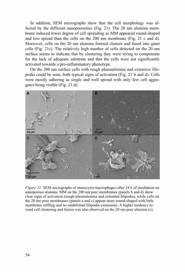
54
In addition, SEM micrographs show that the cell morphology was af-fected by the different nanoporosities (Fig. 21). The 20 nm alumina mem-brane induced lower degree of cell spreading as MM appeared round-shaped and less spread than the cells on the 200 nm membrane (Fig. 21 c and d). Moreover, cells on the 20 nm alumina formed clusters and fused into giant cells (Fig. 21c). The relatively high number of cells detected on the 20 nm surface seems to indicate that by clustering they were trying to compensate for the lack of adequate substrate and that the cells were not significantly activated towards a pro-inflammatory phenotype.
On the 200 nm surface cells with rough plasmalemma and extensive filo-podia could be seen, both typical signs of activation (Fig. 21 b and d). Cells were mostly adhering as single and well spread with only few cell aggre-gates being visible (Fig. 21 d).
Figure 21. SEM micrographs of monocytes/macrophages after 24 h of incubation on nanoporous alumina. MM on the 200 nm pore membranes (panels b and d) show clear signs of activation (rough plasmalemma and extended filipodia), while cells on the 20 nm pore membranes (panels a and c) appear more round-shaped with little membrane ruffling and no established filipodia extensions. A higher tendency to-ward cell clustering and fusion was also observed on the 20 nm pore alumina (c).

55
Release of pro-inflammatory cytokines TNF-α and IL-1ß are potent inflammatory cytokines capable of activating more inflammatory cells and enhancing the localized concentration of cyto-kines and growth factors. These cytokines are important indicators of MM activation and also contribute to the repair phase of tissue healing [118, 119].
Results showed that MM on the 200 nm alumina membranes secreted significantly higher levels of IL-1ß than the cells attached to the 20 nm alu-mina (Fig. 22a). The same tendency was observed for TNF-α secretion (Fig. 22b). Hence, the analysis of the pro-inflammatory cytokines confirmed the distinct activation states of MM on 20 and 200 nm membranes depicted by the microscopy analyses.
LDH activity was measured to determine if cytokine release was caused by the loss of cell integrity as a consequence of different cytotoxic effects by the biomaterials. Cell viability on both alumina membranes was comparable with Thermanox, a commonly used tissue culture substrate. Additionally, the studied materials showed no significant signs of toxicity. Thus, results indi-cated that cytokine release was a consequence of cell activation rather than a passive release caused by disruption of cell integrity.
The effect of surface topography on MM adhesion and activation can be explained by two mechanisms: an indirect effect of nanotopography on pro-tein adsorption in terms of amount, conformational changes, orientation and exposure of binding sites, and/or a direct effect of the nanotopography on cell behavior with cells being capable of sensing nanometric structures and responding to these features [19-21] .
Innumerable studies dealing with the role of biomaterial-adsorbed pro-teins on monocyte/macrophage behaviour have been done [120, 121]. How-ever, to elucidate the effect of specific adsorbed proteins on MM adhesion and activation from the literature is not a simple task. In vitro studies present a wide spectrum of materials where chemistry and topography have not al-ways been varied in a systematic and truly comparable manner. In addition, there is a great variation in the use of proteins as well as their concentrations and the conditions for the adsorption processes [122-128].

56
Figure 22. Cytokines released by MM after 24 h of incubation with 20 nm and 200 nm pore alumina. A significant difference between the two materials was observed for IL-1ß release (p<0.001), as well as a clear tendency for TNF-α.
There is a consensus about the role of fibrinogen and C3 in promoting
MM adhesion to biomaterial surfaces [129, 130]. However the effect of ad-sorbed proteins on cytokine release varies between different studies. For instance, Shen et al. describes that fibrinogen adsorbed on several polymer surfaces does not promote TNF-α release [125]. They propose that monocyte integrin receptors participate mostly in cell adhesion, rather than directly activating MM for cytokine release. On the contrary, Gretzer [131] describes a fibrinogen-mediated pro-inflammatory effect with increased monocyte secretion of IL-1ß and TNF-α and decreased secretion of IL-10. Interactions of adsorbed IgG with monocyte Fc receptors are also documented to pro-mote the release of IL-1ß and TNF-α by adherent monocytes [132]. Jenney and Anderson [123] proposed that preadsorbed IgG promotes long term

57
macrophage adhesion, an effect that is mediated by Fab and F (ab´)2 frag-ments instead of the Fc fragment.
In our previous work we demonstrated that 200 nm alumina adsorbed sig-nificantly higher amounts of the plasma proteins IgM, IgG, C3 and C1q, than the 20 nm membrane (paper I) [93]. Thus the results on MM adhesion pre-sented in this paper prove that not only the amount of adsorbed proteins but also the protein conformation and orientation play an important role for cel-lular adhesion. Furthermore, MM seem to “sense” the topography and as a result expose different membrane receptors [19, 133]. Therefore, we hy-pothesize that the reduced number of adherent cells on the 200 nm alumina could be related to both protein conformation and MM “sensing” the nanotopography, leading to less favourable exposure of binding-receptors. Instead MM seem to adopt a configuration favourable for exposure of activa-tion-receptor binding sites (e.g. receptors for Fc) and as a result they become highly activated thus producing high levels of cytokines.
Concerning the increased cell attachment on the 20 nm alumina, we speculate that the surface promotes not only exposure of MM adhesion-receptor binding sites, but cells also express receptors favouring giant cell formation. However despite the high cell number the cytokine release is much less pronounced than on the 200 nm membrane. Hence as proposed by Young el al [134], greater activation energy is most likely required to acti-vate the receptors of monocytes/macrophages attached to the 20 nm porous surface as compared to the 200 nm surface.
C5a and C3a are anaphylatoxins generated when the complement system is activated. C5a stimulates the release of inflammatory cytokines by MM [33]. C3a has also been described to enhance TNF-α and IL-1ß synthesis by adherent monocytes [135]. As blood exposure to 200 nm alumina leads to much higher complement activation than when exposed to the 20 nm pore membranes [93], the increased levels of C5a and C3a could also contribute to the observed difference in cytokine secretion between the membranes.
The present work shows that by changing the nanopore size of alumina it is possible to modulate MM behaviour. In addition it opens the possibility of exploiting some of the beneficial functions of macrophages in promoting biocompatibility and avoiding harmful behaviour.

58
Conclusions The data presented in this work highlights the significance of biomaterial nanotexture for inflammatory response. Additionally it emphasizes the im-portance of characterizing material topography in the nanometer range.
Results showed that a difference in nanoporesize of 180 nm in diameter has a huge effect on complement and platelet activation, as well as on mono-cyte/macrophage behaviour.
Alumina with 200 nm pores promotes higher complement activation than alumina with 20 nm pores.
Platelets also respond differently depending on the poresize. They adhere and become activated when in contact with 20 nm pores. However, no plate-let microparticles are generated on this surface. On the 200 nm pore size alumina platelets adhere and then “bounce off”, so called transient contact, which is enough to activate the platelets and stimulate the generation and adhesion of microparticles. The fact that PMP are absent in the fluid phase is a huge advantage as the amplification of non-desirable reactions will not take place. Finally alumina with 200 nm pores is less thrombogenic than alumina with 20 nm pores.
The time course study of blood activation identified platelet/PMP adhe-sion and thrombospondin-1 release as the main contributors to the different kinetics of blood activation by the two nanoporous alumina membranes.
A clear difference in monocyte/macrophage adhesion and activation was also found between the membranes. Few but highly activated cells adhered to the 200 nm pore alumina in contrast to many but less activated mono-cytes/macrophages on the 20 nm pore surface.
Finally, the question arises: Which pore size would be more favourable to use on a biomaterial? Alumina with 20 nm pores seems more promising since it induces lower complement activation and less monocyte/macrophage activation. However, the high thrombogenecity makes it a less favourable candidate for use in blood contact devices. Hence, the choice will depend on the specific application. Additionally in vivo studies are required to give a more complete picture.
Nevertheless, regardless of the specific clinical applications of these types of surfaces the results presented in this thesis underline the role of nanotex-ture on dictating inflammatory responses and imply that nanotopography can be exploited to subtly control the inflammatory potential of biomaterials.

59
Future perspectives In vitro tests are the first level of evaluating biomaterial biocompatibility since they are less expensive, allow for direct comparison of different mate-rials, a large number of factors can be examined simultaneously and vari-ables can be more easily controlled [136]. However, the ultimate test of any biomaterial will be its performance in vivo. The exciting results presented in this work push forward the investigation towards in vivo experiments.
Implantation of the alumina membranes in animal models is therefore the next step. A first approach will be to measure acute inflammatory responses after implantation in the peritoneal cavity of mice. After approximately 16 h the materials will be explanted and the membranes and surrounding fluid will be analyzed by looking at cell recruitment and activation. The response will be the result of the interplay between the different systems in the body.
Moreover, in vivo studies can also be designed to obtain information on long- term host response to the alumina membranes e.g. fibrous capsule for-mation. To obtain such information subcutaneous implantation over 2-4 weeks could be done, followed by histological analyses of the implanted membranes and of the surrounding tissues.
The results concerning PMP production and adhesion exclusively after blood contact with 200 nm pore alumina triggers questions such as “will only certain types of PMP adhere to this membrane and does it depend on their antigen characteristics?”.
Due to the increasing evidence of platelet microparticles as pathogenic markers [45, 47, 48], the development of nanoporous alumina as a diagnostic tool has great potential. It is however primarily essential to classify type and origin of the microparticles. PMP could be produced by using different agonists (e.g. calcium ionophore, thrombin receptor activating peptide (TRAP), epinephrine or collagen), and after characterizing them with flow cytometry, they would be incubated with 200 nm pore sized alumina. In this way one could reveal the antigen composition of the microparticles that ad-here to this membrane. The experiment could then be taken to the next level by producing a set of membranes with different pore sizes between 100 and 300 nm in order to evaluate if by changing the nanoporosity one can target specific microparticle adhesion.

60
Acknowledgements
First of all I would like to express my gratitude to Dr. Marjam Ott and Prof. Jan Carlsson for giving me the opportunity to come and work with them. Marjam, the best supervisor one can wish for! It has been great to work with you and not to be forgotten, I have learnt a lot. Thank you for being so posi-tive and encouraging. Thanks for your friendship and the nice times that we spent together. I´ll never forget our “reflections” about science that after a while ended up as reflections about anything and everything �. Jan: thanks for your enthusiasm, all the brainstorming and for reminding me that science can be a lot of fun. Dr. Jaan Hong, my co-supervisor, thank you for stimulating discussions and for always taking the time to help me in the lab. It has been a lot of blood experiments.... I also would like to thank: Prof. Karin Caldwell for always having your door open and for your valua-ble comments to my thesis All present and former colleagues and friends at Surface Biotechnology, specially Fernanda, Karin, Jörgen, Lena, Torgny junior, Tomas, Adam, Ro-bert, Hans and Olov for the pleasant company and the friendly working cli-mate. Olov: a special thanks to you for all the help with my pictures and most im-portantly for the nice cafecito. Fer: thanks for your friendship and for always listening. Uppsala and I miss you. Ulla Widenberg: thanks for being so kind and helpful. The Physical Chemistry group, Karin, Malin, Anna, Victor, Per and Johan for the nice lunch company.
61
I am very grateful to the people at the Department of Oncology, Radiology and Clinical Immunology, Division of Clinical Immunology for their kind-ness and lot of help. It has been very nice to work at your department! Prof. Bo Nilsson for valuable discussions and Prof. Kristina Nilsson Ekdahl for taking the time to read my thesis and your valuable comments. Graciela Elgue and Javier Sanchez for the nice time in the lab practicing Spanish. Graciela: ¿Qué habría hecho yo en el Rudbeck sin tu ayuda? Gracias, che! Javier: gracias por tu interés en mi proyecto y por tus consejos. Lillemor Funke, many thanks for your help with reagents, protocols, every-thing! Jennie Bäck, thanks for your help with the platelet analysis protocols and for drawing blood for my experiments. Linda Mathsson, thanks for your advice regarding monocyte isolation. Also many thanks to Leif Ljung, at the Dept. of Medical Cell Biology for all the help with the SEM studies. Jan Grawé and Dirk Pacholsky at the Cell Analysis Core Facility, Rudbeck Laboratory thanks for excellent technical assistance with the flow cytometry studies and with the confocal microscope. Pacho: thanks for all the hours you spent working with my pictures. Prof. Matteo Santin, School of Pharmacy and Biomolecular Sciences, Uni-versity of Brighton, UK, thank you for welcoming me to your laboratory, for introducing me to the work with macrophages and for your valuable contri-bution to my article. Prof. Andreas Heilmann, Andreas Hoess and Annika Thormann, Fraunhofer Institute for Mechanics of Materials, Halle, Germany, thank you for answer-ing all my questions about nanoporous alumina fabrication. Prof. Liping Tang, Biomedical Engineering Program, University of Texas at Arlington, USA, thank you for sharing your knowledge about in vivo stu-dies and for stimulating discussions. Parisa Lotfi: thank you for staying late in the lab helping me to finish my work in Arlington and for the nice Texas sightseeing. A special thanks to all the people who kindly donated blood for the experi-ments.

62
I would like to thank those outside the University doors, who also contri-buted to this thesis with their support, encouragement and friendship: The Biochemistry Department in Montevideo: Miriam, Paula, Gabriela, Ce-cilia, Karen, Carmen, Beatriz, Laura and Batista for keeping contact during all these years. So many nice memories about my time in Bioquímica. Special thanks to Carmen “mi mamá científica” for getting me starting with research work and to Laura for the yearly visits to Uppsala. My friends at home, José, Alejandro, Diego, Seba, Darling, Bettina, Horacio: because of you, every visit to Uruguay seemed like I had never left. El próximo asadito lo hacemos en NP! A super special thanks to “The Ladies”, Yamila, Claudia, Andrea and Ange-la for always being there for me. Your emails have made it so easy to feel close to all of you. I´m so lucky to have friends like you! A warm thanks to Fari, Jan, Mina, Sascha, Marjam and Nora for making me feel part of the family. The Andersson family, thanks for your warm welcome to the family and for making Sweden my second home. Finally, thanks to my main source of love and encouragement: my brother Javier, my mother and Peter. Javi: thanks for always being there. Mamá: You are such a great mum! There is no need to write in Spanish to you because your English has become so good. I´m very proud of you! Peter: Du fyller mina dagar med glädje.
Natalia
63
Summary in Swedish
Det är allmänt känt att in vivo system styrs av molekylärt beteende på nano-meternivå. Nanovetenskapen skapar därför många intressanta möjligheter inom medicin och biologi. Inom nanoforskningen utvecklas teknik som kan ”skaka hand” med kroppens celler t.ex. genom att på ett kontrollerat sätt styra adsorptionen av proteiner samt påverka deras konformation och orien-tering på en yta för att sedan ge avsedd effekt i kontakt med specifika celler. Ett exempel på lovande medicinska tillämpningar av nanoforskningen är implantat med ytor som är formgivna ända ned på nanonivå för att implanta-ten smidigt skall kunna integreras med kroppsegna vävnader och organ.
Biokeramen aluminiumoxid har använts som implantat material i stor om-fattning beroende på dess korrosionstålighet och biokompatibilitet dvs låga påverkan på och av den omkringliggande biologiska miljön Denna typ av aluminiumoxid är normalt sintrad och har därför en relativt oregelbunden yta. Ett annat sätt att tillverka aluminiumoxid är att använda en metod som kallas anodisering och sedan länge använts i industrin för att i stor skala till låg kostnad tillverka korrosionsresistenta aluminiumoxid delar. Metoden utförs mha elektrolytisk oxidation i en syra tex fosforsyra. Som resultat er-hålls hexagonalt packade rader av cylindriska celler med parallella nanopo-rer (10-400 nm i diameter, beroende på betingelserna). Detta på nanonivå extremt välorganiserade material har visats sig optimalt för att studera nano-strukturers påverkan på proteiner och celler.
Ett biomaterials interaktion med biologisk vävnad är ett komplicerat fe-nomen som i ett första skede består av adsorption av proteiner på materialets yta. Detta är en dynamisk process som involverar en serie konformations-ändringar och utbyten av proteiner. Utbytet påverkas av den totala protein-koncentrationen samt materialegenskaper såsom ytenergi, kemisk samman-sättning och ytstruktur. De efterföljande reaktionerna, dvs aktivering av blo-dets olika kaskadsystem samt rekrytering, inbindning och aktivering av blodceller regleras av adsorberade proteiner. Förändringar beträffande typ, storlek, konformation och orientering av dessa proteiner har en kraftfull in-verkan på cellers beteende vid biomaterial-vävnadsgränssnittet.
I denna avhandling har in vitro-studier utförts för att studera betydelsen av aluminiumoxidens nanoporositet vad gäller den inflammatoriska respon-sen. Aluminiumoxidmembran med två porstorlekar (20 och 200 nm i diame-ter) har använts. Komplement- och trombocytaktivering har utvärderats lik-som monocyt/makrofag respons.

64
Aluminiumoxid-membran inkuberades med helblod i en blodkammarmo-dell. Denna in vitro modell gjorde det möjligt att analysera protein och cellinfästning till membranytorna samt de aktiveringsprodukter som bildades i blodet. Blodet analyserades med avseende på produkter från komplement- (C3a och sC5b-9), trombocyter- (ß-thromboglobulin, thrombospondin-1, trombocytmikropartiklar) och koagulationsaktivering (trombin-antitrombin komplex). Ytadsorberade proteiner eluerades och ”dot blotting” utfördes för att bestämma förekomsten av IgG, IgM, C1q och C3. Trombocytadhesion till materialytorna detekterades med svepelektronmikroskopi samt immunocyto-kemisk färgning och konfokalmikroskopi. Särskild uppmärksamhet ägnades åt cellmorfologin, mikropartikelinbindning och uttryck av P-selektin. Trom-bocytaktivering och vidhäftning utvärderades även i trombocytrik plasma (PRP).
I en separat studie följdes kinetiken för komplement- och trombocytakti-vering i upp till 4 timmar.
Slutligen undersöktes aktivering och morfologi av humana monocyter i kontakt med nanoporös aluminiumoxid genom att studera dessas morfologi samt frisättning av proinflammatoriska cytokiner.
Resultaten visade att aluminiumoxid med 200 nm porer orsakade högre komplementaktivering än aluminiumoxid med 20 nm porer. Blod som inku-berats med aluminiumoxid med 200 nm porer gav två gånger högre C3a och fem gånger högre sC5b-9 utsläpp jämfört med 20 nm membran. Dot blot resultaten visade dessutom att alla studerade proteiner adsorberade mer till 200 nm aluminiumoxid än till 20 nm membran. Proteinanalysresultaten kor-relerar således väl med de höga halter av aktiveringsprodukter som hittades efter inkubation med 200 nm membran.
Trombocyternas reaktion på nanoporös aluminiumoxid visade sig även den vara kraftigt beroende av porstorlek. Aktiverade trombocyter kunde observeras på membran med 20 nm porer men gav inga spår av mikropartik-lar. Denna yta visade även ökad benägenhet att aktivera koagulation. På ytan med 200 nm porer fanns endast ett fåtal trombocyter men istället kunde en mängt kluster av mikropartiklar ses. På denna yta har trombocyterna alltså en förmåga att ”fastna” aktiveras och sedan lämna ytan igen. Denna yta hade dessutom lägre benägenhet att aktivera koagualtion.
Inkubering med trombocytrik plasma visade avvikande resultat jämfört med helblod. Ytan med 20 nm porer var ”ren” dvs inga trombocyter hade fastnat. På ytan med 200 nm porer kunde däremot fortfarande kluster av mikropartiklar observeras. Dessa resultat tyder på att röda blodkroppar och/eller leukocyter har en kraftfull påverkan på trombocyternas vidhäft-ningsförmåga på nanoporös aluminiumoxid. Detta resultat visar även vikten av att arbeta med helblod jämfört med system med separerade blodkompo-nenter.

65
I kinetikstudien identifierades trombocyter/trombocytmikropartikel-inbindning och frisättning av trombospondin-1 som de största bidragsgivarna till de olika effekter som de två nanoporösa aluminiumoxidmembranen gav upphov till. På membranet med 20 nm porer ökade antalet trombocyter som funktion av inkubationstiden till skillnad från aluminiumoxid med 200 nm porer där ingen ökning kunde observeras. För membran med 20 nm porer registrerades ett signifikant utsläpp av trombospondin-1 efter 60 min. För aluminiumoxid med 200 nm porer skedde detta inte förrän efter 240 min inkubering i helblod.
En tydlig skillnad i monocyt/makrofag-inbindning och aktivering påvisa-des mellan de två membranen. Få men mycket aktiverade celler band till membran med 200 nm porer i motsats till många men mindre aktiverade monocyter/makrofager på 20 nm membranen. Detta konstaterades både ge-nom inspektion av cellmorfologi samt genom mätning av de inflammatoriska cytokinerna, TNF-α and IL-1ß.
Det är troligt att den största anledningen till de observerade skillnaderna mellan testmaterialen beror på vilka proteiner som adsorberas, hur mycket samt deras konformation och orientering. Olika pordiameter medför en skill-nad i tillgänglig yta för proteiner beroende på deras storlek och form. Stora proteiner kan binda till insidan av 200 nm porerna medan de bara binds till den makroskopiska ytan på 20 nm membran. Således ger 200 nm alumini-umoxid en högre tillgänglig ytarea. Man kan också spekulera i att på 200 nm membran har proteinerna större frihet att anta olika konformationer vilket kan göra att specifika bindningsställen blir tillgängliga. Dessa fenomen i kombination med att cellerna direkt känner av och reagerar på den nanomet-riska strukturen är sannolikt ”förklaringen” till de observerade proinflamma-toriska skillnaderna mellan aluminiumoxid med 20 nm porer och alumini-umoxid med 200 nm porer.
Sammanfattningsvis verkar aluminiumoxid med 20 nm porer vara en bätt-re kandidat för biomaterial eftersom den uppvisar mindre komplementakti-vering och orsakar ett mindre aktiverat tillstånd hos vidhäftande monocy-ter/makrofager. Men den högre trombogeniciteten hos detta membran jäm-fört med 200 nm aluminiumoxid gör det till en sämre kandidat för använd-ning i blodkontakt. Därför kommer valet att avgöras av det specifika användningsområdet. Oberoende av den kliniska tillämpningen för dessa typer av ytor understryker resultaten som presenteras i denna avhandling den roll nanotexturen spelar för det inflammatoriska svaret, vilket innebär att nanotopografi kan utnyttjas genom att på ett subtilt sätt kontrollera de in-flammatoriska egenskaperna hos ett biomaterial.

66
References
1. Ratner, B., Biomaterials science: an interdisciplinary endeavor, in
Biomaterials Science: An Introduction to Materials in Medicine, B. Ratner, et al., Editors. 2004, Academic Press: San Diego. p. 1-9.
2. Alexander, H., et al., Classes of materials used in medicine, in Bio-materials Science: An Introduction to Materials in Medicine, B. Ratner, et al., Editors. 1996, Academic Press: San Diego. p. 37-83.
3. Ratner, B. and S. Bryant, Biomaterials: Where we have been and where are we going. Annu Rev Biomed Eng, 2004 6: p. 41-75.
4. Tirrell, M., E. Kokkoli, and M. Biesalski, The role of surface science in bioengineered materials. Surface Science, 2002. 500: p. 61-83.
5. Park, J., C. Gemmell, and J. Davies, Platelet interactions with tita-nium: modulation of platelet activity by surface topography. Bioma-terials, 2001. 21: p. 2671-2682.
6. Eriksson, C., J. Lausmaa, and H. Nygren, Interactions between hu-man whole blood and modified TiO2 surfaces: influence of surface topography and oxide thickness on leukocyte adhesion and activa-tion. Biomaterials, 2001. 22: p. 1987-1996.
7. Flemming, R., et al., Effects of synthetic micro and nano-structures surfaces on cell behaviour. Biomaterials, 1999. 20: p. 573-588.
8. Curtis, A. and C. Wilkinson, Topographical control of cells. Bioma-terials, 1997. 18: p. 1573-1583.
9. Curtis, A. and C. Wilkinson, Nanotechniques and approaches in biotechnology. Trends in Biotechnology, 2001: p. 97-101.
10. Goldberg, M., R. Langer, and X. Jia, Nanostructured materials for applications in drug delivery and tissue engineering. J Biomater Sci Polymer Edn, 2007. 18: p. 241-268.
11. Whitesides, G., The right size in nanobiotechnology. Nature Bio-technology, 2003. 21: p. 1161-1165.
12. Webster, T., et al., Nano-biotechnology: carbon nanofibers as im-proved neural and orthopaedic implants. Nanotechnology 2004. 15: p. 48-54.
13. Webster, T., R. Siegel, and R. Brizios, Osteoblast adhesion on na-nophase ceramics. Biomaterials, 1999. 20: p. 1221-1227.
14. Yim, E. and K. Leong, Significance of synthetic nanostructures in dictating cellular response. Nanomedicine: Nanotechnology, Biolo-gy and Medicine, 2005. 1: p. 10-21.

67
15. Price, R., K. Haberstroh, and T. Webster, Enhanced functions of osteoblasts on nanostructured surfaces of carbon and alumina. Med Biol Eng Comput, 2003. 41: p. 372-375.
16. Curtis, A., et al., Cells react to nanoscale order and symmetry in their surroundings. IEEE Transactions on nanobioscience, 2004. 3: p. 61-65.
17. Nguyen, K., et al., Cellular and molecular responses of smooth mus-cle cells to surface nanotopography. Journal of Nanoscience and Nanotechnology, 2007. 7: p. 2823-2832.
18. Karlsson, M., et al., Nanoporous aluminum oxide affects neutrophil behaviour. Microscopy Research and Technique, 2004. 63: p. 259-265.
19. Kriparamanan, R., et al., Nanotopography: Cellular responses to nanostructured materials. J Nanosci Nanotechnol 2006. 6: p. 1905-1010.
20. Stevens, M. and J. George, Exploring and engineering the cell sur-face interface. Science, 2005. 310: p. 1135-1138.
21. Martinez, E., et al., Effects of artificial micro- and nano-structured surfaces on cell behaviour. Ann Anat, 2009. 191: p. 126-135.
22. Anderson, J., Inflammation, wound healing and the foreign body response, in Biomaterials Science: An introduction to materials in medicine, H.A. Ratner BD, Schoen FJ, Lemons JE, Editor. 2004, Academic Press: San Diego. p. 296-304.
23. Anderson, J.M., A. Rodriguez, and D.T. Chang, Foreign body reac-tion to biomaterials. Semin Immunol, 2008. 20(2): p. 86-100.
24. Tang, L. and J. Eaton, Inflammatory response to biomaterials. Am J Clin Pathol, 1995. 103: p. 466-471.
25. Schoen, F., Introduction Host reactions to biomaterials and their evaluation, in Biomaterials Science: An introduction to materials in medicine, H.A. Ratner BD, Schoen FJ, Lemons JE, Editor. 2004, Academic Press: San Diego. p. 293-296.
26. Horbett, T., The role of adsorbed proteins in tissue response to bio-materials, in Biomaterials Science: An Introduction to Materials in Medicine, H.A. Ratner BD, Schoen FJ, Lemons JE, Editor. 2004, Academic Press: San diego. p. 237-246.
27. Wilson, C., et al., Mediation of biomaterial-cell interactions by ad-sorbed proteins: A review. Tissue Engineering, 2005. 11: p. 1-18.
28. Wahlgren, M. and T. Arnebrant, Protein adsorption to solid surfac-es. TIBTECH, 1991. 9(201-208).
29. Kasemo, B., Biological Surface Science. Surface Science, 2002. 500: p. 656-677.
30. Kasemo, B. and J. Gold, Implant surfaces and the interface processes. Adv Dent Res 1999. 13: p. 8-20.
31. Walport, M., Complement, in Immunology, I. Roitt, J. Brostoff, and D. Male, Editors. 1998, Mosby International Ltd: London. p. 43-59.

68
32. Gorbet, M. and M. Sefton, Biomaterial-associated thrombosis: roles of coagulation factors, complement, platelets and leukocytes. Bio-materials, 2004. 25: p. 5681-5703.
33. Nilsson, B., et al., The role of complement in biomaterial-induced inflammation. Molecular Immunology, 2007. 44: p. 82-94.
34. Wetterö, J., Acute inflammation on model biomaterial surfaces. 2002, Linköping University: Linköping
35. Andersson, J., Complement activation triggered by biomaterial sur-faces. 2003, Uppsala University: Uppsala.
36. Marcus, A., Platelets: Their role in hemostasis, thrombosis and in-flammation, in Inflammation: Basic Principles and Clinical Corre-lates, J. Gallin and R. Snyderman, Editors. 1999, Lippincott Wil-liams & Wilkins: Philadelphia. p. 77-95.
37. Vogler, E.A. and C.A. Siedlecki, Contact activation of blood-plasma coagulation. Biomaterials, 2009. 30(10): p. 1857-69.
38. Hong, J., et al., Material-specific thrombin generation following contact between metal surfaces and whole blood. Biomaterials, 2005. 26(12): p. 1397-403.
39. Sanchez, J., et al., Measuring the degree of plasma contact activa-tion induced by artificial materials. Thrombosis Research, 2002. 105: p. 407-412.
40. Gorbet, M.B. and M.V. Sefton, Leukocyte activation and leukocyte procoagulant activities after blood contact with polystyrene and po-lyethylene glycol-immobilized polystyrene beads. J Lab Clin Med, 2001. 137(5): p. 345-55.
41. Savage, B. and Z. Ruggeri, Platelet thrombus formation in flowing blood, in Platelets, A. Michelson, Editor. 2002, Academic Press: San Diego. p. 215-228.
42. Bouchard, B., et al., Interactions between platelets and the coagula-tion system, in Platelets, A. Michelson, Editor. 2002, Academic Press: San Diego. p. 229-253.
43. Horstman, L. and Y. Ahn, Platelet microparticles: a wide angle perspective. Critical reviews in Oncology/Hematology, 1999. 30: p. 111-142.
44. Nieuwland, R. and A. Sturk, Platelet derived microparticles, in Platelets, A. Michelson, Editor. 2002, Academic Press: San Diego. p. 255-265.
45. Simak, J. and P. Gelderman, Cell membranes microparticles in blood and blood products: potentially pathogenic agents and diag-nostic markers. Transfusion medicine reviews, 2006. 20: p. 11-26.
46. Nomura, S., Y. Ozaki, and Y. Ikeda, Function and role of micropar-ticles in various clinical settings. Thrombosis Research, 2008. 123: p. 8-23.
47. Boulanger, C., N. Amabile, and A. Tedgui, Circulating micropar-ticles: A potential prognosis marker for atherosclerotic vascular disease. Hypertension, 2006. 48: p. 180-186.

69
48. Ardoin, S., J. Shanahan, and D. Pisetsky, The role of microparticles in inflammation adn thrombosis. Scand J Immunol, 2007. 66(159-165).
49. Broberg, M., C. Erikksson, and H. Nygren, GPIIb/IIIa is the main receptor for initial platelet adhesion to glass and titanium surfaces in contact with whole blood. J Lab Clin Med, 2002. 139: p. 163-172.
50. Tsai, W., et al., Platelet adhesion to polyestyrene-based surfaces preadsorbed with plasma selectively depleted in fibrinogen, fibro-nectin, vitronectrin or von Willebrand’s factor. J Biomed Mater Res, 2002. 60: p. 348-359.
51. Grunkemeier, J., et al., The effect of adsorbed fibrinogen, fibronec-tin, von Willebrand factor and vitronectin on the procoagulant state of adherent platelets. Biomaterials, 2000. 21: p. 2243-2252.
52. Broberg, M. and H. Nygren, Platelet interactions with surface-adsorbed plasma proteins: exposure of CD62P induced by von Wil-lebrand factor. Colloids and Surfaces B: Biointerfaces, 1998. 11: p. 67-77.
53. Gemmell, C., E. Yeo, and M. Sefton, Flow cytometric analysis of material-induced platelet activation in a canine model:elevated mi-croparticle levels and reduced life span. J Biomed Mater Res, 1997. 37: p. 176-181.
54. Gemmell, C., et al., Platelet activation in whole blood by artificial surfaces: identification of platelet-derived microparticles and acti-vated platelet binding to leukocytes as material-induced activation events. J Lab Clin Med, 1995. 125: p. 276-287.
55. Sims, P. and T. Wiedmer, The response of human platelets to acti-vated components of the complement system. Immunology Today, 1991. 12: p. 338-342.
56. Sims, P. and T. Wiedmer, Induction of cellular procoagulant activity by the membrane attack complex of complement. Seminars in Cell Biology, 1995. 6: p. 275-282.
57. Peerschke, E., K. Reid, and B. Ghebrehiwet, Platelet activation by C1q results in the induction of alpha-IIb/ßIIIa (GPIIb-IIIa) and the expression of P-selectin and procoagulant activity. J Exp Med, 1993. 178: p. 579-587.
58. Beelen, R., M. Betjes, and E. Kamperdijk, Macrophages: General Aspects, in Immunopharmacology of macrophages and other anti-gen-presenting cells, C. Bruijnzeel-Koomen and E. Hoefsmit, Edi-tors. 1994, Academic Press: London. p. 1-6.
59. Rook, G. and F. Balkwill, Cell-mediated Immune Reactions, in Im-munology, I. Roitt, J. Brostoff, and D. Male, Editors. 1998, Mosby International Ltd: London. p. 121-138.
60. Xia, Z. and J. Triffitt, A review on macrophages responses to bioma-terials. Biomedical Materials 2006. 1: p. R1-R9.
61. Thompson, G., Porous anodic alumina: fabrication, characteriza-tion and applications. Thin Solid Films, 1997. 297: p. 192-201.

70
62. Jessensky, O., M. Muller, and U. Gösele, Self-organized formation of hexagonal pore structures in anodic alumina. J Electrochem Soc, 1998. 145: p. 3735-3740.
63. Lanzerotti, L., et al., Nucleation and growth of porous anodic films on aluminium. Nature Biotechnology, 1978. 272: p. 433-435.
64. Thormann, A., et al., Nanoporous aluminium oxide membranes for filtration and biofunctionalization. Small, 2007. 6: p. 1032-1040.
65. Karlsson, M., et al., Initial in vitro interaction of osteoblasts with nano-porous alumina. Biomaterials, 2003. 24: p. 3039-3043.
66. Wieneke, H., et al., Synergistic effects of a novel nanoporous stent coating and tacrolimus on intima proliferation in rabbits. Catheteri-zation and Cardiovascular Interventions, 2003. 60: p. 399-407.
67. La Flamme, K., et al., Biocompatibility of nanoporous alumina membranes for immunoisolation. Biomaterials, 2007. 28: p. 2638-2645.
68. Mendonca, G., et al., Nanostructured alumina-coated implant sur-face: effect on osteoblast-related gene expression and bone-to-implant contact in vivo. Int J Oral Maxillofac Implants, 2009. 24(2): p. 205-15.
69. Björk, I. and U. Lindahl, Mechanism of the anticoagulant action of heparin. Molecular and cellular biochemistry, 1982. 48: p. 161-182.
70. Christensen, K., et al., Heparin coating of the stent graft-effects on platelets, coagulation and complement activation. Biomaterials, 2001. 22: p. 349-355.
71. Johnell, M., et al., Coagulation, fibrinolysis, and cell activation in patients and shed mediastinal blood during coronary artery bypass grafting with a new heparin-coated surface. J Thorac Cardiovasc Surg, 2002. 124(2): p. 321-32.
72. Johnell, M., et al., Cell adhesion and tissue factor upregulation in oxygenators used during coronary artery bypass grafting are mod-ified by the Corline Heparin Surface. Scand Cardiovasc J, 2002. 36(6): p. 351-7.
73. Johnell, M., R. Larsson, and A. Siegbahn, The influence of different heparin surface concentrations and antithrombin-binding capacity on inflammation and coagulation. Biomaterials, 2005. 26(14): p. 1731-9.
74. Hong, J., et al., A new in vitro model to study interaction between whole blood and biomaterials. Studies of platelet and coagulation activation and the effect of aspirin. Biomaterials, 1999. 20: p. 603-611.
75. Steward, M. and D. Male, Immunological techniques, in Immunolo-gy, I. Roitt, J. Brostoff, and D. Male, Editors. 1998, Mosby Interna-tional Ltd: London. p. 386.
76. Nilsson Ekdahl, K., et al., Generation of iC3 at the interface be-tween blood and gas. Scand J Immunol, 1992. 35: p. 85-91.

71
77. Mollnes, T., et al., Quantification of the terminal complement com-plex in human plasma by an enzyme-linked immunosorbent assay based on monoclonal antibodies against a neoantigen of the com-plex. Scand J Immunol 1985. 22: p. 197-202.
78. Longobardi Givan, A., Flow Cytometry First Principles. 2nd ed. 2001, New York: Wiley-Liss, Inc.
79. Nomura, S. and S. Fukuhara, Platelet microparticles, in Platelets and megakaryocytes, Methods in molecular biology, J. Gibbins and M. Mahaut-Smith, Editors. 2004, Humana Press Inc: Totawa. p. 260-277.
80. Rasband, W.S., ImageJ. 1997-2006, U. S. National Institutes of Health, Bethesda Maryland, USA.
81. Clemetson, K., Platelets receptors, in Platelets, A. Michelson, Edi-tor. 2002, Academic Press: San Diego. p. 65-84.
82. Karlsson-Parra, A., et al., A simple immunoenzyme batch staining method for the enumeration of peripheral human T lymphocyte sub-sets. J Immunol Methods, 1983. 64: p. 85-90.
83. Grunkemeier, J., W. Tsai, and T. Horbett, Hemocompatibility of treated polystyrene substrates: contact activation, platelet adhesion, and procoagulant activity of adherent platelets. J Biomed Mater Res, 1998. 41: p. 657-670.
84. Gemmell, C., A flow cytometric immunoassay to quantify adsorption of complement activation products (iC3b, C3d, SC5b-9) on artificial surfaces. J Biomed Mater Res, 1997. 37: p. 474-480.
85. Cheung, A., et al., Activation of complement by hemodialysis mem-branes: polyacrylonitrile binds more C3a than cuprophan. Kidney Int, 1990. 37: p. 1055-1059.
86. Tengvall, P., A. Askendal, and I. Lundström, Ellipsometric in vitro studies on the activation of complement by human immunoglobulins M and G after adsorption to methylated silicon. Colloids and surfac-es B: Biomaterials, 2001. 20: p. 51-62.
87. Tengvall, P., A. Askendal, and I. Lundström, Studies on protein adsorption and activation of complement on hydrated aluminium surfaces in vitro. Biomaterials, 1998. 19: p. 935-940.
88. Andersson, J., et al., Binding of C3 fragments on top of adsorbed plasma proteins during complement activation on a model bioma-terial surface. Biomaterials, 2005. 26: p. 1477-1485.
89. Wetterö, J., et al., On the binding of complement to solid artificial surfaces in vitro. Biomaterials, 2002. 23: p. 981-991.
90. Godo, M. and M. Sefton, Characterization of transient platelet con-tacts on a polyvinyl alcohol hydrogel by video microscopy. Biomate-rials, 1999. 20: p. 1117-1126.
91. Gemmell, C., Activation of platelets by in vitro whole blood contact with materials: increases in microparticle, procoagulant activity, and solubleP-selectin levels. J Biomater Sci Polymer Edn, 2001. 12: p. 933-943.

72
92. Gemmell, C., et al., Platelet activation in whole blood by artificial surfaces: identification of platelet-derived microparticles and acti-vated platelet binding to leukocytes as materal-induced activation events. J Lab Clin Med, 1995. 125: p. 276-287.
93. Ferraz, N., et al., Nanoporesize affects complement activation. J Biomed Mater Res, 2008. 87A: p. 575-581.
94. Nygren, H. and M. Broberg, Specific activation of platelets by sur-face-adsorbed plasma proteins. J Biomater Sci Polymer Edn, 1998. 9: p. 817-831.
95. Gemmell, C., M. Sefton, and E. Yeo, Platelet-derived microparticle formation involves glycoprotein IIb-IIIa. The Journal of Biological Chemistry, 1993. 268: p. 14586-14589.
96. Roach, P., D. Farrar, and C. Perry, Surface tailoring for controlled protein adsorption: effect of topography at the nanometer scale chemistry. J Am Chem Soc, 2006. 128: p. 3939-3945.
97. Sutherland, D., et al., Influence of nanoscale surface topography and chemistry on the functional behaviour of an adsorbed model ma-cromolecule. Macromol Biosci, 2001. 1: p. 270-273.
98. Denis, F., et al., Protein adsorption on model surfaces with con-trolled nanotopography and chemistry. Langmuir 2002. 18: p. 819-828.
99. Minelli, C., et al., A micro-fluidic study of whole blood behaviour on PMMA topographical nanostructures. J Nanobiotechnology, 2008. 6: p. 3.
100. Krishnaswamy, S., et al., Assembly of prothrombinase complex. Methods in enzymology, 1993. 222: p. 260-280.
101. Sinauridze, E., et al., Platelet microparticle membranes have 50- to 100-fold higher specific procoagulant activity than activated plate-lets. Thromb Haemost 2007. 97: p. 425-434.
102. Perez-Pujol, S., P. Marker, and N. Key, Platelet microparticles are heterogeneous and highly dependent on the activation mechanism: studies using a new digital flow cytometer. Cytometry Part A, 2007. 71A: p. 38-45.
103. Tolentino, A. and W. Bahou, Thrombin receptors, in Platelets, A. Michelson, Editor. 2002, Academic Press: San Diego p. 117-138.
104. Hoffman, M. and D.M. Monroe, 3rd, A cell-based model of hemos-tasis. Thromb Haemost, 2001. 85(6): p. 958-65.
105. Reed, G., Platelet secretion, in Platelets, A. Michelson, Editor. 2002, Academci Press: San Diego. p. 181-195.
106. Klinger, M., Inflammation, in Platelets, A. Michelson, Editor. 2002, Academic Press: San Diego. p. 459-467.
107. Alkhamis, T., R. Beissinger, and J. Chediak, Red blood cell effect on platelet adhesion and aggeregation in low-stress shear flow. Myth or fact? ASAIO Trans, 1988. 34: p. 868-873.

73
108. Valles, J., et al., Erythrocytes metabolically enhance collagen-induced platelet responsiveness via increased thromboxane produc-tion, adenosine diphosphate release, and recruitment. Blood, 1991. 78: p. 154-162.
109. Thor, A., et al., The role of whole blood in thrombin generation in contact with various titanium surfaces. Biomaterials, 2007. 28(6): p. 966-74.
110. Bergseth, G., et al., A novel enzyme immunoassay for plasma throm-bospondin: Comparison with beta-thromboglobulin as platelet acti-vation marker in vitro and in vivo. Thrombosis Research 2000. 99: p. 41-50.
111. Wolff, R., E. Plow, and M. Grinsberg, Interaction of thrombospon-din with resting and stimulated human platelets. J Biol Chem, 1986. 261: p. 6840-6846.
112. Broberg, M. and H. Nygren, Von Willebrand factor, a key protein in the exposure of CD62P on platelets. Biomaterials, 2001. 22: p. 2403-2409.
113. Rodriguez, S., et al., Fibrinogen adsorption, platelet adhesion and activation on mixed hydroxyl-/methyl-terminated self-assembled monolayers. Biomaterials, 2006. 27: p. 5357-5367.
114. Ferraz, N., et al., Influence of nanoporesize on platelet adhesion and activation. J Mater Sci: Mater Med, 2008. 19: p. 3115-3121.
115. Ferraz, N., J. Hong, and M. Karlsson Ott, Procoagulant behavior and platelet microparticle generation on nanoporous alumina. Jour-nal of Biomaterials Applications, 2009.
116. Horbett, T., Principles underlying the role of adsorbed plasma pro-teins in blood interactions with foreign materials. Cardiovasc Pa-thol, 1993. 2: p. 137S-148S.
117. Attaluri, A., et al., Evaluation of nano-porous alumina membranes for hemodyalisis application. ASAIO Journal, 2009. 55: p. 217-233.
118. van Furth, R., Human monocytes and cytokines. Res Immunol, 1998. 149: p. 719-720.
119. Clark, R., Wound repair: overview and general considerations, in The molecular and cellular biology of wound repair, R. Clark, Edi-tor. 1996, Plenum: New York. p. 3-50.
120. Thomsen, P. and C. Gretzer, Macrophage interactions with modified materials surfaces. Current Opinion in Solid State and Materials Science, 2001. 5: p. 163-176.
121. Kao, W., Evaluation of protein-modulated machophage behavior on biomaterials: designing biomimetic materials for cellular engineer-ing. Biomaterials, 1999. 20: p. 2213-2221.
122. Kao, W., J. Hubbell, and J. Anderson, Protein-mediated macro-phage adhesion and activation on biomaterials: a model for mod-ulating cell behavior. J Mater Sci: Mater Med, 1999. 10: p. 601-605.
123. Jenney, C. and J. Anderson, Adsorbed IgG: A potent adhesive sub-strate for human macrophages. J Biomed Mater Res, 2000. 50: p. 281-290.

74
124. Jenney, C. and J. Anderson, Adsorbed serum proteins responsible for surface dependent human macrophage behavior. J Biomed Ma-ter Res, 2000. 49: p. 435-447.
125. Shen, M., et al., Effects of adsorbed proteins and surface chemistry on foreign body giant cells formation, tumor necrosis factor alpha release and procoagulant activity of monocytes. J Biomed Mater Res, 2004. 70A: p. 533-541.
126. Shen, M. and T. Horbett, The effects of surface chemistry and ad-sorbed proteins on monocyte/macrophage adhesion to chemical modified polystyrebe surfaces. J Biomed Mater Res, 2001. 57: p. 336-345.
127. Bonfield, T., E. Colton, and J. Anderson, Plasma protein adsorbed biomedical polymers: Activation of human monocytes and induction of interleukin1. J Biomed Mater Res, 1989. 23: p. 535-548.
128. Bonfield, T., et al., Cytokine and growth factor production by mono-cytes/macrophages on protein preadsorbed polymers. J Biomed Ma-ter Res, 1992 26: p. 837-850.
129. Tang, L., et al., Molecular determinants of acute inflammatory res-ponses to biomaterials. J Clin Invest, 1996. 97(5): p. 1329-34.
130. McNally, A. and J. Anderson, Complement C3 participation in mo-nocyte adhesion to different surfaces. Proc Natl Acad Sci USA, 1994. 91: p. 10119-10123.
131. Gretzer, C., Macrophage-Material Surface Interactions. 2000, Göteborg University: Göteborg.
132. van de Winkel, J. and P. Capel, Human IgG Fc receptor heterogene-ity: molecular aspects and clinical implications. Immunol Today, 1993 14: p. 215-221.
133. Wojciak-Stothard, B., et al., Guidance and activation of murine ma-crophages by nanometric scale topography. Experimental cell re-search, 1996. 223: p. 426-435.
134. Young, T., D. Lin, and L. Chen, Human monocyte adhesion and activation on crystalline polymers with different morphology and wettability. J Biomed Mater Res, 2000. 50: p. 490-498.
135. Takabayashi, T., et al., A new biologic role for C3a and C3a desArg: regulation of TNF-alpha and IL-1 beta synthesis. J Immunol, 1996. 156(9): p. 3455-60.
136. Lindon, J., L. Kushner, and E. Salzman, Platelet interaction with artificial surfaces: in vitro evaluation. Methods in enzymology, 1989. 169: p. 104-117.


���.��,�������.�������������������� �������������� ������������������� ������� ����������������������������������������
�����*���/�������!�� ������������������������
������������������������!�� ������������������������0.������.��,�����0�� � ������ ��������� �+����������1���2������������������������������3�����4���2���������������+������02������� �����������������+ ������������������ ����������/�����5����������,�� ���������.������/����������������!�� ������������������������167�����8�� ���0&--%0��������2��� +������ ���������95����������,�� ���������.������/����������������!�� ������������������������:1;
/����+ ���*� +��������1 1�� ��*�+�*��* *��,��$$-�$'
������������������� ���������� �
����