述 职 报 告 - srm institute of science and technology · conjunctivitis the most common...
TRANSCRIPT

conjunctivitis
CHAPTER-IV
BYJ. jayasutha
lecturer department of pharmacy practice
Srm college of pharmacySRM UNIVERSITY
Conjunctivitis
The most common extraocular disorderEtiology:infection of microorganismphysical injurieschemical injuriesallergic disorderimmunological disordernutritional deficiency
Conjunctivitis Classification
According to the cause: bacterial, chlamydial, viral, fungal, allergic conjunctivitis
According to the course: acute, subacute and chronic

Conjunctivitis Clinical manifestation
Symptoms◦ Foreign body sensation◦ Scratching◦ Burning ◦ Fullness around the eyes◦ Itching and tearing ◦ pain and photophobia

Signs of conjunctivitis
Hyperemia Tearing Exudation Pseudoptosis Papillary hypertrophyChemosis Follicless Pseudomembranes Ligneous conjunctivitisGranulomas Phlyctenules Preauricular lymphadenopathy
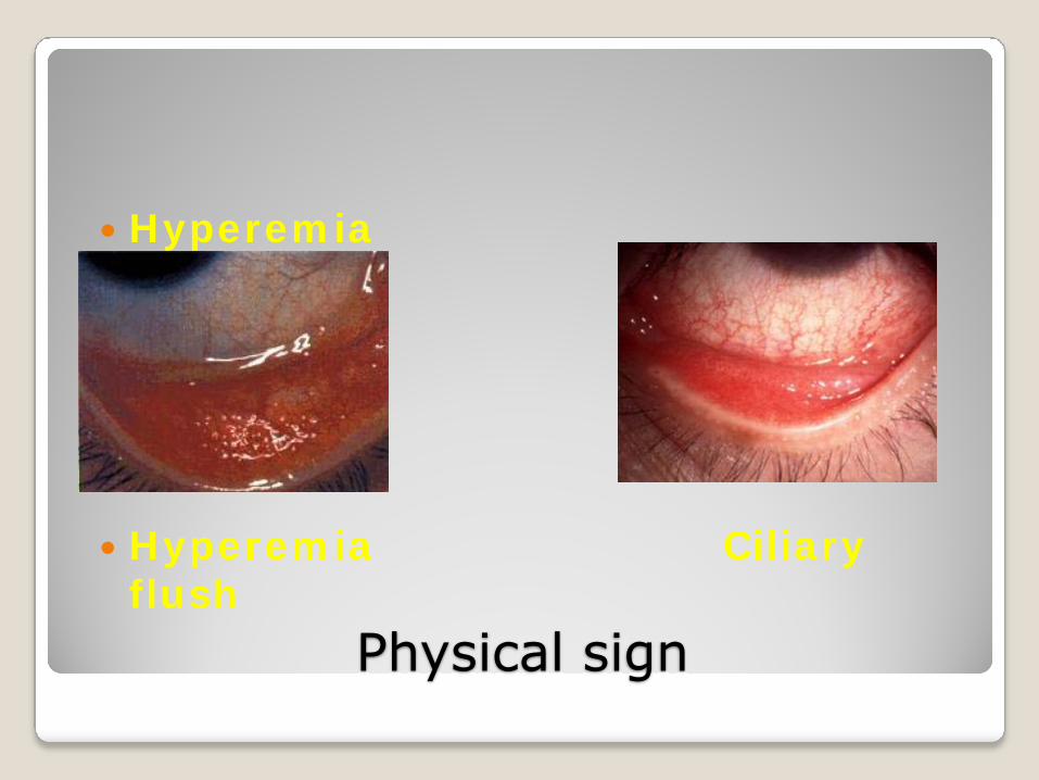
Physical sign
Hyperemia
Hyperemia Ciliary flush
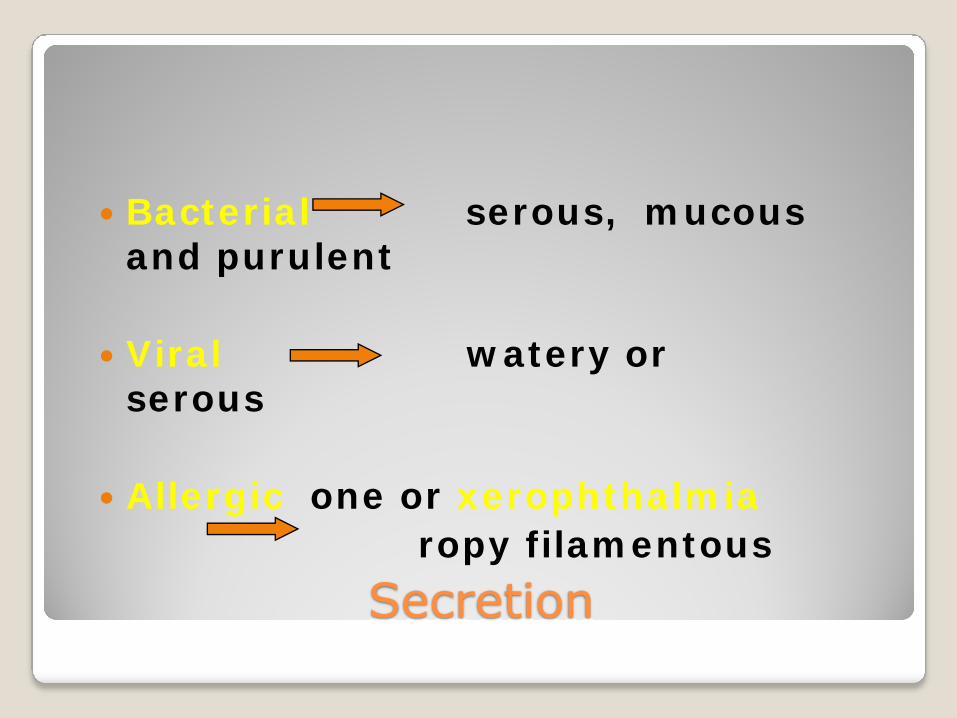
Secretion
Bacterial serous, mucous and purulent
Viral watery or serous
Allergic one or xerophthalmiaropy filamentous
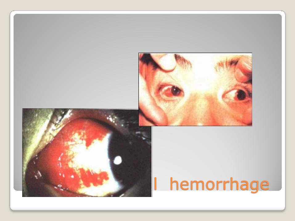
Subconjunctival hemorrhage

Physical sign
papillary hyperplasia: palpebral conjunctival
epithelium
follicular formation: accumulation of lymphocyte
beneath the conjunctival epithelium
Physical sign
pseudomembrane or membrane : the exudation rich in fibrin from palpebral conjunctiva .
Pseudomembrane: in baby and children, adenoviral, neonatal inclusion, streptococcal conj.
True membrane: diphtheritic conj.
Conjunctivitis Examination and diagnosis
Clinical examination Cytologic examinationsmear of conjunctival and scaling smear of conjunctiva
Bacteriological examinationbacterial culture and drug sensitive
testVirus isolation and its antigenic detection

Conjunctivitis Principle treatment
Remove pathogenic cause, take local phamacotherapy as major, systemic treatment as supplement if necessary1)instillation of eyedrops2)instillation of ointment3)washing of conjunctival sac4)systemic treatmentPrevention

Hyperacute Bacterial conjunctivitis
Hyperacute purulent conjunctivitis with the strongest infectivity and large destructibilityEtiology: diplococcus gonorrhoeaeadult: auto infectionchildren: touch infectionnewborn: direct infection

Hyperacute Bacterial conjunctivitis
Clinical findings1)incubation period: 10h-2, 3d, acute
onset2)opthalmalgia, photophobia, tearing3)swelling of the eyelids
palpebral and bulbar hyperemia and chemosis
secretion: serous-bloody-purulent-nong lou yan
inflammatory pseudomembrane preauricular lymphadenectasis corneal ulcer and perforation
Hyperacute Bacterial conjunctivitis
Diagnosis:clinical findings lab examination(Gram’ stain, G-
diplococcus)
Treatment:topical and systemic one is the same
important
Preventionbe isolated to avoid infection and
epidemic

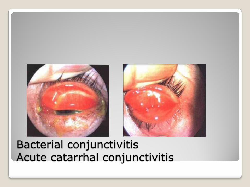
Bacterial conjunctivitisAcute catarrhal conjunctivitis
Clinical finding:acute onset(1-3days), both eyetearing, foreign body and burning sensationconjunctival hyperemia, purulent secretion, palpebral swelling, spots of subconjunctival hemorrhage
Ill process: 2 weeks
Bacterial conjunctivitisAcute catarrhal conjunctivitis
Bacterial conjunctivitisChronic catarrhal conjunctivitis
Etiologybacterial infection:acute-chronic or infection of
bacterial with weak toxicity◦ non-infectious
environment factors: dust, chemical smoke or gas and irritating eye drugs◦ complicated from other disorders

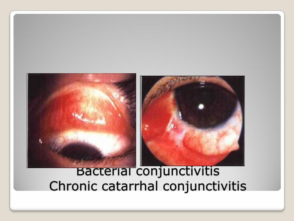
Bacterial conjunctivitisChronic catarrhal conjunctivitis
Clinical finding:chronic onset, both eyeitching, foreign body and asthenopia or no symptomsconjunctival hyperemiamucous secretionpapillary and follicle hyperplasia
Treatment: give management according to different causes
Bacterial conjunctivitisChronic catarrhal conjunctivitis

Chlamydial conjunctivitisChlamydin psittaci:Chlamydia trachomatis:antigen:ABCBa DEFGHIJK
trachoma genitourinary system inclusion
conjunctivitis
clinical findingsAcute or subacute stage(1-2mon):photophobia, tearing, foreign body sensation1)palpebral and bulbar conjunctival hyperemia2)ropy secretion3)papillary hyperplasia, follicles formation4)corneal epithelitisbe cured without scar left

clinical findings
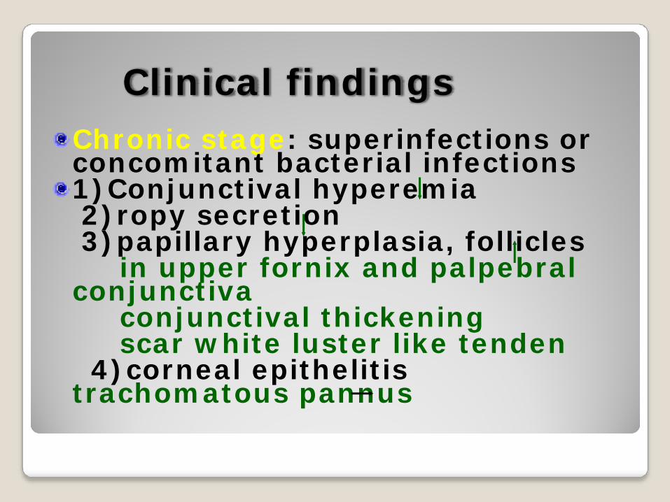
Clinical findingsChronic stage: superinfections or concomitant bacterial infections1)Conjunctival hyperemia2)ropy secretion3)papillary hyperplasia, follicles
in upper fornix and palpebral conjunctiva
conjunctival thickening scar white luster like tenden
4)corneal epithelitis trachomatous pannus

Clinical findings
Chronic stage
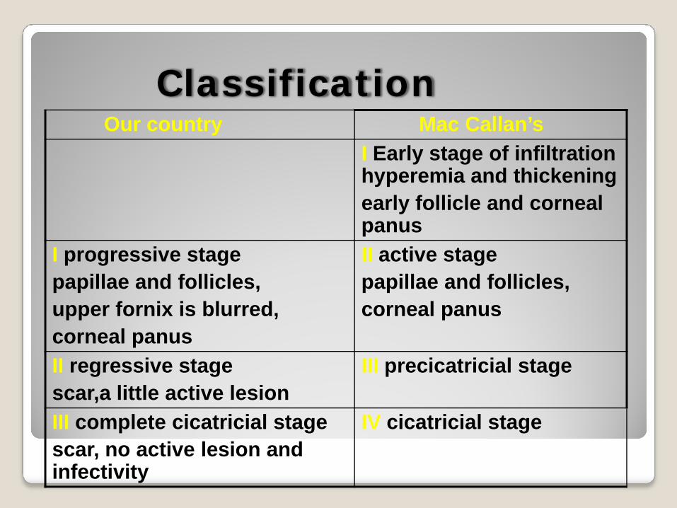
ClassificationOur country Mac Callan’s
I Early stage of infiltration hyperemia and thickeningearly follicle and corneal panus
I progressive stagepapillae and follicles, upper fornix is blurred,corneal panus
pc
II active stageapillae and follicles, orneal panus
II regressive stagescar,a little active lesion
III precicatricial stage
III complete cicatricial stagescar, no active lesion and infectivity
IV cicatricial stage

Equela and complication
Entropion and trichiasisBlepharopatosisSymblepharon (lower fornix)Parenchymatous xerosis of conjunctivaChronic dacryocystitisCorneal pannus

Diagnosis1)the vessels of upper fornix and palpebral conjunctiva are blurred, congested, papillary hyperplasia or follicle formation or both2)corneal pannus3)scar4)trachomatous inclusion
Diagnosison the basis of the first plus one
of other threeantigenic test
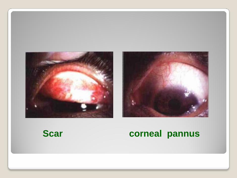
Scar corneal pannus
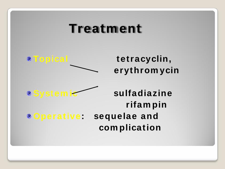
Treatment
Topical tetracyclin, erythromycin
Systemic sulfadiazinerifampin
Operative: sequelae and complication
Viral conjunctivitisEpidemic keratoconjunctivitis
Acute onset, strong infectivity, may be sporadic or epidemicEtiology: adenovirus, type 8, 19, 29 and 37.

Viral conjunctivitisEpidemic keratoconjunctivitis
Clinical findings:1)incubation period: 5-7 days2)foreign body sensation, itching,
pain, photophopia and tearing3)palpebral edema, conjunctival
hyperemia and chemosis, less andwatery secretion, follicles in palpebraland fornix conjunctiva, preauricularlymphadeectasis and tenderness4)be cured after one week
exacerbate: superfial punctatekeratitis

Viral conjunctivitisEpidemic keratoconjunctivitis
Diagnosis:Acute folliclar cinjunctivitis superfial punctate keratitis preauricular lymphadenectasisneutrophialTreatment: no specific drug1)antiviral:topical(mainly) and systemic-acyclic 2)antibiotic
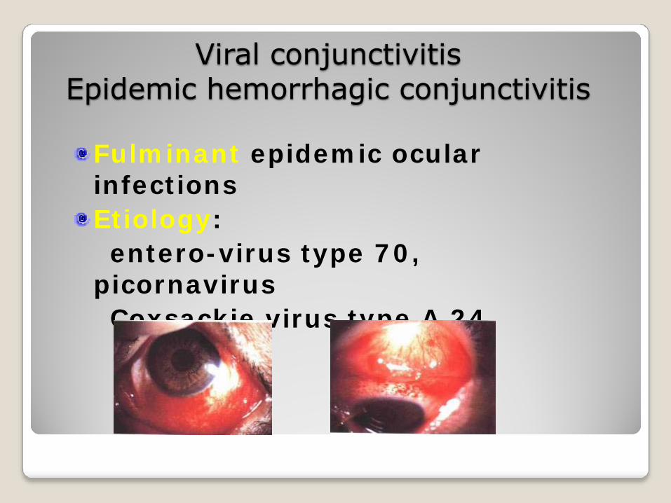
Viral conjunctivitisEpidemic hemorrhagic conjunctivitis
Fulminant epidemic ocular infectionsEtiology:entero-virus type 70,
picornavirusCoxsackie virus type A 24
Viral conjunctivitisEpidemic hemorrhagic conjunctivitis
Clinical findings:1)incubation period: 24hr2)ill course: self-limited, 10d or
shorter3)ophthalmagia, foreign body
sensation, photophopia and tears4)eyelid and conjunctiva red and
swollen, watery secretion, follicular hyperplasis of palpebral conjunctiva, patchy hemorrhage on bulbar conjunctiva, preauricular lymphadenectasis 5) Transient fine punctate epithelial keratitis
Immunologic conjunctivitis Vernal conjunctivitis (Clinical findings)
Symptom:extreme itchingSign1)palpebral type:papillary hyperplasia in the
upper palpebral conjunctiva that like oval flat cobblestone, eosinophillia in secretion 2)corneal limbal type:collid tubercles at the corneal
limbus3)mixed type:
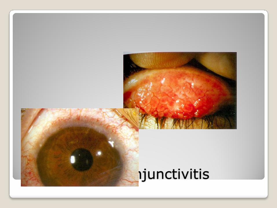
Vernal conjunctivitis
Vernal conjunctivitisTreatment:1)self-limited, no vision affected2)general treatment:keep away proble sensitinogen3)medical treatment:natrii cromoglycascorticosteroid
Allergic conjunctivitis
Immediated allergic antigen:pollen, contact lens, etc.Delayed one: various drug
Clinical findings:immediate type: dermatitis
of palpebral skin, blepharitis, mild infiltrative conjunctivitis
Allergic conjunctivitis
Lab examination:degenerative epithelial cell, few polynuclear cells and mononuclear cells in secretionTreatment:1)find out and get rid of
sensitinogen2)corticosteroid3)3% boric solution4)anti-allergic agents
Phlyctenular keratoconjunctivitis
Etiology: delayed reaction to protein of microorganism, mostly to mycobecterium tuberculosis and staphylococcus aureusClinical findings: herpetic tubercle may appear on the bulbar conjunctiva or limbus.
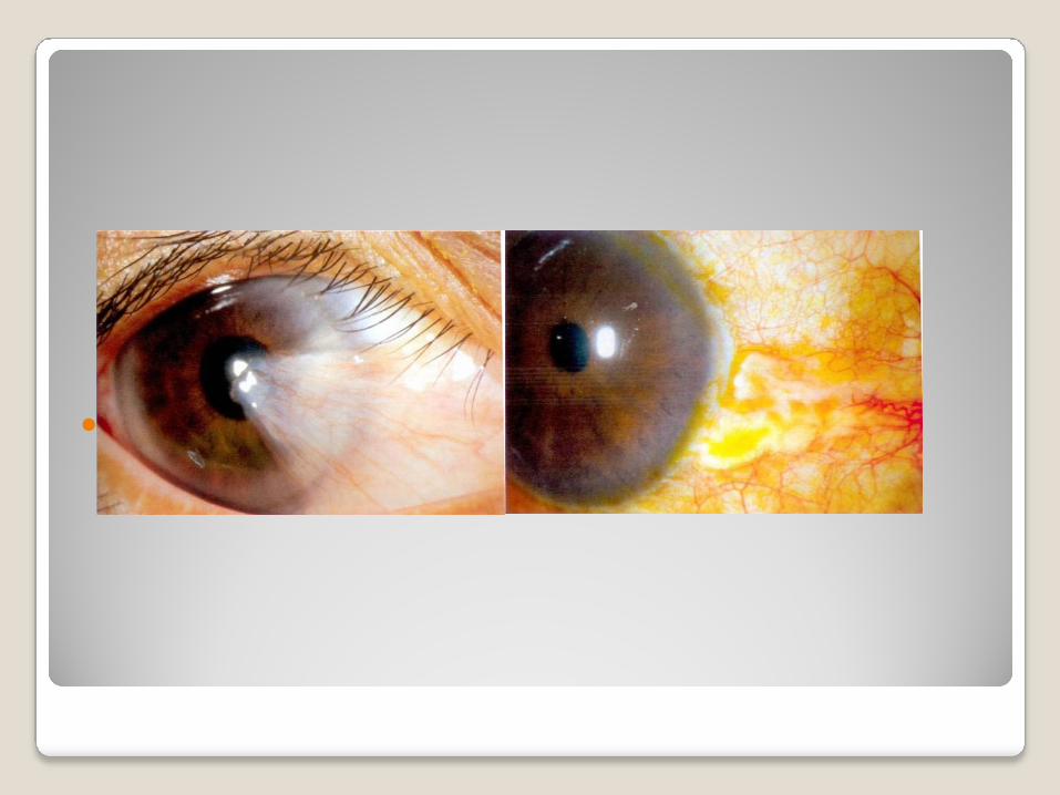
PterygiumEtiology: unclear, outdoor workClinical findings:1)hypertrophic bular conjunctiva
and its subconjunctival tissue invade onto the cornea with the shape of tiangle2)composed of head, neck, body.3)progressive, stationary4)differentiated with
pseudopterygiumTreatment: operation

Pingueculae
A degenerative lesion of the bulbar conjunctiva caused by the effect of ultraviolet raysClinical findings:a kind of white–yellow
amorphous subepithelial deposition near to the limbusTreatment: no needed
Pterygium Pingueculae
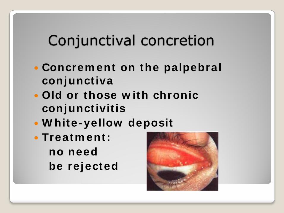
Conjunctival concretion
Concrement on the palpebral conjunctivaOld or those with chronic conjunctivitisWhite-yellow depositTreatment:no needbe rejected
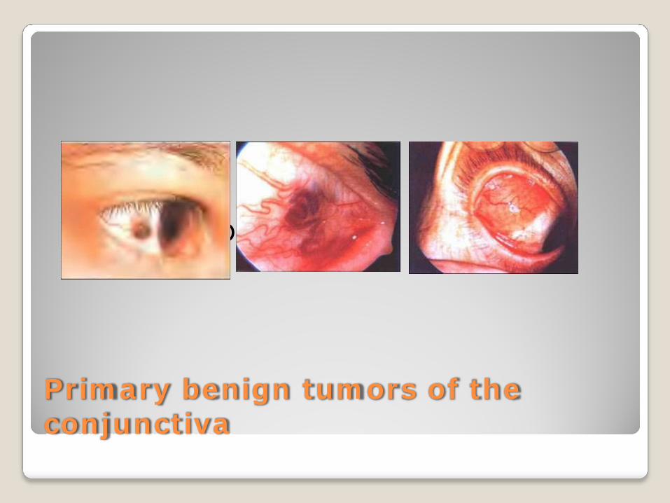
Primary benign tumors of the conjunctiva
nevi Dermolipoma

Subconjunctival hemorrhage
Caused by vascular rupture beneath the bulbar conjunctiva or by osmotic increase of vascular wallTreatment:1)find out the cause2)good explanation