# urologists and nephrologists both treat ... - doctor 2015
TRANSCRIPT


1 | P a g e
# Urologists and nephrologists both treat kidney problems. Urologists are surgical
specialists while Nephrologists are medical specialists who prescribe nonsurgical
medical treatments for kidney problems.
# Urinalysis is applied clinically to evaluate and diagnose patients. It completes the
physical exam ( not part of investigation but a must for the examinations ). It is a mirror
for inside the kidneys.
# Normal urine must not have blood ,ketones ,glucose and proteins( -ve blood, -ve
ketones, -ve glucose ,-ve protein )and its pH 5-8 , rare RBCs (<1 /High Power Field , we
can accept up to 3 /HPF but not more) , may contain few calcium oxalate crystals but
not others (source of oxalate : proteins, nuts, greens, chocolate). Opposite to the
previously mentioned will be abnormal urine.
# Urinalysis is of two ways: dip-stick and microscopy after taking
an early midstream urine sample.
# Dipsticks in general must contain squares to test for blood,
ketones, glucose, proteins ( basics , we can find extra things). We
dip the stick in the urine tube then we need to wait for 30 sec for
chemical reactions to occur. It’s done manually or automatically.
# For microscopy, urine sample of 10-12 mL must be centrifuged
under a specific speed for 3-5 min to separate supernatant and
precipitate in order to concentrate the abnormalities. We get rid of
urine except for few drops to remix them with the precipitate. After
that we take drops to see them under microscope.
# We don’t need centrifugation in cases where urine is totally
abnormal ( totally red , turbid(concentrated ) , UTI
patients).
# Most common cells seen in urine samples are RBCs
(small, round, devoid of nucleus) and WBCs (large,
granulated, segmented nuclei(neutrophil))

2 | P a g e
# RBCs in urine indicate abnormality around UT not kidneys
themselves. In the case of kidneys RBCs will be dysmorphic.
# Glomerular filtration barrier : capillaries “ endothelium”, GBM, podocytes. RBCs are
large to squeeze through this barrier. In the case of glomerular
nephritis we can find RBCs ( destruction of GBM will allow RBCs
to try to squeeze and they will loose their outer border to become
dysmorphic).
- Dysmorphic RBCs in urine ( Glomerular Nephritis) look like sickle cells in blood.
# We can also find yeasts which are normally found in
females. If the field is full of budding yeasts this indicates
an infection. It is seen in immunocompromised patients
(on prednisolone or cortisol , cancer patients on
chemotherapy , diabetic patients, HIV)
>> Immunosuppressive situations will change the normal flora and increase the chance
for infections with abnormal bugs.
# Normally we can find squamous epithelial cells at the
junctions(orifices)near the skin and transitional cells of
urinary system. So we can accept 1-2 cells more than
that there may be tumors ( bulbar or penile ).
>> Increased proliferation of squamous cells will appear
as spots of cells in the field.
Casts in ortho means جبيرة while in € means قالب.
# Hyaline is a protein normally secreted by tubular cells of the kidneys and removed
with urine. Sluggish in flow will lead to stagnation of this protein
and obstruction.
# Hyaline casts which appear transparent are totally normal even if
they were a lot in the field they don’t indicate pathology rather ----
3 | P a g e
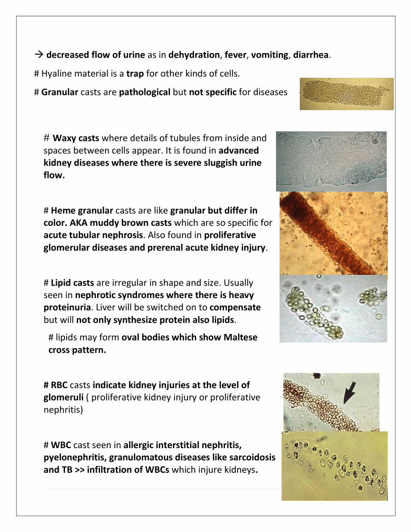
decreased flow of urine as in dehydration, fever, vomiting, diarrhea.
# Hyaline material is a trap for other kinds of cells.
# Granular casts are pathological but not specific for diseases
# Waxy casts where details of tubules from inside and spaces between cells appear. It is found in advanced kidney diseases where there is severe sluggish urine flow.
# Heme granular casts are like granular but differ in color. AKA muddy brown casts which are so specific for acute tubular nephrosis. Also found in proliferative glomerular diseases and prerenal acute kidney injury.
# Lipid casts are irregular in shape and size. Usually seen in nephrotic syndromes where there is heavy proteinuria. Liver will be switched on to compensate but will not only synthesize protein also lipids.
# lipids may form oval bodies which show Maltese cross pattern.
# RBC casts indicate kidney injuries at the level of glomeruli ( proliferative kidney injury or proliferative nephritis)
# WBC cast seen in allergic interstitial nephritis, pyelonephritis, granulomatous diseases like sarcoidosis and TB >> infiltration of WBCs which injure kidneys.

4 | P a g e
# We can see bacteria in the sample. If the sample was fresh we can see WBCs moving to phagocytose bacteria.
# Bacteria doesn’t indicate UTI for sure because sample is concentrated. UTI must be accompanied with symptoms and +ve culture.
# Not centrifuged sample with bacteria in the field indicates UTI for sure.
Crystals
# Calcium oxalate crystals if more than 1-2 this indicates alcohol
(Glutaraldehyde – antifreeze )poisoning. They are so diagnostic.
#other crystals: Uric acid crystals , triple phosphate crystals(coffin lids appearance ), cystine crystals, Ammon Biurate , crystals in HIV patients who are taking medication for HIV …
# We can also see mucus.
# In case of retrograde ejaculation we can find sperms.
Note : Na in urine is not a part in urinalysis. To test for Na we need to order spot test for Na in urine.
# Urinalysis is most useful when there is already abnormal urine or acute kidney injury. It’s important for differential diagnosis.
# To evaluate kidneys we order 2 things : kidney function test which is a blood test for creatinine , urea ,basic electrolytes and Urinalysis.
Case 1
- 22 years old female students found to have +2 blood on a routine urine analysis (it must be 0), no proteinuria and she was not menstruating, blood pressure and serum creatinine are normal. We repeated her urinalysis within one month and we found +3 blood.
5 | P a g e
Urinalysis will be helpful in diagnosing.
If microscopy shows dysmorphic RBCs and casts she may have proliferative Glomerulonephritis, thin basement membrane disease or lupus nephritis (because she’s a young female). If there is no dysmorphic RBCs and casts we need imaging to look for kidney stones or anatomical abnormality as polycystic kidney disease or she may have non-proliferative GN as minimal change disease.
Case 2
- 64 years old man, smoker with +2 blood on routine urine analysis, he had normal urine 1 year earlier, currently taking ACE inhibitors. He had protein and blood in urine and abnormal creatinine in blood.
Kidney stones at this age are less common. We think about tumors first.
Differential diagnosis ( tumors first then stones )
If there is dysmorphic RBCs we think of GN , if not we think about the anatomy of UT.
Note: Stones commonly occur at 20-30 years with genetic predisposition ,abnormal flow and crystals in urine.