reproducibility.and.power. - warwick
TRANSCRIPT

Reproducibility and Power
Thomas Nichols Department of Sta;s;cs & WMG
University of Warwick
“Reproducible Neuroimaging” Educa;onal Course OHBM 2015
slides & posters @ http://warwick.ac.uk/tenichols/ohbm

Power & Reproducibility
• Power Review • Prac;cal Power Methods for Neuroimaging • Why you should care (Reproducibility)
Power: 1 Test
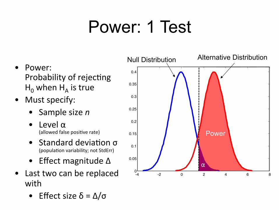
• Power: Probability of rejec;ng H0 when HA is true
• Must specify: • Sample size n • Level α
(allowed false posi;ve rate) • Standard devia;on σ
(popula;on variability; not StdErr) • Effect magnitude Δ
• Last two can be replaced with • Effect size δ = Δ/σ
0 2 4 6 8 -4 -2 0
0.05
0.1
0.15
0.2
0.25
0.3
0.35
0.4
Power
Null Distribution Alternative Distribution
α

Power: Sta;s;c vs. Data Units • 10 subjects
• % BOLD stdev σ = 0.5
−5 0 5 10
α = 0.050T thresh = 1.83
Statistic Value
densi
ty
−1 −0.5 0 0.5 1 1.5 2
α = 0.050
∆ thresh = 0.29
Percent Change
densi
ty
t =x̄
s/
pn
x̄
s/
pn
� t
⇤↵
One-‐Sample T-‐test…
Reject H0 if…
x̄ � t
⇤↵ ⇥ s/
pn
Equivalently, reject H0 if…
= t⇤↵
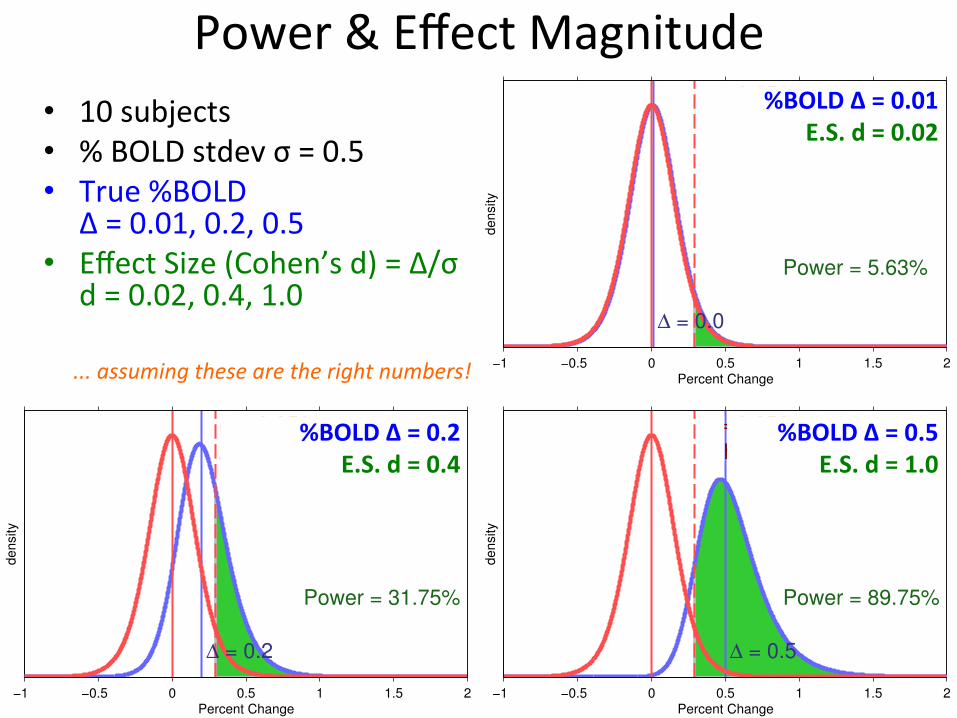
Power & Effect Magnitude • 10 subjects
• % BOLD stdev σ = 0.5 • True %BOLD
Δ = 0.01, 0.2, 0.5 • Effect Size (Cohen’s d) = Δ/σ
d = 0.02, 0.4, 1.0
−1 −0.5 0 0.5 1 1.5 2Percent Change
densi
ty
α = 0.050
∆ thresh = 0.29
∆ = 0.5
Power = 89.75%
−1 −0.5 0 0.5 1 1.5 2Percent Change
densi
ty
α = 0.050
∆ thresh = 0.29
∆ = 0.0
Power = 5.63%
−1 −0.5 0 0.5 1 1.5 2Percent Change
densi
ty
α = 0.050
∆ thresh = 0.29
∆ = 0.2
Power = 31.75%
... assuming these are the right numbers!
%BOLD Δ = 0.01 E.S. d = 0.02
%BOLD Δ = 0.5 E.S. d = 1.0
%BOLD Δ = 0.2 E.S. d = 0.4

Power: 100,000 Tests? • Set α to reflect mul;ple tes;ng (easy part)
– E.g. FWE, for a given search volume & smoothness • MNI mask, FWHM [8 8 8]mm, 3301 RESELs t* = 4.93, then α = 0.0000004
• Alterna;ve: δ1, δ2, δ3, …, δ99,999, δ100,000 (hard part) – Must consider structure of alterna;ve – These 10 voxels ac;ve at Δ, and those other 20… – Oh, and don’t forget to specify σ1, σ2, σ3 … too!

Practical Power Estimation: Clinical Trial ‘Primary Outcome’
• Define ‘Primary Outcome’ – E.g. Average %BOLD in amygdala
• Compute outcome Δ, σ – Previous literature – Pilot data
• E.g. compute %BOLD in amygdala for each subject
• Compute mean, SD • Set α
– Uncorrected α if taking clinical trial approach – α reflec;ng mul;ple tes;ng, otherwise
• Compute power – Matlab, or your favorite power app (e.g. G*power)
ROI Mask
Compute Average BOLD
per subject
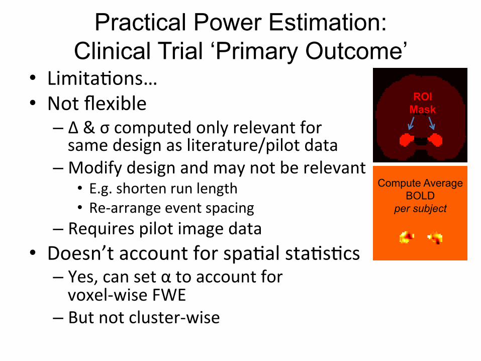
Practical Power Estimation: Clinical Trial ‘Primary Outcome’
• Limita;ons… • Not flexible
– Δ & σ computed only relevant for same design as literature/pilot data
– Modify design and may not be relevant • E.g. shorten run length • Re-‐arrange event spacing
– Requires pilot image data • Doesn’t account for spa;al sta;s;cs
– Yes, can set α to account for voxel-‐wise FWE
– But not cluster-‐wise
ROI Mask
Compute Average BOLD
per subject
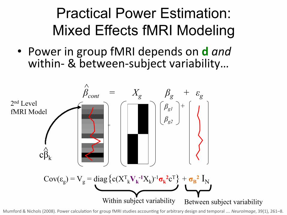
Practical Power Estimation: Mixed Effects fMRI Modeling
• Power in group fMRI depends on d and within-‐ & between-‐subject variability…
Cov(εg) = Vg = diag{c(XTkVk-1Xk)-1σk2cT} + σB2 IN
^
βcont ^
=
βg1
βg2
+
= Xg βg + εg
Within subject variability Between subject variability
2nd Level fMRI Model
cβk
Mumford & Nichols (2008). Power calcula;on for group fMRI studies accoun;ng for arbitrary design and temporal …. NeuroImage, 39(1), 261–8.

Practical Power Estimation: Mixed Effects fMRI Modeling
• Requires specifying – Intra-‐subject correla;on Vk – Intra-‐subject variance σk2 – Between subject variance σB2 – Not to men;on Xk, c & d
• But, then gives flexibility – Can can consider differ designs
• E.g. shorter runs more subjects. • Op;mize event design for op;mal power
Cov(εg) = Vg = diag{c(XTkVk-1Xk)-1σk2cT} + σB2 IN
Within subject variability Between subject variability
0 0.02 0.04 0.06 0.08 0.1 0.12 0.14 0.16Time (hours)
0 5 10 150
10
20
30
40
50
60
70
80
90
100
Number of on/off (20s/10s) cycles (TR=2.5)
Pow
er (%
)
24 subjects22 subjects20 subjects18 subjects16 subjects14 subjects12 subjects
Mumford & Nichols (2008). Power calcula;on for group fMRI studies accoun;ng for arbitrary design and temporal …. NeuroImage, 39(1), 261–8.

Practical Power Estimation: Mixed Effects fMRI Modeling
• Toolbox to es;mate all this from exis;ng data – Jeaneye Mumford’s http://fmripower.org!
Works with FSL & SPM!

Practical Power Estimation: RFT Cluster-wise Inference
• All previous methods ignored space or worked voxel-‐wise only
• Cluster-‐wise inference is popular – Gives greater sensi;vity – Though, pizalls (Woo, et al. NeuroImage, 2014)
• Power for RFT Cluster Inference – Can provide power given a mask of signal
– Or provide maps of ‘local power’
Hayasaka et al. (2007). Power and sample size calcula;on for neuroimaging studies by non-‐central random field theory. NeuroImage, 37:721-‐30.

RFT Cluster-wise Inference
• PowerMap by Satoru Hayasaka
http://sourceforge.net/ projects/powermap"
Hayasaka et al. (2007). Power and sample size calcula;on for neuroimaging studies by non-‐central random field theory. NeuroImage, 37:721-‐30.
Mapping Local Power
FWE vs Primary Outcome Power

Practical Power Estimation: Peak Power
• Mixed-‐effects & RFT Cluster both complicated • Peak inference
– Considers only local maxima above threshold u – Make P-‐values for peak height (SPM8+)
• Peak power calcula;ons simpler to specify – Mean peak height
• Can translate to/from d if needed
– Size of ac;vated region – Plus, mask size, FWHM smoothness
Durnez, Moerkerke, & Nichols, T. E. (2014). Post-‐hoc power es;ma;on for topological inference in fMRI. NeuroImage, 84, 45–64.

Practical Power Estimation: Peak Power
• NeuroPower by Joke Durnez – Es;mates the 2 key parameters from pilot data’s T map – Creates power curves
http://neuropower.shinyapps.io/neuropower

Power Tools Redux
• Primary ‘Clinical Trials’ outcome – Easy but not flexible – Doesn’t reflect image-‐wise analyses actually done
• Mixed effects power – Flexible, harder to specify
• RFT Cluster power – Only method for widely used cluster inference
• Peak power – Easy to specify
• But, again, why do we care?

Power Dangers • Retrospec;ve Power
– Power is a probability of a future true posi;ve – Can’t take current data (e.g. t=1.3) and say “What was my power for this result?”
• Es;ma;ng Effect Sizes – Voodoo correla;ons!
• Effect size at peak is biased – Circularly defined as the best effect
– Must use independent ROIs • Independent data, contrasts • Anatomical ROI
Puzzlingly high correlations in fMRI studies of emotion, personality, and social cognition. Persp. on Psych. Science, 4, 274-290

Power & Replicability • I got a significant result, who cares about
power!? • Law of Small Numbers aka “Winner’s Curse” – Small studies over-‐ es;mate effect size
• A Low N study… • Has low power, likely to fail • Significant if…
• Randomly-‐high effect, or • Randomly-‐low variance
• Low power = hard to replicate!
Effe
ct S
ize
(Log
Odd
s R
atio
)
Sample Size (Log of Total N in Meta Analysis)
• 256 meta analyses − For a binary effect (odds ratio) − Drawn from Cochran database
• Lowest N, biggest effect sizes!
Ioannidis (2008). “Why most discovered true associa;ons are inflated.” Epidemiology, 19(5), 640-‐8.

Low N studies: The Dark Side • Suppressed studies & Biased effects
– P>0.05 not published – Biases that afflict small studies more than large studies
Effe
ct S
ize
(Log
Odd
s R
atio
)
Sample Size (Log of Total N in Meta Analysis)
Effe
ct S
ize
(Log
Odd
s R
atio
)
Sample Size (Log of Total N in Meta Analysis)
File drawer problem (Unpublished non-‐significant studies)
Bias (Fishing or Vibra;on Effects)

Foci per contrast
Den
sity
0 5 10 15 20 25
0.00
0.05
0.10
0.15
●
●●
●
●
●
●
●
●
●
●
●
●●
●●
●●
●●
● ● ● ● ● ●
Self-‐Promo;on Alert
• Es;ma;ng the size of the “File Drawer” in fMRI
• Use meta-‐analysis foci counts to infer number of missing (0 count) studies
• About 1 study missing per 10 published – 9.02 per 100 95% CI (7.32, 10.72)
– Varies by subarea
Counts Per Contrast Empirical & Fiyed Distribu;on
2,562 Studies from BrainMap One contrast per study randomly selected
Pantelis Samartsidis, et al. Poster 4038-‐W “Es;ma;ng the prevalence of ‘file drawer’ studies”

Vibration Effects • Sloppy or nonexistent analysis protocols
– You stop when you get the result you expect – These “vibra;ons” can only lead to inflated false posi;ves
• Afflicts well-‐intended researchers – Mul;tude of preprocessing/modelling choices
• Linear vs. non-‐linear alignment • Canonical HRF? Deriva;ves? FLOBS?
“Try voxel-wise whole brain, then cluster-wise, then if not getting good results, look for subjects with bad movement, if still nothing, maybe try a global signal regressor; if still nothing do SVC for frontal lobe, if not, then try DLPFC (probably only right side), if still nothing, will look in literature for xyz coordinates near my activation, use spherical SVC… surely that’ll work!”
Does your lab have wri?en protocols?

Power failure: Buyon et al. • Meta-‐Analysis of (non-‐imaging) Neuroscience Meta-‐Analyses • Recorded median power per meta-‐analysis
– Median median power 21%
Buyon, et al. (2013). Power failure: why small sample size undermines the reliability of neuroscience. Nat. Rev. Neuros, 14(5), 365–76.
50% of all
neuroscience studies have
at most a 1-‐in-‐5 chance of replica;ng!

Buyon et al’s Recommenda;ons
• Do power calcula;ons • Disclose methods & findings transparently • Pre-‐register your study protocol and analysis plan
• Make study materials and data available – Check out hyp://neurovault.org !
• Work collabora;vely to increase power and replicate findings
Buyon, et al. (2013). Power failure: why small sample size undermines the reliability of neuroscience. Nat. Rev. Neuros, 14(5), 365–76.

Power Conclusions
• Power = Replicability – Best gauge on whether you’ll find the effect again
• “Primary outcome” power – Good way to appease grant reviewers – Doesn’t reflect how we usually analyze data
• Whole image-‐wise power possible – Now various tools to choose from