· web viewpostoperative fistula and other complications in pancreatic surgery: a retrospective...
TRANSCRIPT

Postoperative fistula and other complications in pancreatic surgery:A retrospective analysis of 255 pancreatic resections
Nick De WeverStudent number: 01309484
Supervisor: Prof. dr. Frederik Berrevoet
A dissertation submitted to Ghent University in partial fulfilment of the requirements for the degree of Master of Medicine in Medicine
Academic year: 2017 – 2018

“The author and the promotor give the permission to use this thesis for consultation and to copy
parts of it for personal use. Every other use is subject to the copyright laws, more specifically the
source must be extensively specified when using results from this thesis.”
Date
(signature student) (signature promotor)
Name (student) Name (promotor)


PREFACE
I am proud to present this dissertation which was written to fulfill the requirements for the degree
of Master of Medicine in Medicine at Ghent University. I was engaged in the process of data
acquisition, data analysis and writing of this dissertation from March 2016 until December 2017.
First and foremost, I would like to thank my promotor Professor Frederik Berrevoet (MD, PhD,
Associate Professor General and HPB Surgery at Ghent University Hospital) for his guidance and
constructive support during this research process. I am also thankful to have had the chance to
observe some of the pancreatic surgeries live.
Although this dissertation was written individually, the experimental part of the research including
data acquisition and statistical analysis was performed together with my colleague Sybren
Vanwynsberghe. I would like to thank him for his enthusiasm and efforts throughout the research
process. Discussing the results with you has been helpful in formulating conclusions.
Finally, my parents also deserve special thanks for reading this dissertation, providing helpful
comments and suggestions. Thank you for giving me the opportunity to complete my medical
education and pursue a career as a healthcare professional.
I hope you enjoy reading this dissertation.
Nick De Wever

CONTENTS
1 Abstract..............................................................................................................................1
2 Samenvatting.....................................................................................................................3
3 Introduction........................................................................................................................5
3.1 Research question and aim......................................................................................5
3.2 Problems in pancreatic surgery.................................................................................6
3.2.1 Mortality.................................................................................................................6
3.2.2 Morbidity................................................................................................................7
3.3 Background.............................................................................................................16
3.3.1 Pancreatic Surgery..............................................................................................16
3.3.2 Pancreatic cancer................................................................................................17
4 Patients and methods......................................................................................................21
4.1 Study design...........................................................................................................21
4.2 Patients and data collection....................................................................................22
4.3 Database construction and variable selection.........................................................22
4.4 Specific variable definition.......................................................................................24
4.5 Surgical approach...................................................................................................26
4.6 Postoperative care..................................................................................................27
4.7 Statistical analysis...................................................................................................28
4.8 Literature search.....................................................................................................28
5 Results.............................................................................................................................29
5.1 Database descriptive statistics................................................................................29
5.2 Postoperative pancreatic fistula (popf)....................................................................29
5.2.1 Patient characteristics by grade of fistula............................................................30

5.2.2 Univariate analysis of risk factors for clinically relevant POPF...........................30
5.2.3 Univariate analysis of risk factors for all grades of POPF...................................33
5.3 Management of POPF............................................................................................35
5.4 Other postoperative complications..........................................................................35
6 Discussion.......................................................................................................................36
6.1 Database and patient characteristics......................................................................36
6.2 Postoperative pancreatic fistula (POPF).................................................................36
6.2.1 Incidence of POPF after PD and DP...................................................................36
6.2.2 Risk factors for POPF..........................................................................................39
6.3 Other postoperative complications (DGE & PPH)...................................................45
7 Conclusion.......................................................................................................................46
8 References......................................................................................................................48
Appendix 1: List of abbreviations...............................................................................................i

1 ABSTRACT
Background and aim: Pancreatic surgery is the only potentially curative treatment for
pancreatic cancer. However, morbidity of these operations remains high with complications
in 30-50% of all patients. Postoperative pancreatic fistula (POPF), delayed gastric emptying
(DGE) and postpancreatectomy hemorrhage (PPH) are the most important complications as
they are associated with mortality, delay in adjuvant therapy and impaired quality of life.
POPF is the most feared as it is the one most frequently associated with catastrophic
consequences making it the leading risk factor for postoperative death and longer hospital
stay with increased hospital costs. Analyzing the series of pancreatic surgeries performed at
Ghent University Hospital gives the opportunity to compare incidences and outcomes of
these complications with literature data, which provides feedback to the surgeons as it could
potentially indicate aspects of care needing specific attention. Secondly, investigating the
associations between patient or surgery related risk factors and the occurrence of POPF
could help to identify high risk patients, serving as a future guidance for directing strategies
to reduce complication rates. The main research question and goal can be summarized as
the evaluation of the incidence of POPF, the determining risk factors and the therapeutic
measures in relation to morbidity and mortality.
Methods: Between January 2010 and November 2015, 255 pancreatic resections for benign
or (pre)malignant lesions were performed at Ghent University Hospital. This included 198
(pylorus-preserving) pancreaticoduodenectomies (PD) and 57 distal pancreatectomies (DP).
A large database with variables including the occurrence of POPF, DGE, PPH and many
others was constructed retrospectively, using the data that were available in the electronic
patient records. A selection of variables from this database was made, based on literature
review and current evidence concerning the association between these variables and the
occurrence of POPF. Statistical analysis was then performed on those possible risk factors
or protective factors for POPF.
Results: Fifty-three patients (20.8%) developed a fistula after pancreatic surgery, including
18 patients (7.1%) with a clinically relevant grade B or C fistula (CR-POPF). Twenty-four
patients (12.1%) developed POPF after PD, including 8 patients (4%) with CR-POPF.
Twenty-nine patients (50.9%) developed POPF after DP, including 10 patients (17.5%) with
CR-POPF. There was a significant difference in the occurrence of POPF as well as the
occurrence of CR-POPF in the group of patients who had undergone PD compared to those
who had undergone distal pancreatectomy (P<0.001 and P=0.002 for POPF and CR-POPF
respectively).
1

Univariate analysis showed that soft pancreatic texture was a significant risk factor for any
grade of POPF after PD (P<0.001), but not for CR-POPF. Age, gender, BMI, diabetes,
neoadjuvant therapy, drain perdu, DGE and PPH showed no significant association with
POPF after pancreaticoduodenectomy.
Univariate analysis of the same variables in the group having undergone DP, showed that
diabetes was a significant protective factor for any grade of POPF after DP (P=0.034), but
not for CR-POPF. All other variables showed no significant association with POPF after DP,
although there was a trend towards significance for delayed gastric emptying (P=0.062).
Altogether, 25.6% of all patients undergoing pancreatic surgery developed at least one of the
three major complications. There were 47 cases (18.5%) of DGE, with no significant
difference between the PD group (18.8%) and the DP group (17.5%). PPH occurred in 12
patients (4.7%) with no significant difference between the PD group (5.6%) and the DP group
(1.8%).
Discussion: Our study shows a low rate of POPF and CR-POPF after PD compared to other
published series. This may be attributable to high surgeon experience and the invariable use
of the same anastomotic technique. On the other hand, the incidence of POPF after distal
pancreatectomy was high compared to other institutions, but the majority of these were
grade A biochemical leaks and thus not clinically relevant. The incidence of CR-POPF after
DP was indeed comparable to current literature data. Closure of the pancreatic remnant with
a stapler is used which is the best method based on current evidence. The fact that lipase
levels were used instead of amylase levels to detect POPF, does not jeopardize the
observation that CR-POPF rate in this series is low compared to other published data.
Soft pancreatic texture was found to be a significant risk factor for POPF after PD, supporting
the use of this variable in fistula risk scores. DGE was present in 40% of the patients
suffering from CR-POPF after DP, but it is not a significant risk factor according to our
analysis. High BMI was found to be a significant risk factor for POPF after DP, but this result
needs careful interpretation as there is a high risk for confounding influences. Future studies
might benefit from measuring serum albumin to clarify the seemingly contradictive effects of
BMI and serum albumin on POPF rate as they are both markers for nutritional status.
Diabetes showed to be a significant protective factor against POPF after DP. The percentage
of diabetic patients was higher in the group of patients without POPF after both PD and DP.
Our results therefore support the suggested protective effect of diabetes in pancreatic
surgery, but further research is still required.
The incidence of DGE in Ghent University Hospital was comparable to other reported single-
center studies, and the occurrence of PPH was a little lower than in most series. These
findings suggest that no major flaws in surgical technique or postoperative care are present.
2

3
2 SAMENVATTING
Achtergrond en doelstelling: Pancreaschirurgie blijft tot op vandaag de enige potentieel
curatieve behandeling bij pancreaskanker. Chirurgische resecties van de pancreas gaan
echter nog steeds gepaard met een hoge incidentie aan complicaties (30-50%).
Pancreasfistels (POPF), vertraagde maaglediging (DGE) en postoperatieve bloeding (PPH)
vormen daarbij de belangrijkste complicaties gezien ze geassocieerd zijn met oponthoud van
verdere behandeling, verminderde levenskwaliteit en toegenomen sterfte. Vooral POPF is
een gevreesde complicatie omdat het de belangrijkste risicofactor is voor sterfte, verlengde
hospitalisatieduur en toegenomen kosten. Het analyseren van een reeks pancreasoperaties
in het UZ Gent laat ons toe de incidentie van deze complicaties na te gaan en te vergelijken
met andere ziekenhuizen, wat een feedback geeft naar de chirurgen en eventuele
aandachtspunten in de zorg kan identificeren. Verder heeft het onderzoeken van patiënt of
operatie gerelateerde risicofactoren voor fistels tot doel om mensen met een verhoogd risico
te identificeren, zodoende een leidraad te verschaffen voor eventuele preventieve
maatregelen. De doelstelling van dit onderzoek bestaat dus vooral uit het evalueren van de
incidentie van complicaties na pancreaschirurgie in het UZ Gent, alsook het bepalen van de
risicofactoren en de therapeutische strategieën.
Methoden: Tussen januari 2010 en november 2015 ondergingen 255 patiënten een
pancreasoperatie in het UZ Gent omwille van een benigne of (pre)maligne letsel. Dit omvatte
198 (pylorussparende) pancreaticoduodenectomieën (PD) en 57 kop- en/of staartresecties
(DP). Een grote database met gegevens van deze patiënten werd retrospectief opgesteld op
basis van de gegevens in het elektronisch patiëntendossier. Na literatuuronderzoek werden
een aantal variabelen uit deze database opgehaald, waarvan gedacht werd dat zij potentiële
risicofactoren of beschermende factoren kunnen zijn voor fistels. Vervolgens werd
statistische analyse van deze variabelen verricht om hun associatie met fistels na te gaan.
Resultaten: Drieënvijftig patiënten (20,8%) ontwikkelden een fistel na de operatie, waarvan
18 patiënten (7,1%) een klinisch relevante graad B of C fistel (CR-POPF). Vierentwintig
patiënten (12,1%) ontwikkelden en fistel na PD, waaronder 8 patiënten (4%) een CR-POPF.
Negenentwintig patiënten (50;9%) ontwikkelden een fistel na DP, waaronder 10 patiënten
(17,5%) een CR-POPF. Er was dus een significant hoger aantal fistels en ook CR-POPF na
DP in vergelijking met PD (P<0,001 en P=0,002 voor POPF en CR-POPF respectievelijk).
Univariate analyse toont aan dat een zachte textuur van het pancreasweefsel een
significante risicofactor is voor het ontwikkelen van een fistel na PD (P<0,001), maar niet
voor het ontwikkelen van CR-POPF. Leeftijd, geslacht, BMI, neoadjuvante therapie, drain
perdu, DGE en PPH waren niet significant geassocieerd met POPF na PD.
4

Univariate analyse voor dezelfde variabelen na DP toont dat diabetes een significant
beschermende factor is voor fistels (P=0,034), maar niet voor CR-POPF. Alle andere
variabelen toonden geen significante associaties met de incidentie van fistels na DP, hoewel
vertraagde maaglediging wel een trend naar significantie toonde (P=0,062).
In totaal ontwikkelden 25,6% van alle patiënten tenminste één van de drie hoofdcomplicaties
na hun operatie. Er waren 47 gevallen (18,5%) van vertraagde maaglediging, waarbij geen
significant verschil tussen PD (18,8%) en DP (17,5%) werd vastgesteld. Bloedingen kwamen
slechts bij 12 patiënten (4,7%) voor, waarbij ook geen significant verschil werd vastgesteld
tussen PD (5,6%) en DP (1,8%).
Discussie: Deze studie toont een lage incidentie van POPF en CR-POPF na PD in
vergelijking met andere series. Dit is waarschijnlijk te verklaren door de uitgebreide ervaring
van de chirurgen en het systematisch toepassen van dezelfde anastomosetechniek. De
incidentie van POPF na DP is daarentegen wel hoog ten opzichte van andere series, maar
de meerderheid bedroeg de klinisch weinig relevante graad A fistels. De incidentie van CR-
POPF was wel vergelijkbaar met literatuurgegevens. Bij DP werd de pancreasrest gesloten
met een stapler, wat op basis van de huidige evidentie de beste methode lijkt. Het feit dat
lipase in plaats van amylase gebruikt werd om POPF aan te tonen, doet geen afbreuk aan
de observatie dat de incidentie CR-POPF in onze serie laag is ten opzichte van andere data.
Een zachte textuur van de pancreas blijkt een significante risicofactor te zijn voor POPF na
PD, wat het gebruik van dit kenmerk in risicoscores voor fistels ondersteunt. Veertig procent
van de mensen met een CR-POPF na DP hadden ook last van DGE, maar dit was geen
significante risicofactor volgens onze analyse. Een hoog BMI blijkt wel een significante
risicofactor te zijn voor POPF na DP, maar dit resultaat moet met de nodige omzichtigheid
geïnterpreteerd worden gezien het hoge risico op verstorende variabelen. Toekomstig
onderzoek dient naast BMI ook albuminespiegels in het serum te bepalen gezien er
tegenstrijdige resultaten worden gepubliceerd over deze twee factoren, terwijl beide merkers
van de nutritionele toestand zijn. Diabetes bleek een significant beschermende factor te zijn
voor fistels na DP. Het percentage diabetici was steeds hoger in de groep zonder fistels ten
opzichte van de groep met fistels en dit zowel na PD als na DP. Deze resultaten
ondersteunen dus het gesuggereerde protectieve effect van diabetes, maar verder
onderzoek is nog steeds nodig.
De incidentie van DGE in deze studie was vergelijkbaar met andere monocentrische studies
en de incidentie van PPH was zelfs iets lager dan in de meeste series. Dit toont aan dat er
geen duidelijke tekortkomingen zijn in de gebruikte chirurgische techniek en de
postoperatieve zorg.
5

3 INTRODUCTION
3.1 RESEARCH QUESTION AND AIM
Pancreatic surgery is the domain which is responsible for the surgical treatment of pancreatic
disease including cancer, pancreatitis and trauma. The importance of this surgical field is
illustrated by some epidemiological data concerning the indications for which surgery is
performed. Pancreatic cancer accounts for an estimated global death toll between 200 000
and 330 000 persons per year and it is the fourth leading cause of cancer deaths in the
developed countries (1-3). Surgical resection of the tumor remains the only potentially
curative treatment for this global health issue.
The second major indication for pancreatic surgery is acute pancreatitis, which is one of the
most frequent causes for hospital admission for gastrointestinal problems, implying a
significant impact on hospitalization costs. Acute pancreatitis has an annual incidence
between 13 and 45 per 100 000 persons with an observed increase of 20% over the past 10
years (4, 5). Overall mortality of acute pancreatitis is 2%, but it approaches 30% among
patients with severe forms and persistent organ failure (5). This last category often requires
surgical interception. The third major indication for pancreatic surgery is chronic pancreatitis.
The incidence of this disease in Europe is estimated to be 4-13 per 100 000 per year, with an
increased incidence reported in several studies (6-8). Surgery is one of the basic treatment
strategies in chronic pancreatitis as nearly 50% of the patients undergo surgery at some
point during the disease progression (8).
These epidemiologic data clearly prove that the three main indications for pancreatic surgery
are important and prevalent diseases with a high impact on general population health.
However morbidity after these operations remains high with reported complications in about
30% to 50% of the patients in most large series (9-14). This indicates the importance of a
better understanding of these complications and their prevention and management options
as they are responsible for delay in adjuvant therapy and result in increased hospital costs
and length of hospital stay (10, 14, 15). Most importantly, they negatively impact the quality
of life in patients who frequently already have a limited life expectancy. It is therefore that this
study aims to investigate these postoperative complications. Analysis of a series of
pancreatic surgeries that have been performed at Ghent University Hospital will enable us to
compare outcomes and postoperative complications with literature data and identify potential
flaws in the perioperative care and management of these complications. This is a good
feedback to the surgeons as it can indicate aspects needing specific attention in the
6

multidisciplinary management of these complex patients. Secondly, we want to investigate
the correlations between patient or surgery related risk factors and the occurrence of certain
complications in order to identify high risk patients. In time, this can serve as a guidance for
directing future strategies to reduce complication rates in these high risk individuals. As
postoperative pancreatic fistula remains one of the most frequent and most feared
complications after pancreatic resections, the study focusses on the risk factors, occurrence
and management of this complication in particular.
The main research question and goal of this dissertation can therefore be summarized as the
evaluation of the incidence of different types of pancreatic fistula after pancreatic resection,
the determining risk factors and the therapeutic measures in relation to morbidity and
mortality. Secondary goals were to evaluate the incidence of delayed gastric emptying and
postpancreatectomy hemorrhage.
The contribution of the two students to this study consisted of completing the missing data in
a large database of patients that underwent pancreatic surgery in Ghent University Hospital
based on electronic patient records. They subsequently identified relevant variables for
analysis, based on current literature data and performed statistical analysis for the entire
patient cohort. All steps in the research process were performed under the supervision and
guidance of Professor F. Berrevoet (Associate Professor General and HPB Surgery at Ghent
University Hospital).
3.2 PROBLEMS IN PANCREATIC SURGERY
3.2.1MORTALITY
Historically, the mortality rates of pancreatic surgery, and more specific pancreatic
resections, were very high. Until the 1960s, mortality rates after Whipple surgery were more
than 25%. Only relatively recently have these mortality rates dropped to <5% and even <2%
in high-volume centers (9, 16, 17). This significant decrease in mortality rates only started
from the 1980s onwards, and can be explained by centralization of these procedures in
specialist high-volume centers, accurate indications, improvements in surgical technique and
progress in perioperative care (13, 18-20). Although there is large variation among studies
considering the term ‘high-volume’, the relationship between hospital and surgeon volume
and the outcomes of pancreatic resections has now been well proven (9, 21). Nowadays,
pancreatic resections are relatively safe procedures in terms of mortality.
Of course, the eventual goal of surgery for pancreatic cancer is to obtain a curative resection
and thus cure the patient. However, despite the advances in the safety of these procedures,
the 5-year survival rate for pancreatic cancer is still only approximately 20% within this
7

selected group of patients that qualify for surgery (22, 23). On top of these poor survival
outcomes, only 20% of the patients are eligible for surgery at the time of diagnosis (9, 10,
24). Thus we can conclude that on this day pancreatic surgery is still not able to cure
pancreatic cancer and that early diagnosis remains the largest obstacle in improving survival
rates.
3.2.2MORBIDITY
3.2.2.1 COMPLICATIONS AFTER PANCREATIC SURGERY
Although mortality has improved drastically over the last decades, morbidity still remains high
with reported complications in about 30% to 50% of the patients in most large series (9-14).
However, postoperative complications are influenced by the type of surgery, as well as the
underlying indication for pancreatic resection and this has to be taken into account when
comparing different studies. Not only do postoperative complications negatively influence
quality of life for these patients, they also delay adjuvant therapy and result in increased
hospital costs and length of hospital stay (10, 14, 15). Morbidity remains a very important
problem and a challenge within the field of pancreatic surgery, even in large-volume
specialist centers (25, 26).
The postoperative complications can be divided into general surgical complications and
pancreas-specific complications. General complications that are a risk in all types of major
abdominal surgery include sepsis, surgical site infection and respiratory complications. The
most important pancreas-specific complications on the other hand include pancreatic fistula,
bleeding and delayed gastric emptying. It is known that pancreas-specific complications can
also induce secondary general complications such as sepsis and wound infection. In the past
decades, lack of standardized definitions compromised the reporting of these surgical
complications, making it difficult to compare studies. Therefore, the International Study
Group of Pancreatic Surgery (ISGPS) has constructed definitions for pancreatic leaks (27),
bleeding (28) and delayed gastric emptying (29).
A. Postoperative pancreatic fistula (POPF) Postoperative pancreatic fistula (POPF) is one of the most frequent and most feared
complications after pancreatic surgery. In 2005, the International Study Group for Pancreatic
Fistula (ISGPF) defined POPF as a failure of healing/sealing of a pancreatic-enteric
anastomosis or a parenchymal leak not directly related to an anastomosis such as one
originating from the raw pancreatic surface (27). This internationally accepted definition was
created with the intention to solve the problems in comparison of studies because of the
large heterogeneity in the definitions of POPF which resulted in large variations in reported
8

incidences, ranging from 2% to more than 35%. The all-inclusive definition of POPF is a
drain output of any measurable volume of fluid on or after postoperative day 3 with an
amylase content greater than 3 times the upper normal serum value. The study group also
proposed a grading system for POPF, based on the clinical impact of the complication
(Grade A, B or C) (9, 27). The parameters used for POPF grading can be seen in table 1.
(For further information on the ISGPF definition for POPF, see ‘patients and methods’).
Despite the advantages of a uniform grading system that allows the comparison of data, the
ISGPF grading has also been a subject of criticism. Some authors argue that the practical
implications are limited, as grade A POPF has no clinical implications and the distinction
between clinically relevant grade B and grade C fistula may be artificial (30).
Table 1 Parameters for POPF grading (ISGPF classification)
Criteria No fistula Grade A fistula Grade B fistula Grade C fistulaDrain amylase <3 times normal
serum amylase>3 times normal serum amylase
>3 times normal serum amylase
>3 times normal serum amylase
Clinical condition Well Well Often well Bad/ill appearingSpecific treatment* No No Yes/No YesUS/CT (if obtained) Negative Negative Positive/
negativePositive
Persistent drainage (>3 weeks)
No No Usually yes Yes
Sepsis No No No YesReadmission No No Yes/No Yes/NoReoperation No No No YesDeath related to POPF No No No YesSigns of infection No No Yes YesUS, ultrasonography; CT, Computed tomography; * partial or total parenteral nutrition, antibiotics, enteral nutrition, somatostatin analogue and/or minimal invasive drainage. Adapted from Bassi et al. (27) and from Callery et al. (14).
POPF is feared because it is the complication most frequently associated with catastrophic
consequences such as sepsis, hemorrhage and intra-abdominal abscesses making it the
leading risk factor for postoperative death, longer hospital stay and increased hospital costs
(13, 15, 31). It is also linked with the other important complications such as delayed gastric
emptying, wound infection and ileus. Surprisingly the rate of POPF has not declined
significantly in the last decades, even in high-volume centers (14). It is clear that POPF
remains one of the most important problems in pancreatic surgery and has a large impact on
the quality of life of these patients. It is for these reasons that the focus of this study lies on
pancreatic fistula as this clearly indicates the importance of more research addressing this
issue.
I. Clinical aspects:The presence of a postoperative pancreatic fistula can usually be suspected based upon
nonspecific clinical features such as unexplained fever, nausea and vomiting, abdominal
pain, constipation, infected wound dehiscence with erythema and swelling, and delayed
9

gastric emptying. Actually, every deviation from the expected postoperative course should
raise suspicion for the presence of POPF (30, 32). Another important clinical sign is an
effluent appearance of the drain fluid, which allows for the diagnosis of POPF to be made by
analysis of the amylase or lipase levels. Observation of the drain fluid is also important
because large drain volumes or abnormal coloration may indicate the presence of a fistula
and demand further attention. The importance of the drain fluid in the early detection of
POPF is illustrated by Traverso and Colleagues (33), who showed that a drainage volume of
more than 30ml after postoperative day 5 together with a high amylase content had a 59%
positive predictive value for clinically relevant POPF. A daily drainage volume of more than
200ml increased the predictive value to 84%, clearly indicating the impact of a large drainage
volume (14, 30, 32).
II. Imaging:Routine imaging after pancreatic surgery to screen for POPF is not recommended as the
ISGPF definition is based on the amylase levels of the drain fluids. Furthermore, cross-
sectional imaging can be false-positive in patients lacking any clinical signs suggestive of
POPF. If there is a deviation from the expected postoperative course however, cross-
sectional imaging is commonly used to detect peripancreatic fluid collections, which are
significantly correlated with POPF (32). Contrast-enhanced CT is the preferred technique as
it has a sensitivity of 63% and a specificity of 83% for the diagnosis of POPF by identifying
undrained fluid collections around the pancreaticojejunostomy site (34). Detecting these
abdominal fluid collections is important to allow for percutaneous drainage in the
management of the POPF (see ‘management’). Fistulography has also shown to be a useful
imaging technique as it is readily available and cost-effective, limiting the radiation dose to
which the patient is exposed (30, 32).
III. ManagementNutritional support:
Most patients with POPF have an increased catabolic status and above normal basal energy
consumption. In addition, the high exocrine excretion through the fistula causes additional
loss of electrolytes and nutritional components resulting in deficiencies (35). As a result, it is
not surprising that nutritional support is an important element in postoperative care and
conservative therapy for POPF (32).
However, as oral feeding triggers pancreatic exocrine secretion, it is obvious that this is not
desirable in patients who suffer from POPF. Patients after pancreatic resection therefore
receive total parenteral nutrition (TPN) or enteral nutrition (EN). The rationale for the use of
10

TPN is that it is shown to inhibit pancreatic exocrine secretion. However, it also eliminates
the release of gastrointestinal hormones, which attributes to the negative long-term effects of
TPN on the gastrointestinal tract, including negative functional and morphological changes
(36-38). It has also been shown that TPN increases the incidence of wound infection and
sepsis. Enteral nutrition on the other hand does not show these negative effects because it
inhibits pancreatic excretions while at the same time possibly stimulating the release of
gastrointestinal hormones which further contributes to the pancreatic inhibition (37). Klek and
colleagues (39) showed that EN more than doubled the probability of fistula closure,
shortened the closure time and led to faster recovery and lower costs as compared to TPN,
making it a more favorable strategy.
Somatostatin analogues:
Somatostatin is a natural peptide that, among other functions, inhibits pancreatic exocrine
secretion. It was therefore suggested that it could enhance POPF closure and reduce closure
time (40). Due to the very short half-life of somatostatin, synthetic analogues such as
octreotide and lanreotide were developed. Many studies have analyzed the effects of these
somatostatin analogues on the occurrence and closure of POPF. A recent meta-analysis by
Gans SL et al. (40) concluded that no significant advantage could be observed in terms of
fistula closure rate.
Interventional techniques:
Interventional radiology has shown increased applications in the management of POPF by
image-guided percutaneous or EUS-guided transmural drainage of peripancreatic fluid
collections associated with POPF (41-43). Percutaneous drainage was able to manage 85%
of the POPF patients successfully, implying no need for reoperation (44, 45). However, this
technique shows some obvious disadvantages compared to EUS-guided drainage, including
the need for catheter monitoring, skin irritation, infections and prolonged hospital stay (46).
Possible advantages of EUS-guided drainage include the feasibility to create a precise cyst-
gastrostomy and allow rapid evacuation resulting in quicker symptom resolution. Multiple
studies have shown equal effectiveness between the two techniques, although it should be
noted that these are retrospective studies with a substantial risk of selection bias, as is
remarked by Malleo and colleagues (32, 46-49).
Surgical treatment:
Luckily, surgical treatment is only required in a minority of severe postoperative fistula which
cannot be managed adequately using non-surgical and supporting care. The indications for
reoperation include inaccessible, septic fluid collections, peritonitis, and necrosis (30, 32).
Several different management strategies for the pancreatic stump are used, depending on
11

the intraoperative findings and the patient’s clinical condition (50-52). These include
debridement of the peripancreatic region, repair of the leakage, construction of a new
anastomosis, resection of the anastomosis or completion pancreatectomy. However,
anastomotic revision or completion pancreatectomy should be avoided if possible (30, 32).
Occasionally, an enterocutaneous fistula with wound infection can develop, requiring the
diversion of pancreatic secretions from the wound and the use of negative pressure therapy
(30).
IV. Risk factorsConsidering the importance of POPF, a lot of effort has gone into identifying possible risk
factors for this complication, which is key to successful prevention and management
strategies in high risk patients. In general, risk factors for POPF development can be divided
into three categories: patient-related factors, disease-related factors and operative risk
factors (14). As disease-related and patient-related risk factors are non-modifiable, most
prevention strategies have focused on identifying operative risk factors and subsequently
develop surgical techniques associated with a lower risk for POPF development.
These surgery-associated factors, including the management of the pancreatic remnant, the
type of pancreatic anastomosis, the use of a stent across the anastomosis and the use of
somatostatin analogues and/or fibrin sealants are further discussed in the paragraph on
prevention. Another operative risk factor is large intraoperative blood loss (>1500ml) which
was associated with higher POPF rates (14).
On the other hand, we have patient- and disease-related risk factors which allow for the
identification of high risk patients. Disease-related risk factors include soft pancreatic
parenchyma, small pancreatic duct size (<3mm) and the pathological diagnosis (14, 26, 53).
Patient-related risk factors include high age (>70), male gender, history of coronary artery
disease, jaundice and decreased creatinine clearance. A positive history for diabetes or
neoadjuvant chemoradiotherapy on the other hand have shown to be protective against
POPF (14, 53). These risk factors will be covered more extensively in the discussion of this
dissertation.
V. PreventionAs noted before, somatostatin analogues showed no significant advantages in the treatment
of existing POPF. Other studies however, have focused on the prophylactic use of
somatostatin analogues in the prevention of POPF. The recently published position
statement of the International Study Group of Pancreatic Surgery (ISGPS) (54) finally
concludes that somatostatin and its analogues may reduce perioperative complications such
12
as POPF but not mortality, as was also the conclusion of an earlier meta-analysis (55) and
the Cochrane review of 2013 (56).
Secondly, a lot of research has focused on the relation between surgical
strategies/techniques and the incidence of POPF. Recently, the ISGPS has published a
position statement on the current evidence concerning the association between surgical
techniques, the use of tissue sealants, stenting, prophylactic abdominal drainage,… and the
occurrence of POPF. In this dissertation only a brief resume is provided but we refer to the
ISGPS publication for a more detailed overview (54).
RCTs and meta-analyses comparing the anastomosis of the pancreas to the stomach (PG)
versus the anastomosis to the jejunum (PJ) show varying results and conclusions regarding
the risk of POPF. These conflicting results can be explained by the heterogeneity in the
studies that were analyzed, as some studies for example did not yet apply the 2005 ISGPF
definition for POPF. Not only the place of the pancreatic anastomosis is a subject of
discussion, there is also no consensus on the technique best used to construct this
anastomosis. Variations on the anastomotic technique such as duct-mucosa or end-to-side
invagination haven’t shown a clear advantage to each other in PJ. However, this is largely
the result of difficulties in the comparison of studies because of confounding variables and
absence of standardization. Various techniques such as duct-to-gastric-mucosa and gastric
partition have also been described for PG, but again there is no clear evidence showing
superiority of one technique over the other which could also be attributed to the lack of
standardization (54).
Another factor in the surgical management that has been analyzed for its potential to
decrease the incidence of POPF, is stenting across the pancreatic anastomosis. Although
there are multiple RCTs showing reduced rate of POPF, there is no high-quality evidence of
it. Moreover, a recent Cochrane systematic review by Dong Z. and colleagues (57) shows
uncertainty regarding the role of stenting in decreasing the rate of POPF.
Prophylactic postoperative drainage has been the subject of controversy for years with
conflicting data concerning its benefits towards postoperative complications (58, 59). More
recently however, the concept of selective drainage in high-risk patients was assessed and
the ISGPS states that current evidence allows the avoidance of prophylactic drainage, with
selective drainage and early drain removal in high-risk patients (54).
13

Finally, there has been a lot of research concerning the use of tissue sealants and patches.
The ISGPS concluded that no advantages in the prevention of POPF could be supported by
high-level evidence (54).
B. Delayed gastric emptying Delayed gastric emptying (DGE) is one of the most frequent complications after pancreatic
resections. The ISGPS defined delayed gastric emptying as the inability to tolerate a regular
oral diet 1 week postoperatively (29). A large variability in incidences has been reported,
ranging from 19% to 60% of the patients (9). Noorani and colleagues (60) reported an
incidence of 40% of the patients undergoing pancreaticoduodenectomy. After distal
resections of the pancreas, the reported incidence was lower, but still reached one quarter of
the patients. Although it is a less serious complication than POPF and not life-threatening, it
is still one of the most troublesome postoperative complications after pancreatic resections.
This postoperative complication is important because it may lead to the prolonged need for a
nasogastric drain and nutritional support, and results in longer hospital stay, compromising
the quality of life (9, 60-62).
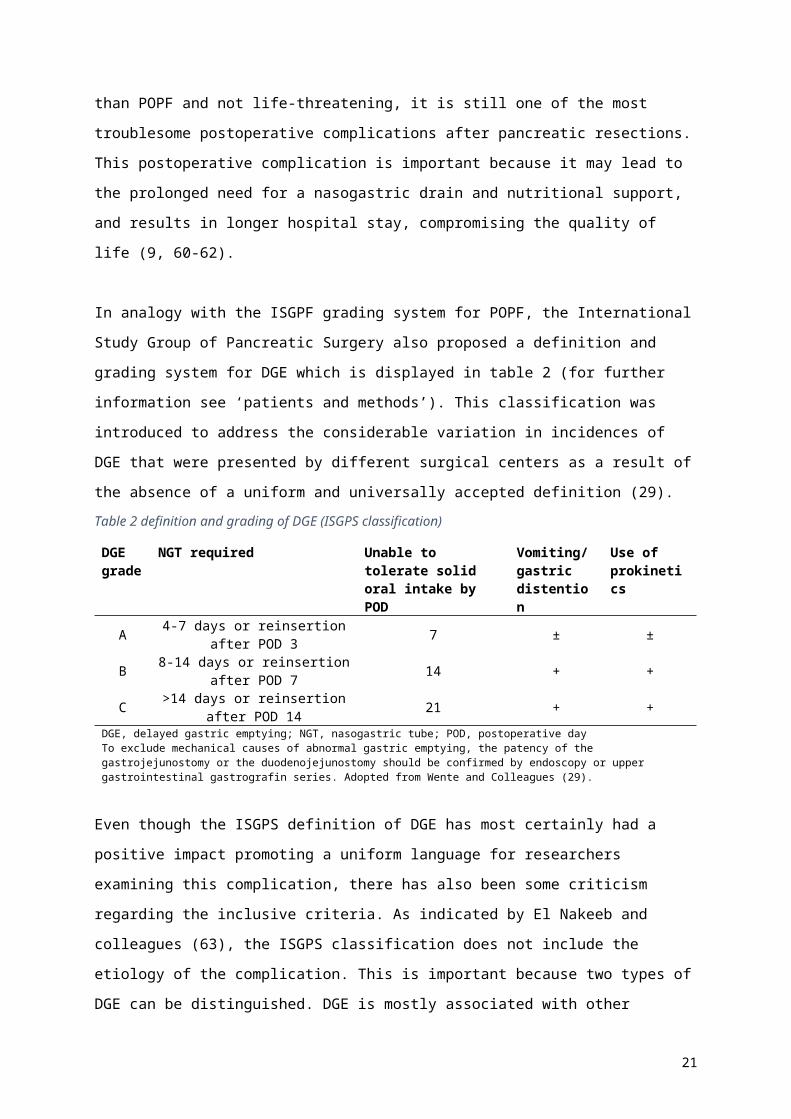
In analogy with the ISGPF grading system for POPF, the International Study Group of
Pancreatic Surgery also proposed a definition and grading system for DGE which is
displayed in table 2 (for further information see ‘patients and methods’). This classification
was introduced to address the considerable variation in incidences of DGE that were
presented by different surgical centers as a result of the absence of a uniform and universally
accepted definition (29).
Table 2 definition and grading of DGE (ISGPS classification)
DGE grade
NGT required Unable to tolerate solid oral intake by POD
Vomiting/gastric distention
Use of prokinetics
A 4-7 days or reinsertion after POD 3 7 ± ±
B 8-14 days or reinsertion after POD 7 14 + +
C >14 days or reinsertion after POD 14 21 + +
DGE, delayed gastric emptying; NGT, nasogastric tube; POD, postoperative dayTo exclude mechanical causes of abnormal gastric emptying, the patency of the gastrojejunostomy or the duodenojejunostomy should be confirmed by endoscopy or upper gastrointestinal gastrografin series. Adopted from Wente and Colleagues (29).
Even though the ISGPS definition of DGE has most certainly had a positive impact promoting
a uniform language for researchers examining this complication, there has also been some
criticism regarding the inclusive criteria. As indicated by El Nakeeb and colleagues (63), the
ISGPS classification does not include the etiology of the complication. This is important
because two types of DGE can be distinguished. DGE is mostly associated with other
14

postoperative complications such as POPF, which is referred to as secondary DGE.
However, in the situation where DGE occurs without any associated complications, it is
referred to as primary DGE. It is likely that these two types of DGE somewhat differ in
etiology and require a different therapeutic approach.
Another point of criticism regards the inclusive design of the criteria of the ISGPS definition.
Healy and colleagues (64) argue that these criteria might overestimate the true incidence of
this complication. They therefore proposed to add exclusion criteria to the definition in order
to more accurately characterize and research genuine DGE.
Since DGE is highly frequent and troublesome, there has been a lot of interest in identifying
possible risk factors for the development of this postoperative complication. However, as is
noted by Wu and colleagues (62), it is important to bear in mind that studies describing these
risk factors are often biased and influenced by confounding factors such as the route of
construction, the administration of prokinetic drugs and the postoperative care, which are not
uniform among the published studies (65-67). Nevertheless, there is strong evidence for the
association between certain risk factors and the occurrence of DGE.
First of all, DGE is associated with some preoperative risk factors including age, cholangitis,
pancreatic fibrosis, diabetes mellitus and malnutrition. Also, it has been observed that
preoperative drainage of the bile ducts reduced DGE, which could possibly be explained by
the fact that preoperative hyperbilirubinemia is a general risk factor for postoperative
complications (60, 61).
Secondly, there has been research on the association between operative factors and the
incidence of DGE. It was shown that an antecolic construction of the jejunostomy played
some role in decreasing the incidence of DGE (68-70). A possible explanation would be that
an antecolic construction reduces the risk of obstruction and increases the mobility of the
stomach, while at the same time posing a barrier from the pancreas (61). Another operative
factor of possible influence is the impact of pylorus preservation. Results on this matter are
inconclusive so far, as some studies indicate a higher incidence of DGE after pylorus-
preserving pancreaticoduodenectomy than after a classical Whipple procedure, while others
reported a lower incidence (69, 71-73). A meta-analysis by Wu and colleagues (62) showed
no significant difference between pylorus-preserving and pylorus-resecting techniques.
Finally, DGE is most strongly associated with postoperative intra-abdominal complications.
This is also referred to as secondary DGE. Postoperative pancreatic fistula, which is the
most feared complication after pancreatic resection, remains the main cause of secondary
DGE both in pancreaticoduodenectomy and in distal resections (60, 61, 74-76).
15

C. Postoperative hemorrhage Postoperative hemorrhage is a third major complication after pancreatic surgery. Although it
is less common than pancreatic fistula or delayed gastric emptying, it has a high mortality
rate of up to 50%, emphasizing the importance of this complication. In analogy to POPF and
DGE, the ISGPS proposed a definition for postpancreatectomy hemorrhage (PPH) as to
reduce the variability in reported hemorrhage rates that were the consequence of differences
in the definition used by the surgeons. PPH was defined by the use of 3 parameters: onset,
location and severity. Additional grading was based upon the clinical impacts (grade A, B and
C) (28). The incidence of PPH (using the ISGPS definition) is between 1 and 10% in most
series, with high reported mortality rates ranging from 11 to 50% (77-79). The importance of
this complication becomes evident when considering the fact that it is responsible for up to
almost 40% of overall mortality after pancreatic resections (9, 28).
According to the ISGPS definition, PPH can be classified as early (≤24h after operation) or
late (>24h after operation) (28). Early bleeding is mostly considered to be the result of
technical failure during the surgery (inadequate hemostatic control and nonsecured vessels).
Hemorrhage in the late postoperative phase is multifactorial and may be the result of tissue
edema due to infection with vascular erosion, ulcers, pseudoaneurysms or failure of an
anastomosis. Multiple factors predisposing to PPH have been identified, including POPF, bile
leak, intraabdominal infections and abscesses (77, 80). Wellner et al. (81) found nine factors
to be independently predictive for severe grade C PPH in all types of pancreatic resections.
These included POPF, high age and BMI, male sex, portal vein resection, multivisceral
resection and intraoperative transfusion.
Mild hemorrhage can usually be treated conservatively and often even remains unnoticed.
Severe hemorrhage on the other hand requires more invasive interventions such as
interventional endoscopy (for intraluminal bleeding), angiography (for extraluminal bleeding)
or operative hemostatic control (80). Although a universally accepted treatment algorithm is
not available, some algorithms have been proposed. For these algorithms, we refer to
Wellner and colleagues (81) or Gao and colleagues (82).
16

3.3 BACKGROUND
3.3.1PANCREATIC SURGERY
3.3.1.1 HISTORICAL EVOLUTION AND CURRENT TRENDS
Pancreatic surgery is the domain which is responsible for the surgical treatment of pancreatic
disease including cancer, pancreatitis and trauma. Historically, developments and
advancements in pancreas surgery were mainly driven by the search for curative treatment
for pancreatic cancer (10, 16). From 1940 to 1970 there were no improvements in the results
of Whipple surgery and morbidity and mortality remained very high with 40-60% and 20-40%
respectively and a 5-year survival rate of less than 5%. Only from the 1980s onwards
outcomes following PD began to improve because of centralization of these complex
procedures in large-volume centers (18-20). Today, mortality rates after Whipple surgery are
less than 5% (even less than 2%) in high-volume centers, whereas this was still more than
25% in the 1960s. Significant improvement in postoperative morbidity has also been
achieved with a decrease from <60% in the 1960s to under 35% nowadays (9, 10, 16, 17).
However, it is clear that despite these advancements in pancreas surgery, the morbidity (and
mortality) rates are still very high.
Current trends in pancreas surgery comprise the expansion of surgical indications. Patients
that would previously have been classified as inoperable are now operated on (16, 24).
There is also a movement towards minimally invasive surgery using laparoscopy and robotic
techniques. The first laparoscopic pancreaticoduodenectomy was performed in 1994 by
Gagner and Pomp for chronic pancreatitis. There is more experience with laparoscopic distal
pancreatectomy because of its lack of anastomoses and lesser risk of damaging large
vessels. Laparoscopic pancreaticoduodenectomy has a higher degree of technical difficulty
and requires a long learning curve. However, it can be performed safely by specialized,
experienced surgeons. Robotic-assisted pancreatectomy makes an easier transition from
open surgery compared to laparoscopy. So far the limited experience with these minimally
invasive techniques looks promising (16, 83, 84).
There are quite some advancements in the surgical technique. However, the main barrier
and challenge for future pancreatic surgeons remains the difficulty of early diagnosis of
pancreatic cancer (PC). Although PC was the main driver in the development of pancreatic
surgery, it is not the sole indication for surgical intervention nowadays. Surgical treatment of
acute and chronic pancreatitis are also part of the domain, as well as the approach of
traumatic injuries of the pancreas (24, 85, 86).
17

3.3.1.2 TYPES OF SURGERY AND INDICATIONS
There are many surgical procedures of the pancreas, depending on the specific indications.
Pancreatic cancer and pancreatitis are the two main indications for pancreatic surgery
nowadays.
For PC, the surgical technique is based on the location of the lesion.
Pancreaticoduodenectomy (PD) is used for the treatment of lesions in the head, neck or
uncinated process. This procedure, also known as the Whipple procedure, involves the
removal of the pancreatic head, the gall bladder with the bile duct, the duodenum and the
pylorus of the stomach. In a pylorus-preserving PD, the stomach is left intact (87). After the
resection three reconstructions are made. These include a pancreaticojejunostomy,
choledochojejunostomy and gastrojejunostomy (88). If the lesion is located in the body or tail
of the pancreas, a distal or left partial pancreatectomy (sometimes accompanied by a
splenectomy) is performed (87).
3.3.2PANCREATIC CANCER
3.3.2.1 EPIDEMIOLOGY
Having an understanding of the epidemiology and trends in pancreatic cancer (PC) can lead
to clues on etiological factors and is therefore crucial in the development of prevention
strategies (89). Pancreatic cancer is the 9th most common cancer in the United States and
the fourth leading cause of cancer death (1, 10). According to Yabar et al. (17), it has even
surpassed breast cancer and is therefore, as of 2016, the third leading cause of cancer
deaths in the US. The amount of deaths due to pancreatic cancer is currently increasing and
it is therefore predicted that in the next decade, PC will likely surpass colorectal cancer and
become the second leading cause of cancer death (1, 17). Pancreatic cancer is a major
health problem in North-America.
In Europe pancreatic cancer deaths are almost just as high and they are also increasing,
making PC the fourth leading cause of cancer deaths in the developed countries altogether
(1, 2). Malvezzi et al. made a European cancer mortality prediction for 2015, putting PC in
the fourth place for cancer mortality in both men and women (90). Estimates of the global
impact of pancreatic cancer on mortality differ, but with an estimated death toll between 200
000 and 330 000 persons per year, it can be stated that it is a global health issue (1, 3). The
developed countries have the highest incidence and mortality rates for pancreatic cancer
worldwide (89).
18

Incidence rates vary considerably between different parts of the world. The highest incidence
is seen in Northern America and Western Europe (7,4 per 100 000 people per year), while
the lowest incidence is almost ten-fold lower and is found in Middle Africa and South-Central
Asia (1 per 100 000 per year). The largest part of this variation is attributed to differences in
exposure to known and suspected environmental and lifestyle factors such as smoking.
However, part of the variation can be explained by differences in diagnostic capacities. The
same variation is seen for pancreatic cancer mortality rates, which can be explained by high
fatality of these malignant tumors (89, 91, 92).
Pancreatic cancer incidence rates have increased from the 1950’s through the 1980’s, with a
levelling off since then, which is most likely due to decreased smoking. However, recent
increasing trends in the European Union indicate that other factors such as obesity, physical
inactivity and dietary factors also play a role (89, 93).
3.3.2.2 DIAGNOSIS
Early diagnosis is essential in the outcome of PC because of the malignant potential of this
tumor, characterized by rapid invasion and metastatic spread, with surgical resection being
the only possible curative treatment. However, most PC have no symptoms in early stage,
making early detection a challenge (1, 10).
The initial clinical presentation of a patient with PC and the accompanying symptoms are
related to the location of the tumor in the pancreas (10, 17). Tumors in the head, neck and
uncinated process mostly lead to obstruction of the pancreatic and common bile duct. This
results in obstructive jaundice in 75% of the cases. Obstruction also often leads to acute
pancreatitis and steatorrhea because of the impaired exocrine pancreatic function. Tumors in
the body and tail of the pancreas are most commonly associated with abdominal and back
pain, new-onset diabetes and nausea (10, 17, 94). In general, there are about 12 alarm
symptoms that should raise a doctor’s suspicion for PC. These include weight loss,
abdominal pain, nausea, bloating, dyspepsia, new-onset diabetes, changes in bowel habit,
pruritus, lethargy, back pain, shoulder pain and jaundice (95). Obstruction of the bile ducts
can also lead to a palpable gallbladder, also known as Courvoisier’s sign, which is a classic
sign in PC (1, 10).
If there is a clinical suspicion for PC, further diagnostic investigations will be performed to
confirm this suspicion. First of all, tumor markers such as CA19-9, CA125 and CEA are used.
However, these markers only have limited sensitivity and specificity (96). A lot of new tumor
markers are currently being examined and will be introduced by genomic epigenetic and
19

proteomic techniques. These markers include cytokines, chemokines, miRNA levels and
even autoantibodies (94). Secondly, imaging techniques are extremely important for early
detection, determination of the location and invasion of the tumor and for staging. Computed
tomography (CT) is used for diagnosis, assessment of resectability and vascular invasion
and for the diagnosis of metastatic lesions. It has a sensitivity of 83% and a specificity of 63-
75%. However, endoscopic ultrasonography (EUS) is now considered the best method for
diagnosing PC. Both retrospective and prospective studies reported superiority over CT with
a sensitivity of 98-100%. EUS-guided fine needle aspiration has a high diagnostic accuracy
of more than 85-90% (1, 94).
3.3.2.3 STAGING AND RESECTABILITY
When the diagnosis of pancreatic cancer is made, the next step is to determine tumor size,
location, local and distant spreading. This process is called staging and results in a TNM-
based classification of the tumor. Conventional staging for pancreatic duct adenocarcinoma
(PDA) is based on the American Joint Cancer Committee (AJCC) TNM-staging system (7th
edition). This staging correlates with overall survival and thus provides prognostic information
(97). However, in PC the ultimate goal of staging is to determine surgical resectability of the
tumor. Therefore, the National Comprehensive Cancer Network (NCCN) published
guidelines in 2015 for a surgically relevant staging system which divides nonmetastatic
disease in three groups (resectable, borderline resectable and locally advanced). These
NCCN guidelines have recently been updated (98). In pancreatic cancer this assessment is
specifically critical since surgical resection is the only potentially curative therapy. The
primary factor in determining if a patient qualifies for surgical resection is whether or not the
surgery can result in a reasonable chance at microscopically negative resection margins.
This implies that only 20% of the patients qualify for surgery at the time of diagnosis (9, 10,
24).
Staging is primarily accomplished by different imaging techniques. The most important
elements that determine resectability are tumor size, location of the tumor within the
pancreas, vascular involvement, lymphadenopathy, anatomic variations and presence of
metastasis. Computed Tomography (CT) is most frequently used for staging but endoscopic
evaluation using EUS is also used. Despite this extensive staging, 10-25% of the patients
undergo an unnecessary laparotomy because unresectable disease (e.g. peritoneal or liver
metastasis) is discovered upon exploration of the abdomen (99, 100).
20

3.3.2.4 THERAPY
Treatment options for pancreatic cancer include surgery, neoadjuvant therapy, adjuvant
chemotherapy, radiation therapy and palliative care. The choice for one therapy or another
will largely depend on the stage of the tumor at diagnosis and the patient’s preferences (1).
Surgery is the cornerstone of therapy for PC because it is the only treatment leading to a
potential cure and can result in significant longer survival as compared to other treatment
options. However, less than 20 % of the patients qualify for surgery at the time of
presentation, making patient selection critical (9, 10). Surgical resection largely consists of
two distinct operations (and some variations), based on the anatomic location of the tumor in
relation to the superior mesenteric vein/portal vein axis. A pancreaticoduodenectomy
(Whipple procedure) is typically performed for cancers of the pancreatic head, neck and
uncinated process. A variant on this procedure is the pylorus preserving
pancreaticoduodenectomy. In this variant of the classical Whipple, the stomach as well as
3cm of the proximal duodenum are preserved. It has been shown that this would reduce
complications such as dumping and delayed gastric emptying, but a recent meta-analysis
shows no significant differences in morbidity and mortality (10, 101, 102). For cancers
located in the body or tail of the pancreas, a distal pancreatectomy can be performed, which
is sometimes combined with a splenectomy (9, 10, 17).
More recently, neoadjuvant therapy has found its access in the treatment of PC, because it
has some possible advantages over adjuvant therapy. First, the chance of delivering a full
chemo dose is bigger when given before surgery. Secondly, neoadjuvant therapy may be
more effective because poor drug delivery to the tumor bed after resection is observed and it
also has low sensitivity to radiation (1). This therapy is most commonly suggested for
borderline resectable tumors, because it results in a greater percentage of patients
eventually undergoing surgery and leads to a higher rate of R0 resections (1, 10, 17).
Metastatic disease is present in 50% of the patients at presentation. These patients can only
be managed by chemotherapy and this treatment will be palliative (17). Current evidence
suggests that both FOLFIRINOX and gemcitabine plus nab-paclitaxel are effective therapies,
if the patient can tolerate them. Further research is needed to see if the combination of
chemotherapy with radiotherapy shows additional benefits to chemotherapy alone (1).
New therapeutic strategies are on the horizon. Targeted therapies including small molecules
and antibodies have already proven to be successful in the management of some other
malignancies. The same applies to immunologic therapies. However, their efficacy in
pancreatic cancer is yet to be proven (10, 17).
21

Last but not least, palliative care is extremely important. In this stage, the focus will be on the
patient’s comfort. This is achieved through surgical and endoscopic therapies, but
chemotherapy and radiotherapy are also used (1, 10).
4 PATIENTS AND METHODS
4.1 STUDY DESIGN
We conducted a retrospective analysis of a series of 255 pancreatic surgeries (classical
Whipple, pylorus-preserving Whipple, distal pancreatectomy) performed for various
indications at the department of general and hepatobiliary surgery of the Ghent University
Hospital. All surgeries were performed between January 2010 and November 2015. The
study protocol (EC/2016/0892) was approved by the Medical Ethics Committee of the Ghent
University Hospital on 07/27/2016. The retrospective analysis of an existing database has
the advantages of being relatively rapid and inexpensive. The database was not designed
specifically for this research and contains many more variables than the ones we used in our
analysis, allowing for other research questions to be investigated using the same data. Of
course, this study design also shows some clear disadvantages, including the fact that many
of the data were derived from electronic patient records leading to frequent missing values.
Other disadvantages concerning this type of research are the predetermined selection of the
study population and the way variables were recorded.
Before starting our analysis we reviewed whether all of the variables needed for answering
our research question were available in the first place. Secondly, we revised the quality and
accuracy of the data and, after completing the missing data through the electronic patient
records, concluded that the quality and availability of these data was sufficient to obtain
reliable and representative results. As the database included all patients who successively
underwent pancreatic surgery between January 2010 and November 2015, we also assumed
that the population selection of this database adequately represented the general population
undergoing pancreatic surgery. In other words, despite the disadvantages and difficulties that
are inherent to this study design, we ensured that this had only a minimal impact on the
study results, thus supporting the reliability of our findings and conclusions.
4.2 PATIENTS AND DATA COLLECTION
Between January 2000 and November 2015, 906 patients underwent pancreatic surgery at
the Department of General and Hepatobiliary surgery at Ghent University Hospital. A large
22
database was constructed using the data that were available in the electronic patient
records. No additional investigations were performed and no extra contact with the patients
was required. Since all of the information consisted of early perioperative data and no long-
term follow-up data were used, there was no requirement to obtain informed consent.
4.3 DATABASE CONSTRUCTION AND VARIABLE SELECTION
All data were initially collected in a database using Filemaker Pro. These included 906
surgeries performed between 2000 and 2015 for various indications such as pancreatic
cancer, pancreatitis, benign lesions of the pancreas and trauma. We selected all cases from
January 2010 until the end of November 2015, resulting in a database of 454 pancreatic
surgeries. All cases before 2010 were excluded because of the substantial amount of
missing data in these patients. The database was then converted into an Excel file and an
SPSS file for further analysis.
More than 50% of the cases were (pylorus-preserving) Whipple procedures and almost 15%
were body and/or tail resections. The remaining 34% included a variation of different surgical
procedures that were each individually only performed a limited number of times. Among
others, this group included necrosectomies for acute pancreatitis, drainage procedures (Frey,
Partingon-Rochelle, Puestow) for chronic pancreatitis, enucleation of cystic lesions,
explorative laparotomies for trauma and double derivations in a palliative setting. Because of
the heterogeneity of this group and the fact that some of these procedures weren’t relevant
for the aim of this study (e.g. palliative double derivation), we decided to exclude these
cases. This resulted in a series of 288 patients who underwent a (pylorus-preserving or
classical) Whipple procedure OR a body and/or tail resection. Nine more patients were
excluded because of inconsistency between the database and the electronic patient record,
leaving 279 cases. Finally it was decided to exclude surgeries performed for pancreatitis
(n=24) and focus only on (pre)malignant pancreatic lesions, resulting in a final database of
255 patients. This series can be divided into 198 cases of (pylorus-preserving)
pancreaticoduodenectomies and 57 pancreatic body and/or tail resections.
23

Thorough data cleaning in SPSS version 24.0 (IBM Corp. Released 2016. IBM SPSS
Statistics for Macintosh, Version 24.0. Armonk, NY: IBM Corp.) was performed prior to any
statistical analysis of the database. First, the variable ‘CODEFORTYPEOFSURGERY’ was
recoded into a dichotomous variable ‘CODEFORTYPEOFSURGERY_dc’. This ensured that
the different terms ‘classical Whipple, pylorus-preserving Whipple, PPPD, extended Whipple,
…’ referring to the same type of surgery were all comprised by the same attribute ‘Whipple
(pylorus-preserving + non pylorus-preserving + extended)’. In accordance, this also ensured
that the terms ‘body + tail resection’ and ‘tail resection’ were comprised by the same attribute
‘Pancreatic corpus and/or tail resection’. Similarly, we recoded the variables ‘FistelEarly_DC’
and ‘GENDER_dc’ into dichotomous variables. The new variable ‘FistelEarlyMetTypeA_DC’
included type A fistula. As type A fistula are shown to have no clinical impact, a second
dichotomous variable was constructed, excluding type A fistula as to only withhold those
which are clinically relevant (type B and C). Further data cleaning was performed using the
‘Codebook’ function in SPSS which allowed us to see the frequencies of all values for a
particular variable. This enabled us to identify false or impossible values and correct or
recode them. This method was used for the variables ‘Diabetes’, ‘Aspectpancreas’,
‘Drainperdu’, ‘neoadjuvantetherapie’, ‘postpancreatectomy hemorrhage’ and
‘DelayedGastricEmptying’, which were subsequently recoded into dichotomous variables as
described before.
As mentioned in the introduction, the goal of this study was to analyze postoperative
complications after pancreatic surgery in a high-volume institution, with the emphasis on
All resections for pancreatitis (both acute and
chronic) were excluded (24 cases).
N = 255
Nine cases were excluded because of inconsistencies
between the database and the electronic patient records.
N = 279
All types of resections other than (pylorus-preserving)
Whipple procedures and distal pancreatectomies were
excluded (166 cases).
N = 288
N = 454
All pancreatic resections before January
2010 were excluded (452 cases).
N = 906All pancreatic resections performed between January 2000 and the end of
November 2015
24
postoperative pancreatic fistula as this is one of the major complications that still exists in
pancreatic surgery. A second goal was to analyze the existence of correlations between
certain risk factors (patient related risk factors, surgical risk factors and disease related risk
factors) and the occurrence of these fistula, as well as other postoperative complications.
Therefore, prior to the selection of any variables for statistical analysis, we conducted a
literature search on the risk factors for postoperative pancreatic fistula.
Eventually, we made a selection of variables based on a number of studies that reviewed
current evidence concerning the association between these possible risk factors and the
occurrence of POPF (14, 31, 103, 104). The variables that were eventually selected for
analysis included age, sex, type of surgery, presence of an early pancreatic fistula (within 1
month after surgery), presence of a late pancreatic fistula, type of POPF, pancreatic texture,
pancreatic duct size, body mass index (BMI), T-stadium in the TNM classification, lymph
node invasion, neoadjuvant therapy, Somatostatin use, operating time, weight loss,
postoperative bleeding, (wound) infection, and delayed gastric emptying.
4.4 SPECIFIC VARIABLE DEFINITION
A. Postoperative pancreatic fistula (POPF): In most studies today, postoperative pancreatic fistula are defined, based on the definition
proposed by the International Study Group for Pancreatic Fistula (ISGPF) (27). In 2005,
ISGPF defined POPF as a failure of healing/sealing of a pancreatic-enteric anastomosis or a
parenchymal leak not directly related to an anastomosis such as one originating from the raw
pancreatic surface. The all-inclusive definition of POPF is a drain output of any measurable
volume of fluid on or after postoperative day 3 with an amylase content greater than 3 times
the upper normal serum value. The study group also proposed a grading system for POPF,
based on the clinical impact of the complication (Grade A, B or C). For more details on this
definition, we refer to Bassi et al. (27). POPF grade A is a transient fistula with no clinical
impact nor any delay in hospital discharge. POPF grade B are fistula that require additional
management such as repositioning the drains and the continuation of parenteral nutrition.
This grade of fistula usually delays hospital discharge, although patients can be sent home
with the drains still in place. The most sever type of POPF is classified as grade C. These
require major adjustment of the standard management, including invasive procedures and
sometimes even re-exploration of the abdomen. This grading system is also summarized in
table 1 (see introduction).
25
However, in this study the ISGPS definition of POPF was not strictly followed as lipase levels
were used to identify POPF instead of amylase. In consultation with our clinical biologists, it
was decided that the amylase levels determined on the drain fluid are also dependent on the
drainage volume and are hence inaccurate. Therefore, lipase levels were measured on the
drain fluid instead. A threshold lipase level of 1000 Units/liter in the drain fluid on
postoperative day 3 was used to identify pancreatic fistula in our patient cohort. Grading of
the POPF was performed according to the ISGPS grading system. Additionally, we
subdivided the variable POPF into two distinct variables ‘early POPF’ and ‘late POPF’. In this
study, ‘early’ was defined as the occurrence of POPF within the first 30 postoperative days.
All fistula that occurred or still existed beyond this time were denoted as ‘late POPF’.
B. Postpancreatectomy hemorrhage (PPH): Postoperative bleeding can be suspected clinically and is diagnosed by imaging techniques
or reoperation. In this study, postpancreatectomy hemorrhage was defined, based on the
definition proposed by the International Study Group of Pancreatic Surgery (ISGPS) in 2007
(28). Their definition of PPH is based on three aspects, being the time of onset, the location,
and the severity of the bleeding.
1. The time of onset can be ‘early’ (≤24 hours after the end of the operation) or ‘late’ (>24
hours).
2. The location of the hemorrhage can be ‘intraluminal’ (e.g. pancreatic surface,
anastomoses, gastric/duodenal ulcer/erosion, or hemobilia) or ‘extraluminal’ (e.g. arterial
or venous vessel, operating field, external suture or staple line, or pseudoaneurysm).
3. The severity of the bleeding can be either ‘mild’ or ‘severe’. Mild bleeding is defined as a
small or medium volume blood loss with no or minimal clinical impairment, no need for
invasive intervention, and successful conservative treatment. Severe bleeding is a larger
volume blood loss and potentially life threatening clinical impairment accompanied by
tachycardia, hypotension, and/or oliguria. Treatment involves the need for blood
transfusion and/or invasive treatment.
For more details on this definition we refer to Wente et al. (28).
C. Delayed gastric emptying (DGE): In this study, delayed gastric emptying or gastroparesis was defined, based on the definition
proposed by the International Study Group of Pancreatic Surgery (ISGPS) in 2007 (29). They
state that “DGE is the inability to return to a standard diet by the end of the first postoperative
week and includes prolonged nasogastric intubation of the patient”. Again, three different
grades (A, B, and C) were proposed based on the clinical impact and the postoperative
management. This grading system is summarized in table 2 (see introduction).
26

DGE is present if a nasogastric tube is required after 4 to 7 days postoperatively, or if a
reinsertion of the nasogastric tube was necessary after removal of the tube by postoperative
day 3, and the patient can’t tolerate a solid oral diet by postoperative day 7. This is defined
as grade A DGE. “DGE is classified as grade B if the nasogastric tube is required from
postoperative day 8 until 14, if reinsertion of the nasogastric tube was necessary after
postoperative day 7, or if the patient cannot tolerate unlimited oral intake by POD 14. Finally,
the most severe grade of DGE is grade C, which implies that nasogastric intubation cannot
be discontinued or has to be reinserted after postoperative 14, or the patient is unable to
maintain unlimited oral intake by postoperative day 21.”. For more details on this definition
and clinical grading, we refer to Wente et al. (29).
D. Pancreatic texture: Pancreatic texture is a subjective judgement made by the surgeon, based on palpation of the
pancreatic tissue. In this study a distinction was made between ‘soft’ and ‘firm’ texture of the
pancreatic parenchyma.
4.5 SURGICAL APPROACH
The 255 patients included in this study underwent different types of pancreatic resections:
twenty-one patients underwent a classic Whipple procedure, 177 underwent a pylorus-
preserving Whipple procedure and 57 underwent a body and/or tail resection. A general
overview of the surgical technique will be described in the following section. However, it
should be taken into account that a variation in surgical steps was used, depending on the
indication or any difficulties that occurred during the procedures, or depending on anatomic
variations in individual patients.
For a pylorus-preserving Whipple procedure, the patient was positioned in a dorsal decubitus
fashion and a median laparotomy or a transverse incision was made. The omental bursa was
opened to find the pancreas and a Kocher maneuver was performed. The hepatoduodenal
ligament was dissected and isolated from the common bile duct, dividing it at the bifurcation
of the left and right hepatic duct. Vascular structures were dissected free and the
gastroduodenal artery was divided, followed by the transection of the duodenum just past the
pylorus. The portal vein was mobilized above and below the pancreatic body. Then, the
duodenojejunal flexure was made free and the pancreatic body was divided over a clamp.
Prolene 5/0 sutures were used to ensure hemostasis. The jejunum was divided at this point
and an omega loop was prepared. The jejunum was pulled under the vascular pedicle and
the vascular structures were divided, dissecting the pancreatic head and the uncinated
process. The resection piece was removed in one piece and sent to pathology.
27

Reconstruction was started by performing a pancreaticojejuneostomy in two layers, using
Prolene 4/0 single stitches and separate duct to mucosa stitches. Then, a
hepaticojejuneostomy was performed at 35 cm with single stitches using PDS 5/0. Finally,
the duodeno-enterostomy was performed in two layers with transmesocolic sutures using
Vicryl 3/0 suturing material. Two Ch24 drains and a penrose drain were placed in a classical
fashion, followed by the closure of the abdomen in a layered fashion using PDS 1 for the
fascia, Vicryl 2/0 sutures for the subcutaneous tissue and Monocryl 3/0 for the intradermal
closure of the skin. The classic Whipple procedure was performed in an analogous fashion,
but instead of transecting the duodenum past the pylorus, an antrectomy was performed. By
consequence, reconstruction was achieved by a gastro-enterostomy instead of a duodeno-
enterostomy.
For a body and tail resection of the pancreas, the patient was positioned in a dorsal
decubitus fashion and a subcostal incision extended to the left was made. The omental bursa
was opened to find the pancreas and the splenic flexure of the colon was released. The
pancreas was dissected free from adjacent structures and a tunnel was created, followed by
the mobilization of the spleen. The splenic and hepatic artery were identified. The splenic
artery and vein were then ligated using Prolene 4/0 and were subsequently divided. A linear
stapler TL 60 was used to transect the pancreatic body and hemostasis was ensured using
Prolene 4/0 sutures. The pancreas was further mobilized from medial to lateral and the short
gastric arteries were divided proximal of the stomach. Resection of the pancreatic body and
tail, together with the spleen was then completed. A penrose drain was placed under the
pancreatic stump and a silicone drain was placed in the splenic compartment. Finally, the
abdomen was closed in a layered fashion analogous as described for the pylorus-preserving
Whipple procedure.
4.6 POSTOPERATIVE CARE
At Ghent University Hospital, the standard postoperative care protocol after pancreatic
resections starts with total parenteral nutrition smofkabiven 12N on postoperative day 1. On
postoperative day 3, the lateral drain on the right side is removed and the nasogastric tube is
withdrawn 10 cm. Primperan was also started on this day (1 ampule of 10mg/2ml, 3 times a
day) unless contraindicated. On postoperative day 5, lipase was measured on penrose and
drain fluids. If the lipase was negative, indicating that no POPF was present, the right medial
drain was removed and the penrose drain was mobilized. If lipase was positive however, the
penrose was fixated and 30mg Sandostatine LAR was administered. The dose of
somatostatine was divided in half on postoperative day 6 and stopped on postoperative day
28
7. From postoperative day 6, the patient was further mobilized and per oral feeding was
started as soon as het nasogastric tube was removed. Food intake was build up in a
stepwise fashion until the patient was ready for discharge.
4.7 STATISTICAL ANALYSIS
Statistical analysis was performed using IBM SPSS version 24.0 (IBM Corp. Released 2016.
IBM SPSS Statistics for Macintosh, Version 24.0. Armonk, NY: IBM Corp.). First, descriptive
analysis of the entire database was performed using the ‘CODEBOOK’ function. Counts,
percentages, means, standard deviations and quartiles were calculated for all relevant
variables. The same process was then repeated separately for those patients who underwent
(pylorus-preserving) Whipple surgery, and once more for patients who underwent a distal
pancreatectomy. Finally, we also performed descriptive analysis on the patients who
developed POPF, using the same method.
Secondly, univariate analysis of categorical variables was performed using a Chi-Square test
(or Fisher’s Exact test when needed). Analysis of continuous variables such as ‘Age’ and
‘BMI’ was performed using the Mann-Whitney U test or an independent t-test. Normality tests
such as the Kolmogorov-Smirnov and the Shapiro-Wilk test were first used to identify
whether a parametric or a non-parametric test had to be used. Significance was defined as a
p-value ≤0.05, implying that a 5% chance of a type 1 error was accepted (⍺ = 5%).
4.8 LITERATURE SEARCH
Before the start of this study, a literature search was performed to expand our background
knowledge on pancreatic surgery, POPF, DGE, PPH, pancreatic cancer and so on. This
information was used in the introduction of this dissertation. Throughout the rest of the study,
additional searches were performed when needed. These literature searches were
performed using Pubmed, Embase and Web Of Science, giving access to major biomedical
databases including Embase, Medline, Science Citation Index Expanded, and others.
A first search was usually performed using Pubmed, because this allowed the identification
of appropriate MeSH-terms for the intended literature search. A limited number of relevant
publications was retrieved, using a combination of MeSH-terms and Major Subject Headings.
These publications were scanned on relevance by reading the abstract and were then
included or excluded accordingly. Additional searches were performed, both in Pubmed and
the other search engines, using a free text-string to identify the most recent literature that
hadn’t been indexed in Medline yet.
29

5 RESULTS
5.1 DATABASE DESCRIPTIVE STATISTICS
A final database of 255 pancreatic surgeries for (pre)malignant pancreatic lesions was
obtained (see flowchart patients and methods). This series consisted of 198
pancreaticoduodenectomies, of which 177 (89%) were pylorus-preserving. The other 57
surgeries were distal pancreatectomies, consisting of 29 (51%) tail resections and 28 (49%)
body and tail resections. There were slightly more male patients (56.5%) compared to female
patients (43.5%) and the mean age was 64.7 (± 11.17). Other baseline variables are shown
in table 3. Statistical analysis did not reveal significant differences in these patient
characteristics between the different types of surgery.
Table 3 Patient characteristics
Patient characteristics and baseline variables
All patients (n = 255)
(pylorus-preserving) pancreaticoduodenectomy
(n = 198)Body and/or tail
resection (n = 57)
Gender
(male/female)144/111 (1.30) 111/87 (1.28) 33/24 (1.38)
Age (years) 64.7 ± 11.2 65.3 ± 10.8 62.5 ± 12.2
Body mass index (%
>25kg/m2)47.9% 45.8% 54.3%
Diabetes (%) 18% 17.2% 21.1%
Data are given as the mean ± standard deviation (SD) unless indicated otherwise.
5.2 POSTOPERATIVE PANCREATIC FISTULA (POPF)
Table 4 shows the rate of postoperative pancreatic fistula in the complete study cohort, as
well as in the group undergoing (pylorus-preserving) pancreaticoduodenectomy and the
group undergoing distal pancreatectomy separately. Using the ISGPS criteria, 202 out of 255
patients (79.2%) did not develop a pancreatic fistula, and 35 patients (13.7%) only
experienced a transient biochemical leak (grade A), whereas a clinically relevant fistula
(grade B or C) occurred in 18 patients (7.1%). There were significantly more clinically
relevant fistula in the distal resection group (17.5%) compared to the (pylorus-preserving)
Whipple group (4.0%) (P = 0.002).
30

Table 4 Incidence of POPF
Postoperative pancreatic fistula
All patients (n = 255)
(pylorus-preserving) pancreaticoduodenectomy
(n = 198)Body and/or tail
resection (n = 57)
All grades n(%) 53 (20.8%) 24 (12.1%) 29 (50.9%)
Clinically relevant*
POPF n(%)
18 (7.1%) 8 (4.0%) 10 (17.5%)
Grade A n(%) 35 (13.7%) 16 (8.1%) 19 (33.3%)
Grade B n(%) 17 (6.7%) 7 (3.5%) 10 (17.5%)
Grade C n(%) 1 (0.4%) 1 (0.5%) 0 (0%)
* Grade B and C POPF are considered clinically relevant whereas grade A fistula is only a biochemical leak.
5.2.1PATIENT CHARACTERISTICS BY GRADE OF FISTULA
Table 5 shows four patient characteristics of the 255 patients classified by the ISGPS
grading system for POPF, grouping ‘no fistula’ and ‘grade A’ fistula together because only
grade B and C POPF are considered clinically relevant. There were no significant differences
in these patient characteristics between those developing clinically relevant POPF and those
who did not.
Table 5 Patient characteristics by grade of fistula
Patient characteristics No fistula or grade A fistula (n=237)
Clinically relevant fistula grade B or C (n=18)
Gender (male/female) 134/103 (1.30) 10/8 (1.25)
Age (years) 64.6 ± 11.35 66.1 ± 8.58
Body mass index (kg/m2) 24.7 ± 4.07 25.1 ± 2.23
Diabetes (%) 18.1% 16.7%
Data are given as the mean ± standard deviation (SD) unless indicated otherwise.
5.2.2UNIVARIATE ANALYSIS OF RISK FACTORS FOR CLINICALLY RELEVANT
POPF
A. All pancreatic surgeries Univariate analysis was performed to identify predictive risk factors for the development of
clinically relevant (grade B or C) postoperative pancreatic fistula. Ten variables displayed in
table 6 were tested, showing that there was a significant difference in clinically relevant
fistula between the group of patients who had undergone (pylorus-preserving)
pancreaticoduodenectomy, compared to those that had undergone distal resections (P =
0.002). For delayed gastric emptying, a trend towards significance could be observed, with
almost 39% of the patients with grade B or C POPF having some degree of DGE compared
to 17% in the group without clinically relevant POPF (P = 0.052). There was no significant
31
difference in gender ratio between those with and without clinically relevant POPF. There
was no significant difference between the group without POPF or with grade A POPF
compared to the grade B/C group in terms of Body mass index, percentage with diabetes,
age, neoadjuvant therapy, drain perdu, pancreatic texture or postpancreatectomy
hemorrhage.
Table 6 univariate analysis of predictive risk factors for clinically relevant POPF
Predictive factors No fistula or grade A fistula (n=237)
Clinically relevant fistula grade B or C
(n=18)P value
Gender (Male/Female)* 134/103 10/8 0.935
Body mass index (kg/m2)‡ 24.7 ± 4.07 25.1 ± 2.23 0.658
Diabetes (%)* 18.1% 16.7% 1
Age (years)‡ 64.6 ± 11.35 66.1 ± 8.58 0.832
Type of surgery
(Whipple/distal resection)*190/47 (4.04) 8/10 (0.80) 0.002
Neoadjuvant therapy (%)* 4.3% 0% 1
Drain perdu (%)* 19.2% 11.1% 0.539
Pancreatic texture
(hard/soft)*82/74 (1.11) 3/7 (0.43) 0.203
DGE (%)* 16.9% 38.9% 0.052
PPH (%)* 5.1% 0% 1
* Chi-Square test or Fisher’s Exact test (where appropriate)‡ Mann-Whitney U test (after normality tests)Data are given as the mean ± standard deviation (SD) unless indicated otherwise.
B. (pylorus-preserving) Whipple procedures Univariate analysis was performed again with the same variables (except ‘type of surgery’) to
identify risk factors for clinically relevant POPF after (pylorus-preserving) Whipple procedures
only. No significant predictive factors for clinically relevant POPF after
pancreaticoduodenectomy were found, as shown in table 7.
32

Table 7 Univariate analysis of risk factors for clinically relevant POPF after (pylorus-preserving) Whipple
Predictive factors No fistula or grade A fistula (n=190)
Clinically relevant fistula grade B or C
(n=8)P value
Gender (Male/Female)* 107/83 (1.29) 4/4 (1) 0.739
Body mass index (kg/m2) ‡ 24.8 ± 4.15 24.7 ± 2.94 0.930
Diabetes (%)* 17.4% 12.5% 1
Age (years)‡ 65.4 ± 10.86 63.2 ± 9.54 0.413
Neoadjuvant therapy (%)* 4.3% 0% 1
Drain perdu (%)* 24.1% 25% 1
Pancreatic texture
(hard/soft)*75/69 (1.09) 1/5 (0.2) 0.114
DGE (%)* 18.0% 37.5% 0.173
PPH (%)* 5.8% 0% 1
* Chi-Square test or Fisher’s Exact test (where appropriate)‡ Mann-Whitney U test (after normality tests)Data are given as the mean ± standard deviation (SD) unless indicated otherwise.
C. Body and/or tail resections Univariate analysis was performed with the same variables to identify risk factors for clinically
relevant POPF after distal pancreatectomies only. The variable ‘drain perdu’ was not
analyzed in distal resections as it is not used in this type of surgery. No significant predictive
factors for clinically relevant POPF after distal pancreatectomy were found. There was a
trend towards significance for delayed gastric emptying (P = 0.062).
Table 8 Univariate analysis of risk factors for clinically relevant POPF after distal resection
Predictive factors No fistula or grade A fistula (n=47)
Clinically relevant fistula grade B or C
(n=10)P value
Gender (Male/Female)* 27/20 (1.35) 6/4 (1.5) 1
Body mass index (kg/m2)‡ 24.6 ± 3.84 25.5 ± 1.38 0.654
Diabetes (%)* 21.3% 20.0% 1
Age (years)‡ 61.3 ± 12.74 68.4 ± 7.42 0.131
Neoadjuvant therapy (%)* 4.3% 0% 1
Pancreatic texture
(hard/soft)*7/5 (1.4) 2/2 (1) 1
DGE (%)* 12.8% 40% 0.062
PPH (%)* 2.1% 0% 1
* Chi-Square test or Fisher’s Exact test (where appropriate)‡ Mann-Whitney U test (after normality tests)Data are given as the mean ± standard deviation (SD) unless indicated otherwise.
33

5.2.3UNIVARIATE ANALYSIS OF RISK FACTORS FOR ALL GRADES OF POPF
A. All pancreatic surgeries Univariate analysis was performed with the same ten variables to identify risk factors for
postoperative pancreatic fistula of any grade. The results are shown in table 9. Again, there
was a significant difference in the occurrence of POPF in the group of patients who had
undergone (pylorus-preserving) pancreaticoduodenectomy, compared to those that had
undergone distal resections (P<0.001). Though pancreatic texture did not prove to be a
significant predictive risk factor for clinically relevant POPF, it is a significant factor in POPF
when including all grades of POPF (P = 0.009). BMI showed to be significantly higher in the
group with POPF (P = 0.041).
Table 9 Univariate analysis of risk factors for all grades of POPF
Predictive factors No fistula(n=202)
Grade A, B or C POPF (n=53) P value
Gender (Male/Female)* 113/89 (1.27) 31/22 (1.41) 0.739
Body mass index (kg/m2) ‡ 24.5 ± 4.17 25.8 ± 2.92 0.041
Diabetes (%)* 19.3% 13.2% 0.304
Age (years)‡ 64.7 ± 11.46 64.6 ± 10.06 0.645
Type of surgery
(Whipple/distal resection)*174/28 (6.21) 24/29 (0.83) <0.001
Neoadjuvant therapy (%)* 4% 3.8% 1
Drain perdu (%)* 19.1% 17.0% 0.725
Pancreatic texture
(hard/soft)*77/61 (1.26) 8/20 (0.4) 0.009
DGE (%)* 16.9% 24.5% 0.204
PPH (%)* 5.1% 0% 1
* Chi-Square test or Fisher’s Exact test (where appropriate)‡ Mann-Whitney U test (after normality tests)Data are given as the mean ± standard deviation (SD) unless indicated otherwise.
B. (pylorus-preserving) Whipple procedures Univariate analysis was performed within the cohort of (pylorus-preserving) Whipple
procedures to identify predictive risk factors for all grades of POPF after this specific type of
procedure. The results are presented in table 10. Soft pancreatic texture showed to be a
significant risk factor for the development of any grade of POPF (P < 0.001). All other
variables showed no significance as predictive factors.
34
Table 10 Univariate analysis of risk factors for all grades of POPF after (pylorus-preserving) Whipple
Predictive factors No fistula(n=174)
Grade A, B or C POPF (n=24) P value
Gender (Male/Female)* 98/76 (1.29) 13/11 (1.18) 0.842
Body mass index (kg/m2)‡ 24.6 ± 4.22 25.5 ± 3.18 0.392
Diabetes (%)* 17.2% 16.7% 1
Age (years)‡ 65.3 ± 11.07 65.6 ± 8.72 0.747
Neoadjuvant therapy (%)* 4.1% 4.2% 1
Drain perdu (%)* 22.2% 37.5% 0.101
Pancreatic texture
(hard/soft)*74/58 (1.28) 2/16 (0.13) <0.001
DGE (%)* 17.8% 25% 0.408
PPH (%)* 5.8% 0% 1
* Chi-Square test or Fisher’s Exact test (where appropriate)‡ Mann-Whitney U test (after normality tests)Data are given as the mean ± standard deviation (SD) unless indicated otherwise.
C. Body and/or tail resections Univariate analysis was performed within the subgroup of patients that had undergone distal
resections to identify predictive risk factors for the development of all grades of POPF after
body and/or tail resections specifically. As can be seen in table 11, diabetes showed to be a
significant protective factor for the development of any grade of POPF (P = 0.044). On the
other hand, higher BMI showed to be a significant risk factor for POPF (P = 0.034). All other
variables showed no significance as predictive factors.
Table 11 Univariate analysis of risk factors for all grades of POPF after distal resection
Predictive factors No fistula (n=28)
Grade A, B or C POPF (n=29) P value
Gender (Male/Female)* 15/13 (1.15) 18/11 (1.64) 0.516
Body mass index (kg/m2)‡ 23.4 ± 3.84 26.1 ± 2.75 0.034
Diabetes (%)* 32.1% 10.3% 0.044
Age (years)‡ 61.2 ± 13.35 63.8 ± 11.13 0.523
Neoadjuvant therapy (%)* 3.6% 3.4% 1
Pancreatic texture
(hard/soft)*3/3 (1) 6/4 (1.5) 1
DGE (%)* 10.7% 24.1% 0.297
PPH (%)* 2.1% 0% 1
* Chi-Square test or Fisher’s Exact test (where appropriate)‡ Mann-Whitney U test (after normality tests)Data are given as the mean ± standard deviation (SD) unless indicated otherwise.
35

5.3 MANAGEMENT OF POPF
Out of 24 patients developing POPF after PD, only 8 had clinically relevant grade B or C
POPF and needed specific treatment. All these patients with CR-POPF were given
Sandostatine-LAR. Two patients required additional surgical reëxploration and drain
placement on top of that. One month postoperatively, six patients still had persisting POPF.
The fistula rate after distal pancreatectomy was 50.9%, with 29 patients developing POPF.
Ten of them had clinically relevant grade B or C POPF and required specific treatment. All
these patients with CR-POPF were given Sandostatine-LAR. One patient required additional
antibiotic treatment and one patient underwent surgical wound exploration and debridement
of a wound infection caused by the presence of the fistula. Seven of these patients (70%)
with CR-POPF still had persisting POPF one month postoperatively.
5.4 OTHER POSTOPERATIVE COMPLICATIONS
In total 25.6% of all patients undergoing pancreatic surgery had at least one of the three
most common and most important complications, being clinically relevant POPF, DGE and
PPH. This was not significantly different between the two surgical groups, with 29.8% of the
patients in the PD group having at least one complication compared to 24.4% of the patients
in the distal resection group (P = 0.406). In total there were 47 cases (18.5%) of delayed
gastric emptying, with no significant difference between the PD group (18.8%) and the DP
group (17.5%). Postpancreatectomy hemorrhage was the least frequent complication,
occurring in 12 (4.7%) patients. Again, no significant difference in bleeding rate was
observed between the PD group (5.6%) and the DP group (1.8%).
Table 12 Postoperative complication rates
Postoperative complications
All patients (n=255)
(pylorus-preserving) pancreaticoduodenectomy
(n=198)
Body and/or tail resections
(n=57)P value
Complication*
n(%) 65 (25.6%) 17 (29.8%) 48 (24.4%) 0.406
DGE n(%) 47 (18.5%) 37 (18.8%) 10 (17.5%) 0.832PPH n(%) 12 (4.7%) 11 (5.6%) 1 (1.8%) 0.310* This variable comprises patients who developed clinically relevant POPF or DGE or PPH
36

6 DISCUSSION
6.1 DATABASE AND PATIENT CHARACTERISTICS
As described in the paragraph on database construction, this study involved a series of 255
patients who underwent pancreatic surgery for the removal of benign and (pre)malignant
tumors. All cases with pancreatitis as a surgical indication were excluded as to ensure that
the patient sample was as homogeneous as possible. However, doing this reduced the
sample size significantly. Furthermore, the absence of certain patient data, which is the
primary disadvantage of any retrospective study design, resulted in missing values which
further decreased sample sizes, thus increasing the risk of insufficient power for accurate
statistical analyses. However, the sample sizes in our study are comparable to those of other
single center studies such as the one by Gagnière et al. (105), including 191 patients and a
study by Yang et al. (106) including 62 patients. This allows comparison with these studies to
be made. Nevertheless, it is recognized that the limited sample size is one of the biggest
limitations in this study, implying that values showing a trend towards significance are
probably worth being further evaluated in larger patient cohorts.
Patient characteristics such as age, gender, mean BMI etc. were very similar in values
compared to other published patient cohorts undergoing pancreatic surgery (such as the two
mentioned above), which indicates that the potential confounding influence of these variables
was reduced to a minimum.
6.2 POSTOPERATIVE PANCREATIC FISTULA (POPF)
6.2.1INCIDENCE OF POPF AFTER PD AND DPOne of the main aims of this study was to analyze the occurrence of POPF, which is the
most important complication after pancreatic resections, in Ghent University Hospital and
compare this to other literature data. However, it should be noted that in this study lipase
levels were used instead of amylase levels to determine the presence of POPF. In other
words, the ISGPS definition was not followed completely which could be a point of criticism.
Nevertheless, lipase levels have shown a high correlation with amylase levels and are also a
good method to identify pancreatic fistula. A study in 2012 by Facy and colleagues (107)
compared the use of lipase for POPF detection with the use of amylase as described by
ISGPS. They found that a threshold lipase level of 1000U/l yielded a sensitivity of 93% for
the detection of clinically relevant POPF. By definition, the use of amylase has a 100%
sensitivity. This could mean that some POPF were missed using lipase instead of amylase.
37

However, in the same study a separate analysis was done identifying patients with CR-POPF
based upon clinical and radiological finding, independent of their drain amylase levels. In this
group, they found that “a lipase threshold of 1000U/l yielded a higher sensitivity and
specificity than amylase” (107). These findings may suggest that the use of lipase instead of
amylase in this study could be an advantage in detecting clinically relevant POPF, implying
that our CR-POPF rates are overestimated compared to other studies using the ISGPS
definition.
In our patient cohort (n=255), a total of 53 patients (20.8%) developed a POPF but only 18
patients (7.1%) developed a clinically significant (grade B or C) POPF. It has to be noted that
this patient cohort consisted of 198 PD’s and 57 DP’s. The surgical techniques of these
operations differ significantly, as patients undergoing PD end up with an anastomosis
between the pancreatic remnant and the jejunum or stomach, while patients undergoing DP
do not have such anastomosis and end up with a free distal remnant of the pancreas. It is
clear that this has significant implications towards the mechanisms and risks of developing a
pancreatic leak, which is reflected in the different POPF rates for these two types of surgery.
Following (pylorus-preserving) pancreaticoduodenectomy, 12.1% of our patients developed a
fistula. A recent study by Gagnière et al. (105) reports a POPF rate of 36.6% (198 PD’s),
Guilbaud et al. (108) report a POPF rate of 38.3% after PD (86 patients). A larger
monocentric study by Hu et al. (109) including 539 PD’s had a POPF rate of 49.9%. There
are also some larger studies that analyze POPF rate on pooled data, such as the study by
McMillan et al. (110) describing a 27.2% POPF rate after PD (1533 patients). A large series
of 2000 PD’s performed at Johns Hopkins Hospital describes a 16% POPF rate (111). Even
though all of these studies use the ISGPF criteria for the definition of POPF, there is still a
large variation in fistula rates. Our study shows a fairly low rate of POPF after Whipple
surgery, but we have to acknowledge that no data considering the indications for surgery
were analyzed. This could be of importance since a study by Aranha et al. (112) in 2006
showed that there is a significant difference in POPF rate following PD according to the
nature of the pancreatic tumor. For example the POPF rate after PD for an ampullary
neoplasm, a duodenal neoplasm or a serous cystic neoplasm was more than 30 percent,
whereas the POPF rate was as low as 6% after PD for pancreatic cancer. Differences in the
distribution of tumor types within the cohort could therefore influence the global POPF rate.
Nevertheless, as patients were not included/excluded based upon the underlying pathology,
the distribution of tumor types in this series is not expected to be significantly different from
other series.
38

Within the group of 24 people who developed a POPF following (pylorus-preserving) PD,
only 8 fistula (4%) were clinically relevant (grade B or C) and only 1 patient needed surgical
reintervention (grade C POPF). These percentages are again lower than other reports such
as the multicenter analysis by Kawai et al. (113) which reports a 14.4% clinically relevant
POPF rate in a series of 1239 PD’s or the multicenter report of more than 4000 PD’s by
McMillan et al. (114) reporting a clinically relevant POPF rate of 11.1%. In 2013, Topal et al.
(115) conducted a large multicenter randomized controlled clinical trial involving 8 Belgian
centers with adequate experience in pancreatic surgery but not including Ghent University
Hospital. They found a CR-POPF rate of 19.8% following PD with pancreaticojejunostomy
(PJ) and a CR-POPF rate of 8% following PD with pancreaticogastrostomy (PG), therefore
concluding the superiority of the latter technique. However, even in the group where PG was
performed, their CR-POPF rate is still twice as high as in our series, although PJ was used
as the standard anastomotic technique for all patients in Ghent University Hospital. This
could possibly be explained by a difference in experience between surgeons. Although all
surgeons contributing to the Belgian multicenter analysis were reported to have several
years of experience with PD, one of the inclusion criteria was that they had performed a
minimum of 5 PG and 5 PJ anastomoses. In Ghent University Hospital PJ is used as the
standard anastomotic technique, implying that the surgeons had significantly higher
experience with this technique compared to the minimum of 5 procedures described above. It
is plausible that the surgeon’s experience with one technique over the other is more
important in influencing CR-POPF rate than the suggested superiority of PG over PJ.
Twenty-nine out of 57 patients (50,9%) developed a fistula following pancreatic corpus
and/or tail resection. This fistula rate is fairly high compared to other reports such as the one
by Goh et al. (116) reporting a POPF rate of 31% and the multicenter analysis by McMillan et
al. (110) reporting a POPF rate of 34.5% after DP. Fistula rates following DP differ
significantly between institutions despite the fact that all of these publications used the
ISGPF classification to define POPF. Despite the high overall pancreatic fistula rate, only 10
patients (17.5%) within our cohort developed a clinically significant (grade B or C) POPF
whereas studies by Goh et al. (116) and McMillan et al. (110) report 14% and 15% rates for
clinically relevant POPF respectively. A recent multi-institution analysis of more than 2000
DP’s by the Distal Pancreatectomy Study Group reported a CR-POPF rate of 15,1% (117).
These reported rates are not so different from our own results, especially when taking into
account a correction for using lipase versus amylase based incident rates. A large
monocentric series of 302 consecutive DP’s published by Kleeff et al. (118) reported an even
lower incidence of 11.6% clinically relevant POPF. In conclusion, the incidence of any grade
of POPF after DP in our institution is quite high, but the majority of these concerned grade A
39

POPF (biochemical leaks). The incidence of clinically relevant grade B and C POPF on the
other hand is relatively comparable to the rates published by other institutions. In analogy
with the relatively low CR-POPF rates after PD, it could have been expected that CR-POPF
rates after DP would be lower than average as well given the extensive experience of the
surgeons. However, as distal pancreatectomy is technically less demanding and does not
involve constructing a pancreatic anastomosis, we believe that this type of surgery is less
prone to influences from surgeon experience. Closure of the pancreatic remnant was
performed using a linear stapler with progressive closure and additional sutures were used if
thought necessary by the surgeon. This technique is in accordance with current literature
data showing that stapler closure or the combined use of stapler and sutures is associated
with significantly lower POPF rates than hand-sewn closure. Various other strategies such as
transection of the pancreas with bipolar scissors, harmonic scalpels or radiofrequency
ablation devices are being tested but at the present time data are too limited to draw any
conclusions (119).
6.2.2RISK FACTORS FOR POPFSince POPF is frequently associated with other major complications making it the leading
risk factor for postoperative death, it is of paramount importance to be able to predict which
patients are at risk of developing POPF (13, 15, 31, 34). Based upon literature research and
the data that were available in our retrospective study, ten potential risk factors for POPF
were identified and analyzed within this patient cohort.
In most studies, the 2005 ISGPF definition for POPF together with its grading system
proposing a distinction between grade A, B and C POPF, is applied. However, this grading
system also became subject of criticism with some authors questioning the practical
relevance of grade A POPF (30). An article by Hackert et al. (120) in 2016 further
accentuated the need for an update of the ISGPS definition and grading system for POPF
which was published soon after (121). As multiple validation studies showed that there was
no difference between grade A POPF and the absence of fistula, it was decided to redefine
the former grade A POPF as biochemical leaks (122, 123). The definition of POPF became
more strict as this implied that the condition of increased amylase activity found in the drain
fluid had to impact clinical outcome in order to be defined as a fistula (121).
As these changes in the 2016 update of the POPF definition were indeed found to be
relevant towards clinical practice, it was decided to also analyze the selected ten risk factors
using this revised grading system. Former grade A fistula (now biochemical leaks) were
therefore analyzed together with ‘no fistula’ so that this group could be compared to those
40

patients suffering from clinically relevant grade B or C fistula (CR-POPF). It is primarily these
clinically relevant POPF that are potentially life-threatening complications, therefore making
the analysis of CR-POPF related risk factors more relevant for clinical practice as compared
to the identification of risk factors for all grades of POPF (including the former grade A).
Using univariate analysis, all ten variables were compared between the group of patients
without any fistula and the group with grade A, B or C POPF. The same method was then
used to compare the variables between the group without CR-POPF (no fistula or
biochemical leak) and CR-POPF grade B or C. In first instance, the analysis was performed
on the total patient cohort, which allowed us to confirm the type of surgery (meaning PD
versus DP) as a risk factor. Distal pancreatectomy showed to be a significant risk factor for
POPF compared to pancreaticoduodenectomy (P<0.001). The same was true for clinically
relevant pancreatic fistula (P = 0.002). As mentioned before, this could be attributed to the
differences in surgical technique with the remaining of a pancreatic stump after DP versus an
anastomosis of the pancreatic duct to bowel or stomach after PD. This finding is confirmed in
the study by McMillan et al. (110) who found a significantly higher POPF rate after DP
compared to PD. Soft pancreatic texture and high BMI (>25kg/m2) both showed to be
significant risk factors for POPF (P = 0.009 and P = 0.041 respectively) irrespective of the
type of surgery performed, but this result is not clinically relevant as the same observations
did not apply to CR-POPF. Also, these findings are less meaningful as this analysis included
all patients. For reasons previously explained, further analysis of risk factors for POPF was
performed for PD and DP separately.
Further univariate analysis was performed for (pylorus-preserving) PD and for DP separately
using the variables male gender, BMI, diabetes, age, neoadjuvant therapy, drain perdu, soft
pancreatic texture, DGE and PPH as potential risk factors. Soft pancreatic texture was the
only significant risk factor for all grades of POPF after PD (P<0.001). This finding has also
been described in numerous other studies, making it a widely accepted major risk factor for
POPF which is explained by the increased technical difficulty of performing an anastomosis
on a soft pancreatic gland (53, 124, 125). Pancreatic texture has also been implemented in
POPF risk scores such as the one proposed by Callery et al. (126). Despite its general
acceptance as a risk factor, soft pancreatic parenchyma was not found to be associated with
clinically relevant POPF after PD in our patient cohort (P = 0.114). However, care should be
taken making conclusions based upon this finding as there were only 8 patients suffering
from CR-POPF after PD, thereby reducing the power of the statistical analysis. It is more
plausible that statistical significance for this well-known risk factor could be proven in a larger
patient group, as was also found by Pratt et al. (127).
41

The same argumentation can be used to explain the fact that soft pancreatic texture was also
found to be no significant risk factor for POPF as well as CR-POPF following distal
pancreatectomy. More specifically, there was only a very small sample size with 10 patients
in the group with fistula and 6 patients in the group without fistula after DP (we did not have
data on pancreatic texture for the remaining 41 patients in this cohort) which did not allow for
statistical significance to be proven. As for CR-POPF, data on pancreatic texture were only
available for 4 out of 10 patients suffering from grade B or C fistula. It is however important to
note that no anastomosis has to be made during DP, thus implying that soft pancreatic
parenchyma does not necessarily influence the technical difficulty of the procedure in the
same way as it does for PD. On the other hand, one can assume that the closure of the distal
remnant of the pancreas will likely be more fragile and prone to leakage if the pancreatic
gland is soft because it is less efficient in holding onto the sutures or staples. In general, the
evidence for soft pancreatic texture as a risk factor for POPF following DP is less established
compared to PD, but a recent meta-analysis by Peng and colleagues (128) did show a
significantly higher incidence of POPF.
One of the problems with evaluating pancreatic texture as a risk factor for POPF or using soft
pancreatic texture in risk scores, is the fact that this parameter is subjectively judged by the
surgeon through direct intraoperative palpation of the gland. However, in recent years there
has been research in using imaging techniques to evaluate pancreatic texture preoperatively
in a non-invasive and quantitative (objective) way. According to Harada et al. (129) an
ultrasound elastographic technique called acoustic radiation force impulse imaging (ARFI)
can be used to evaluate tissue hardness by measuring the shear wave velocity (SWV). The
SWV was shown to correlate significantly with pancreatic fibrosis (and thus with harder
pancreatic texture). There was a significant positive association between soft pancreatic
texture, defined as SWV< 1.54 m/s, and the incidence of POPF. The advantage of using this
technique is that it is fast and non-invasive and it is readily available as it is used in clinical
practice to determine hepatic fibrosis. More recently, Kuwahara et al. (130) suggested the
use of endoscopic ultrasonography-guided elastography (EUS-EG) to evaluate pancreatic
texture by measuring the Mean Elasticity (ME). A ME >70 was shown to be an independent
predictor for POPF after PD. Moreover, this cut-off value showed to be superior in predicting
POPF compared to the pancreatic texture as defined by the surgeon’s intraoperative
palpation of the gland. As was also argued by the authors of this article, one of the
disadvantages of ARFI imaging is the inability to visualize the pancreas and measure the
SWV in obese patients. This problem is overcome by using EUS-EG, giving it an advantage
over transabdominal ultrasonographic elastograpic techniques despite its more invasive
42

nature. Finally, there have also been some studies concerning the use of MRI in evaluating
the pancreatic texture. A recent publication by Hong et al. (131) showed that the apparent
diffusion coefficient (ADC) in diffusion-weighted imaging (DWI) negatively correlated with
fibrosis (and thus harder texture) of the pancreatic gland. Other, more advanced MRI
techniques have also been investigated to evaluate pancreatic fibrosis (and therefore gland
texture). Shi and Colleagues (132) found that 3D-Magnetic resonance elastography could
possibly be used to measure the stiffness of the pancreatic gland. Yoon and colleagues
(133) showed that intravoxel incoherent motion (IVIM) diffusion-weighted (DW) imaging could
provide information on the amount of fibrosis in the pancreatic gland, and that the IVIM DW
imaging parameter ‘perfusion fraction’ was significantly correlated with POPF. However,
these studies are all very recent and only limited data are available. To the best of our
knowledge, there are currently no studies that compare these different imaging techniques
and their ability to assess the pancreatic gland texture or predict the risk for POPF. In
conclusion, it seems attractive to implement transabdominal ultrasonography with ARFI in
Ghent University Hospital to determine pancreatic texture as this non-invasive technique is
readily available and can be performed within a few minutes during the presurgical
evaluation. The major advantage of this is the fact that it makes the parameter ‘pancreatic
texture’ available prior to surgery and thus would allow to adapt the surgical strategy based
on the preoperative POPF risk estimate. Due to its more invasive nature, performing an
EUS-EG of the pancreas may rather be positioned as a technique to be used for patients
who have to undergo EUS anyhow, than as a standard routine. MRI techniques for
determining pancreatic fibrosis and gland texture can be useful but their additional value may
not be enough to outbalance their inherently higher costs and waiting times, making it harder
to implement them in clinical practice.
Male gender was not significantly associated with POPF or CR-POPF, although multiple
other studies show significant correlation with an increased fistula rate (14, 53, 109). The
same can be said for age, DGE and PPH, which showed no significant association to fistula
rate but have each been proposed as possible risk factors in one or more studies (14, 53,
134). However, DGE did show a trend towards significance for developing clinically relevant
POPF after DP (P = 0.062). Forty percent of the patients with CR-POPF also suffered from
delayed gastric emptying. This association between DGE and CR-POPF has been reported
repeatedly (63, 135). In a recent study by Liu et al. (136) CR-POPF and intra-abdominal
collection were found to be significantly correlated to DGE in univariate analysis. Moreover,
they were identified as independent risk factors using a multivariate logistic regression
model. This association can be explained by the fact that DGE after pancreatic surgery is
43

most commonly caused by intra-abdominal complications and specifically by POPF, which is
referred to as secondary DGE (60).
High BMI (>25kg/m2) was not significantly associated with fistula rate after PD. On the other
hand it was a significant risk factor for POPF (but not for CR-POPF) after distal pancreatic
resections (P = 0.034). A similar association between BMI and POPF after DP was found by
Seeliger and colleagues (137) who showed a 6-fold increased risk for POPF with a BMI >25.
This also supports the reported association between high BMI (>25kg/m2) and intra-
abdominal morbidity rate following DP by Sledzianowsky et al. (138) since POPF is one of
the major factors contributing to intra-abdominal complications in this setting. However, these
results are not sufficient to draw any conclusions because there is a possibility of
confounding influences. For example, high BMI might influence the extent of fibrosis present
in the pancreatic gland as was proposed by Kawaida and colleagues (139). Another
possibility is the increased technical difficulty of any procedure in obese patients. Seemingly
contradictive, there have been some studies indicating low serum albumin levels as an
independent risk factor for POPF after DP, although they did not use the ISGPS definition for
POPF (140, 141). As BMI and serum albumin are both markers for nutritional status, one
could assume that both malnutrition and obesity are possible risk factors for POPF. As no
serum albumin levels were measured in our patient cohort, it was not possible to evaluate
these factors. Eventually, we were also unable to show significant association between high
BMI and clinically relevant POPF after DP although this might be attributed to a lack in
statistical power which is why further analysis with augmented patient cohorts is needed to
draw clear conclusions on this matter. A recent meta-analysis by Peng and colleagues (128)
did identify high BMI as a significant risk factor for POPF following DP, but this analysis had
some serious limitations considering the heterogeneity of the included data because the cut-
off value for ‘high BMI’ differed between included studies.
Unlike the previously mentioned variables, neoadjuvant chemotherapy was analyzed as a
potential protective factor in our patient cohort. However no significant correlation with POPF
or CR-POPF could be found. This is in contrast with findings of Cheng et al. (142) who
retrospectively compared a group receiving neoadjuvant chemoradiotherapy followed by PD
to a group undergoing classical PD. They found a significantly lower fistula rate in those
patients that received neoadjuvant chemoradiotherapy. However, when comparing these
results it should be noted that patients in our study only received neoadjuvant chemotherapy
(Folfirinox and Avastin) without neoadjuvant radiotherapy. This explanation is supported by
the findings of Ishikawa et al. (143) in 1991 who found neoadjuvant radiotherapy to be a
protective factor for POPF because it supposedly decreased exocrine pancreatic function.
44

Also, the number of patients receiving neoadjuvant treatment in our cohort (n=12) was
probably too small to detect any association with POPF.
Another potentially protective factor is diabetes. This indeed proves to be a significant
protective factor for POPF after DP (p = 0.044). However, diabetes was not significantly
associated with a lower occurrence of clinically relevant fistula after DP. Also no significant
association was observed between diabetes and both the occurrence of POPF and CR-
POPF after (pylorus-preserving) pancreaticoduodenectomy. Despite the lack of statistical
significance, it was observed that the percentage of diabetic patients in the group without
POPF was always higher than in the group of patients who did develop this complication.
This could possibly indicate that insufficient power for the statistical analysis did not allow for
statistical significance to be proven and by consequence the possibility of diabetes being a
protective factor for POPF development cannot be ruled out based on these data. Our finding
that diabetes is a significant protective factor POPF after DP is contradicted by the before
mentioned meta-analysis by Peng and colleagues (128). However, there are a lot of
conflicting findings in published literature and the association between diabetes and POPF
after both PD and DP is still a matter of debate. Several studies even indicate that a history
of diabetes increased the likelihood of developing postoperative fistulas, rather than being
protective (144, 145). On the contrary, there are also several studies, including a meta-
analysis by Xia and colleagues (146) that show a clear association between the absence of
diabetes and an increased incidence of POPF although this was not proven to be significant
in multivariate regression models (53, 109, 147). Our findings suggest that the protective
effect of diabetes could indeed be true and an explanation for this is provided by Mathur et
al. (147). They found that diabetes was associated with significantly less fat and more
fibrosis in the pancreas. The presence of POPF on the other hand, was found to be
associated with significantly more fat and less fibrosis in the pancreatic gland thereby
explaining the possible protective effect of diabetes. Furthermore, they also remarked the
fact that intuitively one could assume that higher fat content of the pancreas implies a softer
pancreatic parenchyma, which in itself is an established risk factor for POPF. Finally, another
group showed that increased fibrosis in the pancreatic gland, is associated with decreased
exocrine activity which would be an additional argument to suggest a protective effect of
diabetes (148). Taken together, there are some arguments that support the possible
protective effect of diabetes and our results do not refute this hypothesis, but more large
series are needed to make definite conclusions.
45

6.3 OTHER POSTOPERATIVE COMPLICATIONS (DGE & PPH)
Morbidity after pancreatic resections is still as high as 30-50% and this is also caused by
complications such as DGE and PPH, which by consequence also require attention. As
mentioned in the introduction, DGE is the second most important postoperative complication
as it is highly troublesome and results in longer hospital stay, compromising quality of life (9,
60-62). As the importance of DGE is clear, it was one of the aims of this study to assess the
occurrence of DGE in Ghent University Hospital and compare these results to literature data.
The total complication rate (including POPF, DGE and PPH) was 29.8% after (pylorus-
preserving) PD and 24.4% after DP. There was no significant difference between these two
groups of pancreatic resections. Delayed gastric emptying was observed in 37 patients
(18.8%) following PD and 10 patients (17.5%) following DP. Reported incidences of DGE
differ significantly but this DGE rate after PD is comparable to those reported by other single-
center studies such as those by El Nakeeb et al. (63) and Liu et al. (136) which report a DGE
rate of 17.9% and 36.2% respectively. The occurrence of DGE after DP is usually lower
compared to PD, which could possibly be explained by the fact that no alteration of the
normal bowel anatomy is made in DP. Reported incidences of DGE after DP vary from 9%
by Yamamato et al. (149) to 24% by Glowka et al. (135). The large variation in these
reported incidences is probably explained by the influence of many confounding factors such
as the administration of prokinetic drugs and postoperative care which are not uniform across
institutions (62). Another factor that influences reported DGE rates is the used definition, as
not all publications use the ISGPS definition (135).
A third very important complication after pancreatic surgery is postoperative hemorrhage. In
2007, the ISGPS suggested a definition for postpancreatectomy hemorrhage (PPH) with the
intention of creating an internationally accepted definition for this severe complication (28).
Although the incidence of PPH is lower than other complications its significance becomes
clear when reflecting the fact that it is responsible for up to 40% of overall mortality in
pancreatic surgery, which illustrates why it was one of the aims of this study to assess the
incidence of PPH in Ghent University Hospital (9, 28). PPH rate after PD was 5.6%
compared to 1.8% after DP, which implied no significant difference in PPH rate by type of
surgery. This is in accordance with a large series published by Correa-Gallego et al. (78)
who also did not find any significant difference in PPH between PD and DP.
Postpancreatectomy hemorrhage rate in this study was found to be in the same range as
other recent reports. Wellner and colleagues (81) found a PPH rate of 7.2% in a large series
of more than 1000 pancreatic resections and Chen et al. (77) and Manas-Gomez et al. (79)
46

found a PPH rate of 8.8% and 16.8% respectively. It can be concluded that the PPH rate in
Ghent University Hospital is relatively low since all of these studies used the same ISGPS
definition of PPH. Future research should focus on further identifying risk factors for PPH and
defining optimal treatment modalities.
7 CONCLUSION
In conclusion, the initial research question of the dissertation can now be answered. The first
goal was to investigate the incidence of POPF in Ghent University Hospital and compare this
to other literature data. The observed (CR)-POPF rate after Whipple surgery is fairly low
compared to other series which can probably be attributed to high surgeon experience and
the consistent use of the same anastomosis technique. CR-POPF rate after distal
pancreatectomy is comparable to other series and this type of surgery is probably less prone
to influences from surgeon experience. Progressive closure of the pancreatic remnant with a
linear stapler was used and this is the best technique according to current literature data but
more research on alternative techniques is needed.
The second aim of this research was to investigate potential risk factors for POPF. First of
all, distal pancreatectomy is accompanied by a significantly higher incidence of POPF than
Whipple surgery and could therefore be seen as a ‘risk factor’ itself. Secondly, soft
pancreatic texture is a significant risk factor for POPF after PD, which supports its use in
POPF risk scores. A larger patient cohort would probably also show a significant correlation
with CR-POPF after PD and with POPF after DP. In the same context, it could be beneficial
to implement transabdominal ultrasonography with ARFI in Ghent University Hospital to
determine pancreatic texture objectively, making this parameter available prior to surgery
and thus allowing the adaptation of surgical strategy based on preoperative POPF risk
estimates. Thirdly, DGE showed a trend towards significance for developing CR-POPF after
DP, indicating that it is probably worth investigating this association in a larger sample size.
Furthermore, High BMI showed to be a significant risk factor for POPF after DP but the risk
for confounding influences is high. Serum albumin levels should also be measured in future
studies to evaluate the effects of nutritional status in more detail. Finally, diabetes proved to
be a significant protective factor for POPF after DP and the percentage of diabetic patients in
the group without POPF was always higher than in the group who did develop this
complication. Multiple explanations supporting these findings have already been proposed in
other studies and these results warrant further research to make more solid conclusions
concerning the conflicting data found in current literature.
47

The third aim of this retrospective study was to investigate the incidence of DGE and PPH
after pancreatic surgery in Ghent University Hospital and compare this to other literature
data. The incidence of DGE is comparable to other reported series but further research
concerning the underlying mechanisms of this complication is desirable. The PPH rate on the
other hand, is relatively low compared to other data, indicating that no significant flaws in
surgical technique or postoperative care are present. Future research should focus on
identifying risk factors for PPH and defining optimal treatment modalities.
This study is of value as the presented series of pancreatic surgeries adds to the available
data on POPF and its risk factors, thereby expanding the evidence to draw definite
conclusions on the association between certain patient, disease or operative risk factors and
the occurrence of complications. Future research should focus on analyzing which treatment
options are optimal for different grades of POPF, using end points such as fistula closure
time, duration of hospitalization and in hospital mortality. Another benefit of this study is that
the three most important complications (POPF, PPH and DGE) were analyzed within the
same patient cohort, allowing the investigation of associations between these complications.
However, it would have been of added value if we had analyzed potential risk factors for
DGE and PPH as well. In general, the biggest limitation of this study is its retrospective
design. Inherent to this study design was the problem of frequent missing data for some
variables, which substantially limited the statistical power of certain analyses. Also, the
variables that were available for analysis were determined when this database was created,
implying the absence of some relevant data such as serum albumin levels. The use of lipase
instead of amylase to identify POPF might also be a point of criticism, as it is not in
accordance with the ISGPS definition. However, it should be taken into account that quite
possibly the use of lipase implies that our CR-POPF rates are overestimated compared to
other studies using the ISGPS definition.
48

8 REFERENCES
1. Kamisawa T, Wood LD, Itoi T, Takaori K. Pancreatic cancer. Lancet. 2016;388(10039):73-85.2. Barone E, Corrado A, Gemignani F, Landi S. Environmental risk factors for pancreatic cancer:
an update. Arch Toxicol. 2016;90(11):2617-42.3. Amundadottir LT. Pancreatic Cancer Genetics. Int J Biol Sci. 2016;12(3):314-25.4. Manohar M, Verma AK, Venkateshaiah SU, Sanders NL, Mishra A. Pathogenic mechanisms
of pancreatitis. World J Gastrointest Pharmacol Ther. 2017;8(1):10-25.5. Forsmark CE, Vege SS, Wilcox CM. Acute Pancreatitis. N Engl J Med. 2016;375(20):1972-81.6. Duggan SN, Ni Chonchubhair HM, Lawal O, O'Connor DB, Conlon KC. Chronic pancreatitis: A
diagnostic dilemma. World J Gastroenterol. 2016;22(7):2304-13.7. Majumder S, Chari ST. Chronic pancreatitis. Lancet. 2016;387(10031):1957-66.8. Tillou JD, Tatum JA, Jolissaint JS, Strand DS, Wang AY, Zaydfudim V, et al. Operative
management of chronic pancreatitis: A review. Am J Surg. 2017.9. Clancy TE. Surgery for Pancreatic Cancer. Hematol Oncol Clin North Am. 2015;29(4):701-16.10. Eskander MF, Bliss LA, Tseng JF. Pancreatic adenocarcinoma. Curr Probl Surg.
2016;53(3):107-54.11. Hartwig W, Werner J, Jager D, Debus J, Buchler MW. Improvement of surgical results for
pancreatic cancer. Lancet Oncol. 2013;14(11):e476-85.12. Shrikhande SV, D'Souza MA. Pancreatic fistula after pancreatectomy: evolving definitions,
preventive strategies and modern management. World J Gastroenterol. 2008;14(38):5789-96.13. Ridolfi C, Angiolini MR, Gavazzi F, Spaggiari P, Tinti MC, Uccelli F, et al. Morphohistological
features of pancreatic stump are the main determinant of pancreatic fistula after pancreatoduodenectomy. Biomed Res Int. 2014;2014:641239.
14. Callery MP, Pratt WB, Vollmer CM, Jr. Prevention and management of pancreatic fistula. J Gastrointest Surg. 2009;13(1):163-73.
15. Bassi C, Butturini G, Molinari E, Mascetta G, Salvia R, Falconi M, et al. Pancreatic fistula rate after pancreatic resection. The importance of definitions. Dig Surg. 2004;21(1):54-9.
16. Griffin JF, Poruk KE, Wolfgang CL. Pancreatic cancer surgery: past, present, and future. Chin J Cancer Res. 2015;27(4):332-48.
17. Yabar CS, Winter JM. Pancreatic Cancer: A Review. Gastroenterol Clin North Am. 2016;45(3):429-45.
18. Gordon TA, Bowman HM, Tielsch JM, Bass EB, Burleyson GP, Cameron JL. Statewide regionalization of pancreaticoduodenectomy and its effect on in-hospital mortality. Ann Surg. 1998;228(1):71-8.
19. Kagedan DJ, Goyert N, Li Q, Paszat L, Kiss A, Earle CC, et al. The Impact of Increasing Hospital Volume on 90-Day Postoperative Outcomes Following Pancreaticoduodenectomy. J Gastrointest Surg. 2017;21(3):506-15.
20. van der Geest LG, van Rijssen LB, Molenaar IQ, de Hingh IH, Groot Koerkamp B, Busch OR, et al. Volume-outcome relationships in pancreatoduodenectomy for cancer. HPB (Oxford). 2016;18(4):317-24.
21. Gooiker GA, van Gijn W, Wouters MW, Post PN, van de Velde CJ, Tollenaar RA. Systematic review and meta-analysis of the volume-outcome relationship in pancreatic surgery. Br J Surg. 2011;98(4):485-94.
22. Winter JM, Brennan MF, Tang LH, D'Angelica MI, Dematteo RP, Fong Y, et al. Survival after resection of pancreatic adenocarcinoma: results from a single institution over three decades. Ann Surg Oncol. 2012;19(1):169-75.
23. Schnelldorfer T, Ware AL, Sarr MG, Smyrk TC, Zhang L, Qin R, et al. Long-term survival after pancreatoduodenectomy for pancreatic adenocarcinoma: is cure possible? Ann Surg. 2008;247(3):456-62.
24. Hines OJ, Reber HA. Pancreatic surgery. Curr Opin Gastroenterol. 2009;25(5):460-5.25. Jilesen AP, van Eijck CH, in't Hof KH, van Dieren S, Gouma DJ, van Dijkum EJ. Postoperative
Complications, In-Hospital Mortality and 5-Year Survival After Surgical Resection for Patients with a Pancreatic Neuroendocrine Tumor: A Systematic Review. World J Surg. 2016;40(3):729-48.
49
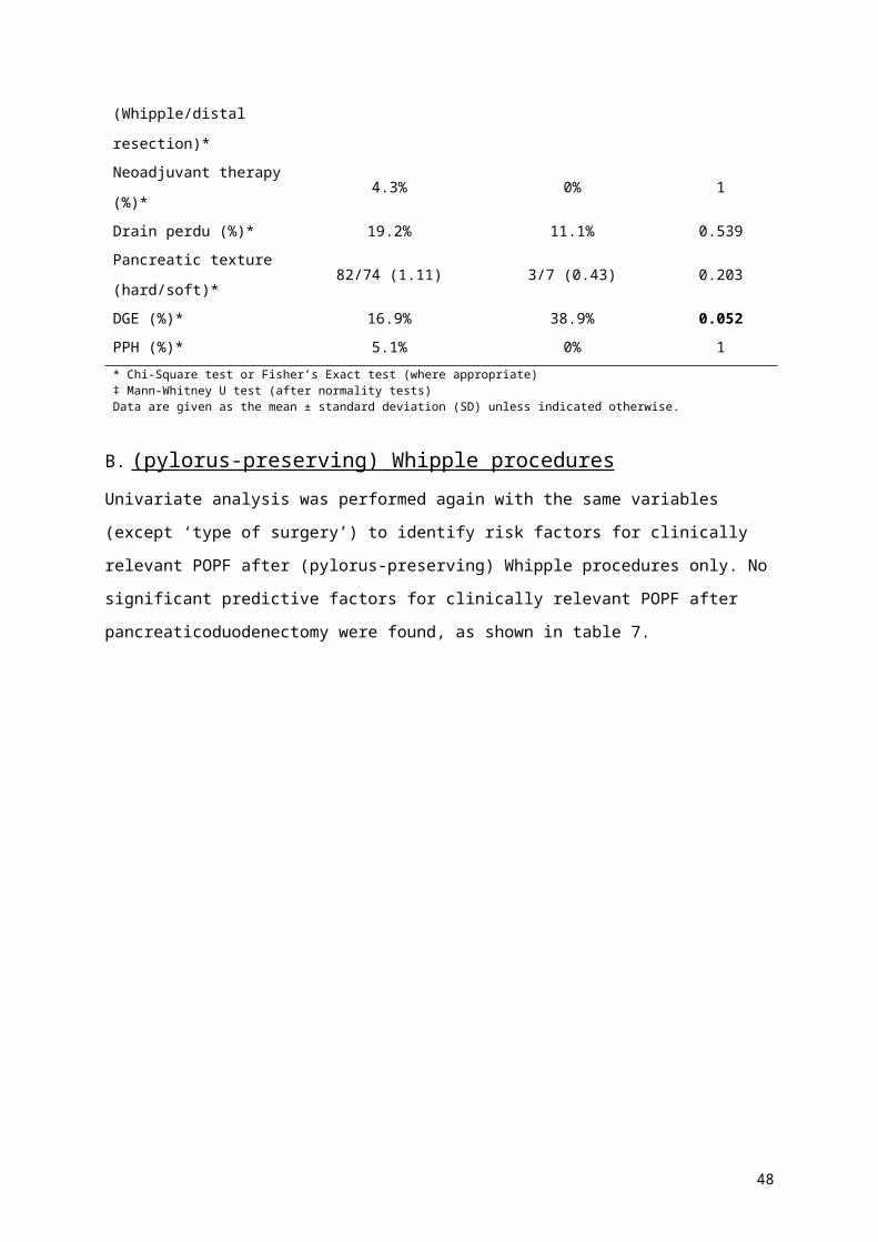
26. Soreide K, Labori KJ. Risk factors and preventive strategies for post-operative pancreatic fistula after pancreatic surgery: a comprehensive review. Scand J Gastroenterol. 2016;51(10):1147-54.
27. Bassi C, Dervenis C, Butturini G, Fingerhut A, Yeo C, Izbicki J, et al. Postoperative pancreatic fistula: an international study group (ISGPF) definition. Surgery. 2005;138(1):8-13.
28. Wente MN, Veit JA, Bassi C, Dervenis C, Fingerhut A, Gouma DJ, et al. Postpancreatectomy hemorrhage (PPH): an International Study Group of Pancreatic Surgery (ISGPS) definition. Surgery. 2007;142(1):20-5.
29. Wente MN, Bassi C, Dervenis C, Fingerhut A, Gouma DJ, Izbicki JR, et al. Delayed gastric emptying (DGE) after pancreatic surgery: a suggested definition by the International Study Group of Pancreatic Surgery (ISGPS). Surgery. 2007;142(5):761-8.
30. Matthews JB. Prevention, evaluation, and treatment of leaks after pancreatic surgery. J Gastrointest Surg. 2011;15(8):1327-8.
31. Lai EC, Lau SH, Lau WY. Measures to prevent pancreatic fistula after pancreatoduodenectomy: a comprehensive review. Arch Surg. 2009;144(11):1074-80.
32. Malleo G, Pulvirenti A, Marchegiani G, Butturini G, Salvia R, Bassi C. Diagnosis and management of postoperative pancreatic fistula. Langenbecks Arch Surg. 2014;399(7):801-10.
33. Shinchi H, Wada K, Traverso LW. The usefulness of drain data to identify a clinically relevant pancreatic anastomotic leak after pancreaticoduodenectomy? J Gastrointest Surg. 2006;10(4):490-8.
34. Bruno O, Brancatelli G, Sauvanet A, Vullierme MP, Barrau V, Vilgrain V. Utility of CT in the diagnosis of pancreatic fistula after pancreaticoduodenectomy in patients with soft pancreas. AJR Am J Roentgenol. 2009;193(3):W175-80.
35. Bassi C, Malleo G. Pancreas: Postoperative pancreatic fistula: use of enteral nutrition. Nat Rev Gastroenterol Hepatol. 2011;8(8):427-8.
36. Fong YM, Marano MA, Barber A, He W, Moldawer LL, Bushman ED, et al. Total parenteral nutrition and bowel rest modify the metabolic response to endotoxin in humans. Ann Surg. 1989;210(4):449-56; discussion 56-7.
37. O'Keefe SJ. Physiological response of the human pancreas to enteral and parenteral feeding. Curr Opin Clin Nutr Metab Care. 2006;9(5):622-8.
38. Spiller RC, Trotman IF, Higgins BE, Ghatei MA, Grimble GK, Lee YC, et al. The ileal brake--inhibition of jejunal motility after ileal fat perfusion in man. Gut. 1984;25(4):365-74.
39. Klek S, Sierzega M, Turczynowski L, Szybinski P, Szczepanek K, Kulig J. Enteral and parenteral nutrition in the conservative treatment of pancreatic fistula: a randomized clinical trial. Gastroenterology. 2011;141(1):157-63, 63 e1.
40. Gans SL, van Westreenen HL, Kiewiet JJ, Rauws EA, Gouma DJ, Boermeester MA. Systematic review and meta-analysis of somatostatin analogues for the treatment of pancreatic fistula. Br J Surg. 2012;99(6):754-60.
41. Bartoli E, Rebibo L, Robert B, Fumery M, Delcenserie R, Regimbeau JM. Efficacy of the double-pigtail stent as a conservative treatment for grade B pancreatic fistula after pancreatoduodenectomy with pancreatogastric anastomosis. Surg Endosc. 2014;28(5):1528-34.
42. Reddymasu SC, Pakseresht K, Moloney B, Alsop B, Oropezia-Vail M, Olyaee M. Incidence of pancreatic fistula after distal pancreatectomy and efficacy of endoscopic therapy for its management: results from a tertiary care center. Case Rep Gastroenterol. 2013;7(2):332-9.
43. Sohn TA, Yeo CJ, Cameron JL, Geschwind JF, Mitchell SE, Venbrux AC, et al. Pancreaticoduodenectomy: role of interventional radiologists in managing patients and complications. J Gastrointest Surg. 2003;7(2):209-19.
44. Munoz-Bongrand N, Sauvanet A, Denys A, Sibert A, Vilgrain V, Belghiti J. Conservative management of pancreatic fistula after pancreaticoduodenectomy with pancreaticogastrostomy. J Am Coll Surg. 2004;199(2):198-203.
45. Sanjay P, Kellner M, Tait IS. The role of interventional radiology in the management of surgical complications after pancreatoduodenectomy. HPB (Oxford). 2012;14(12):812-7.
46. Kwon YM, Gerdes H, Schattner MA, Brown KT, Covey AM, Getrajdman GI, et al. Management of peripancreatic fluid collections following partial pancreatectomy: a comparison of percutaneous versus EUS-guided drainage. Surg Endosc. 2013;27(7):2422-7.
47. Azeem N, Baron TH, Topazian MD, Zhong N, Fleming CJ, Kendrick ML. Outcomes of endoscopic and percutaneous drainage of pancreatic fluid collections arising after pancreatic tail resection. J Am Coll Surg. 2012;215(2):177-85.
50

48. Onodera M, Kawakami H, Kuwatani M, Kudo T, Haba S, Abe Y, et al. Endoscopic ultrasound-guided transmural drainage for pancreatic fistula or pancreatic duct dilation after pancreatic surgery. Surg Endosc. 2012;26(6):1710-7.
49. Tilara A, Gerdes H, Allen P, Jarnagin W, Kingham P, Fong Y, et al. Endoscopic ultrasound-guided transmural drainage of postoperative pancreatic collections. J Am Coll Surg. 2014;218(1):33-40.
50. Balzano G, Pecorelli N, Piemonti L, Ariotti R, Carvello M, Nano R, et al. Relaparotomy for a pancreatic fistula after a pancreaticoduodenectomy: a comparison of different surgical strategies. HPB (Oxford). 2014;16(1):40-5.
51. Denbo JW, Orr WS, Zarzaur BL, Behrman SW. Toward defining grade C pancreatic fistula following pancreaticoduodenectomy: incidence, risk factors, management and outcome. HPB (Oxford). 2012;14(9):589-93.
52. Gangl O, Froschl U, Hofer W, Huber J, Sautner T, Fugger R. Unplanned reoperation and reintervention after pancreatic resections: an analysis of risk factors. World J Surg. 2011;35(10):2306-14.
53. Lin JW, Cameron JL, Yeo CJ, Riall TS, Lillemoe KD. Risk factors and outcomes in postpancreaticoduodenectomy pancreaticocutaneous fistula. J Gastrointest Surg. 2004;8(8):951-9.
54. Shrikhande SV, Sivasanker M, Vollmer CM, Friess H, Besselink MG, Fingerhut A, et al. Pancreatic anastomosis after pancreatoduodenectomy: A position statement by the International Study Group of Pancreatic Surgery (ISGPS). Surgery. 2017;161(5):1221-34.
55. Connor S, Alexakis N, Garden OJ, Leandros E, Bramis J, Wigmore SJ. Meta-analysis of the value of somatostatin and its analogues in reducing complications associated with pancreatic surgery. Br J Surg. 2005;92(9):1059-67.
56. Gurusamy KS, Koti R, Fusai G, Davidson BR. Somatostatin analogues for pancreatic surgery. Cochrane Database Syst Rev. 2013(4):CD008370.
57. Dong Z, Xu J, Wang Z, Petrov MS. Stents for the prevention of pancreatic fistula following pancreaticoduodenectomy. Cochrane Database Syst Rev. 2016(5):CD008914.
58. Adham M, Chopin-Laly X, Lepilliez V, Gincul R, Valette PJ, Ponchon T. Pancreatic resection: drain or no drain? Surgery. 2013;154(5):1069-77.
59. Van Buren G, 2nd, Bloomston M, Hughes SJ, Winter J, Behrman SW, Zyromski NJ, et al. A randomized prospective multicenter trial of pancreaticoduodenectomy with and without routine intraperitoneal drainage. Ann Surg. 2014;259(4):605-12.
60. Noorani A, Rangelova E, Del Chiaro M, Lundell LR, Ansorge C. Delayed Gastric Emptying after Pancreatic Surgery: Analysis of Factors Determinant for the Short-term Outcome. Front Surg. 2016;3:25.
61. Qu H, Sun GR, Zhou SQ, He QS. Clinical risk factors of delayed gastric emptying in patients after pancreaticoduodenectomy: a systematic review and meta-analysis. Eur J Surg Oncol. 2013;39(3):213-23.
62. Wu W, Hong X, Fu L, Liu S, You L, Zhou L, et al. The effect of pylorus removal on delayed gastric emptying after pancreaticoduodenectomy: a meta-analysis of 2,599 patients. PLoS One. 2014;9(10):e108380.
63. El Nakeeb A, Askr W, Mahdy Y, Elgawalby A, El Sorogy M, Abu Zeied M, et al. Delayed gastric emptying after pancreaticoduodenectomy. Risk factors, predictors of severity and outcome. A single center experience of 588 cases. J Gastrointest Surg. 2015;19(6):1093-100.
64. Healy JM, Kunstman JW, Salem RR. Proposal and critical appraisal of exclusion criteria to the international study group for pancreatic surgery definition of delayed gastric emptying. J Am Coll Surg. 2015;220(6):1036-43 e1.
65. Kawai M, Tani M, Hirono S, Miyazawa M, Shimizu A, Uchiyama K, et al. Pylorus ring resection reduces delayed gastric emptying in patients undergoing pancreatoduodenectomy: a prospective, randomized, controlled trial of pylorus-resecting versus pylorus-preserving pancreatoduodenectomy. Ann Surg. 2011;253(3):495-501.
66. Ohwada S, Satoh Y, Kawate S, Yamada T, Kawamura O, Koyama T, et al. Low-dose erythromycin reduces delayed gastric emptying and improves gastric motility after Billroth I pylorus-preserving pancreaticoduodenectomy. Ann Surg. 2001;234(5):668-74.
67. Balzano G, Zerbi A, Braga M, Rocchetti S, Beneduce AA, Di Carlo V. Fast-track recovery programme after pancreatico- duodenectomy reduces delayed gastric emptying. Br J Surg. 2008;95(11):1387-93.
51

68. Nikfarjam M, Kimchi ET, Gusani NJ, Shah SM, Sehmbey M, Shereef S, et al. A reduction in delayed gastric emptying by classic pancreaticoduodenectomy with an antecolic gastrojejunal anastomosis and a retrogastric omental patch. J Gastrointest Surg. 2009;13(9):1674-82.
69. Tani M, Terasawa H, Kawai M, Ina S, Hirono S, Uchiyama K, et al. Improvement of delayed gastric emptying in pylorus-preserving pancreaticoduodenectomy: results of a prospective, randomized, controlled trial. Ann Surg. 2006;243(3):316-20.
70. Murakami Y, Uemura K, Sudo T, Hayashidani Y, Hashimoto Y, Nakagawa N, et al. An antecolic Roux-en Y type reconstruction decreased delayed gastric emptying after pylorus-preserving pancreatoduodenectomy. J Gastrointest Surg. 2008;12(6):1081-6.
71. Patel AG, Toyama MT, Kusske AM, Alexander P, Ashley SW, Reber HA. Pylorus-preserving Whipple resection for pancreatic cancer. Is it any better? Arch Surg. 1995;130(8):838-42; discussion 42-3.
72. Horstmann O, Markus PM, Ghadimi MB, Becker H. Pylorus preservation has no impact on delayed gastric emptying after pancreatic head resection. Pancreas. 2004;28(1):69-74.
73. Bassi C, Falconi M, Molinari E, Salvia R, Butturini G, Sartori N, et al. Reconstruction by pancreaticojejunostomy versus pancreaticogastrostomy following pancreatectomy: results of a comparative study. Ann Surg. 2005;242(6):767-71, discussion 71-3.
74. Lermite E, Pessaux P, Brehant O, Teyssedou C, Pelletier I, Etienne S, et al. Risk factors of pancreatic fistula and delayed gastric emptying after pancreaticoduodenectomy with pancreaticogastrostomy. J Am Coll Surg. 2007;204(4):588-96.
75. Park YC, Kim SW, Jang JY, Ahn YJ, Park YH. Factors influencing delayed gastric emptying after pylorus-preserving pancreatoduodenectomy. J Am Coll Surg. 2003;196(6):859-65.
76. Park JS, Hwang HK, Kim JK, Cho SI, Yoon DS, Lee WJ, et al. Clinical validation and risk factors for delayed gastric emptying based on the International Study Group of Pancreatic Surgery (ISGPS) Classification. Surgery. 2009;146(5):882-7.
77. Chen JF, Xu SF, Zhao W, Tian YH, Gong L, Yuan WS, et al. Diagnostic and therapeutic strategies to manage post-pancreaticoduodenectomy hemorrhage. World J Surg. 2015;39(2):509-15.
78. Correa-Gallego C, Brennan MF, D'Angelica MI, DeMatteo RP, Fong Y, Kingham TP, et al. Contemporary experience with postpancreatectomy hemorrhage: results of 1,122 patients resected between 2006 and 2011. J Am Coll Surg. 2012;215(5):616-21.
79. Manas-Gomez MJ, Rodriguez-Revuelto R, Balsells-Valls J, Olsina-Kissler JJ, Caralt-Barba M, Perez-Lafuente M, et al. Post-pancreaticoduodenectomy hemorrhage. Incidence, diagnosis, and treatment. World J Surg. 2011;35(11):2543-8.
80. Rajarathinam G, Kannan DG, Vimalraj V, Amudhan A, Rajendran S, Jyotibasu D, et al. Post pancreaticoduodenectomy haemorrhage: outcome prediction based on new ISGPS Clinical severity grading. HPB (Oxford). 2008;10(5):363-70.
81. Wellner UF, Kulemann B, Lapshyn H, Hoeppner J, Sick O, Makowiec F, et al. Postpancreatectomy hemorrhage--incidence, treatment, and risk factors in over 1,000 pancreatic resections. J Gastrointest Surg. 2014;18(3):464-75.
82. Gao F, Li J, Quan S, Li F, Ma D, Yao L, et al. Risk Factors and Treatment for Hemorrhage after Pancreaticoduodenectomy: A Case Series of 423 Patients. Biomed Res Int. 2016;2016:2815693.
83. Dai R, Turley RS, Blazer DG. Contemporary review of minimally invasive pancreaticoduodenectomy. World J Gastrointest Surg. 2016;8(12):784-91.
84. Zhang YH, Zhang CW, Hu ZM, Hong DF. Pancreatic cancer: Open or minimally invasive surgery? World J Gastroenterol. 2016;22(32):7301-10.
85. Conwell DL, Banks PA. Chronic pancreatitis. Curr Opin Gastroenterol. 2008;24(5):586-90.86. Mitchell RM, Byrne MF, Baillie J. Pancreatitis. Lancet. 2003;361(9367):1447-55.87. Nassour I, Choti MA. Pancreatic Operations. JAMA. 2016;316(18):1932.88. Wayne MG, Jorge IA, Cooperman AM. Alternative reconstruction after
pancreaticoduodenectomy. World J Surg Oncol. 2008;6:9.89. Ilic M, Ilic I. Epidemiology of pancreatic cancer. World J Gastroenterol. 2016;22(44):9694-705.90. Malvezzi M, Bertuccio P, Rosso T, Rota M, Levi F, La Vecchia C, et al. European cancer
mortality predictions for the year 2015: does lung cancer have the highest death rate in EU women? Ann Oncol. 2015;26(4):779-86.
91. Mayor S. Deaths from pancreatic cancer in Europe continue to increase while rates for other cancers fall. BMJ. 2014;348:g2914.
92. Hidalgo M. Pancreatic cancer. N Engl J Med. 2010;362(17):1605-17.
52

93. Karim-Kos HE, de Vries E, Soerjomataram I, Lemmens V, Siesling S, Coebergh JW. Recent trends of cancer in Europe: a combined approach of incidence, survival and mortality for 17 cancer sites since the 1990s. Eur J Cancer. 2008;44(10):1345-89.
94. Goral V. Pancreatic Cancer: Pathogenesis and Diagnosis. Asian Pac J Cancer Prev. 2015;16(14):5619-24.
95. Keane MG, Horsfall L, Rait G, Pereira SP. A case-control study comparing the incidence of early symptoms in pancreatic and biliary tract cancer. BMJ Open. 2014;4(11):e005720.
96. Goonetilleke KS, Siriwardena AK. Systematic review of carbohydrate antigen (CA 19-9) as a biochemical marker in the diagnosis of pancreatic cancer. Eur J Surg Oncol. 2007;33(3):266-70.
97. McIntyre CA, Winter JM. Diagnostic evaluation and staging of pancreatic ductal adenocarcinoma. Semin Oncol. 2015;42(1):19-27.
98. Tempero MA, Malafa MP, Al-Hawary M, Asbun H, Bain A, Behrman SW, et al. Pancreatic Adenocarcinoma, Version 2.2017, NCCN Clinical Practice Guidelines in Oncology. J Natl Compr Canc Netw. 2017;15(8):1028-61.
99. Mayo SC, Austin DF, Sheppard BC, Mori M, Shipley DK, Billingsley KG. Evolving preoperative evaluation of patients with pancreatic cancer: does laparoscopy have a role in the current era? J Am Coll Surg. 2009;208(1):87-95.
100. Tapper E, Kalb B, Martin DR, Kooby D, Adsay NV, Sarmiento JM. Staging laparoscopy for proximal pancreatic cancer in a magnetic resonance imaging-driven practice: what's it worth? HPB (Oxford). 2011;13(10):732-7.
101. Huttner FJ, Fitzmaurice C, Schwarzer G, Seiler CM, Antes G, Buchler MW, et al. Pylorus-preserving pancreaticoduodenectomy (pp Whipple) versus pancreaticoduodenectomy (classic Whipple) for surgical treatment of periampullary and pancreatic carcinoma. Cochrane Database Syst Rev. 2016;2:CD006053.
102. Pitt HA, Grace PA. Cancer of the pancreas. Pylorus-preserving resection of the pancreas. Baillieres Clin Gastroenterol. 1990;4(4):917-30.
103. Machado NO. Pancreatic fistula after pancreatectomy: definitions, risk factors, preventive measures, and management-review. Int J Surg Oncol. 2012;2012:602478.
104. Bruns H, Kortendieck V, Raab HR, Antolovic D. Intraoperative Fluid Excess Is a Risk Factor for Pancreatic Fistula after Partial Pancreaticoduodenectomy. HPB Surg. 2016;2016:1601340.
105. Gagniere J, Abjean A, Franz M, Aumont O, Pereira B, Dupre A, et al. A Normal Preoperative Lipase Serum Level Is an Easy and Objective Risk Factor of Pancreatic Fistula After Pancreaticoduodenectomy. Pancreas. 2017;46(9):1133-40.
106. Yang YM, Tian XD, Zhuang Y, Wang WM, Wan YL, Huang YT. Risk factors of pancreatic leakage after pancreaticoduodenectomy. World J Gastroenterol. 2005;11(16):2456-61.
107. Facy O, Chalumeau C, Poussier M, Binquet C, Rat P, Ortega-Deballon P. Diagnosis of postoperative pancreatic fistula. Br J Surg. 2012;99(8):1072-5.
108. Guilbaud T, Birnbaum DJ, Loubiere S, Bonnet J, Chopinet S, Gregoire E, et al. Comparison of different feeding regimes after pancreatoduodenectomy - a retrospective cohort analysis. Nutr J. 2017;16(1):42.
109. Hu BY, Wan T, Zhang WZ, Dong JH. Risk factors for postoperative pancreatic fistula: Analysis of 539 successive cases of pancreaticoduodenectomy. World J Gastroenterol. 2016;22(34):7797-805.
110. McMillan MT, Christein JD, Callery MP, Behrman SW, Drebin JA, Hollis RH, et al. Comparing the burden of pancreatic fistulas after pancreatoduodenectomy and distal pancreatectomy. Surgery. 2016;159(4):1013-22.
111. Cameron JL, He J. Two thousand consecutive pancreaticoduodenectomies. J Am Coll Surg. 2015;220(4):530-6.
112. Aranha GV, Aaron JM, Shoup M, Pickleman J. Current management of pancreatic fistula after pancreaticoduodenectomy. Surgery. 2006;140(4):561-8; discussion 8-9.
113. Kawai M, Kondo S, Yamaue H, Wada K, Sano K, Motoi F, et al. Predictive risk factors for clinically relevant pancreatic fistula analyzed in 1,239 patients with pancreaticoduodenectomy: multicenter data collection as a project study of pancreatic surgery by the Japanese Society of Hepato-Biliary-Pancreatic Surgery. J Hepatobiliary Pancreat Sci. 2011;18(4):601-8.
114. McMillan MT, Soi S, Asbun HJ, Ball CG, Bassi C, Beane JD, et al. Risk-adjusted Outcomes of Clinically Relevant Pancreatic Fistula Following Pancreatoduodenectomy: A Model for Performance Evaluation. Ann Surg. 2016;264(2):344-52.
53

115. Topal B, Fieuws S, Aerts R, Weerts J, Feryn T, Roeyen G, et al. Pancreaticojejunostomy versus pancreaticogastrostomy reconstruction after pancreaticoduodenectomy for pancreatic or periampullary tumours: a multicentre randomised trial. Lancet Oncol. 2013;14(7):655-62.
116. Goh BK, Tan YM, Chung YF, Cheow PC, Ong HS, Chan WH, et al. Critical appraisal of 232 consecutive distal pancreatectomies with emphasis on risk factors, outcome, and management of the postoperative pancreatic fistula: a 21-year experience at a single institution. Arch Surg. 2008;143(10):956-65.
117. Ecker BL, McMillan MT, Allegrini V, Bassi C, Beane JD, Beckman RM, et al. Risk Factors and Mitigation Strategies for Pancreatic Fistula After Distal Pancreatectomy: Analysis of 2026 Resections From the International, Multi-institutional Distal Pancreatectomy Study Group. Ann Surg. 2017.
118. Kleeff J, Diener MK, Z'Graggen K, Hinz U, Wagner M, Bachmann J, et al. Distal pancreatectomy: risk factors for surgical failure in 302 consecutive cases. Ann Surg. 2007;245(4):573-82.
119. Zhang H, Zhu F, Shen M, Tian R, Shi CJ, Wang X, et al. Systematic review and meta-analysis comparing three techniques for pancreatic remnant closure following distal pancreatectomy. Br J Surg. 2015;102(1):4-15.
120. Hackert T, Hinz U, Pausch T, Fesenbeck I, Strobel O, Schneider L, et al. Postoperative pancreatic fistula: We need to redefine grades B and C. Surgery. 2016;159(3):872-7.
121. Bassi C, Marchegiani G, Dervenis C, Sarr M, Abu Hilal M, Adham M, et al. The 2016 update of the International Study Group (ISGPS) definition and grading of postoperative pancreatic fistula: 11 Years After. Surgery. 2017;161(3):584-91.
122. Kim WS, Choi DW, Choi SH, Heo JS, Kim MJ, Song SC, et al. Clinical validation of the ISGPF classification and the risk factors of pancreatic fistula formation following duct-to-mucosa pancreaticojejunostomy by one surgeon at a single center. J Gastrointest Surg. 2011;15(12):2187-92.
123. Pratt WB, Maithel SK, Vanounou T, Huang ZS, Callery MP, Vollmer CM, Jr. Clinical and economic validation of the International Study Group of Pancreatic Fistula (ISGPF) classification scheme. Ann Surg. 2007;245(3):443-51.
124. Murakami Y, Uemura K, Hayasidani Y, Sudo T, Hashimoto Y, Nakagawa N, et al. A soft pancreatic remnant is associated with increased drain fluid pancreatic amylase and serum CRP levels following pancreatoduodenectomy. J Gastrointest Surg. 2008;12(1):51-6.
125. Wellner UF, Kayser G, Lapshyn H, Sick O, Makowiec F, Hoppner J, et al. A simple scoring system based on clinical factors related to pancreatic texture predicts postoperative pancreatic fistula preoperatively. HPB (Oxford). 2010;12(10):696-702.
126. Callery MP, Pratt WB, Kent TS, Chaikof EL, Vollmer CM, Jr. A prospectively validated clinical risk score accurately predicts pancreatic fistula after pancreatoduodenectomy. J Am Coll Surg. 2013;216(1):1-14.
127. Pratt WB, Callery MP, Vollmer CM, Jr. Risk prediction for development of pancreatic fistula using the ISGPF classification scheme. World J Surg. 2008;32(3):419-28.
128. Peng YP, Zhu XL, Yin LD, Zhu Y, Wei JS, Wu JL, et al. Risk factors of postoperative pancreatic fistula in patients after distal pancreatectomy: a systematic review and meta-analysis. Sci Rep. 2017;7(1):185.
129. Harada N, Ishizawa T, Inoue Y, Aoki T, Sakamoto Y, Hasegawa K, et al. Acoustic radiation force impulse imaging of the pancreas for estimation of pathologic fibrosis and risk of postoperative pancreatic fistula. J Am Coll Surg. 2014;219(5):887-94 e5.
130. Kuwahara T, Hirooka Y, Kawashima H, Ohno E, Yokoyama Y, Fujii T, et al. Usefulness of endoscopic ultrasonography-elastography as a predictive tool for the occurrence of pancreatic fistula after pancreatoduodenectomy. J Hepatobiliary Pancreat Sci. 2017.
131. Hong TH, Choi JI, Park MY, Rha SE, Lee YJ, You YK, et al. Pancreatic hardness: Correlation of surgeon's palpation, durometer measurement and preoperative magnetic resonance imaging features. World J Gastroenterol. 2017;23(11):2044-51.
132. Shi Y, Glaser KJ, Venkatesh SK, Ben-Abraham EI, Ehman RL. Feasibility of using 3D MR elastography to determine pancreatic stiffness in healthy volunteers. J Magn Reson Imaging. 2015;41(2):369-75.
133. Yoon JH, Lee JM, Lee KB, Kim SW, Kang MJ, Jang JY, et al. Pancreatic Steatosis and Fibrosis: Quantitative Assessment with Preoperative Multiparametric MR Imaging. Radiology. 2016;279(1):140-50.
54

134. Matsusue S, Takeda H, Nakamura Y, Nishimura S, Koizumi S. A prospective analysis of the factors influencing pancreaticojejunostomy performed using a single method, in 100 consecutive pancreaticoduodenectomies. Surg Today. 1998;28(7):719-26.
135. Glowka TR, von Websky M, Pantelis D, Manekeller S, Standop J, Kalff JC, et al. Risk factors for delayed gastric emptying following distal pancreatectomy. Langenbecks Arch Surg. 2016;401(2):161-7.
136. Liu QY, Li L, Xia HT, Zhang WZ, Cai SW, Lu SC. Risk factors of delayed gastric emptying following pancreaticoduodenectomy. ANZ J Surg. 2016;86(1-2):69-73.
137. Seeliger H, Christians S, Angele MK, Kleespies A, Eichhorn ME, Ischenko I, et al. Risk factors for surgical complications in distal pancreatectomy. Am J Surg. 2010;200(3):311-7.
138. Sledzianowski JF, Duffas JP, Muscari F, Suc B, Fourtanier F. Risk factors for mortality and intra-abdominal morbidity after distal pancreatectomy. Surgery. 2005;137(2):180-5.
139. Kawaida H, Kono H, Watanabe M, Hosomura N, Amemiya H, Fujii H. Risk factors of postoperative pancreatic fistula after distal pancreatectomy using a triple-row stapler. Surg Today. 2017.
140. Schnelldorfer T, Mauldin PD, Lewin DN, Adams DB. Distal pancreatectomy for chronic pancreatitis: risk factors for postoperative pancreatic fistula. J Gastrointest Surg. 2007;11(8):991-7.
141. Sierzega M, Niekowal B, Kulig J, Popiela T. Nutritional status affects the rate of pancreatic fistula after distal pancreatectomy: a multivariate analysis of 132 patients. J Am Coll Surg. 2007;205(1):52-9.
142. Cheng TY, Sheth K, White RR, Ueno T, Hung CF, Clary BM, et al. Effect of neoadjuvant chemoradiation on operative mortality and morbidity for pancreaticoduodenectomy. Ann Surg Oncol. 2006;13(1):66-74.
143. Ishikawa O, Ohigashi H, Imaoka S, Teshima T, Inoue T, Sasaki Y, et al. Concomitant benefit of preoperative irradiation in preventing pancreas fistula formation after pancreatoduodenectomy. Arch Surg. 1991;126(7):885-9.
144. Srivastava S, Sikora SS, Pandey CM, Kumar A, Saxena R, Kapoor VK. Determinants of pancreaticoenteric anastomotic leak following pancreaticoduodenectomy. ANZ J Surg. 2001;71(9):511-5.
145. Chu CK, Mazo AE, Sarmiento JM, Staley CA, Adsay NV, Umpierrez GE, et al. Impact of diabetes mellitus on perioperative outcomes after resection for pancreatic adenocarcinoma. J Am Coll Surg. 2010;210(4):463-73.
146. Xia X, Huang C, Cen G, Qiu ZJ. Preoperative diabetes as a protective factor for pancreatic fistula after pancreaticoduodenectomy: a meta-analysis. Hepatobiliary Pancreat Dis Int. 2015;14(2):132-8.
147. Mathur A, Pitt HA, Marine M, Saxena R, Schmidt CM, Howard TJ, et al. Fatty pancreas: a factor in postoperative pancreatic fistula. Ann Surg. 2007;246(6):1058-64.
148. Friess H, Malfertheiner P, Isenmann R, Kuhne H, Beger HG, Buchler MW. The risk of pancreaticointestinal anastomosis can be predicted preoperatively. Pancreas. 1996;13(2):202-8.
149. Yamamoto Y, Sakamoto Y, Ban D, Shimada K, Esaki M, Nara S, et al. Is celiac axis resection justified for T4 pancreatic body cancer? Surgery. 2012;151(1):61-9.
55

APPENDIX 1: LIST OF ABBREVIATIONS
Abbreviation explanationADC apparent diffusion coefficient
AJCC American Joint Cancer Committee
ARFI acoustic radiation force impulse imaging
BMI body mass index
CR-POPF clinically relevant postoperative pancreatic fistula
CT computed tomography
DGE delayed gastric emptying
DP distal pancreatectomy
DWI diffusion-weighted imaging
EN enteral nutrition
EUS endoscopic ultrasonography
EUS-EG endoscopic ultrasonography-guided elastography
ISGPF International Study Group for Pancreatic Fistula
ISGPS International Study Group of Pancreatic Surgery
IVIM DW intravoxel incoherent motion diffusion-weighted imaging
ME mean elasticity
MRI magnetic resonance imaging
NCCN National Comprehensive Cancer Network
NGT nasogastric tube
PC pancreatic cancer
PD pancreaticoduodenectomy
PDA pancreatic ductal adenocarcinoma
PG pancreaticogastrostomy
PJ pancreaticojejunostomy
POD postoperative day
POPF postoperative pancreatic fistula
PPH postpancreatectomy hemorrhage
RCT randomized controlled trial
SWV shear wave velocity
TPN total parenteral nutrition
US ultrasonography
i