0. as n. · the journal of biological chemistry 0 1992 by the american society for biochemistry and...
TRANSCRIPT
THE JOURNAL OF BIOLOGICAL CHEMISTRY 0 1992 by The American Society for Biochemistry and Molecular Biology, Inc.
Vol. 267. No. 13, Issue of May 5, pp. 8770-8777, 1992 Printed in U.S.A.
Folding and Oxidation of Recombinant Human Granulocyte Colony Stimulating Factor Produced in Escherichia coli CHARACTERIZATION OF THE DISULFIDE-REDUCED INTERMEDIATES AND CYSTEINE + SERINE ANALOGS*
(Received for publication, October 14, 1991)
Hsieng S. Lul, Christi L. Clogston, Linda 0. Narhi, Lee Anne Merewether, Wayne R. Pearl, and Thomas C. Boone From Amgen Incorporated, Amgen Center, Thousand Oaks, California 91320
The folding and oxidation of recombinant human granulocyte colony-stimulating factor solubilized from Escherichia coli inclusion bodies was investigated. During the folding process, two intermediates, I1 and Iz, were detected by kinetic studies using high perform- ance liquid chromatography. I1 exists transiently and disappears quickly with the concomitant formation of 12. In contrast, IZ requires a longer time to fold into the final oxidized form, N. CuS04 catalysis increases the folding rate of I2 from 11, while CuS04 and elevated temperature (37 “C) have a dramatic effect on the fold- ing rate of N from Iz. These observations suggest the following sequential oxidative folding pathway.
‘1 fast‘’ rate-limiting ’N
Peptide map analysis of the iodoacetate-labeled in- termediates revealed that I1 represents the fully re- duced granulocyte colony-stimulating factor contain- ing 6 free cysteines; I2 is the partially oxidized species containing a single C y ~ ~ ~ - C y s ~ ~ disulfide bond; and N, the final folded form, has two disulfide bonds. The physicochemical properties and biological activities of 11, Iz, N, and several Cys + Ser analogs made by site- directed mutagenesis were further investigated. In guanidine hydrochloride-induced denaturation stud- ies, the disulfide-reduced intermediates and the ana- logs missing either of the disulfide bonds are confor- mationally less stable than those of the wild type mol- ecule or the analog with the free Cys at position 17 changed to Ser. Recombinant human granulocyte col- ony stimulating factor lacking either disulfide bond or both has overall secondary and tertiary structures dif- ferent from those of the wild type molecule and exhibits lower biological activity. These studies show that di- sulfide bond formation is crucial for maintaining the molecule in a properly folded and biologically active form.
The mechanistic study of protein folding is important in understanding the structure and function of proteins (1, 2). The difficulty in elucidating a protein folding pathway lies in measuring the structural properties of intermediate protein
* The costs of publication of this article were defrayed in part by the payment of page charges. This article must therefore be hereby marked “aduertisement” in accordance with 18 U.S.C. Section 1734 solely to indicate this fact.
$ To whom correspondence should be addressed.
species, since they are usually short-lived. As described by Creighton and others (3-7), a model system that includes an oxidative refolding of a disulfide-reduced protein allows one to investigate the pathway of protein folding in detail. There are two possible advantages in investigating a disulfide-con- taining protein. First, the intrachain S-S bond is a natural covalent cross-link which is closely correlated with protein conformation, and the intramolecular disulfide bond forma- tion reflects the proximity of two relevant sulfhydryls in intermediate forms (3-5). Second, proteins in disulfide-re- duced states are not short lived allowing the intermediates to be trapped by chemical modification during folding.
Human granulocyte colony-stimulating factor (hG-CSF)’ is one of the hemopoietic growth factors which plays an impor- tant role in stimulating proliferation, differentiation, and functional activation of blood cells (8). Human G-CSF is capable of supporting neutrophil proliferation in vitro and in vivo (9,lO). The human and murine G-CSF genes have been cloned and characterized and are about 75% homologous (11, 12). Large quantities of recombinant hG-CSF (rhG-CSF) have been produced in genetically engineered Escherichia coli and have been successfully used in human clinical studies to treat cancer patients suffering from chemotherapy-induced neutro- penia (13-15). E. coli-produced rhG-CSF is a 175-amino acid polypeptide chain containing an extra Met (at position -1) at its NH2 terminus. The molecule also contains a free cys- teine at position 17 and two intramolecular disulfide bonds, C y ~ ~ ~ - C y s ~ ~ and C ~ s ‘ j ~ - C y s ~ ~ (16). The two disulfide bonds form two small loops which are separated by 21 amino acids. Like other bacteria-derived recombinant proteins, rhG-CSF produced in E. coli requires an oxidative folding procedure in order to recover its biological activity (17). In this paper, we describe the kinetic study of a folding and oxidation procedure for the reduced rhG-CSF solubilized from the inclusion bodies as well as the isolation and characterization of the disulfide- reduced intermediates. To establish the role of disulfide bond formation in the folding of biologically active rhG-CSF, we also describe the biological and physicochemical characteriza- tion of intermediates and analogs made by site-directed mu- tagenesis at the Cys residues.
The abbreviations used are: hG-CSF, human granulocyte colony- stimulating factor; rhG-CSF, recombinant human granulocyte col- ony-stimulating factor; GdnHC1, guanidine HCI; RP-HPLC, reverse- phase high performance liquid chromatography; DTT, dithiothreitol; TFA, trifluoroacetic acid DTNB, 5,5’-dithiobis(nitrobenzoic acid).
8770

Oxidative folding of hG-CSF 8771
t
N
1
42 4 4 4 6 48 5 0 5 2
Retention Time Wutno)
FIG. 2. Folding of rhG-CSF at 25 "C in CuSO, monitored by RP-HPLC at different times. Chromatograms 1-6,20 min, 1 ,2 ,4 , 8, and 12 h, respectively. Approximately 50 pg of rhG-CSF in folding mixture was injected. Intermediates I1 and I, and the final oxidized form N are indicated. Note that retention times shown in Fig. 2 are slightly different from those in Fig. 1 due to the use of different C-4 columns.
MATERIALS AND METHODS'
RESULTS
Folding Intermediates and Folding Kinetics of rhG-CSF- Fig. 1 shows the RP-HPLC elution profiles of the native and the fully denatured and reduced rhG-CSFs (chromatograms 1 and 2, respectively). Their retention times differ by approxi- mately 2.1-2.5 min. Fully denatured and reduced rhG-CSF in 6 M GdnHCl is retained slightly longer and elutes as a single sharp peak. Fig. 1 also shows that rhG-CSF present in the crude E. coli lysate solubilized in either GdnHCl or Sarkosyl at 2-5 "C elutes as broader peaks (chromatograms 3, and 4, respectively) at slightly earlier retention times than the fully denatured and reduced rhG-CSF. The difference in the chro- matographic elution times among these rhG-CSF forms has allowed us to detect folding intermediates and to study folding kinetics by RP-HPLC.
Shown in Fig. 2 are the RP-HPLC chromatograms of the solubilized rhG-CSF samples prepared at different refolding times during incubation at 25 "C in the presence of CuS04. The populations of the three major rhG-CSF-related species which elute at retention times around 45 to 48 min change dramatically as a function of time. Intermediate I, as depicted in Fig. 2 (chromatogram 1 ) is the starting reduced rhG-CSF. At the 20-min incubation time, Iz has already accumulated to a level of 33% of the total rhG-CSF. At 1, 2, and 4 h, the generation of I 2 has proceeded further with the concomitant disappearance of I, and appearance of the final oxidized form N (Fig. 2, chromatograms 2-4). The folding into form N reaches its maximum in 12 h and is greater than 95% complete (Fig. 2, chromatogram 6).
The initial first order folding rate for conversion of I1 into I, was estimated to be 1.9 X lo-' s" and the initial rate for conversion of Ip into the final oxidized rhG-CSF (form N) to be approximately 3.1 x s-' (Table 1). The half-maximal conversion of II to I2 takes approximately 30 min, while the
Portions of this paper (including "Materials and Methods," Figs. 1, 3-7, and 9, and Tables 1-4) are presented in miniprint at the end of this paper. Miniprint is easily read with the aid of a standard magnifying glass. Full size photocopies are included in the microfilm edition of the Journal that is available from Waverly Press.
half-maximal conversion of I p to N takes almost 4.5 h. Kinetic studies also indicate that the oxidative folding of form N from I, is biphasic. The folding rate from I2 to N during the first phase is faster. The second phase of folding starts at approx- imately 8 h with a much slower rate.
The folding kinetics of rhG-CSF at 25 and 37 "C without addition of copper sulfate were also investigated. As listed in Table 1, the initial rates for Iz formation a t both 25 and 37 "C are similar (1.0 x lo-' s-') and slightly slower than the folding of rhG-CSF in the presence of copper sulfate (1.9 x 10" s-'). However, a t 25 "C the generation of completely oxidized rhG- CSF (form N) is relatively slow (rate = 6.6 X s-'). In this case, I, persists much longer and approximately 20 h are required to reach half-maximal folding of form N from Ip versus 4.5 h in the presence of copper sulfate at 25 "C. At 37 "C the folding of form N from Ip without CuS04 is faster, but the biphasic kinetics becomes apparent. The first phase of oxidation takes about 5 h, while the second phase takes place at approximately 8 h. After 23 h, the recovery of com- pletely oxidized rhG-CSF reaches only about 80%.
Isolation and Structural Characterization of Intermediates- An alkylating agent, iodoacetic acid, was used to trap the intermediates that may be disulfide-reduced. The resulting carboxymethylated derivatives contain more negatively charged carboxymethyl groups than the oxidized rhG-CSF and are separable by ion-exchange HPLC using a sulfoethyl polyaspartamide silica-based column (data not shown).
To estimate the stoichiometry of labeling, the modification was also performed using [3H]C2-iodoacetic acid. Table 2 lists the labeling results for rhG-CSF and the purified intermedi- ates. In the absence and presence of 6 M GdnHC1, native rhG- CSF gives 0 and 1 mol of label/mol of protein, respectively, consistent with our previous observation (16). The fully re- duced and denatured rhG-CSF gives 5 mol of label. The trapped I1 gives 3.74 mol of label and I2 1.9 mol of label/mol of protein. These results indicate that 4 and 2 free cysteinyl residues are available for the labeling in I, and Iz, respectively.
To further characterize the structure of intermediates, the 3H-labeled I, and I2 were reduced with DTT in 6 M GdnHCl and then carboxymethylated with non-radioactive iodoacetate to generate fully denatured and alkylated derivatives, which were then subjected to HPLC peptide mapping. Fig. 3 shows a typical peptide map derived from the Staphylococcus aureus V-8 protease digestion of the I2 derivative. Peptide fractions were pooled and aliquots were analyzed for determination of peptide concentration, radioactivity counting, and NHz-ter- minal sequence analysis. Table 3 summarizes the isolation and characterization of the labeled and unlabeled peptides derived from I1 and 12. For form Ip, only peptides 7 and 8 (Leu47 to Glug3 and to Glug8, respectively) containing
and Cys74 were radioactively labeled. Sequence analysis confirmed that both and Cys74 were labeled, supporting the quantitative data that 2 labeled cysteines are present in I2 (Table 2). For form 11, Cyd4 and CYS~~, found in peptides 7 and 8, as well as and Cys4' in peptide 2 ( L Y s ~ ~ t o G ~ u ~ ~ ) were radioactively labeled. This confirms that the 4 cysteines a t positions 36,42,64, and 74 in I1 are not involved in disulfide bonding.
As indicated in Table 3, peptide 4 is the NH,-terminal peptide of rhG-CSF containing Cys at position 17. Analysis of peptide 4 derived from both intermediates I1 and 1,indicated that no radioactive label was present. The data suggest that, like the native rhG-CSF, both intermediates contain an in- accessible free cysteine a t position 17.
Preparation and RP-HPLC Analysis of rhG-CSF Analogs- The Cys + Ser analogs made by site-directed mutagenesis of

8772 Oxidative folding of hG-CSF
the rhG-CSF gene were recovered by the procedures developed for rhG-CSF including folding and chromatographic separa- tion. rhG-CSF[Cys17 + Ser17] exhibited oxidation and folding similar to those of the wild type rhG-CSF and could be isolated with equivalent recovery. In contrast, ~ ~ G - C S F [ C ~ S ~ ~ , ~ * + Ser36-42] exhibited very slow folding in the absence of CuS04 (greater than 4 days) and was recovered in low yield. The [ C Y S ~ ~ + Ser74] analog lacking the C y ~ ~ ~ - C y s ~ ~ disulfide bond also folded moderately slowly but correctly in the absence of CuS04 (more rapidly but incorrectly in the presence of CuS04), but recovered in low yield.
Fig. 4A shows the elution of purified r h G - S c F [ c y ~ ~ ~ + Ser74] from a C-4 reverse-phase HPLC column. It elutes 2.20 min (chromatograms 2 and 3 at pH 3 and 7, respectively) later than the wild type rhG-CSF (chromatogram 1 ). I2 lacking the C y ~ ~ ~ - C y s ~ ~ disulfide bond elutes essentially at the same re- tention time as the Cys74 + Ser74 analog, suggesting that the molecules have similar hydrophobicities. Recombinant hG- C S F [ C ~ S ~ ~ * ~ * + Ser36*42] (Fig. 4B, chromatogram 4 ) elutes only 0.33 min later than wild type rhG-CSF.
Biological Activity and Physicochemical Properties of the rhG-CSF, Intermediates and Analogs-Table 4 lists the in uitro biological activities of rhG-CSF, folding intermediates, and analogs. The wild type rhG-CSF standard has an activity of approximately 1.0 X 10' units/mg. Carboxymethylated intermediates I1 and I p have only approximately 3 4 % activity of the wild type rhG-CSF. Both ~ ~ G - C S F [ C ~ S ~ ~ , ~ ~ + Ser36.42] and r h G - c S F [ c y ~ ~ ~ + Ser74] also exhibit very low activity (1 and 3%, respectively relative to the wild type molecule). In contrast, rhG-CSF[Cys17 + Ser17] exhibits full in uitro biolog- ical activity.
Conformational stabilities of rhG-CSF intermediates and analogs were compared by denaturation with GdnHCl at pH 7.2. Fig. 5A shows the absorbance spectra of native and denatured rhG-CSFs. It appears that GdnHCl denaturation results in a blue shift of the UV spectrum, reflecting increased exposure of the aromatic amino acids to the polar aqueous solvent. The absorbance difference at 290 nm can thus be used to estimate the effect of a denaturing agent on the conformational stability of rhG-CSF. From the results shown in Fig. 5B, rhG-CSF appears to stay in the native state below 2 M GdnHCl concentration, denatures quickly above 2.5 M denaturant, and is completely unfolded at 3.5 M GdnHCl. This denaturation profile approximates a simple two-state transition. The midpoint of denaturation is at approximately 3 M GdnHCl (Table 4). Also indicated in Fig. 5B is the accessibility of the free Cyd7 at different GdnHCl concentra- tions as determined by DTNB reaction. The increased acces- sibility of CysI7 is coincident with the denaturation of rhG- CSF detected by the change in absorbance at 290 nm.
As shown in Fig. 6, similar GdnHCl denaturation studies were also performed on I], 12, r h G - c S F [ c y ~ ~ ~ + Ser74], and rhG-CSF[Cys"l4* - Ser36*42]. The denaturation transition oc- curs at lower GdnHCl concentration for the intermediates and analogs. A further decrease in absorbance was observed to proceed at higher GdnHCl concentration. The concentra- tions of GdnHCl that are required to achieve a midpoint denaturation for the intermediates and analogs range from 1.4 to 2.1 M (Table 4). The thermodynamic constant for rhG- CSF is 5.4 Kcal/mol, a value typical of a folded globular protein while all of the disulfide-reduced intermediates and analogs have values below 3 kcal/mol.
Fig. 7A shows the far UV CD spectra of the native molecule, t h e r h G - c S F [ c y ~ ~ ~ -+ Ser74] analog, the ~ ~ G - C S F [ C ~ S ~ ~ . ~ * + Ser"fi,42] analog, and the folding intermediates at pH 7.5. rhG- CSF is rich in a-helix, as evidenced by the minima at 222 and
208 nm. All of the molecules tested exhibit a-helical structure, but the native molecule and the ~ ~ G - C S F [ C ~ S ~ ~ , ~ ~ + Ser36,42] molecule contain substantially higher helical content than the other species examined, i.e. 64% helix uersus 38% helix at neutral pH, calculated using the Greenfield-Fasman equation (19). The native and ~ ~ G - C S F [ C ~ S ~ ~ * ~ ' + Ser36s42] are the only species that show an increase in helicity (up to 75%) at pH 3.5 (Fig. 7B). The far UV CD spectra of I1 and I p with or without iodoacetate modification are identical.
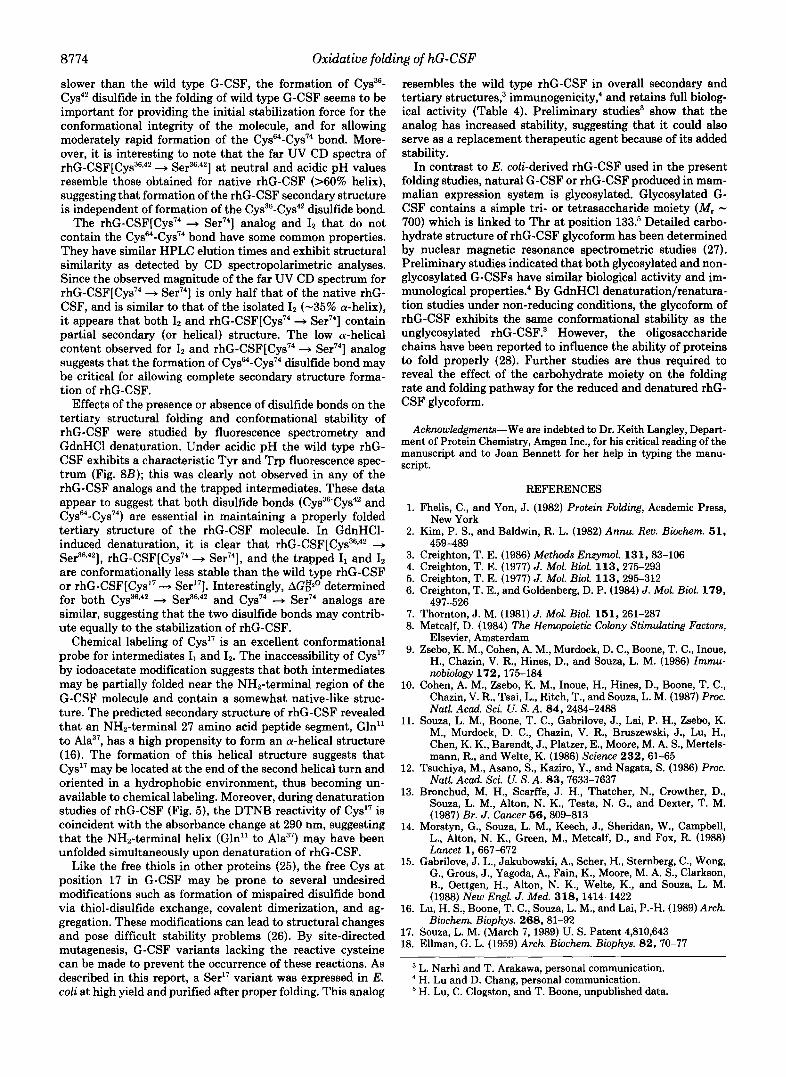
As seen in Fig. 8A, the fluorescence emission spectrum of the rhG-CSF molecule at neutral pH is characterized by a single peak with a maximum at 344 nm, typical of a somewhat solvent-exposed Trp (20). There is no detectable Tyr fluores- cence (around 300 nm). The spectra of the two analogs are similar to that of the native rhG-CSF, although the intensities are greater, indicating that the Trp fluorescence might be somewhat less quenched by the surrounding environment. The spectra of I1 and I2 differ slightly from those of the respective labeled derivatives; the peaks are broader, again reflecting a difference in the environment of the 2 Trp resi- dues.
Fig. 8B shows the fluorescence spectra at pH 3. The Trp peak of native rhG-CSF is still evident, but is greatly de- creased in intensity (the absolute scale of this figure is less than that in A), and a peak at 304 nm, attributable to Tyr, is also present. This suggests that the molecule has undergone a reversible change in conformation so that energy transfer from Tyr to Trp no longer occurs. However, such change occurs only for the native molecule, since the other species show a decrease in the intensity of the Trp fluorescence, but no change in the Tyr fluorescence. The spectrum of the rhG- C S F [ C ~ S ~ ~ , ~ ' + Ser36*42] has a shoulder at 304 nm, indicating a slight acid-induced conformational change.
Fig. 9 shows the hydrodynamic behavior of the native molecule, the Cys74 + Ser74analog, and Cys36*42 + Ser36.42 analog as determined by gel filtration. Recombinant hG-CSF is a very compact molecule ( M , = 15,000 uersus the expected 18,800). While both analogs still elute with an apparent molecular weight smaller than expected, they both elute ear- lier than the native molecule, indicating that without either disulfide bond, the molecule behaves somewhat larger, prob- ably due to an increase in flexibility.
DISCUSSION
The present study demonstrates that the folding of reduced rhG-CSF proceeds through identifiable intermediates I1 and I2 (Fig. 2). The originally fully reduced (I1), partially oxidized (I2), and the final oxidized (N) forms can be separated and quantitated by HPLC for detailed kinetic studies. The disul- fide-reduced intermediates as well as the disulfide-unpaired analogs appear to behave more hydrophobically than the native rhG-CSF.
Copper ion and other trace metals have been reported to catalyze air oxidation of proteins due to their ability to accel- erate thiol oxidation at concentrations ranging from 0.1 and 10 PM (21-24). The optimal Cu2+ concentration for rhG-CSF oxidation is in the range of 20-40 pM (17). The kinetic data show that copper ion increases the folding rate of intermediate IQ from I1 approximately 2-fold and increases the folding rate of N from I2 approximately &fold at 25 "C. Cu2+ also promotes oxidation to greater than 95% completion and shortens the second phase folding time (see "Results"). Increasing the temperature to 37 "C can also accelerate folding and oxidation of the final oxidized form N but does not appear to increase the rate of the second phase of folding. The kinetic studies and the detection of different intermediate folding forms

Oxidative folding of hG-CSF 8773
A
FIG. 8. Fluorescence spectra of rhG-CSF species at pH 7.6 (panel
I I I 1 I I I I I I I I I
<280 300 320 340 360 380 400 420>
A ) and at pH 3.2 (panel B ) . Spectrum I , rhG-CSF standard; spectrum 2, rhG- C S F [ C ~ S ~ ~ + Ser"]; spectrum 3, rhG- C S F [ C ~ S ~ . ~ ' + Ser36,42]; spectrum 4, in-
B
termediate I, derivative; spectrum 5, re- 10 duced rhG-CSF (or intermediate I, de- rivative). 9
- 0 c .- 5 e 7
f 5
e $ 6 - 5
z
s
c - a 4
$ 3
i i : 2
1
I 0 ' I I I I I I I I I I I I I
<280 3do
suggest that the mechanistic pathway for rhG-CSF folding under the described conditions is sequential: I, + Iz * N. The folding from 1, to Iz is fast, while the folding from Iz to N is rate-limiting (Table 1).
1, and Iz were the two predominant intermediates observed during folding and no other species containing intermolecular disulfide forms were detected. The absence of such species was supported by other experimental evidence. For example, a small amount (<5%) of interdisulfide-linked rhG-CSF dimer or aggregate forms is detected by non-reducing sodium dodecyl sulfate-polyacrylamide gel electrophoresis (data not shown). Since levels of these species do not increase during folding, they may be unrelated to the described folding pathway. Moreover, by HPLC analysis, intermediates that contain non- native disulfide bonds were also not evident, indicating that disulfide formation does not occur randomly.
320 340 360 380 460 420> nm
Emission Wavelength
For structural characterization, I, and Iz have to be trapped as stable derivatives at an early time point during folding; these derivatives do not represent the final folded forms. Characterization of the ~ ~ G - C S F [ C ~ S ~ ~ , ~ ' "-* Ser36*42] and rhG- C S F [ C ~ S ~ ~ + Ser74] analogs lacking a single disulfide bond corroborates the findings obtained from physicochemical analysis of the isolated intermediate derivatives since they can be folded and oxidized to their respective stable oxidized states and isolated by rhG-CSF purification procedures.
In the folding studies, the Cys36,42 -+ Ser36,42 analog under- goes a sequential oxidation and folding pathway similar to that observed for the wild type rhG-CSF going from I, (data not shown). This analog is distinct from any of the folding intermediates and exhibits HPLC retention time similar to that of the wild type rhG-CSF (Fig. 4). Since the folding and C y ~ ~ ~ - C y s ~ ~ disulfide bond formation of the Ser36,42 analog is
8774 Oxidative folding of hG-CSF
slower than the wild type G-CSF, the formation of C Y S ~ ~ - Cys4' disulfide in the folding of wild type G-CSF seems to be important for providing the initial stabilization force for the conformational integrity of the molecule, and for allowing moderately rapid formation of the C y ~ ~ ~ - C y s " ~ bond. More- over, it is interesting to note that the far UV CD spectra of rhG-CSF[Cys3'*'' + Ser36.42] at neutral and acidic pH values resemble those obtained for native rhG-CSF (>60% helix), suggesting that formation of the rhG-CSF secondary structure is independent of formation of the C y ~ ~ ~ - C y s ~ ~ disulfide bond.
The rhG-csF[Cy~"~ + Ser74] analog and I2 that do not contain the C ~ s ~ - C y s " ~ bond have some common properties. They have similar HPLC elution times and exhibit structural similarity as detected by CD spectropolarimetric analyses. Since the observed magnitude of the far UV CD spectrum for rhG-cSF[cy~"~ + S ~ I " ~ ] is only half that of the native rhG- CSF, and is similar to that of the isolated I p (-35% a-helix), i t appears that both 12 and rhG-cSF[cy~"~ + Ser"*] contain partial secondary (or helical) structure. The low a-helical content observed for I2 and ~ ~ G - C S F [ C ~ S " ~ + Ser74] analog suggests that the formation of C y ~ ~ - C y s " ~ disulfide bond may be critical for allowing complete secondary structure forma- tion of rhG-CSF.
Effects of the presence or absence of disulfide bonds on the tertiary structural folding and conformational stability of rhG-CSF were studied by fluorescence spectrometry and GdnHCl denaturation. Under acidic pH the wild type rhG- CSF exhibits a characteristic Tyr and Trp fluorescence spec- trum (Fig. 8B); this was clearly not observed in any of the rhG-CSF analogs and the trapped intermediates. These data appear to suggest that both disulfide bonds ( C y ~ ~ ~ C y s ~ ~ and C ~ s ~ ~ - C y s ~ ~ ) are essential in maintaining a properly folded tertiary structure of the rhG-CSF molecule. In GdnHCl- induced denaturation, it is clear that ~ ~ G - C S F [ C ~ S ~ ~ , ~ ~ +
are conformationally less stable than the wild type rhG-CSF or rhG-cSF[cy~'~ + Ser'"]. Interestingly, AG3' determined for both Cys36s42 + Ser36,42 and Cys74 + Ser74 analogs are similar, suggesting that the two disulfide bonds may contrib- ute equally to the stabilization of rhG-CSF.
Chemical labeling of Cys'" is an excellent conformational probe for intermediates I1 and Ip. The inaccessibility of Cys'" by iodoacetate modification suggests that both intermediates may be partially folded near the NHp-terminal region of the G-CSF molecule and contain a somewhat native-like struc- ture. The predicted secondary structure of rhG-CSF revealed that an NH2-terminal 27 amino acid peptide segment, Gln" to Ala3", has a high propensity to form an a-helical structure (16). The formation of this helical structure suggests that Cys'" may be located at the end of the second helical turn and oriented in a hydrophobic environment, thus becoming un- available to chemical labeling. Moreover, during denaturation studies of rhG-CSF (Fig. 5), the DTNB reactivity of Cys'" is coincident with the absorbance change at 290 nm, suggesting that the NHp-terminal helix (Gln" to Ala3") may have been unfolded simultaneously upon denaturation of rhG-CSF.
Like the free thiols in other proteins (25), the free Cys at position 17 in G-CSF may be prone to several undesired modifications such as formation of mispaired disulfide bond via thiol-disulfide exchange, covalent dimerization, and ag- gregation. These modifications can lead to structural changes and pose difficult stability problems (26). By site-directed mutagenesis, G-CSF variants lacking the reactive cysteine can be made to prevent the occurrence of these reactions. As described in this report, a Ser'" variant was expressed in E. coli at high yield and purified after proper folding. This analog
Ser36,42 1, rhG-csF[Cy~"~ + Ser74], and the trapped Il and I2
resembles the wild type rhG-CSF in overall secondary and tertiary structures,3 immunogenicity,4 and retains full biolog- ical activity (Table 4). Preliminary studies3 show that the analog has increased stability, suggesting that it could also serve as a replacement therapeutic agent because of its added stability.
In contrast to E. coli-derived rhG-CSF used in the present folding studies, natural G-CSF or rhG-CSF produced in mam- malian expression system is glycosylated. Glycosylated G- CSF contains a simple tri- or tetrasaccharide moiety (MI - 700) which is linked to Thr at position 133.5 Detailed carbo- hydrate structure of rhG-CSF glycoform has been determined by nuclear magnetic resonance spectrometric studies (27). Preliminary studies indicated that both glycosylated and non- glycosylated G-CSFs have similar biological activity and im- munological properties: By GdnHCl denaturation/renatura- tion studies under non-reducing conditions, the glycoform of rhG-CSF exhibits the same conformational stability as the unglycosylated rhG-CSF.3 However, the oligosaccharide chains have been reported to influence the ability of proteins to fold properly (28). Further studies are thus required to reveal the effect of the carbohydrate moiety on the folding rate and folding pathway for the reduced and denatured rhG- CSF glycoform.
Acknowledgments-We are indebted to Dr. Keith Langley, Depart- ment of Protein Chemistry, Amgen Inc., for his critical reading of the manuscript and to Joan Bennett for her help in typing the manu- script.
REFERENCES
1. Fhelis, C., and Yon, J. (1982) Protein Folding, Academic Press,
2. Kim, P. S., and Baldwin, R. L. (1982) Annu. Reu. Biochem. 5 1 ,
3. Creighton, T. E. (1986) Methods Enzymol. 131,83-106 4. Creighton, T. E. (1977) J. Mol. Biol. 113 , 275-293 5. Creighton, T. E. (1977) J. Mol. Biol. 113 , 295-312 6. Creighton, T. E., and Goldenberg, D. P. (1984) J. Mol. Biol. 179,
7. Thornton, J. M. (1981) J. Mol. Bwl. 161 , 261-287 8. Metcalf, D. (1984) The Hemopoietic Colony Stimulating Factors,
Elsevier, Amsterdam 9. Zsebo, K. M., Cohen, A. M., Murdock, D. C., Boone, T. C., Inoue,
H., Chazin, V. R., Hines, D., and Souza, L. M. (1986) Zmmu-
10. Cohen, A. M., Zsebo, K. M., Inoue, H., Hines, D., Boone, T. C., Chazin, V. R., Tsai, L., Ritch, T., and Souza, L. M. (1987) Proc. Natl. Acad. Sci. U. S. A. 84, 2484-2488
11. Souza, L. M., Boone, T. C., Gabrilove, J., Lai, P. H., Zsebo, K. M., Murdock, D. C., Chazin, V. R., Bruszewski, J., Lu, H., Chen, K. K., Barendt, J., Platzer, E., Moore, M. A. S., Mertels- mann, R., and Welte, K. (1986) Science 232,61-65
12. Tsuchiya, M., Asano, S., Kaziro, Y., and Nagata, S. (1986) Proc. Natl. Acad. Sci. U. S. A. 83,7633-7637
13. Bronchud, M. H., Scarffe, J. H., Thatcher, N., Crowther, D., Souza, L. M., Alton, N. K., Testa, N. G., and Dexter, T. M. (1987) Br. J. Cancer 56,809-813
14. Morstyn, G., Souza, L. M., Keech, J., Sheridan, W., Campbell, L., Alton, N. K., Green, M., Metcalf, D., and Fox, R. (1988) Lancet 1,667-672
15. Gabrilove, J. L., Jakubowski, A., Scher, H., Sternberg, C., Wong, G., Grous, J., Yagoda, A., Fain, K., Moore, M. A. S., Clarkson, B., Oettgen, H., Alton, N. K., Welte, K., and Souza, L. M. (1988) New Engl. J . Med. 318, 1414-1422
16. Lu, H. S., Boone, T. C., Souza, L. M., and Lai, P.-H. (1989) Arch. Biochem. Biophys. 268,Sl-92
17. Souza, L. M. (March 7, 1989) U. S. Patent 4,810,643 18. Ellman, G. L. (1959) Arch. Biochem. Biophys. 82,70-77
New York
459-489
497-526
nobiology 172,175-184
L. Narhi and T. Arakawa, personal communication.
H. Lu, C. Clogston, and T. Boone, unpublished data. ' H. Lu and D. Chang, personal communication.

Oxidative folding of hG-CSF 8775
19. Greenfield, N., and Fasman, G. D. (1969) Biochemistry 8, 4108- 24. Saxena, P., and Wetlaufer, D. B. (1970) Biochemistry 9, 5015-
20. Narhi, L. O., Kenney, W. C., and Arakawa, T. (1991) J. Protein 25. Fernandez-Diez, M. J.9 Osuga, D. T.9 and FeeneY, R. E. (1964)
21. Cecil, R., and Mcphee, J. R. (1959) Adv, protein C b m . 14, 255- 26. Manning, M. c., Patel, K.7 and Borchardt, R. T. (1989) P h r m .
4116 5023
Chem. 10,359-367 Arch. Biochem. Biophys. 107,449-458
389
mun. 13,353-359
Biochem. (Tokyo) 64,449-455 1162
Res. 6 , 903-918
22. Takagi, T., and Isemura, T. (1963) Biochem. Biophys. Res. Com- 27. Oheda, M., Hase, S., Ono, M., and Ikenaka, T. (1988) J. Biochem.
28. Berger, E. G., Buddeke, E., Kamerling, J. P., Kobata, A., Paulson, 23. Yutani, K., Yutani, A., Imanishi, A., and Isemura, T. (1968) J. J. C., and Vliegenthart, J. F. G. (1982) Erperientia 38 , 1129-
(Tokyo) 103,544-546
Supplementary Material to: FOLDING AND OXIDATION OF RECOMBINANT HUMAN GRANULOCYTE COLONY STIMULATING FACTOR PRODUCED IN ESCHERICHIA COLI: CHARACTERIZATION OF THE DISULFIDE-REDUCED INTERMEDIATES AND CVS-SER
and Thomas C. &ne, Amgen Inc., Amgen Center. Thousand Oaks. CA 91 320 ANALOGS. Haleng S. Lu', Chdati L. Clogston, Unda 0. Nerhi, Lee Ann Merewether. Wayne Peari
MATERIALS AN0 METHODS
m k d u c e d rhG-CSF was purified aomrding to methods described previously (t i). Measurement of biolwica) anivifl was perlormed by a slandardized in x h mouse bone marrow m a y (9) (see 'G-CSF bi0lqi.d assay'). Pumy was asseased by sodium dodecy sullete-polyacrylamide gel electro- phoresis (SDS-PAGE) and reverse-phase HPLC. The pmtein is monomeric as judged by gel filtration. The extlnCtion m e r i e m at 280 nm in 20 mM Mdium acetate (OH 4.0 - 7.0) is 16.400 M-1 cm.1.
601ullon (8 M) was obtained fmm Pierce Chemid Co. lodoacelate was lrom Sigma Chemical Co. and Sarkosyl (IauryYerkOalne) was plnhassd from J.T. Baker and DTT from Caibiochem. GdnHCl in
was re€Iyslallized twice in petroleum ether prior to use. [3H]-C2-iodoacetate was oblained from New
&&ks&us pmtease (V-8 strain) was lrom Miles. All 01 the sequendng reagents and England Nudear. HPLC solvew and water were purchased lrom Burdick and JaMon.
solvew were from Applied Biosystems (Foster City, CA).
Mutants 01 IO-CSF induding rhG-CSF[Cyst7-tSert7]. rhG-CSF [Cys36,42+Ser36,42] and rhG-
described previously (14,20). Protein concentrations of the intermediales and analogs were determined C S F [ c y ~ ~ ~ + S e r ' ~ l were mnatwcted as described (17). and purified by procedures smiar to those
the analogs was assarsed by reducinp/non-redudng SDS-PAGE (I 1) and RP-HPLC. A single band at by UV BbSDrbance at 280 nm in 50 mM Tris.HCi. pH 7.2 and calibrated by amino acid analysis. Purity of
Mr t8.000-18,800 on SOS-PAGE was obsarved in all was. The correctness 01 the disulfide bond present in the analogs and the finally oxldized form N was mntirmed by pephde mapping and sequence analysis as described previously (16).
k%%%%%%xpreasion vector. the expression mnditions. cell hawesung. cell breakage, and inclusion body mllection were delailed in two previous repom (I 1.17).
f l ( l t . 1 7 ) was solubilized in 2% Sarkmyl and 50 mM Ttis-HCI. pH 8.0 far 20 min at 25°C. The protein Concentration was 1-2 mgiml.
25T. Al#quots were taken at dinerent time points and immediately subjected to analysis by reverse- CuSO4 was immediately added to a tind mncentralion of 40 PM and the mixture stirred for 16-24 hr at
phase HPLC. Dillerewe in elution times 01 disulfide-reduced intermediates and the final oxldized rhG- CSF allows the separation and quanlltatlon of loldlng lntermedoates generated dunng oxidative folding.
folding rate was eXpRssed by percent disappearance of the intermediates or formation 01 the linal Similar lolding experiments were also performed at 25% and 37% without addition 01 CuS04. The
oxidized form per second.
-F was prepared by adding purified rho-CSF lo 0.1 M Tris-HCi (pH 8.0) mntaining 6 M GdnHCl and 2 mM D n . The reduced rhG-CSF in the cell pellet was prepared by solubilizing the LmU mil pellet in 50 mM Tris-HCI (pH 8.0) in the presence 01 2% Sahosyl at 2-5% for 1 hr or by rolubilizing the protein in 6 M GdnHCi. 0.1 M Tns-HCI (pH 8.0) a1 2.5% for 10 mm and then diluting to t M GdnHCi at 2.5%
. . . . ,
Aliquots of samples duting loldlng were taken at 0 min, 20 min. and 1 hr tlme points and an equal volume 01 0.3 M Tris-HCI (pH 8.3) was added. [3H]-C2-cdoacetic acid [molar ratlo - 20:t] was immediately added to the mlxture. The reamion proceeded for 20 mln at 37'C and w a s terminated by desaning with a Sephedex G-25 mlumn (1 x 15 cm) equilibraled wilh 0.1 M Tris-HCI pH 8.3. TO lunher prily the trapped intermediates, the desalted sample was subjected to ion+xchan& HPLC using a Sulfaethyl Polyaspanamide silica-based HPLC Column (0.46 x 25 Cm the Nest Group) which was equilibrated at 20 mM sodium amtate (pH 5.4) at a flow ram of t m l l n h The elution ;as perloformed by a linear NaCI gredient lrom 0 to 0.3 M over 30 minutes. Radioactivlty and prmein mncemration 01 the pooled fractions were determined lo estimate the stoichlometry of labeling. For determination of protein Concentralion. allquots 01 the pmtein fractions were subjeaed lo quamifative amino actd analysis by
amino acid derivatizer. Phenylthiocalaamfl (PTC-) precolumn derivatiration using an Applied Biosystems Model 420 automatic
The labeled samples Wre funher reduced with DTT ( 5 0 3 molar ratlo) and alkylated with non- radimBaiie iodoacetic add in 6 M GdnHCl and 0.3 M Tris-HCI. pH 8.3 as described above. The final alkylated denvatwe was desaiied using a Sephadex G-25 mlumn (1 x 15 cm) equilibrated with 0.1 N emmonium bicatbonale (pH 8.0) mntaining 2 M urea. These fully alkylated K i -CSF intermediates were subjected to protease digestion and peptide mapping as described below.
were injecled onto a C-4 (Vydac. widepore. 0.46 x 25 cm. 300 A) reverse phase column. Solvent A &%i%%%, the folding intermediates, and the chemcally rnodilied intermedletes (25.75 Fg)
0.1% TFA: rohent 8: TFA: H S : CH3CN (0.1:9.990). The mlumn was initially equilibrated with 75% AR5% Bat a flow rate 01 0.8 mumin. The separation was perlormed by a linear gradlent imm 25% B to 70% B within 50 min and Imm 70% B to 80% B over 5 min on an HP1090 liquid chromatographic Syatem equipped with en autosampler and a phmodiode array detector. Pmteln peaks were detected at 21 5 nm. Peak areas corresponding to rhG-CSF related species were imegrated. If necessary, tractions containing rho-CSF were milected manually lor subsequent analysis.
. . . s d 0.1 M ammonium bicatbonale. pH 8.0 were irmbated with -m (V-8) protease at an enzyme.to-6Yblrate ralio 01 t :25 at 254: for 20 hr. The rnactlon was termmated by injectlng the digest om0 the C-4 column lor peptide map a n a l p s Detalled HPLC peptide mapping Conditions were dewbed previousty (161. Ouantitatlve amino acid analysis Procedure to determine concentration 01 each peplide fraction was identical to that described above.
Sequence analysis of psplides was also prlormed aaOding to previously reponed procedures (16). . .
u 2 9 0 nm at various concentrations of GdnHCl (Pierce Chem. CO.). Samples containing rhG-CSF. intermediate derivatives. and analogs were
then added to a desired mncentralion. Aner 15 mm at room temprature, abwrbance 01 samples at prepered and diluted to appmximateiy 0.3-0.5 mglml in 50 mM sodium phosphate, pH 7.0. GdnHCl was
290 nm was measured lor extinction coer ient determination.
To determine the thermodynamic constants lor denaturatlon. a two-state transition approximallon was used and the free energy change. AGD was calculated lor the reaction. F (folded) + U (unloided). at a given GdnHCl mncemration using the equation.
AGD - -RTlnA. (1)
where A is the observed absolbance at 290 nm, and AF and AU are the absorbances of the folded and
denaturant determined by a lhnear relatmn method (see equation 2).
unfolded statas. respectively. AG:fl was the tree energy lor denaturation in the absence of the
AGD J A@' - m[GdnHCI] (2)
Samples obtalned from denaturation of naliie rhG.CSF at dlnerent GdnHCl concentrations were added to 50 id 01 freshly Prepared DTNB (Sigma Chem. Co.) Solvtion (4 mglml) in 0.1 M sodium phosphate, pH 7.0. Aner incubation at 25°C tor 15 min. the absorbance olthe mixture was measured at 412 nm. The SH mncentralion present In samples was then calculated using E = 1.36 x 10⁴ M"cm"(t8).
. . =ctivity of rhG-CSF tnst samples, an assay employlng 3H-thymidlne uptake in low density non-adherent mouse (lemsle Balb C) bone marrow cells was used (9). Fully actwe rhG-CSF usually gives approximately 1.0 x to8 unitdmg. Under a well-calibrated condot~on. the assay of vanous analogs was performed in tnplicate mth dinerent dilutions.
. . -f the two analogs missing disuillde bonds were concentrated lo 1 mglml in 50 mM Tris, 100 mM NaCI. pH 7 5 using tO.000 moleCular w g h t cuton Centricon device tram Amlmn. The CD spectra of a ponion 01 thew soiutions were analyzed directly, and another panion 01 the soiutions were dialyzed into 1 mM HCI. pH 3.2. The fluorescence spectra, as well as the acidlc CO spectra of the samples at pH 3.2 were then analyzed. The folding Intermediates were obtained in 100 mM ammonlum bicarbonate, pH 7.7. and analyzed immediately in the same
funher oxidation, and then subjected to the treatment described lor the analogs. The fully reduced rhG- buner. The folding intermediates were also modified with non-radioactive iodoacetlc acid to prevent
CSF was prepared by denalumion of rhGCSF in 0.3 M Tris-HCi (pH 7.8) containing 6 M GdnHCl and t o mM DTT for 15 min at 25'C. GdnHCl was then removed by a desalting miumn (Pharmacla G-25, t x 15 cml eauilibraled with 0.1 M ammonium bicarbenate IDH 7.71 WntainlnQ 2 M Urea and 10 mM D n . The sample was funher dialyzed imo either the I O mM iris-HCI buller. pH-7.5. or 1 mM HCI. pH 3.2. both with I O mM DTT. Some precipltate appeared when the analogs and the loldng intermediates were dlalvzed into the t mM HCI. DH 3.2: this was removed and the Drotein concentration 01 the supernatant determined pdor to iriaiysls.' Folded rhG-CSF at 7 mglhl on t mM HCI. pH 3.2 was diluted lo t mglml into the vanous bullers and analyzed as a standard The results With tne standaid obtained wilh both bunen at neuiral pH were identical. and therefore the results wtth the inlermedlates and analogs can be directly compared.
The fluorescence spectra were Oblalned with an SLM-Ammo SPF5OOC spectrof1uOnm~tor controlled
diluted to an absorbance at 280 nm of 0.2 were obtalned by exclttng the protems at 280 nm. Proteins by a Samtmn computer. Slil widths were set at 5 nm. and the emission Spectra of pmtein sdutlons
were diluted imo 10 mM atrate. 100 mM NaCI, pH 7.5 or 2.5. A cuvette with a pathlength 01 0.5 cm was used, and the emission SpecIra were recorded from 280 lo 420 nm.
The far and near UV circular dlchroic spectra were obtalned wfth a Jasm J500C spectropolanmeter mntmlled by a Samrmn computer. A pathlength of I Cm was used for the near UV CD SpecIra. whole a pathiength 01 0.02 cm was used lor the far UV CD spenra.
m g r a p h y was pe89ormed on a TSK2000-SW HPLC coiumn. usmg a flow rate 01 0.5 mllmin. in 0.25 M Wium phosphate. 0.3 M NaCI. pH 7.1. A SpectroPhysiCs HPLC system. consisting 01 an SP-0 oumn. an SP8800 autosamalsr and an SP8450 detector were used on Conlunctlon wnth the
. .
T a l e 1. Initial rate constant lor disappearance of reduced toms and generation 01 oxldnzed forma
Conditions
Folding Pmducts CuS04 + 25% No CuSOq. 25°C No CuS04, 37°C
(Percenvsec)
It + I2b t.9x10.2 1 . o x t o - 2 1 .o x 10.2
I, + NC 3.1 x 6.6 x t .8 x
a The initial rate was estimated Imm the l ~ rs t phase folding kmetiCS. The folding rate of the second folding phase is Slower in all cases and not described here. The error IS appmximately +5%. Estimated from dmappearance of reduced form It that corresponds to the tormallon of intermediate 12. Generation 01 form N. the linal oxidlzed lorm. corresponds to the disappearance of intermedlate 12.

8716 Oxidative folding of hG-CSF
Table 2. Chemical modification 01 rhG-CSF and the isolated iltermeaiates by tritiated iodoacetic acida
Proteins Perturbant added Mole 01 labellmole of protein
1. r-hG-CSF None 0.03 (0)
2. r-hG-CSF 6M guanidine HCI 0.96 (1)
3. Reduced r-hG-CSF 6M guanidine HC112 mM DTT 5.10 (5)
4. Folding lntermediateb
12 None 1.90 (2)
5. Folding intermediateb
11 None 3.74 (4)
a Labeling was pellormed in 0.2 M Trir.HCI, pH 8.2 at 37-10, 15 rnin wlth or without addillon of denaturant and reducing agent (lor details. see Materials and Methods). Both labeled intermedmtes. I, and 12. were further purified by ion exchange chromatography before quantitation (see Materials and Methods).
Table 4. Unfolding parameters and biological aaiwty lor rhG-CSF intermediates and analogs
Biological Activitye
Proteins Mldpoont (M)a A G F O (x 108 unitdmg)
rhG-CSF 2.95: 3.15b 5.4 too ( e o ) rhG-CSF[Cyst7+Sert7p rhG-CSF[Cys36'42+Se,36,42]C 1.65
2.6 5.4 too (*20)
r h G - C S F [ C y ~ ~ ~ + S e r ~ ~ 1 c 2.5
2.10 2.8
rhG-CSF Intermediate l t d rhG-CSF Intermediate 12d 2.05 2.6
t ( f 0 5 )
3 (*I) 5 ( *2)
2.2 3 (*I) 1.40
aCalculated lrom the UV-detected denaturation msults 01 Fig. 2.
CProduced by s11e-dlrected mutagenesis and purlfled according to Table 4. bDetermined Irom half-maximal DTNB labelmg of the tree cysteinyl residue.
dThe lntermediates have been modified by redualon and carboxymethylatlon (see Materials and Methods). eBiologld activity was obtained according to 'Materials and Methods'. NUmbEn In parentheses are the
standard &watton (n25).
1 8
Retention Time (Minutes) 2 0 30 4 0 5 0
Figure 1 RP-HPLC 01 folded and axldlzed rhG-CSF lchromatoaram 1 . 5 no1 denatured and reduced rhG-CSF in GdnHCllDTT (chiamatagrah 2. 15 wi):rhG-CSF solubilized lrom E.& mclus~on b o d m by GdnHCl at O'C (chromatogram 3. 10 pg). and rhG-CSF SOlUblllled lrom L&!nciusion bodies In Sarkosvl al 2.5%
= 35 7 mln) were also detected in chromatograms 3 and 4. (chromatogram 4, 15 pg). E. & iontaminants and the added Sarkosyl (retention lime
" I
i_"-- . ~~ Retention Time (Minutes)
Flgure 3. SlaOhYloCOCCUSumus protease (VB)-derivW HPLC peptide map 01 [3HI-carboxymethyl 12 (lor detalis. see Methods). Approximately 50 pg 01 the peptide mlxture was analyzed. Peak U. a partially digested larger fragment. was not analyzed.
a
"(Mn.ta)
Figure 4. Reverse-phase HPLC analysis 01 rhG-CSF analogs. Panel A: chromatogram 1. rhG-CSF standard: chromatogram 2: rhG- C S F l C y ~ ~ ~ + S e r 7 ~ 1 at pH 3: chromatogram 3: rhG-CSF[Cys74bSer74] at pH 7.0: Panel 6' chromatogram 4: rhG-CSF[Cy536,42,Ser36.42] at pH 7.0; chromatogram 5 : rhG-CSF standard: Inset: expanded vlew 01 chromatograms 4 and 5.
3 0.24 ::::K 4
0.12
0 250 300
GvPnidine ACI (mole)
Flgure 5. A. UV spectra 01 rhG-CSF at 0.6 mglml ,n 20 mM Sodium acetate. pH 7.0 (spearurn 1) and 8" 0.1 M sodium ohosphate, pH 7 0 containing 6 M GdnHCl (spectrum 2).
in ext~na~on (AF-A. see Methods) at 290 nm (open square) and the
functlon of GdnHCl concentration ava~lab81ity 01 free cystem by DTNB labeling (closed square) are a
6. Tttration of GdnHCI-#nduced denaturmon 01 rhG-CSF. The decrease

Oxidative folding of hG-CSF 8777
.0.5 05 1.5 2 5 3.5 4.5 5.5 6.5
Guanidine HCI (mole)
Figure 6. Tttrat8on 01 GdnHCI-induced denaturation 01 rhG-CSF analogs and intermediates Curve 1: rhGCSF [Cys7+Ser7'l: Cuwe 2 intermednate 12: Curve 3: rhGCSF [Cys16.42+Ssr".d2]: Curve 4. inlermediats 11
1
MmuIes
chromatogram 2 rhG-CSF[Cys74~Ser7']: chromatogram 3: rhG-CSF Figure 9. Analyiical HPLC gel tdlralion. chromalogram 1 . rhG-CSF standard:
C y ~ 3 ~ . ~ 2 - S e r J 6 . ~ ~ .