0/faunaofindia.nic.in/pdfvolumes/records/016/03/index.pdf · xiii on the supposed occurrence of the...
TRANSCRIPT
XIII ON THE SUPPOSED OCCURRENCE OF THE MIOCENE GENUS FOSSARULUS
RECENT IN INDIA
By LT.-COL. H. H. GODWIN-AuSTEN, F.R.S.
In the Fauna 0/ Brt'tish India, Mollusca, Freshwater Gastropoda and Pelecypoda, Mr. H. B. Preston, on page 78, places Bithynia costigera, Kiister (= marginata, Chm.) in the fossil genus F ossarulus, following Geoffrey Nevill in his" Hand-List," Vol. II, p.42, with a ? Having recently been looking over Indian species of Bithynia in my own and the Natural History collection this generic position has been brought into question. It appeared to me to be so impossible tha t a fossil genus of Miocene age and European habitat should be still living in Peninsular India. I sought Mr. Bullen Newton's kind help, and he was able to not only show me a Fossarul~f,s but the type of the genus from Dalmatia quoted by G. Nevill. The first glance cleared up a great deal, and showed how great was the difference between the Recent and Fossil shells, in every important .character. It is surp.rising that Nevill ,came to the conclusion he did in r884. He may have had grounds at the time for doing so, certainly at the time he was at work he did not have in Calcutta the type shell of Fossarulus to refer to. Mr. Preston has perpetuated Nevill's determination, when the means of verification were close at hand in the Natural History collection.
To show the difference in question I give the original description of both the genus and its type.
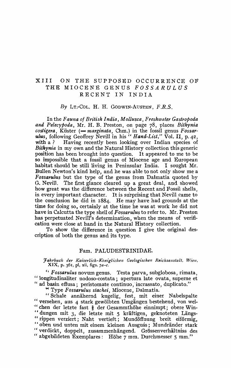
Fam. PALUDESTRINIDAE.
Jahrbuclz del' J<a£serl-ich-Kont'glichen Geologz'schen Rt£c/zsQnstalt, Wien, XIX, p. 361, pI, xii, figs. 7a-c.
" Fossarulus novum genus. 'I'esta parva, subglobosa, rimata, "longitudinaliter nodoso-costata; apertura late ovata, sup erne et rf ad basin effusa; peristomate continuo,' incrassato, duplicato."
« Type Fossarulus stachei, Miocene, Dalmatia. "Schale annahernd kugelig) fest, mit einer N abelspalte
" versehen, aus 4 stark gewolbten Umgangen bestehend, von wel." chen der letzte fast i der Gesammthohe einnimpt; obere Wince dungen mit 3, die letzte mit 5 kraftigen, geknoteten Langs-"rippen verziert; Naht vertieft; Mundoffnung breit eiformig, " oben und unten mit einem kleinen Ausguss; Mundrander stark "verdickt, doppelt, zusammenhangend. Grossenverhaltniss des " abgebildeten Exemplares: Hohe 7 mm. Durchmesser 5 mm."

2IO Records ,oj the 11~dia1t Museum. (VOL. XVI,
Bithynia costigera is a small shell differing considerably from th,e common, widely spread, smooth form in having ribbing on the whorls: 'vi,de figur'e in Conchologia I ndtca, pta te 151, fig. 10. The generic distinction is indicated ,even ,on shell cha,racter and is also to be expected in the animal, which should be examined. It does not occur in Bengal as stated by Preston. At least I hav,e never met witb spe'cimens from the 'Gangetic delta. It is a 'comm.on shell in P,enillsuiar India, recorded by Nevill in his Hand-List from Karnul, Conjeveram and S,. India (30) ex. W T. Blanford and Madras Museum collections; also from Ceylon (30) ex. E. L. and F. Layard col1., together with a subvar. cur,ta, 'G,. Nevill, ob .. tained by him at Bangalore.
The Blanford collection presented toO the British lVIuseum coOntains specimens from South India nam'ed B. su.lcata, Eyd. and Soul.
.As it was so important that the animal of Bt'lhynia cos,tigera should be examined I ask,ed my friend Dr. N. Annandale if he could
FIG. l ,a.-}tossarulus st,ache£, Neufnaycr,. (Enlarged from N,eumay,er's originaifigure).
FIG. Ib.-Myso1'i.a cos,tigera (I{oster) yare curta (Nevill). (Enlarged photograph (x 4) of sheD from type locality).
help me. This he has not only been able to do, but he has most kindly had photographs and drawings made of the shell, radula, and operculum, together with an enlarged photo of Neumayer's original figure .of F ossarulus stachei, which now illustrates this paper,-·- for which I thank him mu'ch .. !
I cannot do better than give in full the result of his examination of specimens he had colle'cted at Bangalore, the original locality of var. cur,ta, Nevill; for th'ey add much to the value of this communication~ , and confirm my idea we are dealit1g with a neyv genus of freshwater shells v,ery distinct from Bithynia. For thIS the name Mysoria se:ems :applicable, if it has not be'en 'Used
1 ~ince this was written ( hav,e obtained fresh specimens' of M. cQstigera in the n~.g~bol1rhood of Madras. The ,animal , so far as appears on a superficial examination, does not differ frOln that of BithJ' ,zt'.a exoept 1n having shorter ten~ tadts" As, . how'ever, I am just startlng ,on a )on~ journey I have not been able to make a de~ailed ,examination.. I hope tha.t Col. Godwin- /\ ustenwiU do this la ter and pltbhsh the results. N . An.naudal,e, 8 .. x-1918.
I9 19·] H. H. GODWIN-AuSTEN: F ossarttlus in India. 211
before. This interesting species has a limited range in Southern India, which was a land surface in pre ... Cretaceous times, during which its early development possibly took place.
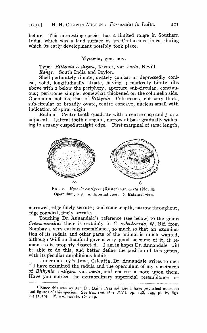
M ysoria, gen. nov.
Type: Bithynia costigera, Kuster, var. curta, Nevill. Range. South India and Ceylon. Shell perforately rimate, ovately conical or depressedly coni
cal, solid, longitudinally striate, having 3 markedly birate ribs above with 2 below the periphery, aperture sub-circular, continuous;' peristome sinlple, somewhat thickened on the columella side. Operculum not like that of Bithynia. Calcareous, not very thick, sub-circular or broadly ovate, centre concave, nucleus small with indication of spiral origin
Radula. Centre tooth quadrate with a centre cusp and 3 or 4 adjacent. Lateral tooth elongate, narrow at base gradually widening to a many cusped straight edge. First marginal of same length,
-6. FIG. 2.-Mysoyia costigera (Kuster) var. curta (Nevill).
Operculum, x 8. a. Internal view. b. External view.
narrower, edge finely serrate; 2nd same length, narrow throughout, edge rounded, finely serrate.
. Touching Dr. Annandale's reference (see below) to the genus Cremnoconchus there is certainly in C. syhadrensis, 'V. Blf. from Bombay a very curious resemblance, so much so that an examination of its radula and other parts of the animal is much \\Tanted, although William Blanford gave a very good account of it, it remains to be properly dissected. I am in hopes Dr. Annandale 1 will be able to do this, and better define the position of this genus, with its peculiar amphibious habits.
Under date 15th June, Calcutta, Dr. Annandale writes to me : " I have examined the radula and the operculum of my specimens of Bithynia costigera var. curta, and enclose a note upon them. Have you noticed the extraordinary superficial resemblance be-
--~.- ---- -------- - - . - --~.-
J. Since this was written Dr. Baini Prashad and I have published notes on and figures of this species. See Rec. Ind. A1us. X V T, pp. 148, 149, p1. iv, fig-so 2-4 (19 10). N. A 11 l1andale, 16-ii- 19.
2I2 Records of the Indian ~luseu1n. [VOL. XVI,
tween the species and Cremnoconchus. "and proceeds to describe the var. curta.
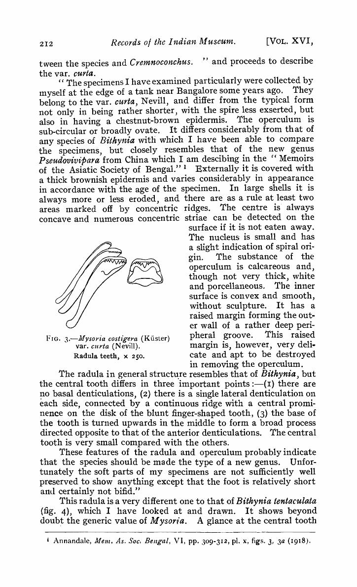
e e The specimens I have examined particularly were collected by myself at the edge of a tank near Bangalore some years ago. They belong to the var. curta, Nevill, and differ from the typical form not only in being rather shorter, with the spire less exserted, but also in having a chestnut-brown epidermis. The operculum is sub-circular or broadly ovate. It differs considerably from that of any species of Bithynia with which I have been able to compare the specimens, but closely resembles that of the new genus Pseudovivipara from China which I am descibing in the" Memoirs of the Asiatic Society of Bengal." 1 Externally it is covered with a thick brownish epidermis and varies considerably in appearance in accordance with the age of the specimen. In large shells it is always more or less eroded, and there are as a rwe at least two areas marked off by concentric ridges. The centre is always concave and numerous concentric striae can be detected on the
surface if it is not eaten away. The nucleus is small and has a slight indication of spiral origin. The substance of the operculum is calcareous and, though not very thick, white and porcellaneous. The inner surface is COll vex and smooth, without sculpture. It has a raised margin forming the outer wall of a rather deep peri-
FIG. 3.-Jlfysoria costigera (Ktister) pher.al groove. This raised yare curta (Nevill). margin is, however, very deli-Radula teeth, x 250 • cate and apt to be destroyed
in removing the opercwum. The radula in general structt1;re resembles that of Bithynia, but
the central tooth differs in three important points :-{r} there are !10 basal denticulations, (2) there is a single lateral denticulation on each side, connected by a continuous ridge with a central prominence on the disk of the blunt finger-shaped tooth, (3) the base of the tooth is turned upwards in the middle to form a broad process directed opposite to that of the anterior denticulations. The central tooth is very small compared with the others.
These features of the radula and operculum probably indicate that the species should be made the type of a new genus. Unfortunately the soft parts of my specimens are not sufficiently well preserved to show anything except that the foot is relatively short and certainly not bifid."
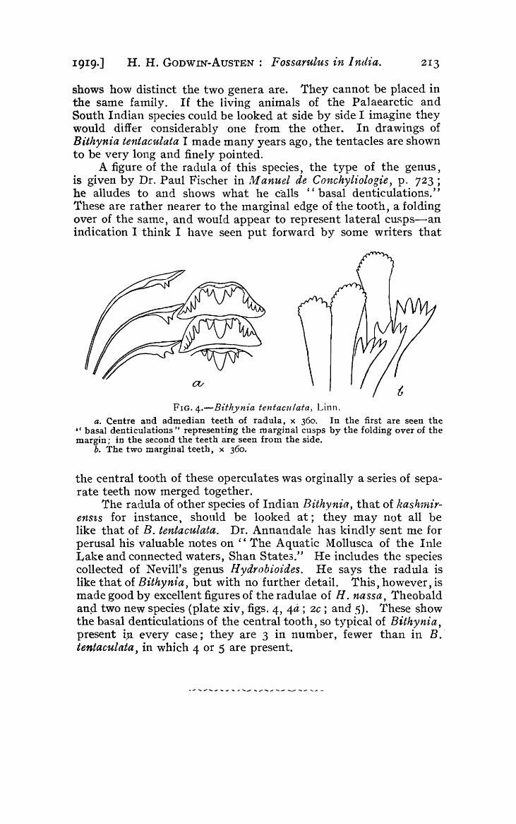
This radula is a very different one to that of Bithynia tentaculata (fig. 4), which I have looked at and dra.wn. It shows beyond doub~ the generic value of Mysorla. A glance at the central tooth
, Annandale, },fem. As. Soc. Bengal, VI, pp. 309-312, pI. X, figs. 3, 3a (1918).
19I 9·] H. H. GODWIN-AuSTEN: Fossarulus in India. 213
shows how distinct the two genera are. They cannot be placed in the same family. If the living animals of the Palaearctic and South Indian species could be looked at side by side I imagine they would differ considerably one from the other. In drawings of Bithynia tentaculata I made many years ago, the tentacles are shown to be very long and finely pointed.
A figure of the radula of this species, the type of the genus, is given by Dr. Paul Fischer in Itl anuel de Conchytiologie, p. 723; he alludes to and shows what he calls "basal denticulations." These are rather nearer to the marginal edge of the tooth, a folding over of the same, and would appear to represent lateral cusps-an indication I think I have seen put forward by some writers that
FIG. 4.-Bithynia tentacu/ata, Linn.
a. Centre and admedian teeth of radula, x 360. In the first are seen the ,e basal denticulations" representing the marginal cusps by the folding over of the margin; in the second the teeth are seen from the side.
b. The two marginal teeth, x 360.
the central tooth of these operculates was orginally a series of separate teeth now merged together.
The radula of other species of Indian Bithynia, that of kash1nirensts for instance, should be looked at; they may not all be like that of B. tentaculata. Dr. Annandale has kindly sent me for perusal his valuable notes on C' The Aquatic l\{ollusca of the lnle Lake and connected waters, Shan State3." He includes the species collected of Nevill's genus Hydrobiol:des. He says the radula is like that of Bithynia, but with no further detail. This, however, is made good by excellent figures of the radulae of H. nassa, Theobald anp. two new species (plate xiv, figs. 4, 4a ; 2C; and 5). These show the basal denticulations of the central tooth, so typical of Bithynia, present iJa every case; they are 3 in number, fewer than in B.' tentaculata, in which 4 or 5 are present.


XIV NOTES FROM ~rHE BENGAL FISHERIES
J-I ABO RAT 0 R Y, N 0 6
EMBRYOLOGICAL AND DEVELOPMENTAL STUDIES OF INDIAN FISHES.
By T. SOUTHWELL, A.R.C.Sc., F.Z.S., Director of Fisheries, Bengal and Bihar and Orissa, and B. PRASHAD D.Se., Superintendent of Fisheries.
(With Plates XVI-XIX.)
This paper consists of four parts; part one deals with two new species of Leptocephalids found in the brackish waters of the Gangetic Delta; in part two we have described the life-history of an Indian Teleost-N otopteyus ch'ttala; the third part is a de'3cription of the egg-capsule of an Indian dogfish, and the fourth consists of descriptions of intra-uterine embryos of some Indian sharks and rays, together with a discussion of various points of general zoological interest resulting from this study.
I. LEPTOCEPHALIDS.
On the occasion of a visit, by one of us, to the Sunderbans, during lVlarch 1918, well preserved specimens of two species of I~eptocephalids were obtained.
Leptocephalus milnei t sp. nov.
(PI. XVI J fig. I.)
~rhis specie:; has the usual band-like form. Description.-Number of segments in a specitnen, I20.
Length 55'4 mm. ; height 8'3 n1m,; head 4 mm,; distance of anus from the end of the tail, I3'2 lnm.; eye 1'1 mrn.; snout l' 3 mm.; post temporal part of the head 1'6 Inn}. ; height 6-7 mm. ; head 13.8 mm.; tail 4'2 times in total le'ngth. Snout rounded" head portion posterior to it slightly convex_ Eye 3'6 times in the head, slightly smaller than the snout and about one and a half times in the post temporal part of the head, Ga p ~ of the lnau th extendin6 behind the eye.
Anus below 79th segment, three times farther from the tip of the snout than from the tail. Very minute teeth are present on the lower and the upper jaws.
Pectorals very small, rounded. Dorsal and anal fins \vith a large nutnber of fin-rays, three to each myosegment.

216 Records oj the Indian ]\1 useUln. [VOL. XVI,
There is a minute piglnent spot at the base of each of the dorsal, caudal, and anal fin-rays\ otherwise the animal~ preserved. in spirit are of a creamy colour. When alive they were quite pellucid, but could just be distinguished swimming in the muddy water.
Specimens obtained in a small beam·trawl at Doorakara, Sunderbans (Gangetic Delta), Bengal, on 15th and 16th of March 1918.
Type-specimens registered in the collection of the Zoological Survey of India} No. F .!Hy1.:§..
We have much pleasure in nanling this species in honour -of Mr. l\Iilne, M.A., I.C.S., Director of Agriculture, Bihar and Orissa, in recognition of much assistance rendered to the Fisheries Department.
Leptocephalus vermicularis t sp. nov.
(PI. XVI, figs. 2, 3.)
This species, instead of having the usual band-like form, is rounded like a worm.
Description. -Number of segments in a specimen, 122. Length 61·2 mm.; height 4"1 mm.; head 4·3 mm.; distance
of anus from the end of the tail, 37'8 mm. ; eye '6 mm. ; snout 1·1
mm"; post temporal part of the head 1·8 mm. Height 14·9, head 14.2, tail 1·6 times in total length. Snout ,acutely rounded, "leading gradually to the post temporal portion which is very broad, even more so than the body. Eye 7.2 times in the head, about half the size of the snout and three times in the post temporal pot· tion of the head.
Gape of the mouth extends a little behind the eye. Anus below the 47th segment, its distance from the tip of the tail being one and a half times the distance from the snout. Minute teeth on the upper jaw, none on the lower. Small rounded pectora~ fins. The dorsal. caudal and anal fins rather small, with a large number of fin-rays, three to each myosegment.
There is a minute black spot at the base of each fill-ray, and a large number of scattered pigme,nt spots, specially collected in groups on the ventral surface of the body. These pigment spots are visible only when specimens are examined under a high magnification, otherwise the specimens appear of a creamy colour when preserved. When alive they were quite pellucid and wriggled very quickly in the muddy water.
Only two specjmens were obtained along with those of the other species described above, in a small beam-trawl at l)oorakara, Sunderhans, Gangetic Delta, Bengal, on the 15th and 16th of March 1918. .
Type-speci.m,ens registered in the collection of the Zoological Survey of India, No. FJ!7/~.

I9I9.] T. SOU1'HWELL & B. PRASHAD: Studies 01 Indian Fishes. 2I7
II. LIFE-HISTORY OF NOTOPTERUS CHITALA (Ham. Buch.)~
(PI. XVI, figs. 4-10.)
During the months of June and July 19I5, Mr. S. M. Mohsin, Superintendent of Fisheries, found eggs of Notopterus cliitala attached to the masonry work: of a bathing ghat on the banks of the river Ganges at Buxar, Bihar. lIe made a few observations on the nature of the nest, the guarding of the nest by the parent fish and the manner in which the eggs are deposited. Mr. lVlohsin collected eggs from the first nest and from other nests which were subsequently found in the vicinity. He also hatched a few eggs in a large earthenware vessel and thus obtained specimens of some of the later larval stages. Our account ~s based on the material collected by lVIJ;'. Afohsin. This material, besides being far from complete, is in a very poor state of preservation. The exact age of the specimens is not stated and canlJ,ot now be ascertained. As, however, nothing is known about the life-history of this, or of any of the nearly related forms, we have thought it advisable to give the folloV\l-ing description even though it is very incomplete. Field notes from Mr. Mohsin's report on the subject are also incorporated, but it should be understood that "re have, as yet, had no opportunity of verifying or extending his investigations.
Breeding habz·ts.-As a result of his observations and local _enquiries Mr. Mohsin arrived at the conclusion that the spawnin:g season of this fish extends from the end of May to the middle of July. This statement must, however, be taken with a certain amount of reservation as we know from experience that the information supplied by fisherme~ is generally inaccurate, and Mr. l\;lohsin's observations were of too limited a character to have enabled him to arrive at a very definite conclusion.
This species prefers to deposit its eggs on solid SUbstances (such as brick-walls, stones, masonry, etc.) close to the banks of the river. The female, when shedding eggs, lies close to the object on which they are to be deposited, the body of the fish being inclined at a certain angle to the vertical. The eggs, being glutinous, adhere firmly to the obj ect on which they are deposited. The male, later on, emits the milt over them. This very simple type of nest was the only one observed in this case. lJsuaUy, from three to five hundred eggs are laid at a time. During the period of laying and hatching, the nest is very carefully guarded by the parent fish and. any intrusion is vigorously resented, fishermen attempting to go near the nest are frequently bitten. Unfortunately, no observations were made as to whether both the male and the female fish guard the eggs, or whether it is done by one of the parent fishes, or by both together, or alternately. Further, nothing is kno\vn as to whether parental care extends to the fry stages or not. According to Mr. Mohsin the eggs hatch out in about two weeks. When hatched the
)
fry have a large yolk-sac, and, during the four to five days

218 Records 0/ the Indian lYl useU1n. [VOL. XVI,
which elapse before this is absorbed, the fry lie quiet and idle .• and do not swim unless disturbed.
The following stages were present in the collection :-(i) Stages with the embryos stilt enclosed in large
globular eggs (figs. 4-6). (ii) Embryos hatching out, some having the egg
membrane still attached. (iii) Stages with the embryos having the yolk-sac in
various stages of absorption (figs. 7-10).
The following descriptions are based on whole Inounts or dissections only, as the specimens were found to be too poorly preserved for section cutting :-
The eggs are of a yellow colour owing to the contained yolk being of this colour in preserved specimens. Nothing is known regarding the colour of this mass in the living eggs. The eggs are large, measuring about 5·2 mm. in diameter. The eggmembrane, on the surface of attachment, is raised up into small projections (fig. 4) by means of which the eggs are attached to stones or other objects in the. nest. Some clusters of from three to five eggs were also found adhering to one another by their sides, and these also showed similar surface proj ections.
On the following page we have given in a tabular form the sizes of five of the later stages and other details of measurement of various organs, etc., in the respective stages. Other details will be found in the detailed description.
Stage I (fig. -4}.-This stage is a fairly advanced one, the contained embryq having already grown to 7.1 mIn. in length. 'rhe elnbryo lies within the egg-membrane in a slightly coiled position over the yolk-sac, and shows a continuous fin-fold along the dorsal and ventral surfaces and over the tail, the division into the various portions not being marked off at this stage. The head is differentiated but still attached to the yolk-sac ventrally. The eye and the ear are formed, but the pigment has not been deposited as yet in the eye. Lying posterior to and below the eyes is the heart, its demarcation into chambers has already commenced but has not advanced snfficiently for the various divisions to be identified. The notochord has a straight course in the tail, and is not turned upwards. The mouth opening is seen as a slit and the rudiments of the branchial arches are also present. The tube of the alimentary canal and the liver Inass are just distinguishable. The air bladder is present as a small, slightly oval sac. In the body and in the tail region the myocommas of a < shape are present; fifty-seven were counted in a specimen but ill the terrninal portion of the tail their boundaries could not be seen.
Stage 11 (figs. 5-6 ).--'rhis stage is only a little more advanced than the previous one~ and but for the lobes of the brain being better tllarked, the myocommas better developed._ the myosepta having a more wa.vy outline and the eye and the air-bladder being more distitict, there is nothing special to mark in this stage.

Stage Total Maximum Dimensions Head Size of the Size of the Size of the Size of the Length of of the length depth of of the
length. dorsal fin. pectoral fin. anal fin. caudal fin. the air Remarks. larvae, the body, yolk-sac, . bladder.
Diameter III. I.1'S mm. 1'6 mm. 5 mm. 2'1 mm, ... J u~t appear- 6'9 mm, I mm, 1'2 mm. Just hatching out.
Ing.
IV, 14'2 mm. I'S mm, 5'2 X4'S mm. 2'5 mm. .. 'Smm, 7'1 mm. " "
Head separated
S'SX3'6 mm. Sl,ightly indi- 1'6 mm. from yolk-sac.
V, ITI mm, 2'6 mm. 3 mm, 1'5 mm. II mm, 1'2 mm. cated.
VI. 18'4 mm. 3'2 mm, S'2X3'1 mm. 3'3 111m, U ndifferentia- 1'6 m,m. 11'3 mm, I 1'4mm" 2 mm. ted, about '8 I
mm, I
VII, 19'5 mm, 4'1 mm, ... 4'5 111m , 1'5 mm. 2'1 mm, 11'5 mm, 1'9 mm. I 4'3 mm. Last larval stage with no trace of
I an external yolk-sac.
--

220 Records 0/ the Indian M ~4se'tHll. [VOL. XVI,
Stage III.-This is a much more advanced stage than the last one. Some of the larvae have already hatched out while others are still enclosed in the egg-capsule. In hatching, the free posterior portion of the body and the tail (both of which are well developed) are the first to come out of the egg-capsule. The head and the rest of the body, with the large yolk-sac, are then separated out by violent l110vements of the taB. Some of the larvae show the body still enclQsed in the egg-capsule but have the tail protruding.
In this stage only a part of the head is free, the rest is very closely applied along the ventral surface to the yolk-sac, as shown (to some extent) in fig. 7. The mouth is present. as a distinct, horizontal slit and the opercular limits are also marked, specially on the ventral surface. The head, which has not developed to any great extent as yet, is broadest in the region of the hind brain. 'I'his latter structure is well developed, and shows the large cerebellum getting marked from it. The pectoral fins are just appearing and the continuous dorso-ventral ,fin bas, besides increasing in size, begun to show rudiments of the fin-rays, in the caudal region. The supporting elements of the fin-rays {or pterygiophores) are already well developed. There is nothing particular to note about the sense organs. except that the external narial opening is well developed. The gill slits are well advanced and the arches show traces of the development of gill filaments on them. The air-bladder is elongated and sac-like, measuring about 1·2 mm. in length, it shows no constriction.
Stage" IV (fig. 8). -The mouth, which was ventral in the last stage, has, owing to the separation of the head from the yolksac and the better development of the middle portion, shifted to a position far forward and is now lnore or less anterior. The head is becoming marked off as a prominent structure owing to the special development of the optic lobes and· the cerebellum. The eyes are now partly enclosed in the optic capsules and do not protrude as much as in the last stage. The pectoral fins are better developed and the fin-rays are making their appearance both in the pectoral and in the anal fin-portion of the dorsoventral fin. The yolk-sac is being gradually absorbed and has become transformed frorn a rounded to an ovoidal structure. The gill filanlents are better developed and even gill-rackers are developing on the ar('h~s. The outline of the jaws is also indicated.
Stage V (fig. g).-l'his stage, except for showing the beginning of the dorsal fin, is very near the last one. The various organs, ho\vever, are better developed and there is a distinct increase in size.
Stage VI.-This stage has still a fairly massive yolk-sac. It show~ the operCUlum quite separated as a flap on either side and forming the posterior limit of the head. The flexure of the brain is better marked, and the medulla oblongata is much better

IgIg.] T. SOUTHWELL & B. PRASHAD: Studies o/Indian Fishes. 22I
developed. The eye has become still more I enclosed In the capsule. In the skull, the jaws 'are already nearly complete, the enveloping bones a.nd the teeth are beginning to be laid down. In the vertebral column the bqdy of the vertebrae, the neural and the haemal arches are formed in the anterior part of the body region but not further in the region adj 0 ining the tail. So far as the fins are concerned, this is the first stage in· which the finrays have begun to make their appearance in the region of the dorsal fin, though the basal pterygiophores could be distinguished in this situation even in the last stage. In the anal and the caudal fins the rays are already quite well developed and the limit between these two fins is also just indicated by the direction of the finrays. 'fhe .alimentary canal, the liver, and the air-bladder are better developed. In the gills, the fi!aments are larger and are present on a.ll the four gill-arches.
Stage V 1 I (fig. Io).-This is the most advanced stage in the collection and shows no trace of an external yolk·sac. The general colour is milky white in the preserved state. Irregularly scattered cbromatophores of the usual shape and of a brownish colour are present on the head and on the abdominal portion; none, however, can be distinguished in the tail region. No scales are developed as yet, but in sections of a portion of the body-wall, scales can be distinctly seen developi ng in the scale-sacs. There is still a continuous fin-fold, in \vhich the dorsal fin is distinctly marked off about the. middle of the animal. and it has well developed fin rays. The thin covering of the fins is stilt directly in continuation of the original dorso-ventral fin, which latter has become greatly reduced posterior to the dorsal fin but is fairly broad anteriorly. The pectoral fins are much larger and have fully developed fin-rays. The yolk-sac is entirely withdra'wn into the body-cavity and is not visible externally. Unfortunately, the condition of the material at our disposal does not allow of a description of the internal yolk·sac, as the structure may now be termed. The operculum is quite well developed, its posterior boundary lies a little behind the middle of the distance. between the snout and the anus. In the operculum, the opercular, pre-, inter- and sub-opercular eiements are marked off, a'nd ossification has commenced. Fi ve branchiostegal rays are already formed and traces of three others can also be seen. The anus lies at a distance of about one-third the total length from the anterior end. The eye is contained about four times' in the head length and its distance from the snout is equal to its diameter. The external narial opening, \vhich is seen as a distinct aperture in this stage, i~ situated near the middle of the distance between the eye and the snout. In the skull region also, ossification has commenced, but the stage is too young to show the various elements. All the j a wbones are t ho"rever, well developed and teeth are present on the maxillaries, dentaries, vomers and palatines. A fe\v can also be seen on the urohyals.
In the body, the irregular myosepta are to be distinguished

222 Records of the 1 ndian Museum. rVOL. XVI,
only in the tniddle region. The alimentary canal is very short and shows only the beginning of tlie stomach and the pyloric caecae. The liver is better developed. The chambers of the heart are becoming more consolidated and the air-bladder is distinctly notched about the. rniddle. The gills are much better ·developed. The kidneys can be distinguished as faint thickenings, but ·no genital organs can be satisfactorily identifie d.
III. THE EGG-CASE OF CHILOSCYLLIUM GRISEUM.
(PI. XIX, fig. 5.)
In 19I4, Sundar a Raj contributed to the ,., Records 0/ the Indian Mu,se-un,," Vol. X, pp. 3rB-j[9, a note on the breeding habits of Chiloscylli~tm griseu1n, Miill. and Henle. In his note a description of the egg-case of this dogfish was included. The egg-cases were laid in the marine aquarium at Madras in January 19I3. Unfortunately the figure accompanying the note is very poor and, further, is inserted wrong side upwards. Moreover, the egg-cases obtained by one 'of us differ in certain inlportant characters from those described from l'fadras. We have, therefore, thought it advisable'to give a detailed descriptive account, and a good diagram of the egg-case of this fish. Through the courte3 y of Dr. N. Annandale, Director, Zoological Survey of India, we \vere able to compare our specimel1~ with one of the 1\iadra-; specinlen3. now in the collection of the Zoological Survey of India (Indian Museum, Calcutta).
A few words regarding the nomenclature of the Indian species of the genus Chiloscyllium \vould not be out of place ~er~. Day in his "Fishes o/India," p. 726, pl. clxxxviH, fig. 3 (I878L and later in his" Fauna 0/ British India, Fishes," Vol. I, pp. 34-35, fig. I4 (r889), recognized only a single species, viz. C. indicunt (Gme1.), with C. griseu1n, Miill. and Henle and C. plagiosum (Bennet) as synonyms. Tate Regan in his revision of the dogfishes I came to the conclusion that the three species are quite dis~ tinct. The same vie\v was further confirlued by Garnian 2. and ha'5 also been found by us to be quite sound. Sundara Raj in his paper describes the egg-cases as belonging to C. griseU11t = C. indicum of the ' , Fauna" not saying, however, that the two are distinct species. \Ve are indebted to Dr. B. L. Chaudhuri, Assistant Superintendent, Zoological Survey of India for the confirmation of the i~entification and for help in working o'ut the synonymy of the speCIes.
The two egg-cases on which the follo\villg description is based \\'ere obtained in the Gangetic Delta at Port Canning, Bengal, in l\iarch T9r8, from a gravid female. Each oviduct contained a
~ Proc. Zool. Soc, Loudoll, 1908, pp. 1-+i-:~6+, pIs. xi-xiii. iJ,/em. Alus. Comly. Zool. !lal"l'rlrd, XXXVI, p. 66 (19 13).

1919.] T. SOUTHWELL & B. PRASHAD: Studies of Indian Fishes. 223
single fully developed egg-case, besides a large quantity of yellowish fluid secretion surrounding a number of eggs.
'l'he egg-cases vvhen fresh were of a light yellowish colour. The specimens preserved in spirit are dark yellow, t he margins being still darker, whereas the sides are brownish. It is of a quadrangular shape, much broader in the middle than at the ends. Two of the four sides of this quadrangular structure are very much narrower than the other two, and hence the longer sides, instead of being straight, curve inwards near the two ends, and in a contracted specimen, seem to tneet each other. In the middle, the egg-case is lnuch thicker owing to the egg and the yolk contents. Near the upper and lower edges the two surfaces of the chitinolls case meet and are united to form a flat surface, which in contracted specimens is wrinkled. The four angles are prolonged into small thin filaments, which, compared with those of the European species of dogfishes, are rudimentary structures, and would be of little t1se for the attachment of the egg-cases to foreign objects in the sea after these have been laid. But another structure of a different type, and probably more suited to the conditions under which these fishes live, has been developed. Attached to one of the longer sides is a very long (134 mln.) and thick cord of a silky material. Where it joins the egg it broadens out and is attached along a large area on the side. It then gradually. tapers to a cylindrical cord. This long cord would be very useful for Ulooring the egg-cases to any object at the bottom of the sea. A few strands of a white colour also arise from two places on the opposite side.
The two specitnens are of the same size, the measurements of one of these are as foIl ows :-
Maximutll length .. "
breadth
" thickness
IV INTRA-UTERINE EMBRYOS.
60·81um. 31'! tnm. r6 5 n1m,
In this part of the paper we have given descriptions of the intra-uterine embryos of a number of Indian Elasmobranchs. In addition to the material described \ve had before us a nutnher of embryos as to the specific identification of which we are not certain. These, however, were found to be of great use in elucidating certain ge neral conclusions which are given at the end of this paper,
Scoliodon walbeehmi t Bleeker.
(PI. XVII, figs. I, 2, 4, 7 and 8.)
1889. Cllrchorias 7.valbeehmi, Day, Faull. Brit. Iud., Fishes, I, p. 10.
19 13. Scoliodoll walbeehmi, Garman, 1l1em. JIllS. Compo Zool. Harvard, XXXVI. p. II2.
In the young embryo 106 mm. long the head is 110t at all elongate and the snout much less pointed than in the adult. The
Records 0/ the Indian Museum. [VOL. XVI,
snout measured from the mouth is just equal to the length of the mouth. The mouth is slightly narrowed forwards. The extent of the labial folds which are poorly developed is nearly the same on the two jaws. The distance of the nostrils froni the point of the snout is approximately the same as that from the mouth, and the distance between the two nostrils is much less than the length of the mouth. The eyes are large and prominent, much longer than half the length of the mouth and the distance from the nostril or even the width of the gill-opening. The gill-openings are of the same shape as in the adult. The ventrals, second dorsal and the anal fins are of the same type as in fully grown specimens. The pectorals have their outer margin slightly curved and the posterior nearly straight and not at all showing the characteristic appearance of the fins of the adult. The anals in a male embryo of the size noted have stOtli: elongated claspers not reaching the tip of the fins. The caudal fin is broad in the region of the sub-caudal lobe, where there is a distinct notch; a second notch is situated posteriorly at a short distance from the tip.
The placental cord is attached at a point in line \vith the anterior edge of the pectoral fins and midway between them (fig. I).
The colour. of the specimens preserved in spirit is slightly greyish with traces of brown on the fins.
M easure1nents :-Total length Snout to caudal pits Snout to fifth gill-opening Snout to mouth I~ength of placental cord
106 mm. 76'4 mm. 29'8 mm. 10'6 mm. 65 mm.
Placental cor~.-The nomenclature of the parts, the appendicula and other pOInts about the external structure are dealt with in the general .section ~t the end of this paper. Here we will, however, describe the Internal relations with the foetal organs and the histological structure.
The placental cord after entering the body of the embryo is seen to consist of an artery and a vein, the outer wall of the cord is not to be seen inside the body. The artery \vhich is thinner ~n. diameter, passes through the mesentery and, 'as shown in fig. 2, JOIns the dorsal aorta. The venous branch, after a short COllrse opens into the portal vein. '
.The placental cord as ~een in a transverse section (fig. 7) conSIsts of. an artery and a vein surrounded by four main channels and on the outsi~e surrounded. by a wall formed of epithelium tw~ to three cells thIck, and having a thin connective tissue lining inside. The wall of the channels mentioned above is also formed of connective .tissue. The outer wall of the placental cord is raised into elongated tubular processes, the appendicula ~ the structure of these is dealt \\7j th further on. '

1919.] '1'. SOUTH\VELL & B. PRASHAD: Studies of Indian "F'ishes. 225
Two specimens of embryos of this fish were obtained from an adult shark trawled in Portugal Bay, Ceylon, on the 27th of February, I91 r.
Scoliodon sorrakowah (Cuv.).
(Pi. XVII, figs. 6, 9 and 10.)
1889. Carc/zal'ias la ~t'caltdatus, Day, Faun. Brit. Iud., Fishes, I, pp. 9, 10, fig. I.
19 13. Scoliodoll sorrakowah, Garman, Mem. Mus. Compo Zool. Harvard, XXXVI, p. 1I0.
In embryos I35 mm. long the head is slightly depressed, the snout is long, gradually narrowing anteriorly and a little rounded at the end. The distance of the snout from the mouth is much longer tha11 the distance between the eye and the first gill-opening. The nostrils are much nearer the mouth than the snout. The mouth is a little wider than long, rounded in front and with feebly developed labial folds on the lower jaw, none on the upper. l~he teeth are not fully developed.
Et"ns.-'l'he pectorals are much longer than wide and do not reach the origin of the first dorsal; they have the hind margin nearly straight. The base of the first dorsal is nluch longer than the distance between the ventral and the anal, and is nearly equal to that between the anal and the c'audal; it ends slightly in front of the ventrals. 'fhe base of the second dorsal is much less than that of the anal. The caudal is well developed with a large subcaudal lobe. The claspers in male specimens are feebly developed rods.
The attachment of the placental cord is of the same type as in S. walbeehmi described already.
Colour.-The back is of a bluish·grey colour, lighter on the sides and with the ventral surface whitish.
III easurements :--Total length I35 mIn. Snout to caudal pits 72 mm. Snout to fifth gill-opening 36 mn). Snout to mouth 12" I mm. Length of placental cord 95 mm. Yolk-sac placenta Il'S lum. )( 9 mm.
The above description is based on a well developed specimen out of a large series obtained at Puri, Orissa, during the months of June and August, 19I8. There are some younger enibryos as well but these do not show any special peculiarities.
Pristis cuspidatus t Lathatn.
[yu~). Pl'istis cllspidatus, AnriandaIc, .lfem. llld. JI;[us., I l, pp. 5, n.
The external characters of the embryos before us, which were collected by one of us from off the coast of Ceylon, have been

226 Records 01 the I,zdifl1lt M useUtlt. [VOL. XVI,
dealt with at length by Southwell) and later by Hussakof.i
The embryos were all presented to the Colombo Museum, Ceylon, and we are indebted to the Director of the Museum for kindly sending two of them to us. We are thus able to add a few notes about the internal anatomy and especially the disposition and connections of the yolk-stalk.
The liver is yellow-ochre in colour and consists of a large undivided right lobe and a much larger left one, which is divided into two. 'fhe gall-bladder is small. and lies embedded in the left inner lobe of the liver at its upper end; the bile duct after receiving the branches from the liver-lobes opens dorsally into the colon close to its comlnencement. The stomach is large, of a pale yellowish colour and lies on the left side· partly covered by the liver; in the specimen dissected it was found to be quite empty. The duodenum is small and of a bluish-green colour. The colon, \vhich is very large and has a well developed spiral valve, lies on the right side. The contents of the colon were found to be a large qt;1antity of partly digested yolk, which is received from the ,large internal yolk-sac. The internal yolk-sac lies dorsal to. the colon and opens into it close to its commencement. l.'he rectum is bent on itself and has a large pear~ shaped gland opening in to it dorsally.
The specimen dissected was a female, and had well developed kidneys and oviducts, but only -a trace of the ovary was to be seen.
As has been described above there is a large internal yolk-sac connected with the colon internally. This internal yolk-sac is only an enlargement of the end of the yolk-stalk after, it enters the body of the embryo, and forms a sort of reservoir for the yolk from the external yolk-sac before its transference into the colon. Unfortunately the external yolk-sac in both the specimens was cut off and so the relations of the blood vessels of the sac and stalk can not be fully described. At the inner end, where the yolkstalk enters the bony of the embryo, a single artery and a vein were seen. The artery passes dorsally and becomes connected with the dorsal ·aorta, while the vein enters the hepatic portal vein. The other relations are probably the same as are described further on for Rhinobatis coluntnae.
Rhinobatis columnae, Bonaparte.
(PI. XVIII, figs. 1-6.)
1832 -41. Rh. columnae, Bonaparte, Fauna Italica, Pesci, No. 152, plate. 1909. Rh. columnae, Annandale, op. cit., pp. 14-15.
Annandale in the paper cited above has discussed the name etc., of the Indian species. We have before us two stages of ver~ different ages,-one of a shark-like form and the second in which the embryos resenlble the adult in general shape, though still
1 Spol£a Zeylanica, VI, pp. 137-139, I pI. (1910).
'2 Bull. Amer. ~lI1ts . .J.Vat. Hist., XXXI, pp. 32 7-330 , figs. 1,2 (19 12).

1919.] T. SOUTHWELL & B. PRASHAD: Studies 0; Indian Fishes. 227
showing certain ernbryonic characters. We will treat of these stages separately.
I. Shark-like form (fig. I}.-We have two specimens of this stage, one a male and the other a female. Unfortunately in both· cases nearly the whole of the yolk-cord and the yolk-sac are missing.
'l"he embryos have a large nutnber of branchial filaments coming out of the gill-openings ventrally. The eyes are large and proj ect on the sides of the head; the interorbital distance is much longer proportionately than in the adult. The snout is very small and rounded instead of being pointed as in the adult. The mouth owing to the snout hanging forwards comes to lie in a depression. The spiracles are situated just behind the 'eyes and have a slightly ovoid outline. The nasal openings have all the valves as in the adult. The branchial region is only slightly inflated. The pectoral fins are attached laterally by a very small base behind the branchial region, but the anterior edge is already growing forwards to unite with the snout to form the disc. The pelvic fins are very small and so are both the dorsal fins. The claspers in the male specimen are merely Bat lobes of skin. The tail-fin is not well developed as yet.
There is nothing special to note regarding the internal anatomy of this stage; the various points of interest are dealt with further on in the description of the more advanced specimens.
Measurements of the male specimen:Total length Maximutn breadth of the pectoral fins Length of the pectoral fins Distance of the pectoral fins from the
snout Snout measured from the mouth InterQrQital distance Tail
36'5 mm. 12 Inm. 9'2 mm.
7'8 mm. 4'2 mm. 3·8 mm.
21"1 mm.
II. Stage with adult form (figs. 2, 3}.-The snout is not at all pointed and is rather acutely rounded; its length is contained less than six times in the total length; the distance between the outer angles of the nostrils is a little more than half that between the mouth and the end of the snout. The anterior nasal valve is produced far beyond the internal margin of the nostril but does not reach the valve of the opposite side;' there is a large valve arising from the outer angle which is connected with a similar valve from the posterior margin. The valve from the posterior margin has in addition a small lobe arising from its inner surface and covered by the anterior nasal valve. The back is slightly arched owing to the large and swollen branchial region. The pectoral fins are evenly rounded and do not possess the straight margin so clearly shown in Bonaparte's excellent figures of the adult (op. cit) ; the breadth across the ,videst part of the pectoral fins is contained a little more than three times in the total length,

228 Records of the Indian Museum. [VOL. XVI,
The pelvic fins have a rounded tip and do not show the shape characteristic of the adult. In the male specimens the claspers have developed into small rod-like structures, arising at a point about i the length of the fins from the base; and have slightly pointed apices. 'rhe pelvic fins are at this stage proportionately much smaller than in the adult; they arise more ventrally and their tips do not reach the base of the first dorsal;· the distance between their tips and the base of the first dorsal being a little less than that between it and the second dorsal. The rostral ridges are broadly separated. The back is quite smooth but small tubercles are just indicated along the mid-dorsal line, others are scattered in two rows parallel to the middle and a few are al~o to be seen round the orbits.
The mouth is slightly arched. The teeth are very minute; those along the inner and outer ma.rgins of both the jaws are much larger than the others, which are to be seen all over the jaws. The roof of tl:\e pharynx also has a large nUl11ber of s~all denticles.
'I'he colou,r of specimens in spirit is dark yellowish; the fins are mt1ch lighter and appear of a creamy colour; the membran~ connecting the snout with the pectoral fins is light yellow; the ventral surface and the yolk-stalk creamy. The yolk-sac, how·· ever, is dark yellow.
Measurements 0/ a female specimen :-Length 114 mm. Maximum breadth of the disc 38 mm. Length of snout measured
from the mouth 20·5 mm. Distance between the nostrils I2·S mm. Length of the yolk-stalk I6 mm. Yolk-sac 22 mIn. X 28 mm.
Internal anatomy {fig. 4).-We do not propose .dealing with the internal anatomy at length; a few of the outstanding features of general interest alone are described In the pharynx fairly large semilunar openings of the spiracles are to be seen on either side. The oesophagus is small, the stomach is long, having the usual U-shaped forln, with well developed longitudinal folds on its inner walls and a thick valve at the pylorus. The duodenum is very short and, Jike the oesophagus and stomach, quite empty. The colon is very large and has a fully developed spiral valve; the internal yolk-sac opens into it dorsally very near its anterior end on the right side. 'fhe colon. is full of yolk granules. The rectum is a much thinner tube and has a large rectal gland. In the cloacal region of the rectum the oviducts and ureters also· open (fig. 4). The liver and the gall-bladder are fully developed. The former is brownish but the gall-bladder is of the usual greenish ti11ge. 'fhe p~1ncreas and spleen are of a dark yellow colour. The single ovary is as yet poorly developed.
The internal yolk-sac is an ovoidal structure lying slightly
1919.] T. SOUTHWELL & B. PRA3HAD : St~tdies o/Indian Fishes. 229
dorsal and to the right of the colon; it is connected with the external yolk-sac through the yolk-stalk and internally with the colon as has been described above.
Histology 01 the yolk-stalk, etc. (figs. 5, 6) .-As seen in a transverse 'section the yolk-stalk is nearly circular, with a fairly thick wall bounding a spacious internal circular cavity. l'he thick \vall (fig. 6) is formed of :-
(i) A single layer of very flat epithelial cells of epiblastic origin.
(ii) A fairly thick mesoblastic portion, many-layered and with a large number of blood vessels,-both arteries and veins, arranged near the inner periphery in a circle; all the blood vessels are full of blood corpuscles. The cells forming this portion are Inore .01'
less polygonal with slightly wavy wal1s and with a small nucleus. (iii) The innermost hypoblastic layer consisting of a single
layer of flat epithelial cells. The walt of the yolk-sac is also formed of the same three
layers, but the nlesoblastic portion is not so thick and the hypoblastic layer is indistinguishable in some places.
The blood vessels as ascertained by dissection and serial sections were found to unite with Orle another, the arteries with arteries and the veins with veins, until, near the point where the yolk-stalk enters the body of the embryo, only a single large artery and a single vein are to be seen. The connecti ons of th ese blood vessels with those of the embryo are as follows: the artery opens into the dorsal aorta and the vein joins the hepatic portal vein. 'l'he exact arrangement of the finer blood vessels on the yolk- sac could not be followed.
The contents of the yolk-sac and the stalk were minute, nearly circular yolk granules.
The arrangement of the blood vessels and the connections of the yolk-sac point to a. double mode of absorption of its contents, viz. (1) the direct transference of the yolk granules into the colon through the yolk-stalk, and (ii) through the blood vessels.
The above description is based on specimens obtained by dissection from two felnale specimens trawled at the south end of Periya Paar on the coast of Ceylon, on 23rd of February, 19II.
There are nine well preserved specimen~ besides some in poor condition. The t\\'O young shark-like embryos were also obtained fronl the sanle locality on the 8th of December, 1910. Tbe disc of the parent fish measured about 2 feet 10 inches in breadth and there was a single embryo in each oviduct.
Trygon kuhlii (Muller and Henle).
(PI. XIX, fig. I.)
1909. TIJlgOIl kuhlii, Annandale, Opt cit., pp. 3+, 35·
As shown in fig. I the outline of the disc of the single felnale enlbryo before us is a quite regular curve, not at all angulate.

230 ReC01'ds 0/ the Indian M ~tseum. [VOL. XVI,
Anteriorly, owing to the pectoral fins not having grown forwards sufficiently to meet in the middle, there is a very distinct notch on either side separating the fins from the rounded papilla-like tip of the snout. The disc is only slightly longer than broad and the specimen still possesses the original shark~like form) except that the pectoral fins are better developed though not quite lateral in position even yet. The head at this stage is a prominent structure projecting far above the level of the fins, particularly in the region of the fore- and mid-brain. The eyes are large and prominent, hanging outwards. The spiracles are large, broad and more or less semicircular openings, situated one on either side of the head in a lateral rather than "a dorsal situation. The other gillslits, with the large elongated branchial filaments springing out of them, are situated on the ventral surface, but, owing to the thin and transparent skin, can be seen through it from the dorsal surface. The branchial region is only slightly inflated. The.. number of gill-filaments is very large. Only a few, however, are shown in the figure for the sake of clearness; one of these measured over 50' mm. in length. The skin is quite smooth without any tubercles either on the disc or the tail. The pectoral fins show distinct fin-rays. The tail has a rather thin- continuous fin .. membrane on the dorsal and ventral surfaces; on the distal half it is better developed on the ventral than on the dorsal side, but there are no fin-rays to be seen. The yolk-sac is rather small and the yolk-stalk has the same structure as has been described in detail for Rh. columnae (po 229).
The specimen preserved in spirit is of a white colour except for the pectoral fins, which have a brownish tinge. The yolk-stalk is of the same colour as the embryo but the yolk-sac is yellowish. Ventrally the embryo appears brownish owing to the colon shining through it.
M eas'ltrements 0/ the /etnale specimen:-Length of the disc 29· I mm, Maximum breadth of the disc 26 mtn. Interorbital distance 5'5 mm. Snout (measured from the
mouth) 5"3 tnm. Mouth to vent 23'4 mm. ~rail 38.2 mm. Yolk-sac 10 ronl. by 7 mm. Yolk-stalk 16·5 IUlTI.
Internal anatol1ty.-l'he colon is the largest of all the parts of the alimentary canal and lies on' the right side; it has the yol~-stalk opening directly into it on the dorsal side, there being no Internal yolk-sac, In embryos of T bleekeri the dnct, according to Alcock, 1 opens ventrally, but in the specimens of this species and of l' uarnak dissected by us it opens dorsally. The duode-
L AIlIl • ..lfag. ~Vat, His!. (6), IX, p. -1-25 ( 1892 ).
1919.] T. SOUTHWELL & B. PRASHAD: Studies ot Indian Fishes. 23 I
num and the stomach both contain large quantities of coagulated tnaterial of the nature of a secretion from the uterine glands of the mother and probably absorbed through the large spiracles. The rectal gland, it may be remarked, is a very large elong.ated sac opening distally into the rectum. The lobes of the hver, of which the left one is the largest, are of a yellow colour. The gall-bladder is a slnall ~nd thin-walled sac with very little secretion in it. The ovary is not to be distinguished in this stage.
Only a single female specimen of this species was obtained" from a large female trawled at Periya Paar on the coast of Ceylon, on the 7th of February, IgII.
Trygon uarnak (Forsk~l).
1909. T. llarnak, Annandale, Ope cit., pp. 22-24, fig. 2.
The embryo is slightly more advanced than that of T .k~thlii described above. The disc has assumed a more definite form, the pectoral fins having grown further forwards; otherwise the shape of the disc and bead is very similar. The fins-rays also are better dev~loped.
The skin is thicker and a few tubercles along the mid- dorsal line are to be seen. The colour is slightly brovvnish.
The measurements oj the male specimen are as follows :-Length of elisc 25'2 mm. Breadth of disc 20 mm. Interorbital distance 5'6 mm. Snout (measured from the
Inouth) Mouth to vent Tail Yolk-sac Yolk-stalk
5 mm. 17'4 mm. 25 mm. 12 mnl. by 7 mm. 14 mm.
Branchial filaments.-The specimen has .only a few small filaments coming out of the gill .. slits ventrally.
We have only a single male specimen before us, though three were obtained from a large female trawled on the 4th of March s Ig10, at Portugal Bay, Ceylon.
Hypolophus sephen (ForGk£l).
(PI. XIX, fig. 2.)
1916, Hypolopltus sephen, Chaudhuri, Mem. Ind. 1lfus., V, Pp.409-. .po.
Chaudhuri in the paper cited above has given measurements, etc., of two embryos from the Chilka Lake. The specimen before us, also from the Chilka Lake, need not, therefore, be discussed at length; we only mention a few additional facts on the external characters and the internal ,anatomy.

232 Records 01 the Indian M USeUI1J. [VOL. XVI,
The embryo is certainly more advanced th~n those of either of the two species of Trygon described above, even though it shows a large number of filaments hanging out of the gill·slits. (The branchial filaments, the yolk-sac and the yolk-stalk are not shown in the dorsal view o~ this specimen, fig. 2). The pectoral fins have developed further on the tV\·o sides, but the papilla of the snout separating them is quite distinct. The pectoral and pelvic fins have well developed fin-rays. The claspers are as yet only flap-shaped structures. The tail has a distinct fold of skin forming a fin-membrane.
The general shape of the body has become more like the rays in that the head does 110t protrude so much and the body is more depressed to form a fiat disc.
Internal anat01ny.-The colon is relatively smaller as is also the rectal gland. The stomach and the duodenum both contain a large quantity of coagUlated material of the same nature as in T kuhliio The connection of the yolk-stalk with the colon is also similar.
Pteroplatea poecilura (Shaw).
(Pl. XIX, figs. 3) 3a.)
1909. Ptel"oplatea micrura, Annandale, Ope cit., p. 39. 1913. Pteroplatea poecilura, Garman, Ope cit., pp. +12-413 .
. As Garman has shown in the paper cited above the name ·of this Indian species must be P. poecilura, the name P. 1nicrura being confined to the West Indian form.
In the single embryo (fig. 3) before us, the lines of union of the pectoral fins with the snout are still indicated. and the fins have not as yet met in front. The eyes do not protrude so much, the spiracles (fig. 3a) are comparatively smaller than they are in the younger stages figured by Wood-Mason and Alcock, l and the appearance of the embryo is more like that of the adult. There are no branchial filanlents at this stage and the yolk-sac and the yolk-stalk are already absorbed to a very large extent.
Measurements 01 a .fe'lnale specimen :-Length of disc 73 mm. Maximum breadth of disc lIS mm. Interorbital distance 14·4 mm. Snout 12 mm. Mouth to vent 51·2 mm. Tail 6S mm. Yolk-sac 6 0 S mm. >< 4 mm. y"'olk-stalk 9 lnm.
There is a single specimen of this stage before us. It was dissected out of a large ad ul t specimen trawled in Portugal Bay,
------------
I Proc. Roy. Soc., XLIX, p. JSQ, pIs. \'ii, viii (1891).

1919.]'T. SOUTHWELL & B. PRASHAD: Studies of Indian Fishes. 233
Ceylon coast, on 7th Novemher, IgIO. There was a single specimen in the right oviduct.
1'his specitnen, though evidently much older than the ones described by Wood Mason and Alcock, has a vestige of a yolk-sac and yolk-stalk, whereas' the specimens described by these authors had no trace of a yolk-sac or yolk-stalk (p. 364, loco cit.).
[gog. 19[3·
Ig[6.
Aetomylaeus nichofii (Schneider).
(PI. XIX, figs. 4, 4a.)
~Vyliobatis niellhofii, Annandal(>, Ope cit., P.51. Aetomvlaeus Ilichofii, Garman, AI/em. 1Ilus. Compo Zool. Harvard,
XXXVI, p. 436. Aetomylaet.~s llichofii, Chaudhuri, Ope cit., V, p. 4[3.
1'his is a very interesting stage in that it is slightly more advanced in the absorption of the yolk-sac and the yolk-stalk than the embryo of Pteroplatea poecilura described above.
Only the anterior part of the head can be said to be distinct from the disc (figs. 4, 4a). A horn or ten~acle nearly 2 mm. in length projects slightly in front of the spiracles and below the eyes. The rostral fin has a fringe along its posterior margin.
The shape of the spiracles is very characteristic (fig. 4a) and brings into mind the pecl1liar modification brought about for the large trophonemata from the mother's uterllS pouring their secretion into the pharynx. The upper margin of the skin which forms a covering over the spiracles is raised upwards and forwards.
The abdomen is very much swollen ventrally owing to the large colon, which can be seen through the skin. The dorsal fin arises just at the origin of the tail. The claspers in the male specimen are small rod-like structures tapering to a point at their free end; they measure 6 tnm. in length.
The body is quite smooth. The yolk-sac and the yolk-stalk have already been very largely absorbed.
The embryos have a brownish colour. In one of them a dark brown line at a little distance from the margin of the disc and closely following its outline is very distinctly to be seen. 'fhe long tail has a deep chocolate colour banded with yellowish rlngs, ventrally its distal portion is entirely yellowish ..
Measurements 01 a ntale specimen :-Breadth of the disc I I I mm. Mouth' to vent 47 mm. Length of snout 8' 5 mm. Rostral fin II mm. X 7 mm. Interorbital space 16'5 mm. Size of spiracle 13 mm. >< 6 mm. Tail 235 mm. Dialneter of yolk-sac I'S mm. Yolk-stalk 0'4 mm.

234 Records 0/ the Indian Museum. [VOL. XVI,
I nternal a11ato~ny.-The only features worth noting are :-(i) The very large colon with a well-developed spiral valve. The colon measures 21 mm. in length; on being slit open it was found to be filled with yolk granules. Lying dorsal to and opening into the colon is (ii) the large internal yolk-sac. It is connected with the small external yolk-sac through the yolk-~talk. The stomach was quite empty. (iii) The rectal gland is a large structure, (iv) The liver is comparatively small. The specimen dissected was a male and shows the male organs, but not fully developed. Leydig's organ is not quite developed, the vas deferens is also small and not so convoluted.
'rwo specimens, a male and a female, were obtained from a large fish trawled in Portugal Bay on the 16th of February, 1910.
THE YOLK-STALK AND THE PLACENTAL CORD.
A few remarks about these structures Will not be out of place here.. In the sharks, as will be shown further on, the placenta is purely of the nature of a yolk-sac p1acenta, in some more highJ.y evolved than in others. The arrangement and relations of the blood vessels in the yolk-stalk of the Batoids, e.g. in Rhinobatis columnae, are of a type essentially similar to that of the sharks. In the more highly advanced or evolved forms of placenta of sharks such as Scoliodon walbeehmi, the channel of the yolk-sac is obliterated in the later stages of development, owing to there being no yolk to absorb and the channel in the yolk-stalk being therefore unnecessary, and further owing to the blood vessels having developed to a much greater extent. The yolk-stalk now becomes the placental cord and instead of the channel in the yolk-stalk there is now a large artery and a large vein. The cavities of unknown function lying next to the blood vessels described in the account of the structure ot the placental cord of Scoliodon walbeehmi may possibly be the remains of the original channel. Another point worthy of note is that the connection between the yolk-stalk and the intestine of the embryo must be stopped before the transformation of the yolk-stalk into the placental cord takes place. As in the earlier stages of the development of the sharks there is a yolk-sac and a yolk--stalk, the stage where there is a direct communication between the yoll~-sac and the intestine must exist even in forms that later on have a placental arrangement. Unfortunately we have 110 material of the very young stages of these sharks at our disposal that would support these theoretical conclusions.
In their descriptions of the embryos, some authors have designated the yolk-stalk of the aplacental Batoids the umbilical' cord. This apparently is a tnisnomer, as in view of what has been stated above, though the yolk-stalk or the stalk of the yolk-sac is transfornled in the sharks into the placental cord on the development of the placenta, the con verse is never true. In the Batoids with the condition of aplacental viviparity the yolk-stalk has

19I 9.] T. SOUTHWELL & B. PRASHAD: Studies 01 Indian Fishes. 235
persisted as such, and not resulted from a retransformation of the placental cord into a yolk-stalk. Fu~ther, the condition of aplacental viviparity alnongst the Batoids is to be derived directly from that in the oviparous Elasnlobranchs and not that in the viviparous forms with a placental development. In fact, there are two distinct lines of development from the oviparous condition. (i) Viviparity with the development of 3: placenta, (ii) apla~ental viviparity. The correct name, therefore t for the structure in these aplacental viviparous Batoids is the yolk-stalk or the stalk of the yolk-sac.
THE ApPENDICULA OF SOME OF THE INDIAN CARCHARIDAE.
We have thought it necessary to deal with these structures as a whole in the various species that we have had a chance to examine.
Johannes Muller 1 in his admirable resume of all that was known up to I840 regarding th~ uterine structures, etc. in the Selachians does not mention any- such processes in the text, or show any of ·them in the beautiful figures of the various species at the end of his paper. The species dealt with by him are Mustelus laevis, Mustelus vulgaris and Carcharias (Prionodon) sp. Alcock ~ is the only author, so far as we know, who has given an account of these strnctures, and it is to hinl that we owe the very appropriate name of appendicula. He described these structures for Zygaena blochii as follows :-" The placental cords, which were much more delicate, were uniformly covered, except at the extreme foetal end, with flattened, leaf-like, hitohed or trilobed appel1dicula, from one-eighth to one-quarter of an inch in length, each lobe being one· eighth of an inch broad." For the embryos of
. the other two species (viz. Carcharias melanopterus and Carcharias d'U,sS1tmieri), also described in the same paper) no appendicula are lnentioned as being present on the placental cord. There are no other references to these structures in the literature consulted.
We have examined the embryos of the following 'species: Scoliodon sorrakowah, S. pala,ssorah, S. walbeehmi and two other species of Scoliodon, the specific identification of which we are not certain, and of a" Cestracion Sp.8 The results of our study of these structures show that there are at least four different types of appendicula in the species studied.
J'ype (i).-In a single specimen of Scolioclon sp. collected from the coast of Ceylon the placental cord is very long, measuring about 19 em. The placenta is of the usual arborescent type and is attached to a 'portion of the uterine wall which was preserved along with the embryo. The placental cord is fairly thick, 5 mm. in dianleter exclusive of the appendieula; its wall is- thrown into
1 Abhand. Ak. I·Viss. Berlin, 1840, p. 188. 2 Journ. As. Soc. Bengal, LXIX (ii), p. 51 (1890). 3 According to Garman, loc. cit., p. 155, Cestracion is the correct generic
name for what has until recently been known as Zygaena.

Records oj the I ttdian M useunt. [VOL. XVI,
folds which, as ShOWll in pI. xvii, fig. 3, become quite separated here ~nd there to form small fiat processes. These processes seelU to be the starting point for the formation of the more highly evolved types of appendicula described further 011.
Type (ii).-In Scoliodon walbeehmi the appendicula are of a more advanced type. As seen with the naked eye the whole surface of the placental cord is raised up into small tubular processes (pI. xvii, fig. 4). The processes or appendicl1la measure r·4 mm. in length and ·3 mnl. to ·S mm. in breadth. The appendicula 011
being examined with the microscope are seen to be small flattened processes, broad at their free end and gradually narrowing to the point of attachment. Some of them as shown in the figure (where they are shown magnified 25 times) have notches anteriorly in positions where division might have taken place. With the low power of the microscope the wall of the appendiculum, which is formed of many layers of epithelial cells, appears of a much darker co~ol1r. There is, however, in the appendicula of this type no vessel of any kind such as that mentioned by Alcock (loc. cit.) for Z. blochii. Otherwise the appelidicula of Z. blochii are very near those of this type.
Type (iii).~There are two embryos of a species of Scoliodon before us from Madras preserved in situ in the uterus of one side. The placenta in this species is of a type intermediate between the sinlple one found in S. sorrakowah and the more highly evolved arborescent one of S. walbeehmi. The placental cord, which tneasures 72 mm. in length, is thickly covered with appendicula. l'he appendicula as shown in pI. xvii, fig. 5 are elongated, much branched structures; the branches arise from a main axis and the further branching is more or less dichotolnous. The appendicula are about IS mm. in length, but not more than ·2S mm. thick. Each of the daughter branches is swollen at its extremity. No vessels can be seen in preparations of this type of appendicula.
Type (iv).-In S. sorrq,kowah and S. palasorrah the appendicula are elongated threads, simple or forked at a distance from the point o~ origin (pI. xvii, fig. 6); they measure up to 60 tnm. in length. The appendicula in these two species have the same structures as in the other three types, except that there is a blood vessel in each. The placenta in the forms with this type of appendicula is the least .highly ·-evolved, being a trtle yolk-sac placenta, formed by the processes jutti~g out from t4e surface of the YQlk·.sac·and empedding thems'elves in the ute'rine wall
It will be clear from what has been stated that we can trace . , a nearly complete series in the evolution of long thread-like single or branching appendicula from mere projections on the wall of the placental cord. I t may also be noted here that the appendicula may be present or absent in nearly related species of the same genus; for example, though they were described by AlcQck (loc. cit.) for Cestracion blochii, they .are absent on the placental cord of a foetus of anothel' species of Cestracion from the collections of the C Golden Crown' from the Bay of B~ngal. It. should
1919.] T. SOUTH\VELL & B. PRASHAD: Studies 0/ Indian Fishes. 237
also be horne in mind that though we have a nearly complete series frotn small projections on the wall oJ the placental cord to long thread-like appendicula, this does not give us any clue as to the evolution of these structures; nor does it indicate any relationships between the various forms; because in the species with the best- developed appendicula the placenta is of the most primitive and least evolved type and vice versa. Indeed, this last-stated fact seems to show that the forms with a less highly organized type of placenta requiring some other mode of absorption of food have developed these additional structures. The appendicula t if this is so, would be more of the nature of acquired or adaptive structures than indications of any genetic relationships.
Histological styucture.-As seen in longitudinal sections (pl. xvii, fig. 8) the wall of the a ppen dicut a is found to be formed of three to' four layers of more or less polygonal cells; the core of the finger-shaped processes is filled up by loose connective tissue, which reaches up to the walls of the channels in the placental cord. In the connective tissue portion stellate cells can also be distinguished here and there.
Function.-Alcock, with some doubt, considered the appendicula to be of the nature of lymphatic glands, provided the channels of the placental cord be considered as lymphatice5. Their structure and various grades of development, and the blood vessels in the appendicula of S. palasorrah and S. sorrakowah, together with the grades of development of the placenta, tend to show that they might, like villi, serve in absorbing the food lllaterial secreted by the uterine wall of the mother. This secretion, ·as was seen in the case of the specimens at Puri, surrounds the embryos completely, just as the amniotic fluid does in the nlammals.
PLACENTA.
Having already dealt with the placental cord we will no\v record a few observations about the placenta in some of the Indian sharks that we have seen, besides adding some notes about the forms previously described.
It may be stated at the outset that the placenta in these forms is of the nature of a yolk-sac placenta. When all the yolk in the yolk-sac has been absorbed, nourishment nlust be obtained by the embryo from the nlaternal uterus. This is done in a variety of different ways. In the earlier stages in the aplacental forms the branchial filaments are probably of use in absorbing the nutritOtls secretions of the uterus in which the embryos are lying. Later on special processes or trophonemata are developed from the uterine wall and these, entering the spiracles of the foetus, pour the nutritious secretion into the alimentary canal of the embryo. In the placental forms the yolk-sac is utilized for the formation of a placenta and the connection of the yolk-stalk with the intestine becomes ob1i~erated; the blood vessels on the other hand beCOlne specially enlarged and nourishment is taken to the embryo directly through
Records 0/ the I ndian Museum. [VOL. XVI,
the circulation of blood. We have been able to distinguish three distinct grades in the development of the placenta in these forms:-
(i) In S. sorrakowah and S. palasorrah we have the least modified type of placenta. As shown in pI. xvii, fig. 10, it is the original yolk-sac of the typical rounded to slightly ovoid form. At its lower free extremity it has' a number of small protuber ... ances which, as seen in pI. 'xvii, fig. ~, are embe9.ded in the maternal uterine tissue and form a very simple type of yolk-sac. placenta.
(ii) A placenta of a slightly more advanced type is the one mentioned by Miiller, Ope cit., in his description of the placenta of M ustelus laevis and a species of C archarias, where there is a distinct placenta:..like interdigitation of folds of the yolk-sac, and these villi-like projections fit into corresponding depressions in the uterine mucous membrane of the mother like the cotyledons of the ~uminant placenta.
(iii) In a specimen of Scoliodo-n sp. from Ceylon the yolk-sac has practically disappeared as such, and in its place we find that the placental cord broadens out into a flattened structure showing traces of division and transformation into an arborescent mass. The placenta in the two specimens of Scoliodon from Madras is still simpler than this~ and-is of a character intermediate between that of the second type and the one found in Scoliodon from Ceylon. This _type when fully evolved is a fairly 1 arge arborescent structure formed by the continued subdivision of the distal extremity of the placental cord and the remains of the yolk-sac. The blood vessels in the placental cord also divide again and again to supply the various subdivisions of the placenta} which is a highly vascular structure. The placenta is in close connection with a flat highly vascular portion of the maternal uterine wall. Each embryo is connected by a separate placental connection with a separate part of the uterine wall. This type is found in Scoliodon walbeehmi (pI. xvii, fig. I., shows a si de view of only the foetal placental portion with the elD bryo and the placental cord), and has been shown by Alcock to occur in Carchart"as melanopterus, Cestracion blochii and Carcharias dussu1nieri.
BRANCHIAL FILAMENTS.
In the earlier stages of the intra-uterine etnbryos of many Batoids large numbers of delicate and much elongated branchial filaments protruding out of the branchial openings of the embryos have been described by many authors. In the course of our studies we also have found these to be present in a number of species from ,vhich they had not been recorded previously. These filanlents are the greatly elongated gill-processes which issue out of all the branchial slits ventrally except for the spiracles, and a.re so numerous as to form about one-third of the whole volume

1919.] T. SOUTHWELL & B. PRASHAD: Studies 0/ Indian Fishes. 239
of the embryo in Pteroptatea nticrura. 1 In the more advanced embryos, however, the branchial clefts are tightly closed and there are no filaments, those which were formerly present having apparently atrophied. These structures, it thus seems, are present only in the earlier $tages of the embryonic existence.
The structures have been called by many different names such as branchial or gill-filaments (Wood Mason and Alcock) ~ , external gills or gill-filaments (Wood-Mason and Alcock) 3 and trophonelnatous filaments (Chaudhuri).' The name external gills or gill- filaments suggests that they function as gills and may lead to wrong conclusions being drawn as to their being homologous with or even analogous to the external gills of Amphibia. On the other hand the name trophOllematous filaments would lead one to think that they were structures for the absorption of nutriment. But as the name trophonema,ta has been used by Wood-Mason and Alcock for the" narrow, strap-shaped nourishing processes" of the uterine wall of Batoids~ we do not think it desirable that the same oro, an essentially similar name should be given to processes of the embryo. In our opinion the name branchial filaments is the most suited, as, besides showing their origin, it does not suggest or imply any function for these structures.
The branchial filaments have been already described in the descriptions of the various embryonic forms, their histological structure has been admirably treated by Alcock in his description of the " Embryonic History of Pteroplatea micrura " (Ann. Mag. Nat. Hist., Vol. X, pp. 3, 4, 1892), and we have nothing to add to that account.
As to their function, Alcock in the paper cited above considered them to be of use for absorbing the nutriment in the yolksac of the embryo. 'l'heir very elaborate vascular supply, on the other hand, points to their being of the nature of respiratory structures, possibly in addition to their being of use in the absorption of yolk and the free secretions of the maternal uterine wall.
SUMMARY.
In the general observations we have described certain structures developed by Elasmobranch embryos during different periods of their intra-uterine existence. These' structures result in v~ry definite changes in the modes_ of obtaining nourishment, and may be summed up as follows:-
I. In the placental forms, in the earlier embryonic stages, there is no placenta and the yolk-sac functions as such. Later on,
1 Alcock, Ann. Mag. Nat. Hist. (6), X.' p. 2. (1892) •. The correct name of the fish according to Garman as stated preVIOusly IS P. poectluya.
'2 Proc. Roy. Soc., XLIX, p. 363 (1891). B Proc. Roy. Soc., L, p. 204 (1891). ~ Mem. Ind. Mus., V, p. 409 (1916).

Records 01 the Indian M useUln. [VOL. XVI, 1919.J
with the development of the placenta by the modification of the yolk-sac, nourishment is obtained directly from the blood of the mother through the blood-vascular system. In some cases additional structures or appendicula are developed on the placental cord and these probably absorb the secretion of the uterine wall in w'hich the embryos are floating.
II. I n the aplacental forms the yolk-sac persists as such through C\ greater part of the embryonic life and the yolk in it is directly taken into the alimentary canal with or without the intermediation of an internal yolk-sac; possibly, as has been suggested, the branchial filaments also help in its absorption. In the earlier stages, when the branchial filaments are present, these help in the absorption of the secretion of the uterine- glands. The blood vessels in the mesoblastic portion of the yolk-sae are also of use in absorbing the nourishment contained within it. During the later stages of development special processes are developed in some cases from the maternal uterine wall and these processes or trophonemta, entering the embryonic spiracles directly, pour the secretion into the pharynx of the embryo. It may also be mentioned here that during the embryonic portion of the life-history the stomach does not function as such, but merely acts as a channel for the transport of food to the colon, where absorption mostly takes place.
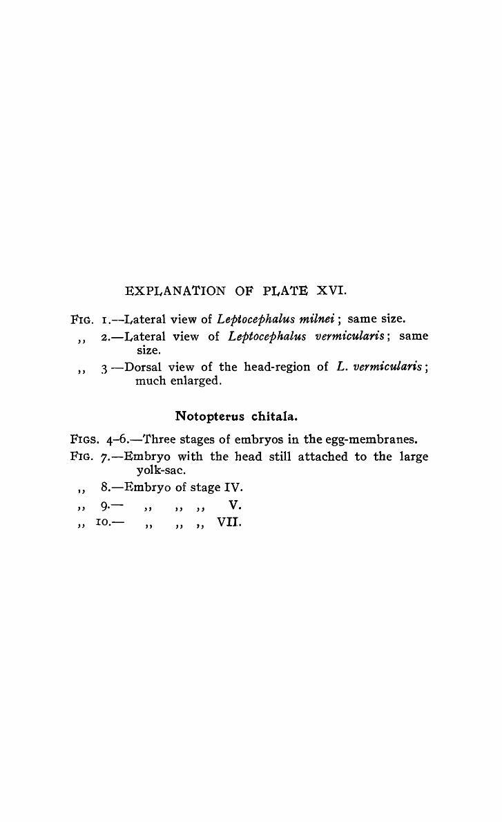
FIG.
"
"
EXPLANATION OF PLATE XVI.
I.--I,ateral view of Leptocephalus milnei; same size. 2.-Lateral view of Leptocephalus vermicularis; same
S1ze. 3 -Dorsal view of the head-region of L. vermicularis;
much enlarged.
N otopterus chitala.
FIGS. 4-6.-~rhree stages of embryos in the egg-membranes. FIG. 7.-Emhryo with the head still attached to the large
yolk-sac. " B.-Embryo of stage IV.
" 9.- " "" v. " IO.- " " " VII.

Rec. Ind. Mus. ,Vol. XVI, 1919.
Plate){VI.
1.
4.
B .P. 8( D.Bagchi del.
A.. Chowdhal':V hth
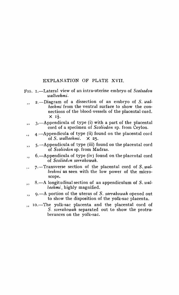
EXPLANATION OF PLATE XVII.
FIG. r.-I/ateral view of an intra-uterine embryo of Scoltodon walbeehmi.
, ,
, ,
, ,
, ,
, ,
2.-Diagram of a dissection of an elnbryo of S. walbeehmi from the ventral surface to show the connections of the blood-vessels of the placental cord. X rt.
3.-Appendicula of type (i) with a part of the placental cord of a specimen of Scoliodon sp, fro111 Ceylon.
4.-Appendicllla of type (ii) found on the placental cord of S. walbeehtni. X 25.
5.-Appendicula of type (iii) found on the placental cord of Scol£odon sp. from Madras.
6.-Appendicula of type (iv) found on the placental cord of Scoliodvn sorrakowah.
" 7.-Transverse section of the placental cord of S. wal-beehnti as seen with the low power of the microscope.
" 8.-A longitudinal section of an appendiculum of S. walbeehmi, highly magnified.
» 9.-A portion of the uterus of S. sorrakowah opened out to show the disposition of the yolk-sac placenta.
" ro.-The yolk-sac placenta and the placental cord of S. sorrakowah separated out to show the protruberances on the yolk-sac.

Rec. Ind. Mus., Vo1.XVf, 1919, Plate: XVII. 1

EXPLANATION OF PLATE XVIII.
Rhinobatis columnae.
FIG. I.-Dorsal view of a very young intra-uterine embryo; the yolk-sac and the yolk-stalk are not shown.
" , ,
"
, ,
, ,
X I! 2.-Dorsal view of an embryo of a much later stage 3.-Ventral view of the above; the tail is not shown. 4.-Diagram of a specimen dissected from the ventral
side to sho\v the relations of the yolk-stalk, etc. X rt.
5.-Transverse section of the yolk-stalk as seen with the low poV\'er of the microscope.
6.-A portion of the above highly magnified to show the constitution of the wall.
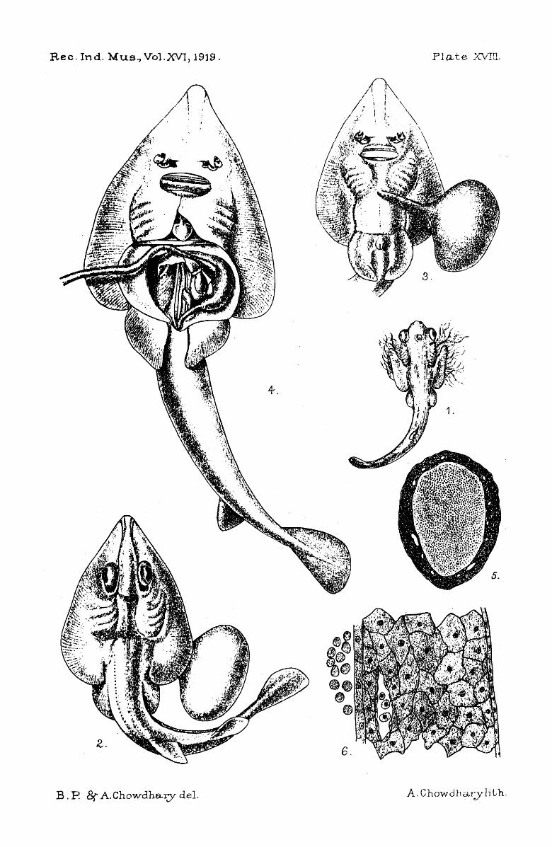
Rec. Ind. Mus., Vo1.XVI, 1919. PIa te )':VIIl.
B. P 8r A.Chowdhary del.

FIG.
"
"
, , "
"
"
EXPLANATION OF PLATE XIX.
l.--Ventral view of an embryo of T kuhlii; only a few of the gill-filaments are shown.
2.-Dorsal view of an embryo of Hyf>olophus sephen; the yolk-sac, yolk-stalk and the branchial filaments are not shown.
3.-Ventral view of a specimen of Pteroplatea poecilura. X t.
3{a).-Lateral view of the head of p. poecilura. 4.-Ventral view of a specimen of Aetomylaeus nichofii.
X i. 4{a).-Lateral view of the head of A. n'icho/ii to show
the characteristic shape of the spiracle. 5.-An egg-case of Chiloscyllium griseum.
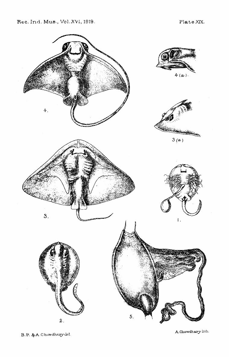
Rcc. Ind. Mus., Vol. XVI, 1919. Plate XIX.
4 (a,,).
2.
B.P. ~A.Chowdh&ty<kl. A.Chowdha.ry lith.

XV SOME GASTROPOD J\iOLLUSCS FROlVI THE GANGE'fIC DELTA
By N. ANNANDALE, D.Sc., F.A .5. B., Director ~ Zoological Survey 0/ India, and B. PRASHAD, D.Se., Superintendent of Fisheries,
Bengal, Bihar and Orissa.
(With Plate XX).
The molluscs des('ribed or discussed in this paper Ii ve ,in the waterways of the Gangetic Delta or lead an amphibious existence on their shores. A large proportion of the species that do so in the delta have already been described by Benson, Stoliczka, Blanford or Nevill, or by more than one of these authors, to all of whom we owe invaluable information about the brackish-water fauna of the Indian estuaries. It is much to be regretted that Nevill did not live to conlplete his Hand List of the Molluscs in the Indian Museum, in which work almost alone exact data as to localities are to be found. We hope later to discuss the invertebrate fauna of the lower part of the delta as a whole; for the present we will consider only those Gastropods about which we have something new to say from a strictly taxonomic point of view.
We have figured the radular teeth of most of the species discussed, but it has seemed to us unnecessary to give elaborate descriptions of these teeth. Our figures, if they are as correct as we believe them to be, should prove much clearer than any written description. We 'have to thank Bahu D. ·N. Bagchi for the accuracy with which he has drawn the figures under our supervision. The figures of the shells have been prepared with equal care by Babu A. C. Chowdhury.
We have also to thank Mr. T. Southwell, Director of Fisheries·, Bengal, Bihar and Orissa, for the facilities that he has ~iven us in collecting specimens. The majority of those on which we base the following notes were collected by Mr. S. W. Kemp and ourselves on a recent trip from Calcutta to Khulna on board the Bengal Fishery launch " Kitty."
Family NERITIDAE.
Genus Dostia t Gray.
n~-t(). Dostia, Gray, SYIl. Contents Brit. JIlts. , 1879. lVeritaeae Mitrulae, von l\iartcns, Ncretilla in Chemitz's Couch.
Cab., pp. 16, 37. . 1915. Septa rz'a , Preston, F'au n. Brit, ind., Fresh'water J.1/01l., p. 6 (Ill
part) .

Records 01 the Indian Museu'1n. [VOL. XVI,
This genus seems to us to be well characterized by the following features :-
(i) The spire is vestigial, but distinctly lateral and spiral. (ti) The shell has no wing-like lateral projections. (iii) The columellar plate extends as a transverse septum
along the aperture of the shell for at least half its length. (iv) Th~ operculum is semicircular and completely exposed.
Preston (Ope cit.) includes species of Dostia in Septaria, Fer., but this genus differs in the still more reduced spire, which is hardly coiled and does not project at its base beyond the bodywhotl; in its much narrower' columellar plate, and specially in the fact that the operculum, which is nearly square, is concealed in the muscles of the foot.
We are convinced by a study of specimens fronl a numqer of different localities that at least three distinct species can be distinguished in the Gangetic Delta alone, and that none of these are synonymous with the true D. violacea (Gmelin). Two of these species have already been described by Benson, but we can find no record of any form with which the third can be identified.
Dostia cornucopia (Benson).
(Plate XX, fig. 2a).
1836. lVeretilla CvYllltCOp£a, Benson, JOUi'I'l. Asiat. Soc. Bengal, Y, p. 7..J.8. 1867. Neretina (Dostia) COY';zucopia, Blanford, Ibid., XXXVI. pt. 2,
p. 60, pl. xii, figs. 23 .... 25.
In this species the shell is relatively large, porcellaneous, thin and high. The columellar plate extends very liftle more than half way across the trne mouth of the shell J i.e. the part occupied by the operculum when the soft parts are retracted. The periostractim is pale olivaceous, sometimes with well-defined transverse zig-zag black lines. The collumellar plate is blackish and there is a blackish margin to the true mouth of the shell.
The differences between shells from the Irrawady Delta and those from that of the Ganges noted by Blanford are not constant.
The operculunl is relatively short, its breadth being two-thirds of its length. The lateral proj ection is stout and blunt, but of considerable length. Its distinctive features in other respects can be seen from our figures.
We have examined specimens of this species from the upper part of the Gangetic Delta, from that of the Irrawady, from the Patani river on the east coast of the Malay Peninsula and from Cochin-China.
Blanford says that in the Irrawady Delta D. cornucopia is usually found on tree-trunks covered- at high-tide by brackish water. In the delta of the Ganges, however, it seems to occur chiefly, if not exclusively, at the edge of creeks of fresh or almost fresh water near the upper limits of tidal influence. The species is, however, scarce in Bengal.

I9I 9.] N. ANNANDALE & B. PRASHAD: Gangetic Delta Molluscs. 243
Dostia depressa (Benson).
(Plate XX, fig. 2b).
1836. Benson, op. cit., p. 748. 1868. Blanford, op. cit.~ p. 60. 1915. Septaria cl'epid1l1al~ia and S. depressa, Preston, Rec.llld. Mus., XI,
P·480.
'fhe shell of this species differs froln that of D. cornucopia in being as a rule narrower in proportion to its length, in having the true mouth still smaller, in the smoother edge of the columellar septum and in colour. The shell substance is rather thicker and has a yellowish tinge. The columellar plate and the outer lip are tinged more or less deeply with orange or coral-red, but this tint is somewhat evanescent. The peri03tracuUl is brownish, s'Jmetimes
~. . .
t·-----····· : I :
a.
Ej. . , l.· '-'.
-....
b TEXT-FIG. I .-Radular teeth of Gangetic species of Dosfta.
(a) D~ depressa, Benson. (b) D. platyconcha. sp. nov.
with transverse greenish lines, an:l o~caC)ional1y marbled with green. \Vhen the periostracum is worn away, the surface of the shell is whitish with transverse zig-zag pink or purple lines and stripes. The operculum differs from that of D. cornucopia in b~ing "relatively broader, in having the lateral process longer and more pointed, and in other characters shown in our figure.
We have seen this species from the deltas of the Ganges and the Irrawady, where it is usually found in slightly brackish water. It is common on the banks of the HugH about twenty miles below Calcutta. Captain R. B" SeYlnour Sewell J I.M.S.) has recently sent us specimens from swampy pools at Calicut on the

244 Records of the Indian l'J useum. [VOL. XVI,
l\lalabar coast and from a small stream twenty-eight miles east of , that place.
We figure (fig. Ia) the radula of a specimen from the HugH. Both the lateral and marginal teeth differ considerably from those of the next species; the marginals being considerably smaller and having the denticulation stronger. The differences between the laterals are of a very complicated nature, and will be understood best by a reference to the figures.
Dostia pIatyconcha, sp. nov.
(Plate XX, figs. I, 2C).
This species differs from both the other Gangetic forms and from all others of the genus known to us in its much thinner and more transluscent shell, its depressed form and non-protruding spire. Its lateral profile is J;egular and forms an arch, less than a semicircle; the spire hardly protrudes beyond the anterior margin of the body-whorl. The spire though very mi tlute is, however, distinctly lateral and spiral. In ventral view the shell is broadly oval, slightly truncated posteriorly. The upper part of the shell is invisible or practically invisible in this view. The columellar plate is separated from the lower margin of the shell, and extends considerably more tban half V\7ay across its true mouth. The margin is irregularly and minutely crenulated. The posterior lip is very broad below the anterior margin of the true mouth of the shell. The dorsal surface is nlarked with well defined and regular transverse striae; Ininute longitudinal- striae can also be detected with a strong lens. The periostracum is dull olivaceotls green with minute black specks and with obscure longitudinal rays. The interior of the shell including the columellar plate has a faint glaucous tinge and is very highly polished.
The operculum is rather short and instead of bearing a single prominent lateral process has two short rather broad tubercles one of \vhich may be obscure.
We figure the radular teeth (fig. rb). Type specimen: M !'!'!±Q in the collection of the Zoological
Survey of India. Locality.-The species is not uncommon in the lower parts
of the Ga~getic Delta, on bushes at the edges of creeks containing brackish water of considerable salinity.
Family LITTORINIDAE.
Genus Littorina, Fernssac.
A number of species of this genus lead an amphibious or almo$t terrestrial existence on the banks of the waterways in the lower part of the Gangetic Delta. At present we will deal with only. three. of these, namely L. melanostoma, Gray, L. s'ubintermed~a, Nevt11 and L. delicatula, Nevill.

19I9.] N. ANNANDALE & B. PRASHAD: Gangetic Delta Molluscs. 245
Littorina melanostoma, Gray.
1882. Litorina melallostoma, Weinkauff, 1/ Die Gatt. Litorina" in Chemitz's Conch. Cab., p .. p.
188,. Littorz'na melanostoma, von 1\iartens, Journ. Lz'nn. Soc. Zool., XXI, p. 170.
This is by far the commonest of the Gangetic Littorinidae. It occurs' in large numbers on grass and bushes exposed at low tide, but submerged when the tide rises. The geographical range extends to the Malay Archipelago,
We figure the radular teeth (fig. 2a) ; their most remarkable feature is the assymetrical form of the inner lateral tooth ..
Littorina subintermedia, Nevill.
(Plate XX, fig. 3).
188..... Littorina conica var. subintermedia, Nevill, Hand-list jJ;lo11. Ind. A/us., II, p. 149.
This form appears to be less like L. conica and the true L. inter1nedia than Nevill thought J and we think it best to regard
h.
C.'
TEXT-FIG. 2.-Radular teeth of Gangetic species of L£ttorina. (a) L. melanostoma, Gray. (b) L. subintermedia, Nevill.
(c) L. delicatula, Nevill. .
it as distinct. The shell is much thinner than that of either of Philippi's species~ and has a slightly transluscent appearance. It is also narrower and more conical, and the sculpture is more delicate. The columella is narrow, but has a very distinct spindleshaped depression upon its surface. The body-whorl is hardly angttlate. The colour of all the shells we have seen is pale-yeltow-
Records 0/ the Indian 1\,[ .,/,seUlI'l. [VOL. XVI,
ish profusely spotted with dull purplish-brown. The spots are transversely elongate in the lower parts of the whorls, and often alternate with shorter spots of a whitish colour. Towards the upper part of the three lowest whorls the dark marks tend to take the form of longitudinal streaks nearly as broad as the pale in terspaces.
The most striking feature of the ranular teeth (fig. 2b) is that the central cusp of the central tooth is very large, with a distinct lateral cusp on either side; the thicker ~ore prominent part of the former extends right across the cusp and occupies more than one half of its area. Most of the denticulations of all the teeth are transversely striate.
The species is only known fronl the lower parts of the Gangetic Delta.
Littorina delicatuIa, Nevill.
(Plate XX, fig. 4).
1884. Littorilla conica var. delicatula, Nevill, Opt cit., p. 150 .
The shell of this fortn is thinner than that of any other species of the genus we have examined, and is always very brightly coloured when fresh. The 'colours, however, as Nevill has noted, are somewhat evanescent. The columella is shorter than in L. subintermedia and the longitudinal depression upon it is much less well defined. The body-,vhorl is distinctly angulate in adult shells owing to one of the ridges being much more strongly developed than the others, but as this ridge is situated not very far from the anterior margin of the whorl, it is not found in young shells.
We figure the radular teeth (fig. 2C). The central tooth ·is smaller than in the other two species we have discussed; its central cusp sOlnewhat resembles that of the central tooth of L. melanosto1na: but is much smaller, and has the membranous marginal portion less well developed. The inner lateral is much more symmetrical.
This species, like the last, is apparently endemic ill the lower parts of the Gangetic Delta. The two are often found together on trees and bushes far above high-tide mark. 'fhey seem to be very largely terrestrial in habits.
Family HYDROBIIDAE.
Genus Stenothyra t Benson.
This is one of the m.ost characteristic genera of the upper estuarine waters of South-eastern Asia. I t is often found in fresh water, but rarely in places permanently unaffected by tidal influence. Both the species here described are from water that was quite fresh at the time of their capture, but is slightly brackish at other seasons.

1919.] N. ANNANDALE & B. PRASHAD: Gangetic Delta Molluscs. 247
Stenothyra echinata t sp. nov.
(Plate XX, fig. 5).
The shell is small, solid, transluscent and pale in colour. It has a very regular conoidal· form except that the ventral surface of the body-whorl is flattened and the anterior part narrowed. The apex is pointed but not acutely. There are si whorls. 'fhe suture is not deeply impressed, but is oblique and irreg~lar; the whorls of the spire increase gradually and evenly. The bodywhorl is broad; in dorsal view it appears to be transverse and almost quadrate, but as seen from pelow it is truncato-ovoid, the truncation being posterior. The mouth of the shell is very small and oblique; it has a regularly oval form. Its rim projects little and the shell is not umbilicate. The whorls of the spire are apparently smooth, but are covered with a deposit in the specimen exatnined. The penultimate whorl and the one behind it bear a single spiral ro\v of sharp, flattened, horny spines, which are directed towards the apex and slightly inwards; they have a golden yellow colour. The body-whorl is sculptured with the spiral punctured lines common to most species of the genus.
The operCUlum is thin and horny, of the normal paucispiral type.
M easure1nents 0/ type-shell (in ntiUimetres).
Length Breadth of body-whorl Length of spire (dorsal view) Breadth of spire (dorsal view) Length of aperture Breadth of aperture
4'0 2'5 1"6 2 1'2 IO
Type-specinten: M 1 t~!I.!! in the collection of the Zoological Survey of India.
Locality.-A single specimen was found among semi-aquatic vegetation at tpe edge of the river Pussur at Khulna, July 22nd, 1918.
The most characteristic feature of the. shell is the row of spines on the basal part of the spire. ()therwise it is very like that of S. deltae (Benson), specimens of which were found with it, but it is Dluch smaller, narrower and less inflated in the bodywhorl and the mouth is a little larger and relatively narrower.
Stenothyra solutat sp. nov.
(Plate XX .• fig. 6).
The shell is of moderate size, thick, globose, with the spire Battened, concave at the apex (fig. 3); the \vhorls are very distinct

Records of the 11l.dian Museum. [VOL. XVI,
and swollen and the body-whorl relatively short and stout, not at all flattened on the ventral surface. The suture is barely impressed.
There are three and a half whorls. The aperture is large, oval, rounded posteriorly very prominent, and with the peristome thick and somewhat plicated concentrically There is 110 umbilicus. The periostracum is olive-green, opaque; the surface is smooth, but not highly polished; minute longitudinal striae and still
TEXT-FIG. 3·-Stenothyra solida, sp. nov., more minute transverse view of the shell-whorls as seen frOln
b striae are present, but punc-a ave. tured lines are entirely absent.
M easurenzents 01 type-shell (in 1nillimetres).
Length - 4· I 2·8 Breadth of body-whorl
Length of spire (dorsal yiew) B~eadth of spire (dorsal view) Length of aperture Breadth of aperture
r·s 2'4 2·0 I-,6
Type-specinten: IVI ll~±~_ In the collection of the Zoological Survey of India. ..
Locality.-A single specimen was found in'flood refuse at the edge of a creek in the Gangetic Delta at Basanti, July 20th, I918.
The species is distinguished by the looseness of its spire, its swollen whorls, the ·:Battened apex, and the absence of punctured spiral lines; the peristome is well developed. Although the shell has a somewhat abnormal 'appearance, we know of no species to which it could be assigned as an aberration.
Genus Bithinella t Moquin-Tandoll.
So far as we have been .able to discover, ,only one species of this genus has as yet been found in India. This ,species is con;. fined to brackish water of considerable but variable _s~linity.
1880.
1884-.
Bithinella miliacea (Nevill).
H)ldrobia (Belgrandia) miliacea, N evil1, rOlll'll. As. Soc. Bengal, XLIX, p. 161, pI. viii, fig. 7,
Hydrobia (Bythillella) nziliacea, Nevill, JJand-Ust Moll. Ind. Mus., II, p. 52. ,
Bithinella cannil1J;e1lsis, Preston, A nn. Mag. Nat. Hist. (7) XIX, p. 2 I 6 (fi g. in te x t ) .

1919.J N. ANNANDALE & B. PRASHAD: Gangetic Delta Molluscs. 249
We have compared Nevill's types with Preston's, and can find no difference. The species appears to be very variable and the varieties described, or rather named; by Nevill in his Randlist are pos~ibly mere individual variations. The var. 1ninor.! however, tnay be worthy of varietal nomenclature.
The species is very common alnong weeds in pools of brackish water at Port Canning.
Family ASSIMINEIDAE.
Genus Assiminea t · Leach.
IR87. Assimt'nea, Bottger, Jahrb. deutsch. ilfalalakozool. Ges., XIV p. IS8.
I880? Assiminea, Heude, "Moll. Terr. ~/em. HI·st. Nat. Chin.," I, p. 82. 1897. Assiminea, vun l\1artens in \Veber's Zool. El'gebell. IVeidel'l. Ost.
Ind., IV, p. 212.
Bottger in his monograph of the genus arrangt'S the species alphabetically; among the Chinese forms Reude recognizes three
c. TEXT-FIG. 4.-Radular teeth of Gangetic species of Assimillea.
((l) A. bl'evicula (Pfr.). (b) A. beddomiana , Nevill. (c) A. trancest'ae (Gray).
divisions, which he calls Euassimineae, Pseudomphalae and Salenolnphalae; while von l\iartens. discussing the species of the Malay Archipelago, divides the genus into two groups, Cyclotrophis and A ssi11tinea! s.s., which is distinguished from Cyclotrophis by the presence of a sharp rim at the inner edge of the umbilicus of the. shell. We have here to consider three species, two of which [A. brevicula (Pfr.) and A. beddomiana. Nevill] belong t<? A ssim£nea, s.s., while the third [A. Irancesiae (Gray) j perhaps represents Cyclotrophis but may ultimately call for generic distinction.
250 Records 0/ the Indian M use'llnt.
Assiminea brevicula (Pfr.).
1~87' Assimt.'nea brevicula, Bottger, Ope cit., p. 163. 1897. Assiminea brevicula, von Martens, Opt cit., p. 21 3.
[VOL. XVI,
Further references will be found in the papers cited. We have nothing fresh to say about the shell or the living
animal. We figure the radular teeth (fig. 4a), they are not unlike those of A. violacea as figured by Hel1de, 1 but the upper part of the central tooth is still more constricted, its cusp is larger, and it has four basal dentictilations on each side; the dentlculations of the later~l teeth are also better developed.
The species is very comnlon on mud between the tide-marks i 11 the lower part of the Gangetic Delta. Its distribution extends from the estuaries of the Ganges to those of Central China.
Assiminea beddomiana, Nevill.
1881. AsshJtinea beddontiana, Nevill, JOUI'Il. As. Soc. Bengal, L (ii), p. 158, pI. vii, fig. ~.
The animal is very like that of .A. brevicula, but the radula differs considerably (fig. 4b), in that the upper part of the central tooth is not constricted, the denticulations at its base are feebly developed and its cusp is smaller. The denticulations of the lateral teeth are also less well developed, and those of the Inarginal, which is more symmetrical, are much smaller and more numerous.
This species occurs with A. breviculll" but is much less abun-dant. It is only known fronl the estuaries of the Ganges and the Irrawady and from intermediate localities.
Assiminea (? Cyclotrophis) francesiae (Gray).
(Plate XX, fig. 7).
1887. Assimiuea francesl, Bott~er, Ope cit., p. 176.
The shell of A. Irancesiae is,_ as Bottger points out, e~tremely variable. We do not think that the different forms named bv Nevill as varieties or sub-varieties should be regarded as distinct . . A.t som,e localities, however, notably at the edge of the river Pussur at Khnlna, many adult shells have a number of ridges or varices running across the lower part of the body-whorl and probably representing periods of aestivation.
The radula (fig. 4c) dtffers considerably from that of the two species we have just discussed, and also from that of A. scalaris, Heude, which on shell characters alone comes in the same group.The chief differences are: (a) there is a minute but elongate bifid
,------,._------tOp. cit., XXI, fig. 4C. 2 See Heude, Opt c£t;, XXI, fig. 5. We have examined a radula of this
~pccies and find that while there are small variations in the denticulation of the teeth, the general outline as shown in Heude's figure is correct.

I919.] N. ANNANDALE & B. PRASHAD: Gangetio Delta Molluscs. 25 I
tooth interpolated between the t\VO laterals on either side; the dental formula thus being 1.3 1.3.1. instead of 1.2.1.2.1. (b) The central tooth is quadrangular and broadly transve~se; it has three stout subequal cusps and a pair of extremely large denticulations arranged side by side at each side of its base. (c) The denticulation of both the lateral teeth is very coarse but short. (d) That of the luarginal tooth is similar to and intermediate between that of A. brevt'cula and A. beddomian.a; there is besides on the outer margin of the tooth a peculiar pointed, but' apparently membranous process.
It is possible that this species may have u1timately to be recognized as a distinct genus, but we have no information as to the rad ular characters of the typical species of CyclotrophisC. papuens£s (Albts.), which was found on the south coast of New Guinea~ As we have shown above, those of the Chinese A. violacea, which would be assigned to this group on shell characters alone, are quite .different from those of A. francesiae.
Falnily MELANIIDAE.
Genus Melania, Lam.
Sub-genus Main waringia, Nevill.
1886. 11Jaill7.Uarillgia, ~ evill, Ifand-List Moll. Iud. Allts., 11, p. 286 .
. A.1though Nevill refers to the only known species as a " new generIc type," he places it as a sub-genus of 114. etania, a view with
TEXT-FIG. s.-Radular teeth of Afain7.varillgia paludom£dea, Nevill. (a) Teeth viewed from above. (b) I~ateral view of marginal tooth.
which we agree. His statel11ent that it is intermediate between that genus and Paludomus seems to be based on the form of the operculunl, and perhaps on the consistency of the shell. Both Blanford, I ho\vever, and the Sarasins It have shown froin different points of view that the former can hardly be regarded as of great generic importance in the Melaniidae. The form of the shell differs litt1e from that of Melania, and we are of the opinion
1 Trans. L£IlIt. Soc., XXIV, pp. 166-167 (1864). . 2 Die SIISs7uIlsser Alo11uskell 'Z'01l Celebes, pp. 5-9 (1898).

Records 01 the Indian Museum. [VOL. XVI,
that the only known species is a depauperated form modified in accordance with an amphibious life partly spent in water of considerable salinity.
The sub-genus may be redescribed as follows, using Nevill's terms to a large extent :-Melaniidae with imperforate, conically produced shell; its spire turretedly acuminate; shell-substance thick and almost porcellaneous, but becoming tnuch thinner in the lower part of the body-whorl; columella strongly arched, not greatly incrassate, outer lip sharp; external surface decorated with deep-cut spiral striae; a thin epidermis present, bearing minute scattered hairs or chaetae. Operculum horny., extremely thin, paucispiral, with the nucleus eccentric.
Animal differing fronl that of the groups Plotia, Striatella, M elanoides, and Tarebia in the following characters :-Foot pointed and produced into a short filamentous process behind; a distinct mid-dorsal groove on posterior part of the foot extending on to the terminal process. Radula (fig. sa) like that of M elan£a, but without a distinct cusp to the median tooth and ,",·ith marginal teeth of peculiar form (see figure Sb).
Type: Ma?~nwaringia paludomidea, Nevill. Distrtbut~on.-Only known from the lower parts of the Gan
getic Delta.
Melania (Mainwaringia) paludomidea t Nevill.
(Plate XX~ fig. 8)'.
Nevill's description of the shell, which is quoted by Preston on p. 37 of his volume in the "Fauna," needs no elaboration. The animal has the characters noted above. Its foot is rather small; the tentacles are very long, slender and tapering, with the eyes situated on distinct prominences at their base externally. The snout is prominent, blunt and shghtly notched in front. The foot and tentacles are transluscent white, suffused with black pigment above; the snout is brownish. The branchial chamber resembles that of Melania, but the branchial folds are exceptionally deep. The osphradium is well developed and ridge-like.
The species is not uncommon between tide-marks in/ the lower parts of the Gangetic Delta. At low-tide it adheres tightly by means of a gummy secretion to the trunks of trees, and particularly to the upright aerating roots of mangroves. When placed in water it emits Q. bubble of air and immediately becomes active. It ,,~ould seem, therefore, to breathe air while in a comatose condition at low-tide, but never to be active while doing so.
Family NASSIDAE.
Genus N assat Lamarck.
'rhe snlall species of this genus that occur in brackish water on the coa~t of India, aU of which have recently been described by Preston, seem to form a very distinct little group, in which

I9I9.] N. ANNANDALE & B. PRASHAD: Gangetic Delta iVf oUuscs. 253
the radula is somewhat simplified, while the she1l is remarkable for its small size and elongate form. These species are-N~ denegabilis, N. orissaensis and N. ennurensis, to all of which we refer in the following note.
N assa ennurensis t Preston.
1916. Nassa orissaens£s yare ennurensz's, Preston f Rec. Ind. lllus., XII, p. 28, figs. 2, 2a.
'fhis form appears to us to be specifically distinct. In addition to the differences between it and the typical IN. orissaensis noted by Preston, we may draw attention to a very distinct feature, well shown in his figures, in the shape of the mouth of the shell, near the posterior extremity of the inner border of which there is always a blunt tooth in fully adult shells of N. ennurensis, while no such projection ever occurs in N. orissaensis. The typical form of the former, which reaches a length of over 10 mm., has
f'l.
h.
c.
TEXT-"FIG. 6.-Radular teeth of brackish water species of Nassa.
(a) N. orissaensis, ,Preston. (b) N. denegabilis, Preston. (c) N. ennurenJis var. depauperata, nov.
been recorded from the outskirts of Calcutta by Preston. A smaller form with a rather broader aperture, and a much thinner shell is common in the lower part of the Gangetic Delta. For this form we propose the name ;-
var. depauperata, nov.
(Plate XX, fig. 9).
We have already stated the characters by which the shell of this form can be distinguished. The animal is very like that of N. orissaensis,i but differs in the following characters: (a) The foot is proportionately narrower and a little mpre deeply notched posteriorly, the two posterior lobes being distinctly pointed. (b) The anterior processes of the foot and also the tentacles are
I See Annandale and Kemp. 1ft/em. Illd. Mus., V, p. 3-+3, fig. I.

254 Records 0/ the I ndia1t M·u,se1t111 .. [VOL. XVI,
rather longer and more slender. (c) The siphon is shorter than the shell.
The whole animal is transluscellt white, there is a tinge of pale grey on the siphon and the dorsal surface of the foot. The eyes are small but quite distinct.
The radula (fig, 6c) is very similar to that of N. denegabilis (fig. 6b) and.N oyissaensis (fig. sa); aU of which ~ve figure. The lateral teeth have two distinct branches, which are quite smooth.
N. orissaensis var. depauperata lives in creeks of brackish \\'ater (sp. gravity corrected to 15°C. l'OII7S-1·0172S) on a nluddy bottom at a depth of 4-6 fathoms. It crawls with extreme ra'pidity both in water and on damp surfaces, and can float adhering shell downwards to the surface film by means of the foot.
Type-specimen: M l.l~±!i in 'the collection of the Zoological Survey of India.
Locality.-Kaikal Maree near Port Canning, Gangetic Delta; 17th July, 1918.
Family RINGICULIDAE.
Genus R·ingicula, Deshayes.
IH4-l-. RUlgicula, Hinds, Proc. Zool. Soc. London, XII, p. 97. 1875. Ringicula apicata, Nevill, JOU1'1l. As. Soc. Bengal, XLIV (ii),
p. 101. 1878. Rillgtcula, Morelet, JOUI'Il. de Conch., (3) XVIII (xxvi), pp.
113-133, pIs. v-viU. 1878. Rt'ngicula (Anatomie de L'animaI), Fischer, Ibid., pp. 1 I.~-I IS. 18~+. Rillgicula, Watson, JOU1'1l. Linn. Soc. London, XVII, _po 291.
Ringicula caeca, Spa nov.
(Plate XX, fig. 10).
The shell is small, very solid and of the usual shining white colour. The spire is narrow, conical and sharply pointed, about half as long as the body-whorl, which is relatively broad and nearly square. The main axis of th~ shell occupies a line much nearer the inner than the outer margin of this whorl, which projects inwards at an angle frotn the base of the spire. The suture is impressed and the whorls somewhat flattened outside it. The Inouth of the shell is contracted but relatively long, extending backwards at an acute angle with the main axis to a point at about four-fifths the length of the body-whorl. The lips are very thick. The outer lip runs nearly straight for the greater part of its length, and turns inwards somewhat abruptly, but at an angle less than a right angle, in front; at the point at \vhich it does so it bears a broad oval prominence. The outer callus extends about half way tip the outer surface of the shell, and is strongly corrugated. The siphonal notch is well developed, but not very prominent. The columella is oblique and has three very prominent folds' the posterior fold is very broad, the median fold ridge-like and
I9I9-J N .. A.NNANDALE & B. PRASHAD: Gangetic Delta fltloltuscs, 255
the anterior fold intermediate, The columellar callus is well developed, The surface of the shell is polished, shining and somewhat opalescent, but is partly covered by a thin blackish-brown deposit, There are two fine but clear-cut spiral ridges 011" the whorls of the spire, extending on to the upper part of the bodywhorl, the central region of which is quite smooth, At the base of the body-whorl there are five other still finer spiral ridges, which are more closely approximated behind than in front.
Measurements of type-s1tell (in millintetres) ,
Length Breadth of body-whorl Breadth of spire at base (dorsal view) Length of spire (dorsal view) Length of aperture Breadth of aperture •
3'25 2'0 1"0
1'0
2'2 o·S
The living animal is devoid of external pigment and is of much the same shade as the shell. There are no eyes. The foot is relatively small, broadly rounded behind, expanding in front into a flattened, bluntly pointed almost triangular process on each side. The cephalic disc is large and extends over the anterior part and the lateral margins of the shell; in front it is notched in the middle line, and it is expanded at each side into a broad flattened process like that of the foot, but larger; the posterior margin is nearly straight, but folded in the middle Hne in such a way as to form an incomplete siphon) ""hich, however, hardly projects at all.
Type-speci1nen: M ll~;t~_ in the collection of the Zoological Survey of India.
Local,,;ty.-Creek at Kaikal Maree, near Port Canning, Gangetic Delta, from a muddy bottom in 4-6 fathonls: specific gravity of water (reduced to ISOC) 1.01725.
The shell resembles that of R. apicata, Nevill, in some respects, but is larger and has the body-whorl much broader, the mouth more contracted and the lateral callus much luore strongly developed.
The animal differs from that of R. auriculata as figured by Fischer (Man. Conch.) p. 36, fig. 322, 1887) in being devoid of eyes, in the different shape of its foot and cephalic disc, and in its much less well-developed cephalic siphon.
Family A URICULIDAE.
GeBus Auricula t Lam.
At least three species of this genus, to which we give a somewhat broad application, occur commonly in the lower parts of the Gangetic Delta, namely-A. auris-judae, Lam., A. gangetica, Benson, and the form we describe below. Benson, Stoliczka and

Records 0/ the Ind£an Museum. [VOL. XVI,
Nevill seem to have regarded this last as a dwarfed fornt of A. gangetica, but it appears to us to be perfectly distinct.
Auricula translucenst sp. nov.
(Plate XX, fig, II), The shell is small, exceptionally thin, colourless, transluscent
and (without the periostracum) semi-hyaline; the periostracum is thin and of a pale greenish olivaceous tint, The shape is elongate-ovate; the apex is bluntly pointed; there are si or 6 whorls, but the apical whorl is usually eroded, The spire is short, less than a third as long as the body-whorl in dorsal view. Though the whorls increase gradually and evenly, the suture is narrowly impressed; it is not markedly oblique. The body-whorl is long and narrow, distinctly spindle-shaped and almost pointed anteriorly; its inner profile is regularly arched, but its outer proHIe, proc~eding from behind forwards, first slopes outwards and backwards for a short distance, then runs almost straight backwards for the greater part of its length, and finally becomes cqncave and slope:; inwards to the .anterior extre111ity. The mouth of the shell is long and narrow, extending backwards for nearly sixsevenths of the length of the body-whorl, curving inwards posteriorly and tapering to a point; anteriorly it is very narrowly rounded, The margin of the outer lip is thin, but there is a slight and perfectly smooth· ridge running parallel to it just inside the mouth, and the anterior margin is slightly thickened, Only two columellar folds are visible; they are not very prominent and are situated in the anterior third of the aperture. The exterl)al surface of. the shell is marked with fine longitudinal, ~ather widely separated curved striae, and with much tiner and more numerous transverse striae; it is also minutely and lightly pitted. The pits or punctures are much larger and deeper on the posterior part of the body-whorl and on the spire than on the anterior two-thirds of the former. The inner surface is highly polished, iridescent, and microscopically transversely striate.
Measurements. at shells (in mt"lli11tetres),
Length of shell Greatest breadth of body-whorl Breadth of spire at base Length of spire Length of aperture Greatest breadth of aperture
Type.
S'S S'I 3"6 3·4
• 2 3 2" I 1·4 1"1
6'2 6· I 1'6 1"5
'8 3°5 2"3 1"2
6'3 r'S
The animal is white and ~ransluscent, with a brownish tinge 011 the tentacles" The. foot IS rounded, slightly emarginate in ~ront and roun~ed beh!nd. The tentacles are long and slightly Inflated at the tips, which are often invaginated when the animal

:919.] N. ANNANDALE & B, PRASHAD: Gangetic Delta Molluscs. 257
is crawling. The eyes are visible as small black spots at the base of the tentacles.
The upper jaw, which is very conspicuous in the living animal, is fully cornified and lunate in form; there is a broad transverse thickened median region, the anterior and posterior regions being almost membranous. The radula is narrowly tongue-shaped (fig. 7a), and bears about 60 transverse rows of teeth; these rows are practically straight, There are about 27 longitudinal rows; the dental formula being 7'6'1"6'7, 1'he central tooth (fig. 7b) is minute, with a single cusp, which is nearly symmetrical and ob-
a"
() f5iJ ~ ~--.----Q hJJ b.
TEXT-FIG, 7.-Radula of AU1,icula t,'anslucens, sp. nov. (a) Radula as a whole. (b) Radular teeth further enlarged.
scurely trilobed. The laterals are similar but much larger, and the marginals differ from the laterals in having the cusps narrower, simpler and directed SOqlewhat inwards.
1'ype ~Pecimen.' M _u1~u- in the collection of the Zoological Survey of India.
Locality,-A single living specimen of this species was taken in a creek at Basanti in the Gangetic Delta, In the collection of the Zoological Survey there are also a number of specimens from Port Canning which had hitherto been confused with A, gangettca.
The shell resembles that of A socotrana, Smith, in sculpture and texture, and that of A. layardi, H. Adams, in shape.

EXPLANATION OF PLATE XX.
The line drawn next to or between the different views of the shells shows the actual length of the specimen.
FIGS. la, Ib, Ic.-Dostia platyconch,a, sp. nov., shell of the typespecimen.
" 2.-0percula of the Gangetic species of Dostia, X 2. (a) D. cornucopia (Benson).
, ,
" , , , ,
"
"
"
(b) D. depressa (Benson). (c) D. platyconcha, sp. nov.
The opercula are drawn froIn actual specimens. That of D. plafJ1concha is always smaller than those of the other two species j but that of n. corl1Ucopz'a is not always smaller than that of D. dep,'essa.
3.-Littorina subintermedia, Nevill, shell of a specimen from the Gangetic Delta.
4.-Littorina delicat-ula, Nevill, shell of a specimen from the Gangetic Delta.
5.-Stenothyra echi1~ataJ sp. nov., shell of the type-specimen. 6.-Stenothyra soluta, sp. nov., shell of the type-specimen. 7.-Lateral view of an expanded living individual of A ssi-
minea /rancesiae (Gray). 8.-M elania (M ainwaringia) paludomidea , Nevill, shell of a
specimen from the lower reaches of the Gangetic Delta.
9.-N assa ennurensis var. depauperata, nov., shell of the typespecimen.
" Io.-Ringicula caeca, sp. nov., shell of the type-specimen. " I I.-Auricula translucens, sp. nov., shell of the type-specimen.

:a1 '~
9
1 I ~ .....
~
:;'11 ~ 21> d
f <: 0 ~
:x <
l ,c ~
..... ~
""" ~
la Ib
7
I I I
MOLLUSCA OF THE GAN'GETIC DELT.\ .