019 ' # '2& *#0 & 8 - intechcdn.intechopen.com/pdfs-wm/21571.pdf · targeting a...
TRANSCRIPT

3,350+OPEN ACCESS BOOKS
108,000+INTERNATIONAL
AUTHORS AND EDITORS115+ MILLION
DOWNLOADS
BOOKSDELIVERED TO
151 COUNTRIES
AUTHORS AMONG
TOP 1%MOST CITED SCIENTIST
12.2%AUTHORS AND EDITORS
FROM TOP 500 UNIVERSITIES
Selection of our books indexed in theBook Citation Index in Web of Science™
Core Collection (BKCI)
Chapter from the book Etioloy , and Pathoph, s ioloy , of Parkinsongs DiseaseDownloaded from: http://www.intechopen.com/books/etioloy , -and-pathoph, s ioloy , -of-parkinson-s-disease
PUBLISHED BY
Worldgs lary est Science'Technoloy , & Medicine
Open Access book publisher
Interested in publishiny with IntechOpen?Contact us at [email protected]
7
Targeting α-Synuclein-Related Synaptic Pathology: Novel Clues for Parkinson’s
Disease Therapy
Arianna Bellucci1 and PierFranco Spano1,2 1Division of Pharmacology, Department of Biomedical Sciences and Biotechnologies and
National Institute of Neuroscience –Italy, School of Medicine, University of Brescia, Brescia (BS),
2IRCCS San Camillo Hospital, Venice, Italy
1. Introduction
Parkinson’s disease (PD) is the most diffuse movement disorder, affecting approximately 6
milion individuals worldwide and presenting tremor, rigidity and bradykinesia as the main
clinical features. The onset of PD typically occur in patients over the age of 50 years and its
incidence slowly progresses with increasing age. Neuropathologically, it is characterized by
loss of striatal-projecting dopaminergic neurons of the substantia nigra pars compacta, and
by the presence of Lewy bodies (LB) and Lewy neurites (LN) (Forno, 1996). To date, LB and
LN are the characteristic neuropathological alterations of another neurodegenerative
disease: Dementia with Lewy bodies (DLB), a form of late-life dementia which eventually
overlaps with Alzheimer’s disease.
It is noteworthy that, despite the staggering impact of PD on society, the available therapeutic
armamentarium for this disease is still limited. Indeed, the gold-standard treatment for PD
from the 60’- the dopamine precursor L-DOPA - induces severe motor side effects and its
efficacy declines with the progression of the disease. The rest of the pharmacological
treatments for PD mainly include drugs that are usually employed in association with L-
DOPA such as dopamine agonists, which however fail to match L-DOPA's efficacy. Best
results are currently achieved with invasive strategies via subcutaneous or intraduodenal
delivery of apomorphine or L-DOPA, or deep brain stimulation of the subthalamic nucleus.
Nonetheless, usually after 15 years of pharmacological and therapeutical interventions, most
of the patients start to present several motor complications, thus requesting multiple adjuvant
strategies such as physiotherapy, hospitalization or social assistance.
For these reasons, in recent years, much effort has been made in order to outline novel therapeutical approaches to cure PD. In particular, interventions such as stem-cell based approaches have been tested as potential treatments for PD. Although results indicate that patients may gain a long-term clinical benefit from the intrastriatal transplantation of human embryonic mesencephalic tissue (Piccini et al., 1999), part of the graft-derived dopaminergic cells develop LB after 11-16 years (Li et al., 2008;Kordower et al., 2008a;Kordower et al., 2008b),
www.intechopen.com
Etiology and Pathophysiology of Parkinson's Disease
138
probably because ┙-synuclein can be transmitted from cell to cell (Desplats et al., 2009), and what’s more, the availability of human mesencephalic tissue is limited. Thus, it became clear that lentiviral vectors are ideal candidates to design novel gene-based disease modifying strategies for neurodegenerative diseases such as PD. Indeed, surgical infusion is currently required for viral vector delivery into the brain and PD is the only neurodegenerative disorder routinely treated with neurosurgery (Bjorklund et al., 2009;Bjorklund and Kirik, 2009;Kaplitt, 2010). Furthermore, several animal models of PD are available to test new therapies. Actually, promising preclinical gene therapy studies focusing on the correction of dopamine deficiency are currently in progress (Azzouz et al., 2002;Jarraya et al., 2009). However, a limitation with this level of analysis is that it sidesteps the main goal that a current therapeutical approach for PD should afford: to block and/or counteract dopamine neuron derangement. Indeed, a disease modifying gene therapy approach is missing, as the mechanisms underlying dopamine neuron derangement in PD are not yet completely clear. For these reasons, unravelling the molecular mechanisms underlying PD onset is essential to discover new therapeutic targets to cure the disease. In recent years, the idea that ┙-synuclein is a causative agent of PD has spread upon the scientific community, the link being that ┙-synuclein is deposited in the pathological hallmark of PD, the LB and that ┙-synuclein mutations are responsible for the onset of familial form of PD (Cookson and van der, 2008;Cookson, 2005). These considerations leave us with the simplest possible sketch of the pathogenesis of PD: ┙-synuclein is an initiator of damage and the final outputs, after many years, are cell loss and LB formation. However, it has to be taken into account that the majority of PD cases are idiopatic, thus: what are the causes underlying the pathological accumulation of ┙-synuclein? Many epidemiological studies have been conducted to verify whether environmental or genetic factors may predispose to the development of PD (Elbaz and Moisan, 2008). From these latter, it became evident that a correlation exists between pesticide or chemicals exposure and PD onset. Furthermore, besides ┙-synuclein, 12 other gene mutations, some of which with unknown function (Belin and Westerlund, 2008) have been found to be responsible for the development of early onset PD. Some of these genes encode mitochondrial associated proteins or members of the ubiquitin-proteasome system. Remarkably, mitochondrial and proteasomal dysfunctions have been linked to the onset of PD (Tritschler et al., 1994). Thus, the overall conclusions of epidemiological studies indicate that PD is a complex, multifactoriated disorder and that ┙-synuclein aggregation, mitochondrial and proteasomal dysfunctions play central pathogenic roles. Noteworthy, the observation that both mitochondrial and proteasomal inhibitors induce ┙-synuclein accumulation as well as dysfunction and degeneration of nigrostriatal dopaminergic neurons “in vivo” (Xiong et al., 2009;Xie et al., 2010) remind to the hypothesis that ┙-synuclein deposition is a central step event during the pathogenesis of PD. This notion has been reinforced by findings showing that the area that degenerate in PD (substantia nigra, striatum and ventral tegmental area) express low levels of ┙-synuclein in physiological conditions (Wersinger et al., 2004), and that ┙-synuclein levels in the substantia nigra decrease with age (Mak et al., 2009) supporting the hypothesis that these regions may be more vulnerable to a pathological increase of ┙-synuclein levels, especially during the aging process. At present, the neuropathological diagnosis of PD and DLB is based on the detection and quantification of LB (Beach et al., 2009b;Beach et al., 2009a;McKeith et al., 1996;McKeith et al., 2005;McKeith, 2006). Indeed, the spreading of LB pathology correlates with the progression of the disease In PD, LB are mainly found at predilection sites of neuronal loss such as the substantia nigra
www.intechopen.com
Targeting α-Synuclein-Related Synaptic Pathology: Novel Clues for Parkinson’s Disease Therapy
139
(Braak et al., 2003;Jellinger, 2009;Halliday and McCann, 2010), thus indicating that LB may be somehow related to nerve cell loss. Later on, it was shown that the number of LB in patients with mild to moderate loss of neurons in the substantia nigra is higher than in patients with severe neuronal depletion, thus indicating that LB-containing neurons may be the dying neurons (Wakabayashi et al., 2007). Furthermore, it seems that the presence of LB is not always related to nerve cell degeneration as every dying nerve cell does not necessarely form LB. Indeed, LB-containing neurons of the substantia nigra don’t undergo apoptotic cell death to a greater degree than the general population and most neurons that undergo cell death do not contain LB (Schulz-Schaeffer, 2010;Tompkins and Hill, 1997). Finally, substantia nigra neurons, whether they contain LB or not, are similarly affected by morphological dendritic abnormalities or biochemical changes, thus indicating that the neurons in general are involved in the disease process (Bergeron et al., 1996;Hill, 1996;Javoy-Agid et al., 1990;Patt et al., 1991;Kramer et al., 2008;Schulz-Schaeffer, 2010). In addition, recent findings indicate that in DLB 90% or even more ┙-synuclein aggregates are located at presynapses in the form of very small deposits, pointing out that not cell
death but rather ┙-synuclein aggregate-related synaptic dysfunctions may cause the neurodegeneration (Schulz-Schaeffer, 2010;Kramer et al., 2008). It may thus be feasible
that PD may be characterized by dying back mechanisms that begin at the synapse and lead to axonal degeneration in the striatum as a consequence of the pathological
accumulation of ┙-synuclein at the synapse. These events may lately drive dopaminergic cells of the substantia nigra toward neurodegeneration. Therefore, ┙-synuclein is central
to the pathogenesis of PD. This view opens the way to a novel question: is it the ┙-synuclein deposition-associated synaptic pathology which may affect the function and
resilience of dopaminergic neurons? Alpha-synuclein is a 140 amino acids protein, a fragment of which has been identified as a non A-┚ component (NAC) in amyloid preparations of Alzheimer’s disease (Ueda et al., 1993). It was firstly described as a brain specific protein (Nakajo et al., 1993;George et al., 1995) and since the initial findings indicated that the protein localizes at presynaptic site and portions of the nucleus in brain neuronal cells (Jakes et al., 1994;Iwai et al., 1995) it was denominated with the acronym “synuclein”. Recombinant ┙-synuclein does not assume a uniform consistent secondary structure in aqueous solution, thus the protein is said to be natively unfolded (Weinreb et al., 1996). Several observations have shown that ┙-synuclein is implicated in the control of synaptic membrane processes and biogenesis (Jenco et al., 1998;Davidson et al., 1998). From these studies, it was hypothesized that ┙-synuclein could play a key role in the modulation of synaptic activity. Consequently, it has been demonstrated that ┙-synuclein interacts with- and modulates the expression, subcellular distribution and activity of numerous synaptic proteins as well as cytoskeletal components (Engelender et al., 1999;Bonini and Giasson, 2005;Chandra et al., 2005;Sousa et al., 2009;Scott et al., 2010;Garcia-Reitbock et al., 2010;Darios et al., 2010;Burre et al., 2010). Alpha-synuclein also shares numerous biochemical and functional similarities with synaptic proteins such as synapsins. Furthermore, it has been found to be implicated in the control of neurotransmitter release (Abeliovich et al., 2000;Gureviciene et al., 2007;Nemani et al., 2010) and since the protein is specifically upregulated in a discrete population of presynaptic terminals of the songbird during a period of song acquisition (George et al., 1995), it has been hypothesized that it may be implicated in synaptic plasticity. Thus, it emerges that ┙-synuclein has a critical role in the control of synaptic activity in specific neuronal populations, rendering it possible to outline a whole new scenario to single out novel
www.intechopen.com
Etiology and Pathophysiology of Parkinson's Disease
140
therapeutical targets among the ┙-synuclein synaptic partners. Modulation of these proteins may open new ways toward the development of disease modifying strategies to cure PD-related synaptic dysfunctions. By performing a critical review of the PD-related ┙-synuclein proteome, this article will outline the most relevant findings defining the specific modulatory effects exerted by ┙-synuclein in the control of synaptic functions in physiological and pathological conditions. The overall conclusions of these studies will spot novel potential therapeutic targets for the development of pharmacological and gene-based strategies aimed at straightening ┙-synuclein-related synaptic dysfunctions as new clues to cure PD.
2. Alpha-synuclein: Biophysical characteristics
Alpha-synuclein was first described in Torpedo californica (Maroteaux et al., 1988). It
belongs to the synuclein family, which includes ┚- and ┛-synucleins. These proteins have a
common amino-terminal sequence containing a different number of repeat regions while
they differ in the carboxy-terminal part (Tofaris and Spillantini, 2005). Alpha-synuclein from
different organisms possesses an high degree of sequence conservation (Clayton and
George, 1998). Three ┙-synuclein isoforms are produced by alternative splicing (Bayer et al.,
1999) in humans. The best known isoform is 140 amino acids in length and constitutes the
whole and major transcript of the protein. Two other isoforms, of 126 and 112 amino acids in
length, derive from the selective deletion of exon 3 and 5, respectively.
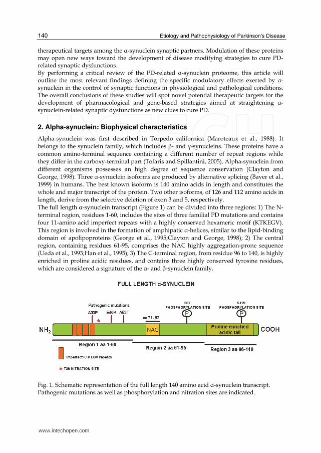
The full length ┙-synuclein transcript (Figure 1) can be divided into three regions: 1) The N-
terminal region, residues 1-60, includes the sites of three familial PD mutations and contains
four 11-amino acid imperfect repeats with a highly conserved hexameric motif (KTKEGV).
This region is involved in the formation of amphipatic ┙-helices, similar to the lipid-binding
domain of apolipoproteins (George et al., 1995;Clayton and George, 1998); 2) The central
region, containing residues 61-95, comprises the NAC highly aggregation-prone sequence
(Ueda et al., 1993;Han et al., 1995); 3) The C-terminal region, from residue 96 to 140, is highly
enriched in proline acidic residues, and contains three highly conserved tyrosine residues,
which are considered a signature of the ┙- and ┚-synuclein family.
Fig. 1. Schematic representation of the full length 140 amino acid ┙-synuclein transcript. Pathogenic mutations as well as phosphorylation and nitration sites are indicated.
www.intechopen.com
Targeting α-Synuclein-Related Synaptic Pathology: Novel Clues for Parkinson’s Disease Therapy
141
The amino acid sequence and subcellular localization of ┙-synuclein indicate that it can interact with lipid membranes. Indeed, the repeat region, mediates reversible binding to acidic phospholipids by making up a conserved apolipoprotein-like class-A2 helix which is associated with a large shift in protein secondary structure from around 3% to about 80% ┙-helical (Davidson et al., 1998). Consistently, it has been observed that ┙-synuclein can inhibit a protein which is localized to plasma membrane and submembraneous vesicles: phospholipase D2, whose activity produces phosphatidic acid, which is implicated in vesicle budding (Jenco et al., 1998). Membrane-bound ┙-synuclein has an high aggregation propensity and seeds the aggregation of the cytosolic form (Lee and Lee, 2002). Lipids can facilitate the incorporation of ┙-synuclein into membranes and influence ┙-synuclein fibril elongation (Gai et al., 2000) suggesting that ┙-synuclein may be strongly implicated in synaptic membrane biogenesis. Furthermore, the key role of ┙-synuclein in membrane-associated processes is supported by findings indicating that ┙-synuclein knock-out mice have enhanced dopamine release at nigrostriatal terminals in response to electrical stimulation, indicating that ┙-synuclein is an activity-dependent negative regulator of dopaminergic transmission (Abeliovich et al., 2000). On this line, it has been shown that depletion of ┙-synuclein results in a decrease in the distal pool of presynaptic vesicles in cultured hippocampal neurons (Murphy et al., 2000). Furthermore, its overexpression reduces neurotransmitter release by inhibiting synaptic vesicle reclustering after endocytosis (Nemani et al., 2010). Cytosolic proteins regulate ┙-synuclein membrane interactions, thus suggesting that cytosolic cofactors may be implicated in disease pathogenesis (Wislet-Gendebien et al., 2006;Wislet-Gendebien et al., 2008). In particular, protein-protein interactions may affect ┙-synuclein ability to bind to synaptic membranes, thus affecting its biochemical properties and modulating its tendency toward aggregation, suggesting that, in the ┙-synuclein synaptic proteome, it could be possible to modulate ┙-synuclein aggregation by selectively regulating the expression of some o its protein partners. The relevance of the formation of protein complexes in the modulation of ┙-synuclein function is supported by several findings which suggest that the protein likely behave like a molecular chaperone. Indeed, some studies have shown that ┙-synuclein shares functional and physical homologies with a family of ubiquitous cytoplasmic chaperones: the 14-3-3 proteins (Ostrerova et al., 1999). Alpha-synuclein can bind 14-3-3 proteins as well as other protein members of its family proteome such as protein kinase C. Thus, it was hypothesized that an increase of ┙-synuclein levels could be harmful for the cells as it could affect signal transduction pathways involved in cell differentiation and survival. On this line, in 2001, Iwata and colleagues showed that ┙-synuclein inhibited mitogen-activated protein (MAP) kinase signaling and accelerated cell death following serum reduction in neuro2a cells. However, results from later studies indicated that overexpression of wild type ┙-synuclein can protect neuronal cells from apoptotic stimuli and delay cell death induced by serum withdrawal (Alves da Costa et al., 2000;Lee et al., 2001b), thus leading to controversial conclusions. Indeed, it has also been reported that ┙-synuclein protects against oxidative stress by inactivating c-jun-N-terminal kinase, which is strongly implicated in stress responses (Hashimoto et al., 2002). However, it is likely that the neuroprotective/toxic effects of ┙-synuclein may be ascribed to a different sensitivity of cells to ┙-synuclein levels. Indeed, it has been shown that at a nanomolar scale TAT-┙-synuclein is neuroprotective against oxidative stress, while at a micromolar scale it is able to aggregate and trigger neurotoxic mechanisms (Albani et al., 2004;Batelli et al., 2008).
www.intechopen.com
Etiology and Pathophysiology of Parkinson's Disease
142
Finally, recent evidence showed that cells from transgenic mice expressing the truncated form of ┙-synuclein are more susceptible to environmental conditions and that overexpression of the wild type from of the protein in neuronal progenitor cells affect their fate of differentiation, thus supporting the notion that full length ┙-synuclein is involved in dopaminergic cell differentiation and survival (Michell et al., 2007;Schneider et al., 2007).
3. Alpha-synuclein pathology in PD and α-synucleinopathies
The term ┙-synucleinopathies is usually employed to define a group of neurodegenerative disorders that show common pathologic proteinaceous accumulation of ┙-synuclein aggregates. In these diseases, ┙-synuclein aggregates are deposited in selective vulnerable populations of neuronal and glial cells (Goedert, 1999;Spillantini and Goedert, 2000;Galvin et al., 2001;Trojanowski and Lee, 2003). From a clinical point of view, ┙-synucleinopathies include symptomatically heterogeneous disorders, among them LB-associated diseased such as PD, DLB, multiple system atrophy (MSA) LB dysphagia as well as neurodegeneration with brain iron accumulation type I, pure autonomic failure, and the LB variant of Alzheimer’s disease (Uversky, 2007). However, since ┙-synuclein has been found to be the main component of LB and LN in the PD brain (Spillantini et al., 1997), and it has been described that mutations in the gene encoding ┙-synuclein (SNCA gene) are responsible for the onset of familiar forms of PD (Polymeropoulos et al., 1997), in the last decade, much effort has been devoted to the characterization of the molecular basis of ┙-synuclein-related neuronal degeneration in PD and DLB. Missense mutations in the gene encoding ┙-synuclein, as well as duplication and triplication of the locus containing the SNCA gene, have been identified in familial forms of PD (Tofaris and Spillantini, 2007;Lee and Trojanowski, 2006). However, familial forms of PD linked to missense mutations are extremely rare and have different clinical and histopathological features, although they may provide insights into pathogenic mechanisms leading to LB formation. Fibril formation is accelerated by the A53T mutated ┙-synuclein with respect to A30P-mutated or wild type form of the protein. However, in fibril-producing conditions, the A30P monomer is consumed more rapidly than the wild type monomer, although less rapidly than the A53T monomer, thus indicating that both mutations trigger the accelerated formation of pre-fibrillar ┙-synuclein oligomers (Conway et al., 2000). A30P-mutated ┙-synuclein display a reduced binding to vesicles, thus increasing the neuropathology and the bioavailability of the protein for aberrant interactions (Choi et al., 2004). The E46K mutation significantly increases the binding of ┙-synuclein to negatively charged liposomes and enhances the rate of filament assembly comparably to the A53T mutation (Greenbaum et al., 2005). The deleterious effects of point mutations and the effect of high expression levels have been investigated “in vivo” in transgenic and viral infected animals. Transgenic mice where the expression of A53T ┙-synuclein has been driven by the mouse prion protein promoter (mPrP) showed accumulation of ┙-synuclein in cell bodies and dystrophic neuritis as well as motor impaiment (Neumann et al., 2002). However, these transgenic mice showed an extensive ┙-synuclein pathology in motor neurons and axons which may account for their behavioural phenotype. Other studies showed that transgenic overexpression of the wild type form of ┙-synuclein under the guidance of the mPrP was not associated to protein accumulation or behavioural deficits. Conversely, transgenic mice overexpressing the A30P mutant ┙-synuclein under the guidance of the Thy1 promoter developed age dependent LB-like changes such as proteinase K-resistant protein aggregates,
www.intechopen.com
Targeting α-Synuclein-Related Synaptic Pathology: Novel Clues for Parkinson’s Disease Therapy
143
neuritic pathology and formation of some argyrophilic and thioflavin S-positive ┙-synuclein inclusions. Remarkably, although mPrP but not Thy1 promoter drives high expression of the transgene in the substantia nigra neurons, the tyrosine hydroxylase (TH)–positive neurons of this area in the mPrP-driven transgenic mice were devoid of ┙-synclein aggregates, and didn’t show loss of striatal dopamine or DAT (Giasson et al., 2002;Lee et al., 2002). In line with these findings another study showed that TH-driven expression of wt or mutant ┙-synuclein didn’t result in the formation of intracellular ┙-synuclein aggregates. From these studies, it became evident that adeno- or lentiviral-mediated expression of ┙-synuclein may represent a powerful tool to induce the selective expression of the protein in dopaminergic neurons of the substantia nigra of adult rodents (Kirik et al., 2002;Klein et al., 2002;Lo et al., 2002). Indeed, overexpression of either wild type or mutant form of the protein led to cellular and axonal pathology associated with loss of nigral neurons, reduction in striatal dopamine levels and motor deficits without the formation of fibrillary inclusions (Lo et al., 2002), thus contradicting evidences from studies on transgenic animals. These observations suggest that dopaminergic neurons are vulnerable to high levels of human ┙-synuclein and that wild type or mutant form of the protein are both toxic. Nonetheless, it may be taken into account that these discrepancies may be related to the high rate of transgene expression that can be achieved by either technology or by the high toxicity which is induced by gene transfection in viral models. To date, neither transgenic nor viral-mediated rodent models expressing wild type or mutated ┙-synuclein show fibrillary inclusions. Thus the mechanisms by which wild type human ┙-synuclein assembles in LB in the substantia nigra of PD patients hasn’t been clarified by using these models. Nonetheless, filamentous ┙-synuclein inclusions have been observed in a Drosophila model in association with loss of dopaminergic cells and locomotor defects (Feany and Bender, 2000). Although studies of genetic mutations in ┙-synuclein helped in the understanding some of the function and pathogenic properties of ┙-synuclein, they only account for a very small proportion of PD cases. Indeed, more than 90% of PD cases are sporadic, thus characterized by the accumulation of insoluble fibrils of WT ┙-synuclein (Spillantini et al., 1997;Spillantini et al., 1998). Therefore, much effort has been made in order to understand what are the alterations which convert wild type ┙-synuclein to a toxic species. It has been shown that wild type ┙-synuclein aggregates form fibrils identical to those isolated from disease brains, even though the rate of fibril formation is slower than that of the mutant form (Serpell et al., 2000;Conway et al., 1998). To date, several post-translational modifications of ┙-synuclein can alter its biophysical properties. Thus, it has been hypothesized that these modifications are implicated in the induction of the fibrillation process (Oueslati et al., 2010). These studies indicate that ┙-synuclein has numerous potential sites for post-translational modifications such as phosphorylation, tyrosine nitration or protein cleavage. In transfected cells, it is constitutively phosphorylated at serine residues 87 and 129, with the latter being the predominant site (Okochi et al., 2000). Residue 129 in ┙-synuclein lies a consensus sequence for casein kinase 1, which is also present in ┚- and ┛-synuclein, and casein kinase 1 and 2 have been found to phosphorylate this site. Alpha-synuclein can be phosphorylated also by several G-protein-coupled receptor kinases, events which reduce the ability of the protein to interact with phospholipids and PLD2 (Pronin et al., 2000). Tyrosine kinase 72syk can phosphorylate the tyrosine residues in the carboxy-terminus of the protein both in vitro and in CHO cells (Negro et al., 2002). The specific phosphorylation of serine 129 in the C-terminal region can decrease the ability of this portion to prevent fibril formation (Fujiwara
www.intechopen.com
Etiology and Pathophysiology of Parkinson's Disease
144
et al., 2002). Other post translational modifications, such as tyrosine nitration of ┙-synuclein, which have been reported in LB, seem to promote fibril formation (Giasson et al., 2000;Hodara et al., 2004). Indeed, it has been shown that the specific nitration of the tyrosine at position 39 in the N-terminal region of ┙-synuclein decreases its ability to bind to lipid vesicles and shows the efficiency of both the proteasome and calpain I to degrade the protein, thus fostering its accumulation. Thus, collectively it seems that modifications of specific regions of ┙-synuclein may differentially affect its tendency toward aggregation. In particular, modifications in the C-terminal part of ┙-synuclein are likely. This is confirmed by the fact that modifications in this region, such as oxidation, nitration and phosphorylation, may influence the propensity of ┙-synuclein to aggregate “in vivo” in a similar way to truncation (Giasson et al., 2000;Fujiwara et al., 2002;Hashimoto et al., 1999). Similarly, polyamines, dopamine or other positively charged molecules can interact with the C-terminal portion of ┙-synuclein and promote its aggregation (Antony et al., 2003;Fernandez et al., 2004;Goers et al., 2003;Norris et al., 2005). Remarkably, it has been reported that LB are rich of C-terminally-truncated ┙-synuclein (Campbell et al., 2001;Baba et al., 1998) which derives from proteolitic cleavage operated by calpain I (Mishizen-Eberz et al., 2005;Dufty et al., 2007). Several studies demonstrated that C-terminally truncated ┙-synuclein has an high tendency towards aggregation both “in vitro” and “in vivo” (Crowther et al., 1998;Tofaris et al., 2003;Tofaris et al., 2006;Bellucci et al., 2011) thus indicating that these ┙-synuclein species could play a role in inducing LB formation. On this line, other findings (Murray et al., 2003) indicate that, while the middle region of ┙-synuclein forms the core of filaments, the negative charges in the carboxy-terminal part of ┙-synuclein counteract its aggregation, thus implying that a lack or biochemical modifications of this region may increase the propensity of the protein to aggregate. It is noteworthy that abnormal protein degradation is another process which has been involved in the formation of LB. Indeed, accumulation of misfolded proteins can overwhelm the ubiquitin-proteasome system, leading to aberrant degradation (Bence et al., 2001;Venkatraman et al., 2004) and binding of ┙-synuclein filaments and soluble oligomers to the proteasome results in marked inhibition of chymotrypsin-like hydrolytic activity (Lindersson et al., 2004). Monomeric WT ┙-synuclein can be directly degraded by the 20S proteasome in a ubiquitin-independent manner (Tofaris et al., 2003) a process that is slowed down by nitrosylation of monomeric ┙-synuclein (Hodara et al., 2004) and that can lead to generation of incompletely degraded, C-terminally truncated ┙-synuclein species (Liu et al., 2005). Furthermore, it has been described that in LB diseases a modified form of ┙-synuclein of 22-24 kDa is the substrate of predominantly mono- or di-unbiquitination (Tofaris et al., 2003). Thus, ubiquitin-dependent degradation is probably not involved in the physiological degradation of ┙-synuclein but rather represents a disease specific pathway activated as a last attempt to unfold and/or degrade misfolded proteins either through the 26S proteasome via poly-ubiquitination, or the lysosome by mono-ubiquitination. This notwithstandings, it has been showed that the direct expression of the chaperone Hsp70 prevents dopaminergic neuronal loss associated with ┙-synuclein toxicity in Drosophila (Auluck et al., 2002) like the overexpression of the co-chaperone carboxyl-terminus of Hsp70-interacting protein (CHIP) decreases the levels and the formations of intracellular ┙-synuclein inclusions via the proteasome and lysosome systems (Shin et al., 2005). Interaction with lipid membranes could also play a role in the conversion of wild type ┙-synuclein to a pathogenic protein, as detergent stable oligomers of ┙-synuclein have been
www.intechopen.com
Targeting α-Synuclein-Related Synaptic Pathology: Novel Clues for Parkinson’s Disease Therapy
145
found in the brains of patients with PD and recombinant ┙-synuclein forms multimers “in vitro” upon exposure to vesicles polyunsaturated fatty acids (PUFA) acyl groups (Perrin et al., 2001). Moreover, exposure of mesencephalic neurons to PUFA increases ┙-synuclein oligomerisation “in vivo” (Sharon et al., 2003). These oligomers precede the formation of insoluble fibrillar aggregates and can bind and permeabilize membrane bilayers through electrostatic and hydrophobic interactions (Volles et al., 2001;Zhu et al., 2003). Conversely, more recent studies have shown that association of ┙-synuclein with biological membranes can also protect the protein from oxidation and nitrosylation, decreasing the formation of aggregates (Trostchansky et al., 2006) . Finally, it has been found that overexpression of wild type and mutant ┙-synuclein in cells isolated from transgenic mice disrupted the vesicular pH and increased cytosolic catechol species, which in turn could trigger oxidative damage (Mosharov et al., 2006). Dopamine, on the other hand, has been found to stabilize oligomeric intermediates (Conway et al., 2001;Mazzulli et al., 2006) which can further disrupt the integrity of synaptic vesicles, thus initiating a vicious circle that eventually leads to aggregation and cell death. Dopamine overload can exacerbate the formation of insoluble ┙-synuclein-positive inclusions and cell death induced by nutrient starvation (Bellucci et al., 2008). Overexpression of wild type, A53T and A30P ┙-synuclein in human dopaminergic neurons led to 2-2.5-fold increase in apoptosis (Xu et al., 2002;Zhou et al., 2002). Thus, from these findings, it seems that dysregulation of dopamine homeostasis may underlie the vulnerability of dopaminergic neurons in PD. However, the possibility that post-translational modifications during disease process may at least partly be a secondary phenomena resulting from aggregation of ┙-synuclein cannot be excluded at present.
4. The α-synuclein synaptic proteome
Several lines of evidence indicate that ┙-synuclein is critically involved in the regulation of synaptic functions (Fortin et al., 2010;Bellani et al., 2010;Sousa et al., 2009;Tofaris and Spillantini, 2007). In particular, it has been shown that ┙-synuclein is loosely associated with the distal pool of synaptic vesicles (Kahle et al., 2000;Lee et al., 2008;Zhang et al., 2008) and with the lipid rafts of the plasma membrane (Fortin et al., 2004), thus indicating that the protein is able to associate with membranes, as confirmed by other studies (Chandra et al., 2003;Davidson et al., 1998;Eliezer et al., 2001;Jo et al., 2000). Nonetheless, it seems that the distribution of ┙-synuclein at synapses is very dynamic and dependent upon neuronal stimulation, with rapid exchanges taking place among neighbouring synapses (Fortin et al., 2005). The critical importance of ┙-synuclein in the modulation of synaptic functions is confirmed by numerous studies shading light upon the ┙-synuclein synaptic proteome. Numerous observations indicate that ┙-synuclein is crucially involved in the regulation of synaptic functions as it interacts with- and modulates key synaptic components including lipidic and protein molecules. Yet, in 2003 Sharon and colleagues (2003) elegantly showed that ┙-synuclein accumulation can influence cellular and brain phospholipids levels, including certain PUFA, thus affecting membrane fluidity and synaptic membrane trafficking. Later on, Jo and coauthors (2004) found that wild type ┙-synuclein does not penetrate to the fatty acyl chains and in isolated synaptosomes, while the A53T mutated form of the protein binds to synaptosomal membranes, increases lipid headgroup packing, induces subtle changes in the lipid interfacial space and decreases the fluidity of the fatty acyl chains. However, a recent
www.intechopen.com
Etiology and Pathophysiology of Parkinson's Disease
146
investigation concerning the lipidomic profiling of different mouse strains showed age and gender related differences that were poorly associated with ┙-synuclein genotype thus weakening the idea that ┙-synuclein-synaptic membrane interactions may underlie the onset of PD-related alterations (Rappley et al., 2009). However, these observations may be justified by the fact that ┙-synuclein is engaged in a foudamentally different mode of membrane interaction than the charge-dependent artificial membrane binding, and the mode of interaction is determined by the intrinsic properties of ┙-synuclein itself and by cytoplasmic context (Kim et al., 2006). Furthermore, it has been shown that nutrient starvation, a condition that mimics the impaired energy metabolism occurring in the aging brain (Bowling and Beal, 1995) induces ┙-synuclein pathological changes, that may lately affect the correct trafficking of synaptic proteins such as the DAT (Bellucci et al., 2008). Thus, we can’t exclude that this effect can be related an ┙-synuclein accumulation-mediated change in synaptic membrane fluidity and composition. This notwithstandings, the modulation of synaptic activity by ┙-synuclein is not solely mediated by the fact that the protein regulates some of the lipidic components of synaptic membrane. Hence, ┙-synuclein has been found to interact with numerous synaptic proteins, and it seems that this critical interaction may affect their specific localization and function. Indeed, ┙-synuclein binds to the actin cytoskeleton (Sousa et al., 2009) and modulates its dynamics, thus participating in the tuning of vesicle release process (Bellani et al., 2010). In 2007, Woods et al. described several new protein partners of ┙-synuclein by phage display and NMR spectroscopy, and among them a key protein implicated in the regulation synaptic vesicle release was identified: synapsin 1a. As a further indication of the occurrence of ┙-synuclein-synapsin 1 interaction, it has been shown that these two proteins are co-trasportated from the cell body to the axon by the slow component-b (Roy et al., 2007;Roy et al., 2008). These observations were lately confirmed by other findings indicating that ┙-synuclein transgenic overexpression results in a significant reduction of synapsin 1, thus impairing vesicle reclustering after exocytosis (Nemani et al., 2010). The authors elegantly discuss that since 1) synapsin 1 and 2 knock out mice show a reduction of the total number of synaptic vesicles, distinct from the primary defect in recycling pool size observed with ┙-synuclein overexpression, and 2) the overexpression of ┙-synuclein causes a more severe defect in recycling pool than the complete loss of synapsins and what’s more, the transgenic mice only show a partial loss of synapsins, the effect of ┙-synuclein is unlikely to reflect the sole loss of synapsin. Rather, the inhibitory effect exerted by ┙-synuclein, may be related also to the modulation of other proteins, such as complexins, proteins involved in a step at or close to fusion with the plasma membrane (Sudhof and Rothman 2009), which have also been found to be reduced by ┙-synuclein overexpression. However, complexins levels are moderately changed in ┙- and ┚-synuclein knockout mice (Chandra et al., 2004), implying that ┙-synuclein is not strikingly involved in their physiological regulation at synaptic sytes. Thus, besides synapsin 1, the expression and subcellular localization of other proteins seem to be affected by ┙-synuclein accumulation. Remarkably, it has been shown that ┙-synuclein is able to restore N-ethylmaleimide sensitive fusion attachment protein receptor (SNARE) proteins (SNAREs) assembly induced by the cysteine-string protein-┙ (CSP-┙) knockout by acting through a downstream mechanism that requires phospholipids binding, thus protecting nerve terminals against CSP-┙ deletion-induced injury (Chandra et al., 2005). The modulation of SNARE complex formation by ┙-synuclein likely occur through a direct interaction with synaptobrevin-2, which involves the C-terminal portion of ┙-synuclein as recently postulated by Burrè et al. (2010). Therefore, C-terminally truncated ┙-synuclein is
www.intechopen.com
Targeting α-Synuclein-Related Synaptic Pathology: Novel Clues for Parkinson’s Disease Therapy
147
likely unable to interact with synaptobrevin-2, and to regulate SNARE complex assembly. Furthermore, ┙-synuclein can indirectly affect SNAREs activation by sequestering arachidonic acid (Darios et al., 2010). Another study showed that transgenic overexpression of ┙-synuclein resulted in the selective reduction of synaptobrevin-2 and synapsin 1 as well as of other synaptic proteins playing a role in exocytosis and endocytosis such as piccolo or endocytosis-regulating proteins such as amphiphysin (Scott et al., 2010). Synphilin, an adaptor molecule which anchors ┙-synuclein to intracellular proteins involved in vesicle transport and cytoskeletal functions (Lee et al., 2004), is another protein member of the synaptic proteome (Engelender et al., 1999;Neystat et al., 2002;Alvarez-Castelao and Castano, 2010). In particular, it has been shown that synphilin-1 binds to ┙-synuclein thus promoting the formation of cytosolic inclusions (Engelender et al., 1999). This effect may be due to the fact that the interaction of the central region of synphilin-1 with the N-terminal region of ┙-synuclein prevents ┙-synuclein degradation by the proteasome (Alvarez-Castelao and Castano, 2010). More recently, a broad proteomic analysis investigated the serine 129 and serine 125 phosphorylation-dependent ┙-synuclein interactions (McFarland et al., 2008). In this paper, authors singled out several other synaptic protein partners such as synaptophysin, SNAP-25-interacting protein, vesicle-associated membrane protein-associated protein (VAPA) A and B and syntaxin-binding protein 1. Finally, it has been shown that ┙-synuclein interacts with and modulate the membrane trafficking of several neuron-specific transporters implicated in neurotransmitter re-uptake from synaptic clefts, among them the DAT (Sidhu et al., 2004;Lee et al., 2001a;Bellucci et al., 2008) as well as the noradrenalin transporter (NET) (Wersinger et al., 2006a) and serotonine transporter (SERT) (Wersinger et al., 2006b). Remarkably, besides dopaminergic neurons of the substantia nigra, the noradrenergic locus ceruleus (oral parts) and motor vagal nucleus as well as serotonergic neurons of the Raphe nuclei are among the most vulnerable neurons in the PD brain (Jellinger, 1991), thus, a loss of DAT, NET or SERT synaptic membrane content, may critically be involved in the onset of PD.
5. Alpha-synuclein-related synaptic pathology at the synapse: Biochemical characteristics and functional consequences
Since many recent reports have highlighted that ┙-synuclein plays a crucial role in the regulation of synaptic functions by modulating lipid synaptic membrane composition and fluidity as well as the trafficking and function of several key synaptic proteins, it has been hypothesized that ┙-synuclein loss of function at the synapse may be one of the pathophysiological mechanisms of PD. Thus, many post-mortem studies on the brain of PD patients as well as investigations on “in vitro” and “in vivo” models of PD showing ┙-synuclein accumulation have been focused on the evaluation of the biochemical and functional consequences of ┙-synuclein accumulation at synaptic sites. Numerous findings indicate that transgenic overexpression of wild type, A53T, A30P and truncated ┙-synuclein results in the degeneration of TH-positive terminals with a reduction of striatal dopamine levels (Rockenstein et al., 2002;Fernagut et al., 2007;Ono et al., 2009;Gao et al., 2008;Gomez-Isla et al., 2003;Nieto et al., 2006;Chen et al., 2006). Interestingly, a couple of these reports described specific alterations of crucial synaptic proteins for dopaminergic neuronal functions such as the DAT (Chen et al., 2006;Magen and Chesselet, 2010). However, results of these studies had different conclusions, with the first one showing a
www.intechopen.com
Etiology and Pathophysiology of Parkinson's Disease
148
reduction of DAT levels in the striatum and nucleus accumbens while the second one demonstrating an increase in DAT density. More recently, it was shown that DAT co-immunoprecipitates with both full length and C-terminally truncated ┙-synuclein in dopaminergic differentiated SH-SY5Y cells and its membrane levels are decreased in parallel with an increase of ┙-synuclein levels and aggregation (Bellucci et al., 2008). In this same research report, authors described that the DAT is retained into ┙-synuclein-immunopositive inclusions following ┙-synuclein aggregation. In addition, the DAT is specifically redistributed in striatal synapses in the brain of a human (1-120) C-terminally-truncated transgenic mouse model of PD (Garcia-Reitbock et al., 2010), thus suggesting that alterations in its subcellular distribution, mediated by the pathological aggregation of ┙-synuclein within striatal dopaminergic synapses, may underlie the beginning of the nigro-striatal dopaminergic loss of function. Hence, a decrease of DAT membrane content due to the fact that the protein is retained into ┙-synuclein intracellular inclusions, may be one of the first central pathologic events leading to dopaminergic neuron loss of function and degeneration. Most importantly, a reduction of the membrane content of the DAT may underlie the onset of PD-related motor symptoms. The critical relevance of ┙-synuclein in modulating DAT levels is also confirmed by recent findings describing that DAT expression is specifically decreased in fetal embryonic nigral grafts showing LB-like pathology, while TH and VMAT2 expression are unaffected (Kordower et al., 2008a). Thus, it emerges that the DAT is one of the protein member of the ┙-synuclein-related synaptic proteome which could be pivotally involved in the onset of PD-related synaptic dysfunctions. However, alterations in other neurotransmitter systems relevant for PD, such as reductions in striatal serotonin (5-HT) or cortical noradrenalin (NE) levels, as well as increased cortical 5-HT and NE levels have also been detected in several ┙-synuclein transgenic mouse models (Magen and Chesselet, 2010). This is a crucial point, as cell loss in PD occurs not only in basal ganglia circuits. Indeed, changes in both 5-HT and NE levels are thought to underlie the manifestation of early symptoms of PD (Simola et al., 2010;Barone, 2010). Furthermore, ┙-synuclein knock out has been demonstrated to impair mobilization of glutamate from the reserve pool, thus confirming that the protein may play a role in the regulation of synaptic exocytotic mechanisms (Gureviciene et al., 2007). However, transgenic overexpression of either wild type or A30P mutated ┙-synuclein does not perturbe the functions of endogenous mouse ┙-synuclein in glutamate mobilization (Gureviciene et al., 2007), thus indicating that the effect of ┙-synuclein-mediated synaptic control is specific for each neurotransmitter pathway, and that monoaminergic neurons are significantly more sensitive to its pathological changes and accumulation. This notwithstandings, it emerges that ┙-synuclein is critically involved in the induction of synaptic dysfunctions not necessarily implicating a direct correlation with the dopaminergic system, because it could affect the basal molecular machinery involved in synaptic release. Remarkably, Garcia-Reitböck and colleagues (2010) have shown that overexpression of C-terminally truncated (1-120) human ┙-synuclein (hsyn120) impaired synaptic vesicle release “in vitro”. In parallel, they found that transgenic overexpression of hsyn120 resulted in the redistribution of SNARE proteins at striatal dopaminergic terminals and coincided with a reduction of dopamine release (Garcia-Reitböck et al., 2010) further supporting that ┙-synuclein can regulate SNARE complex assembly. SNAREs are pivotally involved in synaptic vesicles release. In particular, vesicular (synaptobrevin-2 and synaptotagmin) SNAREs (V-SNAREs) and target membrane-localized (SNAP-25 and syntaxin-1) SNAREs (T-SNAREs) zipper up into an ┙-helical bundle that pulls the two membranes tightly together to exert the force
www.intechopen.com
Targeting α-Synuclein-Related Synaptic Pathology: Novel Clues for Parkinson’s Disease Therapy
149
required for fusion, thus leading to neurotransmitter release (Sudhof and Rothman, 2009). Authors found a co-localization between truncated ┙-synuclein-immunopositive aggregates and SNARE proteins and they showed that both the target-membrane localized SNAREs SNAP-25 and syntaxin-1 were accumulated in the striatum of PD patients, thus indicating that the observations in the SYN120 transgenic mice are of significance for human disease. In line with these findings, Burrè and coauthors (2010) found that triple-knockout mice lacking synucleins develop age dependent neurological impairments, exhibit decreased SNARE-complex assembly, and die prematurely, suggesting that synucleins may function to sustain normal SNARE-complex assembly in presynaptic terminals during aging. However, these results are in conflict with other findings by Darios and colleagues (2010) which showed an inhibitory action of ┙-synuclein on SNAREs which is mediated by its sequestration of arachidonic acid. Indeed, arachidonic acid–stimulated glutamate release from isolated synaptosomes is enhanced in ┙-synuclein C57BL/6S null mice. Thus, these latter two findings display controversial conclusions, although it is clear that, since hsyn120 overexpression results in a redistribution of SNAREs, the formation of these species of the protein may contribute to the induction of synaptic hypofunctions in ┙-synuclein-related diseases.
5.1 Putative pharmacological interventions for α-synuclein-related synaptic dysfunctions It has to be taken into account that the identification of a disease modifying therapy to slow, delay or stop the progression of pathology is one of the unmet needs in the management of PD. Multiple candidate targets and neuroprotective drugs have been proposed in recent years as disease-modifying agents by the analysis of their potential functions in experimental models of PD (Olanow and Kieburtz, 2010). Among these agents, molecules acting at synaptic sites have been investigated. In recent years, much research has been dedicated to dopaminergic agonists that have been hypothesized to possess disease-modifying actions. In particular, several studies in experimental models of PD have shown that, beside their dopamine-like effect on striatal neurons, they may display neuroprotective actions on dopaminergic neurons (Sethy et al., 1997;Le and Jankovic, 2001;Schapira, 2002), and they are also able to block ┙-synuclein aggregation “in vitro” (Ono et al., 2007;Bellucci et al., 2008). In particular, we recently showed that dopamine D2/D3 receptor agonists can reduce the formation of ┙-synuclein+DAT-immunopositive inclusions in experimental models of PD (Bellucci et al., 2008) an effect which may be due to the fact that D2 receptors are able to stimulate DAT membrane translocation (Bolan et al., 2007). Thus, it emerges that the critical interaction of ┙-synuclein with synaptic proteins in dopaminergic terminals may underlie the specificity of ┙-synuclein toxicity in PD. On this line, in the above cited research report we found that cocaine is also able to prevent the formation of ┙-synuclein+DAT-immunopositive inclusions likely acting through three possible mechanisms: 1) the block of dopamine uptake and the consequent formation of dopamine-toxic metabolites that can exacerbate ┙-synuclein aggregation, 2) the increase of extracellular dopamine that may result in an higher stimulation of D2/D3 receptors or 3) the direct stimulation of the membrane translocation of DAT and ┙-synuclein. Indeed, increased DAT and ┙-synuclein membrane translocation occur in the striatum of cocaine abusers, thus indicating that DAT blockers can modulate the trafficking of both proteins and confirming that ┙-synuclein plays a key role in the control of dopamine synaptic tone (Qin et al., 2005). Interestingly, DAT inhibitors have been shown to be able to alleviate specific parkinsonian motor deficits in 1-
www.intechopen.com
Etiology and Pathophysiology of Parkinson's Disease
150
methyl-4-phenyl-1,2,3,6-tetrahydropyridine-treated monkeys (Madras et al., 2006). Thus, drugs acting on protein members of the ┙-synuclein synaptic proteome may be effective for PD. However, the risk for abuse liability strongly unwarrant the establishment of clinical trials with DAT inhibitors. Furthermore, positive trial results with dopaminergic agonists using surrogate neuroimaging biomarkers as primary endpoints could not determinate with certainty if reduced rate of decline of the biomarker was due to a drug-mediated protective effect on dopamine neurons or pharmacologic effects on the biomarker that had nothing to do with their integrity (Rascol and Montastruc, 2000). In addition, a recent clinical report, has shown that dopamine agonist treatment of PD carries a substantial risk of pathological behaviors (Hassan et al., 2011;Ceravolo et al., 2009). These evidences are complicated by the fact that these drugs are usually associated with a wide panel of unwanted side effects such as cardiovascular and gastrointestinal troubles, fibrosis and sudden sleep attacks (Simola et al., 2010). Among the PD-related therapeutic agents monoamine oxidases-B (MAO-B) inhibitors are widely used to attenuate dopamine catabolism and increase dopaminergic tone (Magyar et al., 2004;Simola et al., 2010). Selegiline is the most used MAO-B inhibitor since it can ameliorate motor impairments in early and moderate PD although its efficacy is weaker than L-DOPA (Caslake et al., 2009;Simola et al., 2010) and it has been recently shown to prevent ┙-synuclein aggregation (Ono et al., 2007;Braga et al., 2011), thus opening the way to the possibility that this agent may effectively display disease modifying properties by slowing ┙-synuclein-aggregation and the related synaptic pathology. Entacapone and tolcapone, two Catechol-O-Methyl Transferase (COMT) inhibitors that also counteract dopamine metabolism, have shown to be effective as L-DOPA adjuvant for the control of motor symptoms and of the wearing off on fluctuations phases of PD. Remarkably, both these drugs are able to block fibril formation of ┙-synuclein (Di Giovanni et al., 2010) and therefore could display some beneficial effects by slowing ┙-synuclein-related pathology. However, a strong limitation for the employment of these drugs derives from the fact that they display a wide panel of untoward effects such as vomiting, nausea and hypotension, or in the sole case of tolcapone, hepatotoxicity (Simola et al., 2010). Finally, in line with previously cited evidences indicating that cholesterol metabolites can accelerate ┙-synuclein fibril formation, cholesterol lowering agents have been shown to reduce ┙-synuclein accumulation and the associated neuronal pathology “in vitro” and “in vivo” (Koob et al., 2010). Thus it is likely that these compounds may be able to exert their effects by lowering the ┙-synuclein-related pathology at the synapse, but further investigation is needed in order to clarify these aspects. Nonetheless, these considerations leave us with the simple conclusion that none of the compounds which has been shown to be effective in reducing PD symptoms could slow or delay disease progression. In particular, alternative therapeutic strategies aimed at counteracting the ┙-synuclein-related derangement of dopaminergic synapses may represent a powerful tool to slow or delay PD progression and correct the related simptomathology. Finally, numerous agents have been shown to counteract ┙-synuclein aggregation, ranging from antioxidants, sex hormones or receptor agonists such as nicortine. Thus, it can be hypothesized that these compounds may affect the related synaptic pathology in PD. However, at present, none of these molecules has been demonstrated to be effective in modifying or slowing disease progression.
www.intechopen.com
Targeting α-Synuclein-Related Synaptic Pathology: Novel Clues for Parkinson’s Disease Therapy
151
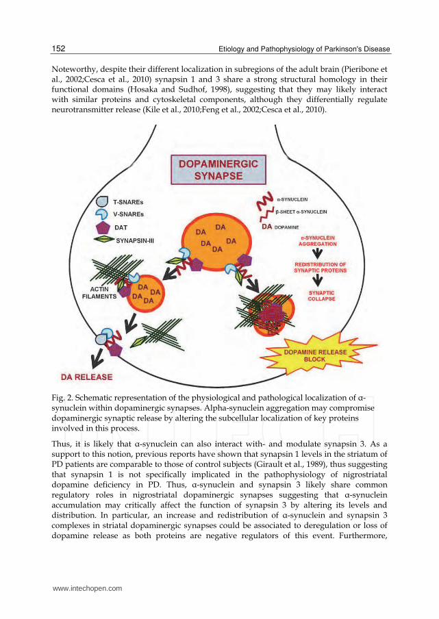
5.2 Gene-based strategies to block or delay synaptic dysfunctions in PD Since dopaminergic neuronal dysfunctions are central to the pathogenesis of PD, it has been recently hypothesized that a restoration of the correct function of dopamine neurons may be achieved by the employment of gene based strategies. In particular, a multicistronic lentiviral vector encoding three crucial genes involved in dopamine synthesis: TH, aromatic L-amino acid decarboxylase and guanosine 5’-triphosphate cyclohydrolase has been produced and tested in experimental models of PD, including MPTP-treated primates. Results of these studies indicate that this gene-based dopamine replacement may be able to correct motor deficits. However, the models used to test these compounds do not exactly reproduce all the features of PD. Indeed, toxin-based models don’t show ┙-synuclein inclusions, or at least significant pathological modifications of ┙-synuclein has been reported only by one group in MPTP-treated squirrel monkeys (McCormack et al 2008). This is not surprising, as mitochondrial dysfunctions have been linked to the induction of ┙-synuclein aggregation, but these features doesn’t necessarily resemble the disease. Indeed, dopaminergic degeneration in toxin-based models is achieved through methods that rapidly and drastically lead to neuronal death and thus they don’t reproduce the exact pathological series of event that leads to this phenomenon in PD patients. Therefore, whether PD onset and progression are dependent on the pathological accumulation of ┙-synuclein at striatal terminals, the gene-based dopamine replacement approach may reproduce the effect of L-DOPA therapy without affectively modifying the pathophysiological aspects of the disease. Indeed, in light of the relevant role played by ┙-synuclein in the control of synaptic activity, a putative disease modifying intervention for PD should block ┙-synuclein-related synaptic dysfunctions. Indeed, its pathological accumulation impairs synaptic release through multiple mechanisms (Figure 2), thus implying that even in the presence of dopamine, the synaptic terminals containing ┙-synuclein aggregates still don’t act. A concept that is reinforced by the fact that ┙-synuclein can be transmitted from cell to cell, thus implying that correction of dopamine deficiency may be uneffective also because of the spreading of ┙-synuclein through adjacent synapses. Thus, again, targeting the ┙-synuclein-related synaptic proteome may represent a powerful tool to block or delay disease progression. As previously described, ┙-synuclein is able to interact with key synaptic proteins, such as DAT and synapsin 1. The synapsins are a family of neuron-associated proteins involved in the regulation of synaptic vesicle release (Cesca et al., 2010;Kile et al., 2010). Synapsin 3 is likely the only isoform which is present in striatal dopaminergic terminals. Indeed, synapsin 1 and 2 have been found to miscolocalize with VMAT2 in the neostriatum of mice (Bogen et al., 2006) although the mRNA for either forms is present in the striatum (Kile et al., 2010). Remarkably, synapsin 3 is a negative regulator of striatal dopamine release (Kile et al., 2010). Although studies reporting a possible involvement of synapsin 3 in the pathogenesis of ┙-synuclein-related disease are lacking, previous studies (Galvin et al., 1999) have shown that synapsin 1 appears to accumulate in ┙-synuclein-immunopositive lesions in axon terminals of the hippocampus of PD patients. Furthermore, synapsin 1 and ┙-synuclein share numerous functional and biochemical similarities. Indeed, besides being both part of the DAT (Maiya et al., 2007) and synaptic proteome (Burre and Volknandt, 2007), they are co-transported from the cell body to the axon by the slow component b as multiprotein complexes without being affected by actin depletion (Roy et al., 2007;Roy et al., 2008) and they can bind to synaptic vesicles and interact with presynaptic membranes and actin cytoskeleton, modulating the dynamics of the actin-based network during the exo-endocytotic cycle (Cesca et al., 2010;Tofaris and Spillantini, 2007).
www.intechopen.com
Etiology and Pathophysiology of Parkinson's Disease
152
Noteworthy, despite their different localization in subregions of the adult brain (Pieribone et al., 2002;Cesca et al., 2010) synapsin 1 and 3 share a strong structural homology in their functional domains (Hosaka and Sudhof, 1998), suggesting that they may likely interact with similar proteins and cytoskeletal components, although they differentially regulate neurotransmitter release (Kile et al., 2010;Feng et al., 2002;Cesca et al., 2010).
Fig. 2. Schematic representation of the physiological and pathological localization of ┙-synuclein within dopaminergic synapses. Alpha-synuclein aggregation may compromise dopaminergic synaptic release by altering the subcellular localization of key proteins involved in this process.
Thus, it is likely that ┙-synuclein can also interact with- and modulate synapsin 3. As a support to this notion, previous reports have shown that synapsin 1 levels in the striatum of PD patients are comparable to those of control subjects (Girault et al., 1989), thus suggesting that synapsin 1 is not specifically implicated in the pathophysiology of nigrostriatal dopamine deficiency in PD. Thus, ┙-synuclein and synapsin 3 likely share common regulatory roles in nigrostriatal dopaminergic synapses suggesting that ┙-synuclein accumulation may critically affect the function of synapsin 3 by altering its levels and distribution. In particular, an increase and redistribution of ┙-synuclein and synapsin 3 complexes in striatal dopaminergic synapses could be associated to deregulation or loss of dopamine release as both proteins are negative regulators of this event. Furthermore,
www.intechopen.com
Targeting α-Synuclein-Related Synaptic Pathology: Novel Clues for Parkinson’s Disease Therapy
153
synapsin 3 increase and redistribution may mechanistically induce the misregulation of other ┙-synuclein/synapsin 3 protein partners such as DAT and synaptobrevin-2 as suggested by recent findings. Therefore, in the scenario of the dopaminergic synapse, deregualtion of synapsin 3 by the pathological aggregation of ┙-synuclein may be a key event compromising synaptic functions as well as the correct tethering and fusion of synaptic vesicles. Another way to improve dopaminergic neuronal functions in relation to ┙-synuclein toxicity at the synapse could be the gene-based delivery of the DAT. Nonetheless, it should be taken into account that, since the DAT can precipitate with ┙-synuclein within intracellular inclusions, delivery of this protein may also foster the pathological aggregation of ┙-synuclein, an event which may be controlled by the administration of dopaminergic agonists which are known to prevent the formation of DAT/┙-synuclein-immunopositive inclusions.
6. Conclusions
A large body of evidence strikingly indicate that ┙-synuclein pathology at the synapse may be crucially involved in PD pathophysiology. Indeed, ┙-synuclein accumulation, pathological modifications and aggregation are able to significantly impair synaptic functions in experimental models of PD. In particular, it appears that ┙-synuclein aggregation at dopaminergic synaptic sites may damage neuronal functions by affecting the correct subcellular distribution of several molecular members of its lipidome and proteome, thus leading to synaptic vesicle stall and to the consequent block of synaptic release. This event may underlie the onset of a perilous axonal degeneration which may retrogradely compromise neuronal resilience, thus leading to dopaminergic cell dysfunctions and death. This view opens the way to the discovery and characterization of novel possible targets to develop drug- and gene-based therapeutical strategies to cure the disease, possibly by modifying and/or slowing disease progression. This is a crucial point, as current therapeutical approaches for PD are aimed at ameliorating disease simptomathology, but none of the present known agent is able to block the progression of the disease. Targeting the ┙-synuclein-related synaptic proteome by drug-based or gene-based therapeutical interventions may thus represent a new frontier to develop disease modifying strategies for PD and other ┙-synucleinopathies.
7. Acknowledgements
We are grateful to Dr. Laura Navarria and Michela Zaltieri for technical assistance and insightful discussion.
8. References
Abeliovich A, Schmitz Y, Farinas I, Choi-Lundberg D, Ho WH, Castillo PE, Shinsky N, Verdugo JM, Armanini M, Ryan A, Hynes M, Phillips H, Sulzer D, Rosenthal A (2000) Mice lacking alpha-synuclein display functional deficits in the nigrostriatal dopamine system. Neuron 25:239-252.
www.intechopen.com
Etiology and Pathophysiology of Parkinson's Disease
154
Albani D, Peverelli E, Rametta R, Batelli S, Veschini L, Negro A, Forloni G (2004) Protective effect of TAT-delivered alpha-synuclein: relevance of the C-terminal domain and involvement of HSP70. FASEB J 18:1713-1715.
Alvarez-Castelao B, Castano JG (2010) Synphilin-1 inhibits alpha-synuclein degradation by the proteasome. Cell Mol Life Sci. 68:2643-2654.
Alves da Costa C, Ancolio K, Checler F (2000) Wild-type but not Parkinson's disease-related ala-53 --> Thr mutant alpha -synuclein protects neuronal cells from apoptotic stimuli. J Biol Chem 275:24065-24069.
Antony T, Hoyer W, Cherny D, Heim G, Jovin TM, Subramaniam V (2003) Cellular polyamines promote the aggregation of alpha-synuclein. J Biol Chem 278:3235-3240.
Auluck PK, Chan HY, Trojanowski JQ, Lee VM, Bonini NM (2002) Chaperone suppression of alpha-synuclein toxicity in a Drosophila model for Parkinson's disease. Science 295:865-868.
Azzouz M, Martin-Rendon E, Barber RD, Mitrophanous KA, Carter EE, Rohll JB, Kingsman SM, Kingsman AJ, Mazarakis ND (2002) Multicistronic lentiviral vector-mediated striatal gene transfer of aromatic L-amino acid decarboxylase, tyrosine hydroxylase, and GTP cyclohydrolase I induces sustained transgene expression, dopamine production, and functional improvement in a rat model of Parkinson's disease. J Neurosci 22:10302-10312.
Baba M, Nakajo S, Tu PH, Tomita T, Nakaya K, Lee VM, Trojanowski JQ, Iwatsubo T (1998) Aggregation of alpha-synuclein in Lewy bodies of sporadic Parkinson's disease and dementia with Lewy bodies. Am J Pathol 152:879-884.
Barone P (2010) Neurotransmission in Parkinson's disease: beyond dopamine. Eur J Neurol 17:364-376.
Batelli S, Albani D, Rametta R, Polito L, Prato F, Pesaresi M, Negro A, Forloni G (2008) DJ-1 modulates alpha-synuclein aggregation state in a cellular model of oxidative stress: relevance for Parkinson's disease and involvement of HSP70. PLoS One 3:e1884.
Bayer TA, Jakala P, Hartmann T, Egensperger R, Buslei R, Falkai P, Beyreuther K (1999) Neural expression profile of alpha-synuclein in developing human cortex. Neuroreport 10:2799-2803.
Beach TG, Adler CH, Lue L, Sue LI, Bachalakuri J, Henry-Watson J, Sasse J, Boyer S, Shirohi S, Brooks R, Eschbacher J, White CL, III, Akiyama H, Caviness J, Shill HA, Connor DJ, Sabbagh MN, Walker DG (2009a) Unified staging system for Lewy body disorders: correlation with nigrostriatal degeneration, cognitive impairment and motor dysfunction. Acta Neuropathol 117:613-634.
Beach TG, White CL, III, Hladik CL, Sabbagh MN, Connor DJ, Shill HA, Sue LI, Sasse J, Bachalakuri J, Henry-Watson J, Akiyama H, Adler CH (2009b) Olfactory bulb alpha-synucleinopathy has high specificity and sensitivity for Lewy body disorders. Acta Neuropathol 117:169-174.
Belin AC, Westerlund M (2008) Parkinson's disease: a genetic perspective. FEBS J 275:1377-1383.
Bellani S, Sousa VL, Ronzitti G, Valtorta F, Meldolesi J, Chieregatti E (2010) The regulation of synaptic function by alpha-synuclein. Commun Integr Biol 3:106-109.
www.intechopen.com
Targeting α-Synuclein-Related Synaptic Pathology: Novel Clues for Parkinson’s Disease Therapy
155
Bellucci A, Collo G, Sarnico I, Battistin L, Missale C, Spano P (2008) Alpha-synuclein aggregation and cell death triggered by energy deprivation and dopamine overload are counteracted by D2/D3 receptor activation. J Neurochem 106:560-577.
Bellucci A, Navarria L, Zaltieri M, Falarti E, Bodei S, Sigala S, Battistin L, Spillantini M, Missale C, Spano P (2011) Induction of the unfolded protein response by alpha-synuclein in experimental models of Parkinson's disease. J Neurochem 116:588-605.
Bence NF, Sampat RM, Kopito RR (2001) Impairment of the ubiquitin-proteasome system by protein aggregation. Science 292:1552-1555.
Bergeron C, Petrunka C, Weyer L, Pollanen MS (1996) Altered neurofilament expression does not contribute to Lewy body formation. Am J Pathol 148:267-272.
Bjorklund A, Bjorklund T, Kirik D (2009) Gene therapy for dopamine replacement in Parkinson's disease. Sci Transl Med 1:2ps2.
Bjorklund T, Kirik D (2009) Scientific rationale for the development of gene therapy strategies for Parkinson's disease. Biochim Biophys Acta 1792:703-713.
Bogen IL, Boulland JL, Mariussen E, Wright MS, Fonnum F, Kao HT, Walaas SI (2006) Absence of synapsin I and II is accompanied by decreases in vesicular transport of specific neurotransmitters. J Neurochem 96:1458-1466.
Bolan EA, Kivell B, Jaligam V, Oz M, Jayanthi LD, Han Y, Sen N, Urizar E, Gomes I, Devi LA, Ramamoorthy S, Javitch JA, Zapata A, Shippenberg TS (2007) D2 receptors regulate dopamine transporter function via an extracellular signal-regulated kinases 1 and 2-dependent and phosphoinositide 3 kinase-independent mechanism. Mol Pharmacol 71:1222-1232.
Bonini NM, Giasson BI (2005) Snaring the function of alpha-synuclein. Cell 123:359-361. Bowling AC, Beal MF (1995) Bioenergetic and oxidative stress in neurodegenerative
diseases. Life Sci 56:1151-1171. Braak H, Del TK, Rub U, de Vos RA, Jansen Steur EN, Braak E (2003) Staging of brain
pathology related to sporadic Parkinson's disease. Neurobiol Aging 24:197-211. Braga CA, Follmer C, Palhano FL, Khattar E, Freitas MS, Romao L, Di GS, Lashuel HA, Silva
JL, Foguel D (2011) The anti-Parkinsonian drug selegiline delays the nucleation phase of alpha-synuclein aggregation leading to the formation of nontoxic species. J Mol Biol 405:254-273.
Burre J, Sharma M, Tsetsenis T, Buchman V, Etherton MR, Sudhof TC (2010) Alpha-synuclein promotes SNARE-complex assembly in vivo and in vitro. Science 329:1663-1667.
Burre J, Volknandt W (2007) The synaptic vesicle proteome. J Neurochem 101:1448-1462. Campbell BC, McLean CA, Culvenor JG, Gai WP, Blumbergs PC, Jakala P, Beyreuther K,
Masters CL, Li QX (2001) The solubility of alpha-synuclein in multiple system atrophy differs from that of dementia with Lewy bodies and Parkinson's disease. J Neurochem 76:87-96.
Caslake R, Macleod A, Ives N, Stowe R, Counsell C (2009) Monoamine oxidase B inhibitors versus other dopaminergic agents in early Parkinson's disease. Cochrane Database Syst RevCD006661.
Ceravolo R, Frosini D, Rossi C, Bonuccelli U (2009) Impulse control disorders in Parkinson's disease: definition, epidemiology, risk factors, neurobiology and management. Parkinsonism Relat Disord 15 Suppl 4:S111-S115.
www.intechopen.com
Etiology and Pathophysiology of Parkinson's Disease
156
Cesca F, Baldelli P, Valtorta F, Benfenati F (2010) The synapsins: key actors of synapse function and plasticity. Prog Neurobiol 91:313-348.
Chandra S, Chen X, Rizo J, Jahn R, Sudhof TC (2003) A broken alpha -helix in folded alpha -Synuclein. J Biol Chem 278:15313-15318.
Chandra S, Fornai F, Kwon HB, Yazdani U, Atasoy D, Liu X, Hammer RE, Battaglia G, German DC, Castillo PE, Sudhof TC (2004) Double-knockout mice for alpha- and beta-synucleins: effect on synaptic functions. Proc Natl Acad Sci U S A 101:14966-14971.
Chandra S, Gallardo G, Fernandez-Chacon R, Schluter OM, Sudhof TC (2005) Alpha-synuclein cooperates with CSPalpha in preventing neurodegeneration. Cell 123:383-396.
Chen L, Thiruchelvam MJ, Madura K, Richfield EK (2006) Proteasome dysfunction in aged human alpha-synuclein transgenic mice. Neurobiol Dis 23:120-126.
Choi W, Zibaee S, Jakes R, Serpell LC, Davletov B, Crowther RA, Goedert M (2004) Mutation E46K increases phospholipid binding and assembly into filaments of human alpha-synuclein. FEBS Lett 576:363-368.
Clayton DF, George JM (1998) The synucleins: a family of proteins involved in synaptic function, plasticity, neurodegeneration and disease. Trends Neurosci 21:249-254.
Conway KA, Harper JD, Lansbury PT (1998) Accelerated in vitro fibril formation by a mutant alpha-synuclein linked to early-onset Parkinson disease. Nat Med 4:1318-1320.
Conway KA, Lee SJ, Rochet JC, Ding TT, Harper JD, Williamson RE, Lansbury PT, Jr. (2000) Accelerated oligomerization by Parkinson's disease linked alpha-synuclein mutants. Ann N Y Acad Sci 920:42-45.
Conway KA, Rochet JC, Bieganski RM, Lansbury PT, Jr. (2001) Kinetic stabilization of the alpha-synuclein protofibril by a dopamine-alpha-synuclein adduct. Science 294:1346-1349.
Cookson MR (2005) The biochemistry of Parkinson's disease. Annu Rev Biochem 74:29-52. Cookson MR, van der BM (2008) Cell systems and the toxic mechanism(s) of alpha-
synuclein. Exp Neurol 209:5-11. Crowther RA, Jakes R, Spillantini MG, Goedert M (1998) Synthetic filaments assembled from
C-terminally truncated alpha-synuclein. FEBS Lett 436:309-312. Darios F, Ruiperez V, Lopez I, Villanueva J, Gutierrez LM, Davletov B (2010) Alpha-
synuclein sequesters arachidonic acid to modulate SNARE-mediated exocytosis. EMBO Rep 11:528-533.
Davidson WS, Jonas A, Clayton DF, George JM (1998) Stabilization of alpha-synuclein secondary structure upon binding to synthetic membranes. J Biol Chem 273:9443-9449.
Desplats P, Lee HJ, Bae EJ, Patrick C, Rockenstein E, Crews L, Spencer B, Masliah E, Lee SJ (2009) Inclusion formation and neuronal cell death through neuron-to-neuron transmission of alpha-synuclein. Proc Natl Acad Sci U S A 106:13010-13015.
Dufty BM, Warner LR, Hou ST, Jiang SX, Gomez-Isla T, Leenhouts KM, Oxford JT, Feany MB, Masliah E, Rohn TT (2007) Calpain-cleavage of alpha-synuclein: connecting proteolytic processing to disease-linked aggregation. Am J Pathol 170:1725-1738.
Elbaz A, Moisan F (2008) Update in the epidemiology of Parkinson's disease. Curr Opin Neurol 21:454-460.
www.intechopen.com
Targeting α-Synuclein-Related Synaptic Pathology: Novel Clues for Parkinson’s Disease Therapy
157
Eliezer D, Kutluay E, Bussell R, Jr., Browne G (2001) Conformational properties of alpha-synuclein in its free and lipid-associated states. J Mol Biol 307:1061-1073.
Engelender S, Kaminsky Z, Guo X, Sharp AH, Amaravi RK, Kleiderlein JJ, Margolis RL, Troncoso JC, Lanahan AA, Worley PF, Dawson VL, Dawson TM, Ross CA (1999) Synphilin-1 associates with alpha-synuclein and promotes the formation of cytosolic inclusions. Nat Genet 22:110-114.
Feany MB, Bender WW (2000) A Drosophila model of Parkinson's disease. Nature 404:394-398.
Feng J, Chi P, Blanpied TA, Xu Y, Magarinos AM, Ferreira A, Takahashi RH, Kao HT, McEwen BS, Ryan TA, Augustine GJ, Greengard P (2002) Regulation of neurotransmitter release by synapsin III. J Neurosci 22:4372-4380.
Fernagut PO, Hutson CB, Fleming SM, Tetreaut NA, Salcedo J, Masliah E, Chesselet MF (2007) Behavioral and histopathological consequences of paraquat intoxication in mice: effects of alpha-synuclein over-expression. Synapse 61:991-1001.
Fernandez CO, Hoyer W, Zweckstetter M, Jares-Erijman EA, Subramaniam V, Griesinger C, Jovin TM (2004) NMR of alpha-synuclein-polyamine complexes elucidates the mechanism and kinetics of induced aggregation. EMBO J 23:2039-2046.
Forno LS (1996) Neuropathology of Parkinson's disease. J Neuropathol Exp Neurol 55:259-272.
Fortin DL, Nemani VM, Nakamura K, Edwards RH (2010) The behavior of alpha-synuclein in neurons. Mov Disord 25 Suppl 1:S21-S26.
Fortin DL, Nemani VM, Voglmaier SM, Anthony MD, Ryan TA, Edwards RH (2005) Neural activity controls the synaptic accumulation of alpha-synuclein. J Neurosci 25:10913-10921.
Fortin DL, Troyer MD, Nakamura K, Kubo S, Anthony MD, Edwards RH (2004) Lipid rafts mediate the synaptic localization of alpha-synuclein. J Neurosci 24:6715-6723.
Fujiwara H, Hasegawa M, Dohmae N, Kawashima A, Masliah E, Goldberg MS, Shen J, Takio K, Iwatsubo T (2002) alpha-Synuclein is phosphorylated in synucleinopathy lesions. Nat Cell Biol 4:160-164.
Gai WP, Yuan HX, Li XQ, Power JT, Blumbergs PC, Jensen PH (2000) In situ and in vitro study of colocalization and segregation of alpha-synuclein, ubiquitin, and lipids in Lewy bodies. Exp Neurol 166:324-333.
Galvin JE, Lee VM, Trojanowski JQ (2001) Synucleinopathies: clinical and pathological implications. Arch Neurol 58:186-190.
Galvin JE, Uryu K, Lee VM, Trojanowski JQ (1999) Axon pathology in Parkinson's disease and Lewy body dementia hippocampus contains alpha-, beta-, and gamma-synuclein. Proc Natl Acad Sci U S A 96:13450-13455.
Gao HM, Kotzbauer PT, Uryu K, Leight S, Trojanowski JQ, Lee VM (2008) Neuroinflammation and oxidation/nitration of alpha-synuclein linked to dopaminergic neurodegeneration. J Neurosci 28:7687-7698.
Garcia-Reitbock P, Anichtchik O, Bellucci A, Iovino M, Ballini C, Fineberg E, Ghetti B, Della CL, Spano P, Tofaris GK, Goedert M, Spillantini MG (2010) SNARE protein redistribution and synaptic failure in a transgenic mouse model of Parkinson's disease. Brain 133:2032-2044.
www.intechopen.com
Etiology and Pathophysiology of Parkinson's Disease
158
George JM, Jin H, Woods WS, Clayton DF (1995) Characterization of a novel protein regulated during the critical period for song learning in the zebra finch. Neuron 15:361-372.
Giasson BI, Duda JE, Murray IV, Chen Q, Souza JM, Hurtig HI, Ischiropoulos H, Trojanowski JQ, Lee VM (2000) Oxidative damage linked to neurodegeneration by selective alpha-synuclein nitration in synucleinopathy lesions. Science 290:985-989.
Giasson BI, Duda JE, Quinn SM, Zhang B, Trojanowski JQ, Lee VM (2002) Neuronal alpha-synucleinopathy with severe movement disorder in mice expressing A53T human alpha-synuclein. Neuron 34:521-533.
Girault JA, Raisman-Vozari R, Agid Y, Greengard P (1989) Striatal phosphoproteins in Parkinson disease and progressive supranuclear palsy. Proc Natl Acad Sci U S A 86:2493-2497.
Goedert M (1999) Filamentous nerve cell inclusions in neurodegenerative diseases: tauopathies and alpha-synucleinopathies. Philos Trans R Soc Lond B Biol Sci 354:1101-1118.
Goers J, Uversky VN, Fink AL (2003) Polycation-induced oligomerization and accelerated fibrillation of human alpha-synuclein in vitro. Protein Sci 12:702-707.
Gomez-Isla T, Irizarry MC, Mariash A, Cheung B, Soto O, Schrump S, Sondel J, Kotilinek L, Day J, Schwarzschild MA, Cha JH, Newell K, Miller DW, Ueda K, Young AB, Hyman BT, Ashe KH (2003) Motor dysfunction and gliosis with preserved dopaminergic markers in human alpha-synuclein A30P transgenic mice. Neurobiol Aging 24:245-258.
Greenbaum EA, Graves CL, Mishizen-Eberz AJ, Lupoli MA, Lynch DR, Englander SW, Axelsen PH, Giasson BI (2005) The E46K mutation in alpha-synuclein increases amyloid fibril formation. J Biol Chem 280:7800-7807.
Gureviciene I, Gurevicius K, Tanila H (2007) Role of alpha-synuclein in synaptic glutamate release. Neurobiol Dis 28:83-89.
Halliday GM, McCann H (2010) The progression of pathology in Parkinson's disease. Ann N Y Acad Sci 1184:188-195.
Han H, Weinreb PH, Lansbury PT, Jr. (1995) The core Alzheimer's peptide NAC forms amyloid fibrils which seed and are seeded by beta-amyloid: is NAC a common trigger or target in neurodegenerative disease? Chem Biol 2:163-169.
Hashimoto M, Hsu LJ, Rockenstein E, Takenouchi T, Mallory M, Masliah E (2002) alpha-Synuclein protects against oxidative stress via inactivation of the c-Jun N-terminal kinase stress-signaling pathway in neuronal cells. J Biol Chem 277:11465-11472.
Hashimoto M, Hsu LJ, Xia Y, Takeda A, Sisk A, Sundsmo M, Masliah E (1999) Oxidative stress induces amyloid-like aggregate formation of NACP/alpha-synuclein in vitro. Neuroreport 10:717-721.
Hassan A, Bower JH, Kumar N, Matsumoto JY, Fealey RD, Josephs KA, Ahlskog JE (2011) Dopamine agonist-triggered pathological behaviors: Surveillance in the PD clinic reveals high frequencies. Parkinsonism Relat Disord 17:260-264.
Hill WD (1996) Altered neurofilament expression does not contribute to Lewy body formation. Am J Pathol 149:728-729.
Hodara R, Norris EH, Giasson BI, Mishizen-Eberz AJ, Lynch DR, Lee VM, Ischiropoulos H (2004) Functional consequences of alpha-synuclein tyrosine nitration: diminished
www.intechopen.com
Targeting α-Synuclein-Related Synaptic Pathology: Novel Clues for Parkinson’s Disease Therapy
159
binding to lipid vesicles and increased fibril formation. J Biol Chem 279:47746-47753.
Hosaka M, Sudhof TC (1998) Synapsins I and II are ATP-binding proteins with differential Ca2+ regulation. J Biol Chem 273:1425-1429.
Iwai A, Masliah E, Yoshimoto M, Ge N, Flanagan L, de Silva HA, Kittel A, Saitoh T (1995) The precursor protein of non-A beta component of Alzheimer's disease amyloid is a presynaptic protein of the central nervous system. Neuron 14:467-475.
Jakes R, Spillantini MG, Goedert M (1994) Identification of two distinct synucleins from human brain. FEBS Lett 345:27-32.
Jarraya B, Boulet S, Ralph GS, Jan C, Bonvento G, Azzouz M, Miskin JE, Shin M, Delzescaux T, Drouot X, Herard AS, Day DM, Brouillet E, Kingsman SM, Hantraye P, Mitrophanous KA, Mazarakis ND, Palfi S (2009) Dopamine gene therapy for Parkinson's disease in a nonhuman primate without associated dyskinesia. Sci Transl Med 1:2ra4.
Javoy-Agid F, Hirsch EC, Dumas S, Duyckaerts C, Mallet J, Agid Y (1990) Decreased tyrosine hydroxylase messenger RNA in the surviving dopamine neurons of the substantia nigra in Parkinson's disease: an in situ hybridization study. Neuroscience 38:245-253.
Jellinger KA (1991) Pathology of Parkinson's disease. Changes other than the nigrostriatal pathway. Mol Chem Neuropathol 14:153-197.
Jellinger KA (2009) A critical evaluation of current staging of alpha-synuclein pathology in Lewy body disorders. Biochim Biophys Acta 1792:730-740.
Jenco JM, Rawlingson A, Daniels B, Morris AJ (1998) Regulation of phospholipase D2: selective inhibition of mammalian phospholipase D isoenzymes by alpha- and beta-synucleins. Biochemistry 37:4901-4909.
Jo E, Darabie AA, Han K, Tandon A, Fraser PE, McLaurin J (2004) alpha-Synuclein-synaptosomal membrane interactions: implications for fibrillogenesis. Eur J Biochem 271:3180-3189.
Jo E, McLaurin J, Yip CM, St George-Hyslop P, Fraser PE (2000) alpha-Synuclein membrane interactions and lipid specificity. J Biol Chem 275:34328-34334.
Kahle PJ, Neumann M, Ozmen L, Muller V, Jacobsen H, Schindzielorz A, Okochi M, Leimer U, van Der PH, Probst A, Kremmer E, Kretzschmar HA, Haass C (2000) Subcellular localization of wild-type and Parkinson's disease-associated mutant alpha -synuclein in human and transgenic mouse brain. J Neurosci 20:6365-6373.
Kaplitt MG (2010) Parkinson disease: Another player in gene therapy for Parkinson disease. Nat Rev Neurol 6:7-8.
Kile BM, Guillot TS, Venton BJ, Wetsel WC, Augustine GJ, Wightman RM (2010) Synapsins differentially control dopamine and serotonin release. J Neurosci 30:9762-9770.
Kim YS, Laurine E, Woods W, Lee SJ (2006) A novel mechanism of interaction between alpha-synuclein and biological membranes. J Mol Biol 360:386-397.
Kirik D, Rosenblad C, Burger C, Lundberg C, Johansen TE, Muzyczka N, Mandel RJ, Bjorklund A (2002) Parkinson-like neurodegeneration induced by targeted overexpression of alpha-synuclein in the nigrostriatal system. J Neurosci 22:2780-2791.
www.intechopen.com
Etiology and Pathophysiology of Parkinson's Disease
160
Klein RL, King MA, Hamby ME, Meyer EM (2002) Dopaminergic cell loss induced by human A30P alpha-synuclein gene transfer to the rat substantia nigra. Hum Gene Ther 13:605-612.
Koob AO, Ubhi K, Paulsson JF, Kelly J, Rockenstein E, Mante M, Adame A, Masliah E (2010) Lovastatin ameliorates alpha-synuclein accumulation and oxidation in transgenic mouse models of alpha-synucleinopathies. Exp Neurol 221:267-274.
Kordower JH, Chu Y, Hauser RA, Freeman TB, Olanow CW (2008a) Lewy body-like pathology in long-term embryonic nigral transplants in Parkinson's disease. Nat Med 14:504-506.
Kordower JH, Chu Y, Hauser RA, Olanow CW, Freeman TB (2008b) Transplanted dopaminergic neurons develop PD pathologic changes: a second case report. Mov Disord 23:2303-2306.
Kramer ML, Behrens C, Schulz-Schaeffer WJ (2008) Selective detection, quantification, and subcellular location of alpha-synuclein aggregates with a protein aggregate filtration assay. Biotechniques 44:403-411.
Le WD, Jankovic J (2001) Are dopamine receptor agonists neuroprotective in Parkinson's disease? Drugs Aging 18:389-396.
Lee FJ, Liu F, Pristupa ZB, Niznik HB (2001a) Direct binding and functional coupling of alpha-synuclein to the dopamine transporters accelerate dopamine-induced apoptosis. FASEB J 15:916-926.
Lee G, Tanaka M, Park K, Lee SS, Kim YM, Junn E, Lee SH, Mouradian MM (2004) Casein kinase II-mediated phosphorylation regulates alpha-synuclein/synphilin-1 interaction and inclusion body formation. J Biol Chem 279:6834-6839.
Lee HJ, Lee SJ (2002) Characterization of cytoplasmic alpha-synuclein aggregates. Fibril formation is tightly linked to the inclusion-forming process in cells. J Biol Chem 277:48976-48983.
Lee M, Hyun D, Halliwell B, Jenner P (2001b) Effect of the overexpression of wild-type or mutant alpha-synuclein on cell susceptibility to insult. J Neurochem 76:998-1009.
Lee MK, Stirling W, Xu Y, Xu X, Qui D, Mandir AS, Dawson TM, Copeland NG, Jenkins NA, Price DL (2002) Human alpha-synuclein-harboring familial Parkinson's disease-linked Ala-53 --> Thr mutation causes neurodegenerative disease with alpha-synuclein aggregation in transgenic mice. Proc Natl Acad Sci U S A 99:8968-8973.
Lee SJ, Jeon H, Kandror KV (2008) Alpha-synuclein is localized in a subpopulation of rat brain synaptic vesicles. Acta Neurobiol Exp (Wars ) 68:509-515.
Lee VM, Trojanowski JQ (2006) Mechanisms of Parkinson's disease linked to pathological alpha-synuclein: new targets for drug discovery. Neuron 52:33-38.
Li JY, Englund E, Holton JL, Soulet D, Hagell P, Lees AJ, Lashley T, Quinn NP, Rehncrona S, Bjorklund A, Widner H, Revesz T, Lindvall O, Brundin P (2008) Lewy bodies in grafted neurons in subjects with Parkinson's disease suggest host-to-graft disease propagation. Nat Med 14:501-503.
Lindersson E, Beedholm R, Hojrup P, Moos T, Gai W, Hendil KB, Jensen PH (2004) Proteasomal inhibition by alpha-synuclein filaments and oligomers. J Biol Chem 279:12924-12934.
Liu CW, Giasson BI, Lewis KA, Lee VM, Demartino GN, Thomas PJ (2005) A precipitating role for truncated alpha-synuclein and the proteasome in alpha-synuclein
www.intechopen.com
Targeting α-Synuclein-Related Synaptic Pathology: Novel Clues for Parkinson’s Disease Therapy
161
aggregation: implications for pathogenesis of Parkinson disease. J Biol Chem 280:22670-22678.
Lo BC, Ridet JL, Schneider BL, Deglon N, Aebischer P (2002) alpha -Synucleinopathy and selective dopaminergic neuron loss in a rat lentiviral-based model of Parkinson's disease. Proc Natl Acad Sci U S A 99:10813-10818.
Madras BK, Fahey MA, Goulet M, Lin Z, Bendor J, Goodrich C, Meltzer PC, Elmaleh DR, Livni E, Bonab AA, Fischman AJ (2006) Dopamine transporter (DAT) inhibitors alleviate specific parkinsonian deficits in monkeys: association with DAT occupancy in vivo. J Pharmacol Exp Ther 319:570-585.
Magen I, Chesselet MF (2010) Genetic mouse models of Parkinson's disease The state of the art. Prog Brain Res 184:53-87.
Magyar K, Palfi M, Tabi T, Kalasz H, Szende B, Szoko E (2004) Pharmacological aspects of (-)-deprenyl. Curr Med Chem 11:2017-2031.
Maiya R, Ponomarev I, Linse KD, Harris RA, Mayfield RD (2007) Defining the dopamine transporter proteome by convergent biochemical and in silico analyses. Genes Brain Behav 6:97-106.
Mak SK, McCormack AL, Langston JW, Kordower JH, Di Monte DA (2009) Decreased alpha-synuclein expression in the aging mouse substantia nigra. Exp Neurol 220:359-365.
Maroteaux L, Campanelli JT, Scheller RH (1988) Synuclein: a neuron-specific protein localized to the nucleus and presynaptic nerve terminal. J Neurosci 8:2804-2815.
Mazzulli JR, Mishizen AJ, Giasson BI, Lynch DR, Thomas SA, Nakashima A, Nagatsu T, Ota A, Ischiropoulos H (2006) Cytosolic catechols inhibit alpha-synuclein aggregation and facilitate the formation of intracellular soluble oligomeric intermediates. J Neurosci 26:10068-10078.
McCormack AL, Mak SK, Shenasa M, Langston WJ, Forno LS, DiMonte DA (2008) Pathological modifications of ┙-synuclein in 1-Methyl-4-Phenyl-1,2,3,6-Tetrahydropyridine (MPTP)-Treated Squirrel Monkeys. J. Neuropathol. Exp. Neurol. 67(8): 793-802.
McFarland MA, Ellis CE, Markey SP, Nussbaum RL (2008) Proteomics analysis identifies phosphorylation-dependent alpha-synuclein protein interactions. Mol Cell Proteomics 7:2123-2137.
McKeith IG (2006) Consensus guidelines for the clinical and pathologic diagnosis of dementia with Lewy bodies (DLB): report of the Consortium on DLB International Workshop. J Alzheimers Dis 9:417-423.
McKeith IG, et al. (2005) Diagnosis and management of dementia with Lewy bodies: third report of the DLB Consortium. Neurology 65:1863-1872.
McKeith IG, et al. (1996) Consensus guidelines for the clinical and pathologic diagnosis of dementia with Lewy bodies (DLB): report of the consortium on DLB international workshop. Neurology 47:1113-1124.
Michell AW, Tofaris GK, Gossage H, Tyers P, Spillantini MG, Barker RA (2007) The effect of truncated human alpha-synuclein (1-120) on dopaminergic cells in a transgenic mouse model of Parkinson's disease. Cell Transplant 16:461-474.
Mishizen-Eberz AJ, Norris EH, Giasson BI, Hodara R, Ischiropoulos H, Lee VM, Trojanowski JQ, Lynch DR (2005) Cleavage of alpha-synuclein by calpain: potential
www.intechopen.com
Etiology and Pathophysiology of Parkinson's Disease
162
role in degradation of fibrillized and nitrated species of alpha-synuclein. Biochemistry 44:7818-7829.
Mosharov EV, Staal RG, Bove J, Prou D, Hananiya A, Markov D, Poulsen N, Larsen KE, Moore CM, Troyer MD, Edwards RH, Przedborski S, Sulzer D (2006) Alpha-synuclein overexpression increases cytosolic catecholamine concentration. J Neurosci 26:9304-9311.
Murphy DD, Rueter SM, Trojanowski JQ, Lee VM (2000) Synucleins are developmentally expressed, and alpha-synuclein regulates the size of the presynaptic vesicular pool in primary hippocampal neurons. J Neurosci 20:3214-3220.
Murray IV, Giasson BI, Quinn SM, Koppaka V, Axelsen PH, Ischiropoulos H, Trojanowski JQ, Lee VM (2003) Role of alpha-synuclein carboxy-terminus on fibril formation in vitro. Biochemistry 42:8530-8540.
Nakajo S, Tsukada K, Omata K, Nakamura Y, Nakaya K (1993) A new brain-specific 14-kDa protein is a phosphoprotein. Its complete amino acid sequence and evidence for phosphorylation. Eur J Biochem 217:1057-1063.
Negro A, Brunati AM, Donella-Deana A, Massimino ML, Pinna LA (2002) Multiple phosphorylation of alpha-synuclein by protein tyrosine kinase Syk prevents eosin-induced aggregation. FASEB J 16:210-212.
Nemani VM, Lu W, Berge V, Nakamura K, Onoa B, Lee MK, Chaudhry FA, Nicoll RA, Edwards RH (2010) Increased expression of alpha-synuclein reduces neurotransmitter release by inhibiting synaptic vesicle reclustering after endocytosis. Neuron 65:66-79.
Neumann M, Kahle PJ, Giasson BI, Ozmen L, Borroni E, Spooren W, Muller V, Odoy S, Fujiwara H, Hasegawa M, Iwatsubo T, Trojanowski JQ, Kretzschmar HA, Haass C (2002) Misfolded proteinase K-resistant hyperphosphorylated alpha-synuclein in aged transgenic mice with locomotor deterioration and in human alpha-synucleinopathies. J Clin Invest 110:1429-1439.
Neystat M, Rzhetskaya M, Kholodilov N, Burke RE (2002) Analysis of synphilin-1 and synuclein interactions by yeast two-hybrid beta-galactosidase liquid assay. Neurosci Lett 325:119-123.
Nieto M, Gil-Bea FJ, Dalfo E, Cuadrado M, Cabodevilla F, Sanchez B, Catena S, Sesma T, Ribe E, Ferrer I, Ramirez MJ, Gomez-Isla T (2006) Increased sensitivity to MPTP in human alpha-synuclein A30P transgenic mice. Neurobiol Aging 27:848-856.
Norris EH, Giasson BI, Hodara R, Xu S, Trojanowski JQ, Ischiropoulos H, Lee VM (2005) Reversible inhibition of alpha-synuclein fibrillization by dopaminochrome-mediated conformational alterations. J Biol Chem 280:21212-21219.
Okochi M, Walter J, Koyama A, Nakajo S, Baba M, Iwatsubo T, Meijer L, Kahle PJ, Haass C (2000) Constitutive phosphorylation of the Parkinson's disease associated alpha-synuclein. J Biol Chem 275:390-397.
Olanow CW, Kieburtz K (2010) Defining disease-modifying therapies for PD--a road map for moving forward. Mov Disord 25:1774-1779.
Ono K, Hirohata M, Yamada M (2007) Anti-fibrillogenic and fibril-destabilizing activities of anti-Parkinsonian agents for alpha-synuclein fibrils in vitro. J Neurosci Res 85:1547-1557.
Ono K, Ikemoto M, Kawarabayashi T, Ikeda M, Nishinakagawa T, Hosokawa M, Shoji M, Takahashi M, Nakashima M (2009) A chemical chaperone, sodium 4-phenylbutyric
www.intechopen.com
Targeting α-Synuclein-Related Synaptic Pathology: Novel Clues for Parkinson’s Disease Therapy
163
acid, attenuates the pathogenic potency in human alpha-synuclein A30P + A53T transgenic mice. Parkinsonism Relat Disord 15:649-654.
Ostrerova N, Petrucelli L, Farrer M, Mehta N, Choi P, Hardy J, Wolozin B (1999) alpha-Synuclein shares physical and functional homology with 14-3-3 proteins. J Neurosci 19:5782-5791.
Oueslati A, Fournier M, Lashuel HA (2010) Role of post-translational modifications in modulating the structure, function and toxicity of alpha-synuclein: implications for Parkinson's disease pathogenesis and therapies. Prog Brain Res 183:115-145.
Patt S, Gertz HJ, Gerhard L, Cervos-Navarro J (1991) Pathological changes in dendrites of substantia nigra neurons in Parkinson's disease: a Golgi study. Histol Histopathol 6:373-380.
Perrin RJ, Woods WS, Clayton DF, George JM (2001) Exposure to long chain polyunsaturated fatty acids triggers rapid multimerization of synucleins. J Biol Chem 276:41958-41962.
Piccini P, Brooks DJ, Bjorklund A, Gunn RN, Grasby PM, Rimoldi O, Brundin P, Hagell P, Rehncrona S, Widner H, Lindvall O (1999) Dopamine release from nigral transplants visualized in vivo in a Parkinson's patient. Nat Neurosci 2:1137-1140.
Pieribone VA, Porton B, Rendon B, Feng J, Greengard P, Kao HT (2002) Expression of synapsin III in nerve terminals and neurogenic regions of the adult brain. J Comp Neurol 454:105-114.
Polymeropoulos MH, Lavedan C, Leroy E, Ide SE, Dehejia A, Dutra A, Pike B, Root H, Rubenstein J, Boyer R, Stenroos ES, Chandrasekharappa S, Athanassiadou A, Papapetropoulos T, Johnson WG, Lazzarini AM, Duvoisin RC, Di IG, Golbe LI, Nussbaum RL (1997) Mutation in the alpha-synuclein gene identified in families with Parkinson's disease. Science 276:2045-2047.
Pronin AN, Morris AJ, Surguchov A, Benovic JL (2000) Synucleins are a novel class of substrates for G protein-coupled receptor kinases. J Biol Chem 275:26515-26522.
Qin Y, Ouyang Q, Pablo J, Mash DC (2005) Cocaine abuse elevates alpha-synuclein and dopamine transporter levels in the human striatum. Neuroreport 16:1489-1493.
Rappley I, Myers DS, Milne SB, Ivanova PT, Lavoie MJ, Brown HA, Selkoe DJ (2009) Lipidomic profiling in mouse brain reveals differences between ages and genders, with smaller changes associated with alpha-synuclein genotype. J Neurochem 111:15-25.
Rascol O, Montastruc JL (2000) [Role of dopaminergic agonists]. Rev Neurol (Paris) 156 Suppl 2 Pt 2:98-104.
Rockenstein E, Mallory M, Hashimoto M, Song D, Shults CW, Lang I, Masliah E (2002) Differential neuropathological alterations in transgenic mice expressing alpha-synuclein from the platelet-derived growth factor and Thy-1 promoters. J Neurosci Res 68:568-578.
Roy S, Winton MJ, Black MM, Trojanowski JQ, Lee VM (2007) Rapid and intermittent cotransport of slow component-b proteins. J Neurosci 27:3131-3138.
Roy S, Winton MJ, Black MM, Trojanowski JQ, Lee VM (2008) Cytoskeletal requirements in axonal transport of slow component-b. J Neurosci 28:5248-5256.
Schapira AH (2002) Dopamine agonists and neuroprotection in Parkinson's disease. Eur J Neurol 9 Suppl 3:7-14.
www.intechopen.com
Etiology and Pathophysiology of Parkinson's Disease
164
Schneider BL, Seehus CR, Capowski EE, Aebischer P, Zhang SC, Svendsen CN (2007) Over-expression of alpha-synuclein in human neural progenitors leads to specific changes in fate and differentiation. Hum Mol Genet 16:651-666.
Schulz-Schaeffer WJ (2010) The synaptic pathology of alpha-synuclein aggregation in dementia with Lewy bodies, Parkinson's disease and Parkinson's disease dementia. Acta Neuropathol 120:131-143.
Scott DA, Tabarean I, Tang Y, Cartier A, Masliah E, Roy S (2010) A pathologic cascade leading to synaptic dysfunction in alpha-synuclein-induced neurodegeneration. J Neurosci 30:8083-8095.
Serpell LC, Berriman J, Jakes R, Goedert M, Crowther RA (2000) Fiber diffraction of synthetic alpha-synuclein filaments shows amyloid-like cross-beta conformation. Proc Natl Acad Sci U S A 97:4897-4902.
Sethy VH, Wu H, Oostveen JA, Hall ED (1997) Neuroprotective effects of the dopamine agonists pramipexole and bromocriptine in 3-acetylpyridine-treated rats. Brain Res 754:181-186.
Sharon R, Bar-Joseph I, Frosch MP, Walsh DM, Hamilton JA, Selkoe DJ (2003) The formation of highly soluble oligomers of alpha-synuclein is regulated by fatty acids and enhanced in Parkinson's disease. Neuron 37:583-595.
Shin Y, Klucken J, Patterson C, Hyman BT, McLean PJ (2005) The co-chaperone carboxyl terminus of Hsp70-interacting protein (CHIP) mediates alpha-synuclein degradation decisions between proteasomal and lysosomal pathways. J Biol Chem 280:23727-23734.
Sidhu A, Wersinger C, Vernier P (2004) alpha-Synuclein regulation of the dopaminergic transporter: a possible role in the pathogenesis of Parkinson's disease. FEBS Lett 565:1-5.
Simola N, Pinna A, Fenu S (2010) Pharmacological therapy of Parkinson's disease: current options and new avenues. Recent Pat CNS Drug Discov 5:221-238.
Sousa VL, Bellani S, Giannandrea M, Yousuf M, Valtorta F, Meldolesi J, Chieregatti E (2009) {alpha}-synuclein and its A30P mutant affect actin cytoskeletal structure and dynamics. Mol Biol Cell 20:3725-3739.
Spillantini MG, Crowther RA, Jakes R, Hasegawa M, Goedert M (1998) alpha-Synuclein in filamentous inclusions of Lewy bodies from Parkinson's disease and dementia with lewy bodies. Proc Natl Acad Sci U S A 95:6469-6473.
Spillantini MG, Goedert M (2000) The alpha-synucleinopathies: Parkinson's disease, dementia with Lewy bodies, and multiple system atrophy. Ann N Y Acad Sci 920:16-27.
Spillantini MG, Schmidt ML, Lee VM, Trojanowski JQ, Jakes R, Goedert M (1997) Alpha-synuclein in Lewy bodies. Nature 388:839-840.
Sudhof TC, Rothman JE (2009) Membrane fusion: grappling with SNARE and SM proteins. Science 323:474-477.
Tofaris GK, Garcia RP, Humby T, Lambourne SL, O'Connell M, Ghetti B, Gossage H, Emson PC, Wilkinson LS, Goedert M, Spillantini MG (2006) Pathological changes in dopaminergic nerve cells of the substantia nigra and olfactory bulb in mice transgenic for truncated human alpha-synuclein(1-120): implications for Lewy body disorders. J Neurosci 26:3942-3950.
www.intechopen.com
Targeting α-Synuclein-Related Synaptic Pathology: Novel Clues for Parkinson’s Disease Therapy
165
Tofaris GK, Razzaq A, Ghetti B, Lilley KS, Spillantini MG (2003) Ubiquitination of alpha-synuclein in Lewy bodies is a pathological event not associated with impairment of proteasome function. J Biol Chem 278:44405-44411.
Tofaris GK, Spillantini MG (2005) Alpha-synuclein dysfunction in Lewy body diseases. Mov Disord 20 Suppl 12:S37-S44.
Tofaris GK, Spillantini MG (2007) Physiological and pathological properties of alpha-synuclein. Cell Mol Life Sci 64:2194-2201.
Tompkins MM, Hill WD (1997) Contribution of somal Lewy bodies to neuronal death. Brain Res 775:24-29.
Tritschler HJ, Packer L, Medori R (1994) Oxidative stress and mitochondrial dysfunction in neurodegeneration. Biochem Mol Biol Int 34:169-181.
Trojanowski JQ, Lee VM (2003) Parkinson's disease and related alpha-synucleinopathies are brain amyloidoses. Ann N Y Acad Sci 991:107-110.
Trostchansky A, Lind S, Hodara R, Oe T, Blair IA, Ischiropoulos H, Rubbo H, Souza JM (2006) Interaction with phospholipids modulates alpha-synuclein nitration and lipid-protein adduct formation. Biochem J 393:343-349.
Ueda K, Fukushima H, Masliah E, Xia Y, Iwai A, Yoshimoto M, Otero DA, Kondo J, Ihara Y, Saitoh T (1993) Molecular cloning of cDNA encoding an unrecognized component of amyloid in Alzheimer disease. Proc Natl Acad Sci U S A 90:11282-11286.
Uversky VN (2007) Neuropathology, biochemistry, and biophysics of alpha-synuclein aggregation. J Neurochem 103:17-37.
Venkatraman P, Wetzel R, Tanaka M, Nukina N, Goldberg AL (2004) Eukaryotic proteasomes cannot digest polyglutamine sequences and release them during degradation of polyglutamine-containing proteins. Mol Cell 14:95-104.
Volles MJ, Lee SJ, Rochet JC, Shtilerman MD, Ding TT, Kessler JC, Lansbury PT, Jr. (2001) Vesicle permeabilization by protofibrillar alpha-synuclein: implications for the pathogenesis and treatment of Parkinson's disease. Biochemistry 40:7812-7819.
Wakabayashi K, Tanji K, Mori F, Takahashi H (2007) The Lewy body in Parkinson's disease: molecules implicated in the formation and degradation of alpha-synuclein aggregates. Neuropathology 27:494-506.
Weinreb PH, Zhen W, Poon AW, Conway KA, Lansbury PT, Jr. (1996) NACP, a protein implicated in Alzheimer's disease and learning, is natively unfolded. Biochemistry 35:13709-13715.
Wersinger C, Banta M, Sidhu A (2004) Comparative analyses of alpha-synuclein expression levels in rat brain tissues and transfected cells. Neurosci Lett 358:95-98.
Wersinger C, Jeannotte A, Sidhu A (2006a) Attenuation of the norepinephrine transporter activity and trafficking via interactions with alpha-synuclein. Eur J Neurosci 24:3141-3152.
Wersinger C, Rusnak M, Sidhu A (2006b) Modulation of the trafficking of the human serotonin transporter by human alpha-synuclein. Eur J Neurosci 24:55-64.
Wislet-Gendebien S, D'Souza C, Kawarai T, St George-Hyslop P, Westaway D, Fraser P, Tandon A (2006) Cytosolic proteins regulate alpha-synuclein dissociation from presynaptic membranes. J Biol Chem 281:32148-32155.
Wislet-Gendebien S, Visanji NP, Whitehead SN, Marsilio D, Hou W, Figeys D, Fraser PE, Bennett SA, Tandon A (2008) Differential regulation of wild-type and mutant
www.intechopen.com
Etiology and Pathophysiology of Parkinson's Disease
166
alpha-synuclein binding to synaptic membranes by cytosolic factors. BMC Neurosci 9:92.
Woods WS, Boettcher JM, Zhou DH, Kloepper KD, Hartman KL, Ladror DT, Qi Z, Rienstra CM, George JM (2007) Conformation-specific binding of alpha-synuclein to novel protein partners detected by phage display and NMR spectroscopy. J Biol Chem 282:34555-34567.
Xie W, Li X, Li C, Zhu W, Jankovic J, Le W (2010) Proteasome inhibition modeling nigral neuron degeneration in Parkinson's disease. J Neurochem 115:188-199.
Xiong N, Huang J, Zhang Z, Zhang Z, Xiong J, Liu X, Jia M, Wang F, Chen C, Cao X, Liang Z, Sun S, Lin Z, Wang T (2009) Stereotaxical infusion of rotenone: a reliable rodent model for Parkinson's disease. PLoS One 4:e7878.
Xu J, Kao SY, Lee FJ, Song W, Jin LW, Yankner BA (2002) Dopamine-dependent neurotoxicity of alpha-synuclein: a mechanism for selective neurodegeneration in Parkinson disease. Nat Med 8:600-606.
Zhang L, Zhang C, Zhu Y, Cai Q, Chan P, Ueda K, Yu S, Yang H (2008) Semi-quantitative analysis of alpha-synuclein in subcellular pools of rat brain neurons: an immunogold electron microscopic study using a C-terminal specific monoclonal antibody. Brain Res 1244:40-52.
Zhou W, Schaack J, Zawada WM, Freed CR (2002) Overexpression of human alpha-synuclein causes dopamine neuron death in primary human mesencephalic culture. Brain Res 926:42-50.
Zhu M, Li J, Fink AL (2003) The association of alpha-synuclein with membranes affects bilayer structure, stability, and fibril formation. J Biol Chem 278:40186-40197.
www.intechopen.com
Etiology and Pathophysiology of Parkinson's DiseaseEdited by Prof. Abdul Qayyum Rana
ISBN 978-953-307-462-7Hard cover, 542 pagesPublisher InTechPublished online 12, October, 2011Published in print edition October, 2011
InTech EuropeUniversity Campus STeP Ri Slavka Krautzeka 83/A 51000 Rijeka, Croatia Phone: +385 (51) 770 447 Fax: +385 (51) 686 166www.intechopen.com
InTech ChinaUnit 405, Office Block, Hotel Equatorial Shanghai No.65, Yan An Road (West), Shanghai, 200040, China
Phone: +86-21-62489820 Fax: +86-21-62489821
This book about Parkinson’s disease provides a detailed account of etiology and pathophysiology ofParkinson’s disease, a complicated neurological condition. Environmental and genetic factors involved inthe causation of Parkinson’s disease have been discussed in detail. This book can be used by basicscientists as well as researchers. Neuroscience fellows and life science readers can also obtain sufficientinformation. Beside genetic factors, other pathophysiological aspects of Parkinson’s disease have beendiscussed in detail. Up to date information about the changes in various neurotransmitters, inflammatoryresponses, oxidative pathways and biomarkers has been described at length. Each section has been writtenby one or more faculty members of well known academic institutions. Thus, this book brings forth both clinicaland basic science aspects of Parkinson’s disease.
How to referenceIn order to correctly reference this scholarly work, feel free to copy and paste the following:
Arianna Bellucci and PierFranco Spano (2011). Targeting α-Synuclein-Related Synaptic Pathology: NovelClues for Parkinson’s Disease Therapy, Etiology and Pathophysiology of Parkinson's Disease, Prof. AbdulQayyum Rana (Ed.), ISBN: 978-953-307-462-7, InTech, Available from:http://www.intechopen.com/books/etiology-and-pathophysiology-of-parkinson-s-disease/targeting-synuclein-related-synaptic-pathology-novel-clues-for-parkinson-s-disease-therapy