03-05-2017 - bosnianpathology.org and...03-05-2017 1 lung cytology ... general-secretary of the...
TRANSCRIPT
03-05-2017
1
LUNG CYTOLOGYPredictive markers and molecular tests
No financial disclosures
Prof. Fernando SchmittDepartment of Pathology and Oncology, Medical Faculty of Porto University
Head of Molecular Pathology Unit, IPATIMUPGeneral-Secretary of the International Academy of Cytology
Pathologists’ role
Historically
• Guiding the surgeons’ hands
• Pattern recognition
• Tumour burden
• Basements (hiding behind a microscope)
2016
• Oncologists’ best mate
• Understanding disease
• Disease biology
• Integral part of the multidisciplinary team
Ancillary Studies in CytologyChallenges
• To select the correct test for a limited sample quantity.
• Avoid jumping from a histological adapted technique directly to cytological material.
• Use appropriate controls for cytological material.
• The variability in material and fixatives is a majorfactor preventing standardization of some proceduresusing cytology.
• Spray and ethanol fixation results in better DNAquality than air drying (but both give reliable results inclinical specimens).
• Due the similarities with histological material, cell-blocks are more easy to handle on the molecular lab.
Pre-analytical issues
Dejmek A. Cancer Cytopathology 2013Schmitt F. Cytopathology 2011
Molecular genetics lab
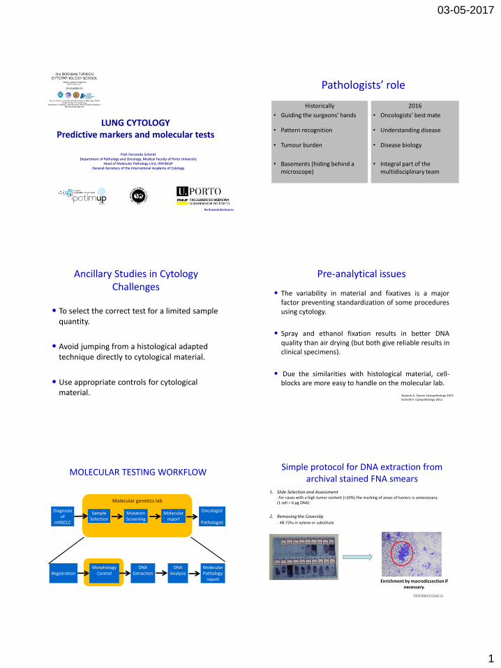
MOLECULAR TESTING WORKFLOW
Diagnosisof
mNSCLC
SampleSelection
MutationScreening
Molecularreport
Oncologist
Pathologist
RegistrationMorphology
ControlDNA
ExtractionDNA
AnalysisMolecularPathology
report
1. Slide Selection and Assessment. For cases with a high tumor content (>20%) the marking of areas of tumors is unnecessary. (1 cell = 6 pg DNA)
Enrichment by macrodissection if necessary.
2. Removing the Coverslip. 48-72hs in xylene or substitute
Simple protocol for DNA extraction from archival stained FNA smears
03-05-2017
2
3. Collecting the Tissue
4. Tissue Lysis and DNA Extraction
Simple protocol for DNA extraction from archival stained FNA smears
Molecular genetics lab
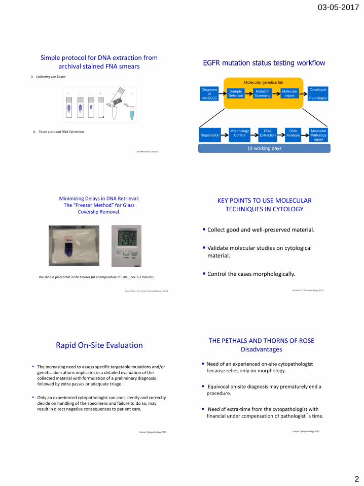
EGFR mutation status testing workflow
Diagnosis
of
mNSCLC
Sample
Selection
Mutation
Screening
Molecular
report
Oncologist
Pathologist
Registration
Morphology
Control
DNA
Extraction
DNA
Analysis
Molecular
Pathology
report
10 working days
Minimizing Delays in DNA Retrieval:The “Freezer Method” for Glass
Coverslip Removal.
Santos GC et al. Cancer Cytopathology 2013
. The slide is placed flat in the freezer (at a temperature of -20ºC) for 1-3 minutes.
• Collect good and well-preserved material.
• Validate molecular studies on cytological material.
• Control the cases morphologically.
KEY POINTS TO USE MOLECULAR TECHNIQUES IN CYTOLOGY
Schmitt FC. Cytopathology 2011
• The increasing need to assess specific targetable mutations and/or genetic aberrations implicates in a detailed evaluation of the collected material with formulation of a preliminary diagnosis followed by extra passes or adequate triage.
• Only an experienced cytopathologist can consistently and correctly decide on handling of the specimens and failure to do so, may result in direct negative consequences to patient care.
Rapid On-Site Evaluation
Cancer Cytopathology 2012
THE PETHALS AND THORNS OF ROSEDisadvantages
• Need of an experienced on-site cytopathologistbecause relies only on morphology.
• Equivocal on-site diagnosis may prematurely end a procedure.
• Need of extra-time from the cytopathologist with financial under compensation of pathologist´s time.
Cancer Cytopathology 2012

03-05-2017
3
PATHOLOGY CONSIDERATIONS FOR GOOD PRACTICE
•Small biopsy and cytology samples should bemanaged not only for diagnosis but also tomaximize the amount of tissue available formolecular studies.
LUNG CYTOLOGY and TUMOR TYPING
Cytology is a powerful tool in the diagnosis of lung cancer, inparticular in the distinction of adenocarcinoma fromsquamous cell carcinoma.
LUNG CYTOLOGY and TUMOR TYPING
LUNG
CARCINOMASmall cell
morphology
YesP63
Positive
No
No
SqCC-
small cell
type
Chromogranin/
Synaptophysin/
CD56-
Positive
Yes SCLC
P40/CK5
TTF1/Napsin AADC
+++
-SqCC
- Or -/+
+++
EGFR
Positive
Yes
Tyrosine
kinase
inhibitors
Yes
NoALK
PositiveCrizotinib
Cancer Cytopathology 2011
03-05-2017
4
POSITIVE CYTOLOGY
NSCLC – NOSNo Clear ADC or SQCC
P40 +TTF1 -
P40-TTF1+
SQCC ADC
EGFR Testing
Negative PositiveTK
inhibitors
ALK
Negative Positive
Crizotinib
ROSRET
(NGS?)
FGFR 1DDR 2
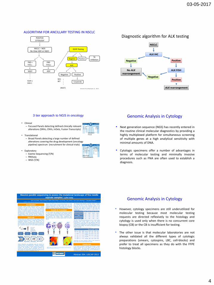
ALGORITHM FOR ANCILLARY TESTING IN NSCLC
Schmitt FC & Machado JC, 2013
Diagnostic algorithm for ALK testing
NSCLC
ALK IHC
Negative Positive
ALK FISH
NegativePositive
No ALKrearrangement
ALK rearrangement
3 tier approach to NGS in oncology
• Clinical– Focused Panels detecting defined clinically relevant
alterations (SNVs, CNVs, InDels, Fusion Transcripts)
• Translational– Broad Panels detecting a large number of defined
alterations covering the drug development (oncology pipeline) spectrum (recruitment for clinical trials)
• Exploratory– Exome Sequencing (T/N)– RNAseq– WGS (T/N)
Oncomine
Focus
Oncomine
CancerResearch
• Next generation sequence (NGS) has recently entered inthe routine clinical molecular diagnostics by providing ahighly multiplexed platform for simultaneous screeningof multiple genes at a high analytical sensitivity withminimal amounts of DNA.
• Cytologic specimens offer a number of advantages interms of molecular testing and minimally invasiveprocedures such as FNA are often used to establish adiagnosis.
Genomic Analysis in Cytology
P
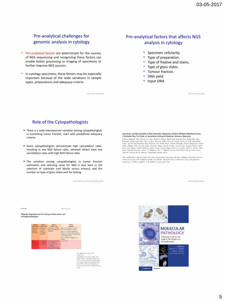
Massive parallel sequencing to assess the mutational landscape of fine needle
aspirate samples: a pilot study
José Luis Costa1, Renê Gerhard1, Esther Diana Rossi2, Luis Cirnes1, Ana Justino1, José Carlos Machado1,3, Fernando Schmitt1,3
1IPATIMUP – Institute of Molecular Pathology and Immunology of the University of Porto, Porto, Portugal 2Anatomic Pathology and Histology-Catholic University of Sacred Heart Rome, Italy 3Medical Faculty of the University of Porto, Porto, Portugal
Introduction
Results & Discussion
ConclusionIn this study we present a workflow that provides a simplegenetic screening tool for FNA samples, in a fast and cost-
efficient manner. This may provide a valuable tool for themanagement of cancer patients, that can be implemented in
most molecular pathology laboratories.
Nowadays, massive parallel sequencing (MPS) is shapingresearch in the field of life sciences. This fast evolving
technology is starting to move into the clinical diagnosticarena. The possibility of performing genomic studies in small
amounts of material obtained, for example, by fine-needleaspiration (FNA), can minimize invasive procedures and allow
the monitoring of cancer, including therapeutic response, withrepeated testing. In this pilot study we aim to address the
possibility of using this technology for assessing the mutationallandscape of FNA samples.
gDNA
10ng of gDNA
739 known mutation in 46
oncogenes and
tumor suppressor genes
Ultra-highMultiplex
PCR
Partia lly digest
primers
Adapter
l igationAmpl i fication
Construct library
3.5 h
Prepare template
4 h
Run sequence
3 h
Analyze data
3 h
MethodsGenomic DNA was isolated from four FNA samples collectedin ThinPrep PreserCyt Solution. A single tube multiplex PCR
amplification designed to detect 739 mutations from 46oncogenes and tumor suppressor genes was performed using
the Ion AmpliSeq Cancer Hotspot Panel assay. All ampliconswere sequenced using the Ion Torrent PGM sequencer (Fig.
1).
Data from the PGM runs were processed using the Ion Torrentplatform specific pipeline software Torrent Suite v3.2. The
mutational landscape of the FNA samples was determinedusing the Torrent Suite Variant Caller plug-in v3.2.4 and the
DNAStar Lasergene software package v10 (DNAStar).
Figure 1. Procedure for Ion Torrent sample sequencing.
Sample 1 Sample 2 Sample 3 Sample 4
Number of mapped reads 866979 869640 1012341 347287
Average base coverage depth
6590 6345 6784 1855
Uniformity of coverage 75% 78% 81% 80%
Coverage at 100x 97% 98% 99% 95%
Table 1. Sequencing runs statistics.
The FNA isolates were obtained from breast tumors (samples1, 2 and 4) and a thyroid follicular tumor (sample 3) (Fig. 2).
High quality genomic DNA was obtained from all samples. Themultiplex PCR-based strategy resulted in the generation of
480 amplicons. This strategy allows multiplexing threesamples in a chip 316 or running a single sample in a chip 314
(Fig. 2).
Variant Caller DNAStar
Sample
GeneRef. Base
Called Base
MutationCovera
geVar. Freq.
Coverage
Var. Freq.
1 PIK3CA A G|Ap.His1047Ar
g7226 80.2 % 998 79.9 %
2 KIT A C|Ap.Met541Le
u2495 49.1 % >1000 50.9 %
2 KDR G T|Gp.Ser1190Ty
r- - 27 40.7 %
4SMAD
4A T|A p.Arg515X 2801 11.3% >1000 12.6 %
Table 2. Mutations identified in the FNA samples using the two analysis tools.
The obtained loading densities of 65-69% resulted in >350000and >2700000 reads, with more than 90% of them on target,
using the chip 314 or 316, respectively (Table 1). Asconsequence, the average base coverage depth was more
than three times higher per sample in a 316 chip, evenmultiplexing three samples, compared to a single sample run
on a 314 chip.
Figure 2. Overview of the results obtained. Upper section: Giemsa staining of the four FNA samples (200x
amplification); Middle section: representation of the multiplex strategy used; Lower section: heat maps
representing the loading density of the sequencing chips.
The use of two different bioinformatic tools for variant callingpresented similar results (Table 2). Only one variant
(p.Ser1190Tyr) in the gene KDR was differently called and
confirmed to be a false-positive. The allele frequency of ±11%of the alteration p.Arg515X in the gene SMAD4 highlights thesensitivity of this strategy. This variant was detected at >2800x
depth of coverage.
Importantly, the three mutations (Table 2) were detected ingenes that can be therapeutically intervened.
Library preparation was performed using the protocol IonAmpliSeq Library Kit (MAN0006735). Template preparation,
which involves the steps of sample emulsion PCR, emulsionbreaking, and enrichment, was performed following the
protocol Ion OneTouch System (PN4468007 Rev. E). TheISPs sample were prepared for sequencing using the protocol
Ion PGM 200 sequencing kit (PN4469714 Rev. C).
Chip 316 Chip 314
Sample 1 Sample 2 Sample 3 Sample 4
Abstract 354, USCAP 2013
• However, cytology specimens are still underutilized formolecular testing because most molecular testingrequests are directed reflexively to the histology andcytology is used only when there is no concurrent corebiopsy (CB) or the CB is insufficient for testing.
• The other issue is that molecular laboratories are notalways validated all the different types of cytologicpreparations (smears, cytospins, LBC, cell-blocks) andprefer to treat all specimens as they do with the FFPEhistology blocks.
Genomic Analysis in Cytology
03-05-2017
5
• Pre-analytical factors are determinant for the successof NGS sequencing and recognizing these factors canenable better processing or triaging of specimens tofurther improve NGS success.
• In cytology specimens, these factors may be especiallyimportant because of the wide variations in sampletypes, preparations and adequacy criteria.
Pre-analytical challenges for genomic analysis in cytology
Chen H. et al. Cancer 2015
Pre-analytical factors that affects NGS analysis in cytology
• Specimen cellularity.• Type of preparation.• Type of fixative and stains.• Type of glass slides.• Tumour fraction.• DNA yield• Input DNA
Chen H. et al. Cancer 2015
Role of the Cytopathologists
• There is a wide interobserver variation among cytopathologistsin estimating tumor fraction, even with predefined adequacycriteria.
• Some cytopathologists demonstrate high cancellation rates,resulting in low NGS failure rates, whereas others have lowcancellation rates with high NGS failure rates.
• The variation among cytopathologists in tumor fractionestimation and selecting cases for NGS is also seen in theselection of substrate (cell blocks versus smears) and thenumber an type of glass slides sent for testing.
Roy-Chowdhuri et al. Cancer Cytopathology 2015 Cancer Cytopathology 2017