0706.1504.pdf
TRANSCRIPT
Free Energy of Activation for the Comorosan Effect
George E. Bassa*, Bernd Meibohma, James T. Daltonb and Robert Sayrec
a College of Pharmacy, University of Tennessee Health Science Center, 800 Madison
Avenue, Memphis, TN 38163, U.S.A. b College of Pharmacy, Ohio State University, 500
West 12th Avenue, Columbus OH 43210, U.S.A. c Rapid Precision Testing Laboratory,
8225 Rockcreek Parkway, Memphis, TN , 38018, U.S.A.
* Author to whom correspondence should be addressed. Email [email protected]
Reaction rate data for lactic dehydrogenase / pyruvate, lactic dehydrogenase / lactate and
malic dehydrogenase / malate enzyme reactions were analyzed to obtain activation free
energy changes of –329, -195 and –221 cal/mole, respectively, for rate increases
associated with time-specific irradiation of the crystalline substrates prior to dissolution
and incorporation in the reaction solutions. These energies, presumably, correspond to
conformational or vibrational changes in the reactants or the activated complex. For the
lactic dehydrogenase / pyruvate reaction, it is estimated that on the order of 10% of the
irradiation energy (546 ± 50 nanometers, 400 footcandles for 5 seconds) would be
required to produce the observed reaction rate increase if a presumed photoproduct is
consumed stoichiometrically with the pyruvate substrate. These findings are consistent
with the proposition that the observed reaction rate enhancements involve photoproducts
of oscillatory atmospheric gas reactions at the crystalline enzyme substrate surfaces
rather than photo-excitations of the substrate molecules, per se.
1

Keywords: enzyme kinetics, irradiation, transition state, free energy, activation energy, Comorosan Effect. 1. Introduction Transition-state theory and its associated free energy of activation serve as the primary
tools in conceptualization of models for enzyme catalysis mechanisms. These models are
becoming increasingly elaborate, and hypothetical, as they struggle to account for the
enzyme’s specificity and efficiency (e.g., see Ma et al., [1]). Modeling necessarily is
based on the assumption that all relevant features of the catalytic process are taken into
consideration. With regard to this point, a body of experimental work referred to as the
Comorosan effect may prove relevant. Here, we present an evaluation of some
implications of that work from a transition-state theory perspective.
The Comorosan effect is a phenomenon in which the initial velocity of an enzymatic
reaction is increased as a consequence of utilizing substrate that had been irradiated in the
crystalline state, for a specific time duration, prior to dissolution and incorporation in the
reaction mixture. This behavior has been observed for the reactions of over twenty
enzymes isolated from multiple sources (see Table 1) and, thus, may reflect a very
common, perhaps even fundamental, property of enzyme catalysis. To date, it has not
been established how the relevant irradiation energy is absorbed by the crystalline
material, how it is transformed on dissolution, nor how it is manifest in producing an
enhanced in vitro enzymatic reaction rate. No assessment of the energetics attendant to
the observed reaction rate stimulation has been reported. For overviews of much of the
published work in this area, see Comorosan et al , [2, 3].
2
Comorosan [4,5] sought to explain the phenomenon as due to photo-excitation of the
irradiated crystalline enzyme substrate molecules, per se, to special “biological
observable” quantum states detectable only with the extreme sensitivity of enzymes. The
purpose of this theoretical investigation is to derive an estimate of the magnitude of the
energy that is involved, particularly with respect to Comorosan’s model. We applied the
transition-state theory of reaction rates to kinetic data published by Comorosan and co-
workers for three enzyme reactions, the lactic dehydrogenase interconversions of
pyruvate and lactate [6] and the malic dehydrogenase conversion of malate to α-
oxaloacetate [7].
In the simplest representation of transition-state theory [8] for an enzymatic reaction, one
has:
k1 k2
Reactants → Complex → Products
where k1 is the rate constant describing formation of the activated complex and k2 that for
its conversion to product. Formation of the transition-state complex requires a free
energy of activation, ΔG*, usually illustrated as a barrier along the reaction coordinate.
The basic modeling assumption made here is that the energy absorbed and transferred
from the irradiated crystals, in whatever form, serves to reduce ΔG*, thereby increasing
the reaction rate. In general, this might be achieved either by raising the free energy level
of the free reactants or of the Michaelis-Menten complex (if considered distinct from the
transition-state complex), or by lowering that of the transition-state complex.
3

The rate, v, of the reaction can be expressed as:
v = [Complex] x (rate of traversing the energy barrier) .
The rate of traversing the energy barrier is given by κKBT/h , where κ is a transmission
coefficient giving the probability that formation of the complex will lead to reaction, KB
is Boltzmann’s constant, T is the absolute temperature and h is Planck’s constant. The
transmission coefficient usually is assumed to be unity or very nearly so, and will be
taken as such here. Thus,
v = KBT/h [Complex] .
The equilibrium constant for the complex, K*, is given by:
K* = [Complex] / [Reactants].
Thus,
v = K* KBT/h [Reactants].
From thermodynamics one has: -ΔG* = RT ln(K*) , or, correspondingly,
K* = exp(-ΔG*/RT).
Thus,
v = (KBT/h) exp(-ΔG*/RT) [Reactants] .
On the other hand, one may write:
v = k1 [Reactants] .
Equating the two expressions for v gives:
k1 = (KBT/h) exp(-ΔG*/RT) .
Or,
-ΔG* = RT ln(k1/α), where α = (KBT/h) .
4

Consider the conversion of pyruvate to lactate catalyzed by lactic dehydrogenase (LDH),
a reaction which includes nicotinamide adenine dinucleotide, reduced (NADH) as a
cofactor. The reaction may be represented as:
Pyruvate + NADH + LDH → Lactate + NAD+ + LDH .
Experimentally, the reaction rate can be assessed spectrophotometrically by recording the
disappearance of NADH at 340 nm. The rate of formation of the products, lactate and
NAD+, is equal to the rate of loss of reactants, pyruvate and NADH. The rate of loss of
pyruvate is equal to that of NADH, which is represented by decreasing absorbance of the
reaction solution at 340 nm. Correspondingly, the LDH / lactate and MDH / malate
reactions can be followed by the conversion of NAD+ to NADH (increasing absorbance
at 340 nm).
Of interest here is the limiting (maximal) rate of reaction, that is, the initial reaction rate,
achieved when all reactants are present in excess relative to the enzyme. This is the
effective rate at an instant after t = 0 and before a significant portion of any reactant can
be removed or product accumulated.
2. Methods In modeling the experimental data at hand, we assumed that irradiation of the crystalline
substrate creates an entity, or precursor thereof, which though unidentified, we shall
designate as Є. Inspection of the published reaction rate curves strongly suggest that it is
consumed in the course of the reaction, the presumed product being here designated Є'.
5

This entity may, or may not, be identifiable with excited molecules of the substrate. In
the subsequent enzyme assay, two simultaneous reactions can proceed:
(1) S + E S:E P + E
and
(2) Є + S + E Є:S:E P + E + Є'
(for notational simplicity, the cofactor, NADH or NAD+, is not represented).
Here, two competing models may be envisioned. If Є corresponds simply to an excited
state of the substrate, as proposed by Comorosan, then the activation energy barrier for
the catalyzed reaction will be reduced as a consequence of increased initial energy of the
substrate. In this case, one would anticipate that the concentration of Є must be some
appreciable fraction of that of the substrate. On the other hand, Є may correspond to an
altogether different chemical species that interacts with the enzyme to alter its
conformation in a manner that increases its catalytic potency, thus lowering the peak of
the activation energy barrier. In this alternate case, the concentration of Є would need be
only a fraction of the concentration of the enzyme, typically orders of magnitude less
than that of the substrate.
Under the conditions of the experiments being examined, concentration of the reactants,
other than Є, are sufficiently high to be treated as constant. For the control reaction
(non-irradiated substrate), the measured reaction rate is zero-order throughout with
respect to both substrate and cofactor (e.g., pyruvate and NADH). Its rate constant is
designated k0. For the reaction involving irradiated substrate, the initial reaction rate is
faster and then decreases (presumed due to consumption or degradation of Є ) to the
6
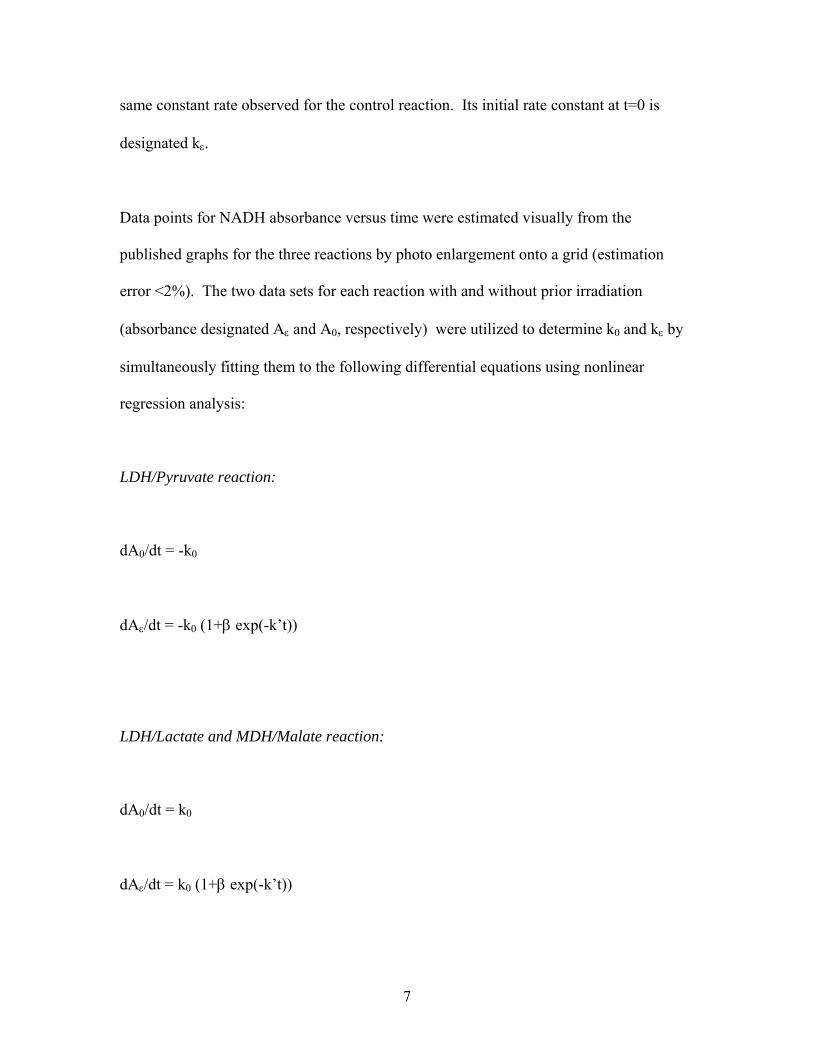
same constant rate observed for the control reaction. Its initial rate constant at t=0 is
designated kε.
Data points for NADH absorbance versus time were estimated visually from the
published graphs for the three reactions by photo enlargement onto a grid (estimation
error <2%). The two data sets for each reaction with and without prior irradiation
(absorbance designated Aε and A0, respectively) were utilized to determine k0 and kε by
simultaneously fitting them to the following differential equations using nonlinear
regression analysis:
LDH/Pyruvate reaction:
dA0/dt = -k0
dAε/dt = -k0 (1+β exp(-k’t))
LDH/Lactate and MDH/Malate reaction:
dA0/dt = k0
dAε/dt = k0 (1+β exp(-k’t))
7

Curve fit values for kε and k0 were converted from absorbance units / sec to moles
NADH / sec noting that for the LDH / pyruvate reaction, the initial absorbance was 0.480
units for the solution containing 0.34 μmoles of NADH.
All calculations were performed using the software package Scientist V.2.01, MicroMath
Inc., Salt Lake City, UT.
For reaction (1),
ΔG*(1) = -RT ln(k0/α) (where α = KBT/h),
and for reaction (2),
ΔG*(2) = -RT ln(kε /α),
so that,
ΔΔG* = ΔG*(2) - ΔG*(1) = -RT ln(kε / k0)
= -594 ln(kε / k0) cal/mole for R = 1.987 cal/deg/mole and T = 299 K.
3. Results The fitted curves for the three reactions are presented in Fig. 1, 2 and 3. The
experimental parameters and calculated reductions in activation free energies, ΔΔG*, are
presented in Table 2.
The LDH / pyruvate data were analyzed further to provide an estimate of the minimum
amount of relevant energy that would have had to be absorbed in the irradiation step,
8

assuming that Є corresponds to photo-excited substrate molecules. The 3 mL reaction
mixture contained 2.2 x 10⁻⁶ moles of pyruvate and 0.025 x 10⁻⁶ g of LDH, estimated to
correspond to 1.8 x 10⁻¹¹ moles of LDH (using MW = 140,000 daltons [9]). Inspection
of the absorbance versus time data for this reaction revealed that the irradiation effect was
associated with a decreased absorbance of 0.030 units that occurs entirely within the first
20 sec of the reaction. Assuming Є corresponds to photo-activated pyruvate molecules
and that 100% of these were converted to lactate, then 2.1 x 10⁻⁸ moles of such molecules
would have been introduced into the reaction mixture. For ΔΔG* = 329 cal/mole, this
implies that the total amount of associated energy transferred into the reaction cuvette
was, minimally, 7.0 x 10⁻⁶ cal. Since one-tenth of the solution of the irradiated crystals
was transferred to the reaction mixture (S. Comorosan, personal communication), the
dissolved crystals would thus have possessed on the order of 7 x 10⁻⁵ cal of transferable
relevant energy. (Data for the slower lactate and malate reactions did not lend
themselves to a corresponding assessment.)
No direct measurements of the amount of irradiation actually absorbed by the crystals
have been reported. In the two reports from which the kinetics data utilized here were
taken, the irradiation intensity was characterized as being 600 lux. However, in a later
publication noting more detailed study of this aspect, Comorosan indicated that the
required irradiation intensity, the illuminance, should be approximately 400 footcandles
[2], corresponding to on the order of 4000 lux. Using this latter figure (assumed more
reliable) along with an estimate of 1 cm2 for the area covered by the sodium pyruvate
crystals and an exposure time of 5 seconds, the total energy to which the crystals were
9

exposed is calculated to be approximately 7.4 x 10⁻⁴ cal. This then would imply that
approximately 10% of the total incident radiation must be captured such as to produce
photo-excited substrate molecules.
4. Discussion On the face of it, one would not expect the crystal irradiation procedure employed in the
studies addressed here to have any measurable impact on a subsequent enzyme reaction
rate. However, Comorosan and co-workers have published 15 primarily experimental
reports involving over 20 different enzymes employing a wide range of reaction rate
determination methodologies all of which display this behavior [10, 11,12,13]. In
addition, 4 collaborative studies [14, 15, 16, 17] conducted in other laboratories and 3
independent studies [18, 19, 20] have been reported. Some of these include single and
double-blind procedures to mitigate against the possibility of experimenter and
procedural bias. While a trivial explanation may underlie the phenomenon, such has not
been uncovered to date. The particular data examined here for LDH / pyruvate are
consistent with other of the above cited reports for that particular reaction, as well as the
body of work as a whole.
Early explanations for the phenomenon assumed that the irradiation procedure placed the
substrate molecule (e.g., pyruvate, malate, etc.) in an excited state that could be detected
(discerned) only by the extreme sensitivity of its enzyme. This model would imply that
the photo-activated entity, here designated Є, is, in fact, a sub-population of the substrate
and would be consumed stoichiometrically in the conversion of substrate to product.
However, it is estimated above that at least 10% of the irradiation incident on the
10

crystalline material would need be absorbed to provide the required activation energy
reduction. This seems highly unlikely, and possibly suggestive of a mysterious light-
matter-biological interaction that lies completely outside contemporary scientific
paradigms. More recently, an alternate model has been proposed [21] wherein the crystal
irradiation process induces oscillatory free radical mediated reactions involving
atmospheric gases at the surface of the crystals. This putative photo-driven process
would be similar to a number of observed temperature-driven oscillatory systems
involving atmospheric gases [22, 23, 24]. On cessation of irradiation, defining the t*
period, much slower dark reactions would lead to relatively stable, water soluble
chemical species which, in turn, are capable of altering reactivity of a particular enzyme.
Thus, a much smaller quantity of the Є species would be required, perhaps only some
fraction of the enzyme molecular concentration rather than the orders of magnitude
higher substrate concentration (here, for the LDH reaction, 1.8 x 10⁻¹¹ vs 2.2 x 10⁻⁶
moles/3 mL).. This behavior would be similar to that observed for the well known action
of nitric oxide, an atmospheric gas photo-product, which can induce over a 40-fold
increase in the rate of conversion of GTP to cAMP by guanylate cyclase [25, 26]. The
body of work on this phenomenon reveals that different enzyme reactions may be
enhanced by different crystal irradiation time periods (e.g., 15 s rather than 5 s exposure).
Accordingly, it should be anticipated that a small set of relevant chemical species are
generated in the photochemical and follow-on dark reactions.
Should this model be proved correct and if the phenomenon indeed reflects a universal
property of enzymes, a new door might be opened to link known physico-chemical
11

processes to pre-biotic evolution and the generalized non-linear dynamics espoused by
others [27, 28] as fundamental to life processes. Moreover, at some level of refinement,
transition state modeling of enzyme reactivity will need to take such a ubiquitous
property into consideration.
12

5. References [1] Ma, B., Kumar, S., Tsai, C., Hu, Z. and Nussinov, R. (2000). Transition-state
ensemble in enzyme catalysis: possibility, reality, or necessity? J. Theor. Biol. 203, 383-
397, doi:10.1006/jtbi.2000.1097.
[2] Comorosan, S. (1976). Biological Observables. In: Progress in Theoretical Biology,
vol. 4 (Rosen, R. ed.) pp. 161-204, New York, Academic Press
[3] Comorosan, S., Jieanu, V., Morlova, I., Paslaru, L., Rosoveanu, P., Toroiman, E., and
Vasilco, R. (1988). A novel interaction between light and matter: a review. Physiol.
Chem. Phys. & Med. NMR 20, 319-328.
[4] Comorosan, S. (1970). New mechanism fo the control of cellular reactions: the
biochemical flip-flop. Nature 227, 64-65.
[5] Comorosan, S. (1975). On a possible biological spectroscopy. Bull. Math. Biol. 37,
419-425.
[6] Comorosan, S., Cru, M. and Vieru, S. (1972). The interaction between enzymic
systems and irradiated substrates. Enzymol. 42, 31-43.
[7] Comorosan, S., Murgoci, P., Sandru, D. and Cru, M (1971c). A new metabolic control
mechanism: III. Physico-chemical aspects of enzyme substrates perturbation. Physiol.
Chem. Phys. 3, 343-352.
[8] Johnson, F.H., Eyring, H. and Stover, B.J. (1974). The theory of rate processes in
biology and medicine. New York, Wiley.
[9] Pesce, A., McKay, R.H., Stolzenbach, F., Cahn, R.D. and Kaplan, N.O. (1964). The
comparative enzymology of lactic dehydrogenase. J. Biol. Chem. 239, 1753-1761.
13
[10] Comorosan, S., Vieru, S., and Sandru, D. (1970). A new approach to control
mechanisms in tumor cells. Europ. J. Can. 6, 393-400.
[11] Comorosan, S., Vieru, S., Crisan, D., Sandru, D., and Murgoci, P. (1971a). A new
metabolic control mechanism: I. Evidence for controlled biochemical networks in yeast
cells. Physiol. Chem. Phys. 3, 1-16.
[12] Comorosan, S., Vieru, S., Crisan, D., Sandru, D, Murgoci, P., Alexandrescu, E.
(1971b). A new metabolic control mechanism: II. Evidence for controlled biochemical
networks in rabbit muscle tissue. Physiol. Chem. Phys. 3, 103-115.
[13] Comorosan, S., Hristea, M. and Murgoci, P. (1980). On a new symmetry in
biological systems. Bull. Math. Biol. 42, 107-117.
[14] Sherman, R.L., Siebert, S.T. and Yee, W.H. (1973). A note on the effect of
electromagnetic field on enzyme substrates. Physiol. Chem. Phys. 5, 49-56.
[15] Goodwin, B.C. and Vieru, S. (1975). Low energy electromagnetic perturbation of an
enzyme substrate. Physiol. Chem. Phys. 8, 89-90.
[16] Bass, G.E. and Crisan, D. (1973). Concerning irradiation-induced biological activity
alterations of tetracycline. Physiol. Chem. Phys. 5, 331-335.
[17] Bass, G.E., Sandru, D., Chenevey, J.E. and Buchovaz, E.T. (1976). The Comorosan
effect: single- and double-blind studies on the urea/urease system. Physiol. Chem. Phys.
8, 253-258.
[18] Bass, G.E. and Chenevey, J.E. (1976). Irradiation induced rate enhancements for the
LDH-pyruvate reaction. Int. J. Quant. Chem. Quant. Biol. Symp. 3, 247-250.
14
[19] Bass, G.E. and Chenevey, J.E. (1977). Substrate irradiation stimulation of the in
vitro lactate-pyruvate interconversion reactions mediated by lactic dehydrogenase.
Physiol. Chem. Phys. 9, 555-562.
[20] Etzler, F.M. and Westbrook, D. (1986). Modulation of reaction kinetics via an
apparently novel interaction of light and matter. Physiol. Chem. Phys. Med. NMR 19,
271-274.
[21] Bass, G.E. (2005). Crystal Irradiation Stimulation of Enzyme Reactivity (CISER): A
Possible Paradigmatic Explanation, 24 Annual Meeting, Society for Scientific
Exploration, Gainesville, FL.
[22] Gray, P., Griffiths, J.F. and Scott, S.K. (1985). Oscillations, glow and ignition in
carbon monoxide oxidation in an open system: I. Experimental studies of the ignition
diagram and the effects of added hydrogen. Proc. R. Soc. Lond. A 397, 21-44.
[23] Cirak, F., Cisternas, J.E., Cuitino, A.M., Ertl, G., Holmes, P., Kevrekidis, I.G., Ortiz,
M., Rotermund, H.H., Schunack, M. and Wolff, J. (2003). Oscillatory thermomechanical
instability of an ultrathin catalyst. Science 300, 1932-1936.
[24] Gray, P. and Scott, S.K. (1990). Chemical Oscillations and Instabilities: Non-Linear
Chemical Kinetics. Clarendon Press, Oxford.
[25] Wolin, M.S., Wood, K.S. and Ignarro, L.J. (1982). Gyanylate cyclase from bovine
lung. J. Biol. Chem. 257, 13312-13320.
[26] Hobbs, A.J. and Ignarro, L.J. (1996). Nitric oxide – cyclic GMP signal transduction
system. Methods Enzymol. 269, 134-148.
[27] Nicolis, G. and Pregogine, I. (1989). Exploring Complexity. W.H. Freeman & Co., New York. [28] Kauffman, S.A. (1993). The Origins of Order. Oxford Univ. Press, New York.
15
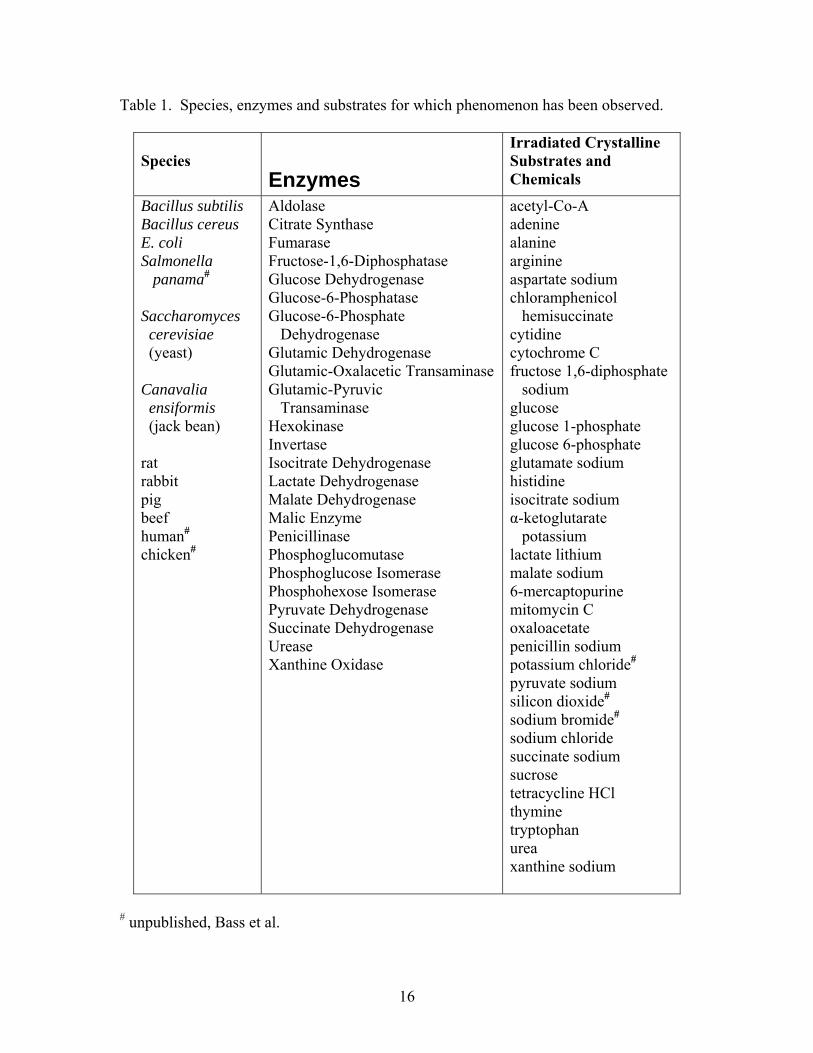
Table 1. Species, enzymes and substrates for which phenomenon has been observed.
Species
Enzymes
Irradiated Crystalline Substrates and Chemicals
Bacillus subtilis Bacillus cereus E. coli Salmonella panama#
Saccharomyces cerevisiae (yeast) Canavalia ensiformis (jack bean) rat rabbit pig beef human#
chicken#
Aldolase Citrate Synthase Fumarase Fructose-1,6-Diphosphatase Glucose Dehydrogenase Glucose-6-Phosphatase Glucose-6-Phosphate Dehydrogenase Glutamic Dehydrogenase Glutamic-Oxalacetic Transaminase Glutamic-Pyruvic Transaminase Hexokinase Invertase Isocitrate Dehydrogenase Lactate Dehydrogenase Malate Dehydrogenase Malic Enzyme Penicillinase Phosphoglucomutase Phosphoglucose Isomerase Phosphohexose Isomerase Pyruvate Dehydrogenase Succinate Dehydrogenase Urease Xanthine Oxidase
acetyl-Co-A adenine alanine arginine aspartate sodium chloramphenicol hemisuccinate cytidine cytochrome C fructose 1,6-diphosphate sodium glucose glucose 1-phosphate glucose 6-phosphate glutamate sodium histidine isocitrate sodium α-ketoglutarate potassium lactate lithium malate sodium 6-mercaptopurine mitomycin C oxaloacetate penicillin sodium potassium chloride#
pyruvate sodium silicon dioxide#
sodium bromide#
sodium chloride succinate sodium sucrose tetracycline HCl thymine tryptophan urea xanthine sodium
# unpublished, Bass et al.
16

Table 2. Reaction Rate Parameters
Reaction enzyme/substrate/cofactor
Substrate μmoles/3 ml
Cofactor μmoles/3 ml
Enzyme U/3 ml
t*, sec
kε,10-9
k0, 10-9
ΔΔG*, cal/mole
Ref
LDH/Na pyruvate/NADH 2.20 0.34 9x10⁻³ 5 4.95 2.84 -329 a LDH/Li lactate/NAD 4.10 3.0 18x10-3 15 4.51 3.25 -195 a MDH/Na malate/NAD 64 3.0 450x10-3 25 35.7 24.6 -221 b Temperature = 299 K for each reaction. k0 = initial reaction rate constant obtained using non-irradiated substrate, moles NADH / sec. kε = initial reaction rate constant obtained using irradiated substrate, moles NADH / sec. t* = duration of crystalline substrate irradiation at 546 nm. ΔΔG* = -594 ln(kε / k0) cal/mole. References: a. S. Comorosan et al., 1972; b. S. Comorosan et al., 1971c. LDH = lactic dehydrogenase; MDH = malic dehydrogenase; NAD = nicotinamide dinucleotide; NADH = nicotinamide dinucleotide, reduced.
17

Fig. 1. Computed Curves for the Lactic Dehydrogenase / Pyruvate Reaction. Data points estimated from Comorosan et al., 1972, Figure 3A. George E. Bass, Bernd Meibohm, James T. Dalton and Robert Sayre
Reaction time (sec)
Abs
orba
nce
(340
nm
)
0 10 20 30 40 50 600.20
0.25
0.30
0.35
0.40
0.45
0.50
w/o substrate irradiation
with substrate irradiation
18

Fig. 2. Computed Curves for the Lactic Dehydrogenase / Lactate Reaction. Data points estimated from Comorosan et al., 1972, Figure 3B. George E. Bass, Bernd Meibohm, James T. Dalton and Robert Sayre
Reaction time (sec)
Abs
orba
nce
(340
nm
)
0 10 20 30 40 50 600.11
0.12
0.13
0.14
0.15
w/o substrate irradiation
with substrate irradiation
19

Fig. 3. Computed Curves for the Malic Dehydrogenase / Malate Reaction. Data points estimated from Comorosan et al., 1971c, Figure 1B., George E. Bass, Bernd Meibohm, James T. Dalton and Robert Sayre
Reaction time (sec)
Abso
rban
ce (3
40 n
m)
0 10 20 30 40 50 600.10
0.15
0.20
0.25
0.30
0.35
0.40
w/o substrate irradiation
with substrate irradiation
20
Acknowledgement
The authors wish to express their appreciation to Dr. Loys Nunez for suggestions and comments during the preparation of this manuscript.
21