0dwhuldo (6, iru0hg&khp&rpp expressing, breast cancer … · an agent for optical imaging...
TRANSCRIPT

An Agent For Optical Imaging Of TrkC-
Expressing, Breast Cancer
Supporting Information
Anyanee Kamkaew,1,2 Feng Li,3 Zheng Li,*,3 and Kevin Burgess*1
1 Department of Chemistry, Texas A & M University, Box 30012, College Station, TX 77842, USA
2 School of Chemistry, Institute of Science, Suranaree University of Technology, Nakhon Ratchasima
30000, Thailand
3 Center for Bioenergetics, Houston Methodist Research Institute, Houston, TX 77030, USA
Materials and Methods ....................................................................................................................2
General Procedures ......................................................................................................................2
Syntheses of Probes 1a and 1a-control ...........................................................................................3
Synthesis of probe 1b ....................................................................................................................11
MTT Cell Viability Assays with 1a in 4T1 cells...........................................................................14
Histochemistry of anti-TrkC on human breast tissue array ...........................................................15
References......................................................................................................................................16
S1
Electronic Supplementary Material (ESI) for MedChemComm.This journal is © The Royal Society of Chemistry 2017

Materials and Methods
General Procedures
All reactions were carried out under an atmosphere of argon. Unless otherwise indicated,
common reagents or materials were obtained from commercial source and used without further
purification. All α-amino acids used were of the L-configuration. Dry DMF, (<50 ppm water)
was purchased from Acros. Tetrahydrofuran (THF), Acetonitrile (MeCN), dichloromethane
(CH2Cl2), and methanol (MeOH) were dried by Mbraun solvent drying system. Other solvents
and reagents were used as received.
NMR spectra were recorded on a Bruker-400 MHz spectrometers (1H at 400 MHz and
13C at 100 MHz) at room temperature unless other mentioned. Chemical shifts of 1H NMR
spectra were recorded and chemical shifts are reported in ppm from the solvent resonance
(CDCl3 7.26 ppm, CD3OD 3.30 ppm, DMSO-d6 2.50 ppm). Data are reported as follows:
chemical shift, multiplicity (s = singlet, br = broad, d = doublet, t = triplet, q = quartet, m =
multiplet), coupling constants, and number of protons. Proton decoupled 13C NMR spectra were
also recorded in ppm from tetramethylsilane (TMS) resonance (CDCl3 77.0, CD3OD 49.1,
DMSO-d6 39.5 ppm). Analytical thin layer chromatography (TLC) was performed on EM
Reagents 0.25 mm silica-gel 60-F plates, and visualized with UV light. Flash chromatography
was performed using silica gel 60 (230–400 mesh). MS were measured under ESI or MALDI
conditions.
Analytical HPLC analyses were carried out on 150 x 4.6 mm C-18 column using gradient
conditions (10 – 90% B, flow rate = 0.75 mL/min). Preparative HPLC was carried out on 100 x
21.2 mm C-18 column using gradient conditions (10 – 70% B, flow rate = 10.0 mL/min). The
eluents used were: solvent A (H2O with 0.1% AcOH) and solvent B (CH3CN with 0.1% AcOH).
S2

The purity of all biologically evaluated compounds is > 95% confirmed by analytical
HPLC.
Throughout the confocal imaging studies, the laser used for excitation was a 633 nm
HeNe, with an emission bandwidth of 700 – 750 nm.
Syntheses of Probes 1a and 1a-control
Scheme S1. Synthesis of 1a.
S3

S4

Synthesis of compound 3
Tyrosine azide1 (41 mg, 0.2 mmol) was dissolved in DMF (0.4 mL), then cooled to 0 oC. EDCI
(41 mg, 0.22 mmol) and HOAt (20 mg, 0.21 mmol) were added to the solution. After stirring at 0
oC for 30 min, Compound 2, synthesized according to previous report,2 (35 mg, 0.04 mmol) was
added to the above suspension followed by iPr2EtN (70 μL, 0.4 mmol). Then, the resulting
solution was warmed to 25 oC and stirred for 24 h. Solvent was removed under reduced pressure.
After that, the residue was purified by reverse phase MPLC using H2O:CH3CN (gradient) to
afford 3 as a green powder (30 mg, 60 %). 1H-NMR (400 MHz, DMSO-d6) δ 10.28 (s, 2H), 9.23
(s, 2H), 8.61 (d, J = 8.9 Hz, 2H), 8.61-8.60 (m, 6H), 8.17 (d, J = 8.8 Hz, 2H), 7.94 (d, J = 7.6 Hz,
2H), 7.68 (t, J = 8.0 Hz, 2H), 7.51 (s, 2H), 7.16-7.09 (m, 6H), 6.69 (d, J = 8.8 Hz, 2H), 4.66-4.61
(m, 2H), 4.09-4.05 (m, 2H), 3.59 (s, 6H), 3.13-3.11 (m, 2H), 3.04-2.99 (m, 6H). 13C (100 MHz,
DMSO-d6) δ 169.4, 162.5, 157.9, 156.6, 143.1, 140.0, 132.5, 132.3, 130.5, 129.6, 128.9, 127.5,
123.7, 120.0, 63.8, 56.1, 54.0, 52.4, 49.0, 42.3. 11B NMR (128 MHz, CDCl3) δ 0.88 (t, J = 32.3
Hz). HRMS (MALDI) calcd for C58H54BF2N13NaO13S2 {M+Na}+ 1276.3364, found 1276.4642.
S5

1H-NMR of compound 3
13C-NMR of compound 3
S6

Synthesis of targeting probe 1a
3 (50 mg, 0.04 mmol) and Boc-Isoleucine alkyne1 (20 mg, 0.09 mmol) were dissolved in DMSO
(1 mL). Then, aqueous solution of CuSO4 (0.1 M, 78 L, 0.008 mmol) and sodium ascorbate
(0.2 M, 156 L, 0.032 mmol) were added to the mixture at 25 °C. The reaction was stirred at 25
°C for 24 h (monitored by C18-TLC using H2O:CH3CN (1:1) as solvents). Solvent was removed
under reduced pressure. After that, the residue was purified by reverse phase MPLC using
H2O:CH3CN (gradient) to afford Boc-1a as a green powder (39 mg, 58 %). Subsequently, Boc-
1a (39 mg, 0.023 mmol) was dissolved in 1,4-dioxane (0.5 mL). Then HCl in 1,4-dioxane (2 M,
0.5 mL) was added into the solution. Reaction mixture was stirred at 25 °C for 1h, then solvent
was removed under reduced pressure to give desired product 1a as green solid quantitatively.
1H-NMR (400 MHz, DMSO-d6) δ 10.32 (br, 2H), 8.85 (br, 2H), 8.37-8.33 (m, 6H), 8.15 (d, J =
8.5 Hz, 2H), 7.92-7.89 (m, 2H), 7.68-7.46 (m, 4H), 7.37 (d, J = 8.8 Hz, 2H), 7.20 (d, J = 8.8 Hz,
2H), 7.12 (d, J = 9.0 Hz, 2H), 6.91 (d, J = 7.2 Hz, 4H), 6.53 (d, J = 7.8 Hz, 4H), 5.69-5.67 (m,
2H), 4.68-4.67 (m, 2H), 4.32 (br, 2H), 3.89 (s, 6H), 3.44-3.31 (m, 2H), 3.04-2.90 (m, 2H), 2.89-
2.86 (m, 2H), 1.93-1.90 (m, 2H), 1.29-1.24 (m, 4H), 1.05-0.90 (m, 2H), 0.89-0.75 (m, 6H), 0.74-
0.68 (m, 6H). 13C (100 MHz, DMSO-d6) δ 169.4, 167.7, 162.5, 156.6, 145.0, 143.0, 142.3,
139.9, 132.5, 132.2, 130.3, 129.2, 128.5, 126.5, 125.3, 124.0, 123.8, 123.6, 120.1, 115.6, 115.5,
115.4, 115.0, 64.9, 56.1, 53.9, 52.4, 51.5, 51.0, 42.6, 37.6, 34.6, 25.6, 18.5, 17.2, 14.2, 14.1, 11.5.
S7

11B NMR (128 MHz, CDCl3) δ 0.88 (t, J = 32.3 Hz). HRMS (MALDI) calcd for
C72H77BF2N15O14S2 {M-H}- 1488.5277, found 1488.6775.
1H-NMR of compound 1a
S8

13C-NMR of compound 1a
11B-NMR of compound 1a
S9

Synthesis of 1a-control
1a-control was synthesized according the same procedure as 1a by using isoleucine azide1 and
Boc-tyrosine alkyne1 as precursors. 1H-NMR (400 MHz, DMSO-d6) δ 9.28 (br, 2H), 8.57 (br,
6H), 8.36 (s, 2H), 8.17 (d, J = 8.7 Hz, 2H), 7.92 (d, J = 7.6 Hz, 2H), 7.73 (d, J = 7.8 Hz, 2H),
7.44-7.41 (m, 4H), 7.15 (d, J = 8.9 Hz, 4H), 6.91 (d, J = 8.3 Hz, 4H), 6.61 (d, J = 8.3 Hz, 4H),
5.43 (d, J = 7.8 Hz, 2H), 4.63 (br, 2H), 3.90 (s, 6H), 3.67-3.64 (m, 2H), 3.43-3.41 (m, 2H), 3.10-
2.98 (m, 2H), 2.34-2.28 (m, 2H), 1.18-1.24 (m, 9H), 1.04-1.02 (m, 6H), 0.84-0.82 (m, 12H). 13C
(100 MHz, DMSO-d6) δ 169.4, 167.5, 162.5, 157.0, 156.7, 145.2, 144.1, 132.3, 130.9, 130.8,
126.9, 126.1, 125.6, 123.6, 123.2, 120.1, 115.7, 115.6, 115.0, 95.4, 80.0, 79.1, 72.9, 72.6, 71.5,
68.1, 67.4, 65.9, 63.3, 60.6, 53.9, 52.7, 49.2, 43.9, 42.6, 42.1, 38.5, 38.3, 29.4, 24.7, 24.5, 18.5,
17.2, 12.7, 11.1, 10.4. 11B NMR (128 MHz, CDCl3) δ 0.90 (t, J = 32.0 Hz). HRMS (MALDI)
calcd for C72H78BF2N15NaO14S2 {M+Na}+ 1512.5253, found 1512.5082.
S10

1H-NMR of compound 1a-control
13C-NMR of compound 1a-control
S11

Synthesis of probe 1b
Scheme S2. Synthesis of 1b.
EDCI (210.9 mg, 1.10 mmol) and HOAt (152.4 mg, 1.12 mmol) were added to a suspension of
azido tyrosine1 (207.2 mg, 1.00 mmol) in 4 mL DMF at 0 oC. After stirring at 0 oC for 30 min, Z
(117.2 mg, 0.127 mmol) was added to the above suspension followed by iPr2EtN (557 μL, 413.6
mg, 3.20 mmol). The resulting solution was stirred at 0 °C for 1 h and warmed to 25 oC and
stirred for overnight. Ethyl acetate (ca. 100 ml) was added to the reaction mixture, and the
resulting suspension was washed with 5% HCl, H2O, sat. NaHCO3 and brine. The organic layer
was separated, dried with Na2SO4, and concentrated to afford green solid. The crude product
was purified by column chromatography on silica gel, and eluted with a mixture of CH2Cl2 and
methanol (98:2, v/v) afforded the desired azide compound (99.1 mg, 81%) as green solid. The
resulting product (38.6 mg, 0.04 mmol) was then mixed with Boc-isoleucine alkyne1 (40 mg,
0.18 mmol) in DMSO (1 mL). Then, aqueous solution of CuSO4 (0.1 M, 78 L, 0.008 mmol)
and sodium ascorbate (0.2 M, 156 L, 0.032 mmol) were added to the mixture at 25 °C. The
reaction was stirred at 25 °C for 24 h (monitored by TLC). Solvent was removed under reduced
S12

pressure. The residue was purified by flash silica chromatography eluting with CH2Cl2:MeOH
(98:2) to yield 36.1 mg of Boc-1b (65 %) as a green powder. After removing a protecting group,
1b was obtained quantitatively as a green solid. 1H-NMR (400 MHz, DMSO-d6) δ 11.14 (s, 2H),
9.27 (s, 2H), 8.54-8.45 (m, 6H), 8.20 (s, 2H), 8.17 (d, J = 8.8 Hz, 2H), 7.95 (d, J = 7.8 Hz, 2H),
7.71 (d, J = 7.8 Hz, 2H), 7.44-7.39 (m, 4H), 7.16 (d, J = 8.9 Hz, 4H), 7.06 (d, J = 8.3 Hz, 4H),
6.62 (d, J = 8.3 Hz, 4H), 5.95-5.90 (m, 2H), 4.39-4.37 (m, 2H), 3.90 (s, 6H), 3.73-3.70 (m, 4H),
2.0-1.8 (m, 2H), 1.36-1.24 (m, 9H), 0.94-0.90 (m, 2H), 0.85-0.89 (m, 6H), 0.76 (d, J = 7.2 Hz,
6H). 11B NMR (128 MHz, CDCl3) δ 0.88 (t, J = 32.5 Hz). HRMS (MALDI) calcd for
C66H67BF2N13O6 {M-H}- 1186.5398, found 1186.1261.
1H-NMR of compound 1b
S13

11B-NMR of compound 1b
HRMS-MALDI of compound 1b
S14

MTT Cell Viability Assays with 1a in 4T1 cells
4T1 cells (5,000-7,000 cells/well, 50 L in completed Dulbecco’s Modified Eagle
Medium (DMEM, Sigma Chemical, St. Louis, MO) were plated on 96-well plates and allowed to
adhere at 37 °C in 5 % CO2 and 95 % air for 24 h. Thereafter, the cells were treated with 50 L
aliquot of each test compounds in cell medium at different concentrations, ranging from 0-25
M. The cells were then incubated for 24 h. The cell’s viability was assessed through an MTT
conversion assay.3 Briefly, 20 L of MTT (5mg/mL, in Hank’s balanced salt solution, HBSS)
were added and the cells were incubated for an additional 3 h. Afterwards, the media were
replaced with 100 L dimethyl sulfoxide (DMSO) to solubilize the blue crystals. The plate was
shaken for 15 min at room temperature before measuring the optical density (OD) at 570 nm
using SpectraMax Plus 384 microplate reader. Cell viability (%) = (mean of OD of treatment
group/mean of OD of control group) × 100.
Figure S1. Cell viability of 1a in 4T1 cells. Targeting probe 1a does not cause toxicity to 4T1
cells up to concentration at 25 M.
S15
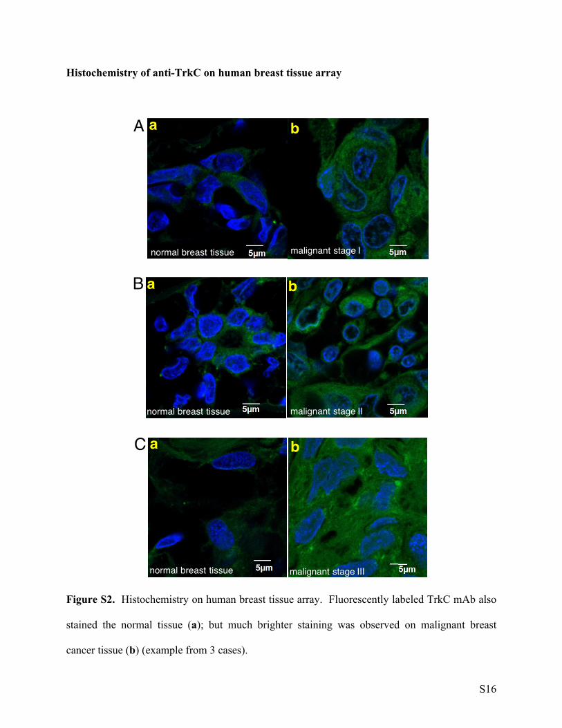
Histochemistry of anti-TrkC on human breast tissue array
Figure S2. Histochemistry on human breast tissue array. Fluorescently labeled TrkC mAb also
stained the normal tissue (a); but much brighter staining was observed on malignant breast
cancer tissue (b) (example from 3 cases).
S16

References
1. Angell, Y.; Chen, D.; Brahimi, F.; Saragovi, H. U.; Burgess, K. A Combinatorial
Method for Solution-Phase Synthesis of Labeled Bivalent β-Turn Mimics. J. Am. Chem. Soc.
2008, 130, 556-565
2. Kamkaew, A.; Burgess, K. Aza-BODIPY Dyes with Enhanced Hydrophilicity. Chem.
Comm. 2015, 51, 10664-10667.
3. Mosmann, T., Rapid colorimetric assay for cellular growth and survival: application to
proliferation and cytotoxicity assays. J. Immunol. Methods 1983, 65 (1-2), 55-63.
S17