1 clostridium difficile mazf toxin exhibits selective, not global
TRANSCRIPT

Clostridium difficile MazF Toxin Exhibits Selective, Not Global,mRNA Cleavage
Francesca P. Rothenbacher,a Motoo Suzuki,e Jennifer M. Hurley,a* Thomas J. Montville,d Thomas J. Kirn,c Ming Ouyang,b andNancy A. Woychika
Department of Molecular Genetics, Microbiology and Immunology, UMDNJ-Robert Wood Johnson Medical School, Piscataway, New Jersey, USAa; Computer Engineering& Computer Science Department, University of Louisville, Louisville, Kentucky, USAb; Department of Pathology and Laboratory Medicine, UMDNJ-Robert Wood JohnsonMedical School, New Brunswick, New Jersey, USAc; Department of Food Science, Rutgers, the State University of New Jersey, School of Environmental and BiologicalSciences, New Brunswick, New Jersey, USAd; and Department of Microbiology, Faculty of Medicine, Kagawa University, Kagawa, Japane
Clostridium difficile is an important, emerging nosocomial pathogen. The transition from harmless colonization to disease istypically preceded by antimicrobial therapy, which alters the balance of the intestinal flora, enabling C. difficile to proliferate inthe colon. One of the most perplexing aspects of the C. difficile infectious cycle is its ability to survive antimicrobial therapy andtransition from inert colonization to active infection. Toxin-antitoxin (TA) systems have been implicated in facilitating persis-tence after antibiotic treatment. We identified only one TA system in C. difficile strain 630 (epidemic type X), designatedMazE-cd and MazF-cd, a counterpart of the well-characterized Escherichia coli MazEF TA system. This E. coli MazF toxin cleavesmRNA at ACA sequences, leading to global mRNA degradation, growth arrest, and death. Likewise, MazF-cd expression in E.coli or Clostridium perfringens resulted in growth arrest. Primer extension analysis revealed that MazF-cd cleaved RNA at thefive-base consensus sequence UACAU, suggesting that the mRNAs susceptible to cleavage comprise a subset of total mRNAs. Inagreement, we observed differential cleavage of several mRNAs by MazF-cd in vivo, revealing a direct correlation between thenumber of cleavage recognition sites within a given transcript and its susceptibility to degradation by MazF-cd. Interestingly,upon detailed statistical analyses of the C. difficile transcriptome, the major C. difficile virulence factor toxin B (TcdB) andCwpV, a cell wall protein involved in aggregation, were predicted to be significantly resistant to MazF-cd cleavage.
Clostridium difficile is a Gram-positive, spore-forming anaer-obe that has recently emerged as an important nosocomial
pathogen. The normal colonic flora usually serves to repress in-fection by this pathogen. However, in patients receiving antimi-crobial therapy, C. difficile populations are able to increase as thenormal flora decreases, typically resulting in infection (C. difficileinfection, or CDI) that manifests clinically as severe diarrhea (8,43). One of the most challenging aspects of C. difficile-associateddisease is the high incidence of recurrent infections, due either toreinfection by spore ingestion or relapse resulting from the germi-nation of spores that remained in the intestinal lumen after theinadequate treatment of the primary infection (37). The incidenceand severity of nosocomial CDI have been increasing in the pastdecade, and treatment is difficult and expensive. The emergence ofthe highly virulent BI/NAP1/027 (toxinotype III) strain accountsfor some of this increase in severity and contributes to rising mor-tality rates. Progressively more cases of community-acquired (ver-sus nosocomial) disease are being documented, and the numberof cases in those who were considered to be at low risk of acquiringinfections (generally healthy individuals who were not in hospitalsor taking antibiotics) is also rising (43).
The virulence of this pathogen is attributed to two major exo-toxins, toxin A and toxin B, and the binary toxin CDT (Clostrid-ium difficile transferase). Contrasting reports on the pathologicalimportance of the toxins have recently been published. One groupfound that toxin B alone was essential for C. difficile virulence (30),while another found that both A�B� and A�B� strains can causedisease in a hamster model (28). Both toxin A and B proteins aremonoglucosyltransferases that cause the disruption of tight junc-tions and the actin cytoskeleton, resulting in the wide-scale de-struction of the intestinal epithelium. The cytotoxic and inflam-
matory pathological features characteristic of the disease areattributed to the physical effects of the toxins. The role of CDT indisease is not well understood but has recently been shown toincrease microtubule formation to presumably facilitate bacterialadherence (47). Surface layer proteins involved in the adherenceof the pathogen to the intestinal mucosa are also considered to beimportant for C. difficile pathogenesis. Surface layer protein A,SlpA, was recently demonstrated to be altered in certain hyper-virulent strains, facilitating tighter C. difficile adherence (43). Fi-nally, the ability to form spores is a major phenomenon associatedwith C. difficile virulence. The spores are highly resistant to desic-cation, chemical shock, and extreme temperatures and are themost important mode of dissemination.
Recent reports suggest a link between pathogenesis and toxin-antitoxin (TA) systems (40, 54, 55). TA systems comprise a stabletoxin and an unstable antitoxin. TA toxins, in contrast to exotox-ins, are intracellular and only affect an essential process, such astranslation or replication within the producing cell (11, 16, 34).The antitoxin is able to sequester the effects of the toxin throughthe formation of a stable complex. When physiological conditions
Received 14 February 2012 Accepted 16 April 2012
Published ahead of print 27 April 2012
Address correspondence to Nancy A. Woychik, [email protected].
* Present address: Jennifer M. Hurley, Department of Genetics, Dartmouth MedicalSchool, Hanover, New Hampshire, USA.
Supplemental material for this article may be found at http://jb.asm.org/.
Copyright © 2012, American Society for Microbiology. All Rights Reserved.
doi:10.1128/JB.00217-12
3464 jb.asm.org Journal of Bacteriology p. 3464–3474 July 2012 Volume 194 Number 13
Dow
nloa
ded
from
http
s://j
ourn
als.
asm
.org
/jour
nal/j
b on
23
Dec
embe
r 20
21 b
y 1.
1.16
5.10
1.

favor the degradation of the antitoxin by proteases, the toxin isfree to act on the cell.
The MazE-MazF TA system has been studied in detail in sev-eral bacteria. Escherichia coli MazF is a sequence-specific endori-bonuclease that cleaves single-stranded mRNA at ACA sequences(53); the MazE antitoxin inhibits the cleavage of mRNA by MazF.MazF facilitates bacterially programmed cell death in E. coli andMyxococcus xanthus which is mechanistically distinct from that ineukaryotic cells (3, 26, 33). Although MazF toxins in two patho-gens (Mycobacterium tuberculosis and methicillin-resistant Staph-ylococcus aureus [MRSA]) are also sequence-specific endoribonu-cleases, they have 5-nucleotide (nt) recognition sequences whichare proposed to selectively target distinct mRNA transcripts fordegradation (54, 55).
Although CDIs cause an estimated 15,000 to 20,000 deaths peryear in the United States (43) and C. difficile treatment results inthe expenditure of billions of health care dollars annually, ourunderstanding of the spectrum of molecular mechanisms (apartfrom its exotoxins) that contribute to its pathogenicity, virulence,and ability to survive antibiotic treatment are not known. To thisend, we studied the characteristics of the MazEF-cd TA system inC. difficile. As in Mycobacterium tuberculosis and Staphylococcusaureus, the MazF-cd toxin appears to selectively target a subset oftranscripts for degradation by the recognition of a 5-nt consensussequence followed by cleavage. Interestingly, this UACAU con-sensus sequence was significantly underrepresented in mRNAs fortoxin B, the cell wall protein CwpV, and two proteases (Lon andClpC) which may play a role in MazE-cd antitoxin degradation.
MATERIALS AND METHODSStrains, plasmids, and reagents. The E. coli strains BL21(DE3) [F� ompThsdS�(r�-m�) dcm gal(DE3) tonA] (Novagen) and BW25113 (lacIq
rrnBT14 �lac-ZWJ16 hsdR514 �araBADAH33 �rhaBADLD78) were used forall protein expression and toxicity studies. E. coli K-12 Mach1 T1 cells[�recA1398 endA1 tonA �80�lacM15 �lacX74 hsdR(rk
� mk�); Invitro-
gen] were used for all cloning experiments. CD3461 (mazF-cd) andCD3462 (mazE-cd) genes were PCR amplified from C. difficile 630genomic DNA (ATCC BAA-1382D-5). mazF-cd was cloned into pBAD33(17) with NdeI/HindIII ends to create pBAD33-mazF-cd. mazE-cd wascloned into pINIII using NdeI/BamHI ends to create pINIII-mazE-cd.mazF-cd and mazE-cd were also cloned into pColdTF-FT using NdeI/HindIII or NdeI/BamHI, respectively, to create pColdTF-FT-mazF-cdand pColdTF-FT-mazE-cd for expression. pColdTF-FT is a derivative ofpColdTF (TaKaRa Bio) and was created by inserting an in-frame flagepitope tag before the NdeI sequence of the multiple cloning site; thisplasmid was a generous gift from Jared Sharp. Cultured E. coli was grownin M9 minimal media supplemented with either 0.2% glucose or 0.21%glycerol at 37°C unless otherwise noted. The working concentrations ofampicillin and chloramphenicol were 100 and 34 �g/ml, respectively. TheDNA sequences of PCR fragments used for cloning were confirmed byautomated DNA sequence analysis. All oligonucleotides used in this studyare listed in Table 1.
Transformation and expression in C. perfringens. The pCM14 plas-mid was created from a pCM7 backbone, a derivative of pCM3 (GenBankaccession no. AB562893). pCM3 contains origins of replication for E. coliand C. perfringens, the cat (chloramphenicol acetyltransferase) gene con-ferring high-level chloramphenicol resistance, and a multiple cloning site.pCM7 was digested with PacI and NheI and ligated to the rrnB terminatorcontaining compatible ends. The bgaR-bgaL promoter fragment was in-serted into the AvrII and NdeI sites. The NdeI site of the bgaR gene wasremoved by site-directed mutagenesis. Truncated genes (such as bla andcop) were removed by mutagenesis. A similar expression plasmid, usingthe same promoter and terminator, was recently published (19). pCM14-
mazF-cd or pCM14 (see Fig. S1 in the supplemental material) was trans-formed into C. perfringens strain 13 as previously described (2). Bacteriawere cultured anaerobically in modified R & S (maltose) medium con-taining 10 �g/ml chloramphenicol at 37°C, and growth was monitored byoptical density. Modified R & S (maltose) medium is based on R & Smedium (14, 42). Induction was performed by the addition of lactose to0.5%.
Preparation of recombinant MazE-cd and MazF-cd. pColdTF-FT-mazF-cd and pColdTF-FT-mazE-cd BL21(DE3) transformants were usedto inoculate 1 liter of M9 liquid medium and grown to an optical densityat 600 nm (OD600) of 0.3 to 0.4. The culture was transferred to a 15°Cwater bath and incubated for 30 min before being induced with 1 mMisopropyl-�-D-thiogalactopyranoside (IPTG) and expressed for 18 h.Cells were subsequently disrupted by sonication, and the protein extractswere applied to nickel-nitrilotriacetic acid (Ni-NTA) resin (Qiagen) topurify the proteins as recommended by Qiagen. Elutions were pooled andsubjected to thrombin cleavage to excise trigger factor (TF)-(His)6 fromthe target protein. Recombinant proteins were further purified over ananti-FLAG resin to remove TF from the flag-tagged MazF-cd. All elutionfractions were visualized by 17.5% sodium dodecyl sulfate-polyacrylam-ide gel electrophoresis (SDS-PAGE) and stained with Coomassie blue.
Determination of MazF-cd-FT activity in vitro. MS2 RNA (1.6 �g)was added to a reaction mixture containing either MazF-cd-FT or MazE-cd-FT in 10 mM Tris-HCl, pH 7.8, for 15 min at 37°C. Increasing ratios oftoxin and antitoxin were preincubated for 30 min at 4°C, followed by 5min at room temperature before being added to MS2 RNA. The reactionproducts were then separated on a 1% agarose-morpholinepropanesulfo-nic acid (MOPS)-formaldehyde gel and visualized by ethidium bromidestaining.
MS2 primer extension analysis. The MazF-cd MS2 cleavage assayswere carried out as previously described (56). Full-length MS2 RNA waspartially digested with or without purified toxin protein MazF-cd-FT andwith or without purified CspA(His)6 protein at 37°C for 15 min. Thedigestion reaction mixture (10-�l total volume) consisted of 0.8 �g ofMS2 RNA substrate, 1.25 �M MazF-cd-FT, 32 �g of CspA(His)6, and 0.5
TABLE 1 Oligonucleotides used in this work
Primername Sequence
B4 5=-GGTCCGTCCCACCGAAGAAC-3=C 5=-GACACGAACGTTTTACGAAG-3=D2 5=-TCTCTATTTATCTGACCGCG-3=E1 5=-TGCATTGCCTTAACAATAAG-3=G 5=-GAGCCGTTGCCTGATTAATG-3=H 5=-AGCGTCAACGCTTATGATGG-3=J1 5=-GACGGCCATCTAACTTGATG-3=K 5=-GCGGAGCGCCTGGCGCC-3=1351 5=-GATCGATTGATCATTCAGG-3=ompF 5=-CCCACAGCAACGGTGTCGTC-3=tufA-1 5=-CTTCCATTTCAACCAGTTCC-3=tufA-2 5=-CCGGCATTACCATCTCTACG-3=NWO1845 5=-GCAACCTAGCTTCTCTAATATAGTAAGATTAGTTTT
ATAATC-3=NWO1760 5=-CGCGGATCCATGGAAATATGTAGTTCTAATAGCAT
AAGAAATATGG-3=NWO1848 5=-GGTTTATAAATTAGGATTTTTAAATTACAAGAGCT
TTGAATATTTAGGC-3=NWO1849 5=-GTTCCTGTCATTCCTTCGTTTAACTTAATAGATAA
TTCAC-3=NWO1861 5=-CTTGGCTCTGATTTCTTCGCTTGCTCTGTTTTAG
ACG-3=NWO1862 5=-CGTTGTTTTTTAACCAACATAAGTGAGTGTTATTT
CCG-3=
MazF-cd Targets mRNAs for Cleavage at UACAU
July 2012 Volume 194 Number 13 jb.asm.org 3465
Dow
nloa
ded
from
http
s://j
ourn
als.
asm
.org
/jour
nal/j
b on
23
Dec
embe
r 20
21 b
y 1.
1.16
5.10
1.
�l of RNase inhibitor (Roche) in 10 mM Tris-HCl (pH 7.8). Primer ex-tensions were carried out at 47°C for 1 h in a 20-�l reaction mixture. Thereactions were stopped by the addition of 12 �l of sequence loading buffer(95% formamide, 20 mM EDTA, 0.05% bromphenol blue, and 0.05%xylene cyanol EF). The samples were incubated at 90°C for 5 min prior toelectrophoresis on a 6% polyacrylamide, 6 M urea gel. Primers were 5=-end labeled with [32P]ATP using T4 polynucleotide kinase. An IPTG-inducible pColdI-cspA plasmid was generously provided by Yoshi Yama-guchi to generate CspA(His)6 pure protein that was purified over Ni-NTAresin (Qiagen).
Analysis of steady-state mRNA levels. Total RNA was extracted usinga hot phenol method. The radiolabeled DNA fragments used for Northernanalysis were derived from PCR products comprising open readingframes (ORFs) of the E. coli genes lpp (major outer membrane lipopro-tein) and ompF (outermembrane porin protein F). The tufA (EF-Tu)-specific fragment was developed using primers that partially overlappedthe fusA upstream gene and ended just before the beginning of the tufAORF, allowing the detection of tufA and not tufB (note that this fragmenthybridizes to two transcripts, one containing tufA and fusA and the othercontaining tufA, fusA rpdG, and rpsL).
In vivo primer extension. Primer extension analysis was carried out aspreviously described (39). Primers used were ompF, tufA-1, and tufA-2.
Statistical analysis of UACAU frequency in the C. difficile tran-scripts. We retrieved the genomic sequence of Clostridium difficile 630from NCBI RefSeq (accession no. NC_009089), as well as plasmidpCD630 (accession no. NC_008226), and extracted all ORFs from therecords. We first calculated the nucleotide composition of each ORF.The probability, p, of the cleavage motif UACAU appearing anywhere inthe ORF is p � (percentage of U)2 � (percentage of A)2 � (percentage ofC). Let L be the length of the ORF. The expected number, E, of the motifsin the ORF is E � p(L � 4). Let K be the actual number of the motifs in theORF. The probability, P, of having K or fewer motifs in the ORF is
P � �i � 0
K
Pi(1 � p)L � 4 � i (L � 4) !
i ! (L � 4 � i) !
An ORF with a very small P value suggests that it has evolved to elim-inate the motif from its sequence.
Target mRNA expression, purification, and reverse transcriptasePCR (RT-PCR). C. perfringens strain 13 DNA was prepared by standardphenol-chloroform-isoamyl alcohol extraction followed by ethanol pre-cipitation. C. perfringens CPE0292 and CPE1662 target genes were PCRamplified from C. perfringens strain 13 DNA. CPE0292 was cloned into
pET21c using BamHI/HindIII ends to make pET21c-CPE1662. CPE1662was cloned into pET21c using NdeI/BamHI ends to make pET21c-CPE0292. Target genes were expressed in E. coli BW25113 cells for 30 minprior to the induction of MazF-cd. Total RNA was extracted with Tri-Reagent, followed by ethanol precipitation. Residual DNA contaminationwas removed using the Turbo DNA-free kit (Ambion). The reverse tran-scription of the isolated RNA was performed using the Superscript IIIfirst-strand synthesis supermix kit (Invitrogen) following the manufac-turer’s protocol. cDNA was synthesized using 0.75 �g of total RNA andeither NWO1845 to detect a single cut region of CPE0292, NWO1849 todetect a region with three cut sites in CPE0292, or NWO1862 to detect aregion with no cuts within CPE1662. The PCR included a single meltingstep at 94°C for 45 s, cooling to either 51.9°C for NWO1845 andNWO1849 or 54.8°C for NWO1862, and then a 40-s extension at 72°C for28 cycles. The following primer pairs were used to detect each region:NWO1845 and NWO1760, NWO1849 and NWO1848, and NWO1862 andNWO1861. All products were stained with ethidium bromide and visualizedon a 1.5% agarose gel.
RESULTSA single MazEF TA system in the genome of a pathogenic C.difficile strain. Any factor that alters the colonic flora increases therisk of C. difficile infection. Antibiotic treatment, especially broadspectrum, is the predominant risk factor for colonization thatleads to C. difficile proliferation and disease (5, 38). The physio-logical and metabolic changes in C. difficile that underlie infectionduring or after antibiotic stress are not known. Since some TAsystems have been linked to the establishment of the persistentstate (25, 27, 50) and the expression of TA toxins in E. coli appearsto facilitate cell survival during stress (16, 34), we initiated studiesaimed at determining if TA systems contribute to C. difficile colo-nization and/or infection. We analyzed the genome of the C. dif-ficile 630 (epidemic type X) strain for the presence of genes encod-ing orthologs of the following cognate antitoxin-toxin pairs usingthe Blastp algorithm: MazEF, ChpBIK, YefM-YoeB, DinJ-YafQ,RelBE, HipBA, HigBA, VapBC, YafNO, HicAB, PrlF-YhaV, andMqsR-YgiT (16, 23, 31, 34, 46, 51). Genes encoding candidatetoxin and antitoxin orthologs were then analyzed for characteris-tic features of TA modules. Only one apparent TA system wasidentified whose genes encoded proteins with size and sequence
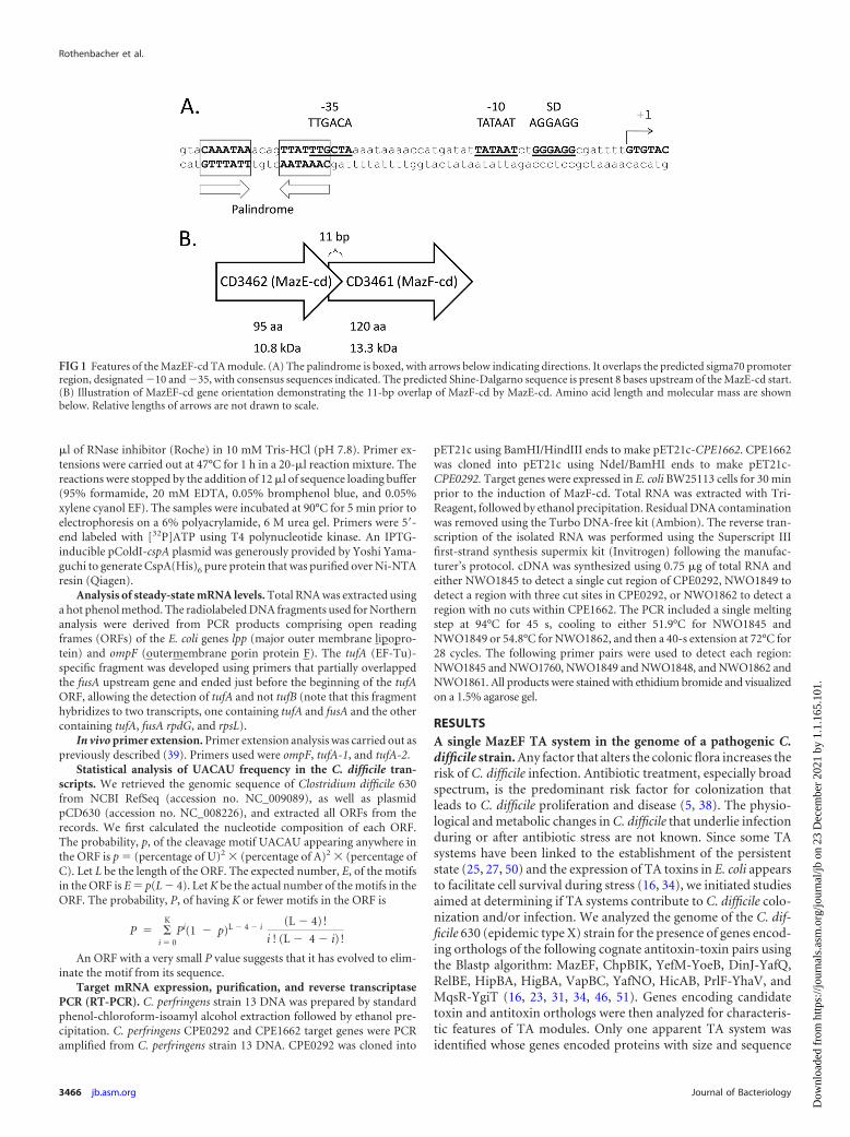
FIG 1 Features of the MazEF-cd TA module. (A) The palindrome is boxed, with arrows below indicating directions. It overlaps the predicted sigma70 promoterregion, designated �10 and �35, with consensus sequences indicated. The predicted Shine-Dalgarno sequence is present 8 bases upstream of the MazE-cd start.(B) Illustration of MazEF-cd gene orientation demonstrating the 11-bp overlap of MazF-cd by MazE-cd. Amino acid length and molecular mass are shownbelow. Relative lengths of arrows are not drawn to scale.
Rothenbacher et al.
3466 jb.asm.org Journal of Bacteriology
Dow
nloa
ded
from
http
s://j
ourn
als.
asm
.org
/jour
nal/j
b on
23
Dec
embe
r 20
21 b
y 1.
1.16
5.10
1.

similarity to E. coli MazE antitoxin (13% identity, 24% similarity;10.8 kDa) and MazF toxin (21% identity, 36% similarity; 13.3kDa); they were designated MazE-cd and MazF-cd, respectively.Characteristic genomic features of TA modules that were also ex-hibited by mazEF-cd are summarized in Fig. 1. The antitoxin andtoxin genes were adjacent, the antitoxin gene preceded the toxingene, the open reading frames overlapped (11 bp in this case), anda palindrome in the promoter region (used for module autoregu-lation) was identified next to the predicted �10 and �35 pro-moter sequences.
MazF-cd is toxic in E. coli and C. perfringens. To date, all E.coli MazF counterparts exhibit the same general enzymatic activ-ity, i.e., they function as single-stranded, sequence-specific en-doribonucleases (sometimes referred to as mRNA interferases)that target mRNA for cleavage. We expressed mazF-cd in E. coliK-12 host cells using the arabinose-inducible vector pBAD33. Aswith E. coli MazF, the expression of MazF-cd led to cell growtharrest (i.e., stasis) (Fig. 2). However, the kinetics of arrest weremarkedly slower for MazF-cd. While growth arrest occurs within15 min of induction for E. coli MazF (1), the expression ofMazF-cd began to significantly slow growth after 2 h and tookapproximately 8 h to reach a state of growth arrest (Fig. 2A). The
longer lag in E. coli cells may be due to lower levels of MazF-cd,since many C. difficile genes are difficult to express in E. coli. Wenext used a functional assay to demonstrate that MazE-cd acts asthe cognate antitoxin to MazF-cd in vivo. MazF-cd toxicity wasrescued by the coexpression of both the toxin and antitoxin genefrom separate inducible plasmids (Fig. 2B). The molecular biolog-ical tools for clostridia are very limited; an inducible vector en-abling the tight on-off expression of genes (especially critical whenexpressing toxic genes, as in our case) is not yet available. Alterna-tively, we created a lactose-inducible plasmid for tightly regulated,inducible toxin expression in C. perfringens. Interestingly, the in-duction of MazF-cd in C. perfringens resulted in an immediateeffect, precluding any increase in the optical density of the toxin-expressing cells (Fig. 2C).
MazF-cd expression leads to mRNA degradation of selectedtranscripts in vivo. Since all characterized MazF toxins act bycleaving mRNA, we first sought to confirm that MazF-cd alsotargets mRNA. We performed Northern analysis to assess steady-state levels of three E. coli mRNAs before and after MazF-cd in-duction: lpp, ompF, and tufA (Fig. 3). Curiously, MazF-cd expres-sion led to the cleavage of two of the three transcripts (ompF andtufA); lpp mRNA was not cleaved. The rate of ompF and tufA
FIG 2 Expression of MazF-cd inhibits growth and MazE-cd prevents toxicity. (A) Growth curve of E. coli BW25113 cells harboring the pBAD33-mazF-cd or pBAD33-mazF-ec plasmid either induced (with arabinose) or uninduced (without arabinose). Optical density was monitored beginning at time zero, the point of induction. Onerepresentative experiment (of three replicates) is shown. (B) pBAD33-mazF-cd and pINIII-mazE-cd constructs were cotransformed into BW25113 cells and streakedonto M9 minimal medium plates containing either 0.015% arabinose (�MazF) or 0.015% arabinose plus 1 mM IPTG (�MazF �MazE). The coinduction of both toxinand antitoxin permits normal growth in these cells (left); however, toxin-only expression prevents cell growth (right). The center diagram identifies the strains in eachsector of the left and right plates. Five of the six sectors are individual transformants of the BW25113 strain containing both pBAD33-mazF-cd and pINIII-mazE-cdplasmids, while the sixth was a control BW25113 strain that contained empty pBAD33 and pINIII plasmids. (C) Growth curve of C. perfringens strain 13 cells harboringpCM14-mazF-cd plasmid either induced (with lactose) or uninduced (without lactose) at time zero. Optical density was monitored immediately after the addition oflactose.
MazF-cd Targets mRNAs for Cleavage at UACAU
July 2012 Volume 194 Number 13 jb.asm.org 3467
Dow
nloa
ded
from
http
s://j
ourn
als.
asm
.org
/jour
nal/j
b on
23
Dec
embe
r 20
21 b
y 1.
1.16
5.10
1.

cleavage was slower and degradation was incomplete relative tothat of E. coli MazF. For example, the lpp, secE, and ompA tran-scripts were completely degraded by E. coli MazF (with no detect-able degradation intermediates) within 20 min of toxin induction(53). In contrast, MazF-cd cleavage resulted in the accumulationof one or more degradation intermediates. These results suggestthat MazF-cd may have lower enzymatic efficiency than its E. colicounterpart, or that it recognizes a more complex RNA consensussequence.
MazF-cd cleaves mRNA at UACAU sequences. We first testedwhether the slower growth inhibition and weaker RNA degrada-tion were consequences of MazF-cd cleavage of mRNA at a longerrecognition sequence relative to the short ACA consensus for E.coli MazF. To this end, we used an in vitro cleavage assay developedin the Inouye laboratory that utilizes a long, single-stranded RNAsubstrate (bacteriophage MS2 RNA) which enabled the determi-nation of the recognition sequences for two M. tuberculosis MazFtoxins, MazF-mt3 and MazF-mt7 (55). MS2 RNA is relativelylarge (3,500 nt) and commercially available, and it comprises anearly equal base content (26% G, 23% A, 26% C, 25% U). A longtest substrate increases the odds for the identification of targetsequences but also tends to form extensive secondary structures(70% of the nucleotides in MS2 are estimated to be doublestranded). Therefore, the major cold shock protein/RNA chaper-one CspA is added at saturating levels to prevent secondary struc-ture formation (22). The site specificity for RNA cleavage byMazF-cd was determined after performing primer extensions witholigonucleotides that enabled us to determine toxin cleavage sitesalong the entire 3.5-kb MS2 RNA.
We prepared recombinant MazF-cd as a fusion to trigger factor(TF) to facilitate robust expression without any toxicity; we usedthe same approach for the production of recombinant MazE-cd.The TF tag was proteolytically removed after the purification ofeach recombinant protein to preclude functional perturbation. Asa pilot experiment, we incubated MS2 RNA with the purified,recombinant MazF-cd alone or with increasing molar ratios ofMazE-cd (Fig. 4). MazF-cd cleaved MS2 RNA into several discrete
products (Fig. 4, lane 4). As expected, the MazE-cd antitoxin alonedid not cleave RNA (Fig. 4, lane 3). Increasing molar ratios of theantitoxin were preincubated with the toxin, and no cleavage wasobserved with the 2:1 and 3:1 antitoxin-to-toxin ratios (Fig. 4,lanes 5 to 7). We then performed primer extensions and identifiedeight cleavage sites (Fig. 5) with an apparent consensus sequenceof UACAU (five of the eight were UACAU and three varied atposition 4, UACUU or UACGU). In vivo primer extension exper-iments (i.e., from RNA prepared from E. coli cells after MazF-cdinduction) were next performed to assess cleavage site specificitywithin the two transcripts (ompF and tufA) that were degraded byMazF-cd in Fig. 3. Primers were designed for nearly completecoverage of the coding regions of each transcript (excluding the 3=ends corresponding to regions near or annealing to the distalprimers), ensuring that each of the UACAU preliminary consen-sus sequences, as well as variants at position 4, were detected (Fig.6). Interestingly, we observed cleavage only at each UACAU se-quence in both transcripts, the predominant cleavage site detectedin vitro. The 1,185-nt tufA coding region contains no UACGUsequences but has one UACUU and two UACAU sequences;MazF-cd only cleaved at the two UACAU sequences in tufA. Sim-ilarly, the 1,089-nt ompF coding region was cleaved only at thesingle UACAU sequence, even though it contained two UACUUsequences. We also did not observe cleavage at either singleUACCU sequence (the alternate substitution at position 4 was notdetected with MS2 RNA) represented in tufA and ompF. Finally,lpp mRNA is devoid of UACAU, UACUU, UACGU, and UACCUsequences, in agreement with our Northern analyses in which nocleavage was detected (Fig. 3). Therefore, under physiologicalconditions, MazF-cd exhibited strict specificity for UACAU se-quences; as with E. coli MazF, the cleavage recognition sequencewas invariant. Regarding the position of cleavage within the 5-ntconsensus, the predominant site (6 of 11) mapped between the Uand A at positions 1 and 2 (U2ACAU). Variation in cleavageposition within the consensus sequence is not unusual, as E. coliMazF also exhibits cleavage before or after the first A of its shortACA consensus sequence (A2CA or2ACA [52]). There is alsoan inherent margin of error of 1 to 2 nt on either side of the actualcleavage site when cleaved RNA product lengths are assessed ac-cording to their alignment with the adjacent DNA sequencing
FIG 3 Expression of MazF-cd in E. coli decreases steady-state levels of ompFand tufA mRNAs but not lpp in vivo. RNA was extracted at the time pointsindicated from BW25113 cells expressing MazF-cd, followed by Northernanalysis. The cleavage of ompF and tufA was detected, and cleavage productsare denoted by arrows. No additional products were detected below thecropped images. No cleavage products were detected for lpp. A minimal levelof cleavage occurred at time zero due to the leaky expression of pBAD33-mazF-cd before induction.
FIG 4 Recombinant MazF-cd cleaves MS2 mRNA, and MazE-cd preventscleavage of MS2 mRNA. Trigger factor (TF) was used as a control for thepurification of recombinant MazE-cd and MazF-cd. Products were visualizedby the addition of ethidium bromide prior to electrophoresis.
Rothenbacher et al.
3468 jb.asm.org Journal of Bacteriology
Dow
nloa
ded
from
http
s://j
ourn
als.
asm
.org
/jour
nal/j
b on
23
Dec
embe
r 20
21 b
y 1.
1.16
5.10
1.

ladder. In summary, our data demonstrate that MazF-cd specifi-cally cleaves mRNA at UACAU consensus sequences in vivo (Ta-ble 2).
MazF-cd shares its pentad cleavage consensus sequence withcounterparts in S. aureus and Bacillus subtilis. Interestingly,MazF orthologs derived from three distinct eubacteria, B. subtilis(35, 36), S. aureus (13, 54), and C. difficile, each cleave mRNA atUACAU. The single transcript tested with MazF from B. subtilis(also known as EndoA) generated the cleavage patternUAUAAU2ACAUA, containing the UACAU consensus (under-lined) (36). Subsequent cleavage analyses for B. subtilis MazF us-ing multiple templates revealed a clear minimal consensus ofUACAU (35). The conservation in consensus recognition se-quences among these three toxins provides a platform that mayaid in the identification of amino acids that contribute to sequencespecificity. The alignment of these three MazF toxins revealed sub-
stantial shared sequence identity that contrasts with marginal sim-ilarity to E. coli MazF (Fig. 7). Curiously, although nuclear mag-netic resonance studies by Li et al. implicate E. coli MazF H28 insubstrate recognition (29), which is consistent with its solventexposure in the X-ray crystal structure of the MazE-MazF com-plex (24), there are no histidines at or near this position in thethree UACAU-cleaving MazF toxins (Fig. 7). In other MazF or-thologs without a histidine at position 28, there is one in proximitythat is instead proposed to contribute to substrate recognition(e.g., H17 of Kid and H23 of ChpBK) (29). However, the threeUACAU-cleaving MazF toxins have an absolutely conserved his-tidine (H63 in MazF-cd) that is predicted as the first residue of�-sheet S4 immediately following loop 2 (Fig. 7). It is possible thatthe position of the histidine influences substrate specificity, asproposed by Li et al. (29). However, they also proposed that therelatively hydrophobic site formed by the binding site created in
FIG 5 In vitro primer extension using an MS2 RNA template suggests a MazF-cd cleavage consensus sequence. Purified MazF-cd was first incubated with MS2target mRNA to allow cleavage. Radiolabeled primers were annealed to the cleaved mRNA, and a reverse transcription reaction was initiated. Reverse transcrip-tase is able to convert the target mRNA into cDNA until it reaches a point of mRNA cleavage. The products of the transcriptional runoff are resolved on ahigh-resolution polyacrylamide gel, and the exact position of RNA cleavage by MazF-cd was accurately deduced by the comparison of the mobility of the primerextension product relative to the mobility of bands comprising the sequencing reactions on the MS2 RNA using the same primer and reverse transcriptase. (A)Four cleavage sites were detected when purified MazF-cd was incubated with MS2 RNA (without the addition of CspA) before performing the extension reaction.Control lanes contain MS2 RNA only, yielding a full-length extension product distant from the MazF-cd cleavage site. (B) Four additional cleavage sites weredetected when purified MazF-cd was incubated with MS2 RNA and CspA before performing the extension reaction. MS2 RNA was added to all four primerextension reaction lanes. �MazF-cd/�CspA denotes that the extension reaction was performed on MS2 RNA only, �MazF-cd/�CspA denotes that CspA andMS2 RNA were incubated before the extension reaction was performed. Full-length MS2 RNA extension products were cropped from the images shown.Cleavage sites/products are designated by arrows, and the sequences flanking the MazF-cd cleavage site are shown below. DNA sequencing ladders using the sameprimer were loaded in the adjacent lanes.
FIG 6 Primer extension of ompF and tufA pinpoints the MazF-cd cleavage site as UACAU. The RNA used for Northern analysis in Fig. 3 was subjected to primerextension analysis; DNA sequencing ladders using the same primer were loaded in the adjacent lanes.
MazF-cd Targets mRNAs for Cleavage at UACAU
July 2012 Volume 194 Number 13 jb.asm.org 3469
Dow
nloa
ded
from
http
s://j
ourn
als.
asm
.org
/jour
nal/j
b on
23
Dec
embe
r 20
21 b
y 1.
1.16
5.10
1.

the MazF dimer (when the S3-S4 loop 2 of one MazF monomerinteracts with the H1 helix of the other) could also directly interactwith the hydrophobic moieties of the RNA substrate (29). Thiswould also be consistent with the sequence relationships shown inour alignment, since residues comprising these two regions areconserved among the three UACAU-cleaving MazF toxins butdistinct from E. coli MazF.
MazF-cd cleavage sites are significantly underrepresented intoxin B and the cell wall surface protein CwpV. C. difficile strain630 contains 3,776 predicted open reading frames (ORFs) (48).We analyzed the overall nucleotide content of the ORFs, deter-mined the probability of the occurrence of the UACAU consensusof each ORF (Table 3, Expected), and compared it to the actualoccurrence (Table 3, Actual) (described in Materials and Meth-ods). The top 20 proteins whose mRNAs contain more UACAUmotifs than predicted (with P values close to 0) are listed in TableS1 in the supplemental material. These proteins are predicted tohave shorter half-lives under conditions where the MazF-cd toxinis activated in vivo. None of these proteins have an obvious role inpathogenesis, nor was there any apparent bias for proteins in-volved in one or more cellular processes. However, the analysis oftranscripts with fewer UACAU motifs than predicted (Table 3)revealed a protein with a prominent role in C. difficile virulence(toxin B) and the aggregation-promoting cell surface protein
CwpV with sequence similarity to known C. difficile adhesins SlpAand Cwp66 (9, 41). Toxin B appears to be an important determi-nant of C. difficile virulence (28, 30). Interestingly, toxin A mRNAcontains 12 predicted cleavage sites, suggesting a differential rateof cleavage between these two important virulence factors. Theprecise role of CwpV is not yet clear; however, it constitutes asignificant proportion of cell surface proteins, it promotes auto-aggregation that may influence the formation of biofilm-like ag-gregates in infected mice (41), and its expression is phase variable(9). Also of note, the protease Lon as well as ClpC (the ATPasecomponent of the Clp proteolytic complex) contain no UACAUmotifs (Table 3). This implicates these proteases in the regulationof MazF-cd activation, since the degradation of MazE-cd wouldresult in excess toxin. In fact, Lon and Clp family members havebeen frequently linked with antitoxin degradation in E. coli (16).
Finally, the UACAU motif is not represented in transcripts for1,303 C. difficile proteins, comprising approximately one-third ofall C. difficile transcripts. We investigated the statistical signifi-cance of this finding. First, the average length of the ORFs that donot have UACAU is 631 nt, and the average length of ORFs havingat least one UACAU is almost twice that, 1,108 nt. Therefore, theabsence of UACAU in the 1,303 C. difficile ORFs is due to theshorter relative ORF length of this subset. Second, we found thatessentially the same number of ORFs (1,305) lacked an AUCUAtest motif with the same base composition as but different orderfrom our real motif. Therefore, the absence of UACAU in a thirdof the C. difficile ORFs is not unexpected given the base composi-tion and length. However, unlike the ACA-cleaving E. coli MazF,MazF-cd is predicted to target selected transcripts because it has alonger recognition sequence.
mRNA transcript stability upon MazF-cd induction is de-pendent on the number of UACAU sequences. To establish ifthere was a relationship between the number of toxin cleavagesites and transcript stability, we induced the expression of targetmRNAs in E. coli and estimated their steady-state levels after thesubsequent induction of MazF-cd using reverse transcriptase PCR(RT-PCR). Two target transcripts were selected for analysis:CPE0292, a carbohydrate kinase family gene, and CPE1662, a Ppx/GppA phosphatase family gene. We chose C. perfringens genes fortargets based on a similar bioinformatic analysis of C. perfringenstranscripts for MazF-cd susceptibility to that previously de-scribed. These two genes were chosen for their differing numbers
TABLE 2 In vitro and in vivo primer extension cleavage sites
Cleavage site and primer name Sequencea
In vitroG 5= CGCGU2ACGUAAAG 3=K 5= GACUU2ACAUCGAA 3=D2 5= GC2UUACUUAAGGG 3=H 5= UUUUAC2AUAAAAC 3=E1 5= UCGU2ACUUAAAUA 3=B4 5= UUUU2ACAUCAAGA 3=1351 5= UCGCU2ACAUAGCG 3=J 5= UUU2UACAUAAACG 3=
In vivoompF 5= UGACU2ACAUCAUC 3=tufA-1 5= UCCGUAC2AUCAUC 3=tufA-2 5= AGUGUA2CAUUCUG 3=
Consensus UACAUa Boldface letters indicate conserved sequences adjacent to detected cut sites.
FIG 7 MazF-cd exhibits significant sequence similarity to counterparts in S. aureus and B. subtilis. EndoA and MazF-sa were compared to each other and to E.coli MazF. Identical amino acids are highlighted in boldface, while conserved substitutions (�1 or greater on the BLOSUM62 alignment score matrix) arehighlighted in gray. Histidines believed to be involved in sequence recognition are boxed. Only the secondary structure motifs relevant to this work are illustratedabove the respective amino acids of E. coli MazF. Black arrows, �-sheets; gray rectangle, -helix; black line, unstructured loops.
Rothenbacher et al.
3470 jb.asm.org Journal of Bacteriology
Dow
nloa
ded
from
http
s://j
ourn
als.
asm
.org
/jour
nal/j
b on
23
Dec
embe
r 20
21 b
y 1.
1.16
5.10
1.
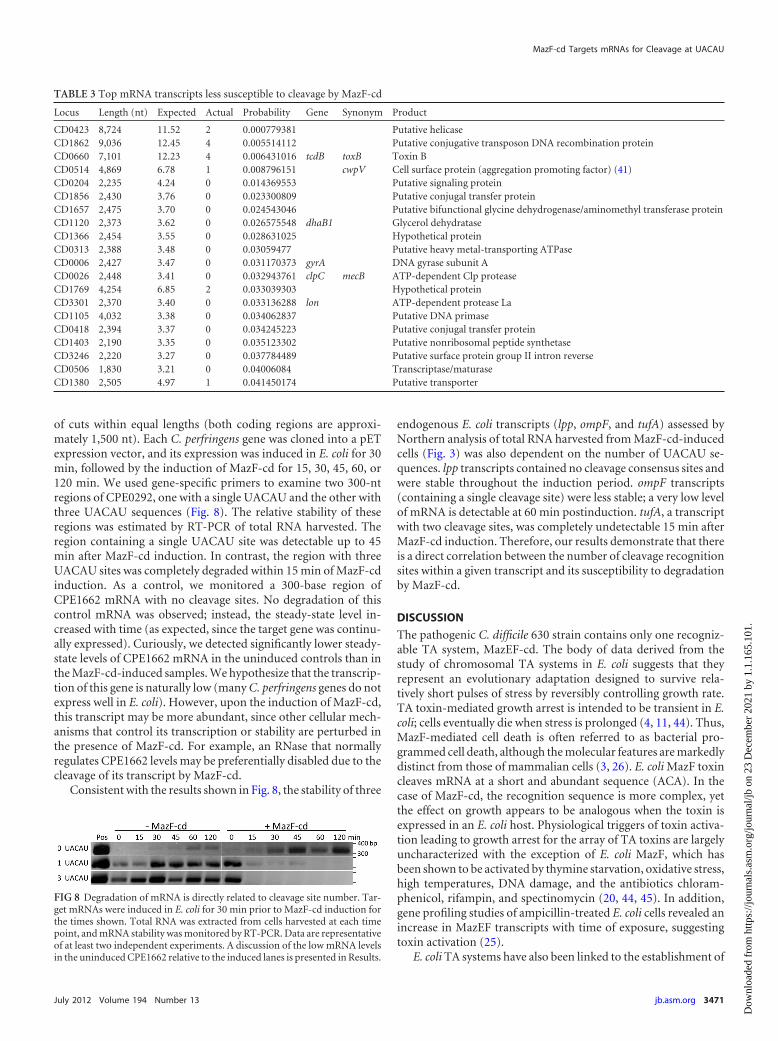
of cuts within equal lengths (both coding regions are approxi-mately 1,500 nt). Each C. perfringens gene was cloned into a pETexpression vector, and its expression was induced in E. coli for 30min, followed by the induction of MazF-cd for 15, 30, 45, 60, or120 min. We used gene-specific primers to examine two 300-ntregions of CPE0292, one with a single UACAU and the other withthree UACAU sequences (Fig. 8). The relative stability of theseregions was estimated by RT-PCR of total RNA harvested. Theregion containing a single UACAU site was detectable up to 45min after MazF-cd induction. In contrast, the region with threeUACAU sites was completely degraded within 15 min of MazF-cdinduction. As a control, we monitored a 300-base region ofCPE1662 mRNA with no cleavage sites. No degradation of thiscontrol mRNA was observed; instead, the steady-state level in-creased with time (as expected, since the target gene was continu-ally expressed). Curiously, we detected significantly lower steady-state levels of CPE1662 mRNA in the uninduced controls than inthe MazF-cd-induced samples. We hypothesize that the transcrip-tion of this gene is naturally low (many C. perfringens genes do notexpress well in E. coli). However, upon the induction of MazF-cd,this transcript may be more abundant, since other cellular mech-anisms that control its transcription or stability are perturbed inthe presence of MazF-cd. For example, an RNase that normallyregulates CPE1662 levels may be preferentially disabled due to thecleavage of its transcript by MazF-cd.
Consistent with the results shown in Fig. 8, the stability of three
endogenous E. coli transcripts (lpp, ompF, and tufA) assessed byNorthern analysis of total RNA harvested from MazF-cd-inducedcells (Fig. 3) was also dependent on the number of UACAU se-quences. lpp transcripts contained no cleavage consensus sites andwere stable throughout the induction period. ompF transcripts(containing a single cleavage site) were less stable; a very low levelof mRNA is detectable at 60 min postinduction. tufA, a transcriptwith two cleavage sites, was completely undetectable 15 min afterMazF-cd induction. Therefore, our results demonstrate that thereis a direct correlation between the number of cleavage recognitionsites within a given transcript and its susceptibility to degradationby MazF-cd.
DISCUSSION
The pathogenic C. difficile 630 strain contains only one recogniz-able TA system, MazEF-cd. The body of data derived from thestudy of chromosomal TA systems in E. coli suggests that theyrepresent an evolutionary adaptation designed to survive rela-tively short pulses of stress by reversibly controlling growth rate.TA toxin-mediated growth arrest is intended to be transient in E.coli; cells eventually die when stress is prolonged (4, 11, 44). Thus,MazF-mediated cell death is often referred to as bacterial pro-grammed cell death, although the molecular features are markedlydistinct from those of mammalian cells (3, 26). E. coli MazF toxincleaves mRNA at a short and abundant sequence (ACA). In thecase of MazF-cd, the recognition sequence is more complex, yetthe effect on growth appears to be analogous when the toxin isexpressed in an E. coli host. Physiological triggers of toxin activa-tion leading to growth arrest for the array of TA toxins are largelyuncharacterized with the exception of E. coli MazF, which hasbeen shown to be activated by thymine starvation, oxidative stress,high temperatures, DNA damage, and the antibiotics chloram-phenicol, rifampin, and spectinomycin (20, 44, 45). In addition,gene profiling studies of ampicillin-treated E. coli cells revealed anincrease in MazEF transcripts with time of exposure, suggestingtoxin activation (25).
E. coli TA systems have also been linked to the establishment of
TABLE 3 Top mRNA transcripts less susceptible to cleavage by MazF-cd
Locus Length (nt) Expected Actual Probability Gene Synonym Product
CD0423 8,724 11.52 2 0.000779381 Putative helicaseCD1862 9,036 12.45 4 0.005514112 Putative conjugative transposon DNA recombination proteinCD0660 7,101 12.23 4 0.006431016 tcdB toxB Toxin BCD0514 4,869 6.78 1 0.008796151 cwpV Cell surface protein (aggregation promoting factor) (41)CD0204 2,235 4.24 0 0.014369553 Putative signaling proteinCD1856 2,430 3.76 0 0.023300809 Putative conjugal transfer proteinCD1657 2,475 3.70 0 0.024543046 Putative bifunctional glycine dehydrogenase/aminomethyl transferase proteinCD1120 2,373 3.62 0 0.026575548 dhaB1 Glycerol dehydrataseCD1366 2,454 3.55 0 0.028631025 Hypothetical proteinCD0313 2,388 3.48 0 0.03059477 Putative heavy metal-transporting ATPaseCD0006 2,427 3.47 0 0.031170373 gyrA DNA gyrase subunit ACD0026 2,448 3.41 0 0.032943761 clpC mecB ATP-dependent Clp proteaseCD1769 4,254 6.85 2 0.033039303 Hypothetical proteinCD3301 2,370 3.40 0 0.033136288 lon ATP-dependent protease LaCD1105 4,032 3.38 0 0.034062837 Putative DNA primaseCD0418 2,394 3.37 0 0.034245223 Putative conjugal transfer proteinCD1403 2,190 3.35 0 0.035123302 Putative nonribosomal peptide synthetaseCD3246 2,220 3.27 0 0.037784489 Putative surface protein group II intron reverseCD0506 1,830 3.21 0 0.04006084 Transcriptase/maturaseCD1380 2,505 4.97 1 0.041450174 Putative transporter
FIG 8 Degradation of mRNA is directly related to cleavage site number. Tar-get mRNAs were induced in E. coli for 30 min prior to MazF-cd induction forthe times shown. Total RNA was extracted from cells harvested at each timepoint, and mRNA stability was monitored by RT-PCR. Data are representativeof at least two independent experiments. A discussion of the low mRNA levelsin the uninduced CPE1662 relative to the induced lanes is presented in Results.
MazF-cd Targets mRNAs for Cleavage at UACAU
July 2012 Volume 194 Number 13 jb.asm.org 3471
Dow
nloa
ded
from
http
s://j
ourn
als.
asm
.org
/jour
nal/j
b on
23
Dec
embe
r 20
21 b
y 1.
1.16
5.10
1.

the persistent state upon antibiotic exposure. The HipA TA toxinwas discovered as a mutant allele that increases the recovery ofpersisters at a 1,000-fold higher level than wild-type cells (32). The�hipBA strain exhibits a 10- to 100-fold decrease in persisters(25), while HipA overexpression causes a 10- to 10,000-fold in-crease in persisters (12, 27, 50). Similarly, RelE or MazF overex-pression increases the level of surviving persisters 10- to 10,000-fold (25) or 100- to 10,000-fold (50), respectively. The deletion ofthe gene encoding the YafQ TA toxin reduces cell survival in E. colibiofilms treated with bactericidal antibiotics; the overproductionof YafQ in E. coli makes biofilm cells more resistant to antibiotics(18).
Although the physiological triggers and role of MazF-cd arestill unclear, extensive studies on E. coli TA systems provide aframework to hypothesize possible roles for MazF-cd in its naturalhost. First, since the expression of MazF-cd in E. coli and C. per-fringens resulted in growth arrest, and since this state has beenlinked to survival during relatively short periods of stress,MazF-cd may be important in (i) aiding survival during coloniza-tion when the inciting antibiotic is still present and/or (ii) facili-tating persistence that leads to recurrent infections.
Two key features of TA systems underlie toxin activation: co-regulation at the transcriptional level and antitoxin instability.More specifically, toxin and antitoxin genes are adjacent and co-regulated in their own operon. Toxin activation is the result of adynamic process that exploits the short half-life of the intrinsicallyunfolded cognate antitoxin (6, 7, 15) that is consequently readilysusceptible to degradation by cellular proteases. Therefore, an in-crease in transcript levels of toxin alone or both antitoxin andtoxin are hallmarks of physiological toxin activation, because eachscenario results in a net increase in toxin levels.
We mined the data sets from two published C. difficile microar-ray studies to determine if they supported a role for MazF-cd inpersistence and/or if any apparent toxin activation could be doc-umented under the conditions used. In the first study, Fairweatherand colleagues tested the effects of clindamycin or the �-lactamamoxicillin (at subinhibitory concentrations to preclude celldeath) on global gene expression (10). They also analyzed the geneexpression profile of C. difficile cells exposed to a subinhibitoryconcentration of metronidazole, the antibiotic traditionally en-listed as first-line therapy for CDI. The effects of other stress con-ditions on global gene expression were also analyzed, allowing thecomparison of the classes of genes whose transcript levels increaseupon exposure to each type of stress (10). In this data set, bothtoxin and antitoxin mRNA levels increased by 25% or more withclindamycin and amoxicillin exposure (Table 4). However, onlythe MazE-cd antitoxin mRNA levels increased in metronidazole-treated C. difficile cells. In contrast to the antibiotic stresses im-parted, there was no significant increase (i.e., relative levels werebelow the 25% increase threshold) in MazEF-cd upon heat, acid,
alkali, and aerobic shock (Table 4). The significance of these datais unclear, because (i) the increase in steady-state toxin and anti-toxin transcript levels was marginal (perhaps due to the subinhibi-tory concentration of antibiotic used), (ii) the antitoxin/toxin ra-tio (1.3 to 1.4) favored the antitoxin with each of the threeantibiotics, and (iii) RNA was also prepared from mid-logarith-mic-phase C. difficile cells, conditions where the transcription ofthe pathogenicity locus (PaLoc; comprising genes for toxins A andB, a putative holin, and two regulatory elements) is repressed byCodY. Consequently, the inherently low level of toxin B mRNAprevented us from seeing any direct effect of MazF-cd on its tran-script levels in vivo. The levels of the CwpV transcripts were essen-tially unchanged with all three antibiotics. In the second study, C.difficile expression profiles were compared before and 30, 60, and120 min after the infection of colorectal epithelial Caco-2 cells(21). There was no statistically significant trend for MazE-cd orMazF-cd gene expression; however, the upregulation of CodY wasagain observed, precluding toxin B expression. Therefore, neitherstudy used conditions that supported a notable trend regardingTA toxin activation.
A battery of primer extension experiments led to the identifi-cation of a clear consensus sequence for MazF-cd cleavage,UACAU. Curiously, the MazF counterparts in B. subtilis and S.aureus possess the same recognition sequence (13, 54). This longerrecognition is predicted to result in more selective mRNA degra-dation than that which occurs with ACA-cleaving E. coli MazF. Infact, in Fig. 8 we demonstrated that the number of UACAU se-quences dictates mRNA stability in the presence of MazF-cd.mRNAs with no UACAU sequences were resistant to MazF-cddegradation; as the number of UACAU sequences increased, sodid the relative rate of transcript degradation. These proof-of-principle experiments enabled us to reliably apply bioinformaticsapproaches to predict which C. difficile transcripts would be pref-erentially protected or degraded upon toxin activation. Nearlyone-third of the C. difficile ORFs (encoding proteins possessing aspectrum of functions) did not contain an UACAU sequence andare consequently protected from MazF cleavage, including theCodY global repressor. We did not detect a common themeamong the functions of proteins whose transcripts have moreUACAU motifs than expected (see Table S1 in the supplementalmaterial), nor did any protein stand out as having an importantrole in pathogenesis. In fact, the majority of hits (17/20) wereannotated as hypothetical proteins or putative counterparts ofknown bacterial proteins.
However, some of the top statistical hits for the protein candi-dates predicted to have transcripts that are resistant or less suscep-tible to MazF-cd cleavage have clear roles in C. difficile pathogen-esis (Table 3). Toxin B, whose gene in the C. difficile PaLoc is amajor determinant of a toxigenic C. difficile strain, is third on thelist. Although the toxin B transcript contains four cleavage sites, its
TABLE 4 mazE-cd and mazF-cd transcripts are upregulated under antibiotic stressa
Gene
Relative transcript level (�SD) after exposure to:
Heat shock Acid shock Alkali shock Aerobic shock Amoxicillin Clindamycin Metronidazole
mazF-cd 0.95 � 0.11 1.01 � 0.20 1.07 � 0.23 1.17 � 0.15 1.50 � 0.15 1.30 � 0.14 1.01 � 0.15mazE-cd 1.04 � 0.15 1.08 � 0.24 1.01 � 0.26 0.95 � 0.18 2.09 � 0.16 1.65 � 0.19 1.40 � 0.29a Shaded values are transcripts upregulated by 25% or more and are adapted from data reported by Emerson et al. (10). Antibiotic concentrations used were 1 mg/ml amoxicillin,50 mg/ml clindamycin, and 0.15 mg/ml metronidazole. Values shown are averages from duplicate microarray experiments.
Rothenbacher et al.
3472 jb.asm.org Journal of Bacteriology
Dow
nloa
ded
from
http
s://j
ourn
als.
asm
.org
/jour
nal/j
b on
23
Dec
embe
r 20
21 b
y 1.
1.16
5.10
1.

relative degradation rate is predicted to be much slower than thatof toxin A (which contains 12). This difference is very provocativeand may represent a novel posttranscriptional method for theregulation of toxin A and B protein production. The cell wallprotein CwpV is fourth on the list (41). Fairweather and col-leagues have demonstrated that CwpV is important for agglutina-tion and hypothesize that CwpV is important for biofilm-like for-mation or adhesion (41). Expression of CwpV is phase variable,and only 0.1 to 10% of C. difficile cells express this protein on thecell surface under standard laboratory conditions (9, 41). Phasevariation in this case is controlled by a novel DNA inversion eventmediated by the site-specific recombinase RecV and inverted re-peats upstream of the gene encoding CwpV, such that transcrip-tion is off in one orientation and on in the other (9, 41). The mostwidely accepted theory for the enlistment of phase variation bypathogens is to evade the host immune system (49). Curiously, thefirst and second hits in Table 3 are annotated as a putative helicaseand a putative DNA recombination protein, respectively. Al-though a recombinase that facilitates CwpV phase variation hasbeen identified (9), these first two hits may have a role in control-ling other, as-yet unknown genes, that may also be turned on andoff through phase variation and play important roles in C. difficilecell survival or virulence.
Future gene profiling experiments using cells in late-exponen-tial/early stationary phase under nutrient-limiting conditions orfrom an expanded panel of antibiotics at higher than subinhibi-tory concentrations should provide useful insights into whetherthe MazEF-cd TA module is upregulated by selected antibioticsand/or other triggers. If so, we should be able to better understandthe physiological role of MazF-cd and assess whether the statisticalpredictions regarding transcript stability upon toxin activation aresupported in vivo.
ACKNOWLEDGMENTS
We thank Ling Zhu for providing reagents and assistance with MS2primer extension analysis and Masayori Inouye for helpful discussionsand expert advice.
This work was supported in part by National Institutes of Health T32training grant AI07403, Virus-Host Interactions in Eukaryotic Cells, fromthe NIAID (to F.P.R. and J.M.H., awarded to G. Brewer).
REFERENCES1. Aizenman E, Engelberg-Kulka H, Glaser G. 1996. An Escherichia coli
chromosomal “addiction module” regulated by guanosine 3=,5=-bispyrophosphate: a model for programmed bacterial cell death. Proc.Natl. Acad. Sci. U. S. A. 93:6059 – 6063.
2. Allen SP, Blaschek HP. 1990. Factors involved in the electroporation-induced transformation of Clostridium perfringens. FEMS Microbiol.Lett. 58:217–220.
3. Amitai S, Kolodkin-Gal I, Hananya-Meltabashi M, Sacher A, Engel-berg-Kulka H. 2009. Escherichia coli MazF leads to the simultaneousselective synthesis of both “death proteins” and “survival proteins.” PLoSGenet. 5:e1000390. doi:10.1371/journal.pgen.1000390.
4. Amitai S, Yassin Y, Engelberg-Kulka H. 2004. MazF-mediated cell deathin Escherichia coli: a point of no return. J. Bacteriol. 186:8295– 8300.
5. Bignardi GE. 1998. Risk factors for Clostridium difficile infection. J.Hosp. Infect. 40:1–15.
6. Buts L, Lah J, Dao-Thi MH, Wyns L, Loris R. 2005. Toxin-antitoxinmodules as bacterial metabolic stress managers. Trends Biochem. Sci. 30:672– 679.
7. De Jonge N, et al. 2009. Rejuvenation of CcdB-poisoned gyrase by anintrinsically disordered protein domain. Mol. Cell 35:154 –163.
8. Dodson AP, Borriello SP. 1996. Clostridium difficile infection of the gut.J. Clin. Pathol. 49:529 –532.
9. Emerson JE, et al. 2009. A novel genetic switch controls phase variableexpression of CwpV, a Clostridium difficile cell wall protein. Mol. Micro-biol. 74:541–556.
10. Emerson JE, Stabler RA, Wren BW, Fairweather NF. 2008. Microarrayanalysis of the transcriptional responses of Clostridium difficile to envi-ronmental and antibiotic stress. J. Med. Microbiol. 57:757–764.
11. Engelberg-Kulka H, Glaser G. 1999. Addiction modules and pro-grammed cell death and antideath in bacterial cultures. Annu. Rev. Mi-crobiol. 53:43–70.
12. Falla TJ, Chopra I. 1998. Joint tolerance to beta-lactam and fluoroquin-olone antibiotics in Escherichia coli results from overexpression of hipA.Antimicrob. Agents Chemother. 42:3282–3284.
13. Fu Z, Donegan NP, Memmi G, Cheung AL. 2007. Characterization ofMazFSa, an endoribonuclease from Staphylococcus aureus. J. Bacteriol.189:8871– 8879.
14. Fuchs AR, Bonde GJ. 1957. The nutritional requirements of Clostridiumperfringens. J. Gen. Microbiol. 16:317–329.
15. Garcia-Pino A, et al. 2010. Allostery and intrinsic disorder mediate tran-scription regulation by conditional cooperativity. Cell 142:101–111.
16. Gerdes K, Christensen SK, Lobner-Olesen A. 2005. Prokaryotic toxin-antitoxin stress response loci. Nat. Rev. Microbiol. 3:371–382.
17. Guzman LM, Belin D, Carson MJ, Beckwith J. 1995. Tight regulation,modulation, and high-level expression by vectors containing the arabi-nose PBAD promoter. J. Bacteriol. 177:4121– 4130.
18. Harrison JJ, et al. 2009. The chromosomal toxin gene yafQ is a determi-nant of multidrug tolerance for Escherichia coli growing in a biofilm.Antimicrob. Agents Chemother. 53:2253–2258.
19. Hartman AH, Liu H, Melville SB. 2011. Construction and characteriza-tion of a lactose-inducible promoter system for controlled gene expressionin Clostridium perfringens. Appl. Environ. Microbiol. 77:471– 478.
20. Hazan R, Sat B, Engelberg-Kulka H. 2004. Escherichia coli mazEF-mediated cell death is triggered by various stressful conditions. J. Bacteriol.186:3663–3669.
21. Janvilisri T, Scaria J, Chang YF. 2010. Transcriptional profiling of Clos-tridium difficile and Caco-2 cells during infection. J. Infect. Dis. 202:282–290.
22. Jiang W, Hou Y, Inouye M. 1997. CspA, the major cold-shock protein ofEscherichia coli, is an RNA chaperone. J. Biol. Chem. 272:196 –202.
23. Jorgensen MG, Pandey DP, Jaskolska M, Gerdes K. 2009. HicA ofEscherichia coli defines a novel family of translation-independent mRNAinterferases in bacteria and archaea. J. Bacteriol. 191:1191–1199.
24. Kamada K, Hanaoka F, Burley SK. 2003. Crystal structure of the MazE/MazF complex: molecular bases of antidote-toxin recognition. Mol. Cell11:875– 884.
25. Keren I, Shah D, Spoering A, Kaldalu N, Lewis K. 2004. Specializedpersister cells and the mechanism of multidrug tolerance in Escherichiacoli. J. Bacteriol. 186:8172– 8180.
26. Kolodkin-Gal I, Hazan R, Gaathon A, Carmeli S, Engelberg-Kulka H.2007. A linear pentapeptide is a quorum-sensing factor required formazEF-mediated cell death in Escherichia coli. Science 318:652– 655.
27. Korch SB, Hill TM. 2006. Ectopic overexpression of wild-type and mu-tant hipA genes in Escherichia coli: effects on macromolecular synthesisand persister formation. J. Bacteriol. 188:3826 –3836.
28. Kuehne SA, et al. 2010. The role of toxin A and toxin B in Clostridiumdifficile infection. Nature 467:711–713.
29. Li GY, et al. 2006. Characterization of dual substrate binding sites in thehomodimeric structure of Escherichia coli mRNA interferase MazF. J.Mol. Biol. 357:139 –150.
30. Lyras D, et al. 2009. Toxin B is essential for virulence of Clostridiumdifficile. Nature 458:1176 –1179.
31. Makarova KS, Grishin NV, Koonin EV. 2006. The HicAB cassette, aputative novel, RNA-targeting toxin-antitoxin system in archaea and bac-teria. Bioinformatics 22:2581–2584.
32. Moyed HS, Bertrand KP. 1983. hipA, a newly recognized gene of Esche-richia coli K-12 that affects frequency of persistence after inhibition ofmurein synthesis. J. Bacteriol. 155:768 –775.
33. Nariya H, Inouye M. 2008. MazF, an mRNA interferase, mediates pro-grammed cell death during multicellular Myxococcus development. Cell132:55– 66.
34. Pandey DP, Gerdes K. 2005. Toxin-antitoxin loci are highly abundant infree-living but lost from host-associated prokaryotes. Nucleic Acids Res.33:966 –976.
MazF-cd Targets mRNAs for Cleavage at UACAU
July 2012 Volume 194 Number 13 jb.asm.org 3473
Dow
nloa
ded
from
http
s://j
ourn
als.
asm
.org
/jour
nal/j
b on
23
Dec
embe
r 20
21 b
y 1.
1.16
5.10
1.

35. Park JH, Yamaguchi Y, Inouye M. 2011. Bacillus subtilis MazF-bs (En-doA) is a UACAU-specific mRNA interferase. FEBS Lett. 585:2526 –2532.
36. Pellegrini O, Mathy N, Gogos A, Shapiro L, Condon C. 2005. TheBacillus subtilis ydcDE operon encodes an endoribonuclease of the MazF/PemK family and its inhibitor. Mol. Microbiol. 56:1139 –1148.
37. Poutanen SM, Simor AE. 2004. Clostridium difficile-associated diarrheain adults. CMAJ 171:51–58.
38. Privitera G, et al. 1991. Prospective study of Clostridium difficile intes-tinal colonization and disease following single-dose antibiotic prophylaxisin surgery. Antimicrob. Agents Chemother. 35:208 –210.
39. Prysak MH, et al. 2009. Bacterial toxin YafQ is an endoribonuclease thatassociates with the ribosome and blocks translation elongation throughsequence-specific and frame-dependent mRNA cleavage. Mol. Microbiol.71:1071–1087.
40. Ramage HR, Connolly LE, Cox JS. 2009. Comprehensive functionalanalysis of Mycobacterium tuberculosis toxin-antitoxin systems: implica-tions for pathogenesis, stress responses, and evolution. PLoS Genet.5:e1000767. doi:10.1371/journal.pgen.1000767.
41. Reynolds CB, Emerson JE, de la Riva L, Fagan RP, Fairweather NF.2011. The Clostridium difficile cell wall protein CwpV is antigenicallyvariable between strains, but exhibits conserved aggregation-promoting function. PLoS Pathog. 7 :e1002024. doi:10.1371/journal.ppat.1002024.
42. Riha WE, Jr, Solberg M. 1971. Chemically defined medium for thegrowth of Clostridium perfringens. Appl. Microbiol. 22:738 –739.
43. Rupnik M, Wilcox MH, Gerding DN. 2009. Clostridium difficile infec-tion: new developments in epidemiology and pathogenesis. Nat. Rev. Mi-crobiol. 7:526 –536.
44. Sat B, et al. 2001. Programmed cell death in Escherichia coli: some anti-biotics can trigger mazEF lethality. J. Bacteriol. 183:2041–2045.
45. Sat B, Reches M, Engelberg-Kulka H. 2003. The Escherichia coli mazEFsuicide module mediates thymineless death. J. Bacteriol. 185:1803–1807.
46. Schmidt O, et al. 2007. prlF and yhaV encode a new toxin-antitoxinsystem in Escherichia coli. J. Mol. Biol. 372:894 –905.
47. Schwan C, et al. 2009. Clostridium difficile toxin CDT induces formationof microtubule-based protrusions and increases adherence of bacteria.PLoS Pathog. 5:e1000626. doi:10.1371/journal.ppat.1000626.
48. Sebaihia M, et al. 2006. The multidrug-resistant human pathogen Clos-tridium difficile has a highly mobile, mosaic genome. Nat. Genet. 38:779 –786.
49. van der Woude MW, Baumler AJ. 2004. Phase and antigenic variation inbacteria. Clin. Microbiol. Rev. 17:581– 611.
50. Vazquez-Laslop N, Lee H, Neyfakh AA. 2006. Increased persistence inEscherichia coli caused by controlled expression of toxins or other unre-lated proteins. J. Bacteriol. 188:3494 –3497.
51. Yamaguchi Y, Park JH, Inouye M. 2009. MqsR, a crucial regulator forquorum sensing and biofilm formation, is a GCU-specific mRNA inter-ferase in Escherichia coli. J. Biol. Chem. 284:28746 –28753.
52. Zhang Y, Zhang J, Hara H, Kato I, Inouye M. 2005. Insights into themRNA cleavage mechanism by MazF, an mRNA interferase. J. Biol.Chem. 280:3143–3150.
53. Zhang Y, et al. 2003. MazF cleaves cellular mRNAs specifically at ACA toblock protein synthesis in Escherichia coli. Mol. Cell 12:913–923.
54. Zhu L, et al. 2009. Staphylococcus aureus MazF specifically cleaves apentad sequence, UACAU, which is unusually abundant in the mRNAfor pathogenic adhesive factor SraP. J. Bacteriol. 191:3248 –3255.
55. Zhu L, et al. 2008. The mRNA interferases, MazF-mt3 and MazF-mt7from Mycobacterium tuberculosis target unique pentad sequences in sin-gle-stranded RNA. Mol. Microbiol. 69:559 –569.
56. Zhu L, et al. 2006. Characterization of mRNA interferases from Myco-bacterium tuberculosis. J. Biol. Chem. 281:18638 –18643.
Rothenbacher et al.
3474 jb.asm.org Journal of Bacteriology
Dow
nloa
ded
from
http
s://j
ourn
als.
asm
.org
/jour
nal/j
b on
23
Dec
embe
r 20
21 b
y 1.
1.16
5.10
1.