1 human papillomavirus and factors associated with
TRANSCRIPT

1
HUMAN PAPILLOMAVIRUS AND FACTORS ASSOCIATED WITH RECURRENCE IN 1
SINONASAL INVERTED PAPILLOMAS FROM POLAND AND SPAIN 2
Fulla M1,2, Szafarowski T3, Frias-Gomez J4,5, Quiros B4,5,6, Clavero O4,5,6, Gomà M2,7, Pavon 3
MA4,5,6, Jurek-Matusiak O3, Lares HR1,2, Mañós M1,2,8, Alemany L4,5,9, Mena M4,5,6*, 4
Gonzalez X1,2* 5
6
1. Department of Otorhinolaryngology, Hospital Universitari Bellvitge, L’Hospitalet de 7
Llobregat, Barcelona, Spain 8
2. Program of Molecular Mechanisms and Experimental Therapy in Oncology, Bellvitge 9
Biomedical Research Institute (IDIBELL), L’Hospitalet de Llobregat, Barcelona, Spain 10
3. Department of Otorhinolaryngology, Faculty of Medicine and Dentistry, Medical University 11
of Warsaw, Poland 12
4. Cancer Epidemiology Research Program, Catalan Institute of Oncology (ICO) –13
L’Hospitalet de Llobregat, Barcelona, Spain 14
5. Epidemiology, Public Health, Cancer Prevention and Palliative Care Program, IDIBELL, 15
L’Hospitalet de Llobregat, Barcelona, Spain 16
6. Centro de Investigación Biomédica en Red de Cáncer (CIBERONC), Instituto de Salud 17
Carlos III, Madrid, Spain 18
7. Department of Pathology, Hospital Universitari Bellvitge, L’Hospitalet de Llobregat, 19
Barcelona, Spain 20
8. University of Barcelona, Barcelona, Spain 21
9. Centro de Investigación Biomédica en Red de Epidemiología y Salud Pública 22
(CIBERESP), Instituto de Salud Carlos III, Madrid, Spain 23
*Co-senior authors 24
25
ABSTRACT 26
Manuscript Click here to access/download;Manuscript;Draft Article HPVSNIP2.docx
Click here to view linked References
1 2 3 4 5 6 7 8 9 10 11 12 13 14 15 16 17 18 19 20 21 22 23 24 25 26 27 28 29 30 31 32 33 34 35 36 37 38 39 40 41 42 43 44 45 46 47 48 49 50 51 52 53 54 55 56 57 58 59 60 61 62 63 64 65
2
Background: Sinonasal inverted papilloma (SNIP) is a benign but locally aggressive tumor 27
that has a tendency toward recurrences and malignant transformation. The role of human 28
papillomavirus (HPV) in SNIP is controversial. 29
Objective: to determine the HPV-DNA prevalence and type distribution in SNIP in two 30
different geographical areas, and to assess the association of HPV infection and other 31
factors to recurrence. 32
Methods: Two retrospective cohorts of SNIP patients from Poland and Spain were 33
evaluated. Demographic, tobacco/alcohol use, clinical and follow-up data were collected. All 34
samples were subject to histopathological evaluation, DNA quality control, and HPV-DNA 35
detection by PCR. HPV-DNA positive samples and a random sample of HPV-DNA negative 36
cases were further subject to p16INK4a analysis. Proportional-hazards models were used to 37
evaluate the risk of recurrence by selected variables. 38
Results: Seventy-nine SNIP patients (46 from Spain diagnosed between 1995 and 2014, 39
and 33 from Poland diagnosed between 2012 and 2017) were included in the study. HPV-40
DNA was detected in four patients (5.1%), two from each group, all four being positive for 41
HPV11. Seventeen patients (21.5%) had recurrence, with a median time to recurrence of 14 42
months. HPV-DNA positivity, toxic habits, Krouse stage or malignant transformation during 43
follow-up were not observed to be associated with a higher risk of recurrence. 44
Conclusion: The low prevalence of HPV-DNA found in SNIP suggests that HPV is not a 45
main etiological factor for SNIP. The absence of association between the herein evaluated 46
factors and recurrence may suggest the involvement of other factors, although further 47
research with larger number of patients and additional biomarkers is warranted. 48
49
INTRODUCTION 50
1 2 3 4 5 6 7 8 9 10 11 12 13 14 15 16 17 18 19 20 21 22 23 24 25 26 27 28 29 30 31 32 33 34 35 36 37 38 39 40 41 42 43 44 45 46 47 48 49 50 51 52 53 54 55 56 57 58 59 60 61 62 63 64 65

3
The sinonasal inverted papilloma (SNIP) is a benign epithelial neoplasm that represents a 51
0.5-4% of primary sinonasal tumors and has an incidence of 0.2-0.7/100.000 people/year. 52
It tends to present local aggressiveness and recurrences after surgical removal and may 53
have a malignant transformation [1]. 54
Within the sinonasal tract, SNIP is more frequently found on the lateral wall of the nasal 55
fossae, although the localization at the ethmoidal and maxillary sinuses is also very frequent. 56
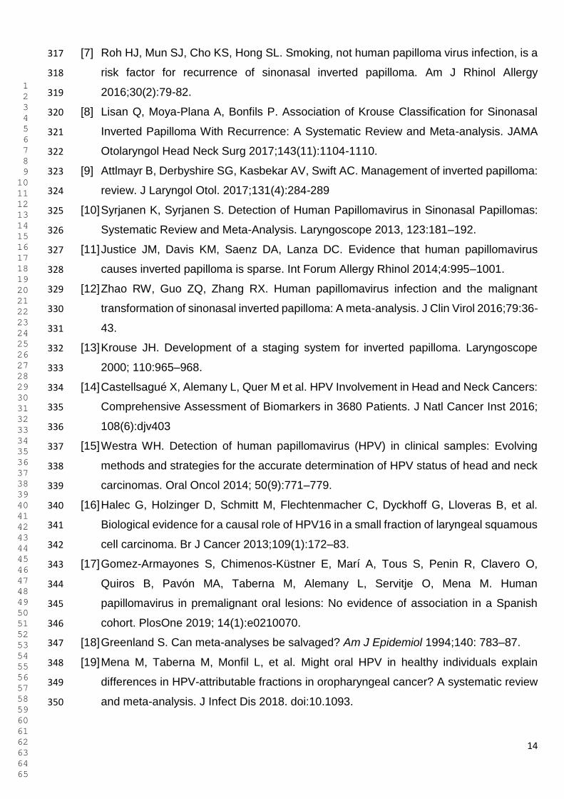
Histologically, SNIP is characterized by invagination of the hyperplastic epithelium, ranging 57
from squamous to ciliated columnar with goblet cells into the underlying stroma (figure 1 and 58
2). 59
SNIP is the most common type of sinonasal papillomas, which are classified according to 60
their histological structure in exophytic or fungiform papilloma, inverted papilloma, and 61
cylindrical or oncocytic papilloma [2]. All types may coexist. SNIPs represent 70% of all 62
sinonasal papillomas [3]. 63
Although histologically benign, SNIP shows a propensity for malignant transformation, most 64
frequently to squamous cell carcinoma (SCC). The rate of malignant association is 65
approximately 10% [4,5]. The factors responsible for malignant transformation are not yet 66
fully elucidated due to the relative low prevalence of SNIPs as well as of SCC arising in the 67
sinonasal tract [4]. SNIP is also particularly prone to recurrence [2] although the postulated 68
factors associated with recurrence such as human papillomavirus (HPV) infection [6], 69
tobacco smoking [7] or Krouse stage [8] have not been consistently corroborated. 70
The recommended treatment for SNIP is complete surgical excision by an endoscopic, open 71
or combined approach [9]. Occupational exposures (organic solvents and welding fumes) 72
have been reported to be implicated in the development of SNIP [2]. Chronic sinonasal 73
inflammation may also have an etiologic relation with SNIP, but the mechanism is still 74
unclear. 75
1 2 3 4 5 6 7 8 9 10 11 12 13 14 15 16 17 18 19 20 21 22 23 24 25 26 27 28 29 30 31 32 33 34 35 36 37 38 39 40 41 42 43 44 45 46 47 48 49 50 51 52 53 54 55 56 57 58 59 60 61 62 63 64 65

4
The establishment of the role of HPV in a fraction of head and neck cancers (HNC) raised 76
interest on the etiologic and prognostic role of HPV in other benign head and neck lesions 77
such as SNIP. In the last decades, several studies have explored the relationship between 78
SNIP and HPV infection, obtaining conflicting results. The detection rates of HPV in SNIP 79
range from 0 to 100% in the world literature [1,2,10]. Such differences have not been 80
explained by differences in geographical regions or HPV detection methods [10]. 81
Recently, last reports about the role of HPV in SNIP are proposing new theories postulating 82
that the infection is not related to the initial pathogenesis of the SNIP but when there is an 83
inflammatory and metaplastic mucosa, the virus is more susceptible to infect it [11]. 84
Moreover, the presence of the virus in the SNIP has been related with a higher risk of 85
recurrences [6] and malignant transformation (especially for high-risk genotypes 16 and 18) 86
[1,12]. The unequivocal establishment of the prognostic HPV role in SNIPs, as well as other 87
nasosinusal lesions, might have implications for tertiary prevention of recurrence or 88
malignant transformation through HPV vaccination. 89
This study aimed to estimate the HPV-DNA prevalence and type distribution in SNIP in two 90
series from two different geographical areas, Spain and Poland, and to analyse risk factors 91
for recurrence and malignant transformation in both groups. 92
93
METHODS 94
Study Design 95
We carried out a retrospective study including two cohorts of all primary SNIPs diagnosed 96
between 1995 and 2014 at the Department of otorhinolaryngology of the Hospital 97
Universitari de Bellvitge (Spain) and between 2012 and 2017 at the Department of 98
Otorhinolaryngology of the Czerniakowski Hospital in Warsaw (Poland). The pathologic 99
diagnosis of the lesions was confirmed by biopsy. Demographic data and information about 100
history of smoking and alcohol use, previous HPV-related pathology, tumor extent by Krouse 101
1 2 3 4 5 6 7 8 9 10 11 12 13 14 15 16 17 18 19 20 21 22 23 24 25 26 27 28 29 30 31 32 33 34 35 36 37 38 39 40 41 42 43 44 45 46 47 48 49 50 51 52 53 54 55 56 57 58 59 60 61 62 63 64 65
5
staging system [13] and follow-up was collected from medical records. Recurrent cases or 102
cases previously undergoing nasal surgeries were excluded. 103
Protocols were approved by the ethics committee of the Catalan Institute of Oncology-ICO 104
(Comité Ètic d’Investigació Clínica de l’Hospital Universitari de Bellvitge, Spain), which 105
required no informed consent to use archived samples. 106
Formalin-fixed, paraffin-embedded (FFPE) blocks processing and histopathological 107
evaluation 108
Protocols have been described elsewhere [14]. Briefly, FFPE blocks were processed under 109
strict conditions to avoid contamination and were re-embedded at ICO whenever necessary. 110
At least four paraffin sections were obtained for each block. First and last sections were 111
used for histopathological evaluation (sandwich method) after hematoxylin and eosin (H&E) 112
staining and the in-between ones for HPV testing and genotyping and expression of p16INK4a. 113
FFPE blocks were processed under strict pre/post polymerase chain reaction (PCR) 114
physical separation, and blank paraffin blocks were systematically tested in parallel to serve 115
as sentinels for contamination as previously published [14]. Pathology review was performed 116
using a form specifically designed by two pathologists for the study (see supplementary 117
material) and blind with respect to the original local diagnosis. It followed a pre-established 118
algorithm for diagnostic consensus involving the two pathologists. First, all pathology slides 119
were reviewed by a trained pathologist at ICO. Samples with discordant diagnosis were 120
further reviewed by the two pathologists for a final evaluation and agreed diagnosis. 121
HPV-DNA Detection and Genotyping 122
The detailed methods used for HPV-DNA detection and genotyping have been reported 123
elsewhere [14]. Briefly, we used SPF-10 PCR and a DNA enzyme immunoassay (DEIA) to 124
test for the presence of HPV-DNA. Virus genotyping was performed using reverse 125
hybridization line probe assay (LiPA25_v1) on all samples testing positive for viral DNA, 126
targeting 25 HPV types with different oncogenic potential. DNA quality was evaluated in all 127
1 2 3 4 5 6 7 8 9 10 11 12 13 14 15 16 17 18 19 20 21 22 23 24 25 26 27 28 29 30 31 32 33 34 35 36 37 38 39 40 41 42 43 44 45 46 47 48 49 50 51 52 53 54 55 56 57 58 59 60 61 62 63 64 65

6
HPV-DNA negative samples by testing for the human tubulin gene [14]. All DEIA and 128
LiPA25_v1 assays were performed at ICO. 129
p16INK4a immunohistochemistry 130
p16INK4a expression was evaluated on all HPV-DNA positive cases and a random sample of 131
HPV-DNA negative cases using the CINtec histology kit (clone E6H4, Roche mtm 132
laboratories AG, Germany), following the manufacturer’s protocol. A pattern of diffuse 133
staining of more than 70% stained cells (nuclear and cytoplasmic) is considered positive for 134
malignant lesions [15], but in our study we assumed as positive a staining between 26 and 135
50% for premalignant lesions in a diffuse or continuous pattern [16,17]. 136
137
Statistical analysis 138
Descriptive statistics were computed for each of the variables analysed. Fisher’s exact test 139
for categorical variables and t-test for continuous variables were used to detect statistically 140
significant differences between the two centres for each variable. Median and range of 141
months to recurrence and months of follow up variables were estimated. To compare 142
medians between groups, qreg (quantile regression) test, equivalent to t-test for means, was 143
employed. A survival analysis was conducted to identify variables associated with 144
recurrence. Cox regression model was performed to estimate hazard ratios and their 95% 145
CI. In order to avoid the possible bias due to the centre where the SNIPs were diagnosed 146
and treated, centre was introduced as a strata variable allowing the baseline hazard function 147
to differ for the different centre. Proportional hazard assumption was also verified. Due to 148
the low number of cases progressing to invasive cancer during follow-up, survival analyses 149
to identify variables associated with malignant transformation could not be performed. All 150
the analyses were performed with STATA 13.1 software. 151
RESULTS 152
1 2 3 4 5 6 7 8 9 10 11 12 13 14 15 16 17 18 19 20 21 22 23 24 25 26 27 28 29 30 31 32 33 34 35 36 37 38 39 40 41 42 43 44 45 46 47 48 49 50 51 52 53 54 55 56 57 58 59 60 61 62 63 64 65

7
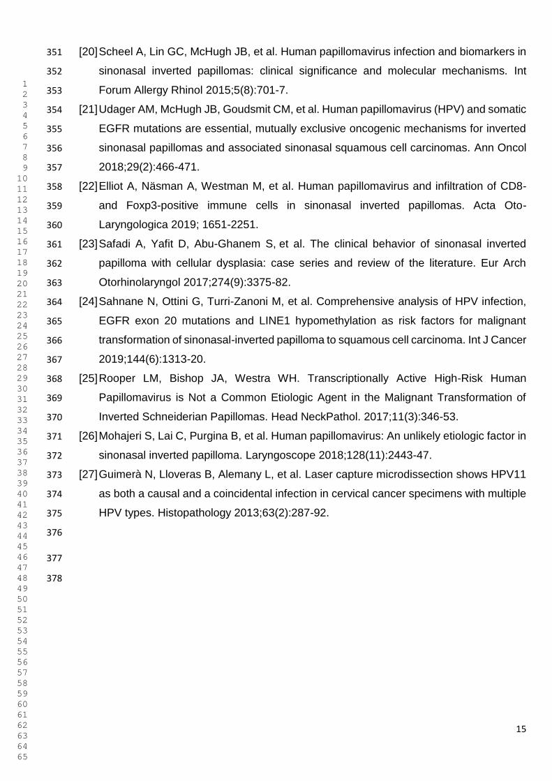
Figure 3 depicts the disposition of SNIP samples collected, processed and tested. The ICO 153
laboratory received 63 samples from Bellvitge Hospital (Spain) and 68 samples from 154
Czerniakowski Hospital (Poland). A total of 79 cases (46 from Spain and 33 from Poland) 155
were included in the final analysis, respectively. 156
The characteristics of the patients are presented in Table 1. Most cases were males (67.1%) 157
and non-drinkers (70.9%) with a mean age of 56.2 years. There was a higher proportion of 158
ever-smokers in the Spanish group (67.4% vs 39.4%, p=0.010) whereas Polish cases were 159
more frequently diagnosed with more advanced Krouse stages (p<0.001). Cases from both 160
centres presented also differences regarding some histopathological variables, with more 161
Polish cases presenting transitional epithelium (p=0.015) and more Spanish cases 162
presenting squamous epithelium (p<0.001) and papillar or exophytic lesion adjacent to SNIP 163
(p<0.001). The median time to follow-up was 76.63 months (range 0.23-174.3) for Spanish 164
cases and 39.1 months (range 6.3-66.5) for Polish ones. Seventeen patients (21.5%) had 165
recurrence, of which 12 were from Spain (26.1%) and 5 from Poland (15.2%), with a median 166
time to recurrence of 14 months (range from 3 to 83). Only two cases (2.5%), both belonging 167
to the Spanish series, progressed to invasive cancer during follow-up. HPV-DNA was 168
detected in two samples (4.3%) in the Spanish series and in two samples (6.1%) in the 169
polish series. All of them were positive for HPV11 and negative for p16INK4a high expression. 170
All HPV-DNA negative cases tested for p16INK4a (20 cases, representing 27% of all HPV-171
DNA negative cases) were also negative for p16INK4a high expression. 172
173
The presence of atypia adjacent to SNIP at diagnosis was the only statistically significant 174
factor associated to recurrence with a crude hazard ratio (HR) of 18.83 (95%CI, 1.71-175
207.65) (Table 2). The recurrence rate was higher in higher Krouse stages (T2 and T3) 176
compared to T1, although not statistically significant. No significant differences in risk of 177
recurrence were found by smoking, alcohol drinking habits or HPV positivity. The four low 178
1 2 3 4 5 6 7 8 9 10 11 12 13 14 15 16 17 18 19 20 21 22 23 24 25 26 27 28 29 30 31 32 33 34 35 36 37 38 39 40 41 42 43 44 45 46 47 48 49 50 51 52 53 54 55 56 57 58 59 60 61 62 63 64 65

8
risk-HPV positive SNIPs were located in the nasal cavity (one in the septum, one in the 179
vestibulum and two in the lateral wall, and among those, one in the lower turbinate and one 180
in the middle turbinate and middle meatus). None of them presented dysplasia. In contrast, 181
most HPV-negative SNIPs were located at the lateral wall (85%), the maxillary sinus (15%) 182
and the ethmoid sinus (20%), some of them affecting more than one location. HPV-positive 183
patients were two males and two females, with a mean age of 37.8 years old at the moment 184
of diagnosis. SNIPs recurred in 25% (1/4) of HPV-positive vs 22.2% (16/72) of HPV-negative 185
lesions (crude HR=3.70, 95%CI 0.44–31.37). Recurrence univariate Cox models for tobacco 186
use and Krouse stage were also performed stratified by centre, and statistically significant 187
differences between both groups were not found. 188
DISCUSSION 189
The etiologic and prognostic role of HPV in SNIP remains unclear, with previous studies 190
reporting HPV detection rates ranging from 0 to 100% [1,2,10,11] as well as contradictory 191
results on the role of HPV infection in recurrence and malignant transformation of SNIP 192
[1,6,12].The differences have not been explained by differences in geographical regions or 193
HPV detection methods [10]. However, to the best of our knowledge, any study has 194
evaluated cases from different geographical regions with the same sample processing and 195
HPV detection protocol. Thus, our results add to current data from a systematic review [10], 196
the only previous publication pooling results of studies from different geographical regions, 197
since systematic reviews and meta-analyses are not exempt from limitations [18]. 198
We herein evaluated the prevalence and prognostic role of HPV in two retrospective cohorts 199
of primary SNIPs from Spain and Poland, as well as additional factors associated to 200
recurrence. We tested the SNIPs cases following a previously validated robust, standardized 201
and international protocol designed to provide estimates of HPV-attributable fractions in 202
HPV-related cancers [14]. 203
1 2 3 4 5 6 7 8 9 10 11 12 13 14 15 16 17 18 19 20 21 22 23 24 25 26 27 28 29 30 31 32 33 34 35 36 37 38 39 40 41 42 43 44 45 46 47 48 49 50 51 52 53 54 55 56 57 58 59 60 61 62 63 64 65
9
Our study is the first to evaluate SNIPs from two different geographical regions which have 204
previously shown marked differences in HPV-AFs in OPC [14]. Our data demonstrates a 205
low HPV-DNA detection (5.1%) in primary SNIPs, similar to the 4.9% estimated at the oral 206
cavity of healthy population [19] or the 4% and 7% estimated at inflammatory nasal polyps 207
and normal sinonasal mucosa, respectively [10], suggesting that HPV is not a main 208
etiological factor for SNIP in either setting herein evaluated. Our HPV prevalence estimates 209
are in accordance with those of previous studies with equivalent number of cases [10,11] 210
although lower than others [7,10,20,21]. The only type found was HPV11, as reported by 211
others [1,7,20]. No high risk types were found in our sample. 212
The prevalence of HPV in SNIP with dysplasia or with SCC adjacent to the lesion is 213
estimated to be higher than in SNIP without dysplasia [1], and more common in recurrent 214
lesions [1]. We only included primary lesions, and only one case from Spain had dysplasia 215
adjacent to the SNIP at diagnosis, and it was HPV positive. Three out of four HPV-positive 216
patients in our series were ever smokers, although the association between the two 217
variables was not statistically significant (p-value=0.631). The low HPV prevalence rates 218
found in our series prevented us to further explore factors associated with HPV positivity. 219
However, we noted a trend for HPV positive cases to affect the nasal cavity rather than the 220
sinus and to involve younger people, in accordance with other studies [22]. 221
We observed some differences between the two groups of patients, with Polish cases 222
diagnosed at more recent periods (due to case selection), more advanced Krouse stages, 223
and presenting a lower proportion of ever-smokers. Differences in some histopathological 224
features were also observed between Polish and Spanish cases. However, due to the low 225
number of cases, we decided to combine both groups to evaluate factors associated with 226
recurrence and to address the differences between groups with the use of the strata function 227
in Stata. Moreover, when accounting for such differences by stratifying the recurrence 228
univariate Cox models for tobacco use and Krouse stage by centre, we did not find 229
1 2 3 4 5 6 7 8 9 10 11 12 13 14 15 16 17 18 19 20 21 22 23 24 25 26 27 28 29 30 31 32 33 34 35 36 37 38 39 40 41 42 43 44 45 46 47 48 49 50 51 52 53 54 55 56 57 58 59 60 61 62 63 64 65

10
statistically significant differences between both groups. Although showing marked 230
differences in HPV-AF in OPC for SNIPs, both series showed similar HPV prevalences, 231
confirming that the variability in HPV detection rates in SNIPs is not explained by their 232
different geographic origins, as it was already hypothesized in a previous metanalysis [10] 233
The presence of dysplasia adjacent to the SNIP at diagnosis was the only factor associated 234
with recurrence (HR: 18.83, 95%CI: 1.71-207.65), as it has been shown in previous studies 235
[23], although only one case contributed to the estimation. A HR of 3.70 (95%CI 0.44-31.37) 236
was observed for recurrence in HPV-positive cases, although not statistically significant. 237
Other factors such as tobacco smoking or Krouse stage did not show any prognostic value 238
for recurrence. However, tobacco use [1,7] and T3 vs T2 Krouse stages [8] have been 239
previously reported to be related to recurrence. The low number of cases evaluated in this 240
study could explain these discrepancies with the literature. A slight decreasing trend for 241
recurrence in more recent years was observed, although it was not statistically significant. 242
Only two cases (2.6%) progressed to invasive cancer during follow-up, and none of them 243
were HPV-positive. Thus, we could not evaluate further the prognostic value of HPV 244
positivity or other factors for malignant transformation. Different risk factors are suspected 245
to be involved in malignant transformation of SNIPs and include HPV infection, tobacco 246
smoking and occupational exposure [24]. In contrast, EGFR mutations have been observed 247
to be a protective factor for malignant transformation of SNIPs [21,24]. However, many 248
previous studies suggesting that HPV infection may play a role as a co-factor in the 249
development of carcinoma ex-SNIP did not use biomarkers of biological activity of HPV such 250
as the presence of E6/E7 mRNA transcripts or p16INK4a expression. Indeed, studies 251
evaluating E6/E7 mRNA transcripts [25] or p16INK4a expression [26] in SNIPs did not find 252
HPV as an etiological driver of SNIP development or progression to SCC. We did not 253
observe p16INK4a expression in any HPV-DNA positive samples as expected, since all of 254
them were positive for low risk genotypes. However, we did not test all HPV-DNA negatives 255
1 2 3 4 5 6 7 8 9 10 11 12 13 14 15 16 17 18 19 20 21 22 23 24 25 26 27 28 29 30 31 32 33 34 35 36 37 38 39 40 41 42 43 44 45 46 47 48 49 50 51 52 53 54 55 56 57 58 59 60 61 62 63 64 65

11
samples for p16INK4a expression. We did neither evaluate further biomarkers of biological 256
activity of HPV on HPV-DNA positive samples such as E6/E7 mRNA positivity nor do use 257
techniques like laser capture microdissection, which combined with highly sensitive PCR 258
allow assignment of a particular HPV genotype to an area of normal or abnormal epithelium 259
[27]. 260
The major limitation of the study was its relatively small sample size, which hampered us to 261
evaluate factors associated with HPV-positivity, recurrence and malignant transformation. 262
However, given the fact that SNIP is a relatively rare entity, few studies have reported results 263
for series with equivalent number of primary SNIP cases consecutively diagnosed in two 264
decades, like ours. Not all HPV-DNA negative cases were evaluated for p16INK4a expression 265
and no evaluation of further biomarkers such as E6/E7 mRNA or EGFR was performed. A 266
recent study showed that EGFR mutations and HPV infection represent essential, 267
alternative oncogenic mechanisms in SNIP and SNIP-associated sinonasal SCC [21]. The 268
study observed that SNIP progression was significantly associated with the presence of HPV 269
infection and the absence of an EGFR mutation. We did not evaluate the prognostic value 270
of the treatment received by the SNIP patient. However, a previous study did not find 271
differences in recurrence by different types of interventions [9]. 272
The low prevalence of HPV-DNA found in SNIPs from two different countries suggests that 273
HPV is not a main etiological factor for SNIP. The absence of association between HPV and 274
the rest of herein evaluated factors and recurrence may suggest the involvement of other 275
factors. Further research with larger number of patients and additional biomarkers is 276
warranted to unequivocally assess the aetiology and prognosis of SNIP. 277
TITLE’S LEGENDS 278
Figure 1. Sinonasal inverted papilloma. Inverted growth pattern and absence of 279
seromucinous glands (hematoxylin-eosin 0’5x) 280
1 2 3 4 5 6 7 8 9 10 11 12 13 14 15 16 17 18 19 20 21 22 23 24 25 26 27 28 29 30 31 32 33 34 35 36 37 38 39 40 41 42 43 44 45 46 47 48 49 50 51 52 53 54 55 56 57 58 59 60 61 62 63 64 65
12
Figure 2. High power image shows non-keratinizing transitional epithelium covered by a 281
layer of ciliated columnar epithelium. Infiltration by neutrophils is seen (hematoxylin-eosin 282
10 x) 283
Figure 3. Flow chart of cases included in the study 284
285
1 2 3 4 5 6 7 8 9 10 11 12 13 14 15 16 17 18 19 20 21 22 23 24 25 26 27 28 29 30 31 32 33 34 35 36 37 38 39 40 41 42 43 44 45 46 47 48 49 50 51 52 53 54 55 56 57 58 59 60 61 62 63 64 65

13
DISCLOURE 286
Cancer Epidemiology Research Program (LA MM JF BQ OC MP) has received sponsorship for 287
grants from Merck and co, Roche, Reig-jofre, IDT, Hologic, GlaxoSmithKline and Seegene. The rest 288
of authors have declared no conflicts of interest. 289
290
FUNDING 291
We thank CERCA Program / Generalitat de Catalunya for institutional support. This study has been 292
funded by the Instituto de Salud Carlos III (ie, the Spanish government), the European Regional 293
Development Fund – A Way to Build Europe, through the projects CIBERESP CB06/02/0073 and 294
CIBERONC,CB16/12/0040, from the Agència de Gestió d’Ajuts Universitaris i de Recerca 295
(2017SGR1085) and from the Department of Health of the Generalitat de Catalunya (PERIS-2016-296
2020, SLT002/16/00404) (personal grants to MM and JFG, respectively). 297
298
REFERENCES 299
[1] Lawson W, Schlecht NF, Brandwein-Gensler M. The role of the human papillomavirus 300
in the pathogenesis of Schneiderian Inverted papillomas: an analytic overview of the 301
evidence. Head Neck Pathol 2008; 2: 49-59 302
[2] Bishop JA. OSPs and ESPs and ISPs, Oh My! An Update on Sinonasal (Schneiderian) 303
Papillomas. Head Neck Pathol 2017;11(3):269-277. 304
[3] Hyams VJ. Papillomas of the nasal cavity and paranasal sinuses. A clinicopathological 305
study of 315 cases. Ann OtolRhinolLaryngol 1971;80(2):192-206. 306
[4] Sung-Lyong Hong, MD; Bae-Hyun Kim, MD; Jung-Hoon Lee, MD; Kyu-Sup Cho, MD; 307
Hwan-Jung Roh, MD. Smoking and Malignancy in Sinonasal Inverted Papilloma. 308
Laryngoscope 2013, 123:1087–1091. 309
[5] M. Re, F. M. Gioacchini, A. Bajraktari, M. Tomasetti, S. Kaleci, C. Rubini, A. Bertini, G. 310
Magliulo, E. Pasquini. Malignant transformation of sinonasal inverted papilloma and 311
related genetic alterations: a systematic review. Eur Arch Otorhinolaryngol 2017; 312
274(8):2991-3000. 313
[6] Beck JC, McClatchey KD, Lesperance MM, Esclamado RM, Carey TE, Bradford CR. 314
Presence of human papillomavirus predicts recurrence of inverted papilloma. 315
Otolaryngol Head Neck Surg 1995; 113:49-55. 316
1 2 3 4 5 6 7 8 9 10 11 12 13 14 15 16 17 18 19 20 21 22 23 24 25 26 27 28 29 30 31 32 33 34 35 36 37 38 39 40 41 42 43 44 45 46 47 48 49 50 51 52 53 54 55 56 57 58 59 60 61 62 63 64 65
14
[7] Roh HJ, Mun SJ, Cho KS, Hong SL. Smoking, not human papilloma virus infection, is a 317
risk factor for recurrence of sinonasal inverted papilloma. Am J Rhinol Allergy 318
2016;30(2):79-82. 319
[8] Lisan Q, Moya-Plana A, Bonfils P. Association of Krouse Classification for Sinonasal 320
Inverted Papilloma With Recurrence: A Systematic Review and Meta-analysis. JAMA 321
Otolaryngol Head Neck Surg 2017;143(11):1104-1110. 322
[9] Attlmayr B, Derbyshire SG, Kasbekar AV, Swift AC. Management of inverted papilloma: 323
review. J Laryngol Otol. 2017;131(4):284-289 324
[10] Syrjanen K, Syrjanen S. Detection of Human Papillomavirus in Sinonasal Papillomas: 325
Systematic Review and Meta-Analysis. Laryngoscope 2013, 123:181–192. 326
[11] Justice JM, Davis KM, Saenz DA, Lanza DC. Evidence that human papillomavirus 327
causes inverted papilloma is sparse. Int Forum Allergy Rhinol 2014;4:995–1001. 328
[12] Zhao RW, Guo ZQ, Zhang RX. Human papillomavirus infection and the malignant 329
transformation of sinonasal inverted papilloma: A meta-analysis. J Clin Virol 2016;79:36-330
43. 331
[13] Krouse JH. Development of a staging system for inverted papilloma. Laryngoscope 332
2000; 110:965–968. 333
[14] Castellsagué X, Alemany L, Quer M et al. HPV Involvement in Head and Neck Cancers: 334
Comprehensive Assessment of Biomarkers in 3680 Patients. J Natl Cancer Inst 2016; 335
108(6):djv403 336
[15] Westra WH. Detection of human papillomavirus (HPV) in clinical samples: Evolving 337
methods and strategies for the accurate determination of HPV status of head and neck 338
carcinomas. Oral Oncol 2014; 50(9):771–779. 339
[16] Halec G, Holzinger D, Schmitt M, Flechtenmacher C, Dyckhoff G, Lloveras B, et al. 340
Biological evidence for a causal role of HPV16 in a small fraction of laryngeal squamous 341
cell carcinoma. Br J Cancer 2013;109(1):172–83. 342
[17] Gomez-Armayones S, Chimenos-Küstner E, Marí A, Tous S, Penin R, Clavero O, 343
Quiros B, Pavón MA, Taberna M, Alemany L, Servitje O, Mena M. Human 344
papillomavirus in premalignant oral lesions: No evidence of association in a Spanish 345
cohort. PlosOne 2019; 14(1):e0210070. 346
[18] Greenland S. Can meta-analyses be salvaged? Am J Epidemiol 1994;140: 783–87. 347
[19] Mena M, Taberna M, Monfil L, et al. Might oral HPV in healthy individuals explain 348
differences in HPV-attributable fractions in oropharyngeal cancer? A systematic review 349
and meta-analysis. J Infect Dis 2018. doi:10.1093. 350
1 2 3 4 5 6 7 8 9 10 11 12 13 14 15 16 17 18 19 20 21 22 23 24 25 26 27 28 29 30 31 32 33 34 35 36 37 38 39 40 41 42 43 44 45 46 47 48 49 50 51 52 53 54 55 56 57 58 59 60 61 62 63 64 65
15
[20] Scheel A, Lin GC, McHugh JB, et al. Human papillomavirus infection and biomarkers in 351
sinonasal inverted papillomas: clinical significance and molecular mechanisms. Int 352
Forum Allergy Rhinol 2015;5(8):701-7. 353
[21] Udager AM, McHugh JB, Goudsmit CM, et al. Human papillomavirus (HPV) and somatic 354
EGFR mutations are essential, mutually exclusive oncogenic mechanisms for inverted 355
sinonasal papillomas and associated sinonasal squamous cell carcinomas. Ann Oncol 356
2018;29(2):466-471. 357
[22] Elliot A, Näsman A, Westman M, et al. Human papillomavirus and infiltration of CD8- 358
and Foxp3-positive immune cells in sinonasal inverted papillomas. Acta Oto-359
Laryngologica 2019; 1651-2251. 360
[23] Safadi A, Yafit D, Abu-Ghanem S, et al. The clinical behavior of sinonasal inverted 361
papilloma with cellular dysplasia: case series and review of the literature. Eur Arch 362
Otorhinolaryngol 2017;274(9):3375-82. 363
[24] Sahnane N, Ottini G, Turri-Zanoni M, et al. Comprehensive analysis of HPV infection, 364
EGFR exon 20 mutations and LINE1 hypomethylation as risk factors for malignant 365
transformation of sinonasal-inverted papilloma to squamous cell carcinoma. Int J Cancer 366
2019;144(6):1313-20. 367
[25] Rooper LM, Bishop JA, Westra WH. Transcriptionally Active High-Risk Human 368
Papillomavirus is Not a Common Etiologic Agent in the Malignant Transformation of 369
Inverted Schneiderian Papillomas. Head NeckPathol. 2017;11(3):346-53. 370
[26] Mohajeri S, Lai C, Purgina B, et al. Human papillomavirus: An unlikely etiologic factor in 371
sinonasal inverted papilloma. Laryngoscope 2018;128(11):2443-47. 372
[27] Guimerà N, Lloveras B, Alemany L, et al. Laser capture microdissection shows HPV11 373
as both a causal and a coincidental infection in cervical cancer specimens with multiple 374
HPV types. Histopathology 2013;63(2):287-92. 375
376
377
378
1 2 3 4 5 6 7 8 9 10 11 12 13 14 15 16 17 18 19 20 21 22 23 24 25 26 27 28 29 30 31 32 33 34 35 36 37 38 39 40 41 42 43 44 45 46 47 48 49 50 51 52 53 54 55 56 57 58 59 60 61 62 63 64 65

HISTOLOGICAL EVALUATION
Identification number ________ Date ______ Pathologist ________
1. HISTOLOGICAL DESCRIPTION OF THE LESION
1.1. Types of epithelium in the sinonasal inverted papilloma (SNIP)
□ Transitional
□ Squamous
□ Columnar
□ Presence of oncocytic cells
□ Hyper-parakeratosis
□ Presence of exophytic or papillary lesion adjacent to SNIP
□ Dysplasia □ Mild □ Moderate □ Severe/Carcinoma in situ
Others:
1.2. Intralesional polymorphonuclear infiltrate:
□ No
□ Yes □ Mild □ Moderate □ Severe
Inflammatory Perilesional infiltrate:
□ No
□ Yes □ Mild □ Moderate □ Severe
1.3. Subepithelial stromal tissue
□ Lax □ Dense
1.4. Other findings:
2. DEFINITIVE DIAGNOSIS OF THE LESION
□ Inverted
□ Oncocytic.
□ Exophytic
□ Non-papillary lesion:
□ Dysplasia □ Mild □ Moderate □ Severe/Carcinoma in situ
Manuscript Click here to access/download;Manuscript;Supplementarymaterial.doc
Click here to view linked References
1 2 3 4 5 6 7 8 9 10 11 12 13 14 15 16 17 18 19 20 21 22 23 24 25 26 27 28 29 30 31 32 33 34 35 36 37 38 39 40 41 42 43 44 45 46 47 48 49 50 51 52 53 54 55 56 57 58 59 60 61 62 63 64 65

3. Slides A&B
□ Same □ Different
4. Control
□ Tissue: _____________
5. Final evaluation
□ Adequate for HPV analysis
□ Repeat sandwich technique
□ Doubtful/Uncertain
□ Discard for HPV analysis
6. External quality control
□ Pathologist _____________
7. Internal quality control
□ Pathologist _____________
Comments
1 2 3 4 5 6 7 8 9 10 11 12 13 14 15 16 17 18 19 20 21 22 23 24 25 26 27 28 29 30 31 32 33 34 35 36 37 38 39 40 41 42 43 44 45 46 47 48 49 50 51 52 53 54 55 56 57 58 59 60 61 62 63 64 65

Figure Click here to access/download;Figure;Figure1.tif
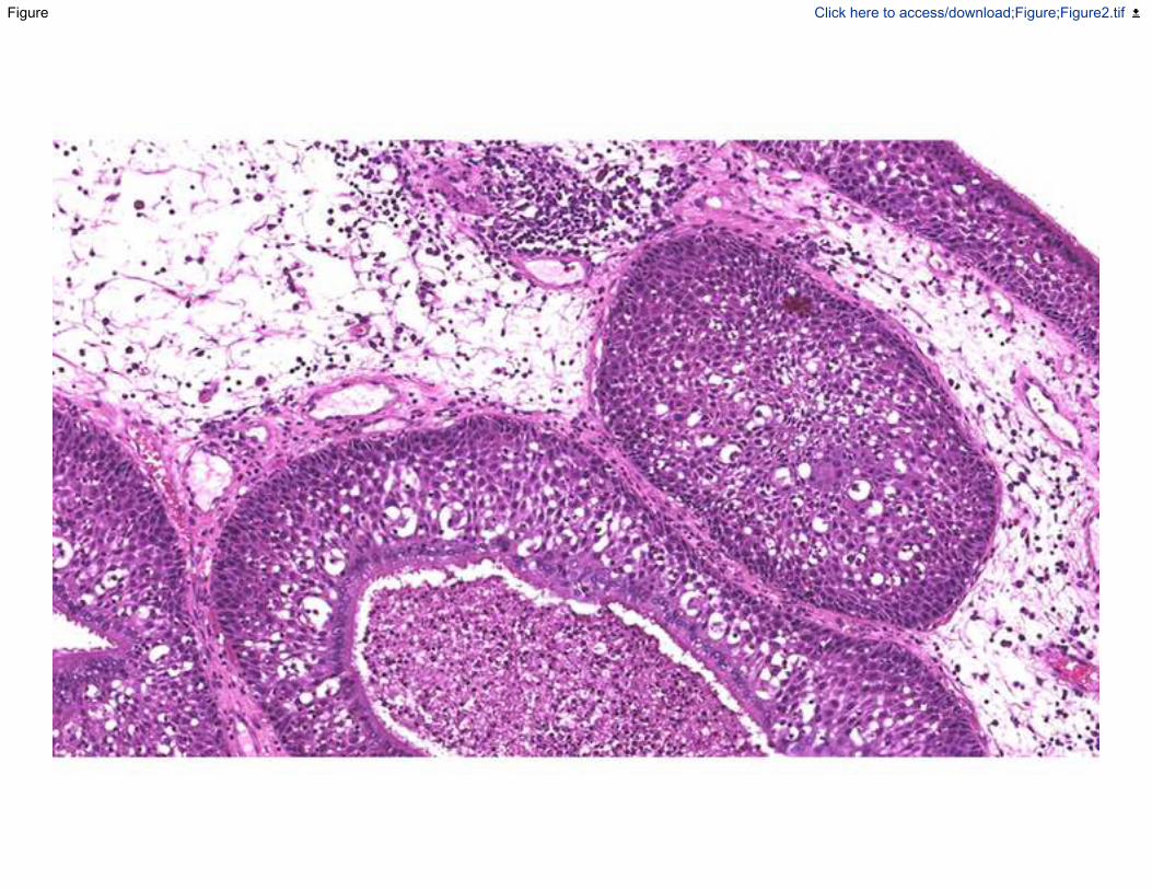
Figure Click here to access/download;Figure;Figure2.tif
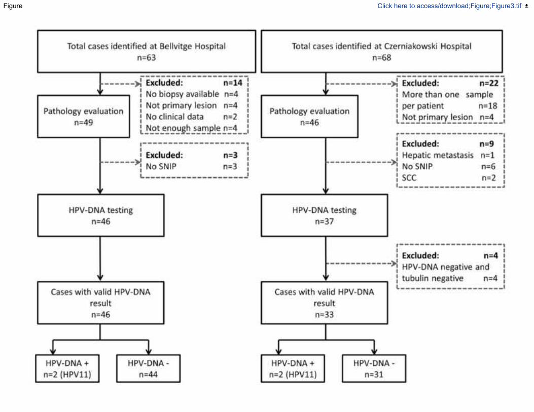
Figure Click here to access/download;Figure;Figure3.tif

Table 1. Demographic and clinical characteristics of SNIP patients included in
the study
Characteristics
Total
SNIPs
(n = 79)
No. (%)a
Spanish
SNIPs
(n = 46)
No. (%)
Polish
SNIPs
(n = 33)
No. (%)
p-valuea
Age at diagnosis
Mean (SD)
Range
56.2 (16.1)
19-91
55.0 (15.0)
19-85
57.8 (17.7)
23-91
0.460
Gender
Male
Female
53 (67.1)
26 (32.9)
34 (73.9)
12 (26.1)
19 (57.6)
14 (42.4)
0.150
Period of diagnosis
1995-2000
2001-2006
2007-2012
2013-2017
7 (8.9)
17 (21.5)
24 (30.4)
31 (39.2)
7 (15.2)
17 (37.0)
22 (47.8)
0 ( 0.0)
0 ( 0.0)
0 ( 0.0)
2 ( 6.1)
31 (93.9)
<0.001
Tobacco use
Never smoker
Ever smoker
Missing
33 (41.8)
44 (55.7)
2 (2.5)
13 (28.3)
31 (67.4)
2 (4.3)
20 (60.6)
13 (39.4)
0 (0.0)
0.010
Alcohol use
Never drinker
Ever drinker
Missing
56 (70.9)
21 (26.6)
2 (2.5)
33 (71.7)
11 (23.9)
2 (4.3)
23 (69.7)
10 (30.3)
0 (0.0)
0.616
Previous history of HPV-related pathology
Yes
No
Missing
3 (3.8)
74 (93.7)
2 (2.5)
2 (4.3)
42 (91.3)
2 (4.3)
1 (3.0)
32 (97.0)
0 (0.0)
1.000
Krouseclassification
1
2
3
4
Missing
33 (41.8)
23 (29.1)
16 (20.3)
4 (5.1)
3 (3.8)
28 (60.9)
11 (23.9)
3 (6.5)
1 (2.2)
3 (6.5)
5 (15.2)
12 (36.4)
13 (39.4)
3 (9.1)
0 (0.0)
<0.001
Transitionalepithelium
Absence
Presence
19 (24.1)
60 (75.9)
16 (34.8)
30 (65.2)
3 ( 9.1)
30 (90.9)
0.015
Squamousepithelium
Absence
Presence
45 (57.0)
34 (43.0)
19 (41.3)
27 (58.7)
26 (78.8)
7 (21.2)
0.001
Columnarepithelium
Absence
Presence
23 (29.1)
56 (70.9)
11 (23.9)
35 (76.1)
12 (36.4)
21 (63.6)
0.316
Hyper-parakeratosis
Absence
Presence
76 (96.2)
3 (3.8)
44 (95.7)
2 ( 4.3)
32 (97.0)
1 ( 3.0)
1.000
Papillar or exophytic lesion adjacent to SNIP
Absence
Presence
61 (77.2)
18 (22.8)
44 (95.7)
2 ( 4.3)
17 (51.5)
16 (48.5)
<0.001
Polymorphonuclear neutrophil inflammatory
intralesional infiltrate
Absence
Presence
2 (2.5)
77 (97.5)
1 ( 2.2)
45 (97.8)
1 ( 3.0)
32 (97.0)
1.000
Polymorphonuclear neutrophil inflammatory perilesional
infiltrate
Absence
Presence
1 (1.3)
78 (98.7)
0 ( 0.0)
46 (100.0)
1 ( 3.0)
32 (97.0)
0.418
Recurrence
No
Yes
Missing
59 (74.7)
17 (21.5)
3 (3.8)
31 (67.4)
12 (26.1)
3 (6.5)
28 (84.8)
5 (15.2)
0 ( 0.0)
0.268
Months of follow up
Median
Range
52.83
0.23-174.33
76.63
0.23-174.33
39.1
6.27-66.47
0.003 b
Months to recurrence
Median
Range
14
3-83
17
9-83
4
3-36
0.469b
Progression to invasive cancer
No
Yes
Missing
74 (93.7)
2 (2.5)
3 (3.8)
41 (89.1)
2 (4.3)
3 ( 6.5)
33 (100.0)
0 ( 0.0)
0 ( 0.0)
0.502
HPV positivity
No
Yes
75 (94.9)
4 (5.1)
44 (95.7)
2 (4.3)
31 (93.9)
2 (6.1)
1.000
SNIP: Sinonasal Inverted Papilloma; SD: Standard deviation; aFischer exact test with the exception of age and months
to recurrence. where t-student test has been used to compare the median values between populations. b: qreg (quantilte
regression): test for equality of medians, equivalent for t-test for medians.
Table1 Click here to access/download;Table;Table1.docx

Table 2. Hazard ratios for recurrence in SNIP patients included
in the study
Characteristics
Total SNIPs
(n = 76)
No. (%)
Recurrences
No. (%)
Crude HR
(95%CI)
Hospital
Bellvitge (Spain)
Czerniakowski (Poland)
43 (56.58)
33 (43.42)
12 (27.91)
5 (15.15)
Ref.
0.81 (0.27–2.39)
Age at diagnosisb
Mean (SD)
56.34 (16.40)
-
1.01 (0.98–1.04)
Gender
Male
Female
51 (67.11)
25 (32.89)
14 (27.45)
3 (12.00)
Ref.
0.45 (0.13–1.58)
Year of diagnosis
Range
1995-2017
-
0.96 (0.86–1.08)
Tobacco use
Never smoker
Ever smoker
32 (42.11)
44 (57.89)
7 (21.88)
10 (22.73)
Ref.
0.97 (0.35–2.64)
Alcohol use
Never drinker
Ever drinker
55 (72.37)
21 (27.63)
12 (21.82)
5 (23.81)
Ref.
1.32 (0.46–3.78)
Previous history of HPV-related pathology
No
Yes
73 (96.05)
3 (3.95)
16 (21.92)
1 (33.33)
Ref.
0.91 (0.12–7.06)
Krouse classification
1
2
3
4
33 (43.42)
23 (30.26)
16 (21.05)
4 (5.26)
8 (24.24)
6 (26.09)
3 (18.75)
0 (0.00)
Ref.
1.70 (0.54–5.29)
1.27 (0.28–5.75)
-
Transitional epithelium
Absence
Presence
18 (23.68)
58 (76.32)
2 (11.11)
15 (25.86)
Ref.
3.01 (0.67–13.56)
Squamous epithelium
Absence
Presence
43 (56.58)
33 (43.42)
11 (25.58)
6 (18.18)
Ref.
0.49 (0.17–1.43)
Columnar epithelium
Absence
Presence
22 (28.95)
54 (71.05)
3 (13.64)
14 (25.93)
Ref.
1.90 (0.54–6.69)
Papillar or exophytic lesion adyacent to SNIP
Absence
Presence
58 (76.32)
18 (23.68)
14 (24.14)
3 (16.67)
Ref.
0.85 (0.20–3.55)
Polymorphonuclear neutrophil inflammatory
intralesional infiltrate (present in 74 cases)
Low
Moderate
67 (90.54)
7 (9.46)
16 (23.88)
1 (14.29)
Ref.
0.52 (0.07–3.96)
Polymorphonuclear neutrophil inflammatory perilesional
infiltrate (present in 75 cases)
Low
Moderate
Severe
56 (74.67)
18 (24.00)
1 (1.33)
12 (21.43)
5 (27.78)
0 (0.00)
Ref.
2.59 (0.79–8.52)
-
Dysplasia at diagnosis adyacent to SNIP
Absence
Presence
75 (98.68)
1 (1.32)
16 (21.33)
1 (100.00)
Ref.
18.83 (1.71–207.65)
Progression to cancer during follow-up
No
Yes
74 (97.37)
2 (2.63)
16 (21.62)
1 (50.00)
Ref.
1.69 (0.22–13.24)
HPV positivity
No
Yes
72 (94.74)
4 (5.26)
16 (22.22)
1 (25.00)
Ref.
3.70 (0.44–31.37)
SNIP: Sinonasal Inverted Papilloma; aThree out of 79 cases did not have information regarding recurrence. bFirst row
shows mean age of the sample with its standard deviation.
Table2 Click here to access/download;Table;Table2.docx