1. introduction 3. classification 4. diagnosis 5 ...reference was also made to other cpg on...
TRANSCRIPT


Management of Osteoarthritis ( Second Edition)
Published by:Malaysia Health Technology Assessment Section (MaHTAS)Medical Development Division, Ministry of Health MalaysiaLevel 4, Block E1, Precinct 1 Federal Government Administrative Centre 62590, Putrajaya, Malaysia
CopyrightThe copyright owner of this publication is MaHTAS. Content may be reproduced in any number of copies and in any format or medium provided that a copyright acknowledgement to MaHTAS is included and the content is not changed, not sold, nor used to promote or endorse any product or service, and not used in an inappropriate or misleading context.
ISBN: 978-967-0399-86-7
Available on the following websites:http://www.moh.gov.myhttp://www.acadmed.org.myhttp://www.msr.my
STATEMENT OF INTENTThese clinical practice guidelines (CPG) are meant to be guides for clinical practice, based on the best available evidence at the time of development. Adherence to these guidelines may not necessarily guarantee the best outcome in every case. Every healthcare provider is responsible for the management of his/her unique patient based on the clinical picture presented by the patient and the management options available locally.
These guidelines were issued in 2013 and will be reviewed in 2017 or sooner if new evidence becomes available.

Management of Osteoarthritis ( Second Edition)
TABLE OF CONTENTS
No. Title Page
Levels of Evidence and Grades of Recommendation i Guidelines Development and Objectives ii Guidelines Development Group iv Review Committee v External Reviewers vi Algorithm on Management of Osteoarthritis vii 1. INTRODUCTION 1 2. EPIDEMIOLOGY & RISK FACTORS 2 2.1 Epidemiology 2 2.2 Risk Factors 3 3. CLASSIFICATION 5
4. DIAGNOSIS 6 4.1 Clinical Features 6 4.2 Diagnostic Criteria 7 5. INVESTIGATIONS 10 5.1 Laboratory Investigations 10 5.2 Imaging 11
6. MANAGEMENT 14 6.1 Non-Pharmacological Treatment 14 a. Education 14 b.LifestyleModification 14 c. Physiotherapy 15 d. Occupational Therapy 17 e. Orthoses 18 6.2 Pharmacological Treatment 19 a. Oral Treatment 19 i. Paracetamol 19 ii. Tramadol 20 iii.Non-steroidalAnti-inflammatoryDrugs& 20 Cyclo-oxygenase-2 Inhibitors iv.Glucosamine&Chondroitin 22 v. Diacerein 24 b. Intra-articular Treatment 24 i. Corticosteroids 24 ii. Viscosupplementation 25 c. Topical Treatment 26

Management of Osteoarthritis ( Second Edition)
No. Title Page
7. ALTERNATIVE TREATMENT 27 8. SURGICAL TREATMENT 28 8.1 Arthroscopic Surgery 28 8.2 High Tibial Osteotomy 28 8.3 Total Joint Replacement 29 8.4 Partial Joint Replacement 29 8.5 Arthrodesis 30 8.6 Recent Advances in Osteoarthritis 30
9. REFERRAL 32 9.1 Rheumatology Referral 32 9.2 Orthopaedic Referral 32 10. PRIMARY PREVENTION 32 11. IMPEMENTING THE GUIDELINES 33 11.1Facilitating&LimitingFactors 33 11.2 Potential Resource Implications 33 REFERENCES 35 Appendix 1 Example of Search Strategy 41 Appendix 2 Clinical Questions 42 Appendix 3 Relationship between Anatomical Site and 43 Possible Physiological Mechanism for Pain in Osteoarthritis Appendix 4 Quadriceps Strengthening Exercise 44 Appendix 5 Hip Strengthening Exercise 45 Appendix 6 Appropriate Measurement for Walking Stick 46 Appendix 7 Joint Protection Principles 47ActivityModificationinPerformingADL Appendix 8 Suggested Medication Dosages and Side Effects 48 Appendix 9 Checklist of OA Management 51 List of Abbreviations 52 Acknowledgement 53 Disclosure Statement 53 Source of Funding 53
TABLE OF CONTENTS

Management of Osteoarthritis ( Second Edition)
LEVELS OF EVIDENCE
SOURCE: US / CANADIAN PREVENTIVE SERVICES TASK FORCE
SOURCE: MODIFIED FROM THE SCOTTISH INTERCOLLEGIATE GUIDELINES NETWORK (SIGN)
Note: The grades of recommendation relates to the strength of the evidenceonwhichtherecommendationisbased.Itdoesnotreflecttheclinical importance of the recommendation.
A
B
C
At least one meta analysis, systematic review, or RCT, or evidence rated as good and directly applicable to the target population
Evidence from well conducted clinical trials, directly applicable to the target population, and demonstrating overall consistency of results; or evidence extrapolated from meta analysis, systematic review, or RCT
Evidence from expert committee reports, or opinions and /or clinical experiences of respected authorities; indicates absence of directly applicable clinical studies of good quality
Level
I
II -1
II-2
II-3
III
Study design
Evidence from at least one properly randomised controlled trial
Evidence obtained from well-designed controlled trials without randomisation
Evidence obtained from well-designed cohort or case-control analytic studies, preferably from more than one centre orgroup
Evidence from multiple time series with or without intervention. Dramatic results in uncontrolled experiments (such as the results of the introduction of penicillin treatment in the 1940s) could also be regarded as this type of evidence
Opinions of respected authorities based on clinical experience; descriptive studies and case reports; or reports of expert committees
i
GRADES OF RECOMMENDATION

Management of Osteoarthritis ( Second Edition)
ii
GUIDELINES DEVELOPMENT AND OBJECTIVES
GUIDELINES DEVELOPMENT
The members of the Development Group (DG) for these Clinical Practice Guidelines (CPG) were from the Ministry of Health (MoH) and Ministry of Higher Education. There was active involvement of a multidisciplinary Review Committee (RC) during the process of the CPG development.
The previous CPG entitled Management of Osteoarthritis 2002 was used as the basis for the development of the present guidelines. A literature search was carried out using the following electronic databases: Guidelines International Network (G-I-N); World Health Organization (WHO), Medline via Ovid, Pubmed, Cochrane Database of Systemic Reviews (CDSR) and International Health Technology Assessment websites (refer to Appendix 1 for Example of Search Strategy). The search was limited to literature published in the last ten years,onhumansandinEnglish.Iftheevidencewasinsufficient,theperiod of publication was extended for another ten years. In addition, the reference lists of all retrieved literature and guidelines were searched to furtheridentifyrelevantstudies.Expertsinthefieldwerealsocontactedto identify further studies. All searches were conducted from 27 October 2011 to 27 September 2012. Literature searches were repeated for all clinical questions at the end of the CPG development process allowing any relevant papers published before 31 July 2013 to be included. Future CPG updates will consider evidence published after this cut-off date. The details of the search strategy can be obtained upon request from the CPG Secretariat.
Reference was also made to other CPG on Osteoarthritis such as The NationalCollaboratingCentreforChronicConditions(2008)&NationalInstitute for Health and Clinical Excellence (2008) – Osteoarthritis: National Clinical Guideline for Care and Management in Adults. The CPG was evaluated using the Appraisal of Guidelines for Research and Evaluation (AGREE) II prior to them being used as references.
A total of 14 clinical questions were developed under different sections. Members of the DG were assigned individual questions within these sections. (Refer to Appendix 2 for Clinical Questions) The DG members met 28 times throughout the development of these guidelines. All literature retrieved were appraised by at least two DG members using Critical Appraisal Skill Programme checklist, presented in evidence tables and further discussed in each DG meetings. All statements and recommendations formulated after that were agreed

Management of Osteoarthritis ( Second Edition)
uponbyboth theDGandRC.Whereevidencewas insufficient, therecommendations were made by consensus of the DG and RC.These CPGarebased largely on the findingsof systematic reviews,meta-analyses and clinical trials, with local practices taken into consideration.
The literature used in these guidelines were graded using the US/Canadian Preventive Services Task Force Level of Evidence (2001), while the grading of recommendation was modified from grades ofrecommendation of the Scottish Intercollegiate Guidelines Network.
On completion, the draft guidelines were sent for review by external reviewers.ItwasalsopostedontheMoHMalaysiaofficialwebsiteforfeedbackfromany interestedparties.Thedraftwasfinallypresentedto the Technical Advisory Committee for CPG, and the HTA and CPG Council MoH Malaysia for review and approval. OBJECTIVES
The aim of these guidelines is to assist clinicians and other healthcare providers in making evidence-based decisions about appropriate managementandtreatmentofOsteoarthritis(OA)specifically:-i. Early recognition and diagnosis, ii. Management, iii. Prevention and referral.
CLINICAL QUESTIONS
Refer to Appendix 2
TARGET POPULATION
Adults with OA
TARGET GROUP/USER
This document is intended to guide healthcare professionals and relevant stakeholders in all healthcare settings including:-i. Doctorsii. Pharmacistsiii. Allied health professionals
HEALTHCARE SETTINGS
Outpatient, inpatient and community settings
iii
iv. Medical students and healthcare traineesv. Professional societiesvi. Patients and carers/non-governmental organisations

Management of Osteoarthritis ( Second Edition)
GUIDELINES DEVELOPMENT GROUP
Chairperson
Dr. Azmillah RosmanSenior Consultant RheumatologistHospital Selayang
Members (alphabetical order)
iv
Datin Dr. Asmahan Mohamed IsmailConsultant RheumatologistHospital Raja Perempuan Zainab II, Kota Bharu
Dr. Asyraf Wong AbdullahConsultant Orthopedic SurgeonHospital Kuala Pilah
Professor Dr. Esha Das GuptaConsultant Rheumatologist International Medical University (IMU), Seremban
Dato’ Dr. Gun Suk ChynConsultant RheumatologistHospital Tuanku Jaafar, Seremban
Dr. Habibah Mohamed YusoofConsultant RheumatologistHospital Selayang
Dr. Hanin Farhana KamaruzamanPrincipal Assistant DirectorHealth Technology Assessment Section, MoH
Dr. Heselynn HusseinConsultant RheumatologistHospital Putrajaya
Dr. Lau Ing SooConsultant RheumatologistHospital Selayang
Mdm. Lim Khee LiPhysiotherapistHospital Kuala Lumpur
Dr. Mohd. Aminuddin Mohd. YusofHead, CPG Unit,Health Technology Assessment Section, MoH
Dr. Mohd. Yusof IbrahimConsultant Orthopedic SurgeonHospital Raja Perempuan Zainab II, Kota Bharu
Dr. Mollyza Mohd. ZainConsultant RheumatologistHospital Selayang
Dr. Muhaini OthmanSenior Consultant Rheumatologist Hospital Serdang
Dr. Norhayati HusseinRehabilitation PhysicianHospital Rehabilitasi Cheras, Kuala Lumpur
Ms.NurhafizaMd.HamzahPharmacistHospital Selayang
Dr. Ong Swee GaikConsultant RheumatologistHospital Kuala Lumpur
Dr. Rosaida Hj. Md. SaidConsultant Gastroenterologist Hospital Ampang
Dr. Sheela TheivanthiranRehabilitation PhysicianHospital Rehabilitasi Cheras, Kuala Lumpur
Dr. Siti Aminah Akbar MericanFamily Medicine SpecialistKlinik Kesihatan Seberang Takir, Terengganu
Dr. Tan Bee EngConsultant RheumatologistHospital Pulau Pinang
Mr. Thillainathan a/l KrishnanOccupational TherapistHospital Selayang
Management of Osteoarthritis ( Second Edition)
REVIEW COMMITTEE
The draft guidelines were reviewed by a panel of experts from both public and private sectors. They were asked to comment primarily on the comprehensiveness and accuracy of the interpretation of evidence supporting the recommendations in the guidelines.
Chairperson
Dr. Yeap Swan SimSenior Consultant RheumatologistSubang Jaya Medical Centre
Members (alphabetical order)
Datin Hjh. Asiah Mohd. HashimHead of Physiotherapy DepartmentHospital Kuala Lumpur
Dr. Bahanordin JaafarConsultant Rehabilitation PhysicianHospital Tengku Ampuan Rahimah, Klang
Dr. Chow Sook KhuanSenior Consultant RheumatologistSunway Medical Centre
Dr. Noorlaili Mohd. TauhidConsultant Family Medicine SpecialistPusat Perubatan Universiti Kebangsaan Malaysia, Kuala Lumpur
Dr. Lee Fatt SoonSenior Consultant GeriatricianHospital Kuala Lumpur Dr. Loh Yet LinConsultant RheumatologistHospital Sultan Ismail, Johor Bahru
Dato’ Dr. N PremchandranSenior Consultant Orthopedic SurgeonHospital Tengku Ampuan Afzan, Kuantan
Mdm. Rokiah AliasOccupational Therapist Hospital Kuala Lumpur
Mdm. Rosminah Mohd. DinDeputyDirectorPharmacyPractice&Development Division, MoH
Dr.SabriMustaffaAfifiPatient Representative
Dato’ Dr. Suresh ChopraSenior Consultant Orthopedic SurgeonHospital Sultanah Bahiyah, Alor Setar
v

Management of Osteoarthritis ( Second Edition)
vi
EXTERNAL REVIEWERS (in alphabetical order)
The following external reviewers provided feedback on the draft:-
Professor Dr. Geoffrey LittlejohnEmeritusDirectorofRheumatology,MonashHealth,Victoria&Associate Professor of Medicine, Monash University, Australia
Associate Professor Dr. Keith Lim Kee TatDirector of RheumatologyUniversity of Melbourne, Australia
Dr. Kong Kok OoiSenior Consultant RheumatologistTan Tock Seng Hospital, Singapore
Dr. Mohammed Shahdan ShahidSenior Consultant RheumatologistPrince Court Medical Centre, Kuala Lumpur
Dato’ Dr. Muhammad Radzi Abu HassanSeniorConsultantPhysician&GastroenterologistHospital Sultanah Bahiyah, Alor Setar
Dr. Nik Azhan Nik MohamedFamily Medicine SpecialistKlinik Kesihatan Jeli, Kelantan
Dato’ Dr. Ramli BabaConsultant Orthopedic SurgeonHospital Selayang
Associate Professor Dr. Wong Kok ThongLecturer, School of PharmacyUniversity of Nottingham Malaysia Campus, Semenyih
Dr. Yusniza Mohd. YusofRehabilitation PhysicianHospital Kuala Lumpur

Management of Osteoarthritis ( Second Edition)
ALGORITHM ON MANAGEMENT OF KNEE & HIP OSTEOARTHRITIS
Refer to Appendix 9 for checklist on management of OA.
vii
Paracetamol ± Topical NSAIDs
Persistent symptoms
Persistent symptoms
Persistent symptoms
• Tramadol • NSAIDs (lowest effective dose,
for the shortest duration) • Selective NSAIDs ± PPI in
patient with high GI risk
Consider intra-articular corticosteroids(especially if knee joint effusion present)
Referral to orthopaedics for evaluation of arthroplasty
Other considerations at any time:- • Glucosamine sulfate • Diacerein • Alternative treatments
Education Weight loss Exercise Physiotherapy Occupational therapy ± Orthoses/assistive devices
Symptomatic osteoarthritis

Management of Osteoarthritis ( Second Edition)
1
1. INTRODUCTION
Osteoarthritis (OA) is a progressive joint disease due to failure in repair of joint damage. This may arise as a result of biomechanical, biochemical and/or genetic factors. The process may involve one or multiple joints.
In the Global Burden of Disease 2010 Study, it was estimated that 251 million people suffered from knee OA worldwide. Musculoskeletal diseases which included OA was the second greatest cause of disability as measured by years lived with disability.1
OA is prevalent in the ageing population. In 2010, WHO estimated that 524 million people were aged 65 or older and this number is expected to triple which represents 16% of the world’s population by 2050.2
In view of its potential public health burden and emergence of more recent advances in the management of OA, it is timely to update the CPG on Management of OA 2002 using evidence-based methodology. It is hoped that this CPG serves as a useful guide in the daily practice of health care providers in various disciplines from the public, academic and private sectors.

Management of Osteoarthritis ( Second Edition)
2
2. EPIDEMIOLOGY & RISK FACTORS
2.1 Epidemiology
There is wide variability of OA prevalence depending on age, gender of population studied and case definition used. The most commonly used case definition is radiographic OA, symptomatic OA and self-reported OA. Symptomatic OA is defined as the presence of the radiographic features of OA in combination with symptoms attributable to it. Not all individuals with radiographic OA have concomitant symptoms; thus radiographic OA has the highest prevalence.3, level III
a. Hand OA
In the Framingham Osteoarthritis Hand OA study, the mean baseline age was 58.9 years. The age-standardised prevalence of hand OA was higher in women (44.2%) than men (37.7%). The prevalence was even higher in erosive (9.9% vs 3.3%) and symptomatic (15.9% vs 8.2%) hand OA. Majority of women (96.4%) and men (91.4%) with hand OA at baseline showed progression at 9-year follow up.4, level II-2
b. Hip OA
The prevalence of symptomatic hip OA in United States of America(USA) was 9.2% among adults age >45 with a slight female preponderance.5, level III In another study, the crude prevalence of radiographic hip OA in Chinese aged 60 - 89 years was 0.9% in women and 1.1% in men. Hip OA was 80 - 90% less frequent in the Chinese than white persons in the USA.6, level III
c. Knee OA
In the Johnston County OA Project of USA, the lifetime risk of developing symptomatic knee OA in at least one knee was 44.7% (95% CI 40.0% to 49.3%) by age 85 years. The lifetime risk is higher in those with history of knee injury and increased BMI.7, level II-2
The prevalence of symptomatic knee OA was 4.9% among adults age >26 years in the Framingham study,5, level III 16.7% among adults age >45 in the Johnston County study,7, level II-2 and 12.1% among adults aged >60 in the NHANES III study.5, level III
In the Beijing Osteoarthritis Study of persons aged 60 years and above, the prevalence of radiographic knee OA was 42.8% in women and

Management of Osteoarthritis ( Second Edition)
3
21.5% in men. Symptomatic knee OA occurred in 15.0% of women and 5.6% of men. Compared with women of the same age in Framingham, women in Beijing had a higher prevalence of radiographic knee OA (prevalence ratio=1.45, 95% CI 1.31 to 1.60) and of symptomatic knee OA (prevalence ratio=1.43, 95% CI 1.16 to 1.75). The prevalence of knee OA in Chinese men was similar to that in their white USA counterparts (prevalence ratio of 0.90 for radiographic OA and 1.02 for symptomatic OA). Possible explanations for these differences range from genetic differences to heavy physical activity among Chinese.8, level III
In the Community Orientated Program for the control of Rheumatic Disease (COPCORD) study in Malaysia which was initiated by ILAR and WHO, 9.3% of adult Malaysians had knee pain and more than half of those examined had clinical evidence of OA. The prevalence ranged from 1.1% to 5.6% in the various ethnic groups. This prevalence is likely to be an underestimate as the study only included those with pain in the past week and not all subjects with knee pain attended the subsequent medical examination. Hip pain was less common, with only 2.2% of the study population affected.9, level III
To better understand the burden of disease, further epidemiological studies are needed to obtain the prevalence and incidence of OA in Malaysia.
2.2 Risk Factors
Multiple risk factors have been associated with the development and progression of OA. The risk factors can be categorised as the following:-
a. Non-modifiable
• Advancing age10, level II-2
• Female [OR=1.8, 95% CI 1.3 to 2.5 (case-control studies), OR=1.9, 95% CI 1.6 to 2.3 (cohort studies)]10, level II-2
• Genetic influence on hand and knee OA in women ranges from 39% to 65% (p<0.001)11, level II-2
• Presence of Heberden’s nodes in hand OA increase the risk for future knee OA, OR=1.4, 95% CI 1.1 to 1.810, level II-2
b. Modifiable
• Body mass index (BMI)10, level II-2
overweight (BMI 25 to 30 kg/m2) [OR=2.6, 95% CI 2.2 to 3.0 (case-control studies), OR=2.0, 95% CI 1.8 to 2.1 (cohort studies)]
Management of Osteoarthritis ( Second Edition)
4
obese (BMI >30 kg/m2) [OR=5.5, 95% CI 4.3 to 7.1 (case-control studies), OR=2.4, 95% CI 2.1 to 2.6 (cohort studies)]
• Previous knee injury [OR=4.7, 95% CI 3.5 to 6.4 (case-control studies), OR=2.8, 95% CI 1.8 to 4.2 (cohort studies)]10, level II-2
• Malalignment contributes to the progression of knee OA, however the results are mixed on whether it contributes to the incidence of the disease12, level II-2
Identifying the modifiable risk factors mentioned above may help in prevention of OA and its progression.

Management of Osteoarthritis ( Second Edition)
5
3. CLASSIFICATION
There are various methods of classifying OA. The disease can be classified by the joint involved such as hand, hip and knee. It can also be classified by aetiology as shown below:-
a. Primary or Idiopathic
Primary OA includes generalised OA, a condition associated with Heberden’s nodes and polyarticular disease. It occurs especially in the hand, with a female preponderance and has a high prevalence in first degree relatives.13
b. Secondary
i. Metabolic such as acromegaly, haemachromatosis and chondrocalcinosis
ii. Anatomic such as slipped femoral epiphysis, Legg-Perthes disease, congenital dislocation of the hip, leg length inequality, hypermobility syndromes and avascular necrosis
iii. Trauma such as joint injury and fracture through a joint or osteonecrosis
iv. Inflammatory such as rheumatoid arthritis, psoriatic arthropathy and septic arthritis

Management of Osteoarthritis ( Second Edition)
6
4. DIAGNOSIS
OA is frequently diagnosed by an overall clinical impression. However, there are diagnostic criteria by American College of Rheumatology and European League Against Rheumatism which can be used as a guide.
4.1 Clinical Features
Clinical features of OA depend on the extent of the disease. Patients may have radiological evidence of OA without clinical symptoms.
Symptoms of OA include:-i. Joint pain - Pain is the most common presenting complaint. It is
usually insidious in onset, of variable intensity through the day, may be intermittent and relapsing, increased by joint use and impact and relieved by rest. Night pain may occur in severe OA. Refer to Appendix 3 for possible mechanisms of pain in OA.
ii. Stiffness - Stiffness may be defined as a sensation of tightening of the involved joint that usually occurs after inactivity, such as in the morning or when arising after sitting for a prolonged period. In contrast to inflammatory arthritis such as rheumatoid arthritis, stiffness in OA usually lasts only a few minutes and almost always less than 30 minutes.
iii. Swelling - There may be fullness and swelling of the joint with or without associated warmth and loss of function.
iv. Gait disturbance - OA of weight-bearing joints if significant is associated with gait disturbance, increased muscle spasm and a reduced quality of life. An affected knee or hip can produce a prominent limp. Impaired function of a weight-bearing joint will cause added stress on the contralateral weight-bearing joints, for example a patient with impaired right knee function and pain will have difficulty with the left hip and vice versa.
v. Bony swelling - In hand OA, hypertrophic bone formation in the interphalangeal joint may result in reduced dexterity and difficulty in performing fine movements such as sewing. OA of the first carpometacarpal (CMC) joint may result in writing difficulties.
vi. Loss of muscle bulk - Inactivity secondary to pain, for example in knee OA, may lead to significant weakness and loss of quadriceps muscle bulk.
vii. Limb deformity - Enlargement of the knee joints may occur resulting in increasing deformity of the knees, such as ‘knock knees’ (valgus) or ‘bowing’ (varus).
viii. Clicking or grinding sensation - There may be a clicking or grinding sensation with joint motion resulting in discomfort or pain.

Management of Osteoarthritis ( Second Edition)
7
ix. Instability - The sensation of instability in the knee or hip may cause the patient to seek assistance in ambulation, such as using a cane or crutch.
Signs of OA include:-i. Gait - OA of weight-bearing joints, for example the hip, knee, ankle
and/or foot leads to altered gait patterns. ii. Tenderness - Tenderness of soft tissues such as synovium,
capsule, bursae and periarticular muscles, or periosteum at the insertion of capsule or ligaments may be present.
iii. Joint swelling - Enlargement of the joint may be due to synovitis, synovial effusion or bone enlargement.
iv. Crepitus - Grinding, crunching or cracking may be present over a joint with OA.
v. Limitation of motion - There may be loss of function with reduced motion as a result of synovitis/effusion or periarticular soft tissue contractures.
vi. Deformity - Deformity may be present in any of the peripheral joints with OA. However, it is most notable in the interphalangeal joints of the hands with enlargement and subluxation, the first CMC joint, the knees (varus/valgus) or the hips (shortened extremity). Deformity may be associated with joint fusion or instability.
4.2 Diagnostic Criteria
The diagnostic criteria for classification of OA are based on the American College of Rheumatology (ACR) criteria. These criteria were formulated according to the affected anatomic areas which include the knee, hand and hip as below:-
a. Hand OA
Table 1. The Diagnostic Criteria for Classification of Idiopathic OA of the Hand Based on the American College
of Rheumatology 1990 Criteria14, level III
DIP=distal interphalangeal MCP=metacarpophalangeal PIP=proximal interphalangeal CMC=carpometacarpal
1 Hand pain, aching or stiffness2 Hard tissue enlargement of ≥2 of 10 selected joints ( 2nd and 3rd DIP, 2nd and 3rd PIP, 1st CMC joints of both hands)3 Fewer than 3 swollen MCP joints4a Hard tissue enlargement of ≥2 of DIP joints OR4b Deformity of ≥2 of 10 selected jointsSensitivity 92%Specificity 98%
DiagnosisCriteria
Clinical only1,2,3 + 4a or 4b

Management of Osteoarthritis ( Second Edition)
8
b. Hip OA
Table 2. The Diagnostic Criteria for Classification of Idiopathic OA of the Hip Based on the American College
of Rheumatology 1991 Criteria15, level III
c. Knee OA
Table 3. The Diagnostic Criteria for Classification of Idiopathic OA of the Knee Based on the American College
of Rheumatology 1986 Criteria16, level III
ESR=erythrocyte sedimentation rate RF=rheumatoid factorSF OA=synovial fluid signs of OA (clear, viscous, or white blood cell count <2,000/mm³)
Must have Knee pain + Knee pain + Knee pain + At least 5 of 9 Osteophytes on At least 3 of 6 of the following x-ray + of the following At least 1 of 3 of the following
1 Age >50 years Age >50 years Age >50 years
2 Stiffness <30 min Stiffness <30 min Stiffness <30 min3 Crepitus Crepitus Crepitus
4 Bony tenderness Bony tenderness
5 Bony enlargement Bony enlargement
6 No palpable No palpable warmth warmth
7 ESR <40
8 RF <1: 40
9 SF OA
Sensitivity 92% 91% 95% 84%Specificity 75% 86% 69% 89% (if 3/6) (if 4/6)
DiagnosisCriteria
Clinical andlaboratory
Clinical andradiographic
Clinicalonly
Must have hip pain + at least 2 from 3 of the following
1 ESR <20 mm/hr
2 Femoral and acetabular osteophytes on X-ray
3 Axial joint space narrowing on X-ray
Sensitivity 89%Specificity 91%
DiagnosisCriteria Clinical, Laboratory and Radiographic

Management of Osteoarthritis ( Second Edition)
9
Knee OA can also be diagnosed using evidence-based recommendations by European League Against Rheumatism (EULAR) as shown in Figure 1:-17, level III
a. background risk (the population prevalence of knee OA)b. risk factors (such as age, gender, BMI and occupation)c. symptoms (persistent knee pain, brief morning stiffness and
functional limitation)d. physical examination (crepitus, restricted movement and bony
enlargemente. plain radiographs as an adjunct
Knee OA
Figure 1. Diagnosis of Knee OA
Background risk
Symptoms Knee pain
Brief morning stiffness Functional limitation
Signs Crepitus
Restricted movement Bony enlargement
Risk factors Age
Gender BMI
Occupation Family history of OA History of knee injury
Radiographic changes Osteophyte
Joint space narrowing Subchondral sclerosis
Subchondral cysts

Management of Osteoarthritis ( Second Edition)
10
5. INVESTIGATIONS
Diagnosis of OA is mainly clinical. Blood investigations and synovial fluid analysis are seldom required except to exclude other diagnosis such as septic, inflammatory and crystal arthropathy. There are various imaging techniques available now; however plain radiography is still the standard imaging for assessment of OA.
5.1 Laboratory Investigations
There are no specific laboratory investigations for diagnosis of OA. Inflammatory markers (ESR, CRP) are likely to be normal or only mildly elevated. Synovial fluid analysis is essentially normal in OA. Refer to Table 4 for interpretation of synovial fluid analysis.
Table 4. Categories of Synovial Fluid Based upon Clinicaland Laboratory Findings
Adapted: 2013 UpToDate, Graphic 76506 version 2069.0 (available at http://www.uptodate.com)
Normal Noninflammatory Inflammatory Septic
Often >3.5
Translucentopaque
Yellow to opalescent
Low3
-2,000
2,000-10,000
≥50
Negative
3-5
High
Nearly equalto blood
HaemorrhagicMeasure
<3.5
Transparent
Clear
High
<200
<25
Negative
1-2
Very low
Yellow
High
200-2,000
<25
Negative
1-3
Very low
Often >3.5
Transparent
Often >3.5
Opaque
Yellow to
Variable
>100,000*
≥75
Often positive
3-5
Variable
Usually >3.5
Bloody
Red
Variable
200-2,000
50-75
Negative
4-6
Similar
Nearly equalto blood
Nearly equalto blood
>25, much lowerthan blood
>25, much lowerthan blood
Volume, mL (knee)
Clarity
Colour
Viscosity
WBC, per mm
PMNs, percent
Culture
Total protein, g/dL
LDH (compared to levels in blood)
Glucose, mg/dL
green
Management of Osteoarthritis ( Second Edition)
11
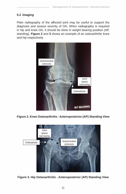
5.2 Imaging
Plain radiography of the affected joint may be useful to support the diagnosis and assess severity of OA. When radiography is required in hip and knee OA, it should be done in weight bearing position (AP, standing). Figure 2 and 3 shows an example of an osteoarthritic knee and hip respectively.
Figure 2. Knee Osteoarthritis - Anteroposterior (AP) Standing View
Figure 3. Hip Osteoarthritis - Anteroposterior (AP) Standing View
Subchondralsclerosis
Jointspace
narrowing
Osteophyte
, !
! !
Subchondralsclerosis
Jointspace
narrowing
Osteophyte

Management of Osteoarthritis ( Second Edition)
12
The Kellgren-Lawrence grading system is the most widely used radiological classification to identify and grade OA (refer to Table 5).
Table 5. Kellgren-Lawrence Grading System
Grade DescriptionGrade I Doubtful narrowing of the joint space, possible osteophytic lippingGrade II Definite osteophytes, possible narrowing of the joint spaceGrade III Moderate multiple osteophytes, definite joint space narrowing, some sclerosis, possible deformity of bone endsGrade IV Large osteophytes, marked joint space narrowing, severe sclerosis and definite bony end deformity.
Adapted: KELLGREN JH, LAWRENCE JS.Radiological assessment of osteo-arthrosis. Ann Rheum Dis. 1957 Dec; 16(4):494-502.
In hand OA, plain radiography may also be useful to distinguishbetween various types of arthritis as shown in Table 6.
Table 6. Radiographic Changes of Interphalangeal Joints and Target Sites Involvement of OA and Other Arthritis
Classical features of OA on plain radiograph includes:-• narrowed joint space • osteophytes• subchondral bone sclerosis • subchondral cysts
Focal narrowing,marginalosteophyte,sclerosis,osteochondralbodies
Subchondralerosion
Proliferativemarginal erosion, retained or increase bone density
Non-proliferativemarginal erosion, osteopenia
Target sites
Osteoarthritis Erosive OA PsoriaticArthritis
RheumatoidArthritis
X-Raychanges
Common Uncommon
Management of Osteoarthritis ( Second Edition)
13
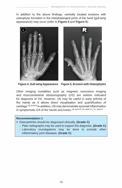
In addition to the above findings, centrally located erosions with osteophyte formation in the interphalangeal joints of the hand (gull-wing appearance) may occur (refer to Figure 4 and Figure 5).
Figure 4. Gull-wing Appearance Figure 5. Erosion with Osteophytes
Other imaging modalities such as magnetic resonance imaging and musculoskeletal ultrasonography (US) are seldom indicated for diagnosis of OA. However, US may be useful in early arthritis ofthe hands as it allows direct visualisation and quantification of cartilage.18, level III In addition, US may demonstrate synovial inflammation in symptomatic OA of the hands and knees.19, level III; 20, level II-2; 21, level III
Recommendation 1• Osteoarthritis should be diagnosed clinically. (Grade C)
Plain radiographs may be used to support the diagnosis. (Grade C) Laboratory investigations may be done to exclude other
inflammatory joint diseases. (Grade C)

Management of Osteoarthritis ( Second Edition)
14
6. MANAGEMENT
The management of OA involves a multidisciplinary approach with the aim to relieve symptoms and improve joint function. It involves non-pharmacological and pharmacological treatment. Refer to Appendix 9 for checklist on management of OA.
6.1 Non-Pharmacological Treatment
a. Education
Patient education is an important non-pharmacological approach in the management of OA. There are various types of patient education programmes and they have to be tailored according to the individual needs, goals and functional capabilities. It should include information of the diagnosis, nature of the disease, therapeutic options and the importance of ongoing patient participation in the disease management.22 - 25, level I Patients who have an understanding of the disease tend to cope better and report less pain.26 The most important goal is to instill a positive attitude.
Recommendation 2• Patient education should form an integral part of osteoarthritis
management. (Grade A)
b. Lifestyle Modification
Lifestyle medicine is defined as the application of environmental, behavioural, medical and motivational principles to the management of lifestyle related health problems.27, level III
Lifestyle modification involves initiating and maintaining lifestyle changes. In hip and knee OA, behavioural changes focus on weight reduction and physical activity or exercise.
i. Weight Reduction
Obesity is an important modifiable risk factor for the development and progression of knee OA. Weight reduction is beneficial in pain reduction and improvement of function.28 - 30, level I In a RCT done by Messier et al., each unit of weight loss will result in 4-fold reduction in the load exerted on the knee per step during daily activities.31, level I There is no evidence available to support the effect of weight loss in hip OA.32

Management of Osteoarthritis ( Second Edition)
15
Recommendation 3• Weight reduction should be emphasised in the management of
patients with knee osteoarthritis and who are overweight. (Grade A)
ii. Physical Activity
Exercise is effective in reducing pain in hip and knee OA. The frequency, intensity, duration and rate of progression of exercise can vary. The amount and intensity of exercise required is uncertain. In order to improve adherence, the following are suggested:-i. Individualised exercise programme ii. Graded type activityiii. Amount of activity based on personal goal settingiv. Feedback on progressv. Appropriate positive reinforcementvi. Problem solving skills incorporated
The EULAR 2013 Recommendations on the Non-Pharmacologic Treatment for Hip and Knee OA recommends that the intensity and duration of exercise should increase over time.32
In hip and knee OA, pacing of activities and/or integrating Activities of Daily Living (ADL) as part of the exercise regime is more effective than usual care but not comparable to standardised exercise.32
In knee OA, aerobic training (walking) is effective in reducing pain (ES=0.48, 95% CI 01.13 to 0.43) and improving physical function (ES=0.35, 95% CI 0.11 to 0.58).33, level I The evidence for mixed exercise programmes, including strengthening, aerobic and flexibility components, in patients with knee OA is conflicting. One type of exercise has not been shown to be better than another (strength, aerobic or mixed exercises).32
Regular non-competitive exercise does not exacerbate OA nor increase the likelihood of requiring joint replacement.
c. Physiotherapy
Physiotherapy can improve muscle strength, balance, coordination and joint mobility. It should be started as soon as possible to improve pain and physical capacity.

Management of Osteoarthritis ( Second Edition)
16
i. Exercise
Exercise programmes should be individualised while taking into consideration patient’s preference and ability to perform the activities.
Land-based exercises include joint range of movement (ROM), muscle strengthening and low impact aerobic exercises. They should be supervised and done regularly (refer to Appendix 4 and Appendix 5). Such exercise has short term benefits in reducing pain (SMD= -0.40, 95% CI -0.50 to -0.30) and improving physical function (SMD= -0.37, 95% CI -0.49 to -0.25) in knee OA.33, level I However in hip OA, the benefit is only seen in pain reduction (SMD= -0.38, 95% CI -0.67 to -0.09) but not in physical function (SMD= -0.10, 95% CI -0.51 to 0.32).34, level I
Aquatic exercise may be advantageous for OA patients. A Cochrane SR showed improvement in pain (SMD=0.19, 95% CI 0.04 to 0.35) and quality of life (SMD=0.32, 95% CI 0.03 to 0.61) for three months only in hip and knee OA. However, there was no statistically significant difference on walking ability.35, level I
In the same review, aquatic exercise was better than land-based exercises in reducing pain in knee OA (SMD=0.86, 95% CI 0.25 to 1.47). However, there was no effect on walking ability and stiffness.35, level I
ii. Transcutaneous Electrostimulation (TENS)
There is a lack of evidence to support the use of transcutaneous electrostimulation for knee OA from the most recent CochraneSR.36, level I However, the ACR 2012 recommends the use of TENS for patients with chronic moderate to severe pain who are not suitable for total knee arthroplasty.37
iii. Thermotherapy
Thermotherapy is commonly used in physical rehabilitation for OA patients. ACR recommends the use of thermal agents for hip and knee OA in combination with exercise supervised by a physiotherapist.37
A SR showed that cold pack usage did not show a significant effect in pain reduction in knee OA (WMD= -1.60, 95% CI -4.53 to1.33).38, level I

Management of Osteoarthritis ( Second Edition)
17
iv. Therapeutic Ultrasound
Therapeutic ultrasound with high frequency vibrations is a modality used for pain relief in patients with hip and knee OA. However, in a Cochrane SR, the effectiveness of this modality in pain reduction and function was inconclusive.39, level I
Recommendation 4• Exercise programmes in hip and knee osteoarthritis must be
individualised, supervised and done regularly. (Grade C)• Land-based or aquatic exercise may be used for short-term benefit in
osteoarthritis. (Grade A)
d. Occupational Therapy
Occupational therapy aims to improve health, prevent disability and help individuals to achieve their optimum functional level and independence in performing ADL.
The evidence suggests that people with pain, difficulty and frustration in performing daily activities and work tasks should be referred early to an occupational therapist for splinting, joint protection training and assistive device provision.40
A NICE-commissioned CPG recommends that assistive devices such as walking sticks and tap turners should be considered as adjuncts to core treatment in OA patients with specific ADL problems.40 The height of the walking stick handle should be at the wrist level when the user is standing. It is important to prescribe appropriate assistive device with proper training to the patients (refer to Appendix 6).
Joint protection and home exercises (JPE) are used in the treatment of hand OA. JPE increases grip strength significantly by 25% (p<0.0005) and global hand function by 65% (p<0.05).41, level I Refer to Appendix 7 for Joint Protection Principles. Thumb splints can help to reduce pain in the thumb and improve hand function.40 Splinting and Joint Protection programme in hand OA can give significant decreases in pain and stiffness, and improvements in daily activities (p<0.05).42, level I However, there is no evidence to support splinting in knee OA.
In a RCT, activity modification or instruction in ADL improved pain (MD= -3.21, 95 % CI -3.45 to -0.70) at 6 weeks.43, level I Activity modification in performing ADL may be helpful in maintaining proper posture and thus reduce pain and disability (refer to Appendix 7).

Management of Osteoarthritis ( Second Edition)
18
Rest and relaxation may help in pain control. The beneficial therapies are:-• Jacobson relaxation: improves pain at end of 8-week treatment
(p<0.05) but the benefit is not sustained.44, level I Jacobson’s training procedure involves tensing and relaxing of muscles alternately, with the intent of developing awareness of the difference. The technique starts from the arm to face working downwards to the shoulders, chest, abdomen, legs and ends with the whole body in complete relaxation. The whole cycle can be performed under 20 minutes.
• Music therapy: improves pain at day 1, day 7 and at 2 weeks (end of treatment) (all p=0.001)45, level I
• Guided Imagery Relaxation (GIR): increases Health Related Quality of Life in women with OA (p=0.023)46, level I
Recommendation 5• Early referral to occupational therapy may be considered for pain
relief and improvement in activities of daily living in osteoarthritis. (Grade A)
e. Orthoses
Orthoses are defined as any medical device added to a person’s body to support, align, position, immobilise, prevent or correct deformity, assist weak muscles or improve function. In knee OA, the general purpose is to decrease pain and improve physical function.
Walking shoes with neutral, contoured orthoses reduce pain and stiffness, and improve function in knee OA at one year (p<0.001).47, level I
Knee braces for medial, lateral or patella-femoral OA have not been shown to reduce pain, improve function or quality of life, even though they are widely used.48 - 49, level I
There is insufficient evidence to recommend the use of orthoses in hip OA.
Recommendation 6• Walking shoes with neutral, contoured orthoses may be offered in:-
knee osteoarthritis (Grade A) hip osteoarthritis (Grade C)
• Knee braces should not be offered in knee osteoarthritis. (Grade A)

Management of Osteoarthritis ( Second Edition)
19
6.2. Pharmacological Treatment
Pharmacological treatments are available in the form of oral, intra-articular and topical.
a. Oral Treatment
Oral treatment consists of:-i. Simple analgesics - paracetamolii. Weak opioid analgesics - tramadoliii. Analgesics with anti-inflammatory properties - Non-steroidal
Anti-inflammatory Drugs (NSAIDs) and Cyclo-oxygenase-2 (COX-2) Inhibitors
iv. Nutraceutical - glucosamine, chondroitin, diacerein
i. Paracetamol
Paracetamol or acetaminophen is classified as a mild analgesic. In a SR of RCTs, paracetamol was superior to placebo in reduction of overall pain [SMD= -0.13 (95% CI -0.22 to -0.04), NNT=16]. However, NSAIDs were more efficacious than paracetamol in total WOMAC score (SMD= -0.25, 95% CI -0.39 to -0.11).50, level I
Similarly, a meta-analysis showed that paracetamol was less efficacious than NSAIDs in overall pain using VAS at rest (WMD= -6.33mm, 95% CI -9.24 to -3.41) and on walking (WMD= -5.76mm, 95% CI -8.99 to -2.52).51, level I
Paracetamol in the extended release formulation is significantly superior to placebo in reducing pain (p=0.012) and improving physical function (p=0.016).52, level I Paracetamol in all formulations is well tolerated and safe.50 - 54, level I
A combination tablet of ibuprofen/paracetamol confers no additional benefit to ibuprofen alone.53, level I
Recommendation 7 • Paracetamol can be used in patients with osteoarthritis. (Grade A)
It should be used as first-line analgesic in mild to moderate pain. (Grade C)

Management of Osteoarthritis ( Second Edition)
20
ii. Tramadol
Tramadol is a synthetic opioid analgesic. In a meta-analysis, tramadol was more efficacious compared to placebo in reducing pain (WMD= -8mm, 95% CI -12.0 to -5.0), and improving stiffness and function (WMD= -0.3, 95% CI -0.5 to -0.2).55, level I
Similarly, oral controlled-release tramadol is also more efficacious in reducing pain (p=0.0009) and improving physical function (p=0.0205) compared to placebo.56, level I However, it is as efficacious as sustained-release diclofenac.57, level I
In patients already on NSAIDs for at least 30 days, addition oftramadol/paracetamol combination tablets for 10 days duration is significantly efficacious in managing painful OA compared to those on placebo.58 - 59, level I
Tramadol in all formulations show no major or significant adverse events.55 - 59, level I Common side effects are dizziness, nausea, vomiting, constipation and drowsiness. This medication has to be used with caution in the elderly.
Recommendation 8 • Tramadol may be used alone or in combination with paracetamol in
patients with osteoarthritis. (Grade A)
iii. NSAIDs and COX-2 Inhibitors
NSAIDs and COX-2 inhibitors reduce production of prostaglandin by inhibiting the enzyme cyclo-oxygenase. They vary in their selectivity for inhibiting different types of cyclo-oxygenase. They are a class of drugs that provide analgesic and anti-pyretic effects and in higher doses, anti-inflammatory effects. COX-2 inhibitors selectively inhibit COX-2 and thus improve gastro-intestinal tolerance.
In a meta-analysis, NSAIDs including COX-2 inhibitors were more efficacious than placebo in reducing short-term (2 - 13 weeks) pain intensity (WMD=0.30, 95% CI 0.24 to 0.39) and functional disability (WMD=0.29, 95% CI 0.18 to 0.40).60, level I
Celecoxib 100 mg and 200 mg BID are as efficacious as diclofenac 50 mg BID and naproxen 500 mg BID in the treatment of hip, knee, or hand OA.61, level I

Management of Osteoarthritis ( Second Edition)
21
In a study using OMERACT-OARSI responder criteria, those who responded within two weeks to etoricoxib 30 mg OD and celecoxib 200 mg OD are highly predictive of later response at 12 weeks.62, level I
• Gastrointestinal (GI) Safety
The relative risks of upper GI complications of several NSAIDs and celecoxib compared to placebo analysed in a meta-analysis ranged from 1.45 to 4.14.63, level II-2
Ulcer complications are seen significantly less frequent in COX-2 inhibitors compared to NSAIDs.61, level I; 64 - 66, level I Comparing concomitant aspirin and non-aspirin users, the GI complications are significantly less in non-aspirin users (p=0.007).61, level I
Proton pump inhibitors are effective in the prevention of NSAID-induced endoscopic duodenal (RR=0.19, 95% CI 0.09 to 0.37) and gastric ulcers (RR=0.40, 95% CI 0.32 to 0.51) at ≥12 weeks when compared to placebo.67, level I
Lansoprazole significantly reduce the risk of gastroduodenal ulcers recurrence in patients with a definite history of GI ulcers requiring long-term NSAIDs therapy (HR=0.25, 95% CI 0.14 to 0.45)68, level I or long-term low dose aspirin therapy (HR=0.10, 95% CI 0.04 to 0.23).69, level I
The risk of GI events is lower in patients receiving celecoxib compared to diclofenac slow release plus omeprazole with HR of 4.3 (95% CI 2.6 to 7.0).70, level I The risk of recurrent ulcer bleeding is also lower in the celecoxib 200 mg BD plus esomeprozole 20 mg BD group compared to the celecoxib 200 mg BD plus placebo group (p=0·0004).71, level I
• Cardiovascular (CV) Safety
One of the main concerns among patients on long-term use of NSAIDs and COX-2 inhibitors is the increased risk of thrombotic CV events.
A meta-analysis showed that naproxen seemed to have the lowest Antiplatelet Trialists’ Collaboration composite outcome of non-fatal myocardial infarction, non-fatal stroke or CV death among users of NSAIDs and COX-2 inhibitors (rate ratio=1.22, 95% CI 0.78 to 1.93).72, level I
The numbers of CV thromboembolic events are low in both celecoxib and diclofenac or naproxen groups at 12 weeks.61, level I Long-term etoricoxib use (20 months) is also associated with comparable CV events with that of diclofenac.73, level I

Management of Osteoarthritis ( Second Edition)
22
• Renal Safety
In the Malaysian CPG on Chronic Kidney Disease (CKD) in Adults, there was conflicting evidence in the association between chronic NSAIDs usage and the development of CKD.74 However, renal function should be monitored regularly in patients on chronic NSAIDs or COX-2 inhibitors treatment.
Recommendation 9 • Non-steroidal anti-inflammatory drugs (NSAIDs) or cyclo-
oxygenase-2 (COX-2) inhibitors can be used in the treatment of osteoarthritis. (Grade A) In patients with high risk of gastrointestinal (GI) complications,
COX-2 inhibitors are preferred to non-selective NSAIDs with proton pump inhibitor (PPI) for primary ulcer prevention. (Grade A)
In patients with previous GI complications:-- NSAIDs or COX-2 inhibitors should be avoided. (Grade C) - combination of COX-2 inhibitors and PPI may be offered for GI
protection if indicated. (Grade A) In patients with renal impairment, NSAIDs and COX-2 inhibitors
should be used with caution. (Grade C)
• Combination therapy with more than one NSAID/COX-2 inhibitor should never be used. There is no benefit in combination therapy and the incidence of side effects may be additive.
• Caution is required when prescribing NSAIDs in the elderly and those with hypertension, cardiovascular disease, renal or hepatic impairment.
• Those who are allergic to one NSAID may also be allergic to others.
iv. Glucosamine and Chondroitin
Glucosamine is an amino sugar and a prominent precursor in the biochemical synthesis of glycosylated proteins and lipids, including glycosaminoglycans (GAG) which is a component of cartilage. Chondroitin sulfate is a sulfated GAG which is usually found attached to proteins as part of a proteoglycan. Glucosamine or chondroitin sulfate is required in the synthesis of the GAG component of cartilage, which provides the rationale for oral supplementation of these compounds in OA.
The glucosamine and chondroitin preparations available in Malaysia are in various combinations, strengths and purities which may affect their efficacy.

Management of Osteoarthritis ( Second Edition)
23
• Glucosamine
Glucosamine, in general, is not consistent in its effect as a structure modifier for OA. In terms of pain reduction, it is statisticallymore efficacious than placebo and can have similar efficacy withNSAIDs.75 - 77, level I
Glucosamine hydrochloride or its combination with chondroitin sulfate is not efficacious as a structure or symptom modifier.75 - 76, level I; 78 - 79, level I
Glucosamine sulfate 1500 mg per day was statistically more efficacious in pain reduction when compared to placebo.76 - 77, level I Pain relieving effect of glucosamine sulfate can be seen by three months after its initiation.80, level I However, the evidence was from pharmaceutical-sponsored studies.
In terms of safety profile, glucosamine is well-tolerated and safe.75 - 78, level I
However, the effect of glucosamine on glucose metabolism needs further research.81, level I
Recommendation 10• Glucosamine sulfate 1500 mg per day may be used as pain relief for
knee osteoarthritis. (Grade C) Evaluation on pain reduction should be done at three months after
initiation of treatment before deciding on its continuation. (Grade C)
• Chondroitin
The evidence on efficacy of chondroitin sulfate in hip andknee OA for pain relief and its structure modifying effect isinconsistent.75 - 82, level I
Chondroitin sulfate 800 mg as a single dose is more efficacious than placebo in pain reduction, improving hand function and morning stiffness in patients with hand OA.83, level I However, its efficacy as a structural modifier is still debatable.84 - 86, level I
In terms of safety profile, chondroitin sulfate is well-tolerated and safe.83, level I
The efficacy of chondroitin in the treatment of hip or knee OA is inconclusive. It may be beneficial for symptomatic relief in hand OA.

Management of Osteoarthritis ( Second Edition)
24
v. Diacerein
Diacerein, a purified anthraquinone derivative, is a drug which inhibits production of interleukin (IL)-1beta, the major proinflammatory cytokine involved in articular cartilage destruction.
In a meta-analysis, diacerein was slightly more efficacious compared to placebo in reduction of pain [n-wtd pooled Glass score=1.31 (95% CI 0.49 to 2.14)] and improvement of joint function [n-wtd pooled Glass score=1.08 (95% CI 0.26 to 1.91)] in knee OA. In addition, diacerein had similar efficacy with NSAIDs in the reduction of pain in knee OA for duration of 16 weeks [(n-wtd pooled Glass score= -0.01 (95% CI -0.76 to 0.74)].87, level I
Besides these, diacerein had significant carry-over effect in pain reduction in knee OA compared to placebo [(n-wtd pooled Glass score=2.71 (95% CI 1.32 to 4.10)] and NSAIDs [(n-wtd pooled Glass score=2.27 (95% CI 1.42 to 3.11)] for 16 weeks duration.87, level I
In general, diacerein had an acceptable safety profile.87 - 90, level I The incidence of adverse events such as diarrhea, abdominal pain, nausea and vomiting was higher compared to placebo (p<0.01), most were mild to moderate91, level I
In view of modest efficacy and common adverse events, more studies are warranted to support the use of diacerein in OA.
Recommendation 11• Diacerein may be used in the treatment of knee osteoarthritis. (Grade C)
b. Intra-articular Treatment
i. Corticosteroids
In a Cochrane systematic review of single/double blinded of 12 randomised controlled trials (n=653), the efficacy and safety of various preparations of intra-articular (IA) corticosteroid in the treatment of knee OA was evaluated.92, level I
IA corticosteroid offered short-term pain relief at one week post-injection in patients with knee OA (WMD in VAS= -21.9 mm, 95% CI -29.9 to -13.9). The NNT calculated was three to four. The effect continued

Management of Osteoarthritis ( Second Edition)
25
to be seen at two weeks (RR=1.8, 95% CI 1.1 to 3.0) and three weeks (RR=3.1, 95% CI 1.6 to 6.0). However, it did not demonstrate improvement in function.92, level I
There were no statistically significant differences detected in the total number of overall withdrawals including lack of efficacy, post-injection flare and local discomfort.92, level I
Recommendation 12• Intra-articular corticosteroid may be used for short-term pain relief in
an acute exacerbation of knee osteoarthritis. (Grade A)
Oral corticosteroid have no role in the treatment of osteoarthritis.
ii. Viscosupplementation
Hyaluronic acid (HA) is a naturally occurring polysaccharide in the synovial fluid and is responsible for the elastoviscosity of synovial fluid. The quantity of HA in the synovial fluid is reduced in the patients who have OA. To improve biomechanical function, different hyaluronic acids were devised for intra-articular injection, commonly called viscosupplementation.There are now several different formulations of viscosupplements (hyaluronan and hylan) produced by different manufacturers and of widely differing molecular weights.
Two SRs showed that HA intra-articular injection may be beneficial in reducing pain, but the effect sizes were small when compared to placebo. In terms of improvement in physical function, the results were conflicting.93 - 94, level I However, the pooled estimates in both SRs have to be interpreted cautiously because of different molecular weights of HA, different injection schedules and poor trial design despite large numbers of studies.
In a pharmacoeconomics analysis by NICE, the above benefits may be offset by the frequency of the HA injections and other indirect costs. Thus, NICE does not recommend the use of viscosupplementation in treatment of knee OA.40
Viscosupplementation is generally well-tolerated and with no significant difference in safety profile between it and placebo.94, level I The risk of overall adverse events is insignificant.93, level I
Management of Osteoarthritis ( Second Edition)
26
Due to a lack of supporting evidence, the CPG is unable to recommend the use of viscosupplementation in the treatment of osteoarthritis.
c. Topical Treatment
Topical treatment is an adjunct or alternative to oral NSAIDs for treatment of OA. The commonly used topical treatment includes NSAIDs, capsaicin and methylsalicylate. Topical analgesics can be in the form of gels, creams and transdermal patches.
Topical NSAIDs reduce knee OA pain at week 2 (p<0.0001), week 3 (p<0.0002)95, level I and at week 4 to 12 (SMD= -0.33, 95% CI -0.48 to -0.18)96, level I compared to placebo.
It is also more efficacious than placebo in reducing stiffness of knee OA (SMD in WOMAC stiffness at week 4 to 12= -0.30, 95% CI -0.45 to -0.15).96, level I
Besides reducing pain and stiffness, physical function is also significantly improved with topical NSAIDs.95 - 97, level I
Generally, topical NSAIDs were found to be safe in adults with OA in a SR of RCTs. The risk of minor skin dryness was higher in topical diclofenac compared to placebo (RR=1.74, 95% CI 1.37 to 2.22). Fewer severe gastrointestinal adverse events were reported in topical NSAIDs compared with oral NSAIDs.98, level I
There were no recent studies found with regards to the usage of capsaicin and methylsalicylate in OA. However, the NICE CPG 2008 recommends that topical capsaicin should be considered as an adjunct to core treatment for knee or hand OA but does not recommend the use of topical methylsalicylate (rubefacients).40
Recommendation 13• Topical non-steroidal anti-inflammatory drugs may be offered in the
treatment of osteoarthritis. (Grade A) It may be used as adjunct therapy in mild to moderate pain. (Grade C)

Management of Osteoarthritis ( Second Edition)
27
7. ALTERNATIVE TREATMENT
Alternative treatments are extensively used in the treatment of OA however it is not based on evidence gathered using scientific methods.
Common alternative treatments that have shown positive results include acupuncture, avocado soybean unsaponifiables (ASU) and ginger.
• Acupuncture
Acupuncture is a traditional treatment which involves inserting needles into meridian points with the intention of influencing energy flow.
In a Cochrane SR, acupuncture significantly improved pain and function (as assessed by the WOMAC scale) in patients with knee OA compared to sham acupuncture:-99, level I
• SMD= -0.29 (95% CI -0.48 to -0.10) for pain at three months and SMD= -0.10 (95% CI -0.21 to – 0.01) for pain at 26 weeks
• SMD= -0.29 (95% CI -0.49 to -0.08) for function at three months • SMD= -0.29 (95% CI -0.50 to -0.09) for total score at three months
No serious adverse events were reported to be associated with acupuncture. Minor side effects were bruising and haematoma.
• Avocado Soybean Unsaponifiables (ASU)
ASU provided pain relief and improvement of function comparedto placebo in chronic stable OA of the hip and knee in a CochraneSR:-100, level I
• SMD= -8.06 (95% CI -11.3 to -4.60) for VAS pain score • SMD= -1.69 (95% CI -2.41 to -0.98) for Lequesne index• SMD=0.71 (95% CI 0.57 to 0.89) for resumption of NSAIDs
ASU had minimal gastrointestinal disturbances and no other serious side effect.
Another meta-analysis supported the above findings with improvement in pain in OMERACT III scale (SMD=0.39, 95% CI 0.01 to 0.76) and function in Lequesne index (SMD=45, 95% CI 0.21 to 0.70).101, level I
• Ginger
In a small RCT of short duration, ginger extract was significantly more efficacious than placebo but less than ibuprofen (p<0.005) in improving pain and function. It was safe without serious side effects.102, level I
Recommendation 14• Acupuncture and avocado soybean unsaponifiables may be used as
an adjunct short-term therapy in osteoarthritis. (Grade A)

Management of Osteoarthritis ( Second Edition)
28
8. SURGICAL TREATMENT
Surgery is considered if the symptoms of the affected joints significantly affect the quality of patients’ life and interfere with ADL. There are five identified key areas which surgeons use to assess the need for surgery:-
• pain (sleep interruption and while resting)• limitations to ADL (walking and self-care)• psychosocial health (psychological well-being)• economic impact and• recent deterioration
The types of surgery that can be offered are:-• Arthroscopic Surgery• High Tibial Osteotomy• Total Joint Replacement• Partial Joint Replacement• Arthrodesis
8.1 Arthroscopic Surgery
Arthroscopic lavage with or without debridement has been used in the treatment of knee OA. Studies showed no additional benefit in terms of pain relief and improvement in joint function compared to optimised physical and medical therapy.103 - 104, level I However, it is indicated in patients with OA associated with mechanical symptoms such as locking, catching or giving way of the joint caused by presence of loose bodies or flaps of meniscus or cartilage.
8.2 High Tibial Osteotomy (HTO)
HTO works best in patients with isolated medial compartment arthritis, particularly if they are younger than 50 years old and have at least 120 degrees of knee flexion. It is an operation where the tibial bone is osteotomised at its upper end and repositioned in order to realign the mechanical axis of the limb away from the diseased area. This allows the knee to glide freely and distribute weight evenly on a more normal compartment.
HTO relieves pain and may delay the progression of OA. However, the disadvantages of this procedure are that the knee may look asymmetrical and may pose a more technically challenging procedure if Total Knee Replacement (TKR) is eventually required.

Management of Osteoarthritis ( Second Edition)
29
8.3 Total Joint Replacement
Total Joint Replacement is indicated in patients with severe OA who have failed to respond to all other therapies. This may result in a dramatic reduction in pain and a significant improvement in ADL.105, level l
However, patients are still advised to avoid certain activities, including jogging and high-impact sports for the rest of their life following surgery to ensure longevity of the implant.
a. Total Knee Replacement (TKR)
In the Canadian Joint Replacement Registry, 94% of TKR was due to degenerative OA.106, level III Active infection is an absolute contraindication whereas young age <55 years, poor compliance, regional pain disorders and unrealistic expectations are relative contraindications. The rate of joint revision ranged between 0% and 13% in studies that reported more than five years follow-up.105, level I
b. Total Hip Replacement (THR)
Indication for THR is similar to TKR. In the Canadian Joint Replacement Registry, 81% of THR was due to degenerative OA.106, level III However, with the advancement in implant design, metallurgy and surgical techniques, THR is also advocated for the younger patient with severe hip OA.
8.4 Partial Joint Replacement
The advantages of partial knee replacement over total knee replacement are less blood loss intraoperatively and less pain post-surgery with faster recovery and better range of motion. The disadvantages include slightly less predictable pain relief and the potential need for subsequent surgery.
a. Unicondylar Knee Replacement (UKR)
UKR differs from TKR in that only a portion of the knee is replaced. It is indicated for patients with knee OA that affect either the medial or lateral compartment and have significant pain on weight bearing. The survival of UKR at 10 years is about 85%.107, level I; 108
b. Patellofemoral Knee Arthroplasty
This surgery is an alternative to TKR for elderly patients with isolated advanced patellofemoral OA.

Management of Osteoarthritis ( Second Edition)
30
c. Bicompartmental Knee Arthroplasty
This type of surgery is an alternative to TKR when advanced medial or lateral compartment OA is associated with significant patellofemoral OA.
8.5 Arthrodesis
The aim of arthrodesis or joint fusion is to fuse the diseased joint in an optimal position. This would make the joint stiff but stable. The most common indication for knee arthrodesis is pain and significant ligamentous instability in an unreconstructable knee following an infection at the site of a knee arthroplasty. Arthrodesis of the knee can provide a stable, painless extremity for young active patients.
In general, arthrodesis is not the preferred surgical option in OA.
Recommendation 15• Arthroscopic lavage and debridement should not be offered as
a treatment in osteoarthritis (OA) of the knee except in selected conditions*. (Grade A)
• Joint Replacement should be offered to patients with severe OA. (Grade A)
*Refer to 8.1
8.6 Recent Advances in Osteoarthritis
a. Intra-articular Stem Cells
Stem cells have the capacity to produce all cell types in an unlimited fashion. Emerging evidence indicates that direct intra-articular injection of stem cells may boost the normally limited reparative process and limit the destructive process.
Malaysian Health Technology Assessment Section (MaHTAS) technology review showed that there was limited evidence on the benefits of stem cells in articular cartilage repair.109
More evidence-based studies are needed before its recommendation as a standard treatment for OA.

Management of Osteoarthritis ( Second Edition)
31
b. Autologous Chondrocyte Implantation (ACI)
Autologous chondrocyte implantation is an approach that has been used to treat symptomatic knee cartilage defects. The aim of this treatment is to replenish cartilage through the recruitment of progenitor cells as potential cartilage precursors, allowing the development into chondrogenic cells and finally cartilage.110, level I
Most of the studies on usage of ACI were done on younger age patients with articular cartilage damage due to secondary causes of OA such as sports injury or trauma. Hence, there is a lack of evidence presently to recommend the use of ACI in the treatment of primary OA.
c. Platelet Rich Plasma (PRP)
Platelet rich plasma (PRP) is a natural concentrate of autologous blood growth factors studied in different fields of medicine in order to test its potential to enhance tissue regeneration.
The preliminary short-term results within 3 to 6 months indicate that treatment with autologous PRP intra-articular injections may be useful for early OA, aiming to reduce pain and improve knee function and quality of life.111, level III However, there is no further improvement after 6 months.111, level III; 112, level II-2
Due to a lack of available evidence, the CPG is unable to recommend the use of intra-articular stem cells, autologous chondrocyte implantation or platelet-rich plasma in the treatment of osteoarthritis.

Management of Osteoarthritis ( Second Edition)
32
9. REFERRAL
9.1 Rheumatology Referral
Osteoarthritis is one of the major causes of disability in adults. Early diagnosis and intervention are important to minimise the impact of short-term and long-term morbidity. Rheumatology opinion should be sought for evaluation of arthritis with unclear diagnosis.
9.2 Orthopaedic Referral
Referral to the orthopaedic surgeon should be made when the patient does not experience satisfactory improvement in terms of pain, stability or function despite adequate pharmacological and non-pharmacological treatment. The patients should be made to understand the reason for referral.
Recommendation 16• Referral of osteoarthritis cases to either rheumatology or orthopaedic
clinic should provide the following information:- Diagnosis Severity and its impact on activity of daily living Co-morbidities that might require further medical assessment Relevant investigation results and current medications (Grade C)
10. PRIMARY PREVENTION
Identifying and modifying the risk factors can help in preventing OA and its progression. Many of these risk factors are of particular importance in weight-bearing joints. Prevention of obesity, weight reduction in the obese and health education pertaining to joint protection techniques including avoidance of trauma to the joints are recommended as measures for primary prevention. Currently, there is no evidence available to recommend the intake of any medication to prevent OA. An important aspect of primary prevention is to identify those individuals at risk.

Management of Osteoarthritis ( Second Edition)
33
11. IMPLEMENTING THE GUIDELINES
It is important to standardise the management of OA at all healthcare levels in Malaysia using an evidence-based CPG in order to manage OA appropriately. It is therefore crucial for healthcare providers to understand the disease as it is a common disease which may result in significant disabilities.
11.1 Facilitating & Limiting Factors
Existing facilitators for application of the recommendations in the CPG include:-
1. Wide dissemination of the CPG to healthcare providers (hard and soft copies)
2. Regular rheumatology update for healthcare providers
Existing barriers for application of the recommendations of the CPG are:-
1. Poor understanding/limited knowledge of OA2. Insufficient resources in the management of OA particularly in
rehabilitation3. Variation in treatment practice and preferences
11.2 Potential Resource Implications
To implement the CPG, there must be strong commitment to:- 1. Ensure widespread distribution of the CPG to healthcare
providers via printed and electronic copies.2. Re-enforce training (with adequate funding) of healthcare
providers by regular seminars or workshops to ensure information is up-to-date
3. Availability of rehabilitation services and trained manpower in OA management including multidisciplinary team at different levels of healthcare
4. Ensure availability of recommended drugs in primary care setting
5. Ensure patient empowerment via patient education materials

Management of Osteoarthritis ( Second Edition)
34
To assist in the implementation of the CPG, the following is proposed as clinical audit indicator for quality management:-
*Those at high risk of GI complications (such as ulceration, bleeding, perforation and obstruction) are:-i. age more than 65 years old orii. on high dose of NSAIDs, aspirin, corticosteroid or anticoagulant or iii. previous history of upper GI complications
Percentage of OA patients referred to rheumatology or orthopaedic clinic with proper documentation
Number of OA patients referred to rheumatology or orthopaedic clinic with proper
documentation within a period of timeX 100%
Number of OA patients referred to rheumatology or orthopaedic clinic
in the same period of time
=
Percentage of patients with symptomatic OA at risk of GI complications* prescribed with NSAIDs alone
X 100%Number of patients with symptomatic OA
at risk of GI complications in the same period of time
=
Number of patients with symptomatic OA at risk of GI complications prescribed with
NSAIDs alone within a period of time

Management of Osteoarthritis ( Second Edition)
35
REFERENCES
1. Vos T, Flaxman AD, Naghavi M, et al. Years lived with disability (YLDs) for 1160 sequelae of 289 diseases and injuries 1990-2010: a systematic analysis for the Global Burden of Disease Study 2010.[Erratum appears in Lancet. 2013 Feb 23;381(9867):628 Note: AlMazroa, Mohammad A [added]; Memish, Ziad A [added]]. Lancet. 2012 Dec 15;380(9859):2163-96.
2. National Institutes of Health, World Health Organization. Global Health and Aging, NIH, Techn. Rep, NIH Publication no. 11-77372011 Contract No.: NIH Publication no. 11-7737.
3. PereiraD,PeleteiroB,AraujoJ,etal.Theeffectofosteoarthritisdefinitiononprevalenceandincidence estimates: a systematic review. Osteoarthritis Cartilage. 2011 Nov;19(11):1270-85.
4. Haugen IK, Englund M, Aliabadi P, et al. Prevalence, incidence and progression of hand osteoarthritis in the general population: the Framingham Osteoarthritis Study. Ann Rheum Dis. 2011 Sep;70(9):1581-6.
5. Lawrence RC, Felson DT, Helmick CG, et al. Estimates of the prevalence of arthritis and other rheumatic conditions in the United States. Part II. Arthritis Rheum. 2008 Jan;58(1):26-35.
6. Nevitt MC, Xu L, Zhang Y, et al. Very low prevalence of hip osteoarthritis among Chinese elderly in Beijing, China, compared with whites in the United States: the Beijing osteoarthritis study. Arthritis Rheum. 2002 Jul;46(7):1773-9.
7. Murphy L, Schwartz TA, Helmick CG, et al. Lifetime risk of symptomatic knee osteoarthritis. Arthritis Rheum. 2008 Sep 15;59(9):1207-13.
8. Zhang Y, Xu L, Nevitt MC, et al. Comparison of the prevalence of knee osteoarthritis between the elderly Chinese population in Beijing and whites in the United States: The Beijing Osteoarthritis Study. Arthritis Rheum. 2001 Sep;44(9):2065-71.
9. Veerapen K, Wigley RD, Valkenburg H. Musculoskeletal pain in Malaysia: a COPCORD survey. J Rheumatol. 2007 Jan;34(1):207-13.
10. Blagojevic M, Jink, C, Jeffery A, et al. Risk factors for onset of osteoarthritis of the knee in older adults: a systematic review and meta-analysis. Osteoarthritis Cartilage. 2010 Jan;18(1):24-33.
11. SpectorTD,CicuttiniF,Baker J,etal.Genetic influencesonosteoarthritis inwomen:a twinstudy. BMJ. 1996 Apr 13;312(7036):940-3.
12. Tanamas S, Hanna, FS, Cicuttini FM, et al. Does knee malalignment increase the risk of development and progression of knee osteoarthritis? A systematic review. Arthritis Rheum. 2009 Apr 15;61(4):459-67.
13. Ministry of Health Malaysia. Management of Osteoarthritis. Kuala Lumpur: MoH; 2002.14. Altman R, Alarcon G, Appelrouth D, et al. The American College of Rheumatology criteria for the
classificationandreportingofosteoarthritisofthehand.ArthritisRheum.1990Nov;33(11):1601-10.15. Altman R, Alarcon G, Appelrouth D, et al. The American College of Rheumatology criteria for the
classificationandreportingofosteoarthritisofthehip.ArthritisRheum.1991May;34(5):505-14.16. AltmanR,AschE,BlochD,etal.Developmentofcriteriafortheclassificationandreportingof
osteoarthritis. Classification of osteoarthritis of the knee.Diagnostic and TherapeuticCriteriaCommittee of the American Rheumatism Association. Arthritis Rheum. 1986 Aug;29(8):1039-49.
17. Zhang W, Doherty M, Peat G, et al. EULAR evidence-based recommendations for the diagnosis of knee osteoarthritis. Ann Rheum Dis. 2010 Mar;69(3):483-9.
18. MollerB,BonelH,RotzetterM,etal.Measuringfingerjointcartilagebyultrasoundasapromisingalternative to conventional radiograph imaging. Arthritis Rheum. 2009 Apr 15;61(4):435-41.
19. KeenHI,WakefieldRJ,GraingerAJ, et al.Anultrasonographic studyof osteoarthritis of thehand: synovitis and its relationship to structural pathology and symptoms. Arthritis Rheum. 2008 Dec 15;59(12):1756-63.
20. D’Agostino MA, Conaghan P, Le Bars M, et al. EULAR report on the use of ultrasonography in painfulkneeosteoarthritis.Part1:prevalenceofinflammationinosteoarthritis.AnnRheumDis.2005 Dec;64(12):1703-9.
21. Conaghan P, D’Agostino MA, Ravaud P, et al. EULAR report on the use of ultrasonography in painful knee osteoarthritis. Part 2: exploring decision rules for clinical utility. Ann Rheum Dis. 2005 Dec;64(12):1710-4.

Management of Osteoarthritis ( Second Edition)
36
22. Kao M-J, Wu M-P, Tsai M-W, et al. The effectiveness of a self-management program on quality of life for knee osteoarthritis (OA) patients. Arch Gerontol Geriatr. 2012 Mar-Apr;54(2):317-24.
23. Coleman S, Briffa NK, Carroll G, et al. A randomised controlled trial of a self-management education program for osteoarthritis of the knee delivered by health care professionals. Arthritis Res Ther. 2012;14(1):R21.
24. Hansson EE, Jonsson-Lundgren M, Ronnheden AM, et al. Effect of an education programme for patients with osteoarthritis in primary care--a randomized controlled trial. BMC Musculoskelet Disord. 2010;11:244.
25. Ravaud P, Flipo RM, Boutron I, et al. ARTIST (osteoarthritis intervention standardized) study of standardised consultation versus usual care for patients with osteoarthritis of the knee in primary care in France: pragmatic randomised controlled trial. BMJ. 2009;338:b421.
26. Pendleton A, Arden N, Dougados M, et al. EULAR recommendations for the management of knee osteoarthritis: report of a task force of the Standing Committee for International Clinical Studies Including Therapeutic Trials (ESCISIT). Ann Rheum Dis. 2000 Dec;59(12):936-44.
27. Egger GJ, Binns AF, Rössner SR. Lifestyle Medicine. Sydney: McGraw-Hill; 2007.28. Bliddal H, Leeds AR, Stigsgaard L, et al. Weight loss as treatment for knee osteoarthritis
symptoms in obese patients: 1-year results from a randomised controlled trial. Ann Rheum Dis. 2011 Oct;70(10):1798-803.
29. Christensen R, Bartels EM, Astrup A, et al. Effect of weight reduction in obese patients diagnosed with knee osteoarthritis: a systematic review and meta-analysis. Ann Rheum Dis. 2007 Apr;66(4):433-9.
30. Messier SP, Loeser RF, Miller GD, et al. Exercise and dietary weight loss in overweight and obese older adults with knee osteoarthritis: the Arthritis, Diet, and Activity Promotion Trial. Arthritis Rheum. 2004 May;50(5):1501-10.
31. Messier SP, Gutekunst DJ, Davis C, et al. Weight loss reduces knee-joint loads in overweight and obese older adults with knee osteoarthritis. Arthritis Rheum. 2005 Jul;52(7):2026-32.
32. Fernandes L, Hagen KB, Bijlsma JW, et al. EULAR recommendations for the non-pharmacological core management of hip and knee osteoarthritis. Ann Rheum Dis. 2013 Jul;72(7):1125-35.
33. Fransen M, McConnell S Exercise for osteoarthritis of the knee. Cochrane Database of Systematic Reviews 2008, Issue 4 Art No: CD004376.
34. Fransen M, McConnell S, Hernandez-Molina G, et al. Exercise for osteoarthritis of the hip. Cochrane Database of Systematic Reviews 2009, Issue 3 Art No: CD007912.
35. Bartels EM, Lund H, Hagen KB, et al Aquatic exercise for the treatment of knee and hip osteoarthritis. Cochrane Database of Systematic Reviews 2007, Issue 4 Art No: CD005523.
36. Rutjes AWS, Nüesch E, Sterchi R, et al Transcutaneous electrostimulation for osteoarthritis of the knee. Cochrane Database of Systematic Reviews 2009, Issue 4 Art No: CD002823.
37. Hochberg MC, Altman RD, April KT, et al. American College of Rheumatology 2012 recommendations for the use of nonpharmacologic and pharmacologic therapies in osteoarthritis of the hand, hip, and knee. Arthritis Care Res. 2012 Apr;64(4):465-74.
38. Brosseau L, Yonge KA, Welch V, et al. Thermotherapy for treatment of osteoarthritis. Cochrane Database of Systematic Reviews 2003, Issue 4 Art No: CD004522.
39. Rutjes AWS, Nüesch E, Sterchi R, et al Therapeutic ultrasound for osteoarthritis of the knee or hip. Cochrane Database of Systematic Reviews 2010, Issue 1 Art No: CD003132.
40. National Collaborating Centre for Chronic Conditions. Osteoarthritis: national clinical guideline for care and management in adults. London Royal College of Physicians; 2008.
41. Stamm TA, Machold KP, Smolen JS, et al. Joint protection and home hand exercises improve hand function in patients with hand osteoarthritis: a randomized controlled trial. Arthritis Rheum. 2002 Feb;47(1):44-9.
42. Boustedt C, Nordenskiold U, Lundgren Nilsson A. Effects of a hand-joint protection programme with an addition of splinting and exercise: one year follow-up. Clin Rheumatol. 2009 Jul;28(7):793-9.
43. Shakoor MA, Taslim MA, Hossain MS. Effects of activity modification on the patients withosteoarthritis of the knee. Bangladesh Med Res Counc Bull. 2007 Aug;33(2):55-9.

Management of Osteoarthritis ( Second Edition)
37
44. Gay MC, Philippot P, Luminet O. Differential effectiveness of psychological interventions for reducing osteoarthritis pain: a comparison of Erikson [correction of Erickson] hypnosis and Jacobson relaxation. Eur J Pain. 2002;6(1):1-16.
45. McCaffrey R, Freeman E. Effect of music on chronic osteoarthritis pain in older people. J Adv Nurs. 2003 Dec;44(5):517-24.
46. Baird CL, Sands LP. Effect of guided imagery with relaxation on health-related quality of life in older women with osteoarthritis. Res Nurs Health. 2006 Oct;29(5):442-51.
47. Barrios JA, Crenshaw JR, Royer TD, et al. Walking shoes and laterally wedged orthoses in the clinical management of medial tibiofemoral osteoarthritis: a one-year prospective controlled trial. Knee. 2009 Mar;16(2):136-42.
48. Hunter DJ, Harvey W, Gross KD, et al. A randomized trial of patellofemoral bracing for treatment of patellofemoral osteoarthritis. Osteoarthritis Cartilage. 2011 Jul;19(7):792-800.
49. Brouwer RW, van Raaij TM, Verhaar JAN, et al. Brace treatment for osteoarthritis of the knee: a prospective randomized multi-centre trial. Osteoarthritis Cartilage. 2006 Aug;14(8):777-83.
50. Towheed T, Maxwell L, Judd M, et al. Acetaminophen for osteoarthritis. Cochrane Database of Systematic Reviews 2006, Issue 1 Art No: CD004257.
51. LeeC,StrausWL,BalshawR,etalAcomparisonof theefficacyandsafetyofnonsteroidalantiinflammatory agents versus acetaminophen in the treatment of osteoarthritis: a meta-analysis. Arthritis Rheum 2004 Oct 15;51(5):746-54.
52. AltmanRD,ZinsenheimJR,TempleAR,etal.Three-monthefficacyandsafetyofacetaminophenextended-release for osteoarthritis pain of the hip or knee: a randomized, double-blind, placebo-controlled study. Osteoarthritis Cartilage. 2007 Apr;15(4):454-61.
53. Doherty M, Hawkey C, Goulder M, et al. A randomised controlled trial of ibuprofen, paracetamol or a combination tablet of ibuprofen/paracetamol in community-derived people with knee pain. Ann Rheum Dis. 2011 Sep;70(9):1534-41.
54. Temple AR, Benson GD, Zinsenheim JR, et al. Multicenter, randomized, double-blind, active-controlled, parallel-group trial of the long-term (6-12 months) safety of acetaminophen in adult patients with osteoarthritis. Clin Ther. 2006 Feb;28(2):222-35.
55. Cepeda MS, Camargo F, Zea C, et al. Tramadol for osteoarthritis: a systematic review and metaanalysis. J Rheumatol. 2007 Mar;34(3):543-55.
56. Thorne C, Beaulieu AD, Callaghan DJ, et al. A randomized, double-blind, crossover comparison oftheefficacyandsafetyoforalcontrolled-releasetramadolandplaceboinpatientswithpainfulosteoarthritis. Pain Res Manag. 2008 Mar-Apr;13(2):93-102.
57. Beaulieu AD, Peloso PM, Haraoui B, et al. Once-daily, controlled-release tramadol and sustained-release diclofenac relieve chronic pain due to osteoarthritis: a randomized controlled trial. Pain Res Manag. 2008 Mar-Apr;13(2):103-10.
58. RosenthalNR,SilverfieldJC,WuSC,etal.Tramadol/acetaminophencombinationtabletsforthetreatmentofpainassociatedwithosteoarthritisflareinanelderlypatientpopulation.JAmGeriatrSoc. 2004 Mar;52(3):374-80.
59. Silverfield JC, KaminM,WuSC, et al. Tramadol/acetaminophen combination tablets for thetreatment of osteoarthritis flare pain: a multicenter, outpatient, randomized, double-blind,placebo-controlled, parallel-group, add-on study. Clin Ther. 2002 Feb;24(2):282-97.
60. Bjordal JM,LjunggrenAE,KlovningA,et al.Non-steroidal anti-inflammatorydrugs, includingcyclo-oxygenase-2 inhibitors, in osteoarthritic knee pain: meta-analysis of randomised placebo controlled trials. BMJ. 2004 Dec 4;329(7478):1317.
61. Singh G, Fort JG, Goldstein JL, et al. Celecoxib versus naproxen and diclofenac in osteoarthritis patients: SUCCESS-I Study.[Erratum appears in Am J Med. 2006 Sep;119(9):801]. Am J Med. 2006 Mar;119(3):255-66.
62. Bingham CO 3rd, Smugar SS, Wang H, et al. Early response to COX-2 inhibitors as a predictor of overall response in osteoarthritis: pooled results from two identical trials comparing etoricoxib, celecoxib and placebo. Rheumatology (Oxford). 2009 Sep;48(9):1122-7.
63. Castellsague J, Riera-Guardia N, Calingaert B, et al. Individual NSAIDs and upper gastrointestinal complications: a systematic review and meta-analysis of observational studies (the SOS project). Drug Saf. 2012 Dec 1;35(12):1127-46.

Management of Osteoarthritis ( Second Edition)
38
64. Laine L, Curtis SP, Cryer B, et al. Assessment of upper gastrointestinal safety of etoricoxib and diclofenac in patients with osteoarthritis and rheumatoid arthritis in the Multinational Etoricoxib and Diclofenac Arthritis Long-term (MEDAL) programme: a randomised comparison. Lancet. 2007 Feb 10;369(9560):465-73.
65. Hunt RH, Harper S, Callegari P, et al. Complementary studies of the gastrointestinal safety of the cyclo-oxygenase-2-selective inhibitor etoricoxib. Aliment Pharmacol Ther. 2003 Jan;17(2):201-10.
66. Hunt RH, Harper S, Watson DJ, et al. The gastrointestinal safety of the COX-2 selective inhibitor etoricoxib assessed by both endoscopy and analysis of upper gastrointestinal events. Am J Gastroenterol. 2003 Aug;98(8):1725-33.
67. Rostom A, Dube C, Wells GA, et al Prevention of NSAID-induced gastroduodenal ulcers. Cochrane Database of Systematic Reviews 2002, Issue 4 Art No: CD002296.
68. Sugano K, Kontani T, Katsuo S, et al. Lansoprazole for secondary prevention of gastric or duodenal ulcers associated with long-term non-steroidal anti-inflammatory drug (NSAID)therapy: results of a prospective, multicenter, double-blind, randomized, double-dummy, active-controlled trial. J Gastroenterol. 2012 May;47(5):540-52.
69. Sugano K, Matsumoto Y, Itabashi T, et al. Lansoprazole for secondary prevention of gastric or duodenal ulcers associated with long-term low-dose aspirin therapy: results of a prospective, multicenter, double-blind, randomized, double-dummy, active-controlled trial. J Gastroenterol. 2011 Jun;46(6):724-35.
70. Chan FKL, Lanas A, Scheiman J, et al. Celecoxib versus omeprazole and diclofenac in patients with osteoarthritis and rheumatoid arthritis (CONDOR): a randomised trial.[Erratum appears in Lancet. 2011 Jul 16;378(9787):228]. Lancet. 2010 Jul 17;376(9736):173-9.
71. Chan FK, Wong VW, Suen BY, et al. Combination of a cyclo-oxygenase-2 inhibitor and a proton-pump inhibitor for prevention of recurrent ulcer bleeding in patients at very high risk: a double-blind, randomised trial. Lancet. 2007 May 12;369(9573):1621-6.
72. Trelle S, Reichenbach S, Wandel S, et al. Cardiovascular safety of non-steroidal anti-inflammatorydrugs:networkmeta-analysis.BMJ.2011;342:c7086.
73. Combe B, Swergold G, McLay J, et al. Cardiovascular safety and gastrointestinal tolerability of etoricoxib vs diclofenac in a randomized controlled clinical trial (The MEDAL study). Rheumatology (Oxford). 2009 Apr;48(4):425-32.
74. Ministry of Health Malaysia. Management of Chronic Kidney Disease. Putrajaya: MoH; June 2011.
75. Wandel S, Juni P, Tendal B, et al. Effects of glucosamine, chondroitin, or placebo in patients with osteoarthritis of hip or knee: network meta-analysis. BMJ. 2010;341:c4675.
76. Black C, Clar C, Henderson R, et al. The clinical effectiveness of glucosamine and chondroitin supplements in slowing or arresting progression of osteoarthritis of the knee: a systematic review and economic evaluation. Health Technol Assess. 2009 Nov;13(52):1-148.
77. Towheed T, Maxwell L, Anastassiades TP, et al. Glucosamine therapy for treating osteoarthritis. Cochrane Database of Systematic Reviews 2005, Issue 2 Art No: CD002946.
78. SawitzkeAD,ShiH,FincoMF,etal.Clinicalefficacyandsafetyofglucosamine,chondroitinsulphate, their combination, celecoxib or placebo taken to treat osteoarthritis of the knee: 2-year results from GAIT. Ann Rheum Dis. 2010 Aug;69(8):1459-64.
79. Sawitzke AD, Shi H, Finco MF, et al. The effect of glucosamine and/or chondroitin sulfate on the progression of knee osteoarthritis: a report from the glucosamine/chondroitin arthritis intervention trial. Arthritis Rheum. 2008 Oct;58(10):3183-91.
80. Pavelka K, Gatterova J, Olejarova M, et al. Glucosamine sulfate use and delay of progression of knee osteoarthritis: a 3-year, randomized, placebo-controlled, double-blind study. Arch Intern Med. 2002 Oct 14;162(18):2113-23.
81. Dostrovsky NR, Towheed TE, Hudson RW, et al. The effect of glucosamine on glucose metabolism in humans: a systematic review of the literature. Osteoarthritis Cartilage. 2011 Apr;19(4):375-80.
82. Kahan A, Uebelhart D, De Vathaire F, et al. Long-term effects of chondroitins 4 and 6 sulfate on knee osteoarthritis: the study on osteoarthritis progression prevention, a two-year, randomized, double-blind, placebo-controlled trial. Arthritis Rheum. 2009 Feb;60(2):524-33.

Management of Osteoarthritis ( Second Edition)
39
83. Gabay C, Medinger-Sadowski C, Gascon D, et al. Symptomatic effects of chondroitin 4 and chondroitin 6 sulfate on hand osteoarthritis: a randomized, double-blind, placebo-controlled clinical trial at a single center. Arthritis Rheum. 2011 Nov;63(11):3383-91.
84. Rovetta G, Monteforte P, Molfetta G, et al. A two-year study of chondroitin sulfate in erosive osteoarthritis of the hands: behavior of erosions, osteophytes, pain and hand dysfunction. Drugs Exp Clin Res. 2004;30(1):11-6.
85. Rovetta G, Monteforte P, Molfetta G, et al. Chondroitin sulfate in erosive osteoarthritis of the hands. Int J Tissue React. 2002;24(1):29-32.
86. Verbruggen G, Goemaere S, Veys EM. Systems to assess the progression of finger jointosteoarthritis and the effects of disease modifying osteoarthritis drugs. Clin Rheumatol. 2002 Jun;21(3):231-43.
87. Rintelen B, Neumann K, Leeb BF. A meta-analysis of controlled clinical studies with diacerein in the treatment of osteoarthritis.[Erratum appears in Arch Intern Med. 2007 Mar 12;167(5):444]. Arc Intern Med. 2006 Sep 25;166(17):1899-906.
88. PavelkaK,TrcT,KarpasK,etal.Theefficacyandsafetyofdiacereininthetreatmentofpainfulosteoarthritis of the knee: a randomized, multicenter, double-blind, placebo-controlled study with primary end points at two months after the end of a three-month treatment period. Arthritis Rheum. 2007 Dec;56(12):4055-64.
89. LouthrenooW,NilganuwongS,AksaranugrahaS,etal.Theefficacy,safetyandcarry-overeffectof diacerein in the treatment of painful knee osteoarthritis: a randomised, double-blind, NSAID-controlled study. Osteoarthritis Cartilage. 2007 Jun;15(6):605-14.
90. Dougados M, Nguyen M, Berdah L, et al. Evaluation of the structure-modifying effects of diacerein in hip osteoarthritis: ECHODIAH, a three-year, placebo-controlled trial. Evaluation of the Chondromodulating Effect of Diacerein in OA of the Hip. Arthritis Rheum. 2001 Nov;44(11):2539-47.
91. Brahmachari B, Chatterjee S, Ghosh A. Efficacy and safety of diacerein in early kneeosteoarthritis: a randomized placebo-controlled trial. Clin Rheumatol. 2009 Oct;28(10):1193-8.
92. Bellamy N, Campbell J,Welch V, et al. Intraarticular corticosteroid for treatment of osteoarthritis of the knee. Cochrane Database of Systematic Reviews 2006, Issue 2 Art No: CD005328.
93. Rutjes AWS, Juni P, da Costa BR, et al. Viscosupplementation for osteoarthritis of the knee: a systematic review and meta-analysis. Ann Intern Med. 2012 Aug 7;157(3):180-91.
94. Bellamy N CJ, Welch V, et al. Viscosupplementation for the treatment of osteoarthritis of the knee. Cochrane Database of Systematic Reviews 2006, Issue 2 Art No: CD005321.
95. Niethard FU, Gold MS, Solomon GS, et al. Efficacy of topical diclofenac diethylamine gel in osteoarthritis of the knee. J Rheumatol. 2005 Dec;32(12):2384-92.
96. Towheed TE. Pennsaid therapy for osteoarthritis of the knee: a systematic review and metaanalysis of randomized controlled trials. J Rheumatol. 2006 Mar;33(3):567-73.
97. Trnavsky K, Fischer M, Vogtle-Junkert U, et al. Efficacy and safety of 5% ibuprofen creamtreatment in knee osteoarthritis. Results of a randomized, double-blind, placebo-controlled study. J Rheumatol. 2004 Mar;31(3):565-72.
98. Makris UE, Kohler MJ, Fraenkel L. Adverse effects of topical nonsteroidal antiinflammatorydrugs in older adults with osteoarthritis: a systematic literature review. J Rheumatol. 2010 Jun;37(6):1236-43.
99. Manheimer E, Cheng K, Linde K, et al. Acupuncture for peripheral joint osteoarthritis. . Cochrane Database of Systematic Reviews 2010, Issue 1 Art No: CD001977.
100. Little CV, Parsons T, Logan S. Herbal therapy for treating osteoarthritis. Cochrane Database of Systematic Reviews 2000, Issue 4 Art No: CD002947.
101. Christensen R, Bartels EM, Astrup A, et al. Symptomatic efficacy of avocado-soybeanunsaponifiables(ASU)inosteoarthritis(OA)patients:ameta-analysisofrandomizedcontrolledtrials. Osteoarthritis Cartilage. 2008 Apr;16(4):399-408.
102. Bliddal H, Rosetzsky A, Schlichting P, et al. A randomized, placebo-controlled, cross-over study of ginger extracts and ibuprofen in osteoarthritis. Osteoarthritis Cartilage. 2000 Jan;8(1):9-12.
103. Moseley B. Arthroscopic surgery did not provide additional benefit to physical and medicaltherapy for osteoarthritis of the knee. J Bone Joint Surg Am. [Comment]. 2009 May;91(5):1281.

Management of Osteoarthritis ( Second Edition)
40
104. KirkleyA,BirminghamTB,LitchfieldRB,etal.A randomized trialofarthroscopicsurgery forosteoarthritis of the knee.[Erratum appears in N Engl J Med. 2009 Nov 12;361(20):2004]. N Engl J Med. 2008 Sep 11;359(11):1097-107.
105. Health Quality Ontario. Total knee replacement: an evidence-based analysis. Ontario Health Technol Assess Ser. 2005;5(9):1-51.
106. Canadian Institute for Health Information. Hip and Knee Replacements in Canada 2008-2009 Annual Report.
107. Spahn G, Hofmann GO, von Engelhardt LV, et al. The impact of a high tibial valgus osteotomy and unicondylar medial arthroplasty on the treatment for knee osteoarthritis: a meta-analysis. Knee Surg Sports Traumatol Arthrosc. 2013 Jan;21(1):96-112.
108. Ministry of Health Singapore. Osteoarthritis of the Knees. Singapore: MoH; 2007.109. Health Technology Assessment Section (MaHTAS) MDD, Ministry of Health Malaysia.
Autologous Peripheral Blood Stem Cells for Articular Cartilage Repair. Serial No. 006/2013.2013.110. Vasiliadis HS, Wasiak J. Autologous chondrocyte implantation for full thickness articular
cartilage defects of the knee. Cochrane Database of Systematic Reviews 2010, Issue 10 Art No: CD003323.
111. Spakova T, Rosocha J, Lacko M, et al. Treatment of knee joint osteoarthritis with autologous platelet-rich plasma in comparison with hyaluronic acid. Am J Phys Med Rehabil. 2012 May;91(5):411-7.
112. Kon E, Buda R, Filardo G, et al. Platelet-rich plasma: intra-articular knee injections produced favorable results on degenerative cartilage lesions. Knee Surg Sports Traumatol Arthrosc. 2010 Apr;18(4):472-9.

Management of Osteoarthritis ( Second Edition)
41
Appendix 1
EXAMPLE OF SEARCH STRATEGY
The following MeSH terms or free text terms were used either singly or in combination, search was limit to English, human and 2001 to cur-rent:-Paracetamol1. osteoarthritis/2. osteoarthritis.tw.3. degenerative arthritis.tw.4. 4.1 or 2 or 35. acetaminophen/6. acetaminophen.tw.7. paracetamol.tw.8. 5 or 6 or 79. 4 and 810. limit 9
Tramadol 1. osteoarthritis/2. osteoarthritis.tw.3. degenerative arthritis.tw.4. 1 or 2 or 35. tramadol/6. tramadol.tw.7. tramadol hydrochloride.tw.8. 5 or 6 or 79. 4 and 810. limit 9
Diacerein1. osteoarthritis/2. osteoarthritis.tw.3. degenerative arthritis.tw.4. 1 or 2 or 35. diacerein.mp. 6. diacerein.tw.7. 5 or 68. 4 and 79. limit 8
Intra-articular corticosteroids1. osteoarthritis/2. osteoarthritis.tw.3. degenerative arthritis.tw.4. 1 or 2 or 35. corticosteroid/6. corticosteroid*.tw.7. glucocorticoid*.tw.8. 5 or 6 or 79. 4 and 810. limit 9
NSAIDs & COX-2 inhibitor 1. osteoarthritis/2. osteoarthritis.tw.3. degenerative arthritis.tw.4. 1 or 2 or 35. non-steroidal anti- inflammatory agents/6. non-steroidal anti- inflammatory agent*.tw.7. NSAID*.tw.8. 5 or 6 or 79. cyclooxygenase 2 inhibitors/10. cyclooxygenase 2 inhibitor*.tw.11. COX-2 inhibitor*.tw.12. 9 or.10 or 11 13. 8 or 1214. 4 and 13 15. limit 14
Glucosamine and Chondroitin1. osteoarthritis/2. osteoarthritis.tw.3. degenerative arthritis.tw.4. 1 or 2 or 3 5. glucosamine/6. glucosamine.tw.7. glucosamine sulphate.tw.8. glucosamine hydrochloride.tw.9. 5 or 6 or 7 or 810. chondroitin/11. chondroitin.tw.12. chondroitin sulphate.tw.13. 10 or 11 or 1214. 9 or 1315. 4 and 1416. limit 15
Viscosupplementation1. osteoarthritis/2. osteoarthritis.tw.3. degenerative arthritis.tw.4. 1 or 2 or 35. hyaluronic acid/6. (hyaluronic adj1 acid).tw.7. (sodium adj1 hyaluron*).tw.8. viscosupplementation/9. viscosupplement*.tw.10. 5 or 6 or 7 or 8 or 9 11. 4 and 1012. limit 11
Topical therapy1. osteoarthritis/2. osteoarthritis.tw.3. degenerative arthritis.tw.4. 1 or 2 or 35. topical NSAIDS.mp. 6. capsaicin.tw.7. methylsalicylate.tw.8. 5 or 6 or 79. 4 and 810. limit 9

Management of Osteoarthritis ( Second Edition)
42
Appendix 2
CLINICAL QUESTIONS
1. What is the epidemiology of OA?2. What are the risk factors of OA?3. What are the signs and symptoms of OA?4. How is OA diagnosed and classified?5. What are the effective diagnostic tests in OA?6. What are the effective & safe non-pharmacological modalities/
lifestyle modifications in OA?7. What are the effective & safe pharmacological treatments in OA?8. What are the effective & safe alternative treatments in OA?9. What are the effective & safe surgical modalities in OA?10. What is the role of intra-articular stem cells, autologous chondrocyte
implantation and platelet-rich plasma in OA?11. When should OA patients be referred for rheumatology/orthopaedic
services?12. What are the primary preventive measures for OA?

Management of Osteoarthritis ( Second Edition)
43
Cartilage (defective orloss)
Menisci
Synovial cavity
Synovium
Subchondralbone
Osteophytes
Joint capsule
Ligaments
Bursae
Muscle
Central nervoussystem
Appendix 3
RELATIONSHIP BETWEEN ANATOMICAL SITE AND POSSIBLE PHYSIOLOGICAL MECHANISM FOR PAIN IN OSTEOARTHRITIS
Anatomical Mechanismsite
Synovial: inflammation induced by cartilage ‘char’ fragments, cartilage crystal shedding, cartilage release of cytokines (e.g. interleukin-1), enzymes (e.g. metalloproteinases)
Subchondral bone: mechanical stress (see below)
Instability: stress on capsule Tear or degeneration: stretch at insertion to the
joint capsule, catch between surfaces
Stretch of joint capsule, transport of inflammatory mediators between synovium and cartilage
Inflammation
Ischemia with increased pressure, decreased oxygen tension and increased pH
Avascular necrosis
Regeneration or repair of infarcted bone
Periosteal elevation
Neural impingement
Stretch from joint distension
Stress at insertion to periosteum and bone
Stress at insertion to periosteum and bone
Inflammation, with or without calcification
Spasm
Nocturnal myoclonus
Mild chronic depression, mild mood fluctuation, fibromyalgia
Ethnic, cultural, coping skills

Management of Osteoarthritis ( Second Edition)
44
Appendix 4
QUADRICEPS STRENGTHENING EXERCISE
Figure A
Lie flat in bed with your legs straight. Bend your ankles and push the back of your knees down firmly against the bed. Hold for five seconds, then return to the original position and relax.
Figure B
Sit on a firm flat surface with one leg bend and keep the other leg straight. Bend your ankle and push the back of your knees down firmly against the bed. Hold for five seconds, then return to the original position and relax.
Figure C
Lie flat in bed with a rolled towel/small cushion under your knee. Bend your ankle and push the back of your knee down firmly against the rolled towel/small cushion (keep knee on the towel/cushion). Hold for five seconds, then return to the original position and relax.
Figure D
Sit on a chair. Straighten your knee and bend your ankle. Hold for five seconds, then return to the original position and relax.

Management of Osteoarthritis ( Second Edition)
45
Appendix 5
HIP STRENGTHENING EXERCISE
Figure A
Stand straight holding to a chair. Bring your leg backwards, keeping your knee straight (do not lean forwards). Hold for five seconds, then return to the original position and relax.
Figure B
Lie on your back with both knees bent. Then, lift your hips up and straighten one leg while shifting the weight over to the bent leg. Hold for five seconds, then return to the original position and relax.
Figure C
Lie on your back with knees bent. Squeeze your buttocks together and lift your bottom off the floor. Hold for five seconds, then return to the original position and relax.
Figure D
Lie face down. Lift one leg up while keeping the other leg straight on the floor. Hold for five seconds, then return to the original position and relax.
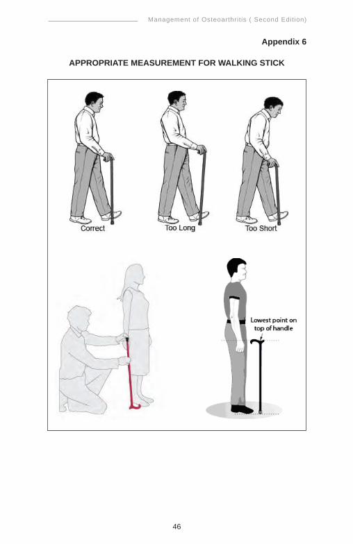
Management of Osteoarthritis ( Second Edition)
46
Appendix 6
APPROPRIATE MEASUREMENT FOR WALKING STICK

Management of Osteoarthritis ( Second Edition)
47
Appendix 7
JOINT PROTECTION PRINCIPLES
Joint protection principles include:-• Resting inflamed joints by reducing load, duration of use and
repetitive movement• Using the largest unaffected muscles and joints to perform a task• Using proper movement techniques for lifting, sitting, standing,
bending and reaching• Using assistive devices and modifications for home equipment to
minimise stress on joints • Plan and organise activities ahead• Using biomechanics and ergonomics to best effect• Simplifying tasks• Recruiting others to help• Making exercise a part of everyday life including exercises which
improve joint range of movement, stamina and strength • Exercise should also be for cardiovascular fitness and to maintain
or improve balance
Source: National Collaborating Centre for Chronic Conditions. Osteoarthritis: national clinical guideline for care and management in adults. London: Royal College of Physicians; 2008
ACTIVITY MODIFICATION IN PERFORMING ADL
• To use walking stick on the opposite hand of the affected knee /more painful knee joint during walking
• To climb stairs, use the railing of the stairs, climb slowly and the body weight should be transferred to the railing by the hand opposite to the affected knee
• To bathe in standing or sitting position with a shower and use high commode in bathroom
• To cook in standing position and if necessary to sit on a chair for a while and use dining table for eating
• To avoid high heeled shoes, sitting on low stool and prolonged walking/sitting/running
• To avoid performing activity in squatting position
Source: Shakoor MA, Taslim MA, Hossain MS. Effects of activity modification on the patients with osteoarthritis of the knee. Bangladesh Med Res Counc Bull. 2007 Aug;33(2):55-9

Management of Osteoarthritis ( Second Edition)
48
App
endi
x 8
SUG
GES
TED
MED
ICAT
ION
DO
SAG
ES A
ND
SID
E EF
FEC
TSD
rug
Cla
ss
Dru
g R
ecom
men
ded
Dos
ages
Si
de E
ffect
s C
autio
n an
d C
ontr
aind
icat
ions
C
omm
ents
Sim
ple
anal
gesi
c P
arac
etam
ol
0.5
– 1
gm, 6
– 8
-hou
rly
Max
: 4 g
m/d
ay
Rar
e bu
t hyp
erse
nsiti
vity
in
clud
ing
skin
rash
may
occ
ur
Hep
atic
impa
irmen
t A
lcoh
ol d
epen
denc
e
Pre
ferr
ed d
rug
parti
cula
rlyel
derly
pat
ient
s
N
on-
sele
ctiv
e N
SAID
s
Ibup
rofe
n
400
– 80
0 m
g, 6
– 8
-hou
rly
Max
: 320
0 m
g/da
y P
eptic
ulc
er
GI b
leed
P
late
let d
ysfu
nctio
n R
enal
impa
irmen
t H
yper
tens
ion
Alle
rgic
reac
tion
in
susc
eptib
le in
divi
dual
s
Incr
ease
in C
VS
eve
nts
Gas
trodu
oden
al u
lcer
A
sthm
a B
leed
ing
diso
rder
R
enal
dys
func
tion
Isch
aem
ic h
eart
dise
ase
C
ereb
ro v
ascu
lar d
isea
seIn
flam
mat
ory
bow
el d
isea
se
Phy
sici
ans
and
patie
nts
shou
ld w
eigh
w
eigh
the
bene
fits
and
risks
of
NS
AID
s th
erap
y
Mef
enam
ic a
cid
250
– 50
0 m
g, 6
– 8
-hou
rly
Max
: 150
0 m
g/da
y D
iclo
fena
c so
dium
50 –
150
mg
daily
,
8
– 12
-hou
rlyM
ax: 1
50 m
g/da
y M
elox
icam
7.
5 –
15 m
g da
ily
Max
: 15
mg/
day
Nap
roxe
n 25
0 –
500
mg,
12-
hour
ly
Max
: 150
0 m
g/da
y N
apro
xen
sodi
um
275
-–55
0 m
g, 1
2-ho
urly
M
ax: 1
650
mg/
day
Piro
xica
m
10 –
20
mg
daily
, in
sing
le o
r di
vide
d do
ses
Max
: 20
mg/
day
Sele
ctiv
e C
OX-
2 in
hibi
tors
Cel
ecox
ib
200
mg
daily
M
ax: 2
00 m
g/da
y (R
ecom
men
ded
daily
m
axim
um d
ose
is 2
00 m
gfo
r OA
and
400
mg
for
infla
mm
ator
y ar
thrit
is)
Ren
al im
pairm
ent
Alle
rgic
reac
tion
in
susc
eptib
le in
divi
dual
s In
crea
se in
CV
S e
vent
s
Isch
aem
ic h
eart
dise
ase
Cer
ebro
vasc
ular
dis
ease
C
ontra
indi
cate
d in
hy
pers
ensi
tivity
to
sulfo
nam
ides
Ass
ocia
ted
with
a lo
wer
risk
of
ser
ious
upp
er G
I sid
e ef
fect
s
Cur
rent
dat
a su
gges
t tha
t
incr
ease
d C
V ri
sk m
ay b
e an
ef
fect
of t
he N
SAID
s/co
xib
clas
s
Phy
sici
ans
and
patie
nts
shou
ld
w
eigh
the
bene
fits
and
risks
of c
oxib
ther
apy
Eto
ricox
ib
60 m
g da
ily
Max
: 90
mg/
day
Hyp
erte
nsio
n R
enal
impa
irmen
t In
crea
se in
CV
S e
vent
s
Unc
ontro
lled
hype
rtens
ion
Is
chae
mic
hea
rtC
ereb
rova
scul
ar d
isea
se
dis
ease
Mel
oxic
am is
a s
elec
tive
CO
X-2
inhi
bito
r at 7
.5 m
gda
ily b
ut n
ot 1
5 m
g da
ily

Management of Osteoarthritis ( Second Edition)
49
Dru
g C
lass
D
rug
Rec
omm
ende
d D
osag
es
Side
Effe
cts
Cau
tion
and
Con
trai
ndic
atio
ns
Com
men
ts
Wea
k op
ioid
Tr
amad
ol
50 –
100
mg,
6 –
8-h
ourly
M
ax: 4
00 m
g/da
y D
izzi
ness
N
ause
a V
omiti
ng
Con
stip
atio
n D
row
sine
ss
Ris
k of
sei
zure
s in
pat
ient
s w
ith h
isto
ry o
f sei
zure
s an
d
with
hig
h do
ses
In
eld
erly
, sta
rt at
low
est d
ose
(5
0 m
g) a
nd m
axim
um o
f 300
lo
wes
t dos
e (5
0 m
g) a
nd
max
imum
of 3
00 m
g da
ily
Inte
ract
ion
with
Tric
yclic
A
ntid
epre
ssan
t, S
elec
tive
Ser
oton
in R
eupt
ake
Inhi
bito
r and
Ser
oton
in
Nor
epin
ephr
ine
Rec
epto
r In
hibi
tor
C
ombi
natio
n of
opi
oid
and
pa
race
tam
ol
Par
acet
amol
325
m
g +
tram
adol
37
.5 m
g (U
ltrac
et®
)
1 –
2 ta
blet
s, 6
– 8
-hou
rly
Max
: 8 ta
blet
s/da
y N
ause
a V
omiti
ng
Dro
wsi
ness
Hep
atic
impa
irmen
t R
enal
impa
irmen
t A
lcoh
ol d
epen
denc
e E
pile
psy
Nut
race
utic
als
Dru
g R
ecom
men
ded
Dos
ages
Si
de E
ffect
s C
autio
n an
d C
ontr
aind
icat
ions
C
omm
ents
Glu
cosa
min
e su
lfate
15
00 m
g in
sin
gle
or
divi
ded
dose
s
Max
: 150
0 m
g/da
y
Nau
sea
Dys
peps
ia
Hea
rtbur
n V
omiti
ng
Con
stip
atio
n D
iarr
hoea
H
eada
che
Ski
n er
ythe
ma
Pru
ritus
Con
train
dica
ted
in p
atie
nts
with
•
alle
rgy
and
hype
rsen
sitiv
ity to
sh
ellfi
sh o
r its
pro
duct
s •
phen
ylke
tonu
ria
Ast
hmat
ic p
atie
nts
may
be
at ri
sk fo
r an
ast
hma
exac
erba
tion
whe
n ta
king
th
e co
mbi
natio
n of
glu
cosa
min
e an
d ch
ondr
oitin
S
peci
al p
reca
utio
n in
dia
bete
s
Cho
ndro
itin
sulfa
te
800
– 12
00 m
g da
ily
or d
ivid
ed d
oses
M
ax: 1
200
mg
Dys
peps
ia
Nau
sea
Vom
iting
Ast
hmat
ic p
atie
nts
may
be
at ri
sk fo
r an
ast
hma
exac
erba
tion
whe
n ta
king
ch
ondr
oitin
A
void
in p
atie
nts
with
pro
stat
e ca
ncer
- -
Dru
g C
lass
D
rug
Rec
omm
ende
d D
osag
es
Side
Effe
cts
Cau
tion
and
Con
trai
ndic
atio
ns
Com
men
ts
Wea
k op
ioid
Tr
amad
ol
50 –
100
mg,
6 –
8-h
ourly
M
ax: 4
00 m
g/da
y D
izzi
ness
N
ause
a V
omiti
ng
Con
stip
atio
n D
row
sine
ss
Ris
k of
sei
zure
s in
pat
ient
s w
ith h
isto
ry o
f sei
zure
s an
d
with
hig
h do
ses
In
eld
erly
, sta
rt at
low
est d
ose
(5
0 m
g) a
nd m
axim
um o
f 300
lo
wes
t dos
e (5
0 m
g) a
nd
max
imum
of 3
00 m
g da
ily
Inte
ract
ion
with
Tric
yclic
A
ntid
epre
ssan
t, S
elec
tive
Ser
oton
in R
eupt
ake
Inhi
bito
r and
Ser
oton
in
Nor
epin
ephr
ine
Rec
epto
r In
hibi
tor
C
ombi
natio
n of
opi
oid
and
pa
race
tam
ol
Par
acet
amol
325
m
g +
tram
adol
37
.5 m
g (U
ltrac
et®
)
1 –
2 ta
blet
s, 6
– 8
-hou
rly
Max
: 8 ta
blet
s/da
y N
ause
a V
omiti
ng
Dro
wsi
ness
Hep
atic
impa
irmen
t R
enal
impa
irmen
t A
lcoh
ol d
epen
denc
e E
pile
psy
Nut
race
utic
als
Dru
g R
ecom
men
ded
Dos
ages
Si
de E
ffect
s C
autio
n an
d C
ontr
aind
icat
ions
C
omm
ents
Glu
cosa
min
e su
lfate
15
00 m
g in
sin
gle
or
divi
ded
dose
s
Max
: 150
0 m
g/da
y
Nau
sea
Dys
peps
ia
Hea
rtbur
n V
omiti
ng
Con
stip
atio
n D
iarr
hoea
H
eada
che
Ski
n er
ythe
ma
Pru
ritus
Con
train
dica
ted
in p
atie
nts
with
•
alle
rgy
and
hype
rsen
sitiv
ity to
sh
ellfi
sh o
r its
pro
duct
s •
phen
ylke
tonu
ria
Ast
hmat
ic p
atie
nts
may
be
at ri
sk fo
r an
ast
hma
exac
erba
tion
whe
n ta
king
th
e co
mbi
natio
n of
glu
cosa
min
e an
d ch
ondr
oitin
S
peci
al p
reca
utio
n in
dia
bete
s
Cho
ndro
itin
sulfa
te
800
– 12
00 m
g da
ily
or d
ivid
ed d
oses
M
ax: 1
200
mg
Dys
peps
ia
Nau
sea
Vom
iting
Ast
hmat
ic p
atie
nts
may
be
at ri
sk fo
r an
ast
hma
exac
erba
tion
whe
n ta
king
ch
ondr
oitin
A
void
in p
atie
nts
with
pro
stat
e ca
ncer
- -
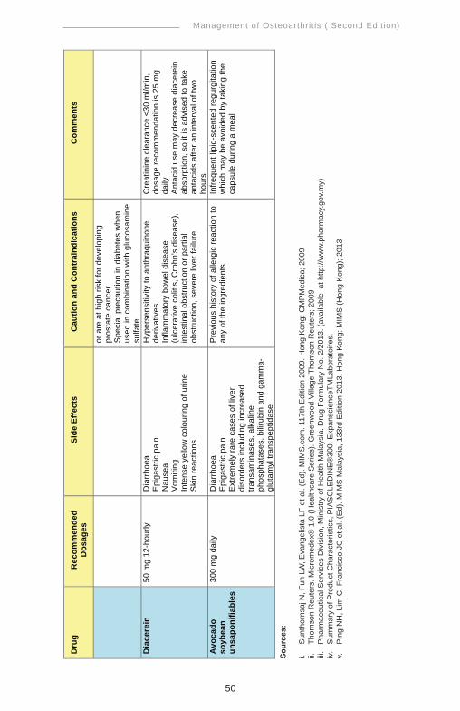
Management of Osteoarthritis ( Second Edition)
50
Sour
ces:
i. S
unth
orns
aj N
, Fun
LW
, Eva
ngel
ista
LF
et a
l. (E
d). M
IMS
.com
. 117
th E
ditio
n 20
09. H
ong
Kon
g: C
MP
Med
ica;
200
9ii.
Th
omso
n R
eute
rs. M
icro
med
ex®
1.0
(Hea
lthca
re S
erie
s). G
reen
woo
d Vi
llage
Tho
mso
n R
eute
rs; 2
009
iii.
Pha
rmac
eutic
al S
ervi
ces
Div
isio
n, M
inis
try o
f Hea
lth M
alay
sia.
Dru
g Fo
rmul
ary
No.
2/2
013.
(ava
ilabl
e a
t http
://w
ww
.pha
rmac
y.go
v.m
y)iv.
S
umm
ary
of P
rodu
ct C
hara
cter
istic
s, P
IAS
CLE
DIN
E®
300.
Exp
ansc
ienc
eTM
Labo
rato
ires.
v.
Pin
g N
H, L
im C
, Fra
ncis
co J
C e
t al.
(Ed)
. MIM
S M
alay
sia,
133
rd E
ditio
n 20
13. H
ong
Kon
g: M
IMS
(Hon
g K
ong)
; 201
3
Dru
g R
ecom
men
ded
Dos
ages
Si
de E
ffect
s C
autio
n an
d C
ontr
aind
icat
ions
C
omm
ents
or a
re a
t hig
h ris
k fo
r dev
elop
ing
pros
tate
can
cer
Spe
cial
pre
caut
ion
in d
iabe
tes
whe
n us
ed in
com
bina
tion
with
glu
cosa
min
e su
lfate
D
iace
rein
50
mg
12-h
ourly
D
iarr
hoea
E
piga
stric
pai
n N
ause
a V
omiti
ng
Inte
nse
yello
w c
olou
ring
of u
rine
Ski
n re
actio
ns
Hyp
erse
nsiti
vity
to a
nthr
aqui
none
de
rivat
ives
In
flam
mat
ory
bow
el d
isea
se
(ulc
erat
ive
colit
is, C
rohn
’s d
isea
se),
inte
stin
al o
bstru
ctio
n or
par
tial
obst
ruct
ion,
sev
ere
liver
failu
re
Cre
atin
ine
clea
ranc
e <3
0 m
l/min
, do
sage
reco
mm
enda
tion
is 2
5 m
g da
ily
Ant
acid
use
may
dec
reas
e di
acer
ein
abso
rptio
n, s
o it
is a
dvis
ed to
take
an
taci
ds a
fter a
n in
terv
al o
f tw
o ho
urs
Avo
cado
so
ybea
n un
sapo
nifia
bles
300
mg
daily
D
iarr
hoea
E
piga
stric
pai
n E
xtre
mel
y ra
re c
ases
of l
iver
di
sord
ers
incl
udin
g in
crea
sed
trans
amin
ases
, alk
alin
e ph
osph
atas
es, b
iliru
bin
and
gam
ma-
glut
amyl
tran
spep
tidas
e
Pre
viou
s hi
stor
y of
alle
rgic
reac
tion
to
any
of th
e in
gred
ient
s In
frequ
ent l
ipid
-sce
nted
regu
rgita
tion
whi
ch m
ay b
e av
oide
d by
taki
ng th
e ca
psul
e du
ring
a m
eal

Management of Osteoarthritis ( Second Edition)
51
Appendix 9
CHECKLIST OF OA MANAGEMENT
1. Education
• Nature of OA Yes No
• Prognosis Yes No
• Treatment option - medication & side effects Yes No
• Self-management Yes No
2. Lifestyle modification
• Weight loss Yes No
• Dietary advice Yes No
• Exercise Yes No
• Footwear Yes No
• Pacing activity Yes No
3. Referral where applicable Yes No
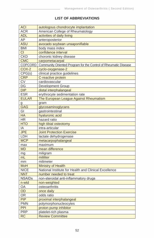
Management of Osteoarthritis ( Second Edition)
52
LIST OF ABBREVIATIONS
ACI autologous chondrocyte implantationACR American College of RheumatologyADL activities of daily livingAP anteroposteriorASU avocado soybean unsaponifiableBMI body mass indexCI confidence intervalCKD choronic kidney diseaseCMC carpometacarpalCOPCORD Community Oriented Program for the Control of Rheumatic DiseaseCOX-2 cyclo-oxygenase-2CPG(s) clinical practice guidelinesCRP C-reactive proteinCV cardiovascularDG Development GroupDIP distal interphalangealESR erythrocyte sedimentation rateEULAR The European League Against Rheumatismg gramGAG glycosaminoglycansGI gastrointestinalHA hyaluronic acidHR hazard ratioHTO high tibial osteotomyIA intra-articularJPE Joint Protection ExerciseLDH lactate dehydrogenaseMCP metacarpophalangealmax maximumMD mean differencemg miligrammL mililitermm milimeterMoH Ministry of HealthNICE National Institute for Health and Clinical ExcellenceNNT number needed to treatNSAIDs non-steroidal anti-inflammatory drugs n-wtd non-weightedOA osteoarthritisOD once dailyOR odds ratioPIP proximal interphalangealPMN polymorphonucleocytesPPI proton pump inhibitorPRP platelet-rich plasmaRC Review Committee

Management of Osteoarthritis ( Second Edition)
53
RCT(s) randomised controlled trial(s)RF rheumatoid factorROM range of movementRR relative riskSF synovial fluidSMD standardised mean differenceSR systematic reviewTENS transcutaneous electrostimulationTHR total hip replacementTKR total knee replacementUKR unicondylar knee replacementVAS Visual Analogue ScaleWBC white blood cellsWHO World Health OrganizationWMD weighted mean differenceWOMAC The Western Ontario and McMaster Universities Arthritis Index
ACKNOWLEDGEMENT
The members of development group of these guidelines would like to express their gratitude and appreciation to the following for their contributions:-• Panel of external reviewers who reviewed the draft• Ms. Zalina Ahmad and Ms. Loong Ah Moi (Nursing Sisters)• Technical Advisory Committee for CPG for their valuable input and
feedback• All those who have contributed directly or indirectly to the development
of the CPG
DISCLOSURE STATEMENT
The panel members of both Development Group and Review Committee had completed disclosure forms. None held shares in pharmaceutical firms or acts as consultants to such firms. (Details are available upon request from the CPG Secretariat)
SOURCE OF FUNDING
The development of the CPG on Management of Osteoarthritis (Second Edition) was supported financially in its entirety by the Ministry of Health Malaysia and Malaysian Society of Rheumatology.

Management of Osteoarthritis ( Second Edition)
54
