1 introduction: biophotonics – visions for better health care · 2 1 introduction: biophotonics...
TRANSCRIPT
Biophotonics: Visions for Better Health Care. Jürgen Popp and Marion Strehle (Eds.)Copyright © 2006 WILEY-VCH Verlag GmbH & Co. KGaA, WeinheimISBN: 3-527-40622-0
1
1Introduction:Biophotonics – Visions for Better Health CareSusanne Liedtke, Michael Schmitt, Marion Strehle, Jürgen Popp1
Since the late 1980s, natural scientists have introduced a multiplicity of newterms and definitions. We had to learn the difference between genes and pro-teins, we have been taken into the miniature world of viruses and prions, andthe newspapers report on a great many new technologies, such as biotech-nology, information technology and nanotechnology, and nowadays even onbionanotechnology. The development of those new technologies combines anincrease of both the scientific and also the technological understanding andknowledge of, for example, life processes and has already led to economicprofit as well as to enormous stock-exchange quotations. However, apart fromundreamed-of possibilities for mankind, the new technologies also entail eth-ical risks and problems as we can conclude from the discussion on the firstcloned sheep “Dolly” or the application of embryonic stem cells. Rather thanentering into a detailed discussion, we will stop here, and instead introduce afairly new discipline of natural science: Biophotonics.
Biophotonics deals with the interaction between light and biological sys-tems. The word itself is a combination of the Greek syllables bios standing forlife and phos standing for light. Photonics is the technical term for all proce-dures, technologies, devices, etc. utilizing light in interaction with any matter.Before we discuss Biophotonics in more detail, we focus first and foremoston the achievements of Photonic Technologies, which are often used synony-mously with Photonics. The advanced control and manipulation of light nowavailable make Photonics as powerful as Electronics. One major goal is to in-corporate even more photonically driven processes into our daily lives. There-fore, Photonics is considered as a key technology of the twenty-first century.
Photonics encompasses the entire physical, chemical and biological lawsof nature, together with technologies for the generation, amplification, con-trol, manipulation, propagation, measurement, harnessing and any other typeof utilization of light. This rather broad definition of Photonics emphasizesthe huge importance of light for our modern human society. Photonics is akey technology for solving momentous problems in the domains of health
1) Corresponding author
2 1 Introduction: Biophotonics – Visions for Better Health Care
care, food production and technology, environmental protection, transporta-tion, mobility, etc. It is a pacemaking technology for other developments suchas communication and production technology, biotechnology and nanotech-nology. Just one brief example of what the near future holds: conventionallighting by light bulbs, neon lamps or fluorescent tubes will be a thing ofthe past. The new wonder is organic light-emitting diodes (OLED). Thoseinnovative diodes provide more efficient and long-lasting illumination, withenergy consumption reduced to a minimum. Thus, the wide-spread appli-cation of these diodes will allow our economy to save billion of Euros bothon the cost of energy and also on consequential costs, such as, for example,the cost of environmental protection. Furthermore, the use of these diodes fordisplays will revolutionize graphic screen technology. In comparison to TFT(Thin Film Transistor) screens, an OLED display does not need backgroundillumination. The display has the thickness of a plastic transparency, andits flexibility. Completely new applications, such as the use as “informationwallpaper”, are in the minds of scientists and advertising experts. However,in other fields of Photonics the future has already started. The informationcarried via the World Wide Web is mainly carried by light. From these fewexamples, we can recognize that photonic technologies are already pervadingour entire life.
Now let us come back to another field in which Photonics is seen as a keytechnology for future scientific and economic progress – the field called “Bio-photonics”. What is special about Biophotonics? In order to answer this ques-tion we have to shed light on life as well as on light itself because, as alreadymentioned, Biophotonics deals with both light and life. Both phenomena willbe important issues in this book. While the “life” aspect will be covered inthe various chapters, a short summary of the properties of light, as well as onthe interactions of light and matter, will be given later on in this introductorychapter. From time immemorial both phenomena have fascinated people. De-spite life and light have being ubiquitous and self-evident, an understandingof the scientific bases of light and life was and is still a special challenge. Aswe can see from history, there was a long and roundabout journey from thefirst description of the nature of light in early antiquity to the photon theoryof Albert Einstein. In a similar but no less complicated way, the phenomenonof life has occupied scientists and philosophers from Aristotle to the modernstudents of genomics in the twenty-first century.
It makes sense to put both life and light together. We can, indeed we must,learn from nature. Nature demonstrates how valuable and fruitful the interac-tion between light and biological systems, and thus Biophotonics as definedabove, can be for life. Consider the “harnessing” of photons. Plants utilizelight via photosynthesis as an energy source. In the process of vision lightgenerates pictures of our environment in our brains by a quite complicated
1 Introduction: Biophotonics – Visions for Better Health Care 3
but very efficient pathway. The scientist dealing with Biophotonics attemptsto understand nature by mimicking the basic principles and taking advan-tage of the same tools. Using this approach, highly innovative future-orientedtechnologies can be brought into the real world. This makes the business quitethrilling. Biophotonics is not a science as an end in itself. In fact it opensundreamed-of possibilities for fundamental research, the pharmaceutical andfood industries, biotechnology, medicine, etc.
The investigation of biological materials by means of innovative opticaltechniques has led to totally new optical technologies, including techniquescapable of, for example, yielding snapshots of cellular conditions and moni-toring dynamic processes. The ultimate goal of Biophotonics is to unravel lifeprocesses within cells, cell colonies, tissues, or even whole organs. Biopho-tonics seeks to provide a comprehensive multidimensional understanding ofthe various processes occurring in an organism. Therefore, Biophotonics com-bines all optical methods to investigate the structural, functional, mechanical,biological and chemical properties of biological material and systems. Theoptical phenomena exploited to gain all this information include all the inter-actions of electromagnetic radiation with living organisms or organic material,such as absorption, emission, reflection, scattering, etc. Other areas of Biopho-tonics use light as a miniaturized tool, e.g., optical tweezers or a laser scalpel.
Why do we use light? What are the advantages of applying light to thestudy of biological matter? The three major advantages are: (1) Light mea-sures without contact, i.e., light allows processes taking place within a livingcell to be studied without disturbing or affecting the biological activity. (2)Light measures more quickly and yields instantaneous information, i.e., thecomplex preparation needed in conventional methods to obtain, for exam-ple, a reliable diagnosis, which may take days, or even weeks, are no longerrequired or can be performed in a much shorter time. (3) Light measuresmore precisely, i.e., optical methods allow ultrasensitive detection, down tothe single-molecule detection necessary for the life science sector.
As already indicated by the title “Biophotonics – Visions for better healthcare” the main focus of this contribution is on the topic “health care”. How-ever, the reader should bear in mind that Biophotonics is not limited to deal-ing with “health care”. The final chapter of this book will mention other topicsand challenges of Biophotonics. With the help of this new discipline we hopeto get a precise understanding of the origin of diseases so that in the future wecan prevent diseases or at least diagnose them more precisely and at an earlierstage so that we can treat them more efficiently. In principle, this looks quitesimple. However, to be successful in achieving these ultimate goals scientistshave to look beyond their own noses. This means that the developers of pho-tonic technologies, mainly physicists, physical chemists, chemists and engi-neers, have to be in close contact with the possible users in biology, medicine,
4 1 Introduction: Biophotonics – Visions for Better Health Care
the pharmaceutical and food industries and environmental research. Other-wise the great potential of Biophotonics cannot be applied. In various con-tributions to this book we shall see that to some extent the innovations ofBiophotonics are based on precise observation of a natural phenomenon by abiologist or physician, which is then transformed with the help of photonictechnologies into new diagnostic methods and technologies. Other progressis based on optical and spectroscopic innovation made by physicists and af-terwards adapted to appropriate biological and medical problems. However,there is still a large gap between those scientists developing optical technolo-gies and those scientists who like to use optical technologies. Thus, bringingtogether the various disciplines is one of the greatest challenges of Biophoton-ics.
1.1The Situation of Biophotonics in Germany and Other Countries
The German government recognized the great potential of Biophotonics, notonly as a key technology but also as a bridging technology, and installed amultidisciplinary operating research network. The main research frameworkBiophotonics, funded by the German Federal Ministry of Education and Re-search (BMBF), has two major common goals.
• Scientific goal: Shedding light on biological processes, i.e. understand-ing life processes on a functional as well as a molecular level. This willallow scientists to understand the origin of diseases and to invent newstrategies to defeat them more effectively and will be the base for realprevention and effective therapies.
• Technological goal: Developing innovative light-based technologies toachieve the above-mentioned goal.
A prerequisite for reaching these two goals is that scientists from the var-ious disciplines mentioned above should work closely together. This meansthat the potential user, e.g. the physician, has to be involved in the scientificand technical development from the very beginning. Another quite importantrequirement is that the industry, with all its technological knowledge and ex-perience, cooperates right from the start. The involvement of both parties isseen as one important key to a successful conclusion.
This multi- and interdisciplinary research should make a strong impact onsociety. First of all, Biophotonics will lead to better health. Diseases, whethercancer or infectious diseases, will be defeated very effectively. Thus, the qual-ity of life will be dramatically improved and the cost of health care consider-ably reduced. Areas in which Biophotonics is already operating successfully
1.1 The Situation of Biophotonics in Germany and Other Countries 5
include pathology, oncology, dermatology, cardiology, urology, ophthalmol-ogy, gastroenterology and dentistry. Secondly, in addition to scientific andtechnical progress, the research framework strives for the important goal ofstrengthening the position of Germany as a centre for technology by creat-ing new employment and protecting highly qualified workers and sustain-able jobs. As a consequence, Biophotonics will become as important as otherleading technologies, such as nanotechnology, genomics and proteomics.
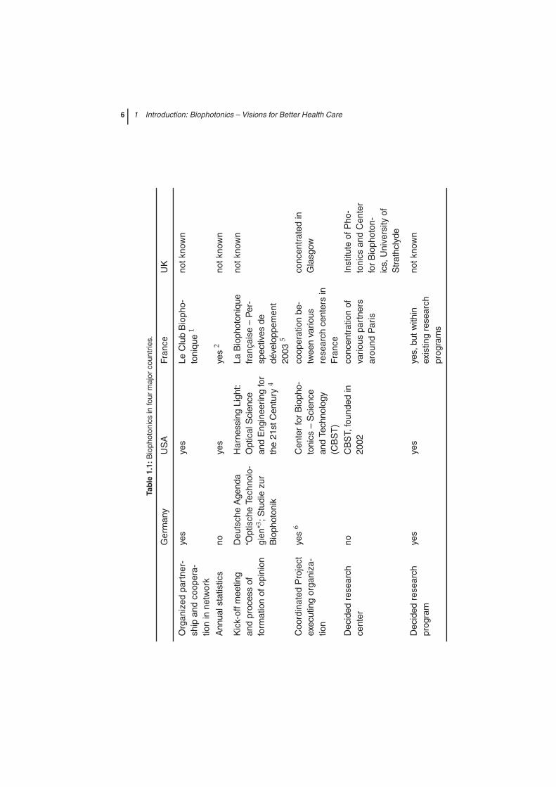
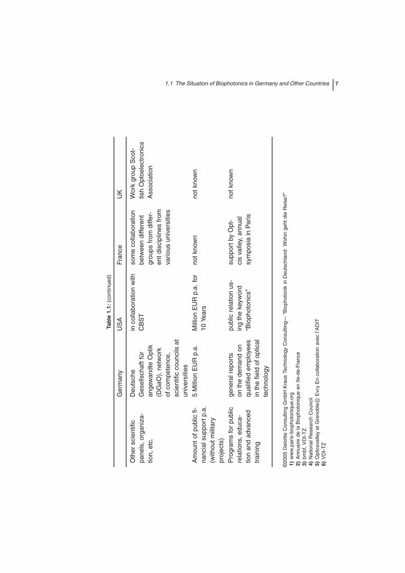
Germany is not the only country to have recognized the importance of Bio-photonics. All over the world scientists from academia and industry are work-ing on this topic. A short overview on the different activities of Germany,USA, France and England is given in Table 1.1 which has been published inreport on Biophotonics in Germany by Deloitte Consulting and Kraus Tech-nology Consulting 2005.
Biophotonics is also very important in other countries, e.g., Canada, Japan,China, Australia and all over Europe. There is almost no country in the worldwhere scientists are not becoming aware of the possibilities and potentialachievement of Biophotonics. The major focus of research activities lies ontopics from medicine and biotechnology. To summarize all these activities isway beyond the scope of this book.
6 1 Introduction: Biophotonics – Visions for Better Health Care
Tabl
e1.
1:B
ioph
oton
ics
info
urm
ajor
coun
trie
s.
Ger
man
yU
SA
Fran
ceU
K
Org
aniz
edpa
rtne
r-sh
ipan
dco
oper
a-tio
nin
netw
ork
yes
yes
LeC
lub
Bio
pho-
toni
que
1no
tkno
wn
Ann
uals
tatis
tics
noye
sye
s2
notk
now
n
Kic
k-of
fmee
ting
and
proc
ess
offo
rmat
ion
ofop
inio
n
Deu
tsch
eA
gend
a“O
ptis
che
Tech
nolo
-gi
en”3 ;
Stu
die
zur
Bio
phot
onik
Har
ness
ing
Ligh
t:O
ptic
alS
cien
cean
dE
ngin
eerin
gfo
rth
e21
stC
entu
ry4
LaB
ioph
oton
ique
fran
çais
e–
Per
-sp
ectiv
esde
déve
lopp
emen
t20
035
notk
now
n
Coo
rdin
ated
Pro
ject
exec
utin
gor
gani
za-
tion
yes
6C
ente
rfo
rB
ioph
o-to
nics
–S
cien
cean
dTe
chno
logy
(CB
ST
)
coop
erat
ion
be-
twee
nva
rious
rese
arch
cent
ers
inFr
ance
conc
entr
ated
inG
lasg
ow
Dec
ided
rese
arch
cent
erno
CB
ST,
foun
ded
in20
02co
ncen
trat
ion
ofva
rious
part
ners
arou
ndP
aris
Inst
itute
ofP
ho-
toni
csan
dC
ente
rfo
rB
ioph
oton
-ic
s,U
nive
rsity
ofS
trat
hcly
de
Dec
ided
rese
arch
prog
ram
yes
yes
yes,
butw
ithin
exis
ting
rese
arch
prog
ram
s
notk
now
n
1.1 The Situation of Biophotonics in Germany and Other Countries 7
Tabl
e1.
1:(c
ontin
ued)
Ger
man
yU
SA
Fran
ceU
K
Oth
ersc
ient
ific
pane
ls,o
rgan
iza-
tion,
etc.
Deu
tsch
eG
esel
lsch
aftf
üran
gew
andt
eO
ptik
(DG
aO),
netw
ork
ofco
mpe
tenc
e,sc
ient
ific
coun
cils
atun
iver
sitie
s
inco
llabo
ratio
nw
ithC
BS
Tso
me
colla
bora
tion
betw
een
diffe
rent
grou
psfr
omdi
ffer-
entd
isci
plin
esfr
omva
rious
univ
ersi
ties
Wor
kgr
oup
Sco
t-tis
hO
ptoe
lect
roni
csA
ssoc
iatio
n
Am
ount
ofpu
blic
fi-na
ncia
lsup
port
p.a.
(with
outm
ilita
rypr
ojec
ts)
5M
illio
nE
UR
p.a.
Mill
ion
EU
Rp.
a.fo
r10
Year
sno
tkno
wn
notk
now
n
Pro
gram
sfo
rpu
blic
rela
tions
,edu
ca-
tion
and
adva
nced
trai
ning
gene
ralr
epor
tson
the
dem
and
onqu
alifi
edem
ploy
ees
inth
efie
ldof
optic
alte
chno
logy
publ
icre
latio
nus
-in
gth
eke
ywor
d“B
ioph
oton
ics”
supp
ortb
yO
pt-
cis
valle
y,an
nual
sym
posi
ain
Par
is
notk
now
n
©20
05D
eloi
tteC
onsu
lting
Gm
bHK
raus
Tech
nolo
gyC
onsu
lting
—“B
ioph
oton
ikin
Deu
tsch
land
:W
ohin
geht
die
Rei
se?”
1)w
ww
.par
is-b
ioph
oton
ique
.org
2)A
nnua
irede
laB
ioph
oton
ique
enIle
-de-
Fran
ce3)
bmbf
,VD
I-T
Z4)
Nat
iona
lRes
earc
hC
ounc
il5)
Opt
icsv
alle
yet
Gre
nobl
e�E
vry
En
colla
bora
tion
avec
I’AD
IT6)
VD
I-T
Z
8 1 Introduction: Biophotonics – Visions for Better Health Care
1.2The Interplay Between Light and Matter: Interactions Allowing Us to UnderstandOur Environment
Before we introduce the marvelous world of Biophotonics we shall first give abrief introduction into the basics of light–matter interactions. The interactionof light with matter, in particular with biological material, i.e., what happensif a light wave or photon hits matter, is an exciting topic. The various light–matter interaction phenomena enable us to observe our environment and areresponsible for the existence of life on earth. What follows is a short and verygeneral introduction into the manifold interplay (e.g. absorption, emission,scattering, reflection, refraction, diffraction, dispersion, polarization, etc.) be-tween light and matter. The advanced reader may therefore skip the followingpages.
Light propagates as electromagnetic waves. Electromagnetic waves arewavelike perturbations (invisible perturbations of the so-called force field)recurring periodically over a certain distance called the wavelength. Lightwaves are described as oscillating electric (E) and magnetic fields (H), whichare perpendicular to each other. Time-dependent changes of the electricfield are always combined with spatial changes of the magnetic field. Simi-larly, time-dependent changes of the magnetic field are connected with spa-tial changes of the corresponding electric field. Electromagnetic waves canpropagate in a vacuum at a speed of c = 299 792.458 km s−1. The time-dependent electric field E can be described as: E(z, t) = E0 cos 2πν(t − z/c)where E(z) = electric field at position z; E0 = maximum electric field; ν = lightfrequency; t = time; c = speed of light. It is just this oscillating electric field thatcan interact with matter and transfer energy to or from matter. The connectionbetween frequency and wavelength is given by λν = c. The spectroscopicallymore common unit wavenumber is defined by ν̃ = 1/λ.
The interaction of electromagnetic radiation with matter can polarize matterand induce a dipole moment. Matter, e.g., molecules, consists of atoms heldtogether by electrons. If the binding electrons are distributed between theatoms, a covalent bond exists. If the binding electrons are located on an atom,ionic bonding is present. These two main models of bonding are extremecases. The only covalent bonds which have no ionic character are those be-tween identical atoms. However, molecules formed between different atomsmay exhibit partial charges due to an asymmetric distribution of the bindingelectrons between the atoms. If no side of the molecule has more negative orpositive partial charge than the other side, the molecule is nonpolar. However,molecules exhibiting an asymmetric partial charge distribution are polar. Anexample of a polar molecule is the water molecule, consisting of two hydro-gen atoms and one oxygen atom held together by two common electron pairs.Since the oxygen atoms attract electrons more than the hydrogen atoms the
1.2 Interplay between light and matter 9
binding electrons in a water molecule are asymmetrically distributed, leadingto a negative partial charge on the oxygen atom and positive partial chargeson the hydrogen atoms. These partial charges make water act as a dipole.The interaction of polar molecules like water with electromagnetic waves of acertain frequency leads to an orientation of the water molecules.
To learn more about electromagnetic waves and their influence on matter,we look first at a plate-type capacitor whose polarity is changing with a cer-tain frequency. Water molecules are aligned within a plate-type capacitor insuch a way that the negative pole of the water molecules points towards thepositively charged plate while the positive pole orients towards the negativeplate. The water dipoles orient themselves according to the applied electricfield of the capacitor, i.e., the water molecules become oriented. This phenom-enon is called orientation polarization. Changing the polarity of the capacitorplates leads to a reorientation of the water molecules.
What happens if nonpolar molecules like nitrogen N2, oxygen O2 or car-bon dioxide CO2 are brought between the plates of a capacitor? Applying anexternal electric field leads to a distortion of the electrons compared to the pos-itively charged atomic nuclei. This distortion creates partial charges, i.e., theelectric field induces a dipole moment. This polarization effect is called dis-tortion polarization. The more easily the electrons can be displaced comparedto the positively charged nuclei the bigger the induced dipole moment. Theinduced dipole moment µind is direct proportional to the applied electric fieldE where the proportionality constant α is called the molecule’s polarizability:µind = αE. α is a measure of how easily electrons can be moved or displacedwithin a molecule. Thus nonpolar molecules within a plate-type capacitoralso become oriented owing to the induced dipole moments. Changing thepolarity of the electric field changes the induced dipole moment accordingly.However, the nonpolar molecules are not reoriented; only the electrons willbe moved in another direction.
The general form of the polarization depends on the frequency of the ap-plied field. Radio frequency radiation (100 MHz) leads to an alignment of po-lar molecules according to the external electric field, i.e., a reorientation of thecomplete molecules (orientation polarization) takes place. The same is true forthe electrons, which can follow the changing electric field much more easilyowing to their low mass. However, higher frequencies lead to a distortion po-larization of the electrons (“induced electric dipole moment”) since moleculesexhibiting a permanent dipole can no longer follow such rapidly oscillatingfields. The induced dipole moment µind oscillates with the same frequency asthe exciting oscillating electric field E: µind = αE0 cos 2πν(t − z/c) where α isthe molecule’s polarizability. Since the oscillating induced dipole moment issimply an oscillating charge, matter radiates light of the same frequency as theinitial exciting radiation field (secondary radiation). This is in total analogy
10 1 Introduction: Biophotonics – Visions for Better Health Care
to a Hertzian dipole acting as a broadcasting and receiving antenna, which isbased on the radiation of electromagnetic waves from a dipole. In the antenna,electrons are driven by a generator to the top or the bottom. This generates acharge distribution similar to a dipole.
The interplay of molecules with light, in particular with visible light, whichpolarizes molecules and leads to the emission of a secondary radiation of thesame frequency as the polarizing field in all directions, is called elastic lightscattering or Rayleigh scattering. It is precisely this undirected scattered ra-diation which enables us to observe our environment in the presence of sun-light or light from a lamp. The difference between the earth and outer space isthat the earth has an atmosphere consisting of molecules which can be po-larized. The darkness in outer space is due to the lack of an atmosphere.Rayleigh scattering scales with the fourth power of the light frequency (ν4),i.e., short wavelengths or high-frequency blue light is scattered significantlymore strongly than low-frequency red or infrared light. Hence the middaysky is blue and the sun appears more yellow or red than it really is. The ν4 de-pendence becomes especially obvious when the path of the sunlight throughthe atmosphere is longest. Thus the rising or setting sun appears especiallyred, since then the less-scattered red light can better reach our eyes.
For a description of microscopic systems like atoms or molecules, quantumeffects need to be considered. The term quantum (Latin, “how much”) refersto discrete units assigned to certain physical quantities, such as the energy ofan atom or molecule. This means that a physical quantity that appears macro-scopically continuous appears in the microscopic world only in well definedvalues that cannot be further divided. The energy of a microscopic systemis quantized, i.e., divided into well defined energy portions. An illustrationof this phenomenon might be a staircase, in which each step marks an en-ergy portion. Molecules exhibit certain movement patterns, i.e., the moleculardegrees of freedom can be classified into translation, rotation and vibration.The energy of all these degrees of freedom is quantized. Similarly, the elec-tron energy in an atom or molecule is quantized. What happens if light in-teracts with such a quantized rotating and vibrating molecule? The energy ofthe molecule’s degrees of freedom are quantized, and the energy of light isquantized too. Light can exhibit properties of both waves and particles. Thisphenomenon is known as wave–particle dualism. Einstein postulated the ex-istence of photons, which are quanta of light energy with particle character.By postulating these photons, Einstein was able to explain the photoelectriceffect which cannot be explained by the wave theory of light. The photoelec-tric effect describes the emission of electrons from matter after the absorptionof high-energy ultraviolet light due to a collision with the particle-like pho-ton. Each photon possesses the energy E = hν where h is Planck’s constant(6.626× 10⁻³ J sec) and ν the frequency of the light, i.e., electromagnetic radi-

1.2 Interplay between light and matter 11
ation of the frequency ν can only carry energy of 0, hν, 2hν, . . .. Only photonspossessing energy above a certain threshold lead to an ejection of electrons.According to the wave theory, the electromagnetic field E exerts an oscillat-ing force on the electrons within the matter. This, however, would mean thatelectrons are ejected with increasing amplitude and not frequency, which iscontrary to experiment. Since the energy of matter, i.e., molecules, and elec-tromagnetic radiation cannot be varied continuously, light can only promotematter from one discrete energy level to an energetically higher one if the pho-ton possesses an energy matching the energy difference between two quan-tum states of the system. This process is called absorption.
Depending on the light wavelength, rotations, vibrations or electrons can beexcited within a molecule. Microwaves can excite a transition between two ro-tational energy states, infrared light is necessary to promote a molecule into ahigher vibrational state and visible or ultraviolet light transfers electrons fromone electronic state in an energetically higher electronic quantum state. Anexcited system, however, can relax into the lowest energy state by releasingthe additional energy in terms of heat via collisions with the surrounding en-vironment or even by the emission of light. This special form of light–matterinteraction is the basis for modern molecular diagnostic procedures in life sci-ences and medicine. In the following we will concentrate on the vibrationaland electronic excitation of molecules, since the excitation of rotations in con-densed matter plays no significant role for diagnostic purposes.
A polyatomic molecule exhibits a multitude of vibrations, but of interestare the so-called normal mode vibrations; an N-atomic molecule has 3N − 6normal modes. How can one derive this little relationship? The moleculardegrees of freedom can be divided into translation, rotation and vibration.Atomic motion through space can be described by the three directions in spacex, y and z. As already mentioned, these three coordinates are therefore enoughto describe the translation motion of an atom, i.e., an atom has three transla-tional degrees of freedom. Rotations and vibrations do not exist for an atom.If we consider a molecule consisting of three atoms, e.g., water H2O, the threeatoms could move independently through space if they were not connectedvia a chemical bond. This independent motion would result in nine transla-tional degrees of freedom. However, we know of course that the atoms of amolecule cannot move independently through space but only the whole mol-ecule as an entity. Therefore we need to subtract the three degrees of freedomdescribing the collective motion of the whole molecule in space from the to-tality of motions, i.e., of the nine degrees of freedom six remain. By takinginto account that a molecule can rotate along the three axes of the coordinatesystem we need to subtract three more degrees of freedom for the rotationalmotion. From the original nine degrees of freedom only three remain. Theseremaining degrees of freedom can be assigned to the vibrational degrees of
12 1 Introduction: Biophotonics – Visions for Better Health Care
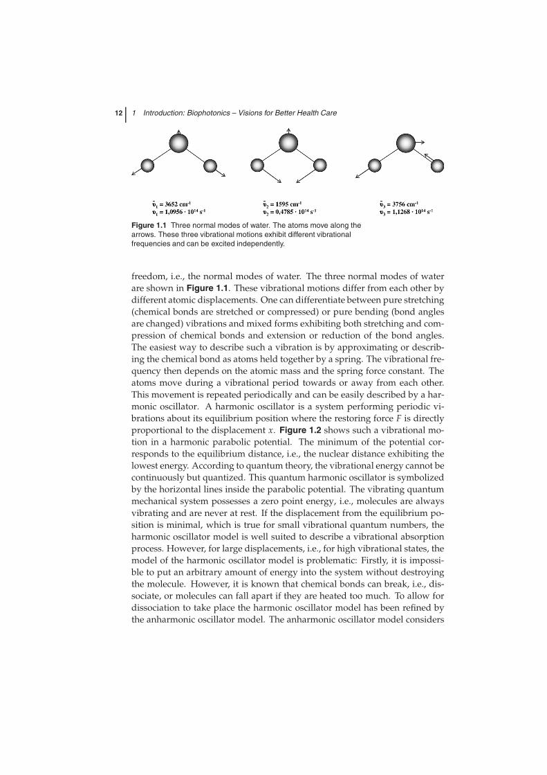
Figure 1.1 Three normal modes of water. The atoms move along thearrows. These three vibrational motions exhibit different vibrationalfrequencies and can be excited independently.
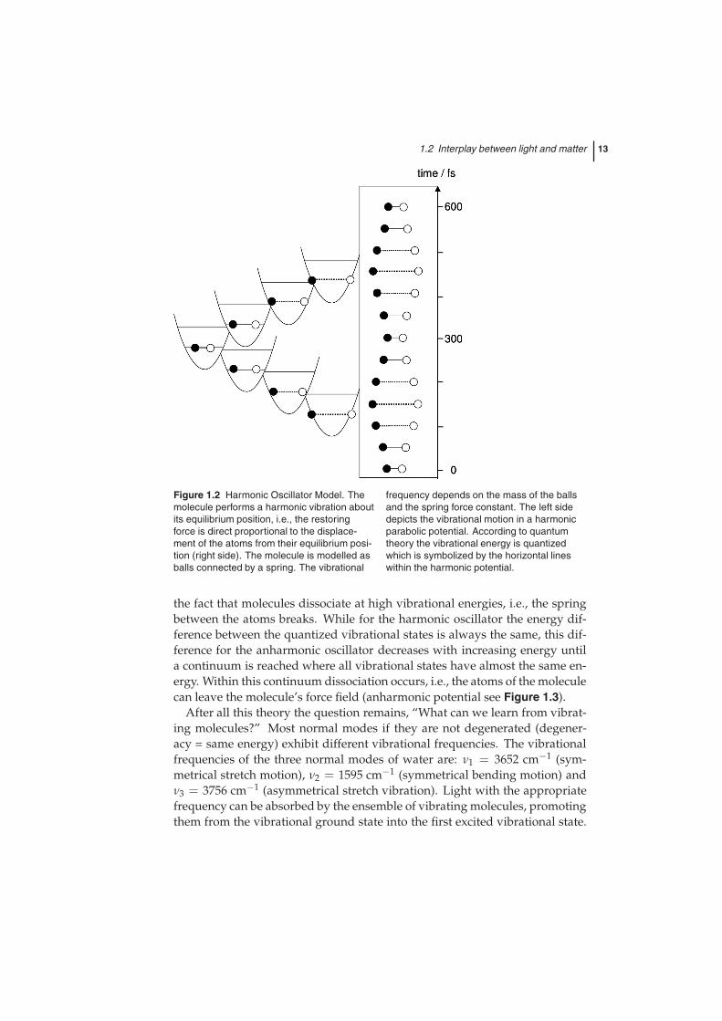
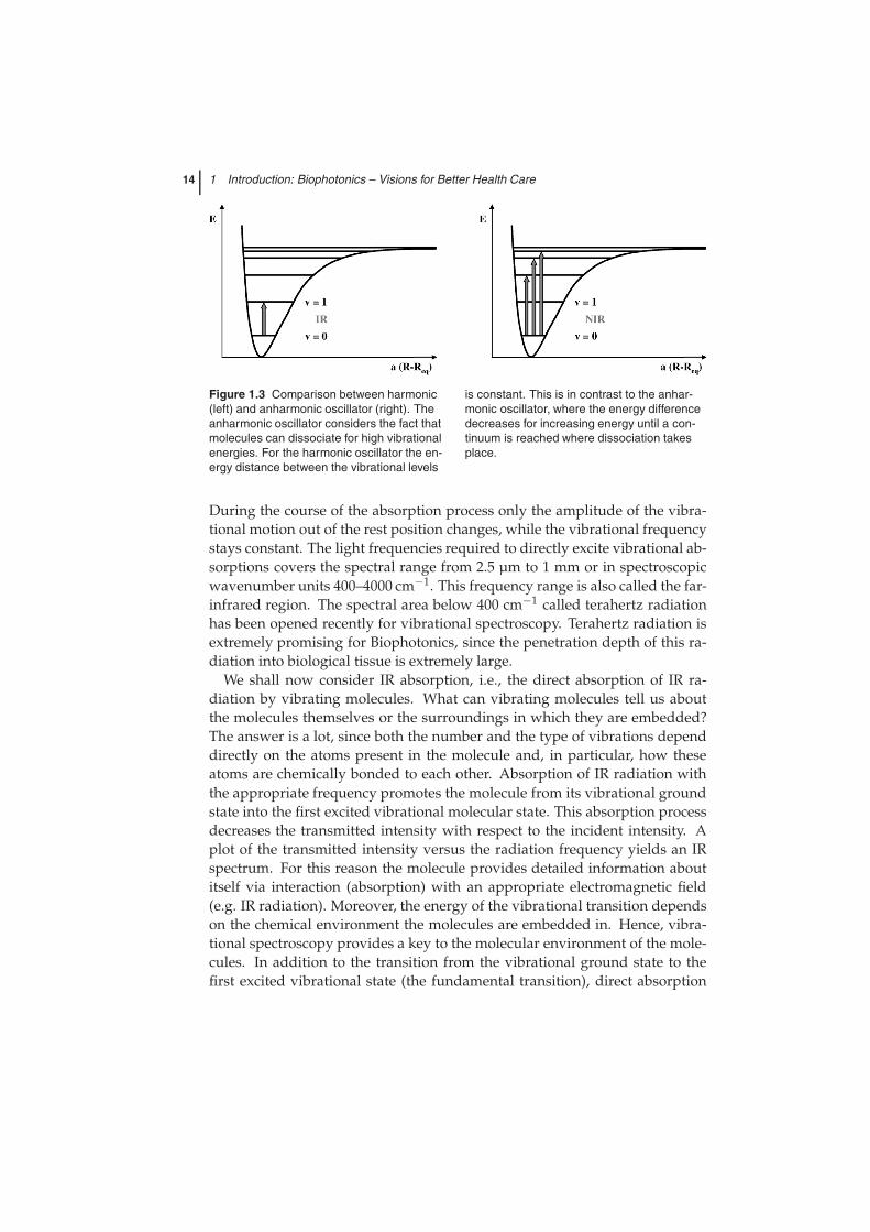
freedom, i.e., the normal modes of water. The three normal modes of waterare shown in Figure 1.1. These vibrational motions differ from each other bydifferent atomic displacements. One can differentiate between pure stretching(chemical bonds are stretched or compressed) or pure bending (bond anglesare changed) vibrations and mixed forms exhibiting both stretching and com-pression of chemical bonds and extension or reduction of the bond angles.The easiest way to describe such a vibration is by approximating or describ-ing the chemical bond as atoms held together by a spring. The vibrational fre-quency then depends on the atomic mass and the spring force constant. Theatoms move during a vibrational period towards or away from each other.This movement is repeated periodically and can be easily described by a har-monic oscillator. A harmonic oscillator is a system performing periodic vi-brations about its equilibrium position where the restoring force F is directlyproportional to the displacement x. Figure 1.2 shows such a vibrational mo-tion in a harmonic parabolic potential. The minimum of the potential cor-responds to the equilibrium distance, i.e., the nuclear distance exhibiting thelowest energy. According to quantum theory, the vibrational energy cannot becontinuously but quantized. This quantum harmonic oscillator is symbolizedby the horizontal lines inside the parabolic potential. The vibrating quantummechanical system possesses a zero point energy, i.e., molecules are alwaysvibrating and are never at rest. If the displacement from the equilibrium po-sition is minimal, which is true for small vibrational quantum numbers, theharmonic oscillator model is well suited to describe a vibrational absorptionprocess. However, for large displacements, i.e., for high vibrational states, themodel of the harmonic oscillator model is problematic: Firstly, it is impossi-ble to put an arbitrary amount of energy into the system without destroyingthe molecule. However, it is known that chemical bonds can break, i.e., dis-sociate, or molecules can fall apart if they are heated too much. To allow fordissociation to take place the harmonic oscillator model has been refined bythe anharmonic oscillator model. The anharmonic oscillator model considers
1.2 Interplay between light and matter 13
Figure 1.2 Harmonic Oscillator Model. Themolecule performs a harmonic vibration aboutits equilibrium position, i.e., the restoringforce is direct proportional to the displace-ment of the atoms from their equilibrium posi-tion (right side). The molecule is modelled asballs connected by a spring. The vibrational
frequency depends on the mass of the ballsand the spring force constant. The left sidedepicts the vibrational motion in a harmonicparabolic potential. According to quantumtheory the vibrational energy is quantizedwhich is symbolized by the horizontal lineswithin the harmonic potential.
the fact that molecules dissociate at high vibrational energies, i.e., the springbetween the atoms breaks. While for the harmonic oscillator the energy dif-ference between the quantized vibrational states is always the same, this dif-ference for the anharmonic oscillator decreases with increasing energy untila continuum is reached where all vibrational states have almost the same en-ergy. Within this continuum dissociation occurs, i.e., the atoms of the moleculecan leave the molecule’s force field (anharmonic potential see Figure 1.3).
After all this theory the question remains, “What can we learn from vibrat-ing molecules?” Most normal modes if they are not degenerated (degener-acy = same energy) exhibit different vibrational frequencies. The vibrationalfrequencies of the three normal modes of water are: ν1 = 3652 cm−1 (sym-metrical stretch motion), ν2 = 1595 cm−1 (symmetrical bending motion) andν3 = 3756 cm−1 (asymmetrical stretch vibration). Light with the appropriatefrequency can be absorbed by the ensemble of vibrating molecules, promotingthem from the vibrational ground state into the first excited vibrational state.
14 1 Introduction: Biophotonics – Visions for Better Health Care
Figure 1.3 Comparison between harmonic(left) and anharmonic oscillator (right). Theanharmonic oscillator considers the fact thatmolecules can dissociate for high vibrationalenergies. For the harmonic oscillator the en-ergy distance between the vibrational levels
is constant. This is in contrast to the anhar-monic oscillator, where the energy differencedecreases for increasing energy until a con-tinuum is reached where dissociation takesplace.
During the course of the absorption process only the amplitude of the vibra-tional motion out of the rest position changes, while the vibrational frequencystays constant. The light frequencies required to directly excite vibrational ab-sorptions covers the spectral range from 2.5 µm to 1 mm or in spectroscopicwavenumber units 400–4000 cm−1. This frequency range is also called the far-infrared region. The spectral area below 400 cm−1 called terahertz radiationhas been opened recently for vibrational spectroscopy. Terahertz radiation isextremely promising for Biophotonics, since the penetration depth of this ra-diation into biological tissue is extremely large.
We shall now consider IR absorption, i.e., the direct absorption of IR ra-diation by vibrating molecules. What can vibrating molecules tell us aboutthe molecules themselves or the surroundings in which they are embedded?The answer is a lot, since both the number and the type of vibrations dependdirectly on the atoms present in the molecule and, in particular, how theseatoms are chemically bonded to each other. Absorption of IR radiation withthe appropriate frequency promotes the molecule from its vibrational groundstate into the first excited vibrational molecular state. This absorption processdecreases the transmitted intensity with respect to the incident intensity. Aplot of the transmitted intensity versus the radiation frequency yields an IRspectrum. For this reason the molecule provides detailed information aboutitself via interaction (absorption) with an appropriate electromagnetic field(e.g. IR radiation). Moreover, the energy of the vibrational transition dependson the chemical environment the molecules are embedded in. Hence, vibra-tional spectroscopy provides a key to the molecular environment of the mole-cules. In addition to the transition from the vibrational ground state to thefirst excited vibrational state (the fundamental transition), direct absorption
1.2 Interplay between light and matter 15
processes can also promote the molecule into the second, third or even highervibrational state, although with much less probability than the fundamentaltransition. These higher transitions are called overtones. The energy requiredto excite overtones moves from the IR region into the mid- (2.5–50 µm) oreven the near-IR (800 nm to 2.5 µm) spectral region. Overtone vibrationalspectroscopy is an important well established method in quality control, butplays only a minor role in the field of health care.
Vibrational transitions can also take place via an inelastic light-scatteringprocess. We have shown already that light can polarize molecules. For thevisible wavelength region, the main contribution to the induced polarizationcomes from electrons, whose distribution relative to the atomic nuclei is dis-torted by the interacting electromagnetic field. Thus this type of light–matterinteraction induces an electric dipole moment within the molecules. The po-larizability α is a measure of how easily the electron distribution can be dis-torted within a molecule. The induced dipole, oscillating with the frequencyof the electromagnetic field, emits an electromagnetic wave in all directions. Ifthe polarizability does not change with time, the frequency of the emitted sec-ondary wave corresponds to the frequency of the oscillating induced dipole,i.e., the frequency of the external electromagnetic wave inducing the dipole(= Rayleigh scattering). However, since molecules are always vibrating, thepolarizability α is not constant over time, but changes according to the dif-ferent vibrational frequencies of the molecule’s normal modes. Therefore, theinduced dipole moment and thus the emitted secondary radiation are alsomodulated by the different vibrational frequencies. Consequently, the sec-ondary radiation emitted by the molecule is a superposition of the excitingfrequency and the various vibrational frequencies of the molecule. Dispers-ing this secondary radiation into its frequency components yields beside thestrong Rayleigh scattering also weak sidebands. The distance between theRayleigh wavelength and the wavelength of the sidebands corresponds to thevibrational frequencies of the molecule. The appearance of these sidebandsarising from an inelastic light-scattering process was first discovered in 1928by C.V. Raman. This so-called Raman Effect marks an indirect approach tothe excitation of molecular vibrations. The Raman Effect can be interpretedquantum mechanically as an inelastic collision between photons and vibratingmolecules. Photons can be scattered from molecules. This scattering processcorresponds to a molecular transition into an extremely short-lived transitionstate, the so-called virtual level (= collective quantum energy state of the entitymolecule and photon). The molecule can subsequently relax from this virtuallevel into the original molecular state or to an energetically excited molecu-lar state. If the scattering process starts from the vibrational ground state andends up in a vibrationally excited state via a transition into the virtual state itis called Stokes–Raman scattering. If the molecules are initially already in a vi-
16 1 Introduction: Biophotonics – Visions for Better Health Care
brationally excited state and are transferred by the scattering process into thevibrational ground state one refers to as anti-Stokes Raman scattering. Sinceat room temperature the vibrational ground state is significantly more pop-ulated than the vibrationally excited states, the Stokes–Raman spectrum of asample is more intense than the anti-Stokes Raman spectrum. For Rayleighscattering, the state before and after the scattering process is the same. Thesescattering processes are classified as two-photon processes since two photonsare involved.
The two vibrational spectroscopic methods Raman and IR absorption spec-troscopy are complementary methods based on two different light–matter in-teraction phenomena and thus exhibit different selection rules. Selection rulesdetermine which vibration of a molecule can be excited by what method. Inthe case of IR absorption, one photon directly promotes the molecule into ahigher vibrational state while the Raman scattering process involves two pho-tons. In order for a molecular vibration to absorb an IR photon, the dipolemoment of the molecule has to change during the course of the vibration, i.e.,only those vibrations which give rise to an oscillating dipole are IR active. Thepolarizability has to change during the vibration so that a molecular vibrationcan be promoted via an inelastic scattering process into a higher vibrationalstate.
Both Raman and IR absorption spectra can be considered as molecular fin-gerprints of the molecules existing in a biological sample. The important roleof vibrational spectroscopy, and in particular Raman spectroscopy, in Biopho-tonics can be found in Chapter 3, where representative examples of IR absorp-tion, Raman spectra and a more detailed introduction to Raman spectroscopy(theory, instrumentation, etc.) is given.
Light scattering can take place over the complete electromagnetic spec-trum, although the scattering power scales with the fourth power of the ex-citing frequency, i.e., short-wavelength radiation is scattered strongly whilelong-wavelength radiation is only scattered poorly. Direct absorption of mi-crowaves or IR radiation can excite rotations or vibrations, respectively. How-ever, the interaction of matter with visible or UV light can also lead to anabsorption of this radiation. In this case, light–matter interaction leads to elec-tronic excitation. We can differentiate between two spectral regions: the regionbetween 200 and 380 nm is called the ultraviolet (UV) region, while the areabetween 380 and 700 nm spans the visible (VIS) wavelengths. What happens ifUV-VIS radiation is absorbed by a molecular system? So far, the description ofIR absorption and Raman scattering has been limited to the electronic groundstate, which was described as a harmonic or anharmonic oscillator (parabolicpotential curves, see also Figures 1.2 and 1.3). Figure 1.4 displays the elec-tronic ground state, denoted S0, as well as the first electronically excited stateS1. In order to simplify the presentation, the harmonic or anharmonic poten-
1.2 Interplay between light and matter 17
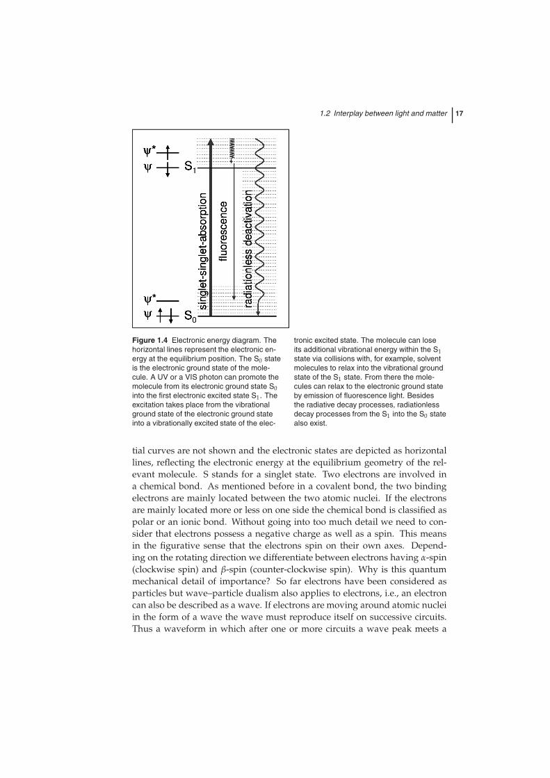
Figure 1.4 Electronic energy diagram. Thehorizontal lines represent the electronic en-ergy at the equilibrium position. The S0 stateis the electronic ground state of the mole-cule. A UV or a VIS photon can promote themolecule from its electronic ground state S0into the first electronic excited state S1. Theexcitation takes place from the vibrationalground state of the electronic ground stateinto a vibrationally excited state of the elec-
tronic excited state. The molecule can loseits additional vibrational energy within the S1state via collisions with, for example, solventmolecules to relax into the vibrational groundstate of the S1 state. From there the mole-cules can relax to the electronic ground stateby emission of fluorescence light. Besidesthe radiative decay processes, radiationlessdecay processes from the S1 into the S0 statealso exist.
tial curves are not shown and the electronic states are depicted as horizontallines, reflecting the electronic energy at the equilibrium geometry of the rel-evant molecule. S stands for a singlet state. Two electrons are involved ina chemical bond. As mentioned before in a covalent bond, the two bindingelectrons are mainly located between the two atomic nuclei. If the electronsare mainly located more or less on one side the chemical bond is classified aspolar or an ionic bond. Without going into too much detail we need to con-sider that electrons possess a negative charge as well as a spin. This meansin the figurative sense that the electrons spin on their own axes. Depend-ing on the rotating direction we differentiate between electrons having α-spin(clockwise spin) and β-spin (counter-clockwise spin). Why is this quantummechanical detail of importance? So far electrons have been considered asparticles but wave–particle dualism also applies to electrons, i.e., an electroncan also be described as a wave. If electrons are moving around atomic nucleiin the form of a wave the wave must reproduce itself on successive circuits.Thus a waveform in which after one or more circuits a wave peak meets a
18 1 Introduction: Biophotonics – Visions for Better Health Care
wave valley is not allowed since then it would interfere destructively withitself and would not survive. This simple picture reveals that only special,discrete waves are acceptable to describe the electronic motion. The spatialdistributions of these special waves (in the strict sense the square modulus oftheses discrete waves) are called orbitals. The discrete solutions lead to quan-tization, and physicists denote the possible single-valued electron waves withquantum numbers. Wolfgang Pauli found that electrons occupying a singleorbital are not allowed to be identical, i.e., the existence of two electrons ex-hibiting exactly the same quantum numbers is not allowed. Thus an orbitalcan be occupied by maximally two electrons differing in their spin state, i.e.,their intrinsic angular momentum. In the case of chemical bonds, the quan-tized electron waves occupied by the bonding electrons are called molecularorbitals. According to the Pauli principle, electrons need to be paired in achemical bond, i.e., one electron has α-spin and the other β-spin. In Figure 1.4the electron having α-spin corresponds to arrow up (↑) and the electron ex-hibiting β-spin is denoted by a down arrow (↓). Paired electrons within theS0 state are denoted by (↑↓). The absorption process promotes an electronicexcitation, i.e., an electron from the S0 state is transferred into an energeticallyhigher lying orbital, the S1 state, while the spin state is conserved. This ex-citation proceeds in analogy to the aforementioned IR absorption process viadirect absorption of an appropriate photon. Thus, electronic excitation takesplace from the vibrational ground state (v = 0) of the electronic ground stateinto a vibrational state v′ of the first excited electronic state. Which vibra-tional states within the electronic excited states are populated depends on thegeometrical rearrangement taking place upon electronic excitation. If we mea-sure, as described above for IR absorption spectroscopy, the ratio between theinitial light intensity I0 and the transmitted intensity I vs. the light wavelengthit is possible to determine the concentration of absorbing molecules within asample via the so-called Lambert–Beer law: E(ν) = ε(ν)cd. The attenuationof light due to an electronic absorption is described by I(ν) = I0 × 10−E. ε(ν)is the molar decade absorption coefficient, E corresponds to the absorption, cis the desired concentration and d represents the thickness of the sample cell.
Now the questions arise, “What happens to electronically excited mole-cules? What is the residence time of the molecules in the excited state?” Anexcited molecule tends to release its additional energy in any form to get backinto the lowest energy state, i.e., the most stable ground state. What happensin detail? If the electronic excitation generated vibrationally excited mole-cules within the excited electronic state, the molecule rapidly releases the ad-ditional vibrational energy via collisions with the surroundings (e.g. solventmolecules) to pass into the vibrational ground state of the excited electronicstate. The time-scale of this ultrafast vibrational relaxation is 10−14–10−12 s. Inpolyatomic molecules, vibrational relaxation can also take place without the
1.2 Interplay between light and matter 19
presence of solvent molecules via a redistribution of the additional vibrationalenergy from one specific mode populated upon electronic absorption to othervibrational modes.
From the vibrational ground state of the excited electronic state the mol-ecule can spontaneously relax to the electronic ground state by emission oflight. This radiational transition is called fluorescence. The time-scale for fluo-rescence to take place is 10−9–10−8 s. Since the time-scale for vibrational relax-ation is orders of magnitudes shorter than that for the emission of fluorescencelight, the emission of molecules in condensed phases or solid state alwaysstarts from the vibrational ground state of the first excited electronic state S1.However, the emission of fluorescence light following an electronic absorptionis not the most common electronic relaxation mechanism but rather an excep-tion. In nature, radiationless transitions are the dominant form of electronicrelaxation. Collisional deactivation processes lead to a decay of the excitedelectronic state S1 directly back into the S0 state. The color of plants, fruits, etc.are not due to the emission of fluorescence following the electronic absorp-tion of light but rather a consequence of light absorption and reflection (videinfra). White light results from a superposition of all wavelengths in the UV-VIS electromagnetic spectrum. A tomato appears red under irradiation withwhite light because it absorbs all colors from the white light spectrum exceptred. Thus the color red is reflected from a tomato.
Besides fluorescence and vibrational relaxation, several other electronic re-laxation mechanisms exist. However, a detailed description of these is beyondthe scope of this general introduction.
So far this short and general introduction of light–matter interactions hasmainly concentrated on single molecules. However, matter is generally notpresent in the form of single molecules but rather as molecular aggregates.The aggregates can exhibit different spatial dimensions and might range froma few associated molecules called clusters via nano- and microparticles tolarge molecular aggregates, e.g., crystals, visible to the naked eye. Nanopar-ticles are aggregates of a few hundreds of molecules or atoms forming dis-crete units with a size in the nanometer range. When light interacts withmatter whose particles are larger than the light wavelength, i.e. larger than300 nm, beside absorption and scattering new light–matter-interaction phe-nomena occur. Such new light–matter interactions are, for example, reflectionand diffraction of light. These mechanisms play an important role in the in-terplay of light with biological matter, e.g., united cell structures or tissues.For these phenomena the aforementioned molecular polarization is of par-ticular importance. In the case of extended matter, with spatial dimensionsgreater than the light wavelength, the polarizability is described as the sumof the molecular properties, i.e., as an averaged value. In total analogy to themolecular polarizability, the bulk polarizability describes the dipole moment
20 1 Introduction: Biophotonics – Visions for Better Health Care
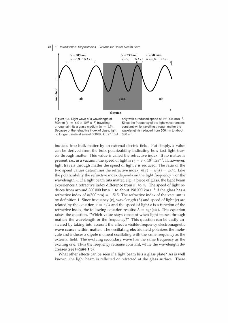
Figure 1.5 Light wave of a wavelength of500 nm (ν = 6.0 × 10¹⁴ s−1) travellingthrough air hits a glass medium (n = 1.5).Because of the refractive index of glass, lightno longer travels at almost 300 000 km s−1 but
only with a reduced speed of 198 000 km s−1.Since the frequency of the light wave remainsconstant while travelling through matter thewavelength is reduced from 500 nm to about330 nm.
induced into bulk matter by an external electric field. Put simply, a valuecan be derived from the bulk polarizability indicating how fast light trav-els through matter. This value is called the refractive index. If no matter ispresent, i.e., in a vacuum, the speed of light is c0 = 3× 10⁸ m s−1. If, however,light travels through matter the speed of light c is reduced. The ratio of thetwo speed values determines the refractive index: n(ν) = n(λ) = c0/c. Likethe polarizability the refractive index depends on the light frequency ν or thewavelength λ. If a light beam hits matter, e.g., a piece of glass, the light beamexperiences a refractive index difference from n1 to n2. The speed of light re-duces from around 300 000 km s−1 to about 198 000 km s−1 if the glass has arefractive index of n(500 nm) = 1.515. The refractive index of the vacuum isby definition 1. Since frequency (ν), wavelength (λ) and speed of light (c) arerelated by the equation ν = c/λ and the speed of light c is a function of therefractive index, the following equation results: λ = c0/(νn). This equationraises the question, “Which value stays constant when light passes throughmatter: the wavelength or the frequency?” This question can be easily an-swered by taking into account the effect a visible-frequency electromagneticwave causes within matter. The oscillating electric field polarizes the mole-cule and induces a dipole moment oscillating with the same frequency as theexternal field. The evolving secondary wave has the same frequency as theexciting one. Thus the frequency remains constant, while the wavelength de-creases (see Figure 1.5).
What other effects can be seen if a light beam hits a glass plate? As is wellknown, the light beam is reflected or refracted at the glass surface. These
1.2 Interplay between light and matter 21
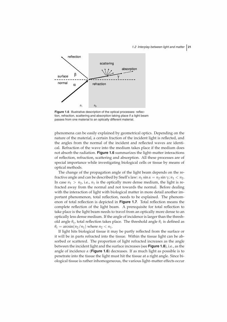
Figure 1.6 Illustrative description of the optical processes: reflec-tion, refraction, scattering and absorption taking place if a light beampasses from one material to an optically different material.
phenomena can be easily explained by geometrical optics. Depending on thenature of the material, a certain fraction of the incident light is reflected, andthe angles from the normal of the incident and reflected waves are identi-cal. Refraction of the wave into the medium takes place if the medium doesnot absorb the radiation. Figure 1.6 summarizes the light–matter interactionsof reflection, refraction, scattering and absorption. All these processes are ofspecial importance while investigating biological cells or tissue by means ofoptical methods.
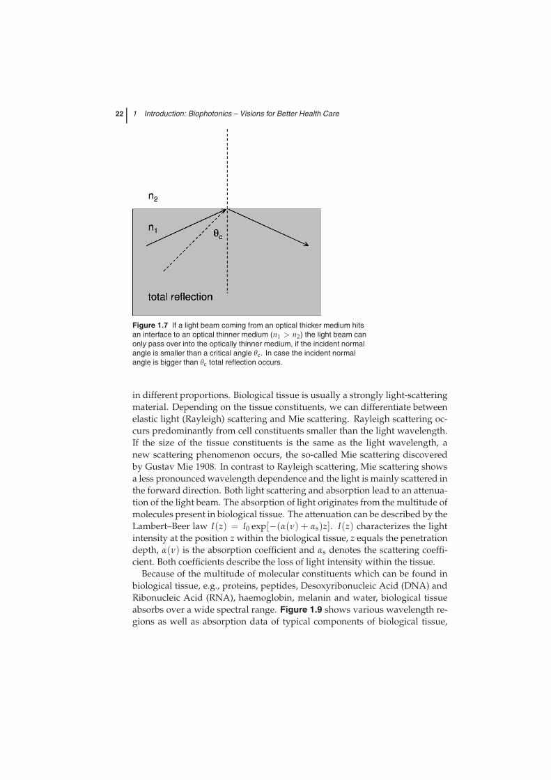
The change of the propagation angle of the light beam depends on the re-fractive angle and can be described by Snell’s law: n1 sin α = n2 sin γ; n1 < n2.In case n1 > n2, i.e., n1 is the optically more dense medium, the light is re-fracted away from the normal and not towards the normal. Before dealingwith the interaction of light with biological matter in more detail another im-portant phenomenon, total reflection, needs to be explained. The phenom-enon of total reflection is depicted in Figure 1.7. Total reflection means thecomplete reflection of the light beam. A prerequisite for total reflection totake place is the light beam needs to travel from an optically more dense to anoptically less dense medium. If the angle of incidence is larger than the thresh-old angle θc, total reflection takes place. The threshold angle θc is defined asθc = arcsin(n2/n1) where n2 < n1.
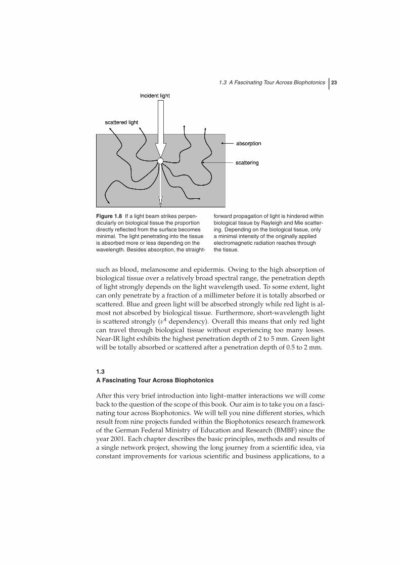
If light hits biological tissue it may be partly reflected from the surface orit will be in parts refracted into the tissue. Within the tissue light can be ab-sorbed or scattered. The proportion of light refracted increases as the anglebetween the incident light and the surface increases (see Figure 1.8), i.e., as theangle of incidence α (Figure 1.6) decreases. If as much light as possible is topenetrate into the tissue the light must hit the tissue at a right angle. Since bi-ological tissue is rather inhomogeneous, the various light–matter effects occur
22 1 Introduction: Biophotonics – Visions for Better Health Care
Figure 1.7 If a light beam coming from an optical thicker medium hitsan interface to an optical thinner medium (n1 > n2) the light beam canonly pass over into the optically thinner medium, if the incident normalangle is smaller than a critical angle θc. In case the incident normalangle is bigger than θc total reflection occurs.
in different proportions. Biological tissue is usually a strongly light-scatteringmaterial. Depending on the tissue constituents, we can differentiate betweenelastic light (Rayleigh) scattering and Mie scattering. Rayleigh scattering oc-curs predominantly from cell constituents smaller than the light wavelength.If the size of the tissue constituents is the same as the light wavelength, anew scattering phenomenon occurs, the so-called Mie scattering discoveredby Gustav Mie 1908. In contrast to Rayleigh scattering, Mie scattering showsa less pronounced wavelength dependence and the light is mainly scattered inthe forward direction. Both light scattering and absorption lead to an attenua-tion of the light beam. The absorption of light originates from the multitude ofmolecules present in biological tissue. The attenuation can be described by theLambert–Beer law I(z) = I0 exp[−(α(ν) + αs)z]. I(z) characterizes the lightintensity at the position z within the biological tissue, z equals the penetrationdepth, α(ν) is the absorption coefficient and αs denotes the scattering coeffi-cient. Both coefficients describe the loss of light intensity within the tissue.
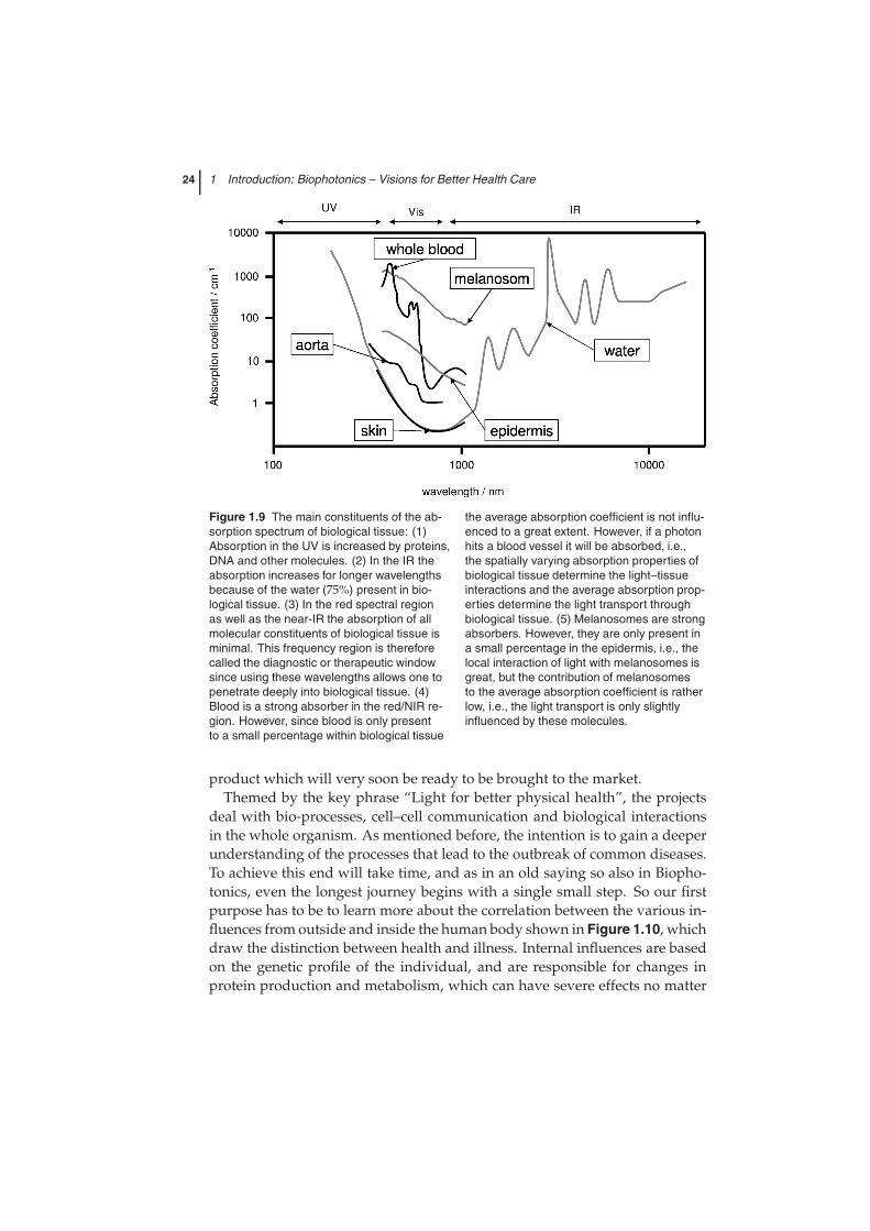
Because of the multitude of molecular constituents which can be found inbiological tissue, e.g., proteins, peptides, Desoxyribonucleic Acid (DNA) andRibonucleic Acid (RNA), haemoglobin, melanin and water, biological tissueabsorbs over a wide spectral range. Figure 1.9 shows various wavelength re-gions as well as absorption data of typical components of biological tissue,
1.3 A Fascinating Tour Across Biophotonics 23
Figure 1.8 If a light beam strikes perpen-dicularly on biological tissue the proportiondirectly reflected from the surface becomesminimal. The light penetrating into the tissueis absorbed more or less depending on thewavelength. Besides absorption, the straight-
forward propagation of light is hindered withinbiological tissue by Rayleigh and Mie scatter-ing. Depending on the biological tissue, onlya minimal intensity of the originally appliedelectromagnetic radiation reaches throughthe tissue.
such as blood, melanosome and epidermis. Owing to the high absorption ofbiological tissue over a relatively broad spectral range, the penetration depthof light strongly depends on the light wavelength used. To some extent, lightcan only penetrate by a fraction of a millimeter before it is totally absorbed orscattered. Blue and green light will be absorbed strongly while red light is al-most not absorbed by biological tissue. Furthermore, short-wavelength lightis scattered strongly (ν4 dependency). Overall this means that only red lightcan travel through biological tissue without experiencing too many losses.Near-IR light exhibits the highest penetration depth of 2 to 5 mm. Green lightwill be totally absorbed or scattered after a penetration depth of 0.5 to 2 mm.
1.3A Fascinating Tour Across Biophotonics
After this very brief introduction into light–matter interactions we will comeback to the question of the scope of this book. Our aim is to take you on a fasci-nating tour across Biophotonics. We will tell you nine different stories, whichresult from nine projects funded within the Biophotonics research frameworkof the German Federal Ministry of Education and Research (BMBF) since theyear 2001. Each chapter describes the basic principles, methods and results ofa single network project, showing the long journey from a scientific idea, viaconstant improvements for various scientific and business applications, to a
24 1 Introduction: Biophotonics – Visions for Better Health Care
Figure 1.9 The main constituents of the ab-sorption spectrum of biological tissue: (1)Absorption in the UV is increased by proteins,DNA and other molecules. (2) In the IR theabsorption increases for longer wavelengthsbecause of the water (75%) present in bio-logical tissue. (3) In the red spectral regionas well as the near-IR the absorption of allmolecular constituents of biological tissue isminimal. This frequency region is thereforecalled the diagnostic or therapeutic windowsince using these wavelengths allows one topenetrate deeply into biological tissue. (4)Blood is a strong absorber in the red/NIR re-gion. However, since blood is only presentto a small percentage within biological tissue
the average absorption coefficient is not influ-enced to a great extent. However, if a photonhits a blood vessel it will be absorbed, i.e.,the spatially varying absorption properties ofbiological tissue determine the light–tissueinteractions and the average absorption prop-erties determine the light transport throughbiological tissue. (5) Melanosomes are strongabsorbers. However, they are only present ina small percentage in the epidermis, i.e., thelocal interaction of light with melanosomes isgreat, but the contribution of melanosomesto the average absorption coefficient is ratherlow, i.e., the light transport is only slightlyinfluenced by these molecules.
product which will very soon be ready to be brought to the market.Themed by the key phrase “Light for better physical health”, the projects
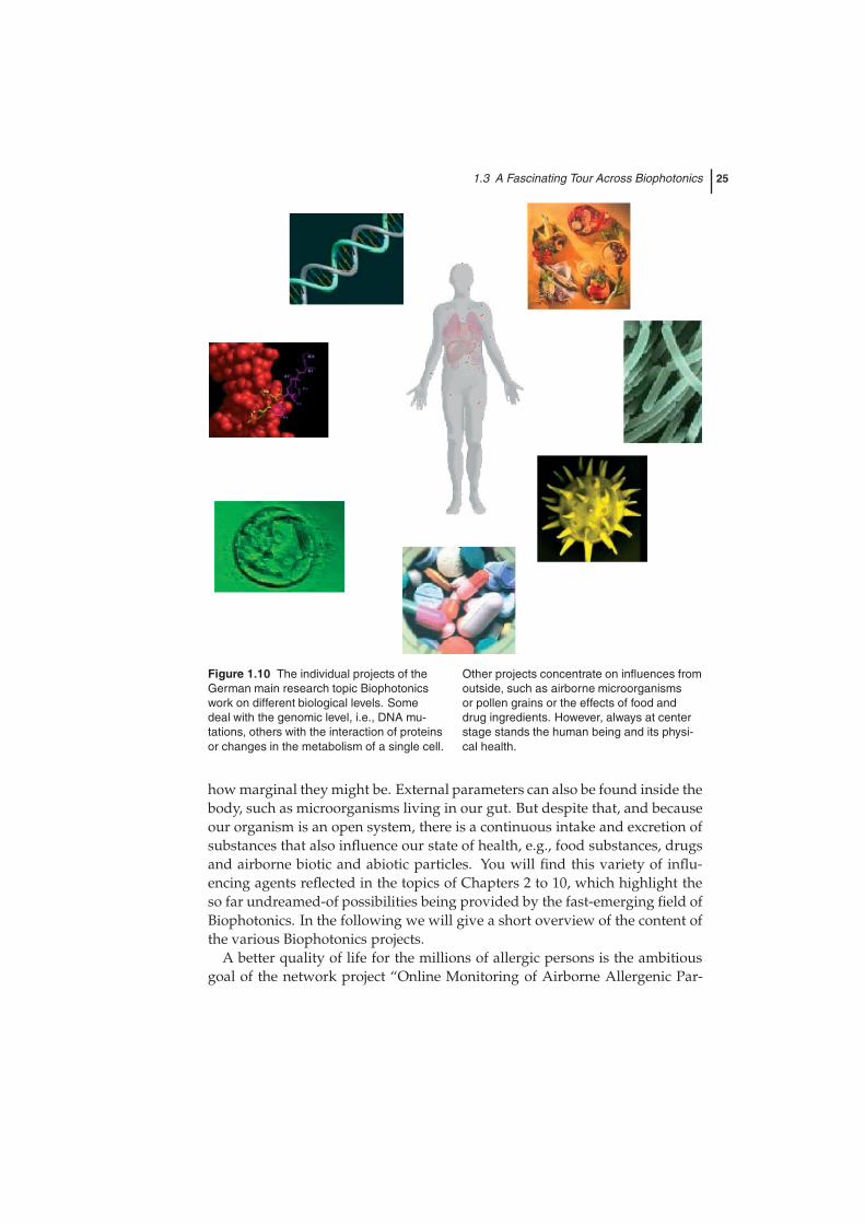
deal with bio-processes, cell–cell communication and biological interactionsin the whole organism. As mentioned before, the intention is to gain a deeperunderstanding of the processes that lead to the outbreak of common diseases.To achieve this end will take time, and as in an old saying so also in Biopho-tonics, even the longest journey begins with a single small step. So our firstpurpose has to be to learn more about the correlation between the various in-fluences from outside and inside the human body shown in Figure 1.10, whichdraw the distinction between health and illness. Internal influences are basedon the genetic profile of the individual, and are responsible for changes inprotein production and metabolism, which can have severe effects no matter
1.3 A Fascinating Tour Across Biophotonics 25
Figure 1.10 The individual projects of theGerman main research topic Biophotonicswork on different biological levels. Somedeal with the genomic level, i.e., DNA mu-tations, others with the interaction of proteinsor changes in the metabolism of a single cell.
Other projects concentrate on influences fromoutside, such as airborne microorganismsor pollen grains or the effects of food anddrug ingredients. However, always at centerstage stands the human being and its physi-cal health.
how marginal they might be. External parameters can also be found inside thebody, such as microorganisms living in our gut. But despite that, and becauseour organism is an open system, there is a continuous intake and excretion ofsubstances that also influence our state of health, e.g., food substances, drugsand airborne biotic and abiotic particles. You will find this variety of influ-encing agents reflected in the topics of Chapters 2 to 10, which highlight theso far undreamed-of possibilities being provided by the fast-emerging field ofBiophotonics. In the following we will give a short overview of the content ofthe various Biophotonics projects.
A better quality of life for the millions of allergic persons is the ambitiousgoal of the network project “Online Monitoring of Airborne Allergenic Par-

26 1 Introduction: Biophotonics – Visions for Better Health Care
ticles” (OMNIBUSS). To allow people suffering from hay fever or asthma anearly normal way of life and to avoid unnecessary intake of pharmaceuticals,a continuously and routinely updated knowledge of pollen concentration inthe air is essential. Until now, pollen forecasting has employed manual mi-croscopy techniques, which are not only time-consuming and labor-intensivebut also provide results of undefined and unsatisfactory quality. As describedin Chapter 2, p. 31ff., OMNIBUSS has developed a new microscope-basedfully automated monitoring method, which is characterized by high tempo-ral resolution, excellent reproducibility, detection limit, recall and precision,and which therefore meets the strong public demand for absolutely reliableof pollen concentration data. The device the project has led to combine con-tinuous sampling and automatic preparation of aerosol, automated particleimaging and automated identification of pollen grains based on mathematicalfingerprints.
Just as in allergy prevention, the presence of airborne biotic particles playsa very important role in connection with clean-room processes. In industrialfood or pharmaceutical production, such bioaerosol may lead to fatal con-sequences. To drastically reduce the time needed for quality assurance oflife science products, traces of contamination need to be tracked down reli-able and rapidly. Chapter 3, p. 89ff., “Online Monitoring and Identificationof Bioaerosols”, shows how the new approach of the research network OMIBcan make a decisive contribution to the monitoring process for a rapid de-tection of aerosol and an identification of airborne microorganisms withoutloss of time. For standard microbiological tests, the microorganisms are col-lected on a growth medium, bred and eventually counted. Under certain con-ditions even the combination of several microbiological tests leads only to anambiguous identification. The authors of Chapter 3 describe how they de-veloped totally new equipment, which combines differentiation of biologicalfrom non-biological particles by means of fluorescence detection with an iden-tification step by vibrational spectroscopy, in particular Raman spectroscopy.This method offers an enormous time gain compared to the conventionalmethods applied until now.
Infectious agents are gaining ground again – and they can not only causeindustry a loss of time and money but each one of us a loss of life. Germssuch as the tuberculosis-causing Mycobacteria are horrifying the world, notonly by their rapid spread around the globe but also by their ability to copewith antimicrobial drugs. One new powerful weapon is presented in Chap-ter 4 called “Novel Singly Labeled Probes for Identification of Microorgan-isms, Detection of Antibiotic-resistant Genes and Mutations, and Tumor Di-agnosis”. “Smart Probes” (Chapter 4, p. 167ff.) are fluorescently labelledDNA-hairpin structures, which have the potential to open a new avenue inmolecular diagnostics by their ability to discriminate between wild type and
1.3 A Fascinating Tour Across Biophotonics 27
resistant bacteria. Besides a detailed description of Smart Probes applicationsin the detection of antibiotic-resistant genes and mutations as well as in tumordiagnosis, the chapter gives a competent general survey of single-moleculefluorescence spectroscopy and 3D fluorescence nanoscopy.
The abbreviation PLOMS (Chapter 5, p. 231ff.) conceals an innovative wayto detect cancer of the colon in its very early stages, thus dramatically increas-ing healing rates simultaneously cutting the expenses in the health system.The authors of Chapter 5 captioned their contribution “Early Diagnosis ofCancer”. They give a very detailed introduction to the epidemiological andbiological background of cancer, the mechanisms of carcinogenesis and theimpact of early cancer diagnosis before outlining their new approach for veryearly detection of the first stages of tumor growth in the colon. This newapproach is based on the fact that the structure of the glycocalyx of normalmucosa cells changes as they degenerate into cancer cells. Like an old blan-ket becoming threadbare, the glycocalyx forms small holes, which allow thescientists of the network project PLOMS to discriminate between healthy anddegenerated cells in the colon via sophisticated labelling strategies.
Chapter 6, p. 301ff., entitled “New Ways for Marker-free Live Cell and Tu-mor Analysis” also deals with early tumor diagnosis. MIKROSO is a networkproject developing digital holographic microscopy as a very new approachfor label-free quantitative imaging of living cells and therefore as a usefultool for seeing the signs that reveal even marginal pathological changes incells and tissue, and for watching very closely the behavior of healthy anddiseased cells. The authors point out the many advantages of digital holo-graphic microscopy in comparison with standard methods of cell microscopy,and describe the various application possibilities of combinations of digitalholographic microscopy with other techniques, such as phase-contrast andfluorescence imaging or laser micromanipulation. In addition, the article pro-vides an introduction to optical coherence tomography as well as to minimallyinvasive holographic endoscopy.
The aim of “Regenerative Surgery” is to heal diseased tissue by full or par-tial reconstruction and to support the regeneration of organs if not actuallyto substitute them. Chapter 7, p. 361ff., gives an account of the medical andbiological background not only of regenerative surgery but also of tissue en-gineering, with a strong emphasis on cell and tissue culture technologies. Thenetwork project MeMo designs novel techniques to improve such cell andtissues cultures by laser optical on-line monitoring. Innovative biophotonictechnologies have been developed for a more efficient evaluation of tissues.Taking as an example the replacement of human cartilage tissue and chon-drocytes, the authors outline the advantages of recent methods like three-dimensional laser-scanning microscopy, fluorescence lifetime measurementsand parallelized two-photon measurement systems for rapid high-resolutiontissue imaging.

28 1 Introduction: Biophotonics – Visions for Better Health Care
Chapter 8, p. 405ff., brings us from the surgery back to the laboratory, andin particular to the “lab on a chip”. Microarrays have been one of the en-abling technologies of the 1990s, and have greatly increased the possibilitiesnot only for basic research in molecular biology but also for the identifica-tion and validation of drug targets. But as a technique which can perform“thousands of reactions on a small chip” as indicated by the chapter title, mi-croarrays demand efficient, reliable and comprehensive analysis methods. Inthis regard, optical systems provide a wide choice of techniques. The scien-tists of the MOBA network project concentrate on terahertz spectroscopy toobtain very rapid high-quality results, previously unknown. The binding ofDNA and other biomolecules can be analyzed to learn more about the geneticprofile of the scanned samples. As well as the specific adjustment of drugsfor individual gene profiles, further applications lie in the areas of bioweaponanalysis and telemedicine. An overview of fluorescence and label-free tech-niques completes this chapter.
A deeper understanding of the processes of life from the molecular structureto the whole organism demands methods that provide very high resolution intime and space. Therefore, analytical tools are required which cope with theneed for minimally invasive measurement techniques featuring not only highprecision and selectivity but also the ability to process a large number of sam-ples at the same time. Chapter 9, p. 477ff., called “Hybrid Optodes” describesthe work of the research network HYBOP aiming for the development of anovel fluorescence-based hybrid technology which will be employed in spa-tiotemporal high-resolution bioprocess analysis. This new technology is basedon indicator-specific polymer surface coatings which supply two kinds of in-formation at the same time by means of optoelectronic measurements. Thus,in each case two parameters, e.g., temperature and oxygen or carbon dioxideand pH, can be measured with high precision simultaneously. The chaptergives an overview of the principles of hybrid optodes and their applicationsand perspectives in biotechnology.
With the vision of an early detection of diseases and tailor-made therapiesthe ODMS network project is consistent with the aims of the main researchtopic “Biophotonics” in general. As described in Chapter 10, p. 519ff., entitled“Digital Microscopy”, ODMS has developed an “Ocularless Digital Micro-scope System” for the on-line in vivo measurement of biological or biomedicalparameters. There are three main areas of application to which the device willbe adapted. One is the target assay development on the cell-culture level, pro-viding a comprehensive visualization of the pharmaceutical effects of a newdrug. Telepathology, that is the transfer of histological medical findings in theform of digitalized data (“virtual slides”), is another application for ODMS.This technology reduces the time-consuming dispatch of samples to differentconsultants. Thirdly, ODMS will aid cell and developmental biological stud-
1.4 Links and Literature about Biophotonics 29
ies. The aims are in particular to minimize stress for the biological probe andto use light to its full capacity.
We hope that with these short summaries we have aroused your curiosityso that you continue to read. But first we should like to thank the Ministryof Education and Research and the Association of German Engineers (VDI)for their financial support of the work of all the network projects and theirexcellent cooperation over the years. In addition, the editors would like tothank all the people who have contributed to this book. Above all, we shouldlike to thank Andreas Thoss and the team at Wiley for providing us with theidea for this project. We appreciate the confidence and patience they haveshown us very much indeed. To cope with the task of editing a book likethis cannot be done without many helping hands. So our gratitude goes tothe authors of the single chapters of this book for all their efforts to make thisvolume a valuable one for scientists all over the world. A truly special wordof thanks goes to our colleagues at the Institute of Physical Chemistry in Jena,who provided us with invaluable help in the matter of technical support andwere able to restore us to a good mood at any time. We must mention byname Kathrin Strehle, Ute Uhlemann, Dana Cialla and Reinhold Gade, butour thanks go also to all the other members of our working group. On a verypersonal note we should like to thank our families for being so understandingduring all the days and nights we were constrained by the work on this book.
1.4Links and Literature about Biophotonics
The following list is far from complete and is only intended to provide theinterested reader with further insight into the rapidly growing subject of bio-photonics:
Journal:
• “Biophotonics International”, Laurin Publishing Company(www.photonics.com)
Selected “Special Issues”:
• Special Issue on Biophotonics, Journal of Physics D: Applied Physics,Volume 36, Number 14, 21 July 2003
• Special Issue on Biomedical Optics, Journal of Physics D: AppliedPhysics, Volume 38, Number 15, 7 August 2005
30 1 Introduction: Biophotonics – Visions for Better Health Care
• “Biophotonics Micro- and Nano-Imaging”, Progress in Biomedical Op-tics and Imaging, Vol. 5, No. 33, in: Proceedings of SPIE, Volume 5462,2004.
Books:
• Faupel, M., Brandenburg, A., Smigielski, P. and Fontaine, J. (Eds): Bio-photonics for Life Sciences and Medicine, FontisMedia, Lausanne/Formatis,Basel, in press.
• Marriott, Gerard (Ed): Biophotonics, Academic Press, San Diego, 2003.
• Prasad, Paras N.: Introduction to biophotonics, Wiley-Interscience,Hoboken, New Jersey, 2003.
• Shen, X. and van Wijk, R. (Eds): Biophotonics: Optical Science and En-gineering for the 21st Century, Springer, Berlin, 2006.
• Wilson, B.C., Tuchin, V.V. and Tanev, S. (Eds): Advances in Biophotonics,NATO Science Series: Life and Behavioural Sciences, Volume 369, IOSPress, Amsterdam, 2005.
Selected Initiatives in Germany:
• Main research topic “Biophotonics” funded by the German Federal Min-istry of Education and Research: http://www.biophotonik.org
• OptecNet: Network consisting of several local initiatives dealing withphotonics and biophotonics, respectively: http://www.optecnet.de
• Another local network, the “Biotech/Life Science Portal Baden Würt-temberg” offers a number of articles in the field of biophotonics:http://www.bio-pro.de