1 mbbs(hk), mrcshub.hku.hk/bitstream/10722/220729/1/content.pdfmedical records of all paediatric...
TRANSCRIPT
Title Page
Management of tracheobronchial obstruction in infants using metallic stents: Long-term outcome
Ling LEUNG1 MBBS(HK), MRCS
Patrick Ho Yu CHUNG1 FRCSEd(Paed), FHKAM(Surg)
Kenneth Kak Yuen WONG1* PhD, FRCSEd(Paed), FHKAM(Surg)
Paul Kwong Hang TAM1 ChM, FRCS, FRCPCH
1 Division of Paediatric Surgery, Department of Surgery, The University of Hong Kong,
Queen Mary Hospital, Hong Kong
*Corresponding author: Dr. Kenneth Kak Yuen WONG
Division of Paediatric Surgery
Department of Surgery
The University of Hong Kong
Queen Mary Hospital
Pokfulam Road
Hong Kong
Tel: (852) 2855 4850
Fax: (852) 2817 3155
Email: [email protected]
No work resembling the enclosed article has been published or is being submitted for
publication elsewhere. We certify that we have each made a substantial contribution so as
to qualify for authorship and that we have approved the contents.
Conflicts of Interest and Source of Funding
No conflicts of interests or source of funding to be disclosed.
Word count of the manuscript body (excluding abstract, keywords, references and figure
legends): 2453
Number of figures: 2
Number of tables: 2

1
Management of tracheobronchial obstruction in infants using metallic stents: Long-
term outcome
Abstract
Introduction:
Tracheobronchial obstruction, although uncommon in the paediatric age group, remains a
challenging problem. We review the long term outcome of endoscopic metallic stenting
in infants with tracheobronchial obstruction.
Materials and methods:
Medical records of all paediatric surgical patients who underwent tracheobronchial
metallic stenting in our centre were reviewed retrospectively from 1996 to 2014. Patients’
demographic data, including etiology, associated anomalies and nature of obstruction
were reviewed. Outcome measures include complications such as re-stenosis, granulation
tissue, stent migration, fractured stent, maximal tracheal diameter achieved, weaning of
ventilator and growth at interval follow-up.
Results:
Twelve balloon-expandable metallic stents were placed in the trachea (n=10) and/or
bronchi (n=2) of 5 patients with a median age of 13 months (range 5 - 30 months).
Etiology of the airway obstruction included congenital tracheal stenosis (n=4), giant
cervical and superior mediastinal lymphatic malformation with tracheobronchomalacia
(n=1). Seven complications were reported (3 patients developed granulation tissue, 2
patients had re-stenosis, 1 stent migrated, 1 stent fractured). All patients survived and
were in good condition with a median follow-up of 16 years (range 11 – 18 years). Three
2
patients weaned off ventilator and oxygen.
Conclusions:
Endoscopic stenting with metallic stent has satisfactory long term outcome in treating
infants with tracheobronchial obstruction.
Keywords:
tracheal stenosis; airway obstruction; metallic stent; self-expandable stent;
tracheobronchomalacia
Article
Introduction
Airway obstruction in paediatric patients remains a challenging problem among clinicians.
Obstruction can be due to various extraluminal, intramural and intraluminal diseases.
Congenital tracheomalacia and stenosis are the most common causes. A number of
surgical treatment options have been advocated. Tracheoplasty can be done either by
tracheal resection and anastomosis or by repair using costal cartilage or pericardial patch.
[1,2] However, these operations may be difficult for small children with respiratory
failure and have a high risk of operative mortality. Aortopexy has been shown to be
effective in cases of short-segment tracheomalacia. [3-5] Endoscopic stenting has been
shown to reduce the need for high risk surgical procedure and prolonged ventilator
dependence in children with diffuse tracheomalacia. In difficult population, endoscopic
stenting has been recommended as an alternative therapeutic option. [6-17] Currently,
there is only scanty information regarding the long term outcome of this procedure. The

3
purpose of this study is to review our experience in treating congenital tracheobronchial
obstruction with endoscopic stent placement.
Materials and methods
Medical records of all paediatric surgical patients who underwent tracheobronchial
stenting in our centre were reviewed retrospectively from 1996 to 2014. Patients’
demographic data, including etiology, associated anomalies and nature of obstruction
were reviewed. Outcome measures include complications such as re-stenosis, granulation
tissue, stent migration, fractured stent, maximal tracheal diameter achieved, weaning of
ventilator and growth at interval follow-up.
Endoscopic stenting was offered to patients who had significant recurrent stenosis
following repeated dilatations. As the number of patients with this condition was very
small, our ENT colleagues did not have the expertise in performing tracheoplasties. As a
result, we were only able to offer endoscopic stenting to patients. In addition it had the
advantage of being minimally invasive. Selection of the stent type depended on the
location of obstruction, the patient's age and body size as well as the availability of the
stent at the time of procedure. Prior to stent placement, the type, location, severity, and
length of the obstruction were assessed by bronchoscopy and computer tomography. In
all of our patients, metallic balloon-expandable Palmaz stents (Johnson & Johnson
Interventional Systems, Warren, NJ) were placed using a Storz rigid paediatric
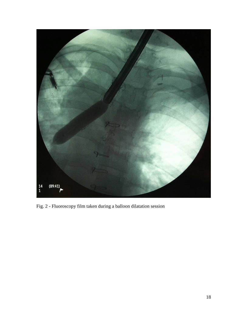
bronchoscope (Karl Storz, Germany) under general anaesthesia. The site for stenting was
determined by simultaneous use of endoscopy and fluoroscopy. The stent, together with a
angioplasty balloon catheter, were passed through the bronchoscope. Under fluoroscopic
4
control, the balloon was inflated by normal saline and the position of expanded stent was
confirmed. (Figures 1&2). The tracheal mucosa was visualized by rigid bronchoscopy.
The balloon was deflated and the catheter was removed.
Results
Five patients presented with respiratory distress in the neonatal or early infantile period.
Patient demographics, the anatomy of tracheobronchial obstruction, number and location
of the stents and complications were summarized in Table 1. Etiology of the airway
obstruction included congenital tracheal stenosis (n=4), giant cervical and superior
mediastinal lymphatic malformation with tracheobronchomalacia (n=1). One patient
(patient 4) had concomitant multiple congenital anomalies including cerebral atrophy and
severe hydrocephalus. The diameter of the obstructed segments ranged from 1 mm to 2
mm and the length from 14mm to the entire trachea.
The median age at first stenting was 13 months (range 5 - 30 months). Four patients had
undergone rigid bronchoscopies and balloon dilatations to expand the airway before stent
placements, ranging from 7 to 19 sessions. One patient (patient 5) with extensive
tracheobronchial compression due to giant cervical and superior mediastinal lymphatic
malformation was initially managed by open excision of the lesion, which subsequently
recurred. A total of twelve Palmaz stents were placed in the trachea (n=10) and/or bronchi
(n=2) in 5 patients. Four patients required placements of more than one stent, these were
placed during repeated bronchoscopic procedures. All stents, except one fractured stent in
patient 5, remained in place. There was no peri-operative mortality.

5
Long term outcome
A summary of long term outcome was presented in table 2. All patients needed regular
bronchoscopic assessments and balloon dilatations to maintain luminal patency. A median
of 25 bronchoscopies (range 21 – 55) and 14 balloon dilatations (range 9 – 40) had been
performed. Gradual increase in tracheal diameter was noted. The median maximal
tracheal diameter achieved was 13 mm (range 8 – 17 mm).
All patients survived and were in good condition with a median follow-up of 16 years
(range 11 – 18 years) from their initial presentation. Three patients weaned off ventilator
and oxygen. Two patients require home oxygen support in the form of positive airway
pressure. All except one patient with profound mental retardation had satisfactory
exercise tolerance for normal daily activity. Three patients had normal body weight and
height, whereas two patients remained below the 3rd percentile.
Patient 1
No complications occurred throughout the eighteen years post-stenting. He remained
ventilator free and enjoyed normal exercise capacity and growth.
Patient 2
In patient 2, two stents were inserted to distal trachea. Stent migration into the left lower
lobe bronchus was noted six years post-stenting, which was managed conservatively. She
was ventilator free and had an exercise tolerance of three flights of stairs.
Patient 3
6
In patient 3, re-stenosis proximal to the stent occurred 2 months after placement of stent
and intensive care was required. It was managed by bronchoscopic dilatations and
dexamethasone injection. On subsequent bronchoscopic reassessments, airway patency
was maintained except granulation tissue was seen requiring removal by endoscopic
cauterization twice. He had mild mental retardation, otherwise he enjoyed normal
exercise capacity.
Patient 4
He had multiple congenital anomalies with profound mental retardation, hydrocephalus
and obstructive sleep apnea. He developed a total of 12 episodes of pneumonia after
bronchoscopies. This could result from his co-existing gastroesophageal reflux disease
and he eventually underwent laparoscopic fundoplication and gastrostomy. Reassessment
bronchoscopy at 4 months post-stenting showed granulation tissue at sub-glottic area and
was removed. He was wheelchair bound and required nocturnal continuous positive
airway pressure ventilation at home.
Patient 5
In patient 5, Palmaz stent was inserted at one and a half year of age. Three months later, a
second stent was inserted in view of a malacic segment of the trachea above the first stent.
However, it was complicated by stent fracture and was managed by balloon dilatation and
adrenaline injection. Reassessment bronchoscopy showed obstructing granulation tissue
over both bronchi requiring bilateral bronchial stents insertion. During her 35th
bronchoscopy, granulation tissue was found to cause a ball-valve effect, and there were

7
migration and fracture of the tracheal stent. Granulation tissue and the stent fragment
were removed by suction and forceps until patency of the tracheal lumen was confirmed.
Despite her difficult clinical condition and stormy hospitalizations, she had normal
growth and intelligence. She required bilevel positive airway pressure ventilation at home,
nonetheless, her normal daily activity was not affected.
Discussion
The first report regarding the use of tracheobronchial stents in children dated back to the
late 1980s. [6,7,18] Endoscopic stents had been used in various clinical conditions
including airway malacia or stenoses, either due to external compression or structurally
abnormal airway walls. Its use in post-operative stenosis after lung transplantation had
also been reported. [13] Such procedures were usually performed in combination with
surgery for the treatment of severe bronchomalacia or for the prevention of post-
tracheoplasty re-stenosis.
In 1995, Zinman [19] showed that tracheal stenting improved ventilatory mechanics in
infants with tracheobronchomalacia. The use of vascular mesh metal prosthesis (Palmaz)
in 16 children was first introduced by Filler et al. [9] The stents were reported to be well
tolerated for up to 6 years.
Different classes of stents have been developed for airway stenosis. They can be broadly
classified into plastic and metallic. For plastic stents, standard silicone stent is easily
removable while Dumon stent is flexible. [20] Silicone stent has major limitations of
being easily collapsible, prone to migration and interruption of mucociliary clearance. Its
use in infants is not well tolerated because of frequent obstruction by secretions. The use
8
of Polyflex stent, a self expanding silicone device, had been reported. However,
migration and mucus impaction occurred in all 12 patients with stenoses. [21]
Concerning metallic stents, the main division is balloon-expandable and self expanding
types. These were originally intended for angiographic applications in adults. Palmaz
stent is made of stainless steel and is balloon expandable. The risks of obstruction by
mucus and migration were less than that of silicone stents. Palmaz stents had been shown
in an experimental trial to provoke an inflammatory reaction and epithelialization. [22]
When compared to self expanding stents, Palmaz stent had the advantage of expansion
under direct observation. Balloon dilatation was performed first to a satisfactory size,
followed by insertion of the Palmaz stent to maintain luminal patency. A major
disadvantage of Palmaz stent was difficult and risky adjustment or removal.
Wallstent, on the other hand, is made of thin wire which allows flexibility. As a result, it
is most frequently seen in relief of vascular compression by self-expanding properties.
Balloon expanding is contraindicating as it may lead to vascular erosion. [11,21,23]
However, self expanding stents might subject the tracheal wall to expanding forces,
leading to significant airway damage.
Nitinol stent composed of a nickel-titanium alloy with ‘‘shape memory effect’’ had been
studied in a few case series. In the recent series by Siegel et al, [24] six out of seven
patients underwent stenting as a salvage procedure following open attempts at airway
reconstruction. Four patients remained decannulated with their stent in place.
Complications included stent migration (23%), re-stenosis (29%), edema (29%), and
granulation (57%). The authors concluded nitinol stents were reserved only as a salvage
procedure in severely complicated airways.
9
Recently, new stents that are more biocompatible were being investigated. [25,26]
Biodegradable materials had been used experimentally for stenting of tracheobronchial
stenosis since 1998. Polydioxanone is a biodegradable polymer that exhibits some shape
memory and dissolves by 15 weeks. In a study by Vondrys et al, eleven stenting
procedures were performed in 4 patients. Three patients needed repeated stenting after
stent absorption. One patient died after withdrawal of care, the 3 survivors were in good
ventilatory condition. [27] There was also a study on the use of Rapamycin-coated stents
to prevent granulation formation [28]. More clinical application of these new stents is
needed to justify the use.
Complications had been reported after Palmaz stents were inserted. Filler et al [9]
reported granulation tissue in six out of seven children stented for malacia. Two patients
required repeated endoscopic excisions and dilatations, two patients underwent placement
of additional stents. The granulation tissue was non-obstructing in two patients and they
were managed conservatively. Furman et al [11] reported major complication of
epithelialization arising from the use of metallic stents, which complicates stent removal.
Other complications including complete erosion of the tracheobronchial wall have been
reported [29,30]. Antón-Pacheco et al [31] reported three out of twelve patients with
tracheal Palmaz stents showed prominent granulation tissue with clinical significance. In
addition, stent migration and stent fracture are not uncommon [12].
Although our series was among the few that had zero mortality, stent related mortality
was reported to be 12.9% by Nicolai [13]. Santoro et al [8] reported two out of three
neonates who underwent Palmaz stenting for tracheobronchomalacia died of sepsis after
the procedure. Recurrent granulation tissue was potentially fatal, Maeda et al [14]

10
reported 5 infants with tracheal stenosis, one died from recurrent granulation tissue
obstructing the trachea and intractable pneumonia after 9 months of palliation. Geller et
al [16] reported 3 mortalities from tracheal haemorrhage and 1 from pulmonary
complications among 9 patients who underwent Palmaz stent insertion for severe
tracheomalacia. In that particular series, seven out of nine patients had co-existing cardiac
disease.
Experience in using silicone stenting had been reported by various centers over the world.
However, the long-term effect of balloon-expandable metallic stents in children is
unknown.
Throughout the past ten years, we have applied the technique on five patients whom we
thought will benefit from stenting. In general, the indication for considering long term
stenting in this series is those who have significant recurrent stenosis following repeated
dilatations. Other indications include extrinsic compression not correctable by surgery or
focal malacia. Compared to other studies we had an acceptable complication rate. [10-
12,32] The most common complication was granulation tissue formation, which occurred
in three of our patients. Patient 5, the girl with a giant cervical and superior mediastinal
lymphatic malformation with tracheobronchomalacia, was particularly challenging to
manage. Having a total of five stents placed to maintain airway patency, she eventually
developed granulation, stent migration and fracture, necessitating multiple bronchoscopic
removals of granulation tissue and stent fragments. The reason why some patients
remained granulation free with normal epithelialization while others developed recurrent
granulation formation was yet to be determined. In patient 5, although the stent was
fractured and migrated, removal would be extremely difficult due to granulation
11
formation. It had been reported by an experimental study by Fraga et al [22] that attempts
to remove stent could be fatal, the stent was found to be incorporated into the fibrotic
tracheal wall and could not be removed even after death. Okuyama et al [33] had reported
one successful removal of stent requiring cardiopulmonary bypass and reconstruction
using slide tracheoplasty. They concluded stent removal by rigid bronchoscope should be
regarded as a dangerous and possibly a fatal procedure.
Despite the relatively low complication rate, our patients required a median of 25
bronchoscopies post stenting under general anesthesia. With concerns about the effects of
general anaesthesia on the developing brain, we believe stenting should be reserved for
complex situations.
Our study was one of the few published series with no reported stent related mortalities.
[7,13,15] In view of high risk nature of this procedure, all our bronchoscopies were
performed by surgeons, paediatric anaesthetists, operating theatre nurses and
radiographers that had previous experiences. Paediatric intensivists were consulted for
provision of post-operative support. We believed a multidisciplinary team approach was
the cornerstone of our satisfactory outcome.
Our study also had the longest follow duration (18 years) published to date. All children
survived and their normal activities were not affected, except one patient who had
profound mental retardation and was wheelchair bound.
Given the rarity of this condition, our experience in this procedure is still limited.
However, the fact that all patients still survive with reasonably good quality of life has
given us much encouragement. Complications can be managed accordingly. The patients
only need to undergo regular bronchoscopic assessments with occasional dilatations. We

12
believe, endoscopic stenting has proved itself being a treatment option to manage
tracheobronchial obstruction, a potentially fatal condition in infants, with satisfactory
long term outcome.
Acknowledgements
The authors gratefully acknowledge Dr Theresa WC Hui from Department of anaesthesia,
Queen Mary Hospital, for her expertise in the management of our patients.
Conflict of Interest
The authors declare that they have no conflict of interest.
References
1. Bailey M, Hoeve H, Monnier P (2003) Paediatric laryngotracheal stenosis: a consensus
paper from three European centres. European archives of oto-rhino-laryngology : official
journal of the European Federation of Oto-Rhino-Laryngological Societies 260 (3):118-
123. doi:10.1007/s00405-002-0526-2
2. Jaquiss RD (2004) Management of pediatric tracheal stenosis and tracheomalacia.
Seminars in thoracic and cardiovascular surgery 16 (3):220-224
3. Greenholz SK, Karrer FM, Lilly JR (1986) Contemporary surgery of tracheomalacia.
Journal of pediatric surgery 21 (6):511-514
4. Schwartz MZ, Filler RM (1980) Tracheal compression as a cause of apnea following
repair of tracheoesophageal fistula: treatment by aortopexy. Journal of pediatric surgery
15 (6):842-848
13
5. Vazquez-Jimenez JF, Sachweh JS, Liakopoulos OJ, Hugel W, Holzki J, von Bernuth G,
Messmer BJ (2001) Aortopexy in severe tracheal instability: short-term and long-term
outcome in 29 infants and children. The Annals of thoracic surgery 72 (6):1898-1901
6. Loeff DS, Filler RM, Gorenstein A, Ein S, Philippart A, Bahoric A, Kent G, Smith C,
Vinograd I (1988) A new intratracheal stent for tracheobronchial reconstruction:
experimental and clinical studies. Journal of pediatric surgery 23 (12):1173-1177
7. Mair EA, Parsons DS, Lally KP (1990) Treatment of severe bronchomalacia with
expanding endobronchial stents. Archives of otolaryngology--head & neck surgery 116
(9):1087-1090
8. Santoro G, Picardo S, Testa G, Formigari R, Marianeschi S, Catena G, Ballerini L (1995)
Balloon-expandable metallic stents in the management of tracheomalacia in neonates.
The Journal of thoracic and cardiovascular surgery 110 (4 Pt 1):1145-1148
9. Filler RM, Forte V, Fraga JC, Matute J (1995) The use of expandable metallic airway
stents for tracheobronchial obstruction in children. Journal of pediatric surgery 30
(7):1050-1055; discussion 1055-1056
10. Filler RM, Forte V, Chait P (1998) Tracheobronchial stenting for the treatment of
airway obstruction. Journal of pediatric surgery 33 (2):304-311
11. Furman RH, Backer CL, Dunham ME, Donaldson J, Mavroudis C, Holinger LD
(1999) The use of balloon-expandable metallic stents in the treatment of pediatric
tracheomalacia and bronchomalacia. Archives of otolaryngology--head & neck surgery
125 (2):203-207
12. Jacobs JP, Quintessenza JA, Botero LM, van Gelder HM, Giroud JM, Elliott MJ,
Herberhold C (2000) The role of airway stents in the management of pediatric tracheal,
14
carinal, and bronchial disease. European journal of cardio-thoracic surgery : official
journal of the European Association for Cardio-thoracic Surgery 18 (5):505-512
13. Nicolai T (2008) Airway stents in children. Pediatric pulmonology 43 (4):330-344.
doi:10.1002/ppul.20790
14. Maeda K, Yasufuku M, Yamamoto T (2001) A new approach to the treatment of
congenital tracheal stenosis: Balloon tracheoplasty and expandable metallic stenting.
Journal of pediatric surgery 36 (11):1646-1649. doi:10.1053/jpsu.2001.27940
15. Kumar P, Bush AP, Ladas GP, Goldstraw P (2003) Tracheobronchial obstruction in
children: experience with endoscopic airway stenting. The Annals of thoracic surgery 75
(5):1579-1586. doi:10.1016/s0003-4975(02)04891-9
16. Geller KA, Wells WJ, Koempel JA, St John MA (2004) Use of the Palmaz stent in the
treatment of severe tracheomalacia. The Annals of otology, rhinology, and laryngology
113 (8):641-647
17. Fayon M, Donato L, de Blic J, Labbe A, Becmeur F, Mely L, Dutau H (2005) French
experience of silicone tracheobronchial stenting in children. Pediatric pulmonology 39
(1):21-27. doi:10.1002/ppul.20136
18. Bugmann P, Rouge JC, Berner M, Friedli B, Le Coultre C (1994) Use of Gianturco Z
stents in the treatment of vascular compression of the tracheobronchial tree in childhood.
A feasible solution when surgery fails. Chest 106 (5):1580-1582
19. Zinman R (1995) Tracheal stenting improves airway mechanics in infants with
tracheobronchomalacia. Pediatric pulmonology 19 (5):275-281
20. Dumon JF (1990) A dedicated tracheobronchial stent. Chest 97 (2):328-332
21. Sommer D, Forte V (2000) Advances in the management of major airway collapse:

15
the use of airway stents. Otolaryngologic clinics of North America 33 (1):163-177
22. Fraga JC, Filler RM, Forte V, Bahoric A, Smith C (1997) Experimental trial of
balloon-expandable, metallic Palmaz stent in the trachea. Archives of otolaryngology--
head & neck surgery 123 (5):522-528
23. McLaren CA, Elliott MJ, Roebuck DJ (2005) Tracheobronchial intervention in
children. European journal of radiology 53 (1):22-34. doi:10.1016/j.ejrad.2004.07.022
24. Siegel B, Bent JP, Ward RF (2013) Endotracheal nitinol stents: lessons from the
learning curve. Otolaryngology--head and neck surgery : official journal of American
Academy of Otolaryngology-Head and Neck Surgery 148 (4):671-677.
doi:10.1177/0194599812474235
25. Sewall GK, Warner T, Connor NP, Hartig GK (2003) Comparison of resorbable poly-
L-lactic acid-polyglycolic acid and internal Palmaz stents for the surgical correction of
severe tracheomalacia. The Annals of otology, rhinology, and laryngology 112 (6):515-
521
26. Rodrigues OR, Minamoto H, Canzian M, Correia AT, Jatene FB (2013)
Biocompatibility of a new device of self-expandable covered and non-covered tracheal
stent: comparative study in rats. Acta cirurgica brasileira / Sociedade Brasileira para
Desenvolvimento Pesquisa em Cirurgia 28 (1):10-18
27. Vondrys D, Elliott MJ, McLaren CA, Noctor C, Roebuck DJ (2011) First experience
with biodegradable airway stents in children. The Annals of thoracic surgery 92 (5):1870-
1874. doi:10.1016/j.athoracsur.2011.07.042
28. Ma X, Hibbert B, Dhaliwal B, Seibert T, Chen YX, Zhao X, O'Brien ER (2010)
Delayed re-endothelialization with rapamycin-coated stents is rescued by the addition of
16
a glycogen synthase kinase-3beta inhibitor. Cardiovascular research 86 (2):338-345.
doi:10.1093/cvr/cvq047
29. Cook CH, Bhattacharyya N, King DR (1998) Aortobronchial fistula after expandable
metal stent insertion for pediatric bronchomalacia. Journal of pediatric surgery 33
(8):1306-1308
30. Wells WJ, Hussain NS, Wood JC (2004) Stenting of the mainstem bronchus in
children: a word of caution. The Annals of thoracic surgery 77 (4):1420-1422.
doi:10.1016/S0003-4975(03)00893-2
31. Anton-Pacheco JL, Cabezali D, Tejedor R, Lopez M, Luna C, Comas JV, de Miguel E
(2008) The role of airway stenting in pediatric tracheobronchial obstruction. European
journal of cardio-thoracic surgery : official journal of the European Association for
Cardio-thoracic Surgery 33 (6):1069-1075. doi:10.1016/j.ejcts.2008.01.034
32. Arda IS, Boyvat F, Otgun I, Guney LH, Hicsonmez A (2007) Preliminary experience
with tracheal stent application in children with tracheal stenosis. European journal of
pediatric surgery 17 (4):241-243. doi:10.1055/s-2007-965122
33. Okuyama H, Kubota A, Kawahara H, Oue T, Nose S, Ihara T (2005) Tracheal
obstruction caused by an expandable metallic stent: a case of successful removal of the
stent. Pediatric surgery international 21 (7):573-575. doi:10.1007/s00383-005-1475-9
17
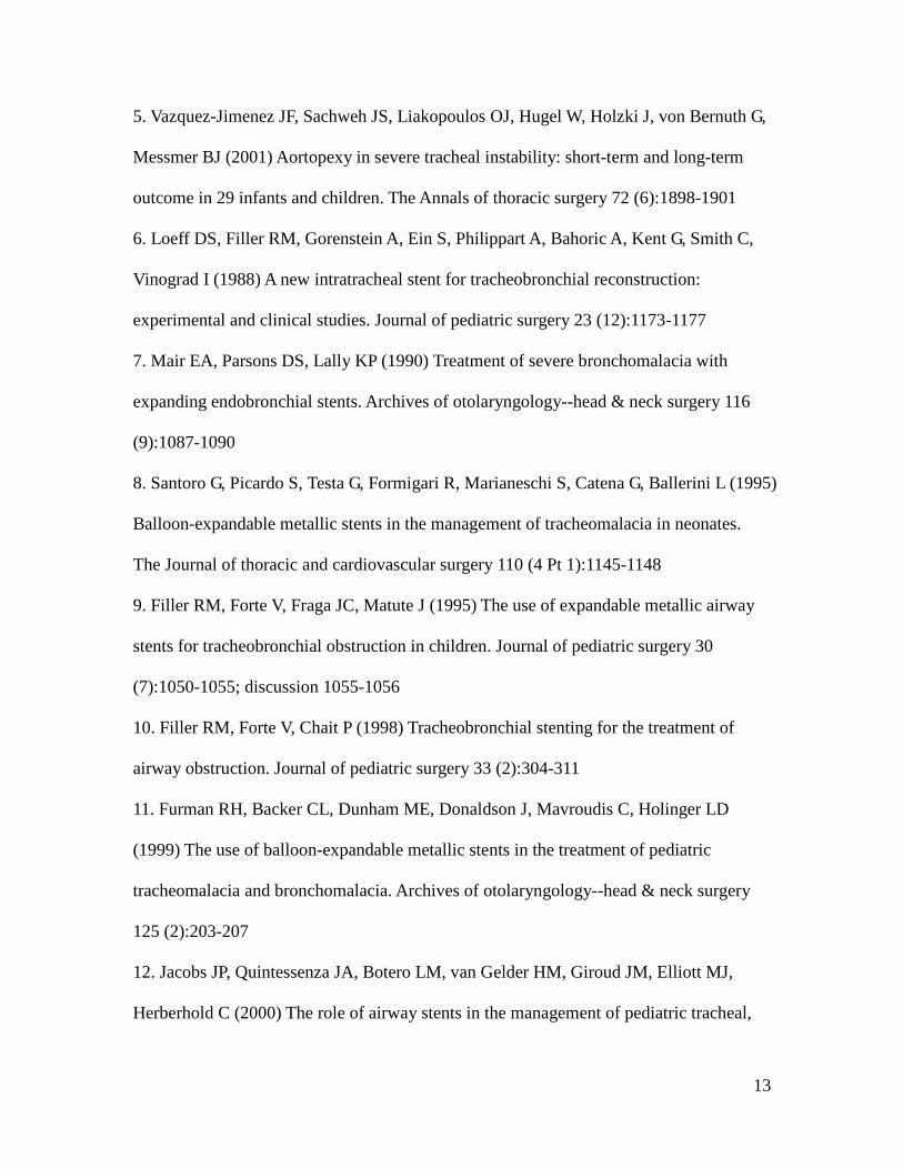
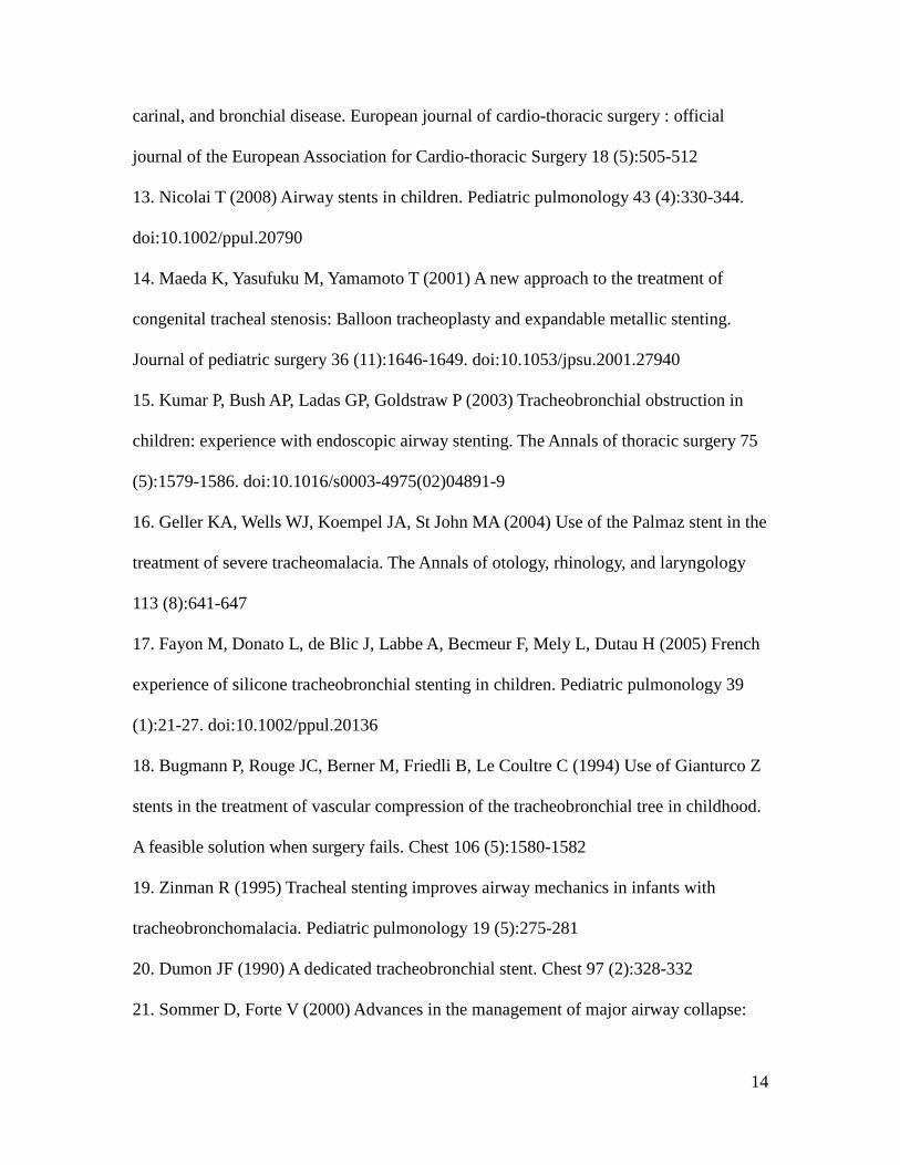
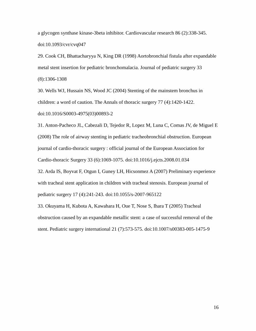
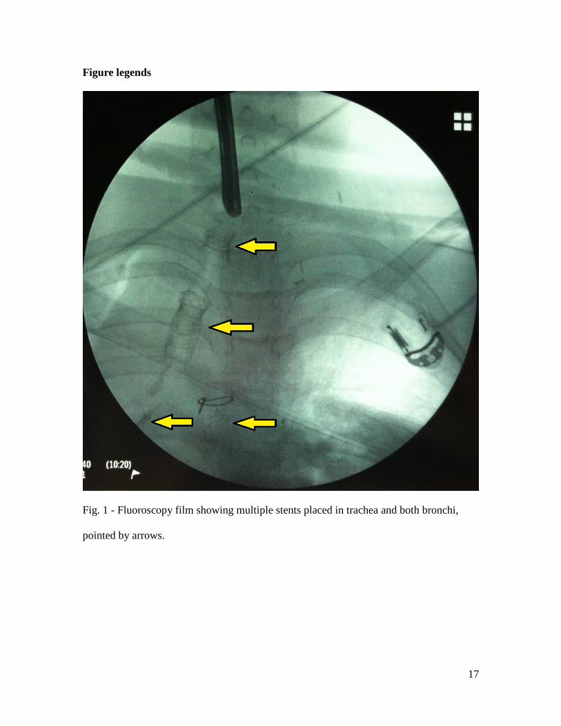
Figure legends
Fig. 1 - Fluoroscopy film showing multiple stents placed in trachea and both bronchi,
pointed by arrows.
18
Fig. 2 - Fluoroscopy film taken during a balloon dilatation session
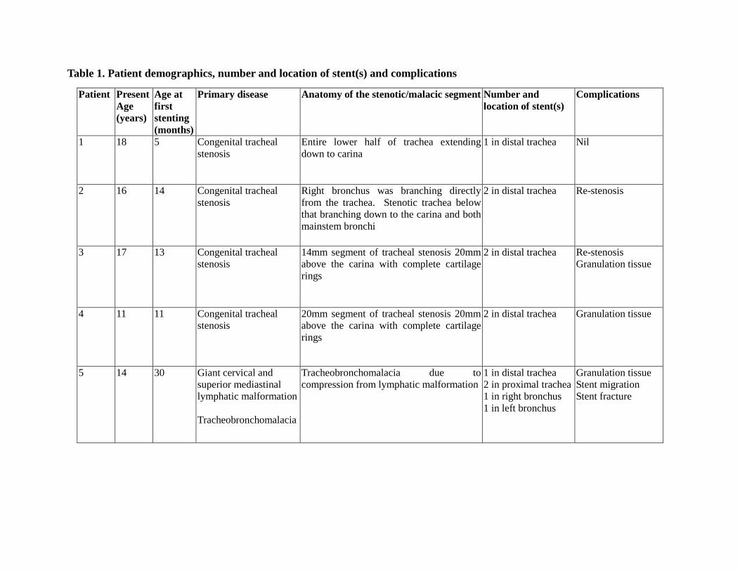
Table 1. Patient demographics, number and location of stent(s) and complications
Patient
Present Age (years)
Age at first stenting (months)
Primary disease Anatomy of the stenotic/malacic segment Number and location of stent(s)
Complications
1 18 5 Congenital tracheal stenosis
Entire lower half of trachea extending down to carina
1 in distal trachea Nil
2 16 14 Congenital tracheal stenosis
Right bronchus was branching directly from the trachea. Stenotic trachea below that branching down to the carina and both mainstem bronchi
2 in distal trachea Re-stenosis
3 17 13 Congenital tracheal stenosis
14mm segment of tracheal stenosis 20mm above the carina with complete cartilage rings
2 in distal trachea Re-stenosis Granulation tissue
4 11 11 Congenital tracheal stenosis
20mm segment of tracheal stenosis 20mm above the carina with complete cartilage rings
2 in distal trachea Granulation tissue
5 14 30
Giant cervical and superior mediastinal lymphatic malformation Tracheobronchomalacia
Tracheobronchomalacia due to compression from lymphatic malformation
1 in distal trachea 2 in proximal trachea 1 in right bronchus 1 in left bronchus
Granulation tissue Stent migration Stent fracture

Table 2. Long term outcome
Patient number
Total number of bronchoscopies
Number of subsequent dilatations
Maximal tracheal diameter achieved (mm)
Oxygen requirement
Exercise tolerance
Growth Follow up duration (years)
1 21
9 14 Nil Normal Normal 18
2 29 16 8 Nil 3 flights of stairs
Below 3rd percentile
16
3 21 14 13 Nil Normal Below 3rd percentile
17
4 25 11 12 Nocturnal continuous positive airway pressure ventilation
Wheelchair bound due to profound mental retardation
10th percentile 11
5 55 40 17 Home bilevel positive airway pressure ventilation
Normal Normal 14