1 pain pathophysiology unraveled (glick) - painweek kandel e, et al, eds. principles of neural...
TRANSCRIPT

Pain Pathophysiology UnraveledDavid M Glick, DC, DAAPM, CPE

Disclosures Nothing to Disclose

Learning ObjectivesDifferentiate between nociceptive and neuropathic painDescribe the process of pain transmission Identify the specific pain pathways that can be acted upon by
certain pharmacotherapies

Classification of PainGood pain vs bad pain
Clinical Pearl

Good PainNociceptive pain: purposeful pain
– Eudynia—being pain linked to normal tissue function or damage –Non-maldynic pain–Adaptive
Bad PainNeuropathic pain: non-purposeful pain
–Maldynia—pain linked to disorder, illness or damage – ie, may be abnormal, unfamiliar pain, assumed to be caused by
dysfunction in PNS or CNS
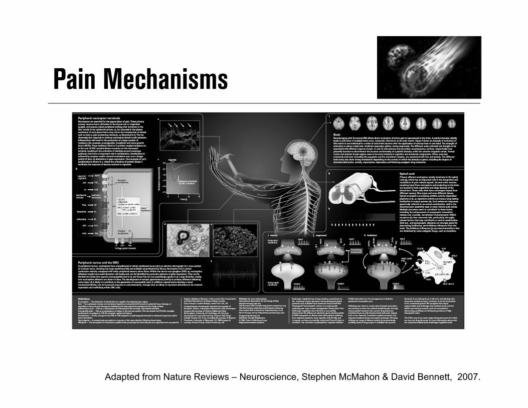
Pain Mechanisms
Adapted from Nature Reviews – Neuroscience, Stephen McMahon & David Bennett, 2007.

General Anatomy of Pain
Adapted from Von Hehn CA, Baron R, Woolf CJ. Deconstructing the neuropathic pain phenotype to reveal neural mechanisms. Neuron. 2012; 23;73(4):638-652.
Cortex and subcortical regions:Perception, sensory, & affective pain components
Thalamus
Brainstem:Descending modulation
Spinal cord:Synaptic transmission, modulation& central sensitization
Periphery:Transmission & peripheral sensitization

Pain Roadmap:Peripheral and Central Nervous System Landmarks
1. Gardner EP, et al. In: Kandel E, et al, eds. Principles of Neural Science. 4th ed. McGraw-Hill Medical; 2000; chapters 21-23.
• Physiologic process involving multiple areas of the nervous system
• Bidirectional• Involves normal as well as
pathological processes• A sensory experience associated
with affective and cognitive responses
• Dynamic (ie, occurring in real time)• Adapts or changes in response to
function–“neuroplasticity”

Common Types of PainNociceptive pain
Inflammatory pain
Neuropathic pain
Adaptive, high-threshold painEarly warning system (protective)
Adaptive, low-threshold painPromotes repair (protective)
Maladaptive, low-threshold painDisease state of nervous system
Noxious stimuli
Dysfunctional pain
Nociceptor sensory neuron
Peripheral nerve damage
Tissue Damage
Injury
Stroke
Normal peripheraltissue and nerves
HeatColdTechanical forceChemical irritants
Neural lesionPositive and negative symptoms
Non-neuropathicNon-inflammatory
Spinal cord
MacrophageMast cellNeutrophilGranulocyte
Functional pain
Adapted from: Woolf CJ. Ann Intern Med. 2004;140:441-451.

Nociceptive vs Neuropathic Pain
1. Bazen PT. Sickle cell disease pain management: evidenced-based clinical practice guidelines enhance health related quality of life. Presented at: American Society of Pain Management Nurses 18th Annual National Conference; September 3-6, 2008; Tucson, AZ.
2. Portenoy RK, Kanner RM. Definition and assessment of pain. In: Portenoy RK, Kanner RM, eds. Pain Management: Theory and Practice. Philidelphia, PA: FA Davis; 1996:4.
Neuropathic Pain
Initiated or caused by primary lesion or dysfunction
in the nervous system
Mixed Type
Caused by a combination of both primary injury and secondary effects
Nociceptive Pain
Caused by activity in neural pathways in response to
potentially tissue-damaging stimuli

Nociceptive vs Neuropathic Pain
1. Portenoy RK, Kanner RM. In: Portenoy RK, et al, eds. Pain Management: Theory and Practice. Philadelphia, PA: FA Davis Company;1996:4. 2. Galer BS, Dworkin RH. A Clinical Guide to Neuropathic Pain. Minneapolis, MN: McGraw-Hill Companies Inc; 2000:8-9.

PerceptionCortex and subcortical regions:sensory, and affective pain components- Behavioral/Limbic
Cortex
RVM
PAG
ThalamusTransductionPeripheral nociceptor converts input to electric charge
ConductionPeripheral nerve synapsing in the dorsal horn
TransmissionSpinal Cord/ Ascending Spinal Pathways
Pain Pathway Steps
Adapted from Scholtz J, Woolf CJ, Nat Neuroscience, 2002,5:1062-1067
PAG = periaqueductal greyRVM = rostral ventromedial medulla

Molecular Elements: Peripheral - Central
TransductionTRPV1, TRPV2, TRPV3, TRPM8ASIC, DRASICMDEG, TREK-1BK1, BK2P2X3
Peripheral sensitizationNGF, TrkATRPV1Nav 1.8PKA, PKC isoforms, CaMK IVErk ½, p38, JNKIL-1, cPLA2, COX2, EP1, EP3, EP4TNF
Membrane excitability ofperipheral afferentsNav 1.8, Nav 1.9K+ channel
Synaptic TransmissionPresynapticVGCCAdenosine-R(mGlu-R)
PostsynapticAMPA/kainite-R, NMDA-R, mGlu-RNK1Nav 1.3K+ channel
Central InhibitionGABA, GABAA-R, GABAB-RGlycine-RNE, 5-HTOpioid receptorsCB1
Signal transductionPKA, PKC isoformsERK, p38,JNK
Gene expressionc-fos, c-jun, CREB,DREAM
Adapted from Scholz J, Woolf CJ, Nature Neuroscience supplement Vol 5, 2002
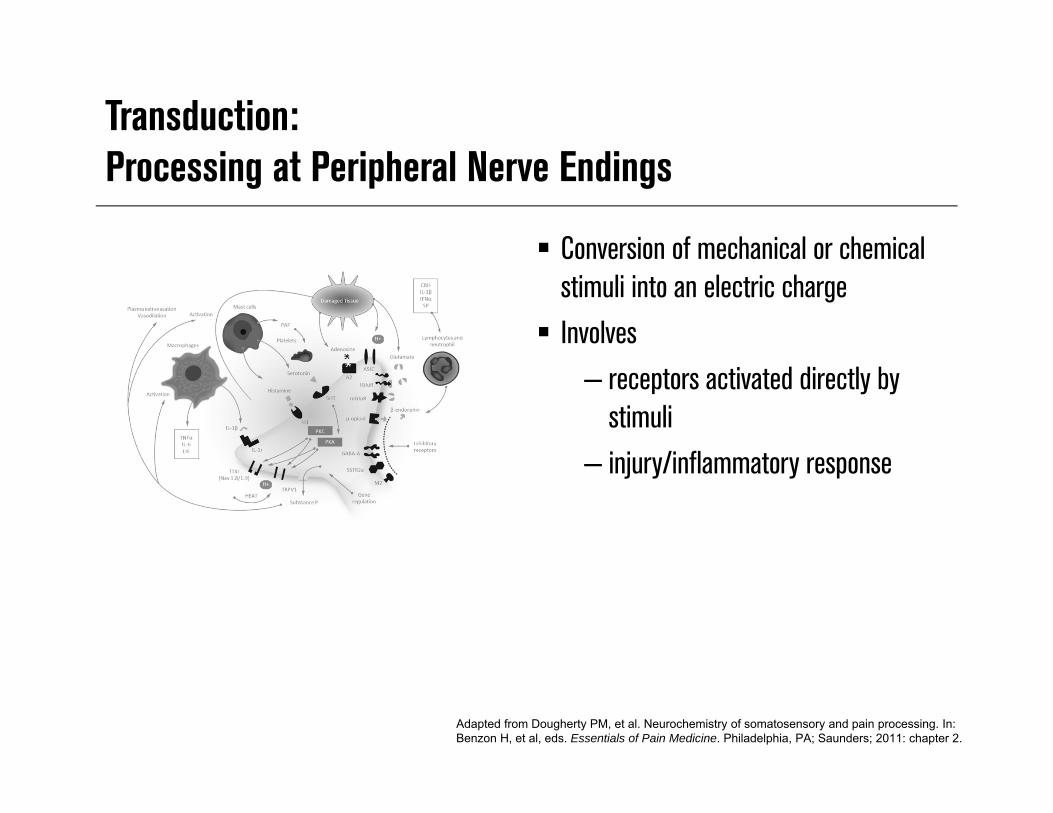
Transduction: Processing at Peripheral Nerve Endings
Conversion of mechanical or chemical stimuli into an electric charge Involves
– receptors activated directly by stimuli
– injury/inflammatory response
Adapted from Dougherty PM, et al. Neurochemistry of somatosensory and pain processing. In: Benzon H, et al, eds. Essentials of Pain Medicine. Philadelphia, PA; Saunders; 2011: chapter 2.
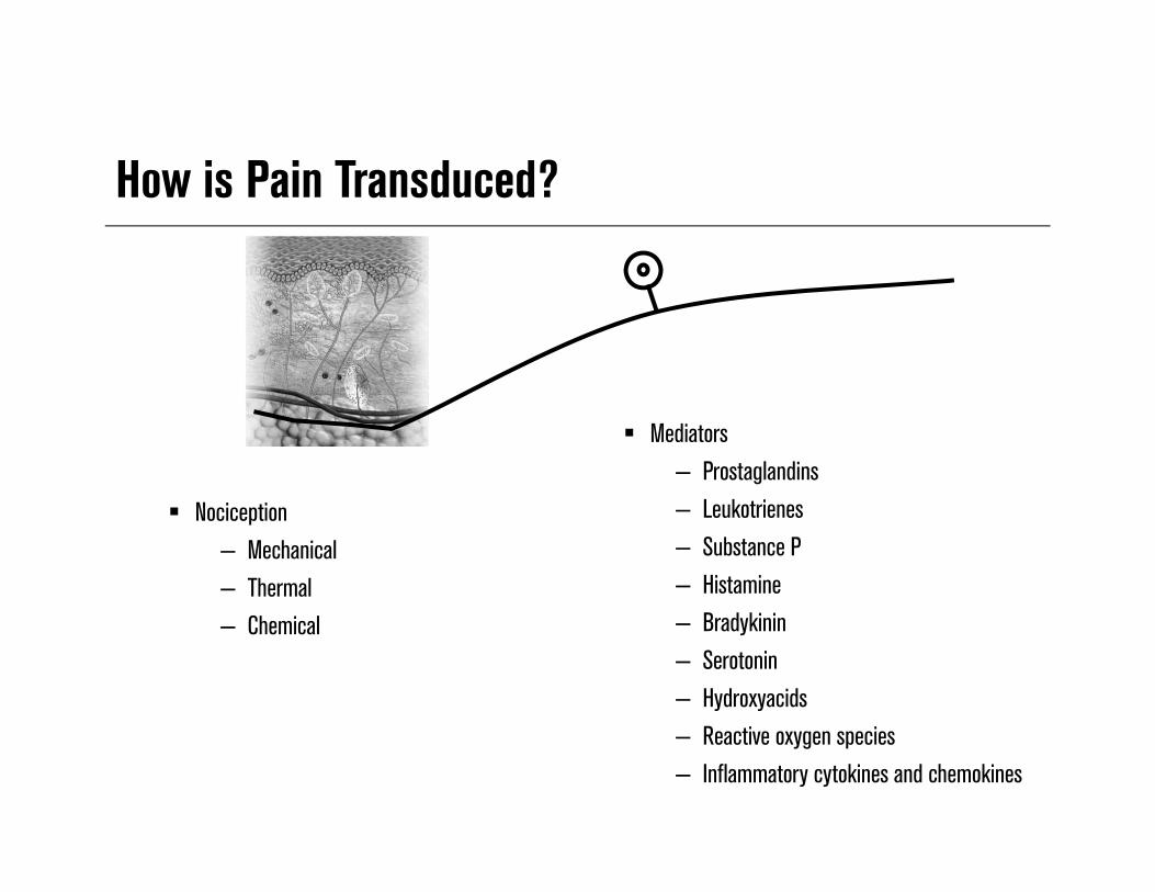
How is Pain Transduced?
Nociception– Mechanical– Thermal– Chemical
Mediators– Prostaglandins– Leukotrienes– Substance P– Histamine– Bradykinin– Serotonin– Hydroxyacids– Reactive oxygen species– Inflammatory cytokines and chemokines
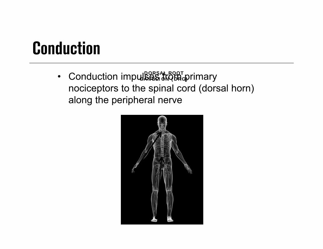
ConductionDORSAL ROOT
GANGLION (DRG)• Conduction impulses from primary nociceptors to the spinal cord (dorsal horn) along the peripheral nerve

Primary Nociception A-delta fibers
– Small receptive fields– Thermal & mechanical– Myelinated– Rapidly conducting
• 10-30 m/sec
– Large diameter
C-fibers– Broad receptive fields– Polymodal– Unmyelinated– Slower conducting
• .5-2.0 m/sec
– Cross sensitized– Small diameter

Peripheral Pain Nociceptors
Bashbaum A, Jessell T, The perception of Pain, In Kendal E, Schwartz J, Principles of Neural Science 4th
ed, New York, McGraw Hill, 2000, 482-483.
Aβ—muscle spindle secondary endings, touch, and kinesthesiaAδ—pain, temperature, crude touch, and pressure
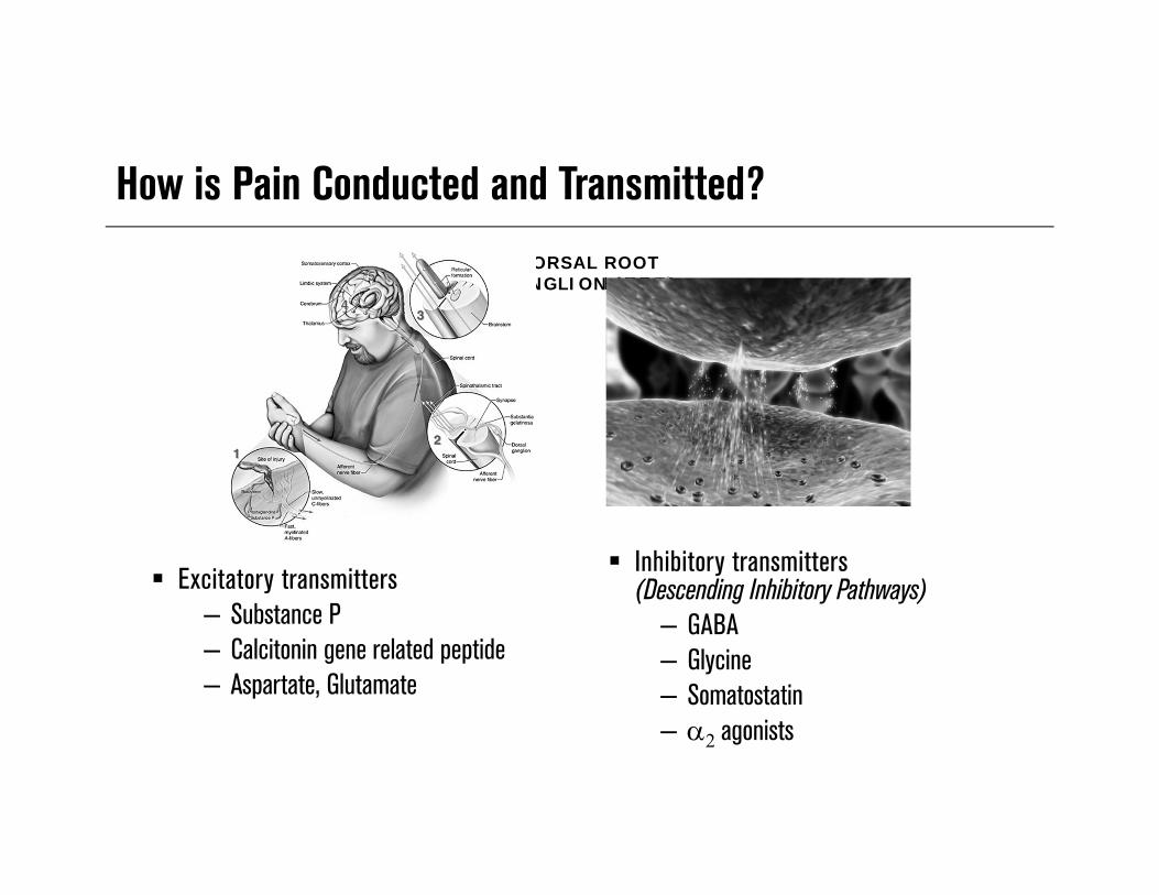
How is Pain Conducted and Transmitted?
Excitatory transmitters– Substance P– Calcitonin gene related peptide– Aspartate, Glutamate
DORSAL ROOT GANGLION (DRG)
Inhibitory transmitters(Descending Inhibitory Pathways)
– GABA– Glycine– Somatostatin– agonists
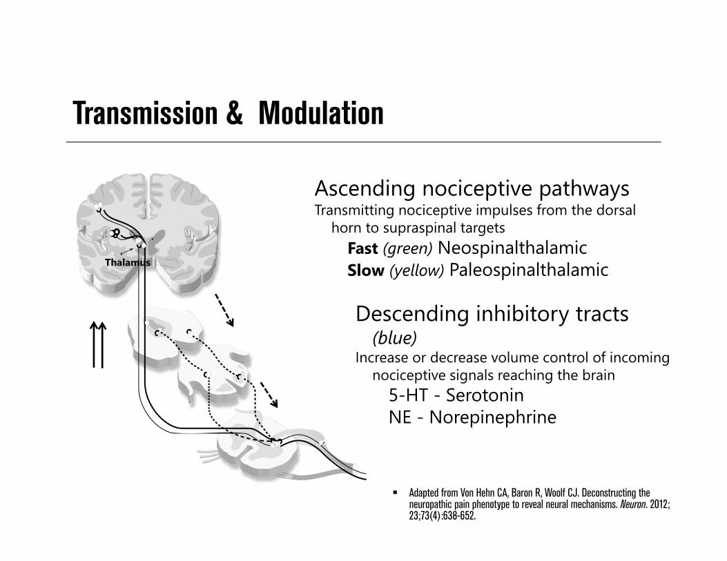
Transmission & Modulation
Adapted from Von Hehn CA, Baron R, Woolf CJ. Deconstructing the neuropathic pain phenotype to reveal neural mechanisms. Neuron. 2012; 23;73(4):638-652.
Thalamus
Ascending nociceptive pathwaysTransmitting nociceptive impulses from the dorsal
horn to supraspinal targetsFast (green) NeospinalthalamicSlow (yellow) Paleospinalthalamic
Descending inhibitory tracts (blue)
Increase or decrease volume control of incoming nociceptive signals reaching the brain
5-HT - SerotoninNE - Norepinephrine
Role of Neuronal Plasticity in Pain Nervous system changes in
– Neuronal structure– Connections between neurons– Quantity/properties of neurotransmitters, receptors, ion channels
Decreases body’s pain inhibitory systems
– Increased Pain Injury, inflammation, and disease are culprits Produces short-term and permanent changes Pivotal to the development of hypersensitivity of inflammatory pain
– Enables NS to modify its function according to different conditions
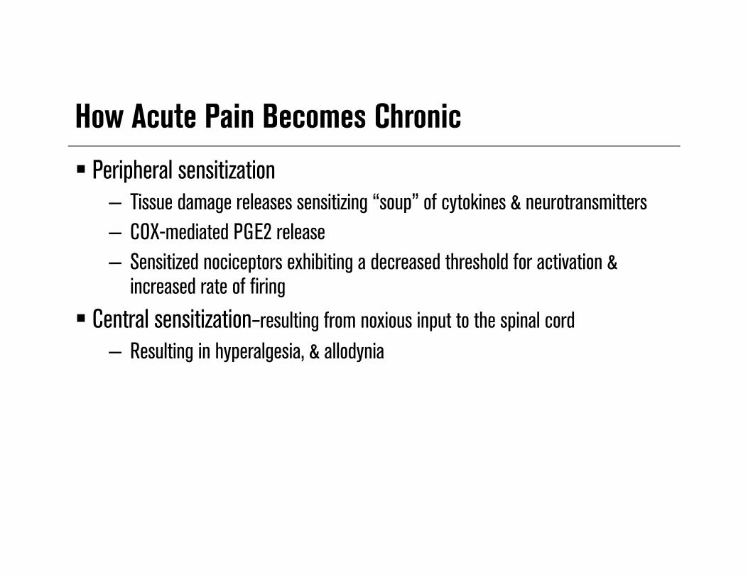
How Acute Pain Becomes Chronic Peripheral sensitization
– Tissue damage releases sensitizing “soup” of cytokines & neurotransmitters– COX-mediated PGE2 release– Sensitized nociceptors exhibiting a decreased threshold for activation &
increased rate of firing Central sensitization–resulting from noxious input to the spinal cord
– Resulting in hyperalgesia, & allodynia

Definitions
Hyperalgesia– Lowered threshold to different
types of noxious stimuli
Allodynia– Painful response to what should
normally be non-painful stimuli

Neuroplasticity in Pain Processing
Innocuous Noxious

Neuroplasticity in Peripheral Pain Transmission

Peripheral Sensitization
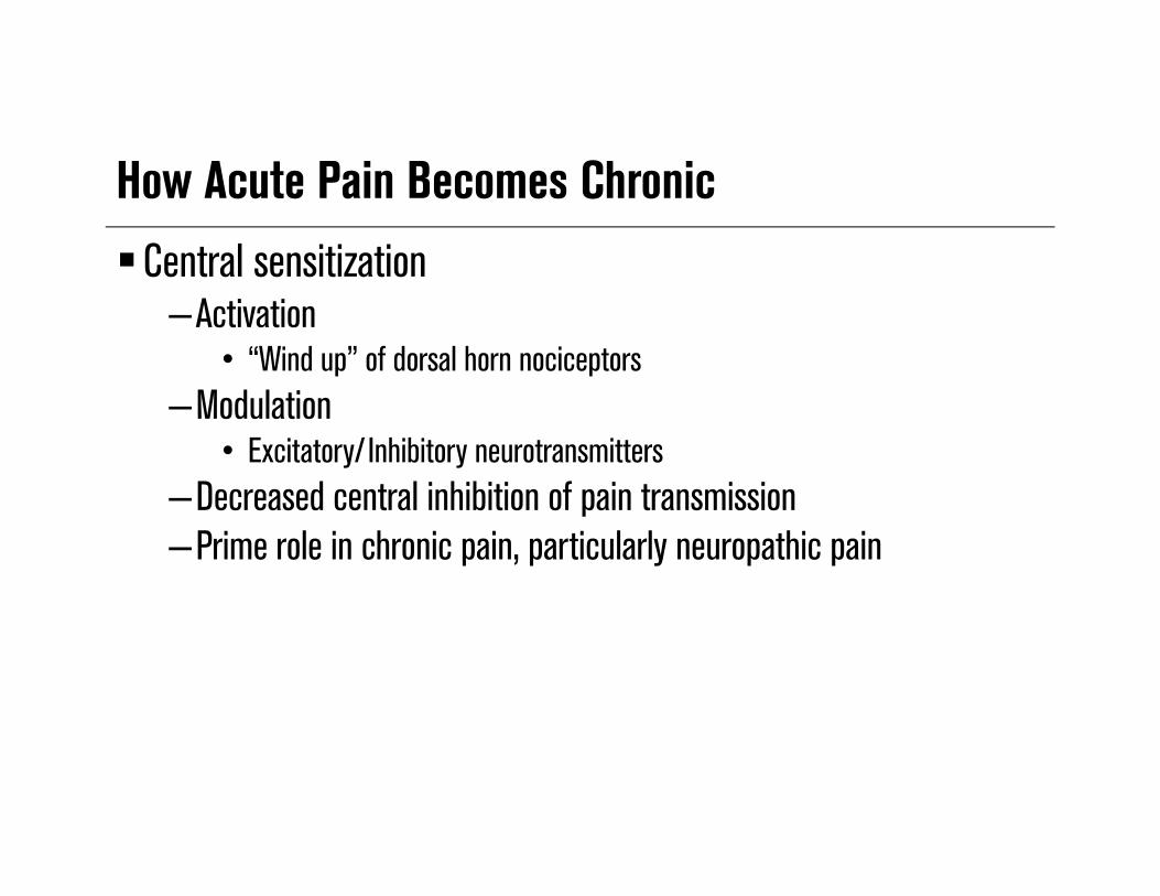
How Acute Pain Becomes ChronicCentral sensitization
–Activation• “Wind up” of dorsal horn nociceptors
–Modulation• Excitatory/ Inhibitory neurotransmitters
–Decreased central inhibition of pain transmission–Prime role in chronic pain, particularly neuropathic pain

Definitions
Wind Up– Causes long-term changes in nociceptive neurons, which become
hyperexcitable such that they respond to lower stimuli• NMDA-type glutamate receptors play an important role in
this process1,2,3,4
– Prolonged opening of the ion channels enables greater influx of calcium and sodium across the post-synaptic membrane and greater excitation of nociceptive neurons2,3
1. Kandel ER, Schwartz JH, Jessell TM, editors. Principles of Neural Science (Fourth Edition). New York: McGraw Hill (Health Professions Division). 2000;472-491.
2. Millan MJ. Progress in Neurobiology 1999;57:1-164. 3. Dickenson AH. Brit J Anaesthesia 1995;75:193-200. 4. Suzuki R and Dickenson AH. Neuroreport 2000;11:R17-21.

Central Sensitization
Afferent first order neuron Dorsal horn neuron
NK-1 = Neurokinin 1 receptor; AMPA = alpha-amino-3-hydroxy-5-methyle 4-isoxazolepropionic acid; NMDA = N-methyl-D-aspartic acid; VGCC = voltage gated sodium channel; TrkB = tropomyosin receptor kinase B; BDNF = Brain derived neurotrophic factor; SP = substance P
Adapted from Schlotz J, Woolf CJ. Nat Neuroscience. 2002;5:1062-1067

Central SensitizationKey influences upon signal propagation
• Excitatory neurotransmitters– Substance P, CGRP,
glutamate• NMDA channel activity
– Glutamate binding– Altering channel activity
• Descending inhibitory tracts– NE/serotonin (5HT)
• Mu opioid receptor
Dorsal Horn
5HT receptor
Adapted from Schlotz J, Woolf CJ. Nat Neuroscience. 2002;5:1062-1067
NK-1 = Neurokinin 1 receptor; AMPA = alpha-amino-3-hydroxy-5-methyle 4-isoxazolepropionic acid; NMDA = N-methyl-D-aspartic acid; VGCC = voltage gated sodium channel; TrkB = tropomyosin receptor kinase B; BDNF = Brain derived neurotrophic factor; SP = substance P; CRGP = Calcitonin gene related peptide
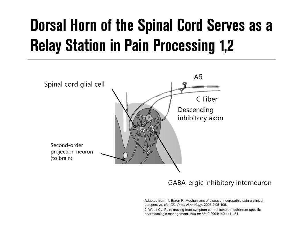
Dorsal Horn of the Spinal Cord Serves as a Relay Station in Pain Processing 1,2
Spinal cord glial cell
Second-orderprojection neuron(to brain)
Aδ
Descending inhibitory axon
GABA-ergic inhibitory interneuron
C Fiber
Adapted from 1. Baron R. Mechanisms of disease: neuropathic pain-a clinical perspective. Nat Clin Pract Neurology. 2006;2:95-106.2. Woolf CJ. Pain: moving from symptom control toward mechanism-specific pharmacologic management. Ann Int Med. 2004;140:441-451.

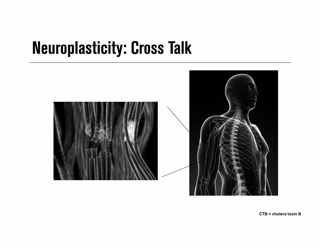
CTB = cholera toxin B
Neuroplasticity: Neural ReorganizationReceptor fields
CTB = cholera toxin B
Neuroplasticity: Cross Talk

Central Sensitization:Neuroplasticity in Spinal Cord Processing
1. Mannion RJ, Woolf CJ: Clin J Pain. 2000;16(3):S151-S153. 2. Ossipov MH, et al. Ann NY Acad Sci. 2000;909:12-24.3. Wieseler-Frank J, et al. Neurosignals. 2005;14:166-174. 4. Guilbaud G, et al. Exp Brain Res. 1992;92:227-245.
• Definition: altered function of neurons or synaptic activity • Mechanisms of central sensitization may include:
– Changes effecting glutamate / NMDA receptors activity• Reduced threshold for activation• Increased availability of glutamate• Increased influx of Na+/Ca+ (receptor open longer)
– Modulation—excitatory/ Inhibitory neurotransmitters – Decreased tone—descending inhibitory pathways2
– Activation/migration of glial cells into the spinal cord3
– Changes in the thalamus and primary somatosensory cortex4

Brain Regions Involved in Pain Processing
Apkarian AV et al, Eur J Pain 2005;9:463-484
Pain and emotion
Pain only
Prefrontal cortexMotor planning
Anterior cingulate cortex Context/Situation of pain
Insular cortexPain judged to the degree and where pain is imagined
AmygdalaEmotional Aspect
HippocampusPain memory/Learning
ThalamusRouting
Somatosensory cortexLocalization

Analgesics That Modify Pain Processes
Perception– Parenteral opioids– agonists– General anesthetics
Conduction– Local anesthetics
• Peripheral nerve, plexus, epidural block
Transmission/Modulation– Spinal opioids– agonists– NMDA receptor antagonists– NSAIDs– NO inhibitors– K+ channel openers
Transduction– NSAIDs– Antihistamines– Membrane stabilizing agents– Local anesthetic cream– Opioids– Bradykinin & serotonin antagonists
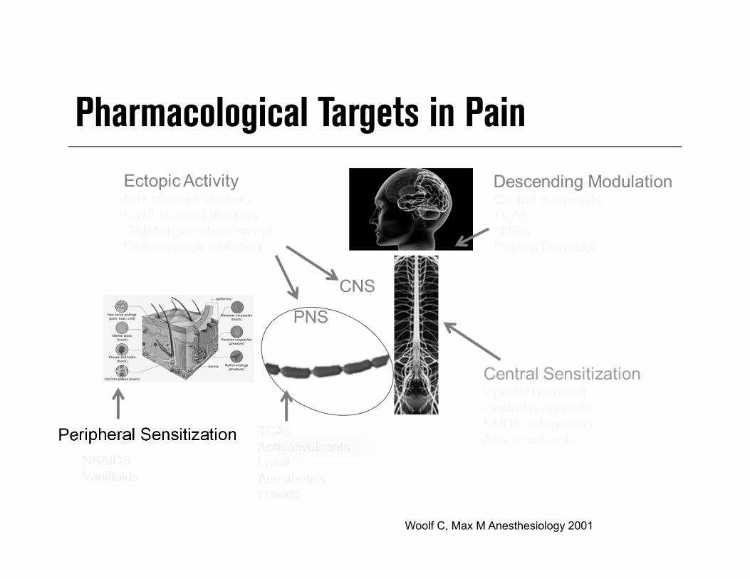
Pharmacological Targets in Pain
Peripheral Sensitization
Woolf C, Max M Anesthesiology 2001

The Chronic Pain ArmamentariumNonopioids
– Acetaminophen– NSAIDs– COX-2 inhibitors
Opioids– Mu-opioid agonists– Mixed agonist-antagonists
Adjuvant analgesics– Antidepressants– Anticonvulsants – Topical agents/local anesthetics
WHO
JC Ballantyne Oncologist 2003:8(6):567-75. © AlphaMed Press; WHO. 2005.
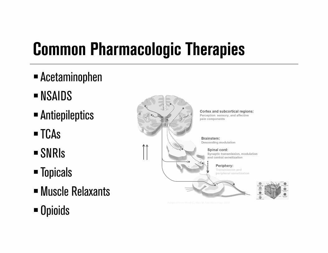
Common Pharmacologic TherapiesAcetaminophenNSAIDSAntiepileptics TCAs SNRIs TopicalsMuscle RelaxantsOpioids
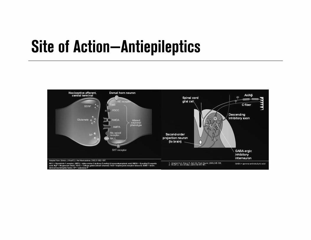
Site of Action—Antiepileptics
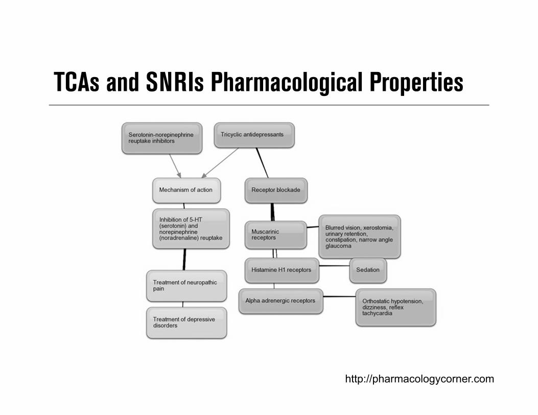
http://pharmacologycorner.com
TCAs and SNRIs Pharmacological Properties

Modulation of Central Sensitization by 5-HT & NE Descending Pathways

Adjuvant Analgesics: TopicalsExamples
– Lidocaine patch 5% , eutectic, mixture of lidocaine and prilocaine– Capsaicin cream/patch– Dicofenac (cream/liquid/patch)
Mechanism of action– Block sodium channels and inhibit generation of abnormal impulses by
damaged nerves– Depletion of peripheral small fibers and therefore substance P release from
sensory nerve endings– Target local inflammatory response
Muscle Relaxants—Spasmolytics Enhancing the level of inhibition
– Mimicking or enhancing the actions of endogenous inhibitory substances, such as GABA
Reducing the level of excitation. Common examples
– Cyclobenzaprine (TCA) methocarbamol, carisoprodol, tizanidine (α-2 agonist), baclofen (GABA agonist), orphenadrine (benzodiazepine)
Common adverse effects– Sedation, lethargy & confusion (cyclobenzaprine), dependence (carisoprodol)
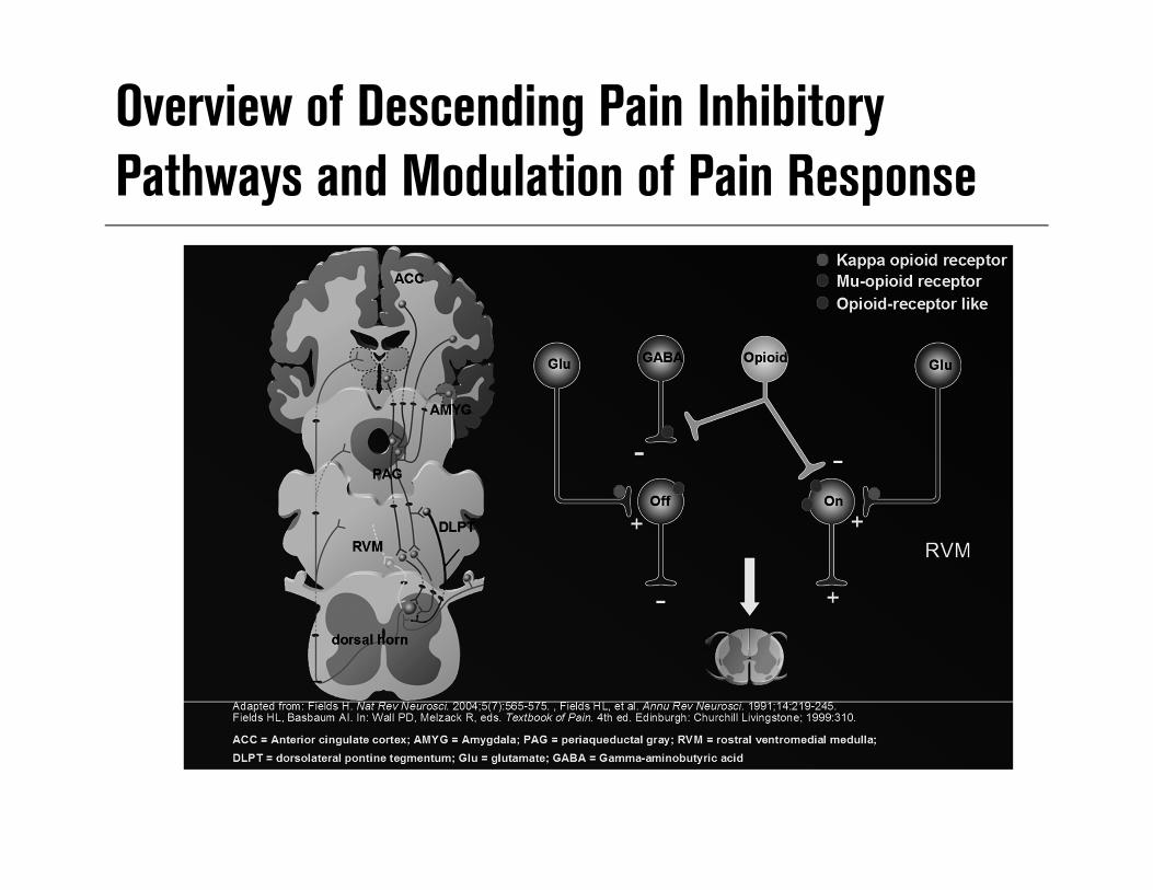
Overview of Descending Pain Inhibitory Pathways and Modulation of Pain Response
Case Study 54-year-old with 3 year history of neck, shoulder and upper extremity pain following
a lifting injury– Current medications
• Fluoxetine• Milnacipran• Gabapentin• Clonazepam• Alprazolam• Robaxin• Tapentadol • Acetaminophen and propoxyphene • Zolpidem • Diclofenac topical• Acetaminophen

Importance for Understanding Pain Mechanisms Allow for rational rather than empirical approach to pain control Foster the development of diagnostic tools to identify specific
pain mechanisms Facilitate pharmacotherapies that act on specific pain pathways and
mechanisms Reduce the number of pharmacotherapies and incidence of drug-related
adverse events Enhances use of non-pharmacologic treatments Improve overall patient care and outcome
Summary Today’s clinicians must possess a working knowledge of the etiology and
mechanisms of pain syndromes– Understanding pain mechanisms/pathophysiology is key to successful pain control
• Reduce the number of medications and incidence of drug-related adverse events – (rationale polypharmacy)
• Many therapeutic options are available – (non-pharmacological)
– Tailoring treatment based on the individual patient and pain type can improve outcomes – Understanding how treatments effect function clinical presentation and function– Do not forget to look for the spear