1 plum pox virus and oxidative stress in apricot centro de...
TRANSCRIPT

1
Running title: Plum pox virus and oxidative stress in apricot
Correspondence to:
Dr. José A. Hernández
Centro de Edafología y Biología Aplicada del Segura
CSIC
Department of Plant Breeding, Apdo. 164
E-30100 Espinardo (Murcia)
SPAIN
FAX: 34 968 396213
E-mail: [email protected]

2
Long-term Plum Pox Virus infection produces an oxidative stress in a
susceptible apricot (Prunus armeniaca L.) cultivar but not in a resistant cultivar
José Antonio Hernández a,*, Pedro Díaz-Vivancos a, Manuel Rubio a, Enrique Olmosb, Alfonso Ros-
Barcelóc A. And Pedro Martínez-Gómeza
aDepartment of Plant Breeding CEBAS-CSIC. P.O. Box 164, 30100 Espinardo-Murcia, Spain.bDepartment of Plant Nutrition CEBAS-CSIC. P.O. Box 164, 30100 Espinardo-Murcia, SpaincDepartment of Plant Biology (Plant Physiology), University of Murcia, E-30100 Murcia (Spain)

3
AbstractThe effect of Plum pox virus (PPV) infection on the response of some antioxidant enzymes
was studied in two apricot cultivars, which behaved differently against PPV infection: cv. Real Fino
(susceptible) and cv. Stark Early Orange (SEO, resistant). In the susceptible cultivar, PPV produced
a decrease in ΦPSII, F’v/F’m and Qp. PPV infection produced a drop in pHMB-sensitive ascorbate
peroxidase, dehydroascorbate reductase and peroxidase in the soluble fraction from susceptible
plants, whereas in the resistant apricot cultivar pHMB-insensitive ascorbate peroxidase,
monodehydroascorbate reductase, glutathione reductase and superoxide dismutase increased.
However, catalase decreased in the soluble fraction from both infected cultivars. Long-term PPV
infection also produced a decrease in the chloroplastic ascorbate-glutathione cycle enzymes only in
the susceptible plants. As a consequence of PPV infection, an oxidative stress, indicated by an
increase in lipid peroxidation and in protein oxidation, was produced only in the leaves from the
susceptible cultivar which was also monitored by the diaminobenzidine-peroxidase coupled H2O2
probe. The loss of ΦPSII, indicative of AOS production, and the decrease in the levels of antioxidant
enzymes in chloroplasts from susceptible plants, could be responsible for the chlorosis symptoms
observed. The results suggest that the higher antioxidant capacity showed by cultivar SEO could be
a consequence of a systemic acquired resistance induced by PPV penetration in stem tissue at the
graft site and could be related, among other factors, to their resistance to PPV.
Key Words: Apricot, antioxidant enzymes, oxidative stress, plum pox virus, virus resistance,
photochemical quenching, Prunus, ultrastructure
Abbreviations - ASC, ascorbate; AOS, activated oxygen species; ASC-GSH cycle, ascorbate-
glutathione cycle; APX, ascorbate peroxidase; CO-protein, carbonil proteins contents; cv. SEO, cv.
Stark Early Orange; DHAR, dehydroascorbate reductase; Fv/Fm, maximum quantum yield of
photosystem II; F’v/F’m, efficiency of excitation energy capture by PSII; GR, glutathione reductase;
MDHAR, monodehydroascorbate reductase; 4-MN, 4-methoxy-1-naphthol; NPQ, non-
photochemical quenching; 1O2; singlet oxygen; .OH, hydroxyl radical; O2.-, superoxide radical;
pHMB, p-hydroxy mercury benzoic acid; PPV, plum pox virus; qp; photochemical quenching; SOD
superoxide dismutase; TBARS, thiobarbituric acid-reactive substances; ΦPSII, quantum yield of
photosystem II photochemistry.

4
Introduction
Sharka, a disease caused by Plum pox virus (PPV) is a serious limiting factor for temperate
fruit production in affected areas, resulting in severe economic losses in Prunus species including
apricot and peach (Kölber 2001). Obtaining Prunus cultivars resistant to sharka is one of the main
objectives of breeders, but the evaluation of programmes for PPV resistance is time-consuming and
very expensive (Martínez-Gómez and Dicenta 2000a). Therefore, biochemical and molecular
markers associated with resistance would be of great interest. These markers will improve the
selection process in the evaluation of a higher number of individuals.
In most incompatible interactions, the rapid induction of highly-localised events imposes
unfavourable conditions for pathogen growth. This defence response culminates in a localized cell
death, called the hypersensitive response (HR), associated with the resistance to pathogen spread
(De Gara et al. 2003). Increased levels of activated oxygen species (AOS), including superoxide
(O2.-) and hydrogen peroxide (H2O2), built up by either enhanced production or decreased
scavenging potential, may contribute to the resistance reaction to pathogens in incompatible
reactions (Alvarez et al. 1988, Adams et al. 1989, Doke and Ohashi, 1988, Thordal-Christensen et
al. 1997). However, very little is known about the oxidative metabolism in plant resistance reactions
to pathogens that do not induce the HR, such as the necrotrophic fungi that invade the plant vascular
system (García-Limones et al. 2002) or some plant viruses such as PPV (Hernández et al. 2001a,
2003, 2004b). Alternatively, increased levels of AOS could also contribute to the symptom
development and pathogenesis in compatible plant-virus interactions, as described recently in PPV-
susceptible peach plants (Hernández et al. 2003, 2004b) and in CMV-infected Cucumis sativus and
ZYMV-infected Cucurbita pepo plants (Riedle-Bauer 2000). In recent work carried out in our
laboratory, we showed that long-term PPV infection produced an oxidative stress in leaves of peach
cv. GF305, characterised by its high susceptibility to PPV, manifested as increases in lipid
peroxidation and protein oxidation, the appearance of oxidative microbursts and effects on
chloroplast ultrastructure (Hernández et al. 2004b).
5
Plants, like other aerobic organisms, are endowed with efficient AOS-scavenging
mechanisms. The primary components of these antioxidant systems include non-enzymatic
antioxidants (carotenoids, ascorbate, glutathione and tocopherols) and enzymes such as SOD, catalase
(EC 1.11.1.6), glutathione peroxidase (GPX, EC 1.11.1.9), peroxidases and the enzymes involved in
the ascorbate-glutathione cycle (ASC-GSH cycle): ascorbate peroxidase (APX, EC 1.11.1.1),
dehydroascorbate reductase (DHAR, EC 1.8.5.1), monodehydroascorbate reductase (MDHAR, EC
1.6.5.4) and glutathione reductase (GR, EC 1.6.4.2). The components of this antioxidant defence
system can be found in different subcellular compartments (Jiménez et al. 1998, Hernández et al.
2000), and they are constitutively expressed to cope with AOS formed under normal conditions.
However, they can also be induced to maintain the lowest possible levels of AOS under both biotic
and abiotic stresses (Hernández et al. 2000, 2001b, García-Limones et al. 2002).
An increasing amount of data supports the hypothesis that a fine regulation of antioxidant
systems is part of the signalling pathways which activate defense responses. However, the diversity
in the systems used for studying plant-pathogen interplay makes it difficult to formulate a clear
picture of whether, and to what extent, changes in antioxidant systems are directly involved in
plants defense responses or are a mere consequence of the oxidative stress occurring in the attacked
cells (de Gara et al. 2003). Several lines of evidence support the regulatory role that cellular
antioxidants, especially GSH and GSH-related enzymes, play in the biochemical and physiological
responses of plants to biotic stress (Gullner et al. 1999, Fodor et al. 1997). In this sense, the
artificial elevation of cellular GSH and the activation of GSH-related enzymes can markedly
suppress necrotic disease symptoms and in some cases also virus multiplication (Gullner et al.
1999). In a recent paper, it has been proposed that a decline in AOS-scavenging capacity may be
required before a rapid increase in virus replication can take place. Phaseolus vulgaris L. plants
treated with the cytokinin dihydrozeatin, salicylic acid or jasmonic acid showed elevated catalase,
GR and peroxidase activities. These treatments, when applied before inoculation with the

6
Potexvirus White clover mosaic virus, inhibited virus replication and symptom development
(Clarke et al. 2002).
In woody plant species, such as apricot, different factors, including their lignified nature, the
inoculation method, and the cycle of growth with periods of dormancy, make the study of the early
response to virus inoculation very difficult. In this work, the effect of long-term PPV infection on
the activity of antioxidant enzymes from apricot cvs. “Real Fino” (susceptible to PPV) and “SEO”
(resistant to PPV) at subcellular levels (cytosol and chloroplasts) was studied. The extent of lipid
peroxidation and protein oxidation, the histochemical detection of H2O2 and the leaf ultrastructure
were also analysed, to determine whether oxidative stress is involved in the development of
symptoms and the pathogenesis of PPV-susceptible apricot plants.
MATERIAL AND METHODS
Plant Material
Plant material assayed included the North American apricot cultivar SEO, characterised as resistant to
PPV, and the Spanish cultivar Real Fino, described as susceptible against virus (Martínez-Gómez and
Dicenta 2000a). Apricot seedlings were grafted on peach GF305 plants, characterised by its
susceptibility to fruit viruses including PPV (Bernhard et al. 1969) and usually used as a rootstock in
PPV-resistance tests on Prunus, both in vivo (Martínez-Gómez and Dicenta 2000a) and in vitro
(Martínez-Gómez and Dicenta 2000b). Ten repetitions from each apricot cultivar were grafted onto
control or infected GF305 rootstocks. Another ten repetitions were kept as control. Two months after
inoculation, seedlings were subjected to an artificial rest period, in a cold chamber at 7 °C, in darkness
for six weeks. Plants were then transferred to an insect-proof greenhouse, and were grown in 2-litre
pots in controlled conditions. Plants were inspected for sharka symptoms 4 weeks after the sprouting of
the buds. Two cycles of growth (two month in the cold chamber and four months in the greenhouse)
per year were analysed, and at least two experiments per cycle was performed. Data were recorded
over two years periods.The environmental conditions in the greenhouse were: temperature between

7
15º and 30ºC during all the year due to the control of temperatures during the summer with a
refrigeration systems, and relative humidity of 60-80%, with a photoperiod of around 16 hour of
light.
PPV isolate
The PPV isolate used was RB3.30, a Dideron Type isolate obtained from the Red Beaut plum
cultivar in Spain, from the PPV collection of the Instituto Valenciano de Investigaciones Agrarias
(IVIA) in Valencia (Spain). This isolate is considered to be representative of the Spanish PPV
population and produces strong sharka symptoms in young leaves, consisting of veinal chlorosis in
peach GF305 and veinal chlorosis and rings in susceptible apricot leaves (Pelet and Bovey 1968).
PPV inoculation procedure
Ten apricot scions per genotype were propagated onto control (healthy) and inoculated (infected)
symptomatic GF305 peach seedlings, one scion per seedling. Scion-grafted trees were forced into
dormancy by subjecting them to 7 ºC and darkness for two months. After this cold-dark treatment,
trees were transferred to an insect-proof greenhouse and were inspected for sharka symptoms four
weeks later. Two cycles of growth were performed over a one-year period. Only plants where the
GF305 rootstock showed clear PPV symptoms were considered to be successfully inoculated.
During each growth cycle, the presence of symptoms in leaves was scored in each leaf of each plant
according to a scale from 0 (no symptoms) to 5 (maximum intensity of symptoms), scale usually used
in the studies of resistance evaluation in apricot (Martínez-Gómez and Dicenta 2000a).
ELISA-DASI test
During the two growth cycles, to verify the presence or absence of the virus an ELISA-DASI (Double
Antibody Sandwich Indirect) was applied to the leaves using 5B monoclonal antibodies (Asensio
1996) against the capside protein of the PPV according with the protocol of Cambra et al. (1994).

8
Samples were incubated at 5 °C for 16 h with polyclonal rabbit antibodies (Real-Durviz. Valencia,
Spain) 1.42 µg/ml in 1% (w/v) Bovine Serum Albumin (BSA) (Boehringer&Mannhein. Barcelona,
Spain)-PBS (0.08% ClNa, 0.002% KH2PO4, 0.3% Na2HPO412H2O, 0.02% CLK). After washing 3
times for 5 min with PBS-Tween-20 (0.5 ml/l Tween-20) the micro-plates were incubated in 1% (w/v)
BSA-PBS with the specific monoclonal antibodies (0.1 µg/ml) (Real-Durviz) at 37 °C for 2h. After
washing 3 times with PBS-Tween-20 samples were incubated in 1% (w/v) BSA-PBS with alkaline
phosphatase-labeled second antibody (0.1 µg/ml) (Real-Durviz) at 37 °C for 2h. Then, micro-plates
were washed again three times (PBS-Tween-20) and were reveled with p-nitrofenolphosphate
colorimetric substrate (Sigma), recording the optical densities (OD) at 405 nm for 60 min. In
accordance with Sutula et al. (1986), samples with OD double that of the healthy control were
considered ELISA-positive.
RT-PCR analysis
RT-PCR analysis was carried out during the two cycles of study using total RNA extracted with the
Rneasy Plant Mini Kit (Qiagen, Hilden, Germany), as described by MacKenzie et al. (1997). Two
specific primers within the coat protein (CP) gene, VP337 (CTCTGTGTCCTCTTCTTGTG)
complementary to 9487-9508 positions of genomic PPV and VP338
(CAATAAAGCCATTGTTGGATC) homologous to 9194-9216 positions, were assayed (Martínez-
Gómez et al. 2003). PCR parameters were: one cycle at 94ºC for 2 min followed by 30 cycles of
94ºC for 30 sec, 55ºC for 30 sec and 72ºC for 30 sec, and finally an extension temperature of 72ºC
for 5 min (Martínez-Gómez et al. 2003). Amplified products were electrophoresed in 1% agarose
gels in 40 mM Tris-acetate and 1 mM EDTA, pH 8.0 (TAE), and stained with ethidium bromide. A
1 Kb Plus DNA Ladder (InvitrogenTM Life Technologies) was used as molecular size standard.
9
Fluorescence measurements
Ten control and ten PPV-infected peach plants were analysed in each cycle. Modulated chlorophyll
fluorescence was measured in dark-adapted peach leaves at midday, using an OS-30 chlorophyll
fluorometer (Optisciences, USA) with an excitation source intensity of 2000 µmol m-2 s-1. The
quantum yield of photosystem II photochemistry (ΦPSII) was calculated empirically as the
fluorescence parameter (Fm’ – Ft)/Fm
’ (Genty et al. 1989), and the maximum quantum yield of
photosystem II (Fv/Fm) as (Fm – Fo)/Fm (Maxwell and Johnson 2000). Non-photochemical
quenching (NPQ) was calculated as a Stern-Vollmer-type quenching (Bilger and Björkman 1990).
The photochemical quenching coefficient, equivalent to the fraction of open PSII reaction centres,
was calculated as qp= (F’m-Ft)/(F’m-F’o) (Maxwell and Johnson 2000).
The efficiency of excitation energy capture by PSII, corresponding to the probability that an
absorbed photon reaches the PSII reaction centres, was calculated in light-adapted leaves as
F’v/F’m= (F’m-F’o)/F’m.
The minimal “dark” fluorescence level following illumination (Fo’) was measured in the presence of
a background far-red light, to favour rapid oxidation of intersystem electron carriers.
Isolation of cell fractions
For the isolation of cell fractions four weeks-old plants were used. All operations were carried out
at 0-4 ºC. Soluble fractions were prepared by homogenising 3 g of fresh leaf material with a mortar and
pestle, with 6 ml of a grinding medium containing 0.35 M mannitol, 30 mM MOPS buffer (pH 7.5), 4
mM L-cysteine, 1 mM EDTA, 5% insoluble PVPP (w/v) and 0.2% (w/v) BSA. For APX activity, 20
mM ascorbate was added. The homogenate was filtered through 2 layers of cheesecloth and
centrifuged at 2200 g for 30 s, to pellet the chloroplast fraction. The supernatant was centrifuged at
12000 g, to discard mitochondria and peroxisomes. Then, the 12000 g supernatant was centrifuged for
20 min at 82000 g. The resulting supernatant obtained was partially purified, in Sephadex G-25 NAP
columns (Amersham Pharmacia Biotech AB, Uppsala, Sweden) equilibrated with the same buffer

10
(with or without 2 mM ascorbate) used for homogenisation, and was considered as the soluble fraction
for use in different assays (Hernández et al. 2004b).
Chloroplasts were prepared by homogenising 5 g, of fresh leaf material, with a mortar and pestle, with
15 ml of a grinding medium containing 0.35 M mannitol, 30 mM MOPS buffer (pH 7.5), 4 mM L-
cysteine, 1 mM EDTA, 5% soluble PVP (w/v) and 0.2% (w/v) BSA (Hernández et al. 2004b). For
APX activity, 20 mM ascorbate was added. The homogenate was filtered through 2 layers of
cheesecloth and centrifuged at 2200 g for 30 s; the resulting pellet was suspended in 0.3 M mannitol,
20 mM MOPS buffer (pH 7.0), 1 mM EDTA and 0.2% BSA (washing medium), with or without 2
mM ascorbate. The suspension was centrifuged at 2200 g for 30 s, and the pellet obtained was
resuspended in 6 ml of the same washing medium. Resuspension medium containing 40% (v/v) Percoll
(Amersham Pharmacia Biotech) was layered under the chloroplasts suspension by slowly pipetting 5
ml into the bottom of the tube (Hernández et al. 2004b). Tubes were centrifuged at 1700 g for 1 min.
The pellet of intact chloroplasts was resuspended in 1 ml of washing medium, without BSA, and used
for enzyme assays.
Assays
Catalase and the ASC-GSH cycle enzymes were measured as described in Hernandez et al.
(1999, 2001a, 2001b). Total peroxidase was analysed according to Pomar et al. (2004). SOD
activity was assayed by the ferricytochrome c method using xanthine/xanthine oxidase as the source
of O2.- radicals (McCord and Fridovich 1969). The extent of lipid peroxidation in leaves was
estimated by determining the concentration of substances reacting with thiobarbituric acid
(TBARS) (Cakmak and Horst 1991). Protein carbonyl content (CO-proteins) was measured by
reaction with 2,4-dinitrophenylhydrazine, as described by Levine et al. (1990).
Histochemical detection of H2O2 in peach leaves

11
The histochemical detection of H2O2 in apricot leaves was performed by using an endogenous,
peroxidase-dependent in situ histochemical staining, in which whole leaves were vacuum-infiltrated
with 0.1 mg mL-1 3,3’-diaminobenzidine in 50 mM Tris-acetate buffer (pH 5.0) and incubated at 25 ºC,
in the dark, for 24 h. Controls were performed in the presence of 10 mM ascorbic acid. Leaves were
rinsed in 80 % (v/v) ethanol for 10 min at 70ºC, mounted in lactic acid:phenol:water (1:1:1, v/v/v) and
photographed directly using an Olympus SZX 12 microscope (Hernández et al. 2001b).
Transmission electron microscopy
For conventional microscopy, samples were fixed for 2.5 h at 4οC in a 0.1 M Na-phosphate-
buffered (pH=7.2) mixture of 2.5% glutaraldehyde and 4% paraformaldehyde (Morales et al. 2001).
Tissue was postfixed with 1% osmium tetroxide for 2h. The samples were then dehydrated in a
graded alcohol series and embedded in Spurr´s resin. Blocks were sectioned on a Reichert
ultramicrotome. Thin sections for transmission electron microscopy were picked up on copper grids
and stained with uranyl acetate followed by lead citrate. The ultrastructure of the tissue was
observed with a Philip Tecnai electron microscope.
RESULTS
Table 1 shows the results obtained in the evaluation of apricot cultivars in the two cycles of study.
Young leaves of cultivars SEO and Real Fino did not show either symptoms or positive ELISA or
RT-PCR in the case of scions grafted onto control (healthy) GF305 peach rootstocks during the two
cycles of study. In addition, young leaves of SEO did not show either symptoms or positive ELISA
or RT-PCR in the case of plants grafted onto inoculated GF305 peach rootstocks. These results
confirmed the resistance of this cultivar. On the other hand, the Spanish cultivar Real Fino appeared
very susceptible to the PPV isolate used. After two cycles of study, all the inoculated plants (grafted
onto inoculated GF305 peach rootstocks) showed leaf sharka symptoms and were ELISA and RT-
PCR positive. The mean intensity of PPV symptoms of all the infected repetitions was around 2.8,

12
confirming the high susceptibility described in this cultivar. In addition, PPV presence was confirmed
by an ELISA-DASI test (presence of PPV coat protein), with a mean optical density (OD) of
ELISA of 1.85, and RT-PCR analysis (presence of PPV nucleic acid) (Fig. 1).
In PPV-inoculated apricot plants no changes in the PSII efficiency (Fv/Fm) were observed
(Table 2). However, a decrease in the quantum yield of PSII electron transport (ΦPSII) and in the
efficiency of excitation energy capture by PSII (F’v/F’m) was produced only in the inoculated
susceptible cultivar (Table 2). The decreases in these parameters could have been caused by a loss
of photochemical quenching (Qp) or by an increase in the non-photochemical quenching (NPQ)
(Foyer & Harbison, 1994). In this case, the decreases in ΦPSII and F’v/F’m was caused by a reduction
in Qp, whereas no significant changes were observed in the NPQ, neither in the susceptible nor in
the resistant cultivars (Table 2).
In apricot plants, p-hydroxy mercury benzoic acid (pHMB)-insensitive APX activity (class
III peroxidase) was detected in both cell fractions (soluble fraction and chloroplasts). Long-term
infection by PPV provoked changes in the antioxidant enzymes in both the soluble and chloroplastic
fractions from apricot leaves. In the soluble fractions from the susceptible cultivar, pHMB-sensitive
APX decreased by nearly 30%, whereas pHMB-insensitive APX did not show significant changes
(Table 3) However, in the resistant cultivar, pHMB-insensitive APX increased as a result of PPV
inoculation (Table 3). PPV infection produced a drop in the DHAR and peroxidase activities in the
soluble fractions from susceptible plants, whereas no significant changes were observed in the
resistant plants (Table 3). However, only in the resistant apricot cultivar were the MDHAR, GR and
SOD activities increased, whereas catalase activity showed a significant decrease in the soluble
fractions from both cultivars (Table 3).
Apart from the soluble fractions, long-term PPV infection also produced an alteration in the
levels of antioxidant enzymes in chloroplasts from apricot plants. In chloroplasts from susceptible
plants, PPV inoculation produced decreases in the ASC-GSH enzymes (Table 4). These decreases
were up to 50% for MDHAR and GR, 40% for DHAR and 30% for pHMB-sensitive APX. In

13
addition to the soluble fractions, pHMB-insensitive APX was also detected in chloroplasts from
apricot plants, and PPV inoculation produced a drop in this activity only in chloroplasts from the
resistant plants (Table 4). Regarding SOD activity, PPV inoculation produced an increase of about
20% in both apricot cultivars, although these changes were not significant (Table 4).
As a consequence of long-term PPV infection, an oxidative stress was produced only in the
susceptible cultivar, as observed by the increase in the leaf lipid peroxidation (50%) and leaf protein
oxidation (150%) (Fig 2a, 2b). However, no significant changes due to PPV inoculation were
observed in the leaves from the resistant apricot cultivar.
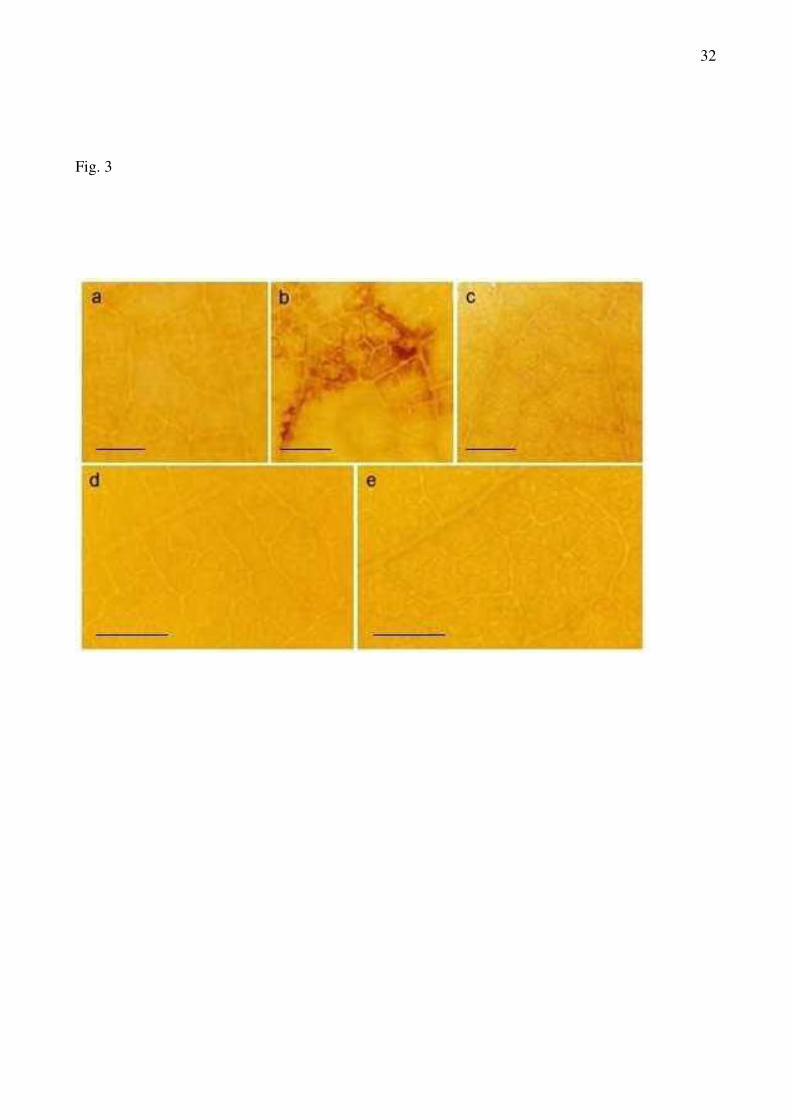
In long-term PPV-infected leaves from susceptible plants, after the staining with DAB
reagent to locate H2O2 production, a red-brown staining in mesophyll cells near the minor veins was
observed (Fig 3b). This staining seemed to be due to H2O2, since it was totally suppressed by 10
mM ascorbic acid, and no staining was observed in control plants or in asymptomatic leaves of
infected plants (Fig 3a; 3c). However, no DAB staining was observed in leaves from control or
inoculated resistant plants (Fig 3d, 3e).
Control plants showed a well-developed ultrastructure, mitochondria, endoplasmic
reticulum, golgi and nucleus. In chloroplasts from the susceptible apricot cultivar, no starch grains
were observed (Fig 4a, b, c, d) in contrast to those observed in the resistant plants (Fig 4e, f, g, h).
PPV infection produced mainly an alteration in chloroplast ultrastructure, and only in susceptible
plants, giving rise to dilated thylakoid membranes (Fig 4d), but other organelles were unaffected.
However, in the PPV-resistant cultivar, no effects were observed in infected plants (Fig 4g,h), in
relation to control plants (Fig 4e, f).
Discussion
The observed results regarding the PPV-resistance of the apricot cultivar SEO and the PPV-
susceptibility of Real Fino, as shown by the strong chlorosis symptoms observed after 4 weeks of
infection, agree with previous data from different authors (Martínez-Gómez and Dicenta 2000a,
14
2000b, Martínez-Gómez et al. 2000). In addition, the ELISA OD values in the case of inoculated
Real Fino were high in comparison with those observed in other apricot cultivars (Cambra et al.
1994), confirming its susceptibility.
Different studies have revealed that plant viruses produce alterations in the photosynthetic
parameters of their host, essentially at the PSII level (Van Kooten et al. 1990, Rahoutei et al. 2000).
PPV produced a decrease in F’v/F’m and ΦPSII in leaves from the susceptible cultivar. In the apricot
cultivar Real Fino, the decrease in F’v/F’m and/or ΦPSII seems to have been due to a loss in Qp. It has
been described that a decrease of Qp is accompanied by an increase in the lifetime of the exciton in
PSII and that this will increase the probability of chlorophyll triplet formation and the associated
formation of 1O2 (Foyer and Harbison 1994). Decreases in Qp and F’v/F’m have been described also
in tobacco plants infected with TMV, PMMoV (pepper mild mottle virus) or PaMMoV (paprika
mild mottle virus) (Van Kooten et al. 1990, Rahoutei et al. 2000). In PPV-infected susceptible
apricot plants, a decrease in Qp was observed, suggesting that under these conditions the production
of AOS, such as 1O2, could be enhanced (Foyer and Harbison 1994). However, in PPV-infected
peach plants, a decrease in the efficiency of excitation energy capture by PSII (F’v/F’m) was
observed, that was accompanied by a drop in NPQ (Hernández et al. 2004b); this could reflect a
diminished capacity for the safe dissipation of excess light energy and, therefore, does not avoid the
production of harmful species, such as 1O2 (Fryer et al. 2002).
In incompatible reactions of plants to viruses, AOS generation may play an important role in
the virus resistance. In the short-term, in infected cells, a rapid accumulation of AOS (especially
H2O2) has been described (Mehdy 1994). The increase in H2O2 levels may cause pathogen
destruction and could be involved as a second messenger in the systemic signal network of plant
cells (Alvarez et al. 1998, Levine et al. 1994). However, in the present work, different factors,
including the use of woody plants, the mode of inoculation (a piece of bark from diseased GF305
peach plants showing strong sharka symptoms) and the time which passed between the subjection
of the plants to the artificial dormancy and the growth of the first expanding leaves, made it difficult
15
to study the short-term responses to PPV infection, and results were obtained for long-term PPV
infection.
As described above, in incompatible plant-virus interaction, AOS generation seems to play
an important role in virus resistance. However, in compatible plant-virus interaction, little is know
about the involvement of AOS in symptom development and pathogenesis or about the effect of
viral infection on the response of antioxidant enzymes. Regarding antioxidant metabolism, in
compatible plant-virus interaction, contradictory results were reported with both induction and
reduction of antioxidant enzymes being described (Riedle-Bauer 2000, Hernández et al. 2001a,
2004b, Clarke et al. 2002, Li and Burrit 2003). The lack of clear trends is probably due to a
combination of variables, including metabolic differences between the plant species studied and
differences in the viral infection processes, the rates of viral movement and/or replication, the
conditions in which the plants were grown and the time of sampling (Li and Burrit 2003).
Most studies on the effect of biotic stress on the activity of antioxidant enzymes have been
conducted with herbaceous plants, and studies with woody plants are less common. Moreover,
information about the effect of PPV infection on the antioxidant systems from apricot plants is very
scarce, being non-existent at subcellular levels.
In apricot plants, p-hydroxy mercury benzoic acid (pHMB)-insensitive APX activity (class
III peroxidase) was detected in both cell fractions (soluble fraction and chloroplasts). These
peroxidases were first described in tea leaves; they exhibited high specific APX activity and
oxidised ASC and organic phenols at comparable rates (Kvaratskhelia et al. 1997). Specific
inhibitors of APX, such as p-chloro-mercury benzoate, hydroxy urea, p-aminophenol or p-hydroxy-
mercury benzoate (pHMB), have only a slight effect on the ASC-dependent peroxidase activity of
this type of peroxidase. In tea leaves, two ascorbate-dependent peroxidases have been described:
TcAPX I and TcAPX II. TcAPX I appears to be an extracellular enzyme exported via the ER, while
TcAPX II is a thylakoid membrane-bound enzyme (Kvaratskhelia et al. 1999). The developmental
regulation of the activity/expression of Class I APXs and Class III POXs seems to be different (de
16
Pinto and de Gara 2004). Class I APXs are generally associated with meristematic and actively
growing tissues, whereas pHMB-insensitive APXs, that should be renamed as Class III APXs to
avoid confusion (Ros Barceló et al. 2005), are generally expressed in non-growing tissues where the
cell wall stiffening process is emerging (de Pinto and de Gara 2004; Ros Barceló et al. 2005). Both
Class III APXs and ascorbate oxidase activities can also acts as ASC removal system in the
apoplastic space. It must be menctioned that ASC itself negatively affect the peroxidase reaction,
which is important for wall stiffening and, in general, for wall differentiation (de Pinto and de Gara
2004; Ros Barceló et al., 2005).
In soluble fractions from inoculated Real Fino plants, significant declines in pHMB-
sensitive APX, DHAR, catalase and peroxidase took place. However, in resistant plants, increases
in pHMB-insensitive APX, MDHAR, GR and SOD occurred. These data indicate that the cytosol
from PPV-resistant apricot plants had a higher capacity to eliminate O2.- and H2O2 and to regenerate
ASC and GSH than that of the PPV-susceptible plants.
Peroxidases were reported to be involved in resistance of Capsicum annuum to CMV
(cucumber mosaic virus) (Candela et al. 1994). However, in the CMV-tolerant pepper cultivar
Gemini F7, tolerance is likely not due to enhanced peroxidase levels (Riedle-Bauer 1998). In the
compatible interactions Cucumis sativus-CMV and Cucurbita pepo-ZYMV (Zucchini yellow
mosaic virus), an important increase in peroxidase activity was produced and all peroxidase
isoforms detected not only functioned as radical scavengers but also catalysed the formation of
H2O2 (Riedle-Bauer 2000). This author suggested that an enhancement of peroxidase activity
contributed to the oxidative stress in systemic plant-virus interactions. However, this capability of
changing the catalytic POX activity is probably a characteristic of specific isoenzymes; whereas,
many other class III isoperoxidases play different role in the plant pathogen interaction (from
increasing cell wall stiffening to be involved in the secondary metabolite biosynthesis. These results
contrast with our data, where the susceptibility to PPV infection correlates with the decrease in
peroxidase activity, that could reflect a disturbance in the cell wall stiffening processes.
17
Catalase activity decreased similarly in soluble fractions from both apricot cultivars. This
observed drop in catalase activity has been described previously in crude extracts from PPV-
infected apricot plants (Hernández et al. 2001a) and a similar decrease has been described in TMV-
infected Nicotiana glutinosa L. plants as well as in Phaseolus vulgaris plants after 5 d of WClMV
infection (Yi et al. 1999, Clarke et al. 2002). In higher plants, catalase is localised mainly in
peroxisomes (del Río et al. 1998). The decrease in catalase observed in both apricot cultivars could
contribute to an increase in peroxisomal H2O2, that could also diffuse through the peroxisomal
membrane into the cytosol (del Río et al. 1998), and the transient accumulation of H2O2 in apricot
leaves could increase the risk of oxidative damage. However, and as mentioned above, the cytosol
from inoculated PPV-resistant plants seems to have a higher capacity to eliminate AOS than that of
the inoculated PPV-susceptible plants, allowing them to cope with this possible H2O2-induced
oxidative stress produced in PPV-infected plants.
In chloroplasts from apricot plants infected by PPV, a differential behaviour was observed,
and only in the susceptible plants did a decrease in the ASC-GSH cycle enzymes occur. Taking into
account that the ASC-GSH cycle is the more important mechanism with respect to elimination of
H2O2 and recycling of ASC and GSH in plants (Asada 1994), and although we have not performed
ASC or GSH analysis, these data suggest that chloroplasts from the susceptible cultivar could have
a lower capacity to scavenge H2O2 and regenerate ASC and GSH than the chloroplasts from the
PPV-resistant cultivar. Several authors have observed a correlation between GSH accumulation and
the resistance to virus infection (Fodor et al. 1997, Gullner et al. 1999). Exposure of tobacco leaf
discs to the cysteine precursor L-2-oxo-4-thiazolidine-carboxylic acid led to a massive
accumulation of GSH as well as reduced TMV coat protein contents and suppression of disease
symptoms in TMV-inoculated tobacco plants (Gullner et al. 1999). In a previous work, and using
crude extracts from two apricot cultivars, foliar DHAR activity increased in response to PPV
infection, but the rise was much higher in the resistant plants (300%) (cv Goldrich) than in the
susceptible ones (only 37%) (cv. Real Fino), suggesting that the inoculated resistant cultivar had a

18
higher capacity for regeneration of ASC than the inoculated susceptible plants (Hernández et al.
2001a). This higher DHAR induction in the PPV-resistant cultivar could contribute to an increased
antioxidant capacity, which could be related, among other factors, to its resistance to PPV.
However, these results differed from those described in the present work, where no changes was
observed in DHAR activity in soluble fractions and chloroplasts from the resistant cultivar, whereas
a decrease was observed in both cell fractions from the susceptible cultivar.These differences could
be due that in the first case, authors used crude extracts from apricot leaves (Hernández et al.
2001a), whereas in this case we used purified cell fractions. These results agree with those recently
described for infected peach plants, susceptible to PPV, where a decrease in chloroplastic GR and
MDHAR took place (Hernández et al. 2004b).
The decrease in the antioxidant enzyme levels observed in the cytosolic and chloroplastic
fractions from PPV-infected susceptible plants produced an oxidative stress, as shown by the
increase in the levels of lipid peroxidation and protein oxidation. Lipid peroxidation and protein
oxidation are the symptoms most easily ascribed to oxidative damage and they are often used as
indicators of oxidative damage (Hernández et al. 2001b, 2004a, 2004b). This PPV-induced
oxidative stress was also manifested as an accumulation of H2O2 in leaves. H2O2 accumulation
occurred only in PPV-infected symptomatic leaves from susceptible plants, but not in
asymptomatic leaves. However, in the resistant cultivar, no oxidative stress was produced, as shown
by the unchanged levels of lipid peroxidation and protein oxidation as well as by the absence of
H2O2 accumulation in leaves. In the inoculated susceptible apricot cultivar, the presence of some
microbursts of H2O2 correlated with the decrease in H2O2-scavenging enzymes (APX, catalase and
peroxidase). PPV infection produced intervenal chlorosis symptoms that could have been induced
by an increased AOS generation, as shown in pea plants subjected to NaCl stress (Hernández et al.
2001b). As described above, the fall in Qp is associated with the AOS formation in chloroplasts
(Foyer and Harbison 1994). This, linked to the decrease in the levels of antioxidant enzymes in the
chloroplasts from PPV-infected susceptible plants, could be responsible for the chlorosis symptoms
19
observed. So, the chlorosis symptoms could be ascribed both to the higher AOS generation and to a
lower capacity for scavenging of AOS, as described previously for peach plants (Hernández et al.
2004b).
PPV infection produced some ultrastructural alterations in the susceptible apricot cultivar.
The presence of dilated thylakoids seems to be a general stress response, because they have been
described previously, both under biotic and abiotic stress (Russo and Martelli 1982, Hernández et
al. 1995). PPV infection also produced ultrastructural alterations in peach cv. GF305: the presence
of dilated thylakoids an increase in the number and size of plastoglobuli and a decrease in the
amount of starch granules (Hernández et al. 2004b). However, the percentage of chloroplasts
showing dilated thylakoids in susceptible apricot plants was lower than that observed in PPV-
infected peach plants. These differences could be due to the fact that peach plants are more
susceptible to PPV infection and showed more chlorosis symptoms than this apricot cultivar.
Resistance to PPV could be associated with a HR at the graft site in SEO cultivar. However,
no HR was observed at the graft site nor in the leaves. On the other hand, the effects on antioxidant
systems from SEO plants could be due to a systemic acquired resistance induced by PPV
penetration in stem tissue at the graft site. However, the oxidative stress observed in susceptible
plants can merely be a consequence of virus infection. This would explain why no oxidative stress
is observed in the resistant cultivar, since no viral particles was detected (see Table 1).
It has been proposed that a decline in free radical scavenging capacity may be required
before a rapid increase in virus replication can take place, and treatments increasing the ability of
plants to scavenge AOS may hinder virus replication (Clarke et al. 2002). If the same situation
occurs in PPV-infected susceptible apricots, the oxidative stress, accompanied by a decrease in
soluble and chloroplastic antioxidant enzymes, could be related to the progress of PPV infection.
The higher antioxidant capacity observed in PPV-inoculated resistant plants, in relation to the PPV-
inoculated susceptible plants, could be important in hindering virus replication, in agreement with

20
results reported for WClMV-infected Phaseolus vulgaris plants (Clarke et al. 2002). In this sense,
the higher antioxidant capacity showed by SEO plants in relation to PPV-susceptible plants, could
be associated, among other factors, to their resistance to PPV.
Acknowledgements – The authors thank Dr. David J. Walker, for his valuable review of this
manuscript and for correction of the English, and Mr. Mariano Gambín, for technical assistance.
This research was supported by grant AGL 2002-02115 from the CICYT (Spanish Ministry of
Education and Science).
REFERENCES
Adams A, Farkas T, Somlayai G, Hevesi M, Kiraly Z (1989) Consequence of O2- generation during
bacterially induced hypersensitive reaction in tobacco: deterioration of membrane lipids.
Physiol Mol Plant Pathol 34: 13-26.
Alvarez ME, Pennell RI, Meijer PJ, Ishikawa A, Dixon RA, Lamb C (1998) Reactive oxygen
intermediates mediate a systemic signal network in the establishment of plant immunity.
Cell 92: 773-784.
Asada K (1994) Production and action of active oxygen species in photosynthetic tissues. In: Foyer
CH, Mullineaux PM (eds) Causes of Photooxidative Stress and Amelioration of Defence
Systems in Plants. Boca Raton, Florida, USA: CRC Press, pp 77-104.
Asensio M. (1996) El virus de la sharka (Plum pox virus). Caracterización, diagnóstico y detección
mediante anticuerpos monoclonales específicos. PhD diss. Universidad Politécnica de
Valencia. Valencia, Spain.
Bernhard R, Marénaud C, Sutic D (1969) Le pêcher GF305 indicateur polyvalent des virus des
espèces a noyau. Ann Phytopathol 1: 603-617.
Bilger W, Björkman O (1990) Role of the xanthophyll cycle in photoprotection elucidated by
measurements of light-induced absorbance changes, fluorescence and photosynthesis in leaves of
Hedera canariensis. Photosynth Res 25: 173-185
Cakmak I, Horst WJ (1991) Effect of aluminium on lipid peroxidation, superoxide dismutase,
catalase and peroxidase activities in root tips of soybean (Glycine max). Physiol Plant 83:
463-468.
Candela ME, Muñoz R, Alcaraz MD, Espín A (1994) Isoperoxidase involvement in the resistance
of Capsicum annuum to infection by cucumber mosaic virus. J Plant Physiol 143: 213-217.

21
Cambra M, Asensio M, Gorris MT, Camarasa E, García JA, Moya JJ, López-Abella D, Vela C, Sanz
A (1994) Detection of Plum pox virus using monoclonal antibodies to structural and non-
structural proteins. Bulletin EPPO 24: 569-578.
Clarke SF, Guy PL, Burritt DJ, Jameson PE (2002) Changes in the activities of antioxidant enzymes
in response to virus infection and hormone treatment. Physiol Plant 114:157-164
del Río LA, Pastori GM, Palma JM, Sandalio LM, Sevilla F, Corpas FJ, Jiménez A, López-Huertas
E, Hernández JA (1998) The activated oxygen role of peroxisomes in senescence. Plant
Physiol 116: 1195-1200
De Gara L, De Pinto MC, Tommasi F (2003) The antioxidant systems vis-à-vis reactive oxygen
species during plant-pathogen interaction. Plant Physio Biochem 41: 863-870
de Pinto,M.C.; De Gara,L. (2004) Changes in the ascorbate metabolism of apoplastic and
symplastic spaces are associated with cell differentiation. Physiol Plant 55:2559-2569.
Doke N, Ohashi Y (1988) Involvement of an O2.- generating system in the induction of necrotic lesions
on tobacco leaves infected with tobacco mosaic virus. Physiol Mol Plant Pathol 32: 163-175
Fodor J, Gullner G, Adám AL, Barna B, Kömives T, Király Z (1997) Local and systemic responses
of antioxidants to tobacco mosaic virus infection and to salicylic acid in tobacco. Plant Physiol
114: 1443-1451
Foyer CH, Harbison J (1994) Oxygen metabolism and the regulation of photosynthetic electron
transport. In: Foyer CH and Mullineaux P (eds) Causes of Photooxidative Stresses and
Amelioration of Defense Systems in Plants. Boca Raton, Florida, USA: CRC Press, pp 1-42.
Fryer MJ, Oxborough K, Mullineaux PM, Baker NR (2002) Imaging of photo-oxidative stress
responses in leaves. J Exp Bot 53: 1249-1254
García-Limones C, Hervás A, Navas-Cortés JA, Jiménez-Díaz RM, Tena M (2002) Induction of an
antioxidant enzyme system and other oxidative stress markers associated with compatible
and incompatible interactions between chickpea and Fusarium oxysporum f. sp. ciceris.
Physiol Mol Plant Pathol 61: 325-337
Genty B, Briantais JM, Baker NR (1989) The relationship between the quantum yield of
photosynthetic electron transport and the quenching of chlorophyll fluorescence. Biochim
Biophys Acta 990: 87-92
Gullner G, Tóbiás I, Fodor J, Kömives T (1999) Elevation of glutathione level and activation of
glutathione-related enzymes affect virus infection in tobacco. Free Rad Res 31:S155-161
Hernández JA, Olmos E, Corpas FJ, Sevilla F, del Río LA (1995) Salt-induced oxidative stress in
chloroplast of pea plants. Plant Sci 105: 151-167

22
Hernández JA, Campillo A, Jiménez A, Alarcón JJ, Sevilla F (1999) Response of antioxidant
systems and leaf water relations to NaCl stress in pea plants. New Phytol 141: 241-251.
Hernández JA, Jiménez A, Mullineaux PM, Sevilla F (2000) Tolerance of pea (Pisum sativum L.) to
long-term salt stress is associated with induction of antioxidant defences. Plant Cell Environm
23: 853-862
Hernández JA, Talavera JM, Martínez-Gómez P, Dicenta F, Sevilla F (2001a) Response of
antioxidative enzymes to plum pox virus in two apricot cultivars. Physiol Plant 111: 313-
321.
Hernández JA, Ferrer MA, Jiménez A, Ros-Barceló A, Sevilla F (2001b) Antioxidant systems and
O2.-/H2O2 production in the apoplast of Pisum sativum L. leaves: its relation with NaCl-
induced necrotic lesions in minor veins. Plant Physiol 127: 817-831.
Hernández JA; Rubio M., Portillo B., Dicenta F., Martínez-Gómez P (2003) Response of
antioxidant enzymes from peach (Prunus persica L. cv GF305) to plum pox virus infection.
Free Rad Res 37(S2):14
Hernández JA, Escobar C, Creissen G, Mullineaux P (2004a) Role of hydrogen peroxide and the
redox state of ascorbate in the induction of antioxidant enzymes in pea leaves under excess
light stress. Funct Plant Biol 31: 359-368
Hernández JA, Rubio M, Olmos E, Ros-Barceló A, Martínez-Gómez P (2004b) Oxidative stress
induced by long-term plum pox virus infection in peach (Prunus persica L. cv GF305).
Physiol Plant 122:486-495.
Jiménez A, Hernández JA, Pastori G, del Río LA, Sevilla F (1998) The role of the ascorbate-
glutathione cycle of mitochondria and peroxisomes in the senescence of pea leaves. Plant
Physiol 118: 1327-1335
Kölber M (2001) Workshop on Plum pox.. Acta Hort 550: 249-255
Kvaratskhelia M, Winkel C, Thorneley RNF (1997) Purification and characterization of a novel
class III peroxidase isozyme from tea leaves. Plant Physiol 114: 1237-1245.
Kvaratskhelia M, Winkel C, Naldrett MT, Thorneley RNF (1999) A novel high activity cationic
ascorbate peroxidase from tea (Camelia sinensis)- A class III peroxidase with unusual
substrate specificity. J Plant Physiol 154:273-282.
Levine IR, Garland D, Oliver C, Amici A, Climent, I, Lenz A, Ahn B, Shaltiel S, Stadtman ER
(1990) Determination of carbonil content in oxidatively modified proteins. Methods
Enzymol 186: 464-478.
Levine A, Tenhaken, R, Dixon R, Lamb C (1994) H2O2 from the oxidative burst orchestrates the
plant hypersensitive disease resistance response. Cell 79: 583-593
23
Li Z, Burritt DJ (2003) The influence of Cocksfoot mottle virus on antioxidant metabolism in the
leaves of Dactylis glomerata L. Physiol Mol Plant Pathol 62: 285-295
MacKenzie DJ, McLean MA, Mukerji S, Green M (1997) Improved RNA extraction from woody
plants for the detection of viral pathogens by reverse transcription-polymerase chain reaction.
Plant Dis 81: 222
Martínez-Gómez P, Dicenta F (2000a) Evaluation of resistance of apricot cultivars to a Spanish isolate
of plum pox potyvirus (PPV). Plant Breeding 119: 179-181.
Martínez-Gómez P, Dicenta F (2000b) In vitro evaluation of apricot (Prunus armeniaca L.) resistance
to plum pox potyvirus. J Hort Sci Biotechnol 75: 259-261
Martínez-Gómez P, Dicenta F, Audergon JM (2000) Behaviour of apricot (Prunus armeniaca L.)
cultivars in the presence of sharka (plum pox potyvirus): a review. Agronomie 20: 407-422
Martínez-Gómez P, Rubio M, Dicenta F, Aparicio F, Pallás V (2003) Comparative analysis of three
diagnostic methods for the evaluation of plum pox virus (PPV) resistance in apricot breeding
programs. Acta Hort 622: 353-357.
Maxwell K, Johnson GN (2000) Chlorophyll fluorescence- a practical guide. J Exp Bot 51: 659-
668.
McCord JM, Fridovich I (1969) Superoxide dismutase: an enzymic function for erythrocuprein. J
Biol Chem 244: 6049-6055.
Mehdy MC (1994) Active oxygen species in plant defense against pathogens. Plant Physiol 105:
467-472.
Morales MA, Olmos E, Torrecillas A, Sanchez-Blanco MJ, Alarcon JJ (2001) Differences in water
relations, leaf ion accumulation and excretion rates between cultivated and wild species of
Limonium sp. grown in conditions of saline stress. Flora 196: 345-352.
Pelet F, Bovey R (1968) Les symptômes de la Sharka sur les pruniers, pruneautiers, abricotiers et
pêchers. Agric Rom 7: 80-84
Pomar F, Novo M, Bernal MA, Merino F, Ros Barceló A (2004) Changes in stem lignins (monomer
composition and crosslinking) and peroxidase are related with the maintenance of leaf
photosynthesis integrity during Verticillium wilt in Capsicum annuum. New Phytol 163: 111-
123
Rahoutei J, García-Luque I, Barón M (2000) Inhibition of photosynthesis by viral infection: Effect
on PSII structure and function. Physiol Plant 110:286-292.
Riedle-Bauer M (1998) Activities of antioxidant enzymes in cucumber plants infected with
cucumber mosaic virus. Phyton 38: 149-157
Riedle-Bauer M (2000) Role of reactive oxygen species and antioxidant enzymes in systemic virus
infections of plants. J Phytopathol 148:297-302.

24
Ros-Barceló A., Gómez-Ros LV, Ferrer MA, Hernández JA (2005) The apoplastic antioxidant
enzymatic system in the wood-forming tissues of trees. Trees Struct Func. (accepted for its
publication)
Russo A, Marterlli GP (1982) Ultrastructure of turnip crinkle- and saguaro cactus virus-infected
tissues. Virology 118: 109-116
Sutula CL, Gillet JM, Morrissey SM, Ramsdell DC (1986) Interpreting ELISA data and
establishing the positive-negative threshold. Plant Dis 70: 722-726
Thordal-Cristensen H, Zhang Z, Wei Y, Collinge DB (1997) Subcellular localization of H2O2 in
plants. H2O2 accumulation in papillae and hypersensitive response during the barley-
powdery mildew interaction. Plant J 11: 1187-1194
Van Kooten O, Meurs C, van Loon LC (1990) Photosynthetic electron transport in tobacco leaves
infected with tobacco mosaic virus. Physiol Plant 80: 446-452.
Yi SY, Yu SH, Choi D (1999) Molecular cloning of a catalase cDNA from Nicotiana glutinosa L.
and its repression by tobacco mosaic virus. Mol Cells 9:320-325

25
Table 1.- Plum pox virus detection in leaves of Real Fino and SEO apricot cultivars grafted onto
control and inoculated GF305 rootstocks during the two cycles of study.
Cycle 1 Cycle 2
Symptomsa ELISAb RT-PCRc Symptomsa ELISAb RT-PCRc
Real Fino
Control 0 (0.0) 0 (0.10) 0 0 (0.0) 0 (0.07) 0
PPV-Inoculated 8 (2.3) 8 (1.64) 8 10 (2.8) 10 (1.85) 10
SEO
Control 0 (0.0) 0 (0.06) 0 0 (0.0) 0 (0.06) 0
PPV-Inoculated 0 (0.0) 0 (0.08) 0 0 (0.0) 0 (0.07) 0
a Number of plants with symptoms and mean intensity (between parenthesis) scored on a scale from
0 (no symptoms) to 5 (maximum intensity).b Number of plants ELISA positive and optical density (between parenthesis) at 405 nm after 60
min.c Number of plants RT-PCR positive.

26
Table 2.- Fluorescence parameters measured in control and long-term PPV-infected apricot leaves.
Data represent means ± SE from at least ten repetitions. Differences from control values are
significant at P<0.05 (a), according to Duncan’s multiple range test.
Fluorescence parameters
Fv/Fm F’v/F’m ΦPSII qp NPQ
Real Fino
Control 0.804±0.022 0.660±0.024 0.066±0.003 0.100±0.008 0.232±0.007
PPV-inoculated 0.782±0.018 0.562±0.040a 0.046±0.002a 0.082±0.002a 0.282±0.024
SEO
Control 0.757±0.020 0.788±0.026 0.069±0.003 0.088±0.004 0.407±0.038
PPV-inoculated 0.733±0.023 0.787±0.016 0.085±0.010 0.108±0.014 0.352±0.040
27
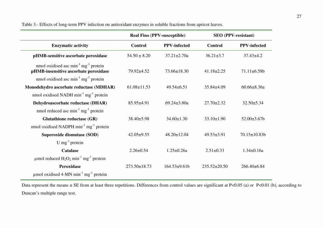
Table 3.- Effects of long-term PPV infection on antioxidant enzymes in soluble fractions from apricot leaves.
Real Fino (PPV-susceptible) SEO (PPV-resistant)
Enzymatic activity Control PPV-infected Control PPV-infected
pHMB-sensitive ascorbate peroxidase
nmol oxidised asc min-1 mg-1 protein
54.50 ± 8.20 37.21±2.70a 36.21±3.7 37.43±4.2
pHMB-insensitive ascorbate peroxidase
nmol oxidised asc min-1 mg-1 protein
79.92±4.52 73.66±18.30 41.18±2.25 71.11±6.58b
Monodehydro ascorbate reductase (MDHAR)
nmol oxidised NADH min-1 mg-1 protein
61.08±11.53 49.54±6.51 35.84±4.09 60.66±8.36a
Dehydroascorbate reductase (DHAR)
nmol reduced asc min-1 mg-1 protein
85.95±4.91 69.24±3.80a 27.70±2.32 32.50±5.34
Glutathione reductase (GR)
nmol oxidised NADPH min-1 mg-1 protein
38.40±5.98 34.60±1.30 33.10±1.90 52.00±3.67b
Superoxide dismutase (SOD)
U mg-1 protein
42.05±9.55 48.20±12.04 49.53±3.91 70.15±10.83b
Catalase
µmol reduced H2O2 min-1 mg-1 protein
2.26±0.54 1.25±0.26a 2.51±0.33 1.34±0.16a
Peroxidase
µmol oxidised 4-MN min-1 mg-1 protein
273.50±18.73 164.53±9.61b 235.52±20.50 266.40±6.84
Data represent the means ± SE from at least three repetitions. Differences from control values are significant at P<0.05 (a) or P<0.01 (b), according to
Duncan’s multiple range test.

28
Table 4.- Effects of long-term PPV infection on antioxidant enzymes in chloroplast suspensions from apricot leaves.
Real Fino (PPV-susceptible) SEO (PPV-resistant)
Enzymatic activity Control PPV-infected Control PPV-infected
pHMB-sensitive ascorbate peroxidase
nmol oxidised asc min-1 mg-1 protein
78.60±12.97 55.47±5.98a 41.99±5.53 40.88±11.46
pHMB-insensitive ascorbate peroxidase
nmol oxidised asc min-1 mg-1 protein
89.46±14.90 95.73±25.50 91.83±7.79 63.40±5.78a
Monodehydro ascorbate reductase (MDHAR)
nmol oxidised NADH min-1 mg-1 protein
56.51±9.77 28.15±4.27a 43.20±7.40 49.31±10.21
Dehydroascorbate reductase (DHAR)
nmol reduced asc min-1 mg-1 protein
31.29±4.82 18.99±4.33a 24.54±2.98 26.61±2.03
Glutathione reductase (GR)
nmol oxidised NADPH min-1 mg-1 protein
24.04±2.15 14.44±4.51a 13.06±3.20 12.04±1.45
Superoxide dismutase (SOD)
U mg-1 protein
17.63±2.75 21.06±3.59 14.76±1.43 17.63±3.89
Data represent the means ± SE from at least three repetitions. Differences from control values are significant at P<0.05 (a), according to Duncan’s
multiple range test.
29
Legend to Figures
Fig. 1.- Amplification products (313 bp) indicative of the presence of PPV, obtained using RT-PCR
for PPV detection in different samples. Lane 1: Real Fino cultivar grafted onto control GF305
seedling, Lane 2: Real Fino cultivar grafted onto GF305 seedling inoculated by PPV and showing
strong sharka symptoms, Lane 3: Start Early Orange cultivar grafted onto control GF305 seedling,
Lane 4: Start Early Orange cultivar grafted onto GF305 seedling inoculated by PPV and showing
strong sharka symptoms. M: Molecular size standard 1 Kb Plus DNA Ladder.
Fig 2.- Effect of long-term PPV infection on lipid peroxidation (given as TBARS) and protein
oxidation in apricot leaves. Data represent the means ± standard errors of at least three replicates.
Differences from control values are significant at P<0.05 (a) or P<0.01 (b), according to Duncan’s
multiple range test. RFc, susceptible control plants; RFi, susceptible PPV-inoculated plants; SEOc,
resistant control plants; SEOi, resistant PPV-inoculated plants.
Fig 3.- Detection of H2O2 generation in leaves from DAB-stained apricot plants. A) susceptible
control plants; B) susceptible PPV-inoculated plants; C) asymptomatic susceptible PPV-inoculated
plants; D) resistant control plants; E) resistant PPV-inoculated plants. Bars = 500 µm.
Fig 4.- Electron micrographs from apricot leaves. a) Palisade mesophyll cells from control
susceptible plants. b) Susceptible control plant. Detail of a chloroplast of a palisade mesophyll cell.
c) Palisade mesophyll cells from susceptible PPV-infected plants. d) Detail of a chloroplast of a
palisade mesophyll cell from PPV-infected susceptible plants showing dilated thylakoids. e)
Palisade mesophyll cells from control PPV-resistant plants. f). Resistant control plant. Detail of a
chloroplast of a palisade mesophyll cell., g) Palisade mesophyll cells from resistant PPV-infected
plants. h) Inoculated control plant. Detail of a chloroplast of a palisade mesophyll cell. Chl,
chloroplast; N, Nucleous, S, strach granules; V, vacuole.

30
Fig. 1
M 1 2 3 4
313 nt
M 1 2 3 4
313 nt

31
Fig. 2
0
2
4
6
8
TB
AR
S (
nm
olx
g´1
FW
) aA
0
20
40
60
80
RFc RFi SEOc SEOi
CO
-pro
tein
s(n
mo
lg-1
FW
)
bB
0
2
4
6
8
TB
AR
S (
nm
olx
g´1
FW
) aA
0
2
4
6
8
TB
AR
S (
nm
olx
g´1
FW
) aA
0
20
40
60
80
RFc RFi SEOc SEOi
CO
-pro
tein
s(n
mo
lg-1
FW
)
bB
0
20
40
60
80
RFc RFi SEOc SEOi
CO
-pro
tein
s(n
mo
lg-1
FW
)
bB
32
Fig. 3

33
Fig 4
a b
c d
e
g
f
V V
N
V
Chl
Chl
Chl
h
V
N
S
S
Chl
V
V
V
S
S