1-s2.0-0002941650900649-main
DESCRIPTION
ddTRANSCRIPT

THE: FRANKE’(_)RT-~lANT)IB~II,AK PLAN13 AN(:liE: ANI) TIIP: FACTAT, PATTERN
ERNEST L. JOHNSON, l).l).S., I~'.A.C.I).,* SAN FRANCISCO, (~,IF.
S OME years ago a great, industrial Icadcr was asked to explain the amazing advancement of American industries. His reply was short, but factual, for
he said: “We progress only when we are capable of using the t,houghts of many. ’ ’ HOW well this applies to the America,11 sciences, and particularly to orthodontics. There can be no doubt that the amazing advancement in the field of orthodontics has been due to the contribution of the thoughts of many. It is hoped,that this work will contribute toward that progress.
Since 1946, when Tweed’ described his method of using the Frankfort- mandibular plane angle (as measured from lateral photographs) in orthodontic diagnosis, classifications, and trcat,ment planning, the use of this particular measurement of facial morphology has rapidly won favor among orthodontists. Downs* included this among ten important measurements taken from cephalo- metric head films, and provided a mean value and maxirnal and minimal values derived from a group of individuals having excellent occlusion. Salzmann” described an instrument for measuring the Frankfort-mandibular plane angle and other angles without the use of roentgenographic equipment. Continued use testifies to the fact that, the Frankfort-mandibular angle is clinically useful. It is therefore not disparaging to remark that, this angle, like other angles, is a measure of the relative position of the points which by definition form the angle. Angles seldom, if ever, provide any true indication or absolute measure of the specific location of the departure from acceptable facial morphology. Angles will always continue to be useful in the assessment of rraniofacial morphology, since it has been demonstrated4 that these angles remain relatively unchanged from birth to maturity, and that to a large extent age differences may therefore be ignored. At the same time it is highly desirable to know where exactly facial morphology veers away from desirable st,andards, and in that search we are often obliged to use methods of measurement other t,han angles.
That there is a change in orthodontic thinking is becoming increasingly more apparent. Brodie” and WylieG have both called for reorientation of ort,ho- dontic t,hinking concerning the basis of deviations from the so-called “normal” facial pattern, and have maintained that these aberrations should not necessarily be considered as defects of growth, nor as undesirable postnatal alterations in craniofacial morphology, nor even as abnormalities. Instead it has been sug- gested by them that man\- of these facial l)attcrns which orthodontists find un- desirable are in reality only the random combination of separate facial parts, each of which is wit,hin the normal range of variation in conformation and size, but which in toto combine to produce an undesirable facial configuration.
This thesis was submitted to the American Roard of Ortho(lontics in pxrtial fulfillment of the author’s requirements for certiflc’ation.
*Demonstrator in Orthodontics, TJniversity of California.
516

~R‘~NI<FOKT-~inSDIRULAR I’LASI? .\SGLE AKD FACIBL PATTERN 517
While it would be unwise to say that orthodontists have accepted all the views stated in the two articles last mentioned, on the other hand there has been at least a tacit acceptance of this philosophy among orthodontists in recent years, as testified by the fact that orthodontists today are more inclined to describe the facial patterns, often quantitatively, rather than explain why they occur. Wylie’ recently advanced a quant.itative method for the assessment of cranio- facial dysplasia in the anteroposterior plane of space, using linear values meas- ured in millimeters rather than angles. With this method, it is possible to localize anteroposterior dysplasia in the cranial base, in the length or lack of it in the maxilla or the mandible, and in the placement of the maxillary dental arch upon its bony base. This system of assessment of anteroposterior dysplasia also has some use in ferreting out cases in which abnormal arch relationships are due to mandibular displacement, as described by Thompson.8
This study is intended to explain, in specific and localized terms, how and why the facial pattern is bad when the Frankfort-mandibular plane angle be- comes increasingly large.
If it is possible to state that some single anatomical factor is responsible for the undesirable facial conformation which is characterized by a steep man- dibular plane angle, then systemati,c measurement of a group of patients properly classified with respect to that angle should demonstrate the fact. Furthermore, if this kind of facial pattern is characterized by two or more anatomical departures from the typical facial morphology, measurement should also demonstrate this and, perhaps, even indicate the interrelationship between the two or more factors involved.
The great variety of differences in human facial pattern made the prelim- inary organization of these data somewhat difficult. It is relatively easy to find isolated cases which will support a particular theory propounded to explain a specific kind of facial pattern, and further search will even make it possible to duplicate with similar cases instances of the same kind which can reinforce the theory further. On the other hand, when 150 cases are assembled, with no preconceived notion to be substantiated, contradictions and exceptions and extremes in facial pattern crop up to make it very obvious that simple and limited explanations for variations in facial morphology can never suffice. Faces vary from one to another and for many different reasons.
One hundred fifty lateral cephalometric head films from the University of California Orthodontic Clinic were used. These were selected at random to give as good as possible a cross section of the orthodontic material presenting to the clinic. Care was taken to select only patients with full components of teeth, as mutilated cases would be misleading. Also, care was taken to be sure there was a fair sample of cases with good occlusion and acceptable facial morphology to act as a control.
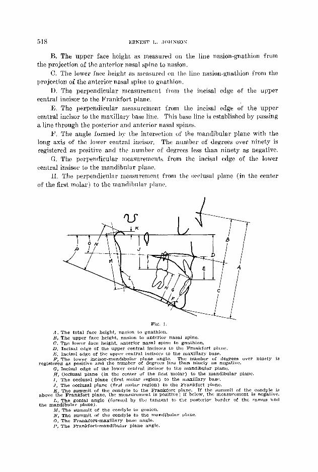
Tracings of these head films were made and the following measurements in millimeters taken as illustrated in Fig. 1:
A. The total face height as measured from nasion to gnathion.
518 ERNEST I,. JOHNSOK
B. The upper face height as measured on the line nasion-gnathion from the projection of the anterior nasal spine to nasion.
C. The lower face height as measured on the line nasion-gnathion from the projection of the anterior nasal spine to gnathion.
D. The perpendicular measurement from the in&al edge of the upper central incisor to the Frankfort plane.
E. The perpendicular measurement from the incisal edge of the upper central incisor to the maxillary base line. This base line is established by passing a line t,hrough the posterior and anterior nasal spines.
F. The angle formed by the interection of the mandibular plane with the long axis of the lower central incisor. The number of degrees over ninety is registered as positive and the number of degrees less than ninety as negative.
G. The perpendicular measurements from the incisal edge of the lower central insisor to the mandibular plane.
H. The perpendicular measurement from the occlusal plane (in the cent,er of the first molar) to t,he mandibular plane.
Fig. 1.
A, The total face height, nasion to gnathion. B, The upper face height, nasion to anterior nasal spine. C, The lower face height, anterior nasal spine to gnathion. D, Incisal edge of the upper central incisors to the Frankfort &me. E, In&al edge of the upper central incisors to the maxillary base. I?, The lower incisor-mandibular plane angle. The number of degrees over ninety is
registered as positive and the number of degrees less than ninety as negative. G, Incis’al edge of the lower central incisor to the mandibular plane. H, Occlusal plane (in the center of the flrst molar) to the mandibular plane. I, The occlusal plane (flrst molar regi’on) to the maxillary base. J, The occlusal plane (flrst molar region) to the Frankfort plane. K, The summit of the condyle to the Frankfort plane. If the summit of the condyle is
above the Frankfort plane, the measurement is positive; if below, the measurement is negative. L, The genial angle (formed by the tangent to the Posterior border of the ramus and
the mandibular plane). M, The summit of the condyle to gonion. N, The summit of the mndyle to the mandibular plane. 0, The Frankfort-maxillary base angle. P, The Frankfort-mandibular plane angle.

FRAi’iKFORT-MANDIBULAR PLANE ANGLE AND FACIAL PATTERN 519
I. The perpendicular measurement from the occlusal plane (in the center of the first molar region) to the maxillary base line.
J. The perpendicular measurement from the occlusal plane (in the center of the first molar region) to the Frankfort plane.
K. The perpendicular measurement from the summit of the eondyle to the Frankfort plane. The point used is the mean between the two condyles. If the summit of the condyle is above the Frankfort plane, the measurement is positive, if below, the measurement is negative.
L. The gonial angle formed by the tangent to the posterior border of the ramus and the mandibular plane.
&I. The length of the ramus from the summit of the condyle to gonion. N. The perpendicular measurement from the summit of the condyle to the
mandibular plane. 0. The angle formed by the Frankfort plane and the maxillary base line
(Frankfort-maxillary base angle) . This angle is positive if the planes converge posteriorly.
P. The angle formed by the Frankfort plane and the mandibular plane (Frankfort-mandibular plane angle).
In order to evaluate the material, it was divided into four groups, deter- mined by the Frankfort-mandibular plane angle (Table I). There were 47 cases with the Frankfort-mandibular plane angle 25O or less, 49 with the Frank- fort-mandibular plane angle between 26’ and 31’ inclusive, 26 with the Frank- fort-mandibular plane angle between 32” and 34” inclusive, and 28 with the Frankfort-mandibular plane angle 35” or more.
A. In looking at the first appraisal “A” of Table I, it will be noted that there is a consistent increase in the total face height (13.28 mm.) as the Frank- fort-mandibular plane angle increases.
B. The slight increase of the upper face height (1.65 mm.) as the Frankfort- mandibular plane angle increases shows the tendency of vertical facial growth to predominate over forward growth in the upper face, thus contributing to the increase in total face height.
C. The very marked and consistent increase in the lower face height (11.63 mm.), as the Frankfort-mandibular plane angle increases, is determined mainly by the vertical growth of the mandibular and maxillary alveolar processes, thus making the lower face an important contributor to the total face height, and in turn making it play a major role in determining the Frankfort-mandibular plane angle.
D. This appraisal of the distance between the Frankfort-mandibular plane and the upper central incisors also shows a consistent and rapid increase, as might be expected, for it includes part of the upper face and all of the vertical growth of the upper alveolar process.
E. As with D, there is a consistent increase of the distance between the incisal edge of the upper central incisors and the maxillary base. This is largely the result of the increase in vertical growth of the upper alveolar process, as the Frankfort-mandibular plane angle increases.

520 IGtNEST I,. JOHSSOS
F. This shows the importance of considering more than the lower incisor mandibular plane relationship when det,ermining the normal location for this tooth. As the Frankfort-mandibular plane angle increases, the lower incisors become more upright. This is a good thing, or they wouId be lying forward in the face when the Frankfort-mandibular plane angle is very large. Tt is proh- ably the muscle pressure of the lower lip t,hat keeps the lower incisors back, even when the bony pattern of the lower jaw would have a predisposing tendency of throwing them forward. Conversely, when the Frankfort-mandibular plane angle is small the lower incisors become more procumbent. In the first group of 47 cases with the Frankfort-mandibular plane angle 25O or less, t,here were 21 cases with the lower incisor mandibular angle between plus IO and plus, 25’. In the fourth group of 28 cases with the Frankfort-mandibular plane angle 35O or more, there were 6 cases with the lower incisor mandibular angle between minus 5 and minus 14O.
TABLEI .___
A. Total face height nasion-gonion B. Goner face height nasion-maxillarv base C. Lower face height maxillary base-“gnathion D. Incisal edge upper central to Frankfort plane E. Incisal edge upper central to maxillary base F. Lower incisor mandibular plane angle G. Incisal edge lower central to mandibular base H. Occlusal plane to mandibular plane
-__ ~~~ FRANKFORT-MANDIBULAR PLANE ANGLE
25" 35" ORLESS 26-31" 32-34O OR MORE I-J=47 N = 49 ~=26 NE28 106.32 109.42 112.46 119.60
48.89 50.00
(first molar region) I. Occlusal plane to maxillary base
(first molar region) J. Oeclusal ulane to Frankfort ulane
50.45 55.87 50.02 27.34 +7.70 40.13 31.00
60.53 62.46 51.55 54.23 28.69 29.42 f5.77 t3.61 42.55 43.42 31.24 31.46
52.10 67.50 55.53 29.64
+0.92 44.60 32.04
(first molar region) 1_ K. Condyle summit to Frankfort plane L. Genial angle M. Condyle summit to gonion N. Condyle summit to mandibular plane 0. Frankfort-maxillary base angle P. Frankfort-mandibular plane angle
20.94 21.55 22.04 22.57
43.28 43.26 43.42 45.00
to.02 122.04
59.98 52.83 to.96 21.97
t1.75 t2.77 t5.17 126.63 128.81 132.46
55.18 54.38 55.64 48.51 45.38 44.25 3-3.00 t4.69 i-6.17 28.34 33.23 38.71
G. The decisive and steady increase (4.47 mm.) in the distance between the incisal edge of the lower central incisors and the mandibular plane illustrates the large part the lower alveolar process contributes to the total face height as the Frankfort-mandibular plane angle increases.
H. The very slight increase (1.04 mm,) in the distance between the oeclusal plane and the mandibular plane in the first molar region shows that most of the increase in vertical height is limited to the profile as the Frankfort-man- dibular plane angle increases.
I. Although progression exists in the distance between the occlusal plane in the first molar region and the maxillary base, it is very slight (1.63 mm.) in comparison to the increase in the total face height.
J. Similar to I, there is a slight progression (1.72 mm.) in the distance between the occlusal plane in the first molar region and the Frankfort plane as the Frankfort-mandibular plane angle increases.

521
A. Total face height (N-GN) 25 26-31 32-34 35
and under and over
N=47 N=49 N=26 N=28
200
190
180
170
160
150
140
130
120 . . .
110 :i: .:,;. .:f. .I.
100 :::. 'a:.: 90
80
70
60
50
40
30
20
10
0
-10
-20
-30
, .:. . .
. :: .*
,LI . . ,::. ::. :: .: . :,s. ,,I,
*... f.:. . . .
X12 .t: i.
.+ : i*
.:. .
B. Upper face height (N-NS) 25 26-31 32-34 35
and under and over
N=47 N=49 N=26 N-18
200
190
180
170
160
150
140
130
120
110
100
90
80
70
60
50
40
30
20
10
0
-10
-20
-30 Fig. 2.

522 ERNEST L. JOH SSOK
C. Lower face height (NS-GN) D. 25 26-31 32-34 35
Inc. edge upper central to (Fr.P.) 25 26-31
and under and over 32-34 35
and under and over
N=47 kl9 N=26 N=28 N=47 N=49 N=26 N=28
200 200
190 190
180 180
170
160
150
140
130
120
110
100
90
80 . . :.
40
30
20
10
0
-10
-20
-39
170
160
150
140
130
120
110
100
90
80
70
60
50
40
30
20
10
0
-10
-20
-30
Fig. 3.

FRANKFORT-MANDIBULAR PLANE ANGLE AND FACIAL PATTERS 523
E. Inc. edge upper central to max. base F. Lower inc. mandibular plane angle 25 26-31 32-34 35 25 26-31 32-34 35
and under and over and under and over
N=47 N=49 N=26 N=26 N=47 N=49 N=26 N&28
200
190
180
170
160
150
140
130
120
110
100
90
80 80
70
60
50
40
10
0
-10
-20
-30
200
190
180
170
160
150
140
130
120
110
100
90
70
60
30
: L . 20
;’ -20
-30
Fig. 4.

G. Inc. edge lower central to mand. base H. Oct. plane to mand. base 25 - 26-31 32-34 35 25 26-31 32-34 35
and under and over and under and over
200
190
180
170
160
150
140
130
120
110
100
90
80
70
60
50
40
30
20
10
0
-10
-20
-30
N=Q8
200
190
180
170
160
150
140
130
120
110
100
90
80
70
60
50
40 r ..;c;: :.r*** ',^.' 30
, 20
10
0
-10
-20
Fig. 5.

FR.2SF;E’ORT-~IIANDIRULAR I’LAXE ANGLE ASD FACIAL PATTERN 525
I. Oct. plane to maxillary base J. Oct. plane to Frankfort plane 25 26-31 32-34 35 25 26-31 32-34 35
and under and over and under and over
N=47
200
190
180
170
160
150
140
130
120
110
100
90
80
70
60
50
40
30 *
20 ,,I$;;:.. y..;.;.,;
.I) 10
0
-10
-20
-30
N-26 ~-26 I?=41 N=49 N=26 IJ=20
200
;90
180
170
160
150
140
130
120
110
100
90
80
70
60
Fig. 6.
50
40
30
20
10
0
-10
-20
-30
526 ERNEST L. JOHNSON
K. Condyle summit to Frankfort 25 L. 26-31 plane Gonial 32-34 35
angle 25 26-31 32-34 35 and under
N=47 N=49
and over and under and over
N=26 N=222 Pi=;67 w9 N=26 N=a
200
190 .
180
170
160
150
140
130
120
110
100
90
80
70
60
50
40
30
20
10
0 #..
.10 .
,20
30
. :
..&g . :
200
190
180
170
160
Fig. 7.
80
70
60
50
40
30
20
10
0
-10
-20
-30

200
190
180
170
160
150
140
130
120
110
527
M. Condyle eummit to gonion N. Condyle summit to mandibular bass 25 26-31 32-34 35
and under and 25 26-31 32-34 35 clver and under and over
N=47 N=49 N-26 N=G23 N=47 N=49 N=26 N=26
100
90
80 .
40
30
20
10
0
-10
-20
-30
200
190
180
170
160
150
140
130
120
110
100
90
80
, . .
“ ! . 60
0
-10
Fig. 8.

528 ERNEST I,. .JOHNSOiX
0. Frankfort-maxillary base angle P. Frankfort-mandibular plane angle 25 26-31 32-34 35 25 26-31 32-34 35
and under and over and under and over
200
190
180
170
160
150
140
130
120
110
100
90
a0
70
60
50
40
30
20
10
0
-10
-zo
-30
N=47 N=49 X=26 N=28 N=fJ N=49 N=26 N=28
200
190
180
170
160
150
140
130
120
110
100
90
80
70
60 .
20
10
0
-10
-20
-30 Fig. 9.

FRASKFORT-MAXDIBULAR PLASE ANGLE AND FACIAL PATTERN 529
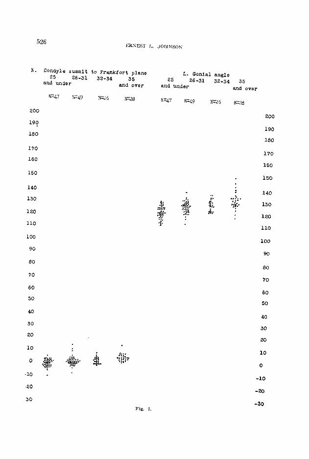
K. As the head of the condyle is progrewively placed further above the Frankfort plane, tending to exaggerate the lack of sufficient vertical height in the ramus, the Frankfort-mandibular plane angle increases. The rapid increase from plus 0.02 to plus 5.17 shows that the location of the head of the condyle in the skull pla.ys a very important part in determining the entire facial pattern, as well as the Frankfort-mandibular plane angle.
IL There is steady and rapid increase in the size of the gonial angle, as the Frankfort-mandibular plane angle increases, which goes hand in hand with the increase in facial height.
&I. Where the ~‘~ankfort-tnandihular plane angle is less than 25”, the mean vertical length of the ramus (condyle summit to gonion) is substantially larger than the means for the other groups; the three remaining groups have mean values for this measurement which are within a millimeter of one another. It was expected that ramus length would have a larger influence on the deter- mination of the Frankfort-mandibular plane angle than is indicated here, and this will be discussed in a later portion of the paper.
N. The distance from the ,condyle summit to t,he mandibular plane dimin- ishes as the mean Frankfort-mandibular plane angle increases. We should expect this for this measurement is the effective length of the ramus, as altered by the increasing gonial angle.
0. The definite and consistent increase in the Frankfort-mandibular base angle shows that as the vertical height of the profile increases, it affects the cant of the palate by carrying the anterior end downward.
P. This shows the mean Frankfort-mandibular plane angle in each of the four divisions of the group of one hundred fifty cases.
Figs. 2, 3, 4, 5, 6, 7, 8, and 9 are graphs showing each of the measurements of the individuals comprising the four groups. They show the wide variation, even within each group, and yet t,he definite trend as expressed in the means for each of the four groups as shown in Table I.
CLINICAL IMPLICATIONS
It appears to me that much of the value of similar studies is often lost to the reader because so frequently the investigator neglects to explain adequately the clinical implications of his study. Hence, I will attempt to present a summary of my thoughts on this survey.
Facial patterns are of utmost, importance in orthodontic prognosis and treatment. There would appear to be little doubt that facial patterns are deter- mined by heredity, in large part, or by other deep-seated factors not susceptible to modification. There may be a great deviation in the individual pattern from the so-called ‘ ‘ normal ’ ’ or ideal type or pattern. Yet, these extremes are in one sense normal for that individual, and many of the individual deviations from the acceptable pattern are not within the realm of orthodontic correction.
As was pointed out earlier in this paper, the magnitude of the Frankfort- mandibular plane angle must be regarded as the result of relatively greater development, in certain areas of the face than others and not as a biological entity in itself.

Wylie’ was careful to point out in his paper on anteroposterior dysplasia that the assessment of dysplasia must involve not. one but all three planes of space, and in that paper he dealt, with the facial dimensions which have to do with depth. A study of Table 1 in this paper indicates that in the plane of space at right angles to the one considered by Wylie, the plane in which vertical dimensions are recorded, the absolute dimensions of height interact with one another to affect appreciably the est,hetics of the fact‘.
The first three values in Table 1: measurements of total face height. upper face height, and lower fac*e height, indicate clearly that in these facial patterns which have been labeled bad by virtue of tile large Frankfort-mandibular plane angle, the facial pattern is disposed vertic*ally, with the height of t,he face longer at the profile in individuals in whom this angle is steep than it is in individuals with more acceptable ph,vsiognomies. The progression of mean values from one group to another is sure and stead,v in total fart height and in lower face height,, but less so with respect to upper face height. This shows rat,her un- mistakably that excessive vertical development of the alveolar process in the anterior segment of the denture is primarily responsible for the facial anomaly. This is further brought out by measurements t,akcn from the incisal edge of the maxillary central incisors to the Frankfort plane and to the maxillary base, respectively, and by the mcasurementj (C: ‘i from the incisal edge of t,he man- dibular central incisors to the plane of the mandibular base.
These figures show that, the facial pattern becomes worse as there is a vertical elongation in the anterior segment of the denture. This has important clinical implications, since cephalometric studies have shown that the depression of teeth, wgardlesx of the segment of the denture involved, is extremely difficult. When the effort to depress teeth is made by means of ordinary orthodontic appliances, the depression of those teeth is pitted against the vertical de- velopment of teeth in other segments of the dental arch, and the vertical development or elongation of t,he teeth and alveolar process is the movement which predominates. Conseqently, when it is determined that an undesirable vertical elongation of the denture in the anterior portion of the face is present, prognosis for attacking the problem directly is not good.
These measurements arc concerned with height at the profile ; measurements taken in the molar region to the Frankfort plane and to the planes chosen to represent maxillary and mandibular bases, respectively, indicate that the vertical elongation is limited primarily to the anterior portion of the denture rather than the molar area.
Finding that the profile is elongated in individuals with steep E’rankfort- mandibular plane angles came as no surprise; on the other hand, it was fully expected that the length of the mandibular ramus as measured from the head of the condyle to the gonial angle (M) would have equal importance in the deter- mination of the Frankfort-mandibular plane angle, i.e., it was expected that t,his ramus length would be relatively short where the angle was steep. As a matter of fact, before the Frankfort-mandibular plane angle came into USC among orthodontists, reference was made at California to this type of case as “short ramus” cases.

FRANKFORT-MLIASDIBULAR PLANE ANGLE AND FACIdL PATTERS 531
The figures published herewith indicate that such an appellation was, always excepting individual cases, a misnomer. The lack of any significant downward progression of mean values indicates that there is not an inverse relationship between the length of the ramus and the magnitude of the Frank- fort-mandibular plane angle as was supposed before t,he data were collected. It appears from these data, however, that an important qualification to that statement must be made. It would seem that the length of the mandibular ramus is of real importance where the Frankfort-mandibular angle is small. This suggests that orthodontists might consider the facial pattern characterized by a low mandibular plane angle as a distinct facial type in itself, with the primary factor responsible bein, v more than average length in the mandibular ramus. Wylie9 pointed out that there are essentially two classes of facial types in which the Frankfort-mandibular plane angle is relatively low. One must be regarded as a pleasing facial type according to present-day standards, for it, is found frequently in the faces of professional models, particularly those whose services are sought by the so-called magazines of “high style. ” In these faces, besides the low Frankfort-mandibular plane angle, one may see the bony outline of the mandible sharply definite beneath the soft tissues, with a gonial angle approaching 90° and gonion itself prominent, with t,he posterior border of the mandibular ramus clearly discernible. In these faces the marked contribution to the facial pattern of a long ramus is obvious.
In the other class of facial types with relatively low mandibular plane angles, there is a. severely closed-bite and the facial pattern cannot be considered good. Here, an increase in the vertical dimension brought about through ortho- dontic treatment brings a profound improvement in facial pattern. While these figures show that the actual length of the ramus from the head of the condyle to the angle of the jaw has less importance than was previously thought, at the same time the effective length of the ramus does play a significant part in estab- lishing the Frankfort-mandibular plane angle. Brodie’” was the first to indicate how anteroposterior placement of the glenoid fossa, determined by lengt,h in the cranial base, could affect t,he anteroposterior relationship of t,he dental arches. Wylie7 allowed for the quantitative appraisal of this factor, and the use of his method reveals the importance of Brodie’s concept. Item K in Table I shows that the location of the glenoid fossa in the vertical plane as related to the Frankfort horizontal plane has a definite influence upon the efective lengt,h of t,he mandibular ramus. The distance between the head of the condyle and the angle of the jaw may be sufficiently large to give good vertical proportions to the face; however, if the glenoid fossa is sufficiently high in the cranium, the effect will be the same as that produced by a short ramus, and, other things being equal, the Frankfort-mandibular plane angle will be steep. The definite progression of mean values aft,er Item K shows that there is a definite relation- ship between the value of the Frankfort-mandibular plane angle and the place- ment of the glenoid fossa vertically in relation to the Frankfort plane.
When 1.50 cases are classified with respect to the size of the Frankfort-man- dibular plane angle, a progression is also seen in the value of the gonial angle (r,) .

532 ERNEST L. .JOHSsOK
The fact that the gonial angle tends to be larger when the Frankfort-mandibular plane angle is large, and when there is more tha.n average vertical development of the anterior portion of t,he face, leads t,o some interesting speculation. One wonders whether the gonial angle is large because there is an inherent tendency in the face of the individual to develop vertically at the profile, with gonial angle size adapting to this situation, or whether the fundamental reason for vertical development at the profile is adaptation of’ the facial pattern to an inherently large genial angle. There seems to be no ready way in which this question could be settled definitively, but offhand it, seems more likely that the height of the alveolar process is adaptive to an inherently large genial angle. If the primary factor in the relationship were a tendency for alveolar development, adjustment in the posterior part of the face could be achieved simply through backward hinging at the temporomandibular joint, without, an,v alterations in the magni- tude of the genial angle. This is but anot,hcr instance in which the relationship between two variables may be observed, and in which it would be unwise to assume any cause and effect relationship.
It is probable that the progression which may be observed with respect to the Frankfort-maxillary base angle (0) may be explained on a basis of marked vertical development at the profile which has been mentioned sufficiently often in this paper. Note, however, that where the Frankfort-mandibular plane angle
is less than 25O the mean value for the FrankforLmaxillary base angle does not form a part of the progression, lendin g further support to the concept that this is a more or less distinct t,ppe of face.
The Frankfort-mandibular plane angle has been shown to be a valuable diagnostic criterion in the analysis of the facial pattern in orthodontic patients. It is not an anatomical ent,ity, however, and in this st,udy 150 patients ha.ve been classified with respect to the Frankfort-mandibular plane angle. An analysis of mean values for different dimensions of the face shows, that a definite progres- sion of mean values may be observed in practically all of the dimensions under study when facial patterns are segregated into different classes baaed on the magnitude of the Frankfort-mandibular plane angle. These progressions of mean values indicate that the Frankfort-mandibular plane angle is affected by vertical development of the alveolar process in the anterior pprtion of the den- ture, by the absolute length of the mandibular ramus, and by the inferosuperior placement of the glenoid fossa in the skull. It is furthermore shown that, statistically speaking, there is a direct relationship between the size of the gonial angle and the size of the Frankfort-mandibular plane angle. Where the Frank- fort-mandibular plane angle is relatively small (i.e., less than 25O), a more or less separate and distinct facial type is encount,ered, which has special conno- tations in esthetics and in prognosis for ort,hodont,ic treatment.
This project was carried on under the direction of Dr. Wendell L. Wylie, while he was Chairman of the Division of Orthodontics at the University of California. Research funds provided by Dr. J. M. Loughridge, of Sacramento, Calif., were drawn upon for the support of this project.
FRAIiKFORT-MANDIBULAR PLANE ANGLE AND FACIAL PATTERN 533
REFERENCES
1. Tweed Charles H . . . The Frankfort-Mandibular Plane Angle in Orthodontic Diagnosis, Ciassifieations, Treatment Planning, and Prognosis, AM. J. ORTHODONTICS AND ORAL SURG. 32: 175~230,1946. -'
2. Downs. William B.: Variations in Facial Relationshios. Their Sinnificance in Treatment and Progno’sis, AM. J. ORTHODONTICS 34: 812-840,L1848. o
3. Salzmann, J. A.: The Maxillator: A New Instrument for Measuring the Frankfort- Mandibular Base Angle, the Incisor-Mandibular Base Angle, and &her Component Parts of the Face and Jaws, AM. J. ORTHODONTICS AND ORAL SURG. 31: 603-618, 1945.
4. Brodie, Allan G.: The Growth Pattern of the Human Head From the Third Month to the Eighth Year of Life, A. J. Anat. 68: 209.262,1941.
5. Brodie, Allan G.: Facial Patterns; a Theme on Variation, Angle Orthodontist 16: 75-87, 1946.
6. Wylie, Wendell L.: Malocclusion-Malady or Malformation, Angle Orthodontist 19: 3-l 1, 1949.
7. Wylie, Wendell L.: The Assessment of Anteroposterior Dysplasia, Angle Orthodontist 17: 97-109, 1949.
8. Thompson. J. R.: Oral and Environmental Factors in Malocclusion of the Teeth. AM. i. ORTHODONTICS 35: 33-53, 1949.
9. Wylie, Wendell L.: Personal communication. IO. Strang, R. H. W.: Textbook of Orthodontia, Philadelphia, 1943, Lea $ Febiger, p. 472.