1 structural modeling and physicochemical characterization
TRANSCRIPT
1
Structural Modeling and Physicochemical Characterization Provide Evidence that
P66 forms a ȕ-Barrel in the Borrelia burgdorferi Outer Membrane
Melisha R. Kenedy1, Amit Luthra
2, Arvind Anand
2, Joshua P. Dunn
1, Justin D. Radolf
2,3,4,5,6, and
Darrin R. Akins1#
Department of Microbiology and Immunology, University of Oklahoma Health Sciences Center,
Oklahoma City, Oklahoma 731041, Departments of Medicine
2, Pediatrics
3, Genetics and
Developmental Biology4, Immunology
5, and Molecular Microbial and Structural Biology
6,
University of Connecticut Health Center, Farmington CT 06030
#Corresponding author: Darrin R. Akins, Ph.D.
Department of Microbiology and Immunology
The University of Oklahoma Health Sciences Center
Oklahoma City, Oklahoma 73104
Phone: (405) 271-2133 x46640
FAX: (405) 271-3117
Running Title: Structural model of B. burgdorferi OMP P66
Keywords: Borrelia burgdorferi, P66, Lyme disease
JB Accepts, published online ahead of print on 6 December 2013J. Bacteriol. doi:10.1128/JB.01236-13Copyright © 2013, American Society for Microbiology. All Rights Reserved.
on February 6, 2018 by guest
http://jb.asm.org/
Dow
nloaded from
2
ABSTRACT 1
The Borrelia burgdorferi outer membrane (OM) contains numerous surface-exposed lipoproteins 2
but a relatively low density of integral OM proteins (OMPs). Few membrane-spanning OMPs of 3
B. burgdorferi have been definitively identified, and none are well characterized structurally. 4
Here, we provide evidence that the borrelial OMP P66, a known adhesin with pore-forming 5
activity, forms a ȕ-barrel in the B. burgdorferi OM. Multiple computer-based algorithms predict 6
that P66 forms a ȕ-barrel with either 22- or 24-transmembrane domains. According to our 7
predicted P66 topology, a lysine residue (K487) known to be sensitive to trypsin cleavage was 8
located within a surface-exposed loop. When we aligned the mature P66 amino acid sequences 9
from B. burgdorferi and B. garinii, we found that K487 was present only in the B. burgdorferi 10
P66 protein sequence. When intact cells from each strain were treated with trypsin, only B. 11
burgdorferi P66 was trypsin-sensitive, indicating that K487 is surface-exposed as predicted. 12
Consistent with this observation, when we inserted a c-Myc tag adjacent to K487 and utilized 13
surface localization immunofluorescence, we detected the loop containing K487 on the surface 14
of B. burgdorferi. P66 was examined by both Triton X-114 phase partitioning and circular 15
dichroism, confirming that the protein is amphiphilic and contains extensive (48%) ȕ-sheet, 16
respectively. Moreover, P66 also was able to incorporate into liposomes and form channels in 17
large unilamellar vesicles. Finally, blue-native PAGE (BN-PAGE) revealed that under non-18
denaturing conditions P66 is found in large complexes of ~400 kDa and ~600 kDa. Outer 19
surface lipoproteins A (OspA) and OspB both co-immunoprecipitate with P66, demonstrating 20
that P66 associates with OspA and OspB in B. burgdorferi. The combined computer-based 21
structural analyses and supporting physicochemical properties of P66 provide a working model 22
on February 6, 2018 by guest
http://jb.asm.org/
Dow
nloaded from
3
to further examine the porin and integrin-binding activities of this OMP as they relate to B. 23
burgdorferi physiology and Lyme disease pathogenesis. 24
on February 6, 2018 by guest
http://jb.asm.org/
Dow
nloaded from
4
INTRODUCTION 25
Lyme disease is currently the most common arthropod-borne infection in the United States and is 26
also prevalent throughout Europe and Asia (1). The disease is caused by pathogenic spirochetes 27
belonging to the Borrelia burgdorferi sensu lato complex. The three major genospecies 28
associated with Lyme disease include B. burgdorferi sensu stricto (hereafter referred to as B. 29
burgdorferi), B. garinii, and B. afzelii (2). Borrelia spirochetes are maintained in nature through 30
an enzootic cycle that includes Ixodes ticks and a mammalian host (1, 3). In humans, Lyme 31
disease typically manifests as an expanding skin rash, termed erythema migrans, which can be 32
followed by cardiac symptoms, nervous system abnormalities, and arthritis (4, 5). 33
B. burgdorferi is a dual-membrane (diderm) organism with both an outer membrane 34
(OM) and a cytoplasmic or inner membrane (IM). The borrelial OM markedly differs from those 35
of typical Gram-negative enteric organisms, such as Escherichia coli (3, 6). For example, the 36
borrelial OM lacks lipopolysaccharide (LPS) (7, 8), the highly inflammatory glycolipid found in 37
Gram-negative bacteria. Furthermore, the surface of B. burgdorferi is decorated with numerous 38
lipid-modified, membrane anchored lipoproteins, whereas surface-exposed lipoproteins are 39
uncommon in typical Gram-negative bacteria (6, 9-11). Most importantly with respect to the 40
current study, freeze-fracture electron microscopy, which visualizes integral OM proteins 41
(OMPs) as intramembranous particles, revealed that the B. burgdorferi OM also contains at least 42
10-fold fewer integral OMPs per µm2 as compared to that of E. coli (12, 13). Few of these outer 43
membrane-spanning proteins have been identified, and none has been structurally characterized 44
to any extent (9). Given that OMPs identified in other diderm organisms, as well as eukaryotic 45
organelles of bacterial origin (e.g., mitochondria and chloroplasts), consist of amphipathic ȕ-46
strands that form ȕ-barrels (14, 15), one would expect that B. burgdorferi OMPs form ȕ-barrels 47
on February 6, 2018 by guest
http://jb.asm.org/
Dow
nloaded from
5
as well. In diderms, the amphipathic nature of the ȕ-barrel OMP precursors allow for the 48
translocation of these polypeptides across the hydrophobic IM. In contrast, IM proteins contain 49
Į-helical transmembrane domains that serve as stop transfer sequences that result in proteins 50
being localized to the IM (16). Furthermore, as with Gram-negative organisms, nutrients must 51
be transferred across the borrelial OM for the spirochete to survive within the host; thus, 52
channels and pores must be present in the borrelial OM to facilitate nutrient acquisition. 53
Moreover, we now know that B. burgdorferi has the machinery necessary to locate and fold ȕ-54
barrel proteins into the borrelial OM. Recent studies have revealed that ȕ-barrel OMPs from E. 55
coli, Neisseria meningitidis and all other diderm bacteria characterized to date are chaperoned 56
into the OM via the multi-protein ȕ-barrel assembly machine (BAM) complex (17-20). The 57
central component of the BAM complex, BamA, is conserved among diderm organisms (21). 58
Importantly, it has been shown that B. burgdorferi has a functional BamA ortholog and at least 59
two Bam accessory proteins (22, 23). Similarly, Treponema pallidum, the spirochete that causes 60
syphilis, also has been shown to contain a BamA ortholog (24). 61
Of the B. burgdorferi OMPs identified to date, P66, encoded by open reading frame 62
bb0603, has been the most extensively studied. BamA is required for efficient transport of P66 63
into the borrelial OM (22). Proteolysis studies have confirmed that P66 has surface-exposed 64
domains, including a putative surface-exposed loop (residues 459-502) near the C-terminus (25-65
29). Among the Borrelia genospecies, sequence variation in the predicted surface-exposed loop 66
is greater than that found throughout the rest of the P66 sequence, indicating that the loop may be 67
under immune selection pressure during mammalian infection (28, 29). Furthermore, Skare et 68
al. demonstrated that native P66 forms pores in lipid bilayer assays (30), and similar properties 69
have been described for P66 from B. garinii and B. afzelii (31). In vitro analyses have implicated 70
on February 6, 2018 by guest
http://jb.asm.org/
Dow
nloaded from
6
P66 as an adhesin that binds specifically to ȕ3-chain integrins (32-34). Although the tertiary 71
structure and final structural conformation of P66 are likely required for interaction with 72
integrins (33), a region encompassing amino acids 150-343 is sufficient for ligand binding (32, 73
34, 35). Interestingly, while P66 is required for infection of mice by tick inoculation (36), a P66 74
mutant replicates normally within dialysis membrane chambers implanted into the peritoneal 75
cavities of rats (36). Thus, the molecule’s contribution to growth and nutrient acquisition during 76
the mammalian phase of the spirochete’s enzootic cycle is unclear. 77
While the functional studies described above have provided insight into the role of P66 in 78
vitro and in vivo, little is still known about the protein’s structure. Herein, we report that P66 is 79
predicted to form a ȕ-barrel and displays the properties expected of an OMP with amphipathic ȕ-80
barrel structure. Additionally, we confirmed that lysine K487 is located in a surface-exposed 81
loop that is protease sensitive and corroborated prior reports that P66 specifically interacts with 82
OspA and OspB. These data support the contention that the B. burgdorferi P66 protein forms a 83
ȕ-barrel despite its lack of sequence homology with known Gram-negative OMPs. Our results 84
provide a working model to further examine the porin and integrin-binding activities of this 85
OMP as they relate to B. burgdorferi physiology and Lyme disease pathogenesis. 86
on February 6, 2018 by guest
http://jb.asm.org/
Dow
nloaded from
7
MATERIALS and METHODS 87
Bacterial strains and growth conditions. Borrelia organisms, including B. burgdorferi B31, B. 88
burgdorferi JD1, B. garinii Pbi, and B. garinii IP90, were cultivated at 34°C in BSK-II medium 89
containing 6% heat inactivated rabbit serum (BSK-II complete, pH 7.6) (37). For surface 90
localization immunofluorescence and Triton X-114 phase partitioning experiments, we utilized 91
the avirulent, high-passage strain B. burgdorferi clone F (cF), which was described previously 92
(38, 39), 93
94
P66 sequence alignment and modeling. The membrane topology of the mature B. burgdorferi 95
P66 protein (GenBank accession number NP_212737) was predicted using the PRED-TMBB 96
server (http://biophysics.biol.uoa.gr/PRED-TMBB/) (40, 41). OM localization and ȕ-barrel 97
predictions were made using the following servers: CELLO (http://cello.life.nctu.edu.tw/) (42), 98
PSORTb (http://www.psort.org/psortb/) (43), HHOMP (http://toolkit.tuebingen.mpg.de/hhomp/) 99
(44), TMBETADISC-AAC (http://rbf.bioinfo.tw/~sachen/OMPpredict/TMBETADISC-100
RBF.php) (45), and BOMP (http://services.cbu.uib.no/tools/bomp/) (46). To predict the 101
structural properties of B. burgdorferi P66, the mature P66 amino acid sequence was analyzed 102
using the TMBpro server (http://tmbpro.ics.uci.edu/) (47), and the resulting pdb file was further 103
analyzed by Swiss-PdbViewer 4.0.2 (48). Amino acid alignments of P66 from B. burgdorferi 104
B31, B. burgdorferi JD1, B. garinii Pbi, and B. garinii IP90 (GenBank accession numbers 105
NP_212737, YP_005805693, AAU07452, and CAA61026, respectively) were generated using 106
MacVector 10.0 (MacVector, Inc., Cary, North Carolina). 107
108
on February 6, 2018 by guest
http://jb.asm.org/
Dow
nloaded from
8
Cloning, purification, and folding of recombinant P66. P66 was amplified from B. 109
burgdorferi B31 genomic DNA using primers P66 5’ 110
(GCGGCTAGCTTAAAGGAAAAAGATATATTTAAAATA) and P66 3’ 111
(GCGCTCGAGGCTTCCGCTGTAGGCTATTT) (restriction enzymes in bold). B. burgdorferi 112
BamA was amplified from genomic DNA with primers BamA 5’ 113
(GCGGCTAGCGTTGAAAATTACAAGGGGAAAA) and BamA 3’ 114
(GCGCTCGAGATATCTCATCTCAATTCCTAAGA). Escherichia coli OmpA was amplified 115
with primers OmpA 5’ (GCGGCTAGCGCTCCGAAAGATAACACCTG) and OmpA 3’ 116
(GCGCTCGAGAGCCTGCGGCTGAGTTACA). For amplification of B. burgdorferi BamA 117
Potra 1, primers BamA P1 5’ (GCGGCTAGCAAGGGGAAAATAAGGGTAT) and BamA P1 118
3’ (GCGCTCGAGTTCTTTTACAATAAAATGTAATAAAAAG) were used. The amplicons 119
were subsequently digested and cloned into the NheI and XhoI sites of pET23a (EMD Millipore, 120
Billerica, MA), which has a C-terminal His-tag. The constructs were transformed into the E. coli 121
strain Rosetta 2 DE3 (EMD Millipore, Billerica MA), and DNA sequencing was performed to 122
verify that the sequence remained unaltered throughout the cloning process. 123
For batch purification of recombinant P66, OmpA, and BamA, 1 L of lysogeny broth was 124
inoculated with 25 ml of overnight culture which then was grown at 37oC to an optical density at 125
600 nm (OD600) of 0.6. Protein expression was then induced with 1 mM IPTG, and the culture 126
was grown for an additional three hours. After induction, the cells were pelleted at 10,000 x g 127
for 15 min at 4oC and resuspended in 25 ml of resuspension buffer (50 mM Tris, 100 mM NaCl, 128
pH 7.6) with 25 µl of protease inhibitor cocktail. The cells were then lysed by sonication and 129
pelleted for 20 min at 20,000 x g. The pellet was resuspended in 10 ml of binding buffer (100 130
mM NaH2PO4, 10 mM Tris, 8 M Urea) at pH 8.0. The suspension was then rotated at room 131
on February 6, 2018 by guest
http://jb.asm.org/
Dow
nloaded from
9
temperature for 30 min and subjected to centrifugation for 30 min at 20,000 x g. After 132
centrifugation, the supernatant was applied to a column containing a 5 ml volume of nickel-133
nitrilotriacetic acid agarose (Qiagen,Valencia, CA) that had previously been equilibrated with 134
binding buffer. After rocking the resin and supernatant for 20 min, the column was washed with 135
100 ml of wash buffer (100 mM NaH2PO4, 10 mM Tris, 8 M Urea) at pH 6.3. The protein was 136
next eluted in 30 ml of elution buffer 1 (100 mM NaH2PO4, 10 mM Tris, 8 M Urea) at pH 5.8 137
and elution buffer 2 (100 mM NaH2PO4, 10 mM Tris, 8 M Urea) at pH 4.5. SDS-PAGE was 138
used to analyze the purified recombinant protein. After protein purification, the protein was 139
concentrated using Amicon-Ultra centrifugal filters (EMD Millipore, Billerica, MA). 140
Treponema pallidum protein TP0453 and E. coli OmpF were purified as described elsewhere 141
(49, 50). 142
To fold recombinant P66, BamA, and OmpA, each protein was incubated in DDM buffer 143
[50 mM Tris pH 7.6, 100 mM NaCl, 2.0% dodecyl-ȕ-D-maltopyranoside (DDM; Affymetrix, 144
Santa Clara, CA)] for 24 hours at 4oC, and the insoluble material was pelleted by centrifugation 145
at 20,000 x g for 30 min at 4oC. The supernatant was removed and analyzed by SDS-PAGE. 146
147
Immunoblotting and antibody production. SDS-PAGE and immunoblotting procedures were 148
performed as previously described (22, 51). Rat polyclonal antibodies recognizing B. 149
burgdorferi P66 and E. coli OmpA were generated by Harlan Bioproducts for Science, Inc. 150
(Madison, WI) and were used at a 1:5,000 dilution for enhanced chemiluminescence. OspA, 151
FlaB, BamA, BB0796, and CspA antibodies were described previously (22, 39, 52-54). 152
Monoclonal mouse OspB antibodies (CB2) have been described elsewhere (55) and were 153
provided by Dr. Jorge Benach (Stony Brook University, Stony Brook, New York). 154
on February 6, 2018 by guest
http://jb.asm.org/
Dow
nloaded from
10
Trypsin surface accessibility assays. To digest surface proteins with trypsin, 2×10⁸
B. 155
burgdorferi B31 cells were subjected to centrifugation at 4,000 × g for 4 min and washed three 156
times in PBS (pH 7.4). The final pellet was resuspended in 1 ml of PBS, and samples were 157
either treated or mock-treated with trypsin (Sigma, St. Louis, MO) resuspended in 0.001 N HCl 158
for one hour at room temperature. Phenylmethylsulfonylfluoride (0.4 mM; Sigma) was added to 159
each sample to stop protease activity, and the samples were pelleted at 10,000 × g for 10 min. 160
The final pellets were prepared for SDS-PAGE and immunoblot analysis using rat anti-P66 161
antibodies. Membranes were also subjected to immunoblot with antibodies to OspB and FlaB 162
for surface and sub-surface controls, respectively. 163
164
Generation of c-Myc B. burgdorferi clones. To express P66 with a c-Myc tag in the K487 165
predicted surface loop, we generated the P66-c-MycK487
construct by cloning p66 and the c-myc 166
tag sequence into pBSV2G (56). To clone the flgB promoter into pBSV2G, the flgB promoter 167
was amplified from pBSV2 with primers flgB F 168
(GCGGGTACCTACCCGAGCTTCAAGGAAGA) and flgB R 169
(GCGGGATCCATGGAAACCTCCCTCATTTAAA). The amplicon was cloned into the KpnI 170
and BamHI sites of pBSV2G. Next, the portion of p66 including the sequence encoding K487 171
and the upstream region was amplified from B. burgdorferi B31 genomic DNA with primers P66 172
F (GCGGGATCCATGAAAAGCCATATTTTATATAAATT) and P66 us R 173
(GCGTCTAGACTTTGTGCTTGTTGAACTTTGT) and inserted into the BamHI and XbaI sites 174
of the vector. The region of p66 downstream of K487 was amplified with primers P66 ds F 175
(GCGGTCGACACCACAACCCCTAATCTGAC) and P66 R 176
(GCGGTCGACTTAGCTTCCGCTGTAGGCTA) and cloned into the SalI site. To generate the 177
on February 6, 2018 by guest
http://jb.asm.org/
Dow
nloaded from

11
c-myc tag, the c-myc tag sequence 178
(GCGTCTAGAGAACAAAAACTTATTTCTGAAGAAGATCTGGTCGACGCG) and the 179
reverse complement sequence were annealed before ligating and cloning the product into the 180
XbaI and SalI sites of the vector. As a control, we also generated a vector from which P66 181
would be expressed with a c-Myc tag at the P66 C-terminus and termed the construct P66-c-182
MycC-terminal
. flgB was inserted into the pBSV2G vector as described above, and the p66 183
sequence without the stop codon was amplified with primers P66 F and P66 ns R 184
(GCGTCTAGAGCTTCCGCTGTAGGCTATTT) and cloned into the BamHI and XbaI sites of 185
pBSV2G. Next, the c-Myc tag was inserted as described above; however, a stop codon was 186
added to the c-myc sequence 187
(GCGTCTAGAGAACAAAAACTTATTTCTGAAGAAGATCTGTAAGTCGACGCG). 188
Finally, a control vector P66-c-Myc- was generated that expressed P66 without a c-Myc tag by 189
amplifying p66 with primers P66 F and P66 R and inserting the amplicon into the BamHI and 190
XbaI sites of pBSV2G in which the flgB promoter was previously inserted into the vector as 191
described above. The three vectors were electroporated into competent B. burgdorferi cF cells as 192
described previously (57). After electroporation, spirochetes were selected with BSK-II 193
complete medium containing gentamicin (40 µg/ml). 194
195
Surface localization Immunofluorescence. Spirochetes were grown to mid-exponential phase 196
and diluted to a final concentration of 5 X 10⁶ cells. Cell suspensions were coincubated for 1 197
hour with rabbit anti-c-Myc antibodies (Sigma) at a dilution of 1:10 and rat anti-FlaB antibodies 198
at a dilution of 1:1000. The cells were then washed three times in PBS. The final pellet was 199
resuspended in 100 µl PBS, and 10 µl of the sample was spotted onto microscope slides and 200
on February 6, 2018 by guest
http://jb.asm.org/
Dow
nloaded from
12
fixed for 10 min with acetone. Samples were subsequently blocked with PBS containing 1% 201
BSA. Samples were then incubated for 45 min with Alexa Fluor 488-conjugated goat anti-rabbit 202
antibodies (1:500; Invitrogen, Carlsbad, CA) and Alexa Fluor 568-conjugated goat anti-rat 203
antibodies (1:500; Invitrogen) before being washed three times with PBS containing BSA. 204
Samples were then mounted in buffered glycerol containing DAPI (Vector Laboratories, 205
Burlingame, CA), sealed with a coverslip, and images were visualized and captured with an 206
Olympus BX-60 fluorescence microscope (Olympus America Inc., Center Valley, PA). As a 207
control, samples were also spotted onto microscope slides and fixed with acetone prior to 208
coincubation with rabbit anti-c-Myc antibodies and rat anti-FlaB antibodies. After these samples 209
were washed three times with PBS containing 1% BSA, they were incubated with secondary 210
antibodies, and prepared as described above. 211
212
Tryptophan fluorescence. Tryptophan emission spectra were measured using a Hitachi F-2500 213
fluorescence spectrophotometer. Protein samples that were either denatured in urea or folded in 214
DDM buffer (described above) were inserted into a 5-mm path length quartz cell at 25°C. The 215
excitation wavelength and bandwidth were 295nm and 2.5 nm, respectively. The tryptophan 216
emission spectra were measured between 300 and 400 nm. Background spectra were also 217
measured using DDM buffer without protein, and the background measurements were subtracted 218
to obtain the final emission curves. 219
220
Circular dichroism spectroscopy. All CD experiments were performed using a Jasco J-715 221
spectropolarimeter (Jasco, Easton, MD). Far-UV CD spectra were recorded at 20°C using a 1 222
mm path-length cuvette, a bandwidth of 1 nm, a time response of 8 s, and a scan rate of 20 223
on February 6, 2018 by guest
http://jb.asm.org/
Dow
nloaded from
13
nm/min. CD spectra for each protein, representing the average of nine scans, were corrected by 224
subtracting the background spectra of the buffer. To assess the secondary structure contents of 225
each protein, we analyzed the spectra using the DICHROWEB server (58, 59). 226
227
Triton X-114 phase partitioning. The amphiphilic properties of P66 from B. burgdorferi cF 228
cells or recombinant protein (10 ȝg) were assessed by Triton X-114 phase partitioning as 229
described elsewhere (22, 53, 60). The resulting aqueous- and detergent-enriched proteins were 230
precipitated with acetone, and the protein was analyzed by SDS-PAGE and immunoblot using rat 231
anti-P66, rat anti-BamA, rat anti-OspA, and rat anti-BB0796 antibodies. 232
233
Preparation of Liposomes. Large unilamellar vesicles (LUVs) were prepared as described (61). 234
To simulate the phospholipid content of the B. burgdorferi outer membrane, a mixture of 1-235
palmitoyl-2-oleoyl-sn-glycero-3-phosphocholine and 1-palmitoyl- 2-oleoyl-sn-glycero-3-236
[phospho-L-serine] (sodium salt) (Avanti Polar Lipids, Inc., Alabaster, AL), (70:30 mol %, 237
respectively) was used. Preparation of liposomes containing Tb(DPA)33-
were performed as 238
previously described (61). 239
240
Liposome floatation assay. Recombinant P66 (400 ng) in DDM buffer was incubated with 750 241
µg of LUVs at room temperature for 1 h in 50 mM acetate buffer. After incubation, 200 mg of 242
sucrose was added to the reaction, and the sample was mixed thoroughly before being transferred 243
to an ultracentrifuge tube. Discontinuous sucrose gradients were made by layering 40% and 6% 244
sucrose on top of the sample in the ultracentrifuge tubes. Samples were centrifuged at 90,000 245
rpm for 1 h at 4°C. The samples were removed from the centrifuge and three fractions were 246
on February 6, 2018 by guest
http://jb.asm.org/
Dow
nloaded from
14
carefully collected from the tube: the liposome (top) layer and two non-liposome (middle and 247
bottom) layers. Each fraction was separated by SDS-PAGE and subjected to immunoblot 248
analysis using rat anti-P66 antibodies. Control experiments were performed using OmpA 249
unfolded protein as well as OmpA protein folded in DDM buffer. Fractions from the control 250
experiments were immunoblotted with rat anti-OmpA antibodies. 251
252
Pore formation assay. Pore formation assays were performed as described (50, 62). Liposomes 253
loaded with Tb(DPA)33-
were diluted in 50 mM Tris (pH 7.5), 100 mM NaCl supplemented with 254
5mM EDTA to a concentration of 100 ȝM total lipids. The sample was equilibrated at 25°C for 255
5 min, and the net initial emission intensity (F0) was determined. Recombinant P66, OmpF, or 256
TP0453 (100 nM final concentration) was incubated with the liposome suspension for 30 min at 257
37°C. Samples were then re-equilibrated to 25°C. The final net emission intensity (Ff) of the 258
sample was determined after subtracting the blank and correcting for dilutions. The fraction of 259
Tb(DPA)33-
quenched was estimated using Ff/F0. 260
261
Heat modifiability. B. burgdorferi whole-cell lysates or recombinant P66 and OmpA folded in 262
DDM buffer per above were boiled in sample buffer (62 mM Tris-HCl [pH 6.8], 10% [vol/vol] 263
glycerol, 100 mM dithiothreitol, 2% sodium dodecyl sulfate, 0.001% bromophenol blue) or 264
incubated at room temperature prior to SDS-PAGE and subsequent immunoblot analysis with 265
P66 or OmpA antibodies. 266
267
Blue-native polyacrylamide gel electrophoresis (BN-PAGE) analysis of native and 268
recombinant P66. B. burgdorferi organisms were pelleted by centrifugation at 20,000 × g for 269
on February 6, 2018 by guest
http://jb.asm.org/
Dow
nloaded from
15
20 min at 4°C. The subsequent pellet was resuspended and incubated overnight at 4°C in 50 mM 270
Tris (pH 7.0), 2% DDM, and 5% PIC. Detergent-insoluble material was removed from the 271
sample by centrifugation at 20,000 × g for 20 min at 4°C. B. burgdorferi lysates (1 × 10⁸ to 5 × 272
109 spirochetes) were analyzed using BN-PAGE (63, 64). Lysates were resolved at 4°C in a 4-273
12% Bis-Tris acrylamide gel (Bio-Rad). For the first 1/3rd
of the gel run, 0.02% Coomassie 274
brilliant blue G-250 (CBB-G250) was added to the cathode buffer [50 mM tricine (pH 7.0) and 275
15 mM bis-tris]. Fresh cathode buffer that did not contain CBB-G250 was then used for the 276
remaining time. The anode buffer consisted of 50 mM bis-tris (pH 7.0) throughout the duration 277
of the run time. After the lysates were resolved, the samples were transferred to nitrocellulose 278
membrane in 50 mM tricine (pH 7.0). Membranes were next subjected to immunoblot using rat 279
anti-P66 antibodies or anti-OspA antibodies. Analysis of recombinant P66 was performed as 280
above except 1µg of protein was incubated on ice for 30 min prior to BN-PAGE. To assess 281
protein migration, the retardation factor (Rf) values were calculated according to the 282
manufacturer’s instructions (Invitrogen). 283
284
Co-immunoprecipitation (co-IP). Cell lysates for co-IP experiments were prepared by 285
harvesting mid-log phase cultures of B. burgdorferi strain B31 5A4NP1 (65) for 20 min at 5,000 286
x g. The cells were washed four times in PBS (pH 7.4), and the final pellet was solubilized in 287
2.5 ml per gram of wet cell weight 1X BugBuster Reagent (EMD Biosciences, Inc., Darmstadt, 288
Germany) supplemented with 10 ȝl per gram wet cell weight Lysonase Bioprocessing Reagent 289
(EMD Biosciences, Inc.) and 20 ȝl of protease inhibitor cocktail (Sigma Chemical Company, St. 290
Louis, MO). Samples were then incubated at room temperature for 20 min and subsequently 291
pelleted at 15,000 x g for 15 min at 4°C. Co-IPs were performed with the prepared lysates using 292
on February 6, 2018 by guest
http://jb.asm.org/
Dow
nloaded from
16
the Pierce Crosslink Immunoprecipitation Kit (Pierce Biotechnologies, Rockford, IL) according 293
to manufacturer’s instructions. The lysates were pre-cleared and then applied in IP/Lysis Buffer 294
to Protein A/G columns treated and crosslinked with 10 ȝl of rat antiserum to P66. After 295
incubation at 4°C for 3 hours, columns were washed and the bound protein eluted in low pH 296
elution buffer. The eluted protein was subjected to SDS-PAGE, and analyzed by immunoblot 297
using rat anti-P66, rat anti-OspA, rat anti-CspA, and monoclonal OspB antibodies. 298
on February 6, 2018 by guest
http://jb.asm.org/
Dow
nloaded from
17
RESULTS 299
P66 is predicted to form a 22- or 24-stranded ȕ-barrel in the B. burgdorferi outer 300
membrane. To assess whether B. burgdorferi P66 is predicted to form a ȕ-barrel in the borrelial 301
OM, we analyzed its sequence using computational algorithms that can predict cellular location 302
of proteins as well as their propensity to form a ȕ-barrel. P66 was predicted by five out of the six 303
computational programs utilized (CELLO (42), PSORTb (43), HHOMP (44), PRED-TMBB (40, 304
41), and TMBETADISC-AAC (45)) to be in a ȕ-barrel conformation and located in the OM. To 305
generate a model of P66 membrane topology, we analyzed the mature P66 amino acid sequence 306
using PRED-TMBB (40, 41), which utilizes three different prediction algorithms (Viterbi, N-307
Best, and Posterior decoding) to identify putative transmembrane domains in protein sequences. 308
As expected of a ȕ-barrel OMP, P66 was predicted to have either 24 membrane-spanning regions 309
according to the Viterbi and N-best algorithms or 22 transmembrane strands according to the 310
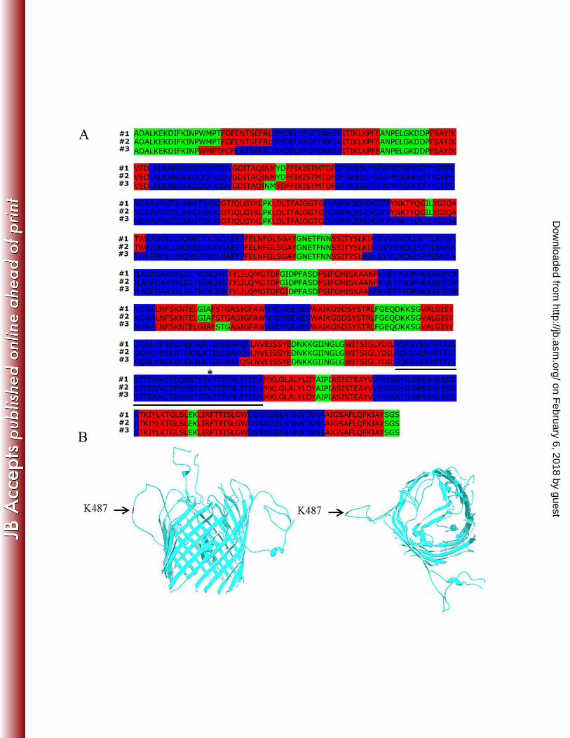
Posterior decoding algorithm (Fig. 1A). Additionally, the N- and C-terminal regions of P66 311
were predicted by all three PRED-TMBB algorithms to be located in the periplasm (Fig. 1A, 312
highlighted in green), which also is typical of ȕ-barrel forming OMPs (66). To predict the 313
tertiary structure of P66, we submitted the mature sequence to the TMBpro server (47), which 314
predicted that P66 forms a ȕ-barrel structure with 24 transmembrane strands (Fig. 1B). Previous 315
proteolysis assays have revealed that P66 has a surface-exposed, trypsin sensitive lysine at 316
position 487 (K487) (28). Consistent with this prior observation, the K487 residue was predicted 317
by all PRED-TMBB algorithms to be in an extracellular domain spanning amino acids 459-498 318
(Fig. 1A). While TMBpro also predicted K487 to be located in an extracellular loop, the loop in 319
this analysis was composed of residues 462-502 (Fig. 1B). 320
on February 6, 2018 by guest
http://jb.asm.org/
Dow
nloaded from
18
Lysine residue K487 is surface-exposed and the target of trypsin degradation. Among the 321
Lyme disease Borrelia, the P66 amino acid sequence is well conserved (28). However, previous 322
reports have shown that greater variability exists within a predicted surface loop of P66 323
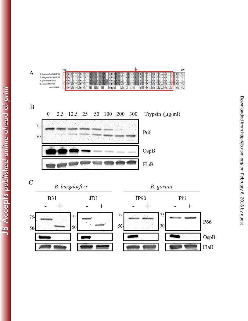
containing the K487 residue (28). When the mature P66 amino acid sequences of strains B. 324
burgdorferi B31, B. burgdorferi JD1, B. garinii IP90, and B. garinii Pbi were aligned, the 325
sequences shared approximately 90% sequence identity between the two borrelial genospecies. 326
However, sequence identity in the surface-exposed loop (amino acids 459-502) (Fig. 2A, boxed 327
in red) was only approximately 70%. Interestingly, we observed that K487, the lysine thought to 328
be the target of trypsin digestion, was detected only in the B. burgdorferi senso stricto strains 329
(Fig. 2A, indicated with red arrow). Therefore, we sought to confirm that K487 is the surface-330
exposed residue targeted by trypsin in the protease experiments. First, we confirmed that when 331
intact B. burgdorferi cells were digested with increasing amounts of trypsin, which specifically 332
cleaves on the carboxyl side of lysine and arginine residues, the 66 kDa full-length protein was 333
reduced to ~52 kDa, the size predicted if cleavage occurs at K487 (Fig. 2B) (28). Next, we 334
incubated B. burgdorferi B31, B. burgdorferi JD1, B. garinii IP90, and B. garinii Pbi cells with 335
trypsin. Consistent with K487 being the trypsin cleavage site, only the B. burgdorferi B31 and 336
JD1 strains yielded the 52-kDa truncated forms (Fig. 2C). Conversely, the B. garinii IP90 and 337
Pbi strains that lacked K487 were resistant to trypsin digestion (Fig. 2C). Control immunoblots 338
for the trypsin sensitivity experiments (Fig. 2B-C) demonstrated that the surface-exposed 339
lipoprotein OspB was degraded by trypsin, while the periplasmic FlaB protein remained intact. 340
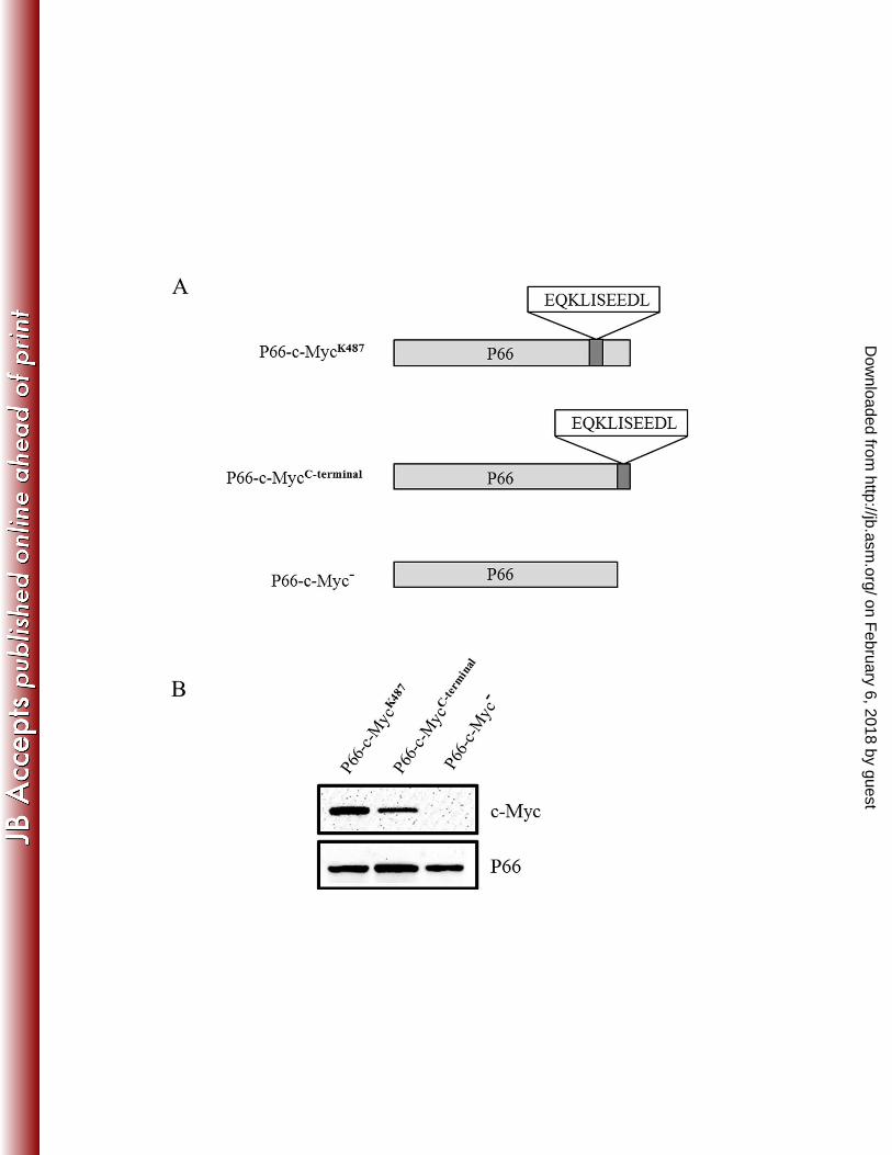
To verify surface localization of the loop containing K487, we inserted a c-Myc tag into 341
the P66 sequence adjacent to K487, and expressed P66 with the c-Myc tag in trans from 342
pBSV2G (Fig. 3A; P66-c-MycK487
). We next performed surface localization 343
on February 6, 2018 by guest
http://jb.asm.org/
Dow
nloaded from
19
immunofluorescence assays with anti-c-Myc antibodies to determine if the c-Myc tag could be 344
detected on the surface of B. burgdorferi cells. As a control, we also expressed the c-Myc tag at 345
the C-terminus of P66 (Fig. 3A; P66-c-MycC-terminal
). Given that P66 is predicted to be folded 346
into an amphipathic ȕ-barrel with its extreme C-terminus located in the periplasm (see Fig. 1A), 347
we would not expect the C-terminus of the protein to be detected on the surface of B. 348
burgdorferi. We also included a control in which P66 was expressed from pBSV2G but without 349
a c-Myc tag (Fig. 3A; P66-c-Myc-). We first confirmed by immunoblot using anti-c-Myc 350
antibodies that strains P66-c-MycK487
and P66-c-MycC-terminal
but not the P66-c-Myc- strain 351
expressed c-Myc (Fig. 3B) and that all strains, as expected, expressed P66 (Fig. 3B). Surface 352
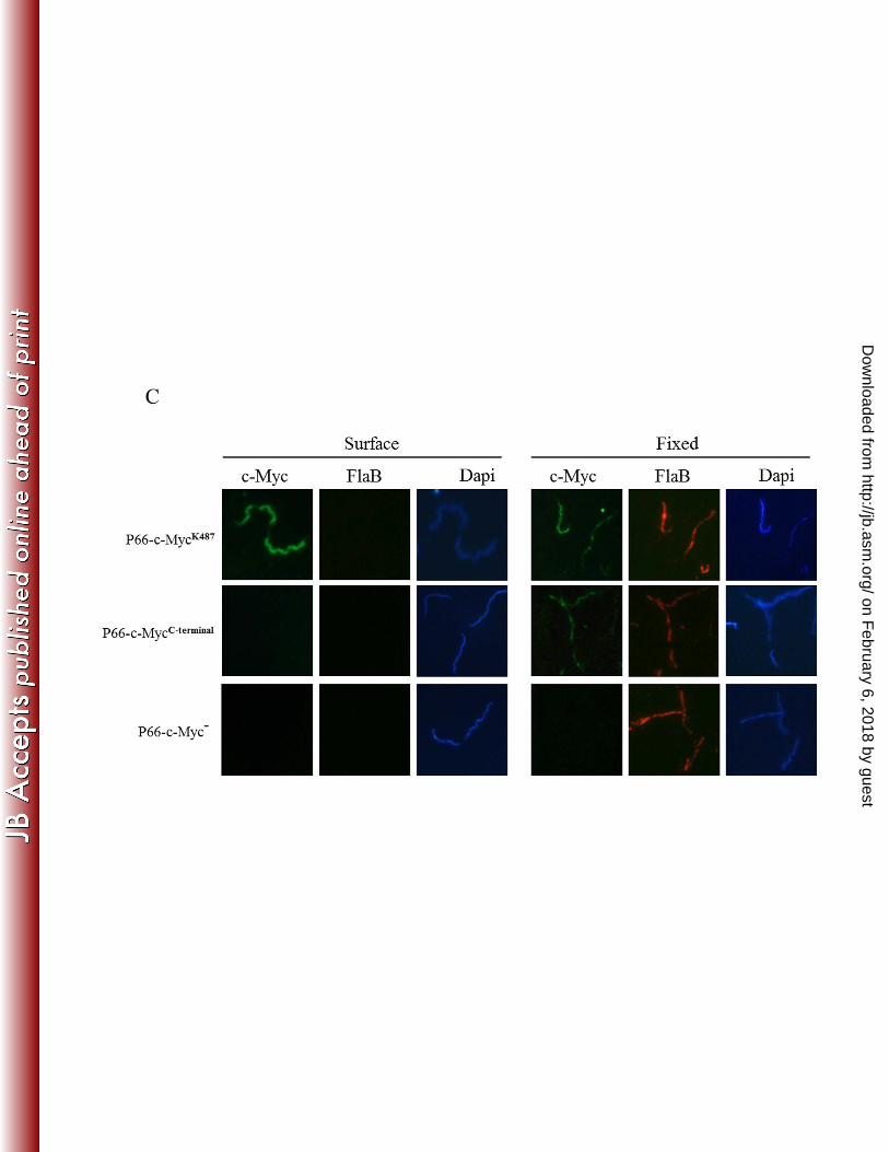
localization immunofluorescence assays were next performed by coincubation of intact cells 353
with anti-c-Myc and anti-FlaB antibodies. By including the periplasmic FlaB protein in the 354
assays, we verified that the borrelial OM remained intact throughout the experiments. DAPI 355
staining of cells also was included for visualization of spirochetes. As shown in Figure 3C, c-356
Myc was detected on the surface of B. burgdorferi when expressed in the putative surface loop 357
containing K487 (P66-c-MycK487
) but not when expressed at the C-terminus of P66 (P66-c-358
MycC-terminal
). As expected, c-Myc antibodies did not detect protein in controls expressing P66 359
without the c-Myc tag (Fig. 3C; P66-c-Myc-). Additionally, the surface localization 360
immunofluorescence assays did not detect FlaB on the surface of the spirochetes (Fig. 3C). To 361
verify that the c-Myc antibodies could bind to c-Myc expressed at the C-terminus of P66, 362
immunofluorescence assays also were performed by coincubating samples with anti-c-Myc and 363
anti-FlaB antibodies after cells had been disrupted and fixed to microscope slides. Under these 364
conditions, P66-c-MycK487
and P66-c-MycC-terminal
, but not the P66-c-Myc- strain, were 365
immunolabeled (Fig. 3C). FlaB antibodies labeled all organisms in the fixed samples, indicating 366
on February 6, 2018 by guest
http://jb.asm.org/
Dow
nloaded from
20
that their outer membranes had been disrupted (Fig. 3C). The combined immunofluorescence 367
assays demonstrate unequivocally that K487 is located in a surface-exposed loop of P66. 368
369
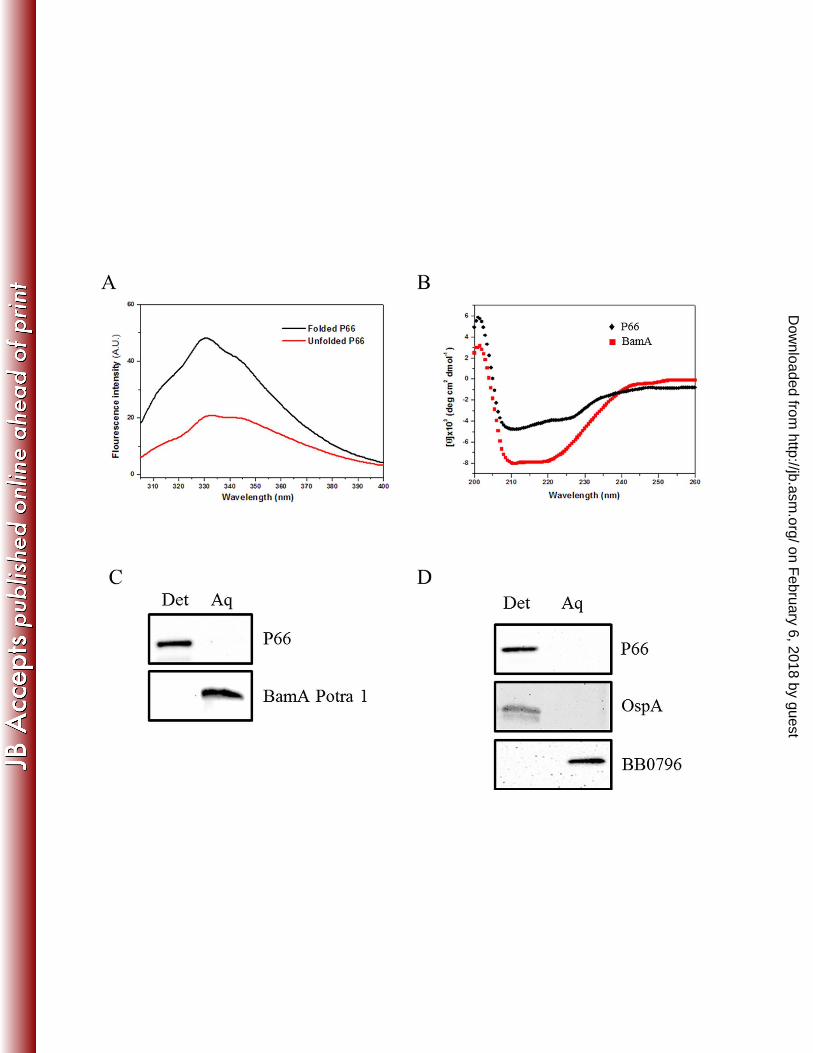
B. burgdorferi P66 is amphiphilic and forms a ȕ-barrel pore. We next sought to determine 370
whether P66 possesses the properties expected of an amphiphilic, ȕ-barrel protein. To this end, 371
we expressed P66 with a C-terminal His-tag in E. coli, purified the recombinant protein, and 372
folded it in detergent (2% DDM). Since P66 contains six tryptophan residues, folding of P66 373
could be monitored using tryptophan fluorescence. This technique utilizes the intrinsic 374
fluorescence properties of the tryptophan residues within the protein (67). As an integral 375
membrane protein folds, a “blue-shift” in the fluorescence emission maximum can be measured 376
as tryptophan residues move from an aqueous environment to a more hydrophobic environment. 377
As shown in Figure 4A, the expected shift was observed during incubation of unfolded P66 in 378
DDM. We next assessed the ȕ-sheet content of folded P66 using circular dichroism (CD) 379
spectroscopy. CD analysis revealed a broad minimum at 211 nm, consistent with extensive ȕ-380
sheet structure (Fig. 4B). As a control, we also included the B. burgdorferi BamA protein, a 381
bipartite protein with N-terminal periplasmic and C-terminal membrane-spanning ȕ-barrel 382
domains (22). Similar to P66, BamA also contained a broad minimum at 211 nm as expected for 383
a protein containing extensive ȕ-sheet (Fig. 4B). To assess the secondary structure contents of 384
P66 and BamA, we further analyzed the spectra using the DICROWEB server (58, 59). Both 385
P66 and the BamA proteins were composed of ~48% ȕ-sheet (data not shown). 386
We also performed Triton X-114 phase partitioning assays to determine whether P66 is 387
amphiphilic. Assays were performed with both recombinant P66 and B. burgdorferi lysates. 388
Both recombinant and native P66 associated with the detergent-enriched phase (Fig. 4C and 4D, 389
on February 6, 2018 by guest
http://jb.asm.org/
Dow
nloaded from
21
respectively). As a control, recombinant BamA Potra 1, a soluble, periplasmic domain of BamA, 390
also was subjected to phase-partitioning with Triton X-114; as expected, it partitioned 391
exclusively into the aqueous phase (Fig. 4C). Triton X-114 phase-partitioning using borrelial 392
whole-cell lysates resulted in partitioning of the amphiphilic, membrane bound lipoprotein OspA 393
into the detergent-enriched phase as expected (Fig. 4D). BB0796, the periplasmic B. burgdorferi 394
Skp ortholog, partitioned with the aqueous phase as previously described (Fig. 4D) (23). 395
To assess whether the folded recombinant B. burgdorferi P66 protein could incorporate 396
into lipid bilayers, we performed liposome floatation assays. Liposomes were generated from a 397
phospholipid mixture based upon the known phospholipid content of B. burgdorferi OM (68) 398
and subsequently incubated with folded P66 before being separated on discontinuous sucrose 399
gradients. Fractions then were subjected to SDS-PAGE followed by immunoblot analysis with 400
P66 antiserum. P66 was detected only in the liposome-containing top fraction (TF), as opposed 401
to the middle and bottom fractions (MF and BF, respectively), which contain unincorporated 402
material (Fig. 4E). Parallel experiments also were performed with E. coli OmpA, an extensively 403
characterized bacterial porin with ȕ-barrel structure (69). Similar to the P66 result, OmpA folded 404
in detergent was detected in the liposome-enriched top fraction (Fig. 4E, bottom panel). 405
Furthermore, we examined the pore-forming capabilities of P66 using pore formation assays in 406
which we measured the efflux of the fluorophore Tb(DPA)33-
from liposomes (50, 61, 62). The 407
fluorophore escaped from liposomes loaded with P66 (Fig. 4F), indicating that the folded, 408
recombinant protein forms a pore. Fluorophore efflux also was detected in liposomes incubated 409
with the E. coli pore-forming protein OmpF (Fig. 4F). Previous studies have demonstrated that 410
the Treponema pallidum protein TP0453 does not permit fluorophore efflux at neutral pH (50). 411
Therefore, we utilized TP0453 as a non-pore forming control; as expected, fluorophore efflux 412
on February 6, 2018 by guest
http://jb.asm.org/
Dow
nloaded from
22
was not observed in TP0453-loaded liposomes (Fig. 4F). The combined physicochemical data 413
strongly suggest P66 forms a ȕ-barrel pore in the OM of B. burgdorferi. 414
Canonical ȕ-barrel OMPs remain folded when solubilized in SDS at room temperature; as 415
a result they migrate at a lower apparent molecular mass when separated by SDS-PAGE as 416
compared to the boiled (i.e., fully denatured) protein, a property termed heat-modifiability (14, 417
70). Having established that P66 has extensive ȕ-structure, is amphiphilic, can incorporate into 418
artificial membranes, and form pores, we also sought to determine if it is heat-modifiable. While 419
folded, recombinant OmpA displayed heat modifiability, recombinant P66 did not (Fig. 4G). 420
Heat modifiability assays were also performed with B. burgdorferi whole-cell lysates, and, like 421
recombinant P66, native P66 was not modified by heat (Fig. 4H). 422
423
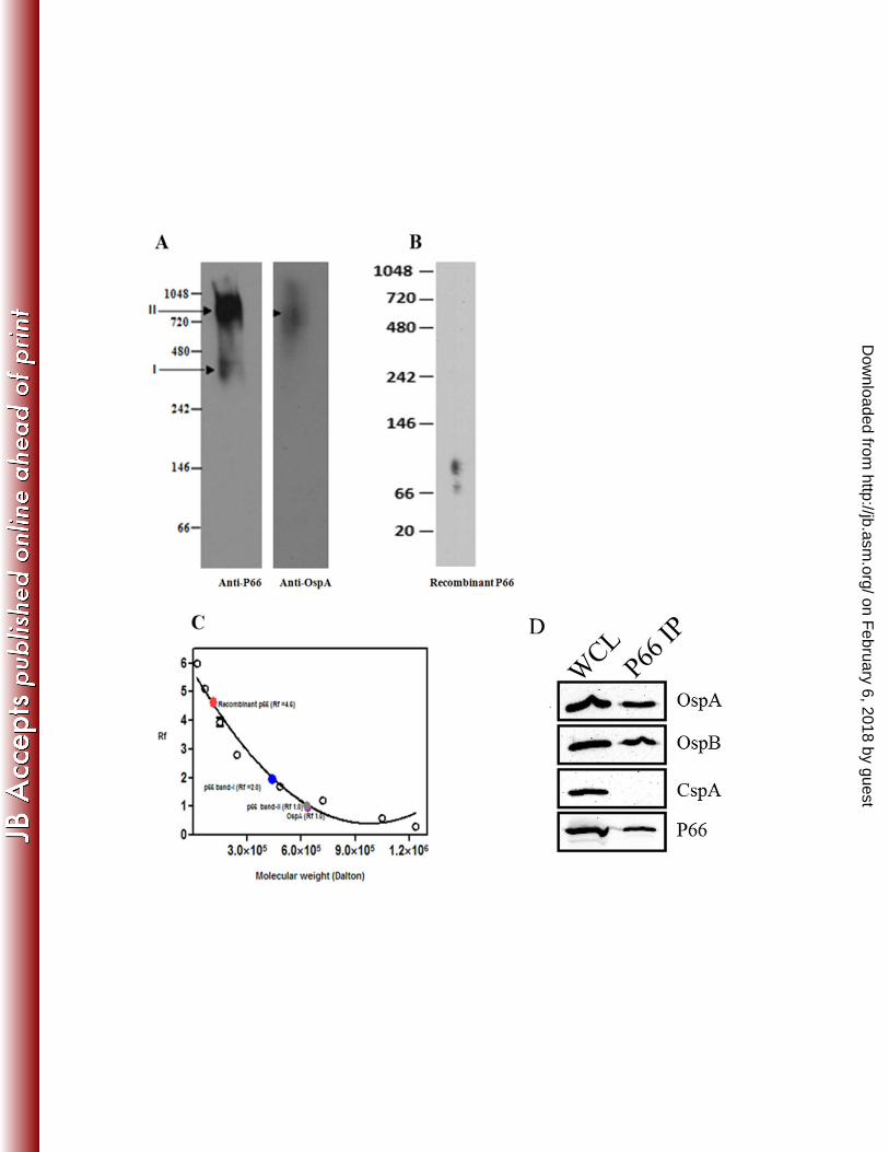
P66 is part of a higher order complex and associates with OspA and OspB in the B. 424
burgdorferi OM. To examine the oligomeric state of B. burgdorferi P66, B. burgdorferi lysates 425
and recombinant P66 were subjected to Blue-native PAGE (BN-PAGE) followed by immunoblot 426
analysis (Fig. 5A and B). The recombinant P66 migrated at 112 kDa (Rf value=4.6) (Fig. 5B 427
and C). After subtracting the molecular weight of the detergent micelle (~50 kDa), the size of 428
the recombinant P66 was estimated at 62 kDa, indicating that it is monomeric. In contrast, 429
native P66 migrated in complexes of ~400 kDa (Band I, Rf value=2.0) and ~600 kDa (Band II, 430
Rf value=1.0) (Fig. 5A and C). Previous studies have shown that OspA and P66 can be co-431
immunoprecipitated after cross-linking of B. burgdorferi cells (71, 72). Consistent with these 432
results, OspA was detected in a high molecular mass complex that migrated similarly to the large 433
~600 kDa P66-Band II complex (Fig. 5A, compare left and right panels). Interactions between 434
P66 and OspB have also been reported (72, 73). Therefore, we next assessed whether P66 435
on February 6, 2018 by guest
http://jb.asm.org/
Dow
nloaded from
23
interacts with both OspA and OspB in B. burgdorferi cells by performing co-436
immunoprecipitation studies using P66 antisera. The eluate from the co-IP experiment was 437
analyzed by immunoblot with OspA and OspB antibodies. As shown in Figure 5D, both OspA 438
and OspB co-immunoprecipitated with P66 suggesting that P66 interacts with OspA and OspB in 439
B. burgdorferi cells. The Factor H binding surface lipoprotein CspA, however, did not co-440
immunoprecipitate with P66 suggesting that the interaction between P66 and both OspA and 441
OspB is specific and that P66 does not interact with all lipoproteins found in the borrelial OM 442
(Fig. 5D). 443
on February 6, 2018 by guest
http://jb.asm.org/
Dow
nloaded from
24
DISCUSSION 444
While it has been well established that the surface of B. burgdorferi is decorated with numerous 445
lipid-modified, membrane anchored proteins (6, 8, 74), integral outer membrane proteins 446
(OMPs) from this pathogenic spirochete are still only poorly characterized. In fact, borrelial 447
OMPs have proved difficult to identify, which is due in part to the fragile outer membrane (OM) 448
of B. burgdorferi, the lack of sequence homology to known OMPs from other Gram-negative 449
bacteria, and the poor immunogenicity of integral OMPs as compared to the highly immunogenic 450
surface lipoproteins of B. burgdorferi (8, 74, 75). Despite the lack of structural data, the 451
existence of B. burgdorferi membrane-spanning OMPs was clearly demonstrated by freeze-452
fracture electron microscopy (12). While it is tempting to assume that these OMPs form ȕ-453
barrels in the borrelial OM, as is the case for Gram-negative OMPs, this has never been 454
examined in B. burgdorferi. Although some B. burgdorferi proteins have been shown to be 455
integral OMPs [P66 (BB0603) (30), P13 (BB0034) (76), BamA (BB0795) (22), BesC (BB0142) 456
(77), BB0405 (53), Lmp1 (BB0210) (78), and DipA (BB0418) (79)], the native structures of 457
these proteins are still unknown. A better understanding of the conformation of these proteins in 458
the borrelial OM will provide important insight into the structure and biogenesis of the borrelial 459
OM and could provide clues as to how these OMPs interact with host proteins. Given that prior 460
studies have shown that P66 forms pores similar to ȕ-barrel porins from other organisms, and 461
also is a known host adhesin protein (30, 32, 36), we chose to characterize the structure and 462
physicochemical properties of P66 using multiple techniques to develop a working model of the 463
P66 tertiary structure. This is an important first step that will provide working hypotheses for 464
future studies aimed at examining the structure-function properties of P66, especially since 465
crystal structures of membrane proteins, such as P66, are inherently difficult to obtain. The 466
combined structural and physicochemical analyses performed here provide compelling evidence 467
on February 6, 2018 by guest
http://jb.asm.org/
Dow
nloaded from
25
that P66 is folded into an amphipathic ȕ-barrel protein that forms a pore and contains several 468
surface-exposed loops that could interact with host proteins during infection. 469
P66, like other known OMPs, incorporates into liposomes, forms pores in large 470
unilamellar vesicles, and has amphiphilic properties, all of which are expected of a ȕ-barrel 471
protein. Furthermore, circular dichroism indicated that the recombinant P66 is composed of 48% 472
ȕ-sheet structure, which was consistent with the computer-based structural analyses. Five out of 473
the six secondary structure models used, along with the structural model prediction, indicated 474
that P66 forms a ȕ-barrel with 22- or 24-transmembrane strands. To date, bacterial OMPs with 475
8-24 transmembrane strands have been identified (14, 66), which indicates that P66 is predicted 476
to contain a relatively large number of putative transmembrane domains. Other bacterial OMPs 477
with 22- and 24-transmembrane domains include the E. coli TonB-dependent transporters (22 ȕ-478
strands), the Pseudomonas aeruginosa sidephore receptors FpvA and FptA (22 ȕ-strands), and E. 479
coli PapC (24 ȕ-strands) (14, 80). Previous reports have also indicated P66 has porin activity 480
with a large pore diameter of 2.6 nm (30). Given that other spirochetal proteins reported to be 481
OM-spanning, pore forming proteins in planar lipid bilayer assays were later revealed to be 482
periplasmic proteins (81-83), we recognized the importance of examining pore formation by P66 483
using a technique other than the planar lipid bilayer assay, especially since the planar lipid 484
bilayer has been misleading in the past. Therefore, we utilized the fluorophore Tb(DPA)33-
and 485
assessed efflux of the fluorophore from liposomes as a measure of pore formation. Moreover, 486
using the fluorophore allowed us to assess pore size. The fluorophore used in our studies, 487
Tb(DPA)33-
, has been shown to have a diameter of approximately 1 nm (62). Thus, we can 488
conclude that P66 does form pores larger than 1 nm, consistent with the previously published 489
report (30). Importantly, the size of the P66 pore is large enough to allow for the influx of many 490
on February 6, 2018 by guest
http://jb.asm.org/
Dow
nloaded from
26
nutrients and amino acids that are required for B. burgdorferi survival. Interestingly, although 491
we found that P66 was not heat modifiable like many other bacterial porin proteins, this 492
observation is consistent with the ȕ-barrel proteins BamA and Msp from the spirochetes T. 493
pallidum and T. denticola, respectively, which also are not heat-modifiable (24, 84). Although 494
we used recombinant protein in our studies and cannot rule out that the protein was not in its 495
native confirmation, recombinant P66 formed pores in membrane vesicles suggesting that the 496
protein was, indeed, folded correctly. The lack of heat modifiability was the only biochemical 497
property that was found to be different between P66 and other ȕ-barrel proteins and may 498
represent a unique property of spirochetal OMPs. 499
P66 was predicted to contain a surface-exposed loop from amino acids 459-502, which 500
contains the trypsin-sensitive lysine residue at position 487 (K487) (27). We have now 501
confirmed that this loop is located on the surface of B. burgdorferi by inserting a c-Myc tag 502
adjacent to K487 and demonstrating that the predicted surface loop is in fact extracellular by 503
surface localization immunofluorescence. To our knowledge, this is the first example in which 504
an epitope tag has been inserted into a surface exposed loop of a borrelial OMP and subsequently 505
used to examine surface exposure of a protein, or protein loop, in B. burgdorferi. Furthermore, 506
when we compared B. burgdorferi and B. garinii, only the P66 of B. burgdorferi contained a 507
lysine residue at position K487. Consistent with this, only the B. burgdorferi P66 protein was 508
observed to be susceptible to treatment with trypsin. Interestingly, within the surface-exposed 509
loop containing the trypsin accessible K487 (28), there also are lysine residues at positions 461 510
and 500 that are conserved among B. burgdorferi and B. garinii. Given that only the B. 511
burgdorferi P66 is cleaved by trypsin it would suggest that K461 and K500 are clearly not 512
accessible to trypsin for degradation. Actually, all of the strains we analyzed have numerous 513
on February 6, 2018 by guest
http://jb.asm.org/
Dow
nloaded from
27
lysine residues that the computer models predict to be located within various extracellular 514
domains connecting adjacent transmembrane ȕ-strands. Thus, it seems that lysine residues in 515
other extracellular loops are protected from trypsin cleavage. While it may seem paradoxical 516
that some lysines predicted to be surface-exposed are susceptible to trypsin cleavage while others 517
are not, complete and partial protease resistance has been described for other ȕ-barrel OM 518
proteins (15, 22, 85-89). In fact, bacterial OMPs such as FomA from Fusobacterium and BamA 519
from numerous organisms have been shown to be at least partially trypsin-resistant despite the 520
presence of numerous lysine and arginine residues throughout their primary sequence and 521
predicted surface loops (17, 22, 24, 87, 90). According to three PRED-TMBB algorithms, the 522
predicted loop in which K487 is located is one of the larger extracellular loops (Fig. 1A), and the 523
lysine residue is predicted to be on the most distal portion of the loop in relation to the ȕ-barrel in 524
the TMBpro structural model (Fig. 1B). It is tempting to speculate that the distal position of 525
K487 in the extracellular loop could make this residue more accessible to trypsin than the other 526
lysine residues. 527
Previous studies have shown that P66-integrin binding requires amino acids 150-343 (33, 528
35); thus, the surface loop of P66 containing the K487 residue does not appear to be required for 529
P66-integrin binding interactions. The region of P66 necessary for integrin binding contains 530
several predicted extracellular loops that can now be examined to better define the integrin 531
binding domain(s) that are surface-exposed in B. burgdorferi. In this regard, the structural model 532
predicted a large extracellular loop (amino acids 307-343) in the integrin binding region that 533
could be important in the P66-integrin interaction during infection. The structural predictions 534
have provided a new working model that can guide the construction of specific mutants to 535
examine in further detail the interaction between P66 and mammalian integrins. 536
on February 6, 2018 by guest
http://jb.asm.org/
Dow
nloaded from
28
Previous experiments have revealed an interaction between P66 and borrelial surface 537
lipoproteins (71-73). According to the BN-PAGE experiments reported here, recombinant P66 538
migrates at a much lower molecular weight than native P66, indicating that P66 is a member of a 539
large protein complex in the borrelial OM. We also observed that P66 and OspA both migrated 540
in a complex of similar size, which may indicate that the two proteins are members of the same 541
multi-component protein complex in the borrelial OM. This conclusion is further supported by 542
Yang and colleagues who observed that P66, OspA, and OspB can be detected in the same B. 543
burgdorferi OM complex, and that formation of the complex is dependent on the presence of P66 544
(72). Furthermore, we have shown that P66 co-immunoprecipitates specifically with OspA and 545
OspB. According to the solved crystal structures of OspA and OspB, both proteins have a C-546
terminal, positively charged cleft with an adjacent cavity that is lined with hydrophobic residues 547
(91, 92). These cavities have been proposed to possibly bind potential extracellular loops of 548
surface proteins. Therefore, it is possible then that the cavities of OspA and OspB bind one or 549
more of the P66 extracellular loops. Although it is not known what function a potential OspA/B-550
P66 complex might play in the life cycle of this spirochete, one would predict that the interaction 551
is most relevant during the tick phase or during transmission from the tick to mammal since 552
OspA and OspB are both dramatically down-regulated during or soon after transmission to the 553
mammal (93-98). For instance, OspA and OspB may bind P66 and protect the extracellular 554
loops of the protein from degradation in the tick environment. As OspA and OspB are down-555
regulated and the spirochete migrates to the mammalian host, however, P66 would no longer 556
bind OspA or OspB, and the surface of P66 would then become accessible for binding other 557
borrelial outer membrane proteins and/or proteins in the mammalian host, such as integrins, as 558
on February 6, 2018 by guest
http://jb.asm.org/
Dow
nloaded from
29
has been previously reported (32). Given that recombinant P66 was able to form pores, it is clear 559
that P66 porin activity does not require interactions with other proteins. 560
A recent report has shown that P66 is essential for infection of mice (36). While the 561
ability of P66 to bind ȕ3 chain integrins and to form pores has been well established (30, 32-34, 562
99-101), the relative contributions of the pore-forming and integrin binding capabilities of P66 to 563
spirochetal virulence are not clear. Although P66 is required for establishing an infection in 564
mice, it is not essential for growth of B. burgdorferi in dialysis membrane chambers (36). This 565
latter observation could indicate that the porin activity of P66 is not required for nutrient 566
acquisition in the mammalian environment. Alternatively, it is possible that the porin function of 567
P66 is redundant with that of another OMP within the restricted environment of the dialysis 568
membrane chamber, which precludes contact between the spirochete and immune effector cells 569
as well as any molecule or nutrient larger than the 8-kDa membrane cutoff (98). Therefore, 570
further studies will be required to elucidate the importance of the porin activity versus the 571
integrin binding activity of P66 as it relates to Lyme disease pathogenesis. 572
As mentioned above, few integral OMPs have been identified or characterized in B. 573
burgdorferi. There are at least 10-fold fewer integral OMPs per µm2
in the B. burgdorferi OM as 574
compared to the E. coli OM as estimated by freeze-fracture electron microscopy (12, 13). 575
Although there is a dearth of information about the structures of borrelial integral OMPs even 576
after more than two decades of study, the seminal observation made here that P66 forms a ȕ-577
barrel structure is of particular importance. Not only is this the first empirical evidence that 578
borrelial OMPs do, in fact, form ȕ-barrels, but the experiments outlined in this study also 579
establish methodological precedents by which other potential borrelial OMPs can be examined. 580
Finally, future P66 studies can now use the framework and structural model provided in this 581
on February 6, 2018 by guest
http://jb.asm.org/
Dow
nloaded from

30
study to examine in further detail the role of the porin/integrin binding of this protein either 582
together or separately in B. burgdorferi virulence and Lyme disease pathogenesis. 583
ACKNOWLEDGEMENTS 584
We would like to thank Dr. Jorge Benach for providing the OspB mAb CB2. This work 585
was supported in part by grants AI059373 and AI085310 from NIH/NIAID to DRA and grants 586
AI26756 and AI29735to JDR. 587
on February 6, 2018 by guest
http://jb.asm.org/
Dow
nloaded from
31
REFERENCES 588
1. Steere, A. C., J. Coburn, and L. Glickstein. 2004. The emergence of Lyme disease. J 589
Clin Invest 113:1093-1101. 590
2. Brisson, D., D. Drecktrah, C. H. Eggers, and D. S. Samuels. 2012. Genetics of 591
Borrelia burgdorferi. Annu.Rev.Genet. 46:515-536. 592
3. Radolf, J. D., M. J. Caimano, B. Stevenson, and L. T. Hu. 2012. Of ticks, mice and 593
men: understanding the dual-host lifestyle of Lyme disease spirochaetes. 594
Nat.Rev.Microbiol. 10:87-99. 595
4. Nocton, J. J. and A. C. Steere. 1995. Lyme disease. Adv.Intern.Med. 40:69-117. 596
5. Steere, A. C. 1989. Lyme Disease. N.Engl.J.Med. 321:586-597. 597
6. Bergstrom, S. and W. R. Zuckert. 2010. Structure, function and biogenesis of the 598
Borrelia cell envelope, p. 139-166. In: D. S. Samuels and J. D. Radolf (eds.), Borrelia: 599
Molecular biology, host interaction and pathogenesis. Caister Academic Press, Norfolk, 600
UK. 601
7. Takayama, K., R. J. Rothenberg, and A. G. Barbour. 1987. Absence of 602
lipopolysaccharide in the Lyme disease spirochete, Borrelia burgdorferi. Infect.Immun. 603
55:2311-2313. 604
8. Fraser, C. M., S. Casjens, W. M. Huang, G. G. Sutton, R. Clayton, R. Lathigra, O. 605
White, K. A. Ketchum, R. Dodson, E. K. Hickey, M. Gwinn, B. Dougherty, J.-F. 606
Tomb, R. D. Fleischmann, D. Richardson, J. Peterson, A. R. Kerlavage, J. 607
on February 6, 2018 by guest
http://jb.asm.org/
Dow
nloaded from
32
Quackenbush, S. Salzberg, M. Hanson, R. van Vugt, N. Palmer, M. D. Adams, J. 608
Gocayne, J. Weidman, T. Utterback, L. Watthey, L. McDonald, P. Artiach, C. 609
Bowman, S. Garland, C. Fujii, M. D. Cotton, K. Horst, K. Roberts, B. Hatch, H. O. 610
Smith, and J. C. Venter. 1997. Genomic sequence of a Lyme disease spirochaete, 611
Borrelia burgdorferi. Nature 390:580-586. 612
9. Kenedy, M. R., T. R. Lenhart, and D. R. Akins. 2012. The role of Borrelia burgdorferi 613
outer surface proteins. FEMS Immunol.Med.Microbiol. 614
10. Kovacs-Simon, A., R. W. Titball, and S. L. Michell. 2011. Lipoproteins of bacterial 615
pathogens. Infect.Immun. 79:548-561. 616
11. Norris, S. J., J. Coburn, J. M. Leong, T. H. Linden, and M. Hook. 2010. 617
Pathobiology of Lyme Disease Borrelia, p. 299-331. In: D. S. Samuels and J. D. Radolf 618
(eds.), Borrelia: Molecular biology, host interaction and pathogenesis. Caister Academic 619
Press, Norfolk, UK. 620
12. Radolf, J. D., K. W. Bourell, D. R. Akins, J. S. Brusca, and M. V. Norgard. 1994. 621
Analysis of Borrelia burgdorferi membrane architecture by freeze-fracture electron 622
microscopy. J.Bacteriol. 176:21-31. 623
13. Lugtenberg, B. and L. van Alphen. 1983. Molecular architecture and functioning of the 624
outer membrane of Escherichia coli and other Gram-negative bacteria. 625
Biochim.Biophys.Acta 737:51-115. 626
on February 6, 2018 by guest
http://jb.asm.org/
Dow
nloaded from
33
14. Fairman, J. W., N. Noinaj, and S. K. Buchanan. 2011. The structural biology of ȕ-627
barrel membrane proteins: a summary of recent reports. Curr.Opin.Struct.Biol. 21:523-628
531. 629
15. Tommassen, J. 2010. Assembly of outer-membrane proteins in bacteria and 630
mitochondria. Microbiology 156:2587-2596. 631
16. Pugsley, A. P. 1993. The complete general secretory pathway in gram-negative bacteria. 632
Microbiol.Rev. 57:50-108. 633
17. Voulhoux, R., M. P. Bos, J. Geurtsen, M. Mols, and J. Tommassen. 2003. Role of a 634
Highly Conserved Bacterial Protein in Outer Membrane Protein Assembly. Science 635
299:262-265. 636
18. Doerrler, W. T. and C. R. H. Raetz. 2005. Loss of Outer Membrane Proteins without 637
Inhibition of Lipid Export in an Escherichia coli YaeT Mutant. J.Biol.Chem. 280:27679-638
27687. 639
19. Werner, J. and r. Misra. 2005. YaeT (Omp85) affects the assembly of lipid-dependent 640
and lipid-independent outer membrane proteins of Escherichia coli. Mol.Microbiol. 641
57:1450-1459. 642
20. Wu, T., J. Malinverni, N. Ruiz, S. Kim, T. J. Silhavy, and D. Kahne. 2005. 643
Identification of a Multicomponent Complex Required for Outer Membrane Biogenesis 644
in Escherichia coli. Cell 121:235-245. 645
on February 6, 2018 by guest
http://jb.asm.org/
Dow
nloaded from

34
21. Silhavy, T. J., D. Kahne, and S. Walker. 2010. The bacterial cell envelope. Cold Spring 646
Harb.Perspect.Biol 2:a000414. 647
22. Lenhart, T. R. and D. R. Akins. 2010. Borrelia burgdorferi locus BB0795 encodes a 648
BamA orthologue required for growth and efficient localization of outer membrane 649
proteins. Mol Microbiol. 75:692-795. 650
23. Lenhart, T. R., M. R. Kenedy, X. Yang, U. Pal, and D. R. Akins. 2012. BB0324 and 651
BB0028 are constituents of the Borrelia burgdorferi ȕ-barrel assembly machine (BAM) 652
complex. BMC.Microbiol. 12:60. 653
24. Desrosiers, D. C., A. Anand, A. Luthra, S. M. Dunham-Ems, M. Ledoyt, M. A. 654
Cummings, A. Eshghi, C. E. Cameron, A. R. Cruz, J. C. Salazar, M. J. Caimano, 655
and J. D. Radolf. 2011. TP0326, a Treponema pallidum ȕ-barrel assembly machinery A 656
(BamA) orthologue and rare outer membrane protein. Mol.Microbiol. 80:1496-1515. 657
25. Barbour, A. G., S. L. Tessier, and S. F. Hayes. 1984. Variation in a major surface 658
protein of Lyme disease spirochetes. Infect.Immun. 45:94-100. 659
26. Probert, W. S., K. M. Allsup, and R. B. LeFebvre. 1995. Identification and 660
characterization of a surface-exposed 66-kilodalton protein from Borrelia burgdorferi. 661
Infect.Immun. 63:1933-1939. 662
27. Bunikis, J., L. Noppa, and S. Bergstrom. 1995. Molecular analysis of a 66-kDa protein 663
associated with the outer membrane of Lyme disease Borrelia. FEMS Microbiol.Lett. 664
131:139-145. 665
on February 6, 2018 by guest
http://jb.asm.org/
Dow
nloaded from
35
28. Bunikis, J., C. J. Luke, E. Bunikiene, S. Bergstrom, and A. G. Barbour. 1998. A 666
surface-exposed region of a novel outer membrane protein (P66) of Borrelia spp. is 667
variable in size and sequence. J.Bacteriol. 180:1618-1623. 668
29. Bunikis, J., L. Noppa, Y. Ostberg, A. G. Barbour, and S. Bergstrom. 1996. Surface 669
exposure and species specificity of an immunoreactive domain of a 66-kilodalton outer 670
membrane protein (P66) of the Borrelia species that cause Lyme disease. Infect.Immun. 671
64:5111-5116. 672
30. Skare, J. T., T. A. Mirzabekov, E. S. Shang, D. R. Blanco, H. Erdjument-bromage, 673
J. Bunikis, S. Bergstrom, P. Tempst, B. L. Kagan, J. N. Miller, and M. A. Lovett. 674
1997. The Oms66 (p66) protein is a Borrelia burgdorferi porin. Infect.Immun. 65:3654-675
3661. 676
31. Barcena-Uribarri, I., M. Thein, A. Sacher, I. Bunikis, M. Bonde, S. Bergstrom, and 677
R. Benz. 2010. P66 porins are present in both Lyme disease and relapsing fever 678
spirochetes: A comparison of the biophysical properties of P66 porins from six Borrelia 679
species. Biochim Biophys Acta 1798:1197-1203. 680
32. Coburn, J., W. Chege, L. Magoun, S. C. Bodary, and J. M. Leong. 1999. 681
Characterization of a candidate Borrelia burgdorferi ȕ3-chain integrin ligand identified 682
using a phage display library. Mol.Microbiol. 34:926-940. 683
33. Defoe, G. and J. Coburn. 2001. Delineation of Borrelia burgdorferi p66 sequences 684
required for integrin ĮIIbȕ3 recognition. Infect.Immun. 69:3455-3459. 685
on February 6, 2018 by guest
http://jb.asm.org/
Dow
nloaded from
36
34. Coburn, J. and C. Cugini. 2003. Targeted mutation of the outer membrane protein P66 686
disrupts attachment of the Lyme disease agent, Borrelia burgdorferi, to integrin Įvȕ3. 687
Proc Natl Acad Sci U S A 100:7301-7306. 688
35. Antonara, S., R. M. Chafel, M. LaFrance, and J. Coburn. 2007. Borrelia burgdorferi 689
adhesins identified using in vivo phage display. Mol.Microbiol. 66:262-276. 690
36. Ristow, L. C., H. E. Miller, L. J. Padmore, R. Chettri, N. Salzman, M. J. Caimano, 691
P. A. Rosa, and J. Coburn. 2012. The ȕ3-integrin ligand of Borrelia burgdorferi is 692
critical for infection of mice but not ticks. Mol.Microbiol. 85:1105-1118. 693
37. Barbour, A. G. 1984. Isolation and cultivation of Lyme disease spirochetes. Yale 694
J.Biol.Med. 57:521-525. 695
38. Eggers, C. H., M. J. Caimano, M. L. Clawson, W. G. Miller, D. S. Samuels, and J. D. 696
Radolf. 2002. Identification of loci critical for replication and compatibility of a Borrelia 697
burgdorferi cp32 plasmid and use of a cp32-based shuttle vector for the expression of 698
fluorescent reporters in the Lyme disease spirochaete. Mol.Microbiol. 43:281-295. 699
39. Kenedy, M. R. and D. R. Akins. 2011. The OspE-related proteins inhibit complement 700
deposition and enhance serum resistance of Borrelia burgdorferi, the lyme disease 701
spirochete. Infect.Immun. 79:1451-1457. 702
40. Bagos, P. G., T. D. Liakopoulos, I. C. Spyropoulos, and S. J. Hamodrakas. 2004. 703
PRED-TMBB: a web server for predicting the topology of ȕ-barrel outer membrane 704
proteins. Nucleic Acids Res. 32:W400-W404. 705
on February 6, 2018 by guest
http://jb.asm.org/
Dow
nloaded from
37
41. Bagos, P., T. Liakopoulos, I. Spyropoulos, and S. Hamodrakas. 2004. A Hidden 706
Markov Model method, capable of predicting and discriminating ȕ-barrel outer 707
membrane proteins. BMC Bioinformatics 5:29. 708
42. Yu, C. S., C. J. Lin, and J. K. Hwang. 2004. Predicting subcellular localization of 709
proteins for Gram-negative bacteria by support vector machines based on n-peptide 710
compositions. Protein Sci. 13:1402-1406. 711
43. Yu, N. Y., J. R. Wagner, M. R. Laird, G. Melli, S. Rey, R. Lo, P. Dao, S. C. 712
Sahinalp, M. Ester, L. J. Foster, and F. S. Brinkman. 2010. PSORTb 3.0: improved 713
protein subcellular localization prediction with refined localization subcategories and 714
predictive capabilities for all prokaryotes. Bioinformatics. 26:1608-1615. 715
44. Remmert, M., D. Linke, A. N. Lupas, and J. Soding. 2009. HHomp--prediction and 716
classification of outer membrane proteins. Nucleic Acids Res. 37:W446-W451. 717
45. Ou, Y. Y., M. M. Gromiha, S. A. Chen, and M. Suwa. 2008. TMBETADISC-RBF: 718
Discrimination of ȕ-barrel membrane proteins using RBF networks and PSSM profiles. 719
Comput.Biol.Chem. 32:227-231. 720
46. Berven, F. S., K. Flikka, H. B. Jensen, and I. Eidhammer. 2004. BOMP: a program to 721
predict integral ȕ-barrel outer membrane proteins encoded within genomes of Gram-722
negative bacteria. Nucleic Acids Res. 32:W394-W399. 723
47. Randall, A., J. Cheng, M. Sweredoski, and P. Baldi. 2008. TMBpro: secondary 724
structure, ȕ-contact and tertiary structure prediction of transmembrane ȕ-barrel proteins. 725
Bioinformatics. 24:513-520. 726
on February 6, 2018 by guest
http://jb.asm.org/
Dow
nloaded from
38
48. Guex, N. and M. C. Peitsch. 1997. SWISS-MODEL and the Swiss-PdbViewer: an 727
environment for comparative protein modeling. Electrophoresis 18:2714-2723. 728
49. Hazlett, K. R., D. L. Cox, M. Decaffmeyer, M. P. Bennett, D. C. Desrosiers, C. J. La 729
Vake, M. E. La Vake, K. W. Bourell, E. J. Robinson, R. Brasseur, and J. D. Radolf. 730
2005. TP0453, a concealed outer membrane protein of Treponema pallidum, enhances 731
membrane permeability. J.Bacteriol. 187:6499-6508. 732
50. Luthra, A., G. Zhu, D. C. Desrosiers, C. H. Eggers, V. Mulay, A. Anand, F. A. 733
McArthur, F. B. Romano, M. J. Caimano, A. P. Heuck, M. G. Malkowski, and J. D. 734
Radolf. 2011. The transition from closed to open conformation of Treponema pallidum 735
outer membrane-associated lipoprotein TP0453 involves membrane sensing and 736
integration by two amphipathic helices. J.Biol.Chem. 286:41656-41668. 737
51. Kenedy, M. R., S. R. Vuppala, C. Siegel, P. Kraiczy, and D. R. Akins. 2009. CspA-738
mediated binding of human factor H inhibits complement deposition and confers serum 739
resistance in Borrelia burgdorferi. Infect.Immun. 77:2773-2782. 740
52. Brooks, C. S., S. R. Vuppala, A. M. Jett, A. Alitalo, S. Meri, and D. R. Akins. 2005. 741
Complement regulator-acquiring surface protein 1 imparts resistance to human serum in 742
Borrelia burgdorferi. J Immunol. 175:3299-3308. 743
53. Brooks, C. S., S. R. Vuppala, A. M. Jett, and D. R. Akins. 2006. Identification of 744
Borrelia burgdorferi outer surface proteins. Infect.Immun. 74:296-304. 745
on February 6, 2018 by guest
http://jb.asm.org/
Dow
nloaded from
39
54. Cox, D. L., D. R. Akins, K. W. Bourell, P. Lahdenne, M. V. Norgard, and J. D. 746
Radolf. 1996. Limited surface exposure of Borrelia burgdorferi outer surface 747
lipoproteins. Proc.Natl.Acad.Sci.U.S.A 93:7973-7978. 748
55. Coleman, J. L., R. C. Rogers, P. A. Rosa, and J. L. Benach. 1994. Variations in the 749
ospB gene of Borrelia burgdorferi result in differences in monoclonal antibody reactivity 750
and in production of escape variants. Infect.Immun. 62:303-307. 751
56. Elias, A. F., J. L. Bono, J. J. Kupko, III, P. E. Stewart, J. G. Krum, and P. A. Rosa. 752
2003. New antibiotic resistance cassettes suitable for genetic studies in Borrelia 753
burgdorferi. J Mol.Microbiol.Biotechnol. 6:29-40. 754
57. Samuels, D. S. 1995. Electrotransformation of the spirochete Borrelia burgdorferi. 755
Methods Mol.Biol. 47:253-259. 756
58. Whitmore, L. and B. A. Wallace. 2004. DICHROWEB, an online server for protein 757
secondary structure analyses from circular dichroism spectroscopic data. Nucleic Acids 758
Res. 32:W668-W673. 759
59. Whitmore, L. and B. A. Wallace. 2008. Protein secondary structure analyses from 760
circular dichroism spectroscopy: methods and reference databases. Biopolymers 89:392-761
400. 762
60. Brusca, J. S. and J. D. Radolf. 1994. Isolation of integral membrane proteins by phase 763
partitioning with Triton X-114. Methods Enzymol. 228:182-193. 764
on February 6, 2018 by guest
http://jb.asm.org/
Dow
nloaded from
40
61. Anand, A., A. Luthra, S. Dunham-Ems, M. J. Caimano, C. Karanian, M. Ledoyt, A. 765
R. Cruz, J. C. Salazar, and J. D. Radolf. 2012. TprC/D (Tp0117/131), a trimeric, pore-766
forming rare outer membrane protein of Treponema pallidum, has a bipartite domain 767
structure. J.Bacteriol. 194:2321-2333. 768
62. Heuck, A. P., R. K. Tweten, and A. E. Johnson. 2003. Assembly and topography of the 769
prepore complex in cholesterol-dependent cytolysins. J.Biol.Chem. 278:31218-31225. 770
63. Schagger, H. and J. G. von. 1991. Blue native electrophoresis for isolation of membrane 771
protein complexes in enzymatically active form. Anal.Biochem. 199:223-231. 772
64. Wittig, I., H. P. Braun, and H. Schagger. 2006. Blue native PAGE. Nat.Protoc. 1:418-773
428. 774
65. Kawabata, H., S. J. Norris, and H. Watanabe. 2004. BBE02 disruption mutants of 775
Borrelia burgdorferi B31 have a highly transformable, infectious phenotype. 776
Infect.Immun. 72:7147-7154. 777
66. Schulz, G. E. 2002. The structure of bacterial outer membrane proteins. Biochimica et 778
Biophysica Acta (BBA) - Biomembranes 1565:308-317. 779
67. Heuck, A. P. and A. E. Johnson. 2002. Pore-forming protein structure analysis in 780
membranes using multiple independent fluorescence techniques. Cell Biochem.Biophys. 781
36:89-101. 782
68. Belisle, J. T., M. E. Brandt, J. D. Radolf, and M. V. Norgard. 1994. Fatty acids of 783
Treponema pallidum and Borrelia burgdorferi lipoproteins. J.Bacteriol. 176:2151-2157. 784
on February 6, 2018 by guest
http://jb.asm.org/
Dow
nloaded from
41
69. Reusch, R. N. 2012. Insights into the structure and assembly of Escherichia coli outer 785
membrane protein A. FEBS J. 786
70. Nakamura, K. and S. Mizushima. 1976. Effects of heating in dodecyl sulfate solution 787
on the conformation and electrophoretic mobility of isolated major outer membrane 788
proteins from Escherichia coli K-12. J.Biochem. 80:1411-1422. 789
71. Bunikis, J. and A. G. Barbour. 1999. Access of antibody or trypsin to an integral outer 790
membrane protein (P66) of Borrelia burgdorferi is hindered by Osp lipoproteins. 791
Infect.Immun. 67:2874-2883. 792
72. Yang, X., K. Promnares, J. Qin, M. He, D. Y. Shroder, T. Kariu, Y. Wang, and U. 793
Pal. 2011. Characterization of Multiprotein Complexes of the Borrelia burgdorferi Outer 794
Membrane Vesicles. J.Proteome.Res. 10:4556-4566. 795
73. LaRocca, T. J., D. J. Holthausen, C. Hsieh, C. Renken, C. A. Mannella, and J. L. 796
Benach. 2009. The bactericidal effect of a complement-independent antibody is 797
osmolytic and specific to Borrelia. Proc.Natl.Acad.Sci.U.S.A 106:10752-10757. 798
74. Casjens, S., N. Palmer, R. van Vugt, W. M. Huang, B. Stevenson, P. Rosa, R. 799
Lathigra, G. Sutton, J. Peterson, R. J. Dodson, D. Haft, E. Hickey, M. Gwinn, O. 800
White, and C. M. Fraser. 2000. A bacterial genome in flux: the twelve linear and nine 801
circular extrachromosomal DNAs in an infectious isolate of the Lyme disease spirochete 802
Borrelia burgdorferi. Mol.Microbiol. 35:490-516. 803
on February 6, 2018 by guest
http://jb.asm.org/
Dow
nloaded from
42
75. Brandt, M. E., B. S. Riley, J. D. Radolf, and M. V. Norgard. 1990. Immunogenic 804
integral membrane proteins of Borrelia burgdorferi are lipoproteins. Infect.Immun. 805
58:983-991. 806
76. Ostberg, Y., M. Pinne, R. Benz, P. Rosa, and S. Bergstrom. 2002. Elimination of 807
channel-forming activity by insertional inactivation of the p13 gene in Borrelia 808
burgdorferi. J Bacteriol. 184:6811-6819. 809
77. Bunikis, I., K. Denker, Y. Ostberg, C. Andersen, R. Benz, and S. Bergstrom. 2008. 810
An RND-type efflux system in Borrelia burgdorferi is involved in virulence and 811
resistance to antimicrobial compounds. PLoS Pathog 4:e1000009. 812
78. Yang, X., T. R. Lenhart, T. Kariu, J. Anguita, D. R. Akins, and U. Pal. 2010. 813
Characterization of unique regions of Borrelia burgdorferi surface-located membrane 814
protein 1. Infect.Immun. 78:4477-4487. 815
79. Thein, M., M. Bonde, I. Bunikis, K. Denker, A. Sickmann, S. Bergstrom, and R. 816
Benz. 2012. DipA, a pore-forming protein in the outer membrane of Lyme disease 817
spirochetes exhibits specificity for the permeation of dicarboxylates. PLoS.ONE. 818
7:e36523. 819
80. Noinaj, N., M. Guillier, T. J. Barnard, and S. K. Buchanan. 2010. TonB-dependent 820
transporters: regulation, structure, and function. Annu.Rev.Microbiol. 64:43-60. 821
81. Blanco, D. R., C. I. Champion, M. M. Exner, H. Erdjument-bromage, R. W. 822
Hancock, P. Tempst, J. N. Miller, and M. A. Lovett. 1995. Porin activity and sequence 823
on February 6, 2018 by guest
http://jb.asm.org/
Dow
nloaded from
43
analysis of a 31-kilodalton Treponema pallidum subsp. pallidum rare outer membrane 824
protein (Tromp1). J.Bacteriol. 177:3556-3562. 825
82. Akins, D. R., E. Robinson, D. V. Shevchenko, C. Elkins, D. L. Cox, and J. D. Radolf. 826
1997. Tromp1, a putative rare outer membrane protein, is anchored by an uncleaved 827
leader peptide to the Treponema pallidum cytoplasmic membrane. J.Bacteriol. 179:576-828
586. 829
83. Mulay, V., M. Caimano, D. Liveris, D. C. Desrosiers, J. D. Radolf, and I. Schwartz. 830
2007. Borrelia burgdorferi BBA74, a Periplasmic Protein Associated with the Outer 831
Membrane, Lacks Porin-Like Properties. The Journal of Bacteriology 189:2063-2068. 832
84. Anand, A., A. Luthra, M. E. Edmond, M. Ledoyt, M. J. Caimano, and J. D. Radolf. 833
2013. The major outer sheath protein (Msp) of Treponema denticola has a bipartite 834
domain architecture and exists as periplasmic and outer membrane-spanning conformers. 835
J.Bacteriol. 195:2060-2071. 836
85. Hoenger, A., J. M. Pages, D. Fourel, and A. Engel. 1993. The orientation of porin 837
OmpF in the outer membrane of Escherichia coli. J.Mol.Biol. 233:400-413. 838
86. Werner, J., A. M. Augustus, and r. Misra. 2003. Assembly of TolC, a structurally 839
unique and multifunctional outer membrane protein of Escherichia coli K-12. J.Bacteriol. 840
185:6540-6547. 841
87. Robert, V., E. B. Volokhina, F. Senf, M. P. Bos, P. V. Gelder, and J. Tommassen. 842
2006. Assembly Factor Omp85 Recognizes Its Outer Membrane Protein Substrates by a 843
Species-Specific C-Terminal Motif. PLoS Biol 4:e377. 844
on February 6, 2018 by guest
http://jb.asm.org/
Dow
nloaded from
44
88. Stegmeier, J. F. and C. Andersen. 2006. Characterization of Pores Formed by YaeT 845
(Omp85) from Escherichia coli. J.Biochem. 140:275-283. 846
89. Sun, G., S. Pal, A. K. Sarcon, S. Kim, E. Sugawara, H. Nikaido, M. J. Cocco, E. M. 847
Peterson, and L. M. de la Maza. 2007. Structural and functional analyses of the major 848
outer membrane protein of Chlamydia trachomatis. J.Bacteriol. 189:6222-6235. 849
90. Puntervoll, P., M. Ruud, L. J. Bruseth, H. Kleivdal, B. T. Hogh, R. Benz, and H. B. 850
Jensen. 2002. Structural characterization of the fusobacterial non-specific porin FomA 851
suggests a 14-stranded topology, unlike the classical porins. Microbiology 148:3395-852
3403. 853
91. Li, H., J. J. Dunn, B. J. Luft, and C. L. Lawson. 1997. Crystal structure of Lyme 854
disease antigen outer surface protein A complexed with an Fab. 855
Proc.Natl.Acad.Sci.(USA) 94:3584-3589. 856
92. Becker, M., J. Bunikis, B. D. Lade, J. J. Dunn, A. G. Barbour, and C. L. Lawson. 857
2005. Structural investigation of Borrelia burgdorferi OspB, a bactericidal Fab target. 858
J.Biol.Chem. 280:17363-17370. 859
93. Schwan, T. G., J. Piesman, W. T. Golde, M. C. Dolan, and P. A. Rosa. 1995. 860
Induction of an outer surface protein on Borrelia burgdorferi during tick feeding. 861
Proc.Natl.Acad.Sci.U.S.A 92:2909-2913. 862
94. Ohnishi, J., J. Piesman, and A. de Silva. 2001. Antigenic and genetic heterogeneity of 863
Borrelia burgdorferi populations transmitted by ticks. Proc.Natl.Acad.Sci.(USA) 98:670-864
675. 865
on February 6, 2018 by guest
http://jb.asm.org/
Dow
nloaded from
45
95. Hefty, P. S., S. E. Jolliff, M. J. Caimano, S. K. Wikel, and D. R. Akins. 2002. 866
Changes in temporal and spatial patterns of outer surface lipoprotein expression generate 867
population heterogeneity and antigenic diversity in the Lyme disease spirochete, Borrelia 868
burgdorferi. Infect.Immun. 70:3468-3478. 869
96. Revel, A. T., A. M. Talaat, and M. V. Norgard. 2002. DNA microarray analysis of 870
differential gene expression in Borrelia burgdorferi, the Lyme disease spirochete. 871
Proc.Natl.Acad.Sci.U.S.A 99:1562-1567. 872
97. Brooks, C. S., P. S. Hefty, S. E. Jolliff, and D. R. Akins. 2003. Global analysis of 873
Borrelia burgdorferi genes regulated by mammalian host-specific signals. Infect.Immun. 874
71:3371-3383. 875
98. Akins, D. R., K. W. Bourell, M. J. Caimano, M. V. Norgard, and J. D. Radolf. 1998. 876
A new animal model for studying Lyme disease spirochetes in a mammalian host-adapted 877
state. J.Clin.Invest. 101:2240-2250. 878
99. Ntchobo, H., H. Rothermel, W. Chege, A. C. Steere, and J. Coburn. 2001. 879
Recognition of multiple antibody epitopes throughout Borrelia burgdorferi p66, a 880
candidate adhesin, in patients with early or late manifestations of Lyme disease. 881
Infect.Immun. 69:1953-1956. 882
100. LaFrance, M. E., J. V. Pierce, S. Antonara, and J. Coburn. 2011. The Borrelia 883
burgdorferi integrin ligand P66 affects gene expression by human cells in culture. 884
Infect.Immun. 79:3249-3261. 885
on February 6, 2018 by guest
http://jb.asm.org/
Dow
nloaded from
46
101. Pinne, M., M. Thein, K. Denker, R. Benz, J. Coburn, and S. Bergstrom. 2007. 886
Elimination of channel-forming activity by insertional inactivation of the p66 gene in 887
Borrelia burgdorferi. FEMS Microbiol.Lett. 266:241-249. 888
889
890
on February 6, 2018 by guest
http://jb.asm.org/
Dow
nloaded from
47
FIGURE LEGENDS 891
892
Figure 1. P66 is predicted to form a ȕ-barrel. A. The B. burgdorferi P66 mature amino acid 893
sequence was analyzed using the PRED-TMBB server. Topology results from algorithms used 894
by PRED-TMBB include Viterbi (#1), N-Best (#2), and Posterior decoding (#3) methods. 895
Predicted transmembrane regions (red), extracellular domains (blue), and periplasmic domains 896
(green) are indicated for each algorithm. The predicted surface-exposed loop containing K487 is 897
noted with a black bar under the sequence while K487 is indicated with an asterisk above the 898
residue. B. TMBpro (http://tmbpro.ics.uci.edu/) predicted tertiary structure of B. burgdorferi 899
P66 depicted as a ribbon model. Both side and top views of the predicted P66 model are shown. 900
The lysine residue (K487) predicted to be sensitive to trypsin cleavage is labeled. 901
902
Figure 2. K487 is a target for trypsin degradation. A. Amino acid alignment of P66 from B. 903
burgdorferi B31, B. burgdorferi JD1, B. garinii IP90, and B. garinii Pbi were generated using 904
MacVector 10.0. The variable region predicted to be surface-exposed is boxed in red with an 905
arrow indicating the lysine residue responsible for trypsin degradation (K487). B. B. 906
burgdorferi cells were washed and incubated with 0-300 ȝg/ml of trypsin. Samples then were 907
immunoblotted with P66 antibodies to assess surface degradation. Equivalent samples also were 908
immunoblotted with monoclonal OspB antibodies to control for protease activity and with 909
antibodies that recognize the periplasmic protein FlaB to ensure that the OM remained intact 910
throughout the proteolysis experiments. C. B. burgdorferi B31, B. burgdorferi JD1, B. garinii 911
IP90, and B. garinii Pbi cells were collected and incubated with 200 µg/ml trypsin before 912
immunoblot with rat anti-P66 antibodies. Samples were also immunoblotted with antibodies 913
on February 6, 2018 by guest
http://jb.asm.org/
Dow
nloaded from
48
against the periplasmic FlaB protein (bottom panel) to ensure membrane integrity and outer 914
membrane lipoprotein OspB antibodies (middle panel) as a positive control for protease activity. 915
Molecular weight standards (in kDa) are shown at left. 916
917
Figure 3. K487 is located on the surface of B. burgdorferi. A. Schematic of c-Myc constructs 918
and controls generated in pBSV2G for surface localization immunofluorescence experiments. 919
P66 expressed in trans from pBSV2G is indicated with a grey bar, and the location of the c-Myc 920
tag is noted with a dark grey bar as well as the c-Myc sequence (EQKLISEEDL). B. Equivalent 921
whole-cell lysates from P66-c-MycK487
, P66-c-MycC-terminal
, and P66-c-Myc- were subjected to 922
immunoblot analysis with rabbit anti c-Myc and rat anti-P66 antibodies. C. For surface 923
localization assays, P66-c-MycK487
, P66-c-MycC-terminal
, and P66-c-Myc- cells were coincubated 924
with rabbit anti c-Myc antibodies and rat anti-FlaB antibodies before being fixed to slides and 925
incubated with the appropriate Alexa Fluor-conjugated secondary antibodies. Samples were also 926
fixed to slides prior to coincubation with rabbit anti c-Myc antibodies and rat anti-FlaB 927
antibodies. All spirochetes were also counterstained with DAPI. 928
929
Figure 4. B. burgdorferi P66 is amphiphilic and forms a ȕ-barrel pore. A. Tryptophan 930
fluorescence emission spectra of recombinant B. burgdorferi P66 in DDM buffer (folded P66, 931
black line) or in denaturation buffer (unfolded P66, red line). B. Circular dichroism spectra of 932
recombinant B. burgdorferi P66 and BamA in DDM buffer. C. Recombinant P66 and 933
recombinant B. burgdorferi BamA Potra domain 1 were subjected to Triton X-114 phase 934
partitioning. The detergent-enriched (Det) and aqueous (Aq) phases were immunoblotted with 935
P66 or BamA antibodies. D. B. burgdorferi whole-cell lysates were subjected to phase 936
on February 6, 2018 by guest
http://jb.asm.org/
Dow
nloaded from
49
partitioning. The detergent-enriched (Det) and aqueous-enriched (Aq) phases were 937
immunoblotted with P66 antibodies. Equivalent amounts of the Det and Aq phases were also 938
immunoblotted with OspA antibodies to provide a detergent-enriched fraction control and 939
BB0796 antibodies to provide an aqueous-enriched fraction control. E. Liposomes simulating 940
the B. burgdorferi OM were incubated with folded P66 or folded OmpA before separation on 941
discontinuous sucrose gradients. Fractions were collected from the top (TF), middle (MF), and 942
bottom (BF) of the gradient before immunoblotting with rat anti-P66 antibodies or rat anti-943
OmpA antibodies. F. Liposomes containing the fluorophore Tb(DPA)33-
were incubated with 944
recombinant B. burgdorferi P66, E. coli OmpF, or T. pallidum TP0453 in buffer supplemented 945
with EDTA and fluorophore efflux was measured as quenched fluorescence. G. Folded P66 and 946
OmpA were incubated at room temperature (- boil) or boiled (+ boil) for 20 min in final sample 947
buffer. The samples were then separated by SDS-PAGE and subjected to immunoblot analysis 948
with either P66 or OmpA antibodies. H. B. burgdorferi whole-cell lysates were incubated at 949
room temperature (- boil) or boiled (+ boil) for 10 min in final sample buffer prior to SDS-PAGE 950
and immunoblot analysis with P66 antibodies. 951
952
Figure 5. P66 forms a large protein complex and interacts with OspA and OspB in B. 953
burgdorferi. Immunoblot analysis of (A) B. burgdorferi whole-cell lysates and (B) recombinant 954
P66 subjected to BN-PAGE. Immunoblots were performed using anti-P66 or anti-OspA 955
antibodies. Molecular weight standards (kDa) are shown at left of panels. C. The retardation 956
factor (Rf) values for the stained bands of M.W. standards, P66 complexes I and II, and 957
recombinant P66 (in Da) were calculated and plotted versus molecular weights using a second 958
order polynomial best fit according to manufacturer’s instruction (Invitrogen). D. B. 959
on February 6, 2018 by guest
http://jb.asm.org/
Dow
nloaded from

50
burgdorferi cells lysates were prepared and subjected to co-immunoprecipitation experiments 960
using rat anti-P66 antibodies. The eluate from the co-immunoprecipitation was analyzed by 961
SDS-PAGE and immunoblot using OspA, OspB, CspA, and P66 antibodies. Whole-cell lysates 962
(WCL) were also included in each immunoblot as a positive control. 963
on February 6, 2018 by guest
http://jb.asm.org/
Dow
nloaded from