1 the perils of pathogen discovery - journal of virology - american
TRANSCRIPT
The Perils of Pathogen Discovery: Origin of a Novel Parvovirus-LikeHybrid Genome Traced to Nucleic Acid Extraction Spin Columns
Samia N. Naccache,a,b Alexander L. Greninger,a,b Deanna Lee,a,b Lark L. Coffey,c Tung Phan,c Annie Rein-Weston,a,b
Andrew Aronsohn,d John Hackett, Jr.,e Eric L. Delwart,a,c Charles Y. Chiua,b,f
Department of Laboratory Medicine, University of California, San Francisco, California, USAa; UCSF-Abbott Viral Diagnostics and Discovery Center, San Francisco, California,USAb; Blood Systems Research Institute, San Francisco, California, USAc; Center for Liver Disease, University of Chicago Medical Center, Chicago, Illinois, USAd; AbbottDiagnostics, Abbott Park, Illinois, USAe; Department of Medicine, Division of Infectious Diseases, University of California, San Francisco, California, USAf
Next-generation sequencing was used for discovery and de novo assembly of a novel, highly divergent DNA virus at the interfacebetween the Parvoviridae and Circoviridae. The virus, provisionally named parvovirus-like hybrid virus (PHV), is nearly identi-cal by sequence to another DNA virus, NIH-CQV, previously detected in Chinese patients with seronegative (non-A-E) hepatitis.Although we initially detected PHV in a wide range of clinical samples, with all strains sharing �99% nucleotide and amino acididentity with each other and with NIH-CQV, the exact origin of the virus was eventually traced to contaminated silica-bindingspin columns used for nucleic acid extraction. Definitive confirmation of the origin of PHV, and presumably NIH-CQV, wasobtained by in-depth analyses of water eluted through contaminated spin columns. Analysis of environmental metagenome li-braries detected PHV sequences in coastal marine waters of North America, suggesting that a potential association between PHVand diatoms (algae) that generate the silica matrix used in the spin columns may have resulted in inadvertent viral contamina-tion during manufacture. The confirmation of PHV/NIH-CQV as laboratory reagent contaminants and not bona fide infectiousagents of humans underscores the rigorous approach needed to establish the validity of new viral genomes discovered by next-generation sequencing.
Over the past 5 years, next-generation sequencing (NGS), oth-erwise known as deep sequencing, has been a remarkably
successful approach for the identification and characterization ofnovel pathogens (1–3). In principle, with the exception of prions(4), all microbial agents that have the potential to cause disease canbe detected in clinical samples on the basis of their specific nucle-otide sequence. The rapidly increasing breadth and scope of mi-crobial sequence reference databases in the research communityhave also facilitated the identification of novel microorganisms (3,5), especially viruses that can exhibit a high degree of sequencedivergence. Some recent examples of the use of NGS technologyfor pathogen discovery include identification of new agents asso-ciated with chronic illnesses, such as cancer (6, 7), screening ofbiologics, such as vaccines, for safety and testing purity (8–10),and outbreak investigation of novel viral pathogens (11–15).
Nevertheless, despite the broad utility of NGS in pathogen dis-covery, the technique is associated with a high risk of inadvertentcontamination (16–18). The use of random instead of targetedprimers to amplify all of the nucleic acid (NA) in clinical samplesand the sheer depth of NGS, which routinely generates millions tobillions of sequences per run, result in significant potential forlaboratory and reagent contamination in addition to sample car-ryover. Concurrent analyses of cases and controls in a blindedfashion to exclude laboratory-derived contamination, collectionof supportive clinical, epidemiologic, and serological data, andrigorous replication studies are thus critical in confirming or re-futing putative associations of candidate novel agents with disease(3). These strategies were previously applied to conclusively de-termine that the retrovirus xenotropic murine leukemia virus-related virus(XMRV) is not associated with chronic fatigue syn-drome or prostate cancer and, in fact, originated as a mouse cellline-derived laboratory contaminant (19–26).
Here we describe the identification and whole-genome assem-
bly of a highly divergent single-stranded DNA (ssDNA) virus sit-uated at the interface between Circoviridae and Parvoviridae bydeep sequencing. The virus, provisionally named parvovirus-likehybrid virus (PHV), was detected in samples from patients withchronic seronegative (non-A-E) hepatitis and diarrhea of un-known etiology. The initial finding of a novel parvovirus-/circo-virus-like agent in these patients was of great interest because theseviruses are known to broadly infect insects, vertebrate animals,and humans (27–29), and specific members, such as parvovirusB19 in humans and porcine circovirus type 2 (PCV2) in pigs, havebeen linked to hepatitis (30–32). Furthermore, a study by Xu et al.recently described the discovery of a hybrid DNA virus in serumsamples from Chinese patients with seronegative hepatitis, namedNIH-CQV, with a sequence nearly identical to that of PHV (33).However, combined findings from follow-up deep sequencing,PCR, and data mining analyses performed in two independentlaboratories and presented here demonstrate that PHV (and pre-sumably NIH-CQV) are in fact laboratory reagent contaminantsand underscore strategies that can be employed in the future torapidly establish the significance and clinical relevance of novelmicrobial agents discovered by NGS.
Received 15 August 2013 Accepted 28 August 2013
Published ahead of print 11 September 2013
Address correspondence to Charles Y. Chiu, [email protected].
Supplemental material for this article may be found at http://dx.doi.org/10.1128/JVI.02323-13.
Copyright © 2013, American Society for Microbiology. All Rights Reserved.
doi:10.1128/JVI.02323-13
The authors have paid a fee to allow immediate free access to this article.
11966 jvi.asm.org Journal of Virology p. 11966–11977 November 2013 Volume 87 Number 22
Dow
nloa
ded
from
http
s://j
ourn
als.
asm
.org
/jour
nal/j
vi o
n 09
Dec
embe
r 20
21 b
y 11
2.18
6.38
.40.
MATERIALS AND METHODSEthics statement. All clinical samples were analyzed under protocols ap-proved by the Institutional Review Boards (IRBs) of University of Cali-fornia, San Francisco (UCSF), and Blood Systems Research Institute(BSRI). Written informed consent was previously obtained for patients innon-A-E hepatitis cohort 1 (University of Chicago), non-A-E hepatitiscohort 2 (BIOLINCC Transfusion-Transmitted Viruses Study) (34), andthe California Encephalitis Project (35) to provide clinical samples forviral analysis. Diarrheal samples from Nigeria, negative plasma for theHIV-spiked sample, and other miscellaneous clinical samples used to ob-tain the data shown in Fig. 2 and in Table S1 in the supplemental materialdid not require consent, as these samples were pre-existing and deidenti-fied, and their use was thus deemed not to constitute human subjectresearch.
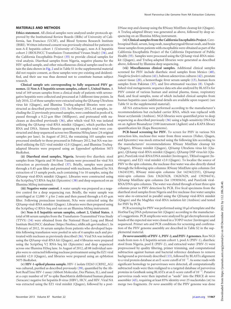
Clinical sample sets corresponding to fully sequenced PHV ge-nomes. (i) Non A-E hepatitis serum samples, cohort 1, United States. Atotal of 169 serum samples from a clinical study of patients with serone-gative hepatitis were collected and processed at 3 different time points. InJuly 2010, 22 of these samples were extracted using the QIAamp UltraSensvirus kit (Qiagen), and Illumina TruSeq-adapted libraries were con-structed as described previously (13). In February 2012, 64 samples, in-cluding the 22 previously processed samples, were combined in pools of 4,passed through a 0.22-�m filter (Millipore), and pretreated with nu-cleases as described previously (36), after which viral NA was isolatedutilizing the QIAamp viral-RNA minikit (Qiagen), which purifies bothRNA and DNA. Sixteen libraries spanning 64 samples total were con-structed and deep sequenced across two Illumina HiSeq lanes (24 originalsamples per lane). In August of 2012, the remaining 105 samples wereprocessed similarly to those processed in February 2012, but NA was iso-lated utilizing the EZ1 viral minikit v2.0 (Qiagen), and Illumina TruSeq-adapted libraries were prepared using an Eppendorf epMotion 5075BioRobot.
(ii) Diarrheal stool samples, Nigeria. Seventy-five diarrheic stoolsamples from Nigeria and 50 from Tunisia were processed for viral NAextraction as previously described (37). Briefly, samples were passedthrough a 0.45-�m filter and pretreated with nucleases, followed by NAextraction of 13 sample pools, each containing 5 to 10 samples, using theQIAamp viral-RNA minikit (Qiagen). Libraries were constructed usingthe ScriptSeq V2 RNA-Seq kit (Epicentre) (38) and deep sequenced on anIllumina MiSeq instrument.
(iii) Negative water control. A water sample was prepared as a nega-tive control for a deep sequencing run. Briefly, the water sample wascentrifuged at 12,000 � g for 2 min and then passed through a 0.45-�mfilter. Following prenuclease treatment, NAs were extracted using theQIAamp viral-RNA minikit (Qiagen). Libraries were then prepared usingthe ScriptSeq v2 RNA-Seq and run on an Illumina MiSeq instrument.
(iv) Non-A-E hepatitis serum samples, cohort 2, United States. Atotal of 88 serum samples from the Transfusion-Transmitted Virus Study(TTVS) (34) were obtained from the National Heart Lung and BloodInstitute BioLINCC database (https://biolincc.nhlbi.nih.gov/studies/). InFebruary of 2012, 16 serum samples from patients who developed hepa-titis following transfusion were pooled in sets of 4 samples each and pre-treated with nucleases as previously described (36). Viral NA was isolatedusing the QIAamp viral-RNA kit (Qiagen), and 4 libraries were preparedusing the ScriptSeq V2 RNA-Seq kit (Epicentre) and deep sequencedacross one Illumina HiSeq lane. In August of 2012, all 88 individual sam-ples were re-extracted following nuclease pretreatment using the EZ1 viralminikit v2.0 (Qiagen), and libraries were prepared using an epMotion5075 BioRobot.
(v) HIV-1-spiked plasma sample. HIV-1 isolate DJ263 (CRF02_AG)was cultured, purified as described previously (39), quantified by the Ab-bott RealTime HIV-1 assay (Abbott Molecular, Des Plaines, IL), and usedat a copy number of 104 to spike BaseMatrix defibrinated human plasma(Seracare) negative for hepatitis B virus (HBV), HCV, and HIV. Viral NAwas extracted using the EZ1 viral minikit (Qiagen), followed by a post-
DNase step and cleanup using the RNeasy MinElute cleanup kit (Qiagen).A TruSeq-adapted library was generated as above, followed by deep se-quencing on an Illumina MiSeq instrument.
(vi) Clinical samples from the California Encephalitis Project. Cere-brospinal fluid, serum, lung swab, nasopharyngeal/throat swab, and braintissue samples from patients with encephalitis were obtained as part of theCalifornia Encephalitis Project of the California Department of PublicHealth (35). Samples were processed using the QIAamp viral-RNA mini-kit (Qiagen), and TruSeq-adapted libraries were generated as describedabove, followed by Illumina deep sequencing.
(vii) Miscellaneous clinical samples. Additional clinical samplesscreened for PHV include diarrheic stool samples from Mexico (40),Naegleria fowleri cultures (41), baboon adenovirus cultures (42), prostatecancer tissue (20), a hemorrhagic fever serum sample (13), human fecessamples from Pakistan (37), and live-attenuated vaccines (8). Unpub-lished viral metagenomic sequence data sets also analyzed by BLASTn forPHV consist of various human and animal plasma, tissue, respiratoryfluid, and fecal samples, none of which included clinical samples fromhuman hepatitis cases (additional details are available upon request) (seeTable S1 in the supplemental material).
All NA extractions were performed according to the manufacturer’srecommendations but excluded carrier RNA, which was replaced withlinear acrylamide (Ambion). NGS libraries were quantified prior to deepsequencing as described previously (36) using a high-sensitivity DNA kiton an Agilent Bioanalyzer 2100 instrument (Agilent) and a KAPA libraryquantification kit (Kapa Biosystems).
PCR-based screening for PHV. To screen for PHV in various NAextraction kits, nuclease-free water from three sources (Fisher, Qiagen,and Epicentre) was mock extracted using the following kits according tothe manufacturers’ recommendations: RNeasy MinElute cleanup kit(Qiagen), RNeasy minikit (Qiagen), QIAamp UltraSens virus kit (Qia-gen), QIAamp viral-RNA minikit (Qiagen), QIAamp DSP virus kit (Qia-gen), PureLink viral-RNA/DNA minikit (Invitrogen), TRIzol reagent (In-vitrogen), and EZ1 viral minikit v2.0 (Qiagen). To localize the source ofPHV to the spin columns, the nuclease-free water was also directly elutedthrough the following spin columns: RNeasy MinElute spin columns (lot136243239), RNeasy mini-spin columns (lot 1423422253), QIAampmini-spin columns (lots 136267628, 136267629, and 139294474),QIAamp MinElute spin columns (lot 139305634), and PureLink viral-RNA/DNA spin columns. The water was eluted through at least three spincolumns prior to PHV detection by PCR. Five fecal specimens from thediarrheal stool samples from Nigeria and five nuclease-free water sampleswere also re-extracted in parallel using the QIAamp viral-RNA minikit(Qiagen) and the MagMax viral-RNA isolation kit (Ambion) and testedfor PHV by PCR.
PCR screening for PHV was performed using 10 �l of template and theHotStarTaq DNA polymerase kit (Qiagen) according to the manufactur-er’s suggestions. PCR amplicons were analyzed by gel electrophoresis andbands of the expected size were cloned in a TOPO vector (Invitrogen) andsequenced. Primer sets and PCR conditions for screening and confirma-tion of the PHV genome assembly are described in Table S2 in the sup-plemental material.
De novo assembly of PHV-1, PHV-2, and PHV-3 genomes. Raw NGSreads from non-A-E hepatitis serum cohort 1, pool A (PHV-1), diarrheicstool from Nigeria, pool E (PHV-2), and extracted water (PHV-3) werepreprocessed by quality filtering, primer trimming, and computationalsubtraction against human and bacterial reference databases to removebackground as previously described (13), followed by BLASTx alignmentto a viral protein database at an E-score cutoff of 10�3. As some reads withsignificant homology to parvoviruses were detected, all computationallysubtracted reads were then realigned to a targeted database of parvovirusproteins in GenBank using BLASTx at an E-score cutoff of 10�2. Putativeparvovirus reads were then inputted as “seeds” into the PRICE de novoassembler (43), requiring at least 85% identity over 25 nucleotides (nt) tomerge two fragments. De novo assembly of the PHV genome was done
Novel Parvovirus-Like Genome from NA Extraction Columns
November 2013 Volume 87 Number 22 jvi.asm.org 11967
Dow
nloa
ded
from
http
s://j
ourn
als.
asm
.org
/jour
nal/j
vi o
n 09
Dec
embe
r 20
21 b
y 11
2.18
6.38
.40.

iteratively using PRICE and manual editing with the Geneious version5.3.4 software package (44).
Screening for PHV in metagenomic data sets. Clinical NGS data setsgenerated in laboratories 1 and 2 (see Table S1 in the supplemental mate-rial) were screened for PHV by BLASTn alignment at E-score cutoffs of10�30 (Illumina reads) or 10�50 (454 pyrosequencing contigs). 454 pyro-sequencing reads were assembled into contigs using the SOAPdenovopackage (45) prior to BLASTn alignment. Publicly available environmen-tal metagenomic data sets deposited in the Sequence Read Archive (SRA),CAMERA (46), and MG-RAST (47) (see Table S3 in the supplementalmaterial) databases were also screened by BLASTn alignment for PHVreads at an E-score cutoff of 10�30.
Coverage maps. Reads from NGS data sets corresponding to de novo-assembled genomes PHV-1 (non-A-E hepatitis serum cohort 1, pool A)and PHV-2 (diarrheal stool, Nigeria, pool E) were aligned using BLASTnat a cutoff of 10�30 and mapped to their respective genomes using Ge-neious version 6.1.2. Coverage maps for other PHV strains were generatedby BLASTn alignment at a cutoff of 10�30 and mapping to the PHV-1genome. The consensus sequence was determined in Geneious by selec-tion of the majority base at each nucleotide position.
Phylogenetic analysis. For construction of the amino acid phylogenytrees, the translated replicase and capsid sequences corresponding to rep-resentative parvoviruses, circoviruses, and circovirus-like viruses werefirst downloaded from GenBank (accession numbers are provided in thesupplemental materials and methods). Multiple sequence alignments in-cluding PHV-1, PHV-2, and NIH-CQV were then performed usingMAFFT with the “auto” option and with default parameters. A phyloge-netic tree was constructed in Geneious version 6.1.2 with PHYML (48)with default parameters. Branch supports were computed in PHYML us-ing an approximate likelihood ratio test (aLRT) approach based on anShimodaira-Hasegawa-like (SH-like) option (49).
Nucleotide sequence accession numbers. The genome sequences ofall 12 PHV strains described in this study (genotypes PHV-1, PHV-1B,PHV-1C, PHV-1D, PHV-2, PHV-3, PHV-4A, PHV-4B, PHV-5, PHV-6A, PHV-6B, and PHV-6C) have been deposited in GenBank as PHVstrains UC1 to UC12 (accession numbers KF170373 and KF214637 toKF214647, respectively). The NGS data sets from which PHV-1, PHV-2,and PHV-3 were assembled were filtered for removal of human reads anddeposited into the GenBank Sequence Read Archive (project accessionnumber PRJNA217527 and SRA accession number SRP029352).
RESULTSDiscovery of PHV in seronegative hepatitis and diarrheal sam-ples. As part of an ongoing investigation into potential viral etiol-ogies for undiagnosed cases of seronegative non-A-E hepatitis,deep sequencing libraries in laboratory 1 at University of Califor-nia, San Francisco (UCSF), were prepared from sera collectedfrom a patient cohort of non-A-E hepatitis in the United States(Fig. 1A). Nucleic acids (NA) were extracted using the QIAampviral-RNA minikit (Qiagen). Metagenomic libraries were pre-pared for unbiased deep sequencing from 64 patient sampleswhich had been split into 16 indexed pools of 4 samples each.Analysis of the resulting NGS data using a previously developedcomputational pipeline for viral pathogen identification (17)revealed multiple divergent sequence reads with homology toparvoviruses. Translated amino acid alignments to parvovirussequences in the GenBank nonredundant protein database(NR) resulted in the identification of a 100-bp read from onepool (pool A), sharing only 48% amino acid identity withAcheta domestica densovirus (ADZ50508.1, 93% query cover-age, E value � 6 � 10�5) and 39% identity with human parvo-virus B19 (ABB36726.1, 93% query coverage, E value � 1 �10�3) in the capsid region (Fig. 1A, asterisk). This read was
selected as a seed for de novo assembly using the PRICE assem-bler (43), which generated a complete viral genome within 9cycles (Fig. 1A). The organization of the viral genome was con-firmed by PCR and Sanger sequencing of targeted regions
FIG 1 De novo assembly of PHV. The PRICE assembler was used to assemble thepartial or full genomes of PHV-1 (A), PHV-2 (B), and PHV-3 (C), correspondingto a sample pool of non-A-E hepatitis sera, a sample pool of diarrheal stool sam-ples, and a negative water control, respectively. The asterisks denote the initialseeds used for de novo assembly. Intermediate contiguous sequences (contigs)generated during the assembly (red bars) are mapped to their corresponding lo-cations on the PHV genome. (D) Genome organization of PHV, showing the openreading frames (ORFs) corresponding to the putative replicase and capsid proteinsand hypothetical 15-kDa protein of unknown function and two 148-nt invertedterminal repeat (ITR) sequences. Regions of the genome that were confirmed bySanger sequencing are represented by black lines. nt, nucleotide.
Naccache et al.
11968 jvi.asm.org Journal of Virology
Dow
nloa
ded
from
http
s://j
ourn
als.
asm
.org
/jour
nal/j
vi o
n 09
Dec
embe
r 20
21 b
y 11
2.18
6.38
.40.

(Fig. 1D). The virus was provisionally named parvovirus-likehybrid virus (PHV), and the initial detected strain from theseronegative hepatitis pool was designated PHV-1.
A separate viral discovery lab, laboratory 2 at the Blood Sys-tems Research Institute (BSRI), independently de novo assembleda novel parvovirus-like virus from NGS data corresponding todiarrheal stool samples from Nigeria (Fig. 1B). Nucleic acid ex-tractions from these samples were also performed using theQIAamp viral-RNA minikit. De novo assembly of the viral genomewas performed in 16 cycles from a single 100-bp read with38% amino acid identity to Acheta domestica densovirus(ADZ50508.1, 96% query coverage, E value � 4 � 10�3) and 39%identity to goose parvovirus (ABI20761.1, 84% query coverage, Evalue � 1 � 10�2) (Fig. 1B, asterisk). Comparison of the assem-bled viral genome with PHV-1 revealed 99% nucleotide identity,and thus this virus was designated parvovirus-like hybrid virus,strain 2 (PHV-2). Strikingly, the whole-genome sequences ofPHV-1 and PHV-2 shared 99% nucleotide and amino acid iden-tity with each other and NIH-CQV, a novel hybrid DNA virusrecently reported by Xu et al. to have been found in Chinese pa-tients with seronegative hepatitis (33).
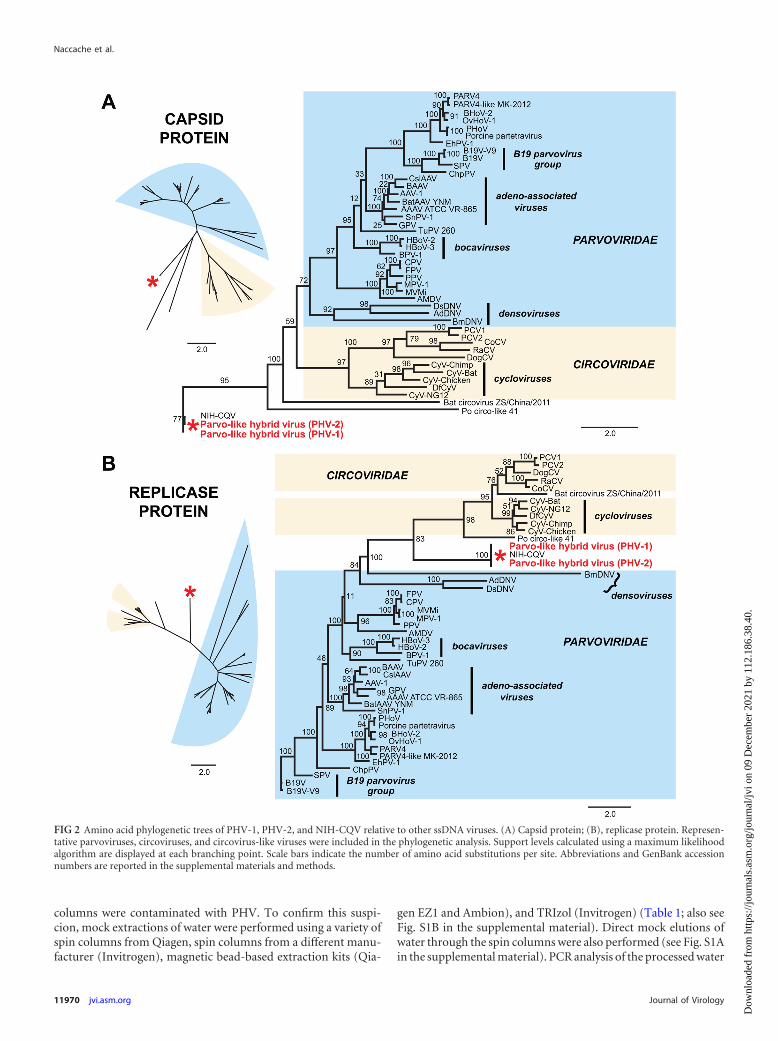
PHV genomic structure and phylogenetic analysis. The as-sembled genome of PHV-1 was found to be 3,636 bp long, with 3open reading frames (ORFs) and 148-nt inverted terminal repeatsat the 3= and 5= ends (Fig. 1D). The ORFs corresponding to theputative replicase and capsid genes were oriented in the same di-rection, and neither shared significant nucleotide identity withany sequence in NIH GenBank by BLASTn alignment. The repli-case gene exhibited remote homology to circoviruses; by BLASTxprotein alignment, �25% of the translated sequence shared 35%amino acid identity with bat circovirus (AEL28794) and �50% ofthe sequence shared 23% amino acid identity to porcine circovi-rus-like (Po-Circo-like) virus 21 (AER30018). The capsid genewas also highly divergent, with only 31% translated amino acididentity over 17% of the gene to the corresponding capsid gene ingoose parvovirus (ACK86566.1). Sequences encoding a conservedP-loop nucleoside triphosphate (NTP)-binding domain (50) andN-terminal parvovirus coat domain (51) were detected in the rep-licase and capsid genes, respectively. The capsid gene also encodeda putative phospholipase A2 (PLA2) motif that is critical for par-vovirus infectivity (52). Bridging PCR using primers spanning thetwo largest ORFs confirmed that the circovirus-like replicase andparvovirus-like capsid genes originated from the same viral ge-nome (Fig. 1D). Multiple attempts using inverse PCR failed todetect evidence of a circular form for PHV. To determine the exactphylogenetic placement of PHV relative to other ssDNA viruses,amino acid phylogenetic analysis of the putative replicase and cap-sid proteins was performed using representative genomes fromthe families Circoviridae and Parvoviridae. The resulting phyloge-netic trees (Fig. 2) revealed that PHV and NIH-CQV are situatedon a deep independent branch that appears to be intermediatebetween the circoviruses and parvoviruses. The closest, albeit dis-tant, relative to PHV and NIH-CQV is Po-Circo-like virus 21, aporcine circovirus-like virus previously identified as part of thefecal virome of pigs at a high-density farm (53).
Failure to detect PHV in re-extracted clinical samples. Innon-A-E hepatitis serum cohort 1, the observation that reads fromPHV-1 were present in all of the indexed pools (Fig. 3A) raised thelikelihood of contamination, either laboratory derived or fromsample cross-contamination. To investigate this possibility, indi-
vidual samples corresponding to the pool from which the PHV-1genome was initially assembled (pool A) were re-extracted using amagnetic-bead-based NA extraction method on an automatedEZ1 instrument (EZ1 viral minikit v2.0) and tested for PHV byspecific PCR. Although PCR of the NA extracted using theQIAamp viral-RNA minikit successfully detected PHV in all ofthe tested samples, NA extracted from the same samples using theautomated instrument tested negative for PHV. These discrepantresults raised doubts as to whether the original clinical samplesactually harbored PHV.
Detection and genome assembly of PHV from multiple sam-ple cohorts. To further investigate the prevalence of PHV in clin-ical samples, BLASTn alignments of 28 metagenomic data setscorresponding to a wide range of clinical sample cohorts wereperformed, using a high-stringency E value of 10�30 (Illumina100-bp or 250-bp short reads) or 10�50 (longer Roche 454 pyro-sequencing reads) for detection of PHV sequences. In non-A-Ehepatitis serum cohort 1, reads aligning to PHV were detected inall pools, with the percentage of total reads per pool being remark-ably similar, between 0.2 and 0.3% (Fig. 3A). PHV sequences wereidentified in multiple additional data sets from laboratories 1 and2 (Fig. 3A and B). Sample data sets positive for PHV had all beenprocessed using a column-based Qiagen NA extraction kit (Fig.3A and B, red text), with most of the detected PHV sequencescorresponding to samples processed from 2011 to the present. NoPHV reads were associated with data sets corresponding to NAisolated using kits from manufacturers other than Qiagen or byother extraction methods (e.g., use of magnetic beads) (Fig. 3Aand B, black text). In overlapping samples from non-A-E hepatitisserum cohorts 1 and 2 that had been extracted using two indepen-dent methods (Fig. 3A), i.e., by using Qiagen column-based andmagnetic bead-based kits, PHV reads were detected only in sam-ples that had been extracted using Qiagen columns (Fig. 3A).Strikingly, a large number of PHV reads were also recovered fromdeep sequencing of a negative water control mock extractedthrough the QIAamp viral-RNA minikit, from which �2/3 of thegenome, designated PHV-3, could be de novo assembled (Fig. 1C).
The average coverage of PHV-1 and PHV-2 achieved by deepsequencing of the sample pools and de novo assembly was 993�and 195�, respectively, and spanned 99 to 100% of the genome(Fig. 4A). Although the complete PHV-3 genome derived fromthe negative water control could not be de novo assembled due toa gap in coverage (Fig. 4A, arrow), the PHV-3 reads mapped toPHV-1 spanned 97% of the genome at 69� coverage. The averagecoverage obtained from representative individually indexed sam-ples or sample pools when mapped to PHV-1 was �150� andspanned 98 to 100% of the genome (Fig. 4B). Consensus se-quences generated from all 12 coverage maps revealed that allassembled viral genomes were remarkably similar (Fig. 5A), di-verging from the PHV-1 reference strain by �1.3%, with the ex-ception of PHV-3 (negative water control), which diverged by4.2% due to gaps in coverage. Notably, all PHV consensus se-quences were found to share 99 to 100% amino acid identity witheach other and with NIH-CQV.
PHV is a viral contaminant of Qiagen spin columns. The de-tection of PHV reads only in metagenomic data sets correspond-ing to clinical samples extracted using Qiagen spin columns (Fig.3A and B), and the assembly of a PHV genome directly from anegative water control that had also been processed using the samespin columns (Fig. 1C), raised the strong possibility that Qiagen
Novel Parvovirus-Like Genome from NA Extraction Columns
November 2013 Volume 87 Number 22 jvi.asm.org 11969
Dow
nloa
ded
from
http
s://j
ourn
als.
asm
.org
/jour
nal/j
vi o
n 09
Dec
embe
r 20
21 b
y 11
2.18
6.38
.40.
columns were contaminated with PHV. To confirm this suspi-cion, mock extractions of water were performed using a variety ofspin columns from Qiagen, spin columns from a different manu-facturer (Invitrogen), magnetic bead-based extraction kits (Qia-
gen EZ1 and Ambion), and TRIzol (Invitrogen) (Table 1; also seeFig. S1B in the supplemental material). Direct mock elutions ofwater through the spin columns were also performed (see Fig. S1Ain the supplemental material). PCR analysis of the processed water
FIG 2 Amino acid phylogenetic trees of PHV-1, PHV-2, and NIH-CQV relative to other ssDNA viruses. (A) Capsid protein; (B), replicase protein. Represen-tative parvoviruses, circoviruses, and circovirus-like viruses were included in the phylogenetic analysis. Support levels calculated using a maximum likelihoodalgorithm are displayed at each branching point. Scale bars indicate the number of amino acid substitutions per site. Abbreviations and GenBank accessionnumbers are reported in the supplemental materials and methods.
Naccache et al.
11970 jvi.asm.org Journal of Virology
Dow
nloa
ded
from
http
s://j
ourn
als.
asm
.org
/jour
nal/j
vi o
n 09
Dec
embe
r 20
21 b
y 11
2.18
6.38
.40.

FIG 3 Screening for PHV in clinical NGS data sets. Sequences corresponding to PHV were identified by BLASTn alignments at an E-score cutoff of 10�30 tosequence reads in NGS data sets processed from 2008 to 2013 in laboratory 1 (A) and laboratory 2 (B). The bar graphs show the percentage of PHV reads in eachdata set, with the number of PHV reads given above the bars. Clinical sample sets extracted using Qiagen spin columns are highlighted using red text; thoseextracted using other methods are shown in black. Abbreviations: UCSF, University of California, San Francisco; BSRI, Blood Systems Research Institute.
Novel Parvovirus-Like Genome from NA Extraction Columns
November 2013 Volume 87 Number 22 jvi.asm.org 11971
Dow
nloa
ded
from
http
s://j
ourn
als.
asm
.org
/jour
nal/j
vi o
n 09
Dec
embe
r 20
21 b
y 11
2.18
6.38
.40.

FIG 4 Mapping of reads from clinical NGS data sets to the PHV genome. The coverage (y axis) achieved at each nucleotide position along the genome (x axis)is plotted. (A) Coverage maps for the independently de novo assembled PHV-1, PHV-2, and PHV-3 genomes. The arrow depicts a break in coverage thatprecluded whole-genome de novo assembly of PHV-3. (B) PHV genomes assembled by mapping of NGS reads from various clinical sample data sets to the PHV-1genome. The inset information includes the name of the clinical data set, as well as the percent (%) and average (avg) genomic coverage achieved. nt, nucleotide.
Naccache et al.
11972 jvi.asm.org Journal of Virology
Dow
nloa
ded
from
http
s://j
ourn
als.
asm
.org
/jour
nal/j
vi o
n 09
Dec
embe
r 20
21 b
y 11
2.18
6.38
.40.

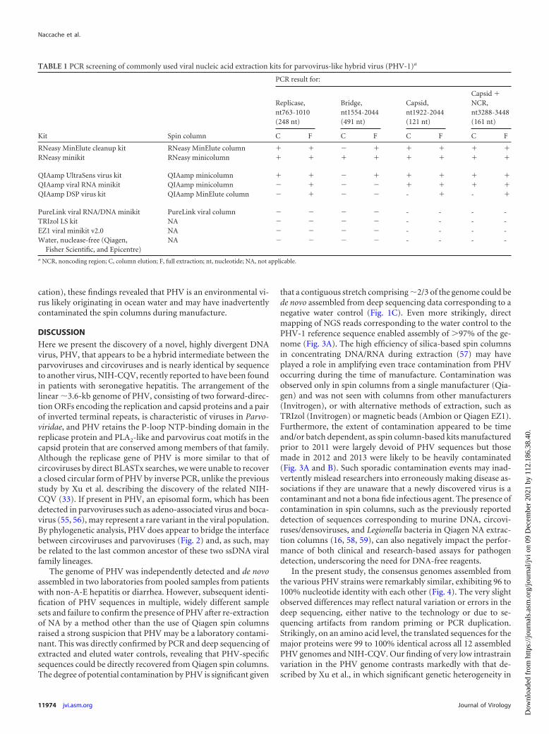
controls using four sets of primers detected PHV only in samplesthat had been processed using Qiagen spin columns, while usingspin columns from other manufacturers or different extractionmethods consistently failed to detect PHV. A subset of PCR am-plicons were Sanger sequenced and confirmed to be �99% iden-tical to PHV-1. The detection of PHV in mock water extractionsthrough Qiagen columns was reproducible in two independentlaboratories (see Fig. S2A in the supplemental material) and withthe use of purified water from multiple sources (Fisher, Qiagen,and Epicentre) (see Fig. S2B), directly implicating Qiagen spincolumns as the source of PHV contamination.
Identification of PHV sequences in environmental samples.To gain further insight into the origins of PHV, publicly availableenvironmental metagenomic sequence data sets in the CAMERA(46) and MG-RAST (47) databases were scanned for evidence ofPHV-related sequences using BLASTn alignments at a high-strin-gency cutoff of 10�30. A total of 78 public data sets containing213,615,095 sequence reads were analyzed, including 8,063,303reads from vertebrate metagenomes, 395,038 reads from plant
metagenomes, 14,922,577 reads from sediment sewage and soilmetagenomes, 6,609,658 reads from freshwater metagenomes,and 189,242,666 reads from marine metagenomes, includingplankton, microbialite, and coral reef metagenomic studies. TwoNGS data sets from marine sources containing Roche 454 pyrose-quencing data were found to harbor 3 PHV sequences, in totalspanning 17% of the PHV-1 genome with 87 to 99% identity (Fig.5C). Interestingly, both data sets corresponded to metagenomicshotgun sequencing of sampled seawater off the Pacific coast ofNorth America, as two of the identified PHV reads were derivedfrom a study of metagenomes in Monterey Bay, California (CAM-ERA project “North Pacific metagenomes from Monterey Bay toOpen Ocean”) (46, 54), and one was from a study of metagenomesin coastal regions off Oregon and Concepción, Chile (CAMERAproject “Microbial initiative in low oxygen areas off Concepciónand Oregon”) (see Table S3 in the supplemental material) (46). Asthe sample processing and generation of these data sets did notinvolve the use of columns or kits manufactured by Qiagen (A. Z.Worden, A. Bertagnolli, and S. Giovannoni, personal communi-
FIG 5 Whole-genome pairwise comparison of PHV strains and NIH-CQV relative to PHV-1. Black vertical bars indicate mismatches or insertions, whilehorizontal lines indicate deletions in the whole-genome consensus sequence relative to PHV-1. nt, nucleotide.
Novel Parvovirus-Like Genome from NA Extraction Columns
November 2013 Volume 87 Number 22 jvi.asm.org 11973
Dow
nloa
ded
from
http
s://j
ourn
als.
asm
.org
/jour
nal/j
vi o
n 09
Dec
embe
r 20
21 b
y 11
2.18
6.38
.40.
cation), these findings revealed that PHV is an environmental vi-rus likely originating in ocean water and may have inadvertentlycontaminated the spin columns during manufacture.
DISCUSSION
Here we present the discovery of a novel, highly divergent DNAvirus, PHV, that appears to be a hybrid intermediate between theparvoviruses and circoviruses and is nearly identical by sequenceto another virus, NIH-CQV, recently reported to have been foundin patients with seronegative hepatitis. The arrangement of thelinear �3.6-kb genome of PHV, consisting of two forward-direc-tion ORFs encoding the replication and capsid proteins and a pairof inverted terminal repeats, is characteristic of viruses in Parvo-viridae, and PHV retains the P-loop NTP-binding domain in thereplicase protein and PLA2-like and parvovirus coat motifs in thecapsid protein that are conserved among members of that family.Although the replicase gene of PHV is more similar to that ofcircoviruses by direct BLASTx searches, we were unable to recovera closed circular form of PHV by inverse PCR, unlike the previousstudy by Xu et al. describing the discovery of the related NIH-CQV (33). If present in PHV, an episomal form, which has beendetected in parvoviruses such as adeno-associated virus and boca-virus (55, 56), may represent a rare variant in the viral population.By phylogenetic analysis, PHV does appear to bridge the interfacebetween circoviruses and parvoviruses (Fig. 2) and, as such, maybe related to the last common ancestor of these two ssDNA viralfamily lineages.
The genome of PHV was independently detected and de novoassembled in two laboratories from pooled samples from patientswith non-A-E hepatitis or diarrhea. However, subsequent identi-fication of PHV sequences in multiple, widely different samplesets and failure to confirm the presence of PHV after re-extractionof NA by a method other than the use of Qiagen spin columnsraised a strong suspicion that PHV may be a laboratory contami-nant. This was directly confirmed by PCR and deep sequencing ofextracted and eluted water controls, revealing that PHV-specificsequences could be directly recovered from Qiagen spin columns.The degree of potential contamination by PHV is significant given
that a contiguous stretch comprising �2/3 of the genome could bede novo assembled from deep sequencing data corresponding to anegative water control (Fig. 1C). Even more strikingly, directmapping of NGS reads corresponding to the water control to thePHV-1 reference sequence enabled assembly of �97% of the ge-nome (Fig. 3A). The high efficiency of silica-based spin columnsin concentrating DNA/RNA during extraction (57) may haveplayed a role in amplifying even trace contamination from PHVoccurring during the time of manufacture. Contamination wasobserved only in spin columns from a single manufacturer (Qia-gen) and was not seen with columns from other manufacturers(Invitrogen), or with alternative methods of extraction, such asTRIzol (Invitrogen) or magnetic beads (Ambion or Qiagen EZ1).Furthermore, the extent of contamination appeared to be timeand/or batch dependent, as spin column-based kits manufacturedprior to 2011 were largely devoid of PHV sequences but thosemade in 2012 and 2013 were likely to be heavily contaminated(Fig. 3A and B). Such sporadic contamination events may inad-vertently mislead researchers into erroneously making disease as-sociations if they are unaware that a newly discovered virus is acontaminant and not a bona fide infectious agent. The presence ofcontamination in spin columns, such as the previously reporteddetection of sequences corresponding to murine DNA, circovi-ruses/densoviruses, and Legionella bacteria in Qiagen NA extrac-tion columns (16, 58, 59), can also negatively impact the perfor-mance of both clinical and research-based assays for pathogendetection, underscoring the need for DNA-free reagents.
In the present study, the consensus genomes assembled fromthe various PHV strains were remarkably similar, exhibiting 96 to100% nucleotide identity with each other (Fig. 4). The very slightobserved differences may reflect natural variation or errors in thedeep sequencing, either native to the technology or due to se-quencing artifacts from random priming or PCR duplication.Strikingly, on an amino acid level, the translated sequences for themajor proteins were 99 to 100% identical across all 12 assembledPHV genomes and NIH-CQV. Our finding of very low intrastrainvariation in the PHV genome contrasts markedly with that de-scribed by Xu et al., in which significant genetic heterogeneity in
TABLE 1 PCR screening of commonly used viral nucleic acid extraction kits for parvovirus-like hybrid virus (PHV-1)a
Kit Spin column
PCR result for:
Replicase,nt763-1010(248 nt)
Bridge,nt1554-2044(491 nt)
Capsid,nt1922-2044(121 nt)
Capsid �NCR,nt3288-3448(161 nt)
C F C F C F C F
RNeasy MinElute cleanup kit RNeasy MinElute column � � � � � � � �RNeasy minikit RNeasy minicolumn � � � � � � � �
QIAamp UltraSens virus kit QIAamp minicolumn � � � � � � � �QIAamp viral RNA minikit QIAamp minicolumn � � � � � � � �QIAamp DSP virus kit QIAamp MinElute column � � � � - � - �
PureLink viral RNA/DNA minikit PureLink viral column � � � � - - - -TRIzol LS kit NA � � � � - - - -EZ1 viral minikit v2.0 NA � � � � - - - -Water, nuclease-free (Qiagen,
Fisher Scientific, and Epicentre)NA � � � � - - - -
a NCR, noncoding region; C, column elution; F, full extraction; nt, nucleotide; NA, not applicable.
Naccache et al.
11974 jvi.asm.org Journal of Virology
Dow
nloa
ded
from
http
s://j
ourn
als.
asm
.org
/jour
nal/j
vi o
n 09
Dec
embe
r 20
21 b
y 11
2.18
6.38
.40.
NIH-CQV corresponding to putative sequence variants betweenpatient samples was observed (33). Although fold coverage mapsfrom that prior study were not presented, it is possible that insuf-ficient sequence coverage and/or errors in the NGS data may haveaccounted for the observed high substitution rates. Notably, inour study, greater sequence variation in the assembled genomeswas observed in PHV-3 (negative water control) and PHV-6C(encephalitis samples, pool C), which had comparatively lowerdepths of coverage than the other PHV genomes (Fig. 3 and 5). Analternative possibility is that there is indeed genetic heterogeneityin PHV/NIH-CQV that reflects natural variation and/or artifac-tual variation arising from lot-to-lot variability in the degree ofspin column contamination.
The finding of laboratory contamination as the origin of PHVsuggests that NIH-CQV, which shares 100% amino acid identitywith PHV, is most likely also a laboratory contaminant. In thestudy by Xu et al., there was 70% PCR positivity in seronegativehepatitis patient samples with an average virus titer of 1.05 � 104
copies/�l (corresponding to 1.05 � 107 copies/ml) yet 0% posi-tivity in healthy blood donors (33). The dichotomy between theseresults and serological detection showing comparable rates of pos-itivity for IgG specific to the C-terminal portion of the NIH-CQVcapsid protein in hepatitis patients and blood donors is striking.The PCR results may be explained by lot-to-lot variability or theuse of a Qiagen extraction kit prior to 2011, as those kits appearedto be less contaminated with PHV/NIH-CQV, while the serolog-ical results may potentially be due to detection of cross-reactiveantibodies by the immunoblot assay. Previously, a serological as-say designed to detect antibodies to p15E of XMRV showed ele-vated seroreactivity in human T-cell lymphotropic virus type 1(HTLV-1)-infected individuals (60), although none of these indi-viduals had detectable antibodies to a second XMRV protein,gp70. Subsequent analysis revealed that a highly conserved se-quence within the immunodominant region of HTLV gp21 that isshared with p15E was likely the source of the cross-reactive anti-bodies elicited by HTLV-1 infection (60). In the study by Xu et al.(33), confirmatory data based on serologic reactivity to multiplenonoverlapping epitopes within a single protein or more than oneviral protein would have provided stronger evidence of infectionby NIH-CQV.
By data mining of publicly available environmental metag-enomic databases, sequences with 100% identity to PHV/NIH-CQV were detected in coastal waters off North America. The rel-atively low number of reads detected is likely due to several factors:(i) high-efficiency concentration of viral DNA in the spin col-umns, (ii) differential rates of PHV abundance in ocean water, and(iii) lower-throughput Roche 454 pyrosequencing rather thanIllumina NGS for data generation. Viral abundance in aquaticecosystems is exceedingly high, with concentrations estimated at�108 per 1 ml (61). In total, approximately 1030 viruses arethought to reside in the world’s oceans, constituting a vast, largelyunsequenced reservoir of genomes. In addition, highly diversessDNA viruses, such as circoviruses and parvoviruses, have beendetected in seawater (62) and in ocean dwellers such as peneidshrimp (63), and viruses are known to infect diatoms (algae) thatare ubiquitous in seawater (64, 65). Taken together, these obser-vations suggest a plausible pathway for how PHV contaminationof the NA spin columns could have occurred. Column-based NApurification is a solid-phase extraction method that binds NA byadsorption to silica, and the silica used in many commercial spin
columns is derived from the cell walls of diatoms (57). If Qiagen’sNA extraction kits and “silica gel membrane technology” involvethe use of diatoms (66), it is plausible that PHV is a virus of dia-toms and had inadvertently contaminated the spin columns dur-ing manufacture. The sporadic contamination observed in thesilica-based spin columns (Fig. 3A and B) may thus be due toseasonal variation in diatom abundance, diatom type, and rates ofviral infection (67). The contamination of spin columns is notconfined to PHV but can also be seen by the presence of sequencescorresponding to phages, circoviruses, and parvoviruses otherthan PHV (16). Further studies will be needed to establish thatPHV is a virus of diatoms. Notably, we did not detect PHV inenvironmental metagenomic data sets corresponding to otheroceanic or environmental communities, which may reflect a lim-ited geographic and temporal distribution for the virus or a biasand/or incompleteness in the publicly available metagenomic da-tabases surveyed. The impact, if any, of these oceanic viruses onhuman health or public safety is unknown.
As the use of molecular methods such as deep sequencing forpathogen discovery becomes more frequent, it is critical that ro-bust strategies be developed to rapidly determine the biologicaland clinical relevance of any new candidate agent. This is espe-cially true with the discovery of novel, potentially transfusion-transmissible viruses in blood that may have an immediate impacton infectious diseases and public health (68), as exemplified by thehigh-profile putative association between XMRV and chronic fa-tigue syndrome that was eventually refuted by rigorous follow-upinvestigation (19–26). In the present study, the confirmation ofPHV as a laboratory reagent contaminant and not a candidateblood-borne infectious agent was made possible by (i) indepen-dent assessment at two research sites, (ii) free and open sharing ofsequence data corresponding to multiple sample cohorts betweenlaboratories, (iii) use of control samples subjected to the sameextraction and deep sequencing steps as experimental samples,(iv) direct PCR confirmation of viral contamination, and (v) datamining of publicly available metagenomic sequence databases de-rived from a vast array of clinical and environmental samples. Ourresults thus strongly call into question any association of the PHVand NIH-CQV viruses with seronegative hepatitis or, indeed, anybona fide infections of humans. Timely reporting of “dediscover-ies” as well as discoveries, by focusing effort and resource invest-ment, is needed to maximize the translational impact of pathogendiscovery to clinical medicine and infectious diseases.
ACKNOWLEDGMENTS
We thank Guixia Yu and Erik Samayoa for expert technical assistance andfor help with archival metadata, Stephanie Yen and Eunice Chen for mul-tiple cohort processing, Chunlin Wang and Xutao Deng for bioinformat-ics assistance, and Jerome Bouquet for comments and editorial sugges-tions. We also thank Alexandra Worden, Anthony Bertagnolli, andStephen Giovannoni for helpful discussions on their environmental met-agenomic data sets deposited in the CAMERA database.
This work is supported by National Institutes of Health (NIH) grantsR01-HL105704 (C.Y.C.), R01-HL105770 (E.D.), a University of Califor-nia Discovery Award (C.Y.C.), and an Abbott Viral Discovery Award(C.Y.C.).
REFERENCES1. Chiu CY. 2013. Viral pathogen discovery. Curr. Opin. Microbiol. http:
//dx.doi.org/10.1016/j.mib.2013.05.001. [E-pub ahead of print.]2. Lipkin WI. 2013. The changing face of pathogen discovery and surveil-
lance. Nat. Rev. Microbiol. 11:133–141.
Novel Parvovirus-Like Genome from NA Extraction Columns
November 2013 Volume 87 Number 22 jvi.asm.org 11975
Dow
nloa
ded
from
http
s://j
ourn
als.
asm
.org
/jour
nal/j
vi o
n 09
Dec
embe
r 20
21 b
y 11
2.18
6.38
.40.
3. Lipkin WI. 2010. Microbe hunting. Microbiol. Mol. Biol. Rev. 74:363–377.
4. Colby DW, Prusiner SB. 2011. Prions. Cold Spring Harb. Perspect. Biol.3:a006833. doi:10.1101/cshperspect.a006833.
5. Relman DA. 1998. Detection and identification of previously unrecog-nized microbial pathogens. Emerg. Infect. Dis. 4:382–389.
6. Kostic AD, Gevers D, Pedamallu CS, Michaud M, Duke F, Earl AM,Ojesina AI, Jung J, Bass AJ, Tabernero J, Baselga J, Liu C, ShivdasaniRA, Ogino S, Birren BW, Huttenhower C, Garrett WS, Meyerson M.2012. Genomic analysis identifies association of Fusobacterium with colo-rectal carcinoma. Genome Res. 22:292–298.
7. Feng H, Shuda M, Chang Y, Moore PS. 2008. Clonal integration of apolyomavirus in human Merkel cell carcinoma. Science 319:1096 –1100.
8. Victoria JG, Wang C, Jones MS, Jaing C, McLoughlin K, Gardner S,Delwart EL. 2010. Viral nucleic acids in live-attenuated vaccines: detec-tion of minority variants and an adventitious virus. J. Virol. 84:6033–6040.
9. Chiu C. 2013. The hidden threat of unidentified agents of disease inhuman and veterinary biologicals. Biologicals 41:129 –130.
10. Chandriani S, Skewes-Cox P, Zhong W, Ganem DE, Divers TJ, VanBlaricum AJ, Tennant BC, Kistler AL. 2013. Identification of a previouslyundescribed divergent virus from the Flaviviridae family in an outbreak ofequine serum hepatitis. Proc. Natl. Acad. Sci. U. S. A. 110:E1407–E1415.
11. Xu B, Liu L, Huang X, Ma H, Zhang Y, Du Y, Wang P, Tang X, WangH, Kang K, Zhang S, Zhao G, Wu W, Yang Y, Chen H, Mu F, Chen W.2011. Metagenomic analysis of fever, thrombocytopenia and leukopeniasyndrome (FTLS) in Henan Province, China: discovery of a new bunyavi-rus. PLoS Pathog. 7:e1002369. doi:10.1371/journal.ppat.1002369.
12. Palacios G, Druce J, Du L, Tran T, Birch C, Briese T, Conlan S, QuanPL, Hui J, Marshall J, Simons JF, Egholm M, Paddock CD, Shieh WJ,Goldsmith CS, Zaki SR, Catton M, Lipkin WI. 2008. A new arenavirusin a cluster of fatal transplant-associated diseases. N. Engl. J. Med. 358:991–998.
13. Grard G, Fair JN, Lee D, Slikas E, Steffen I, Muyembe J-J, Sittler T,Veeraraghavan N, Ruby G, Wang C, Makuwa M, Mulembakani P,Mazet J, Rimoin A, Taylor T, Schneider BS, Simmons G, Delwart E,Wolfe ND, Chiu CY, Leroy E. 2012. A novel rhabdovirus associated withacute hemorrhagic fever in Central Africa. PLoS Pathog. 8:e1002924. doi:10.1371/journal.ppat.1002924.
14. Briese T, Paweska JT, McMullan LK, Hutchison SK, Street C, PalaciosG, Khristova ML, Weyer J, Swanepoel R, Egholm M, Nichol ST, LipkinWI. 2009. Genetic detection and characterization of Lujo virus, a newhemorrhagic fever-associated arenavirus from southern Africa. PLoS Pat-hog. 5:e1000455. doi:10.1371/journal.ppat.1000455.
15. Zaki AM, van Boheemen S, Bestebroer TM, Osterhaus AD, FouchierRA. 2012. Isolation of a novel coronavirus from a man with pneumonia inSaudi Arabia. N. Engl. J. Med. 367:1814 –1820.
16. Lysholm F, Wetterbom A, Lindau C, Darban H, Bjerkner A, FahlanderK, Lindberg AM, Persson B, Allander T, Andersson B. 2012. Charac-terization of the viral microbiome in patients with severe lower respiratorytract infections, using metagenomic sequencing. PLoS One 7:e30875. doi:10.1371/journal.pone.0030875.
17. Greninger AL, Chen EC, Sittler T, Scheinerman A, Roubinian N, Yu G,Kim E, Pillai DR, Guyard C, Mazzulli T, Isa P, Arias CF, Hackett J,Schochetman G, Miller S, Tang P, Chiu CY. 2010. A metagenomicanalysis of pandemic influenza A (2009 H1N1) infection in patients fromNorth America. PLoS One 5:e13381. doi:10.1371/journal.pone.0013381.
18. Cheval J, Sauvage V, Frangeul L, Dacheux L, Guigon G, Dumey N,Pariente K, Rousseaux C, Dorange F, Berthet N, Brisse S, Moszer I,Bourhy H, Manuguerra CJ, Lecuit M, Burguiere A, Caro V, Eloit M.2011. Evaluation of high-throughput sequencing for identifying knownand unknown viruses in biological samples. J. Clin. Microbiol. 49:3268 –3275.
19. Simmons G, Glynn SA, Komaroff AL, Mikovits JA, Tobler LH, HackettJ, Jr, Tang N, Switzer WM, Heneine W, Hewlett IK, Zhao J, Lo SC, AlterHJ, Linnen JM, Gao K, Coffin JM, Kearney MF, Ruscetti FW, Pfost MA,Bethel J, Kleinman S, Holmberg JA, Busch MP, Blood XSRWG. 2011.Failure to confirm XMRV/MLVs in the blood of patients with chronicfatigue syndrome: a multi-laboratory study. Science 334:814 – 817.
20. Lee D, Das Gupta J, Gaughan C, Steffen I, Tang N, Luk KC, Qiu X,Urisman A, Fischer N, Molinaro R, Broz M, Schochetman G, Klein EA,Ganem D, Derisi JL, Simmons G, Hackett J, Jr, Silverman RH, Chiu CY.2012. In-depth investigation of archival and prospectively collected sam-
ples reveals no evidence for XMRV infection in prostate cancer. PLoS One7:e44954. doi:10.1371/journal.pone.0044954.
21. Knox K, Carrigan D, Simmons G, Teque F, Zhou Y, Hackett J, Jr, QiuX, Luk KC, Schochetman G, Knox A, Kogelnik AM, Levy JA. 2011. Noevidence of murine-like gammaretroviruses in CFS patients previouslyidentified as XMRV-infected. Science 333:94 –97.
22. Alter HJ, Mikovits JA, Switzer WM, Ruscetti FW, Lo SC, Klimas N,Komaroff AL, Montoya JG, Bateman L, Levine S, Peterson D, Levin B,Hanson MR, Genfi A, Bhat M, Zheng H, Wang R, Li B, Hung GC, LeeLL, Sameroff S, Heneine W, Coffin J, Hornig M, Lipkin WI. 2012. Amulticenter blinded analysis indicates no association between chronic fa-tigue syndrome/myalgic encephalomyelitis and either xenotropic murineleukemia virus-related virus or polytropic murine leukemia virus. mBio3:e00266 –12. doi:10.1128/mBio.00266-12.
23. Paprotka T, Delviks-Frankenberry KA, Cingoz O, Martinez A, KungHJ, Tepper CG, Hu WS, Fivash MJ, Jr, Coffin JM, Pathak VK. 2011.Recombinant origin of the retrovirus XMRV. Science 333:97–101.
24. Hue S, Gray ER, Gall A, Katzourakis A, Tan CP, Houldcroft CJ,McLaren S, Pillay D, Futreal A, Garson JA, Pybus OG, Kellam P,Towers GJ. 2010. Disease-associated XMRV sequences are consistent withlaboratory contamination. Retrovirology 7:111.
25. Oakes B, Tai AK, Cingoz O, Henefield MH, Levine S, Coffin JM, HuberBT. 2010. Contamination of human DNA samples with mouse DNA canlead to false detection of XMRV-like sequences. Retrovirology 7:109.
26. Sato E, Furuta RA, Miyazawa T. 2010. An endogenous murine leukemiaviral genome contaminant in a commercial RT-PCR kit is amplified usingstandard primers for XMRV. Retrovirology 7:110.
27. Fields BN, Knipe DM, Howley PM (ed). 2013. Fields virology, 6th edWolters Kluwer/Lippincott Williams & Wilkins Health, Philadelphia, PA.
28. Li L, Kapoor A, Slikas B, Bamidele OS, Wang C, Shaukat S, MasroorMA, Wilson ML, Ndjango JB, Peeters M, Gross-Camp ND, Muller MN,Hahn BH, Wolfe ND, Triki H, Bartkus J, Zaidi SZ, Delwart E. 2010.Multiple diverse circoviruses infect farm animals and are commonlyfound in human and chimpanzee feces. J. Virol. 84:1674 –1682.
29. Rosario K, Marinov M, Stainton D, Kraberger S, Wiltshire EJ, CollingsDA, Walters M, Martin DP, Breitbart M, Varsani A. 2011. Dragonflycyclovirus, a novel single-stranded DNA virus discovered in dragonflies(Odonata: Anisoptera). J. Gen. Virol. 92:1302–1308.
30. Hatakka A, Klein J, He R, Piper J, Tam E, Walkty A. 2011. Acutehepatitis as a manifestation of parvovirus B19 infection. J. Clin. Microbiol.49:3422–3424.
31. Mogensen TH, Jensen JM, Hamilton-Dutoit S, Larsen CS. 2010.Chronic hepatitis caused by persistent parvovirus B19 infection. BMCInfect. Dis. 10:246.
32. Resendes AR, Majo N, van den Ingh TS, Mateu E, Domingo M,Calsamiglia M, Segales J. 2011. Apoptosis in postweaning multisystemicwasting syndrome (PMWS) hepatitis in pigs naturally infected with por-cine circovirus type 2 (PCV2). Vet. J. 189:72–76.
33. Xu B, Zhi N, Hu G, Wan Z, Zheng X, Liu X, Wong S, Kajigaya S, ZhaoK, Mao Q, Young NS. 2013. Hybrid DNA virus in Chinese patients withseronegative hepatitis discovered by deep sequencing. Proc. Natl. Acad.Sci. U. S. A. 110:10264 –10269.
34. Mosley JW, Aach RD, Hollinger FB, Stevens CE, Barbosa LH, Nemo GJ,Holland PV, Bancroft WH, Zimmerman HJ, Kuo G, et al. 1990. Non-A,non-B hepatitis and antibody to hepatitis C virus. JAMA 263:77–78.
35. Glaser CA, Honarmand S, Anderson LJ, Schnurr DP, Forghani B,Cossen CK, Schuster FL, Christie LJ, Tureen JH. 2006. Beyond viruses:clinical profiles and etiologies associated with encephalitis. Clin. Infect.Dis. 43:1565–1577.
36. Swei A, Russell BJ, Naccache SN, Kabre B, Veeraraghavan N, PilgardMA, Johnson BJ, Chiu CY. 2013. The genome sequence of lone star virus,a highly divergent bunyavirus found in the Amblyomma americanumTick. PLoS One 8:e62083. doi:10.1371/journal.pone.0062083.
37. Victoria JG, Kapoor A, Li L, Blinkova O, Slikas B, Wang C, Naeem A,Zaidi S, Delwart E. 2009. Metagenomic analyses of viruses in stool sam-ples from children with acute flaccid paralysis. J. Virol. 83:4642– 4651.
38. Li L, Pesavento PA, Leutenegger CM, Estrada M, Coffey LL, NaccacheSN, Samayoa E, Chiu C, Qiu J, Wang C, Deng X, Delwart E. 2013. Anovel bocavirus in canine liver. Virol. J. 10:54.
39. Michael NL, Herman SA, Kwok S, Dreyer K, Wang J, ChristophersonC, Spadoro JP, Young KK, Polonis V, McCutchan FE, Carr J, MascolaJR, Jagodzinski LL, Robb ML. 1999. Development of calibrated viral loadstandards for group M subtypes of human immunodeficiency virus type 1
Naccache et al.
11976 jvi.asm.org Journal of Virology
Dow
nloa
ded
from
http
s://j
ourn
als.
asm
.org
/jour
nal/j
vi o
n 09
Dec
embe
r 20
21 b
y 11
2.18
6.38
.40.

and performance of an improved Amplicor HIV-1 Monitor test with iso-lates of diverse subtypes. J. Clin. Microbiol. 37:2557–2563.
40. Yu G, Greninger AL, Isa P, Phan TG, Martinez MA, de la Luz SanchezM, Contreras JF, Santos-Preciado JI, Parsonnet J, Miller S, DeRisi JL,Delwart E, Arias CF, Chiu CY. 2012. Discovery of a novel polyomavirusin acute diarrheal samples from children. PLoS One 7:e49449. doi:10.1371/journal.pone.0049449.
41. Herman EK, Greninger AL, Visvesvara GS, Marciano-Cabral F, DacksJB, Chiu CY. 2013. The mitochondrial genome and a 60-kb nuclear DNAsegment from Naegleria fowleri, the causative agent of primary amoebicmeningoencephalitis. J. Eukaryot. Microbiol. 60:179 –191.
42. Chiu CY, Yagi S, Lu X, Yu G, Chen EC, Liu M, Dick Jr, Carey EJKD,Erdman DD, Leland MM, Patterson JL. 2013. A novel adenovirus speciesassociated with an acute respiratory outbreak in a baboon colony andevidence of coincident human infection. mBio 4:e00084 –13. doi:10.1128/mBio.00084-13.
43. Ruby JG, Bellare P, Derisi JL. 2013. PRICE: software for the targetedassembly of components of (meta)genomic sequence data. G3 (Bethesda)3:865– 880.
44. Kearse M, Moir R, Wilson A, Stones-Havas S, Cheung M, Sturrock S,Buxton S, Cooper A, Markowitz S, Duran C, Thierer T, Ashton B,Meintjes P, Drummond A. 2012. Geneious Basic: an integrated andextendable desktop software platform for the organization and analysis ofsequence data. Bioinformatics 28:1647–1649.
45. Luo R, Liu B, Xie Y, Li Z, Huang W, Yuan J, He G, Chen Y, Pan Q, LiuY, Tang J, Wu G, Zhang H, Shi Y, Yu C, Wang B, Lu Y, Han C, CheungDW, Yiu SM, Peng S, Xiaoqian Z, Liu G, Liao X, Li Y, Yang H, WangJ, Lam TW. 2012. SOAPdenovo2: an empirically improved memory-efficient short-read de novo assembler. Gigascience 1:18.
46. Sun S, Chen J, Li W, Altintas I, Lin A, Peltier S, Stocks K, Allen EE,Ellisman M, Grethe J, Wooley J. 2011. Community cyberinfrastructurefor advanced microbial ecology research and analysis: the CAMERA re-source. Nucleic Acids Res. 39:D546 –D551.
47. Meyer F, Paarmann D, D’Souza M, Olson R, Glass EM, Kubal M,Paczian T, Rodriguez A, Stevens R, Wilke A, Wilkening J, Edwards RA.2008. The metagenomics RAST server—a public resource for the auto-matic phylogenetic and functional analysis of metagenomes. BMC Bioin-formatics 9:386.
48. Guindon S, Delsuc F, Dufayard JF, Gascuel O. 2009. Estimating maxi-mum likelihood phylogenies with PhyML. Methods Mol. Biol. 537:113–137.
49. Anisimova M, Gascuel O. 2006. Approximate likelihood-ratio test forbranches: a fast, accurate, and powerful alternative. Syst. Biol. 55:539 –552.
50. Lamberg-Allardt C, Wilska M, Saraste KL, Gronlund T. 1990. VitaminD status of ambulatory and nonambulatory mentally retarded childrenwith and without carbamazepine treatment. Ann. Nutr. Metab. 34:216 –220.
51. Marchler-Bauer A, Lu S, Anderson JB, Chitsaz F, Derbyshire MK,DeWeese-Scott C, Fong JH, Geer LY, Geer RC, Gonzales NR, Gwadz M,Hurwitz DI, Jackson JD, Ke Z, Lanczycki CJ, Lu F, Marchler GH,Mullokandov M, Omelchenko MV, Robertson CL, Song JS, Thanki N,Yamashita RA, Zhang D, Zhang N, Zheng C, Bryant SH. 2011. CDD: a
Conserved Domain Database for the functional annotation of proteins.Nucleic Acids Res. 39:D225–D229.
52. Zadori Z, Szelei J, Lacoste MC, Li Y, Gariepy S, Raymond P, Allaire M,Nabi IR, Tijssen P. 2001. A viral phospholipase A2 is required for parvo-virus infectivity. Dev. Cell 1:291–302.
53. Shan T, Li L, Simmonds P, Wang C, Moeser A, Delwart E. 2011. Thefecal virome of pigs on a high-density farm. J. Virol. 85:11697–11708.
54. Monier A, Welsh RM, Gentemann C, Weinstock G, Sodergren E,Armbrust EV, Eisen JA, Worden AZ. 2012. Phosphate transporters inmarine phytoplankton and their viruses: cross-domain commonalities inviral-host gene exchanges. Environ. Microbiol. 14:162–176.
55. Kapoor A, Hornig M, Asokan A, Williams B, Henriquez JA, Lipkin WI.2011. Bocavirus episome in infected human tissue contains non-identicaltermini. PLoS One 6:e21362. doi:10.1371/journal.pone.0021362.
56. Chen CL, Jensen RL, Schnepp BC, Connell MJ, Shell R, Sferra TJ,Bartlett JS, Clark KR, Johnson PR. 2005. Molecular characterization ofadeno-associated viruses infecting children. J. Virol. 79:14781–14792.
57. Boom R, Sol CJ, Salimans MM, Jansen CL, Wertheim-van Dillen PM,van der Noordaa J. 1990. Rapid and simple method for purification ofnucleic acids. J. Clin. Microbiol. 28:495–503.
58. Erlwein O, Robinson MJ, Dustan S, Weber J, Kaye S, McClure MO.2011. DNA extraction columns contaminated with murine sequences.PLoS One 6:e23484. doi:10.1371/journal.pone.0023484.
59. Evans GE, Murdoch DR, Anderson TP, Potter HC, George PM, Cham-bers ST. 2003. Contamination of Qiagen DNA extraction kits with Legio-nella DNA. J. Clin. Microbiol. 41:3452–3453.
60. Qiu X, Swanson P, Tang N, Leckie GW, Devare SG, Schochetman G,Hackett J, Jr. 2012. Seroprevalence of xenotropic murine leukemia virus-related virus in normal and retrovirus-infected blood donors. Transfusion52:307–316.
61. Suttle CA. 2005. Viruses in the sea. Nature 437:356 –361.62. Rosario K, Duffy S, Breitbart M. 2009. Diverse circovirus-like genome
architectures revealed by environmental metagenomics. J. Gen. Virol. 90:2418 –2424.
63. Roekring S, Nielsen L, Owens L, Pattanakitsakul SN, Malasit P, FlegelTW. 2002. Comparison of penaeid shrimp and insect parvoviruses sug-gests that viral transfers may occur between two distantly related arthro-pod groups. Virus Res. 87:79 – 87.
64. Amin SA, Parker MS, Armbrust EV. 2012. Interactions between diatomsand bacteria. Microbiol. Mol. Biol. Rev. 76:667– 684.
65. Nagasaki K. 2008. Dinoflagellates, diatoms, and their viruses. J. Micro-biol. 46:235–243.
66. Karp A, Ingram D, Isaac P. 2001. Molecular tools for screening biodi-versity plants and animals, vol pp xxiv. Kluwer Academic Publishers, Dor-drecht, the Netherlands.
67. Parsons RJ, Breitbart M, Lomas MW, Carlson CA. 2012. Ocean time-series reveals recurring seasonal patterns of virioplankton dynamics in thenorthwestern Sargasso Sea. ISME J. 6:273–284.
68. Stramer SL, Hollinger FB, Katz LM, Kleinman S, Metzel PS, GregoryKR, Dodd RY. 2009. Emerging infectious disease agents and their poten-tial threat to transfusion safety. Transfusion 49(Suppl 2):1S–29S.
Novel Parvovirus-Like Genome from NA Extraction Columns
November 2013 Volume 87 Number 22 jvi.asm.org 11977
Dow
nloa
ded
from
http
s://j
ourn
als.
asm
.org
/jour
nal/j
vi o
n 09
Dec
embe
r 20
21 b
y 11
2.18
6.38
.40.