100 brain barrier science: assessing the landscape€¦ · · 2017-11-26100 brain barrier...
TRANSCRIPT

Page 1 of 51
100 Brain barrier science: Assessing the landscape Lester R. Drewes Department of Biomedical Sciences, University of Minnesota Medical School Duluth, Duluth, Minnesota, USA Introduction: Over a century ago, the term “blood-brain barrier” was coined following the observation that a blood-borne dye is nearly completely excluded from the parenchymal space of the brain for hours while penetrating other organs and tissues within seconds. Since then our understanding of this phenomenon has expanded greatly and encompasses the cell and molecular biology of numerous cell types within the neurovascular unit (NVU) and specific nervous system structures including the cerebral spinal fluid (CSF), spinal cord, retina, cochlea and labyrinth. These advances, combined with the roll of the NVU in disease, suggest a bright future with significant innovative research contributions that impact the neuroscience field. Discussion: A prominent area of brain barrier research is the structure and function of membranes. This includes transporter biology and the mechanisms of cell-cell junction formation, adhesion and transmigration. An exciting advance is the identification of carriers for entry of essential brain nutrients that maintain the neural cell microenvironment. Recent genetic technologies reveal a new class of dysfunctional blood-brain transporters that are associated with neuropathologies. Characterizing transporters that protect the brain by ejecting potentially harmful, blood-borne toxins is especially important for developing successful strategies to deliver therapies for treatment of neurological diseases. Going forward, the expanding societal burden of neurological diseases from addiction to dementia is a challenging worldwide human health issue. Brain barrier science and investigations of all aspects of the neurovascular unit (NVU) will be crucial and at the core of successful therapy development for these diseases. The nature of these investigations will include many features, but prominent among them will be interactions between clinicians and basic scientists, input from patients, inter-laboratory collaborations, development of novel models, application of newly developed technologies and exploitation of databases and sample repositories. Successful implementation of these strategies promise many future exciting and significant discoveries.
Page 2 of 51
101 Impact of exercise on BBB and brain metastases Michal Toborek Department of Biochemistry and Molecular Biology, University of Miami School of Medicine, Miami, FL, USA Introduction: With a growing number of associations between daily activities and cancer development, understanding the contribution of lifestyle choices such as exercise is an emerging issue. Duration, intensity, and metabolic type (aerobic vs. anaerobic) of exercise as well as the cancer cell type are all important in understanding the impact of exercise on tumorigenesis or tumor progression. Aims: Our research focused on the influence of the microenvironment generated by exercise and metastasizing tumor cells at the levels of brain microvessels on the formation and outcome of brain metastases. Tumor cell extravasation into the brain requires passage through the blood-brain barrier (BBB), which is a highly protected microvascular environment sealed by tight junction (TJ) proteins. There is evidence that exercise can modulate oxidation status within the brain microvasculature and protect against tumor cell extravasation and metastasis formation, although the mechanisms are not well understood. Methods: In order to study these events, mature male mice were given access to voluntary exercise on a running wheel (exercise) or access to a locked wheel (sedentary) for five weeks. The average running distance was 9.0 ± 0.2 km/day. Highly metastatic tumor cells (murine Lewis lung carcinoma) were then infused into the brain microvasculature through the internal carotid artery. Results: A significant increase in the level of reactive oxygen species was observed following 48 hours or 3 weeks of tumor cell growth, which was accompanied by a reduction in MnSOD expression in the exercised mice. Activation of the small GTPase Rho was negatively correlated with running distance in the tumor cell infused mice. Immunohistochemical analysis revealed fewer tumor cells extravasating into the brain at both 48 hours and 3 weeks post surgery in exercised mice. Occludin levels were reduced in the sedentary tumor group, but maintained in the exercised tumor group at 48 hours post tumor cell infusion. Discussion: Studies have shown that regular exercise and a healthy lifestyle may reduce the risk for various diseases, including several types of cancer. Mimicking such effects, exercise was introduced in the present study prior to tumor cell exposure as a model of pre-treatment. The exercise paradigm (voluntary wheel running) was chosen to model a trained individual with an active lifestyle. Mice were exercised for 5 weeks (one week of acclimation, followed by 4 weeks used for analysis) in order to avoid early changes, which may be associated with short-duration workouts. Instead, our model aimed to reproduce biological responses, which may develop in trained, regularly-exercised individuals. While mice were running for ~9 hours per 24 hours, the activity and intensity was fully voluntary, making the model relevant to human exercise, which is also voluntary by nature. In our model, significant benefits of exercise were seen primarily in high running mice, ~10-15km/day, which could correspond to highly physically active humans. However, our wheel running model does not take into account other types of exercise that humans do, which contribute to overall health and disease prevention or decreased cancer progression. Many studies have addressed the benefits of exercise on various cancer models; however, our studies specifically focused on the BBB environment, as the BBB is the most prominent of the barriers of the CNS, constituting one of the most impermeable interfaces in the body. While exercise is universally accepted as beneficial to the body, in the context of pathology the picture becomes less clear. It is important to consider the impact of exercise alone as well as the type of exercise model as different intensity of activity may confer a different outcome. In our experiments, exercise impacted tight junctions of brain endothelial cell during tumor development, suggesting that such conditioning can impact BBB integrity and may provide benefits in the early stages of metastases formation. Potential mechanisms of this effect include antioxidative protection and modulatory impact on small GTPases, especially, RhoA GTPase. Conclusion: These results indicate that voluntary exercise may participate in modulating blood-brain barrier integrity thereby protecting the brain during metastatic progression.
Page 3 of 51
102 Impact of metabolic disease on blood-brain barrier structural and immunological integrity Egle Solito William Harvey Research Institute, Barts and the London School of Medicine and Dentistry, Queen Mary University London, London, UK Introduction: Despite clinical evidence of the Blood-Brain Barrier (BBB) being a major target of pathology during metabolic syndrome (MetS) - a cluster of interconnected risk factors for obesity, diabetes as well Alzheimer’s disease and Multiple Sclerosis- its pathophysiological response during sustained metabolic stress is almost unexplored. Impaired BBB functionality allows unrestricted entry of inflammatory cytokines and circulating leukocytes into the brain parenchyma, leading to the activation of brain’s immune cells, with a consequent detrimental effect on neural function (1). A key factor governing BBB tightness is the anti-inflammatory molecule Annexin A1 (ANXA1/AnxA1) (2). We have previously reported that ANXA1 regulates BBB integrity in vitro and in vivo through the regulation of tight junction protein expression and organization. Importantly, exogenous administration of ANXA1 can enhance BBB integrity in vivo, pinpointing the potential to be use therapeutically (3-4). Furthermore, we have shown that ANXA1 limits leukocyte extravasation following binding to- and blockade of- the α4β1 integrin, VLA-4 (the target of the monoclonal antibody Natalizumab) (5). We hypothesize that systemic metabolic overload affects BBB both directly, by compromising its tightness possibly via the intracellular metabolic machinery (named glycolysis, oxidative respiration, fatty acid oxidation), and indirectly, as a result of the low-grade chronic inflammation which accompanies MetS. Aims: We AIM to characterise the early events causing BBB malfunction in MetS, with the ultimate goal of identifying molecular targets/molecules that halt/control cerebrovascular disease progression. Methods: We used a multiple approach characterised by 1) in vivo and ex vivo mouse model of High Fat Diet (high fat + high glucose, HFSD) and clinical samples (blood from patients with diabetes type 1, 2 and MS). Results: Our results in vivo show a clear leakage of BBB post 10 weeks of HFSD compared to Chow diet mainly due to disturbance on essential TJ such as occludin and the basal lamina (Fig.1) expression and organization, as well as in the up regulation of adhesion molecules (e.g. ICAM-1). Interestingly such phenotype was reverted by the ANXA1 chronic treatment. HFSD also had an impact on the metabolism of BBB endothelium as measured by seahorse as well as on the expression of Glucose transporter 1 and cytoskeleton proteins (e.g. tubulin). Likewise the HFD diet affected circulating T cells metabolism making them more glycolytic (pro-inflammatory phenotype) with a consequent higher migration rate through the damaged endothelium into the brain parenchyma contributing to the neuronal damage (e.g. activation of microglia cells).The in vivo animal model results were mirrored by the analysis on diabetes type 1 DT1 and Multiple Sclerosis (RR-MS) patients sera effect on the human brain endothelial cells in vitro. Discussion: Our study represents the first global analysis on the impact of metabolic diseases on the BBB endothelium and circulating T cells. Furthermore our data identify a possible connection between metabolism-autoimmune (DT1) and neurological disorders (MS) and the effects of a perturbed metabolism on the structure and functionality of the BBB endothelium and T cells activation. ANXA 1 treatment is confirmed to have great potential not only in correcting BBB leakage but also modulating T cell tolerance which are impaired in metabolic and autoimmune diseases.
Page 4 of 51
103 The BBB and neurodegenerative diseases: Impact of dietary DHA and insulin Frédéric Calon1, 2, 3 1Faculté de Pharmacie, Université Laval, Québec, QC, G1V 0A6 Canada 2Centre de Recherche du CHU de Québec, Axe Neurosciences, Québec, QC, G1V 4G2, Canada 3OptiNutriBrain International associated Laboratory (NutriNeuro France-INAF Canada) Introduction: There is growing evidence of defects in the blood-brain barrier (BBB) in neurodegenerative diseases. Because of this, but also due to their active role in cerebral homeostasis, brain capillary endothelial cells (BCEC) forming the BBB are being increasingly recognized as potential therapeutic targets. Aims: (1) Gather evidence of BBB defects in human post-mortem tissue and in animal models of Alzheimer’s disease (AD), including on the brain uptake of omega-3 fatty acids (2) Investigate strategies to directly act on BCEC in the BBB to treat neurodegenerative diseases. Methods: Transgenic models, in situ cerebral perfusion, Western immunoblots, immunofluorescence, electron microscopy, brain capillary depletion, etc. Results: (a) The 3xTg-AD mouse model of AD displays reduced cerebrovascular volume, thickening on the basal lamina, reduced glucose uptake but no major changes in permeability. (b) APOE4 transgenics show reduced cerebral vascularization and impaired glucose uptake, compared with APOE2 and APOE3 mice. (c) The brain uptake rate of docosahexaenoic acid (DHA, a key omega-3 polyunsaturated fatty acid) is lower in both 3xTg-AD and APOE4 mice. (d) The apparent amyloid-clearing effect of a single insulin injection suggests that BBB insulin receptors may be a drug target in AD. (e) In vivo investigations suggest that BCEC can be targeted using antibodies undergoing transferrin receptor-induced endocytosis in the 3xTg-AD model. Discussion: Altogether, our work highlights dysfunction in BCEC or in the BBB that could progressively contribute to AD pathogenesis. We propose to consider insulin and transferrin transporters located on the BBB as potential targets of therapeutic interventions in neurodegenerative diseases.
Page 5 of 51
104 Neurovascular effects of nicotine delivered by electronic cigarettes alters stroke outcome Ali E. Sifat1, Bhuvaneshwar Vaidya1, Heidi Villalba1, Mohammad A. Kaisar1, Luca Cucullo1, 2, Thomas J. Abbruscato1, 2 1Department of Pharmaceutical Sciences, School of Pharmacy, Texas Tech University Health Sciences Center, Amarillo, Texas, USA 2Center for Blood Brain Barrier Research, Texas Tech University Health Sciences Center, Amarillo, TX, USA Introduction: Use of electronic cigarettes (eCig) is a growing health concern in both smoking and nonsmoking populations and rigorous studies are needed to understand the effects of the nicotine exposure via eCig on the neurovascular unit (NVU) and stroke outcome. Previous studies by our lab have shown that nicotine exposure significantly alters blood-brain barrier (BBB) transport in ischemia-reperfusion conditions. In the present study, we investigated the effects of acute & chronic nicotine and eCig vaping on ischemic stroke outcome and whole brain and neuronal glucose uptake. Methods: Nicotine was delivered either by osmotic pump (4.5 mg/kg) or by eCig vapor (2.4% nicotine) for 7, 10, 14 and 30 days and focal ischemia was induced by middle cerebral artery occlusion. Glucose transport was estimated with an in situ brain perfusion technique. In vitro primary cortical neurons were exposed to nicotine (10 µM) & cotinine (5 µM) for 1 & 5 days and then subjected to 2 hour oxygen-glucose deprivation (OGD) followed by 24 hour reperfusion to mimic ischemic conditions. Neuronal glucose utilization was measured by radiolabeled deoxy-D-glucose uptake. Immunocytochemistry & MTT assay were done to investigate glucose transporter 3 (glut3) expression & neuronal viability respectively. Brain deoxy-D-glucose uptake was also determined in brain slices exposed to 30 minute OGD utilizing an acute brain slice technique. Results: Nicotine exposure for 7 and 14 days resulted in significant reductions in D-glucose influx rate (Kin) across the BBB. 1 & 5 days of nicotine & cotinine exposure significantly decreases neuronal glucose utilization in OGD-reperfusion conditions which were reversed by a non-specific nicotinic acetylcholine receptor antagonist, mecamylamine. Interestingly, both 10 days and 30 days eCig exposed animals developed worsened stroke outcome, as measured by TTC staining and measurement of neurological deficits. Discussion: These data suggest, from a cerebrovascular perspective, that nicotine & eCig vaping exposure could create an
enhanced glucose deprived state at the NVU which could lead to increased ischemic brain injury
Page 6 of 51
105 Blood-brain barrier disruption in a diet-induced model of Type II diabetes: Treatment with the mitochondrial carbonic anhydrase inhibitor, topiramate Therese S. Salameh1, 2, William G. Mortell1, William A. Banks1, 2 1Geriatrics Research, Education, and Clinical Center, Veterans Affairs Puget Sound Health Care System, Seattle, Washington, USA 2Division of Geriatrics and Gerontology, Department of Medicine, University of Washington, Seattle, Washington, USA Introduction: All forms of diabetes are characterized by chronic hyperglycemia resulting in the development of microvascular and macrovascular complications. Diabetes is also associated with changes in the brain microvasculature leading to the disruption/dysfunction of the blood-brain barrier (BBB). In diabetes, BBB damage is associated with increased oxidative stress caused by hyperglycemia-induced oxidative metabolism of glucose, which can be reduced through inhibition of mitochondrial carbonic anhydrase (mCA). Previously, we demonstrated in a mouse model of type I diabetes, BBB disruption occurred in the cortices, midbrain, and thalamus, and was attenuated using the mCAI, topiramate. Studies have implicated that dietary and metabolic factors can lead to damage of the BBB. Aims: To test the idea that increased metabolism via neurogenesis underlies the type II pattern of BBB disruption. Methods: CD-1 mice were fed low (10%)- and high (60%)-fat diets for up to 8 months. Group 1 received daily subcutaneous injections of 5 mg/kg topiramate from 0-4 months, while group two received topiramate from 4-8 months. Disruption to radiolabeled albumin and sucrose were measured in brain regions after IV injection. Results: After four months on high-fat diet, BBB disruption was observed in the hypothalamus and hippocampus in mice that developed hyperglycemia. Topiramate treatment attenuated BBB disruption in the hypothalamus. In the normal glycemic mice on high-fat diet, topiramate treatment caused BBB disruption in the cortices, midbrain, cerebellum, and thalamus. This disruption is associated with an increase in levels of the hormone resistin, involved in endothelial cell dysfunction. After eight months, BBB disruption was detected in the retina, hypothalamus, and hippocampus, and topiramate treatment attenuated disruption in the hippocampus. Discussion: Type I and type II diabetes display different patterns of BBB disruption. This is important in defining the roles of insulin/leptin in BBB function. mCA is a viable therapeutic target for preventing BBB disruption.
Page 7 of 51
106 Reduced blood-brain barrier transport of docosahexaenoic acid in APP/PS1 mice is associated with increased vulnerability to cognitive deficits from low omega-3 fatty acid diets Yijun Pan1, Kwok HC Choy2, Christopher JH Porter1, Martin J Scanlon3, Jennifer L. Short2, Joseph A Nicolazzo1 1Drug Delivery, Disposition and Dynamics, Monash University, Parkville, Victoria, Australia 2Drug Discovery Biology, Monash University, Parkville, Victoria, Australia 3Medicinal Chemistry and Drug Action, Monash University, Parkville, Victoria, Australia Introduction: Lower levels of the cognitively-beneficial docosahexaenoic acid (DHA) are often observed in Alzheimer’s disease (AD) brains. Brain DHA levels are regulated by blood-brain barrier (BBB) transport of plasma-derived DHA, a process facilitated by fatty acid-binding protein 5 (FABP5). Aims: To investigate if FABP5 expression at the BBB and DHA transport across the BBB is affected in APP/PS1 AD mice, and the impact of dietary DHA (n-3 fatty acids) depletion on the cognitive function of these mice. Methods: In situ transcardiac perfusion was performed in 8 month old APP/PS1 mice to study the BBB transport of 14C-DHA, and brain capillary-enriched fractions were isolated from APP/PS1 mice brains to measure FABP5 expression using ELISA. In addition, APP/PS1 mice (8 months old) were fed a control diet or n-3 fatty acid-depleted diet for 6 months, and the cognitive function was assessed using a battery of behavioral paradigms, such as Y-maze spatial recognition, novel object recognition, and water maze. Results: A 42.1 ± 12.6% decrease in BBB transport of 14C-DHA was observed in APP/PS1 mice relative to wild-type mice, which is associated with a 34.5 ± 6.7% reduction in FABP5 expression in isolated brain capillary-enriched fractions of APP/PS1 mice. Furthermore, short-term spatial and recognition memory deficits were observed in APP/PS1 mice on a 6-month n-3 fatty acid-depleted diet, but not in AD mice on a control diet. This intervention led to a dramatic reduction (41.5 ± 11.9%) of brain DHA levels in AD mice. Discussion: FABP5 deficiency and impaired DHA transport across the BBB are associated with increased vulnerability to cognitive deficits in APP/PS1 mice fed an n-3 fatty acid-depleted diet, in line with our previous studies demonstrating a crucial role of FABP5 in BBB transport of DHA and cognitive function.
Page 8 of 51
107 Targeting C' dots for delivery of small molecule inhibitors Brian Madajewski1, Rupa Juthani2,3, Barney Yoo1, Pei-Ming Chen1, Li Zhang1, Kai Ma4, Sean Carlin1, Michael Overholtzer5, Jason Huse2, Ulrich Wiesner4, Charles Rudin6, Cameron W. Brennan2,3, Michelle Bradbury3,7 1Department of Radiology, Memorial Sloan Kettering Cancer Center, New York, NY, 2Human Oncology & Pathogenesis Program Memorial Sloan Kettering Cancer Center, New York, NY; 3Department of Neurosurgery, Memorial Sloan Kettering Cancer Center, New York, NY; 4Department of Materials Science & Engineering, Cornell University, Ithaca, NY; 5Cell Biology Program Memorial Sloan Kettering Cancer Center, New York, NY; 6Thoracic Oncology Service, Memorial Sloan Kettering Cancer Center, New York, NY; 7Molecular Pharmacology Program, Memorial Sloan Kettering Cancer Center, New York, NY. Introduction: Brain metastases occur in 40% of cancer patients, and are responsible for significant neurological morbidity. One of the present challenges hindering effective treatment of brain metastases by systemically-administered therapy has been the uncertain and limited drug delivery through the blood-brain barrier (BBB), often leaving the brain as the only site of uncontrolled disease. This challenge underscores the need to improve brain penetration for small molecule inhibitor (SMI) therapy beyond the limitations imposed by systemic side effects with standard drug dosing. A clinically-promising approach advances an ultrasmall tumor-targeted nanoparticle drug conjugate (NDC) that can be adapted with SMIs, such as gefitinib, in order to promote enhanced target penetration, intratumoral distributions, and improved therapeutic indices. Aims: To explore the potential of sub-10 nm NDCs to deliver SMIs to brain metastases and malignant brain tumors, we aimed to investigate (1) how cRGDY-targeted C’ dots and NDCs distribute in brain tumors relative to regional BBB permeability, and (2) the efficacy of gefitinib-NDCs to suppress growth of EGFR-mutant non-small cell lung cancer xenografts. Previous work has shown that ultrasmall NDCs, incorporating gefitinib, achieve target inhibition in vitro using EGFRmt-expressing tumor cell lines. Methods: RCAS/tv-a mouse gliomas were established by intracranial injections of RCAS-PDGFB in transgenic TVA-expressing newborn mice. Tumor-bearing animals were co-injected with cRGDY-PEG-C’ dots or a control particle, cRAD-PEG-C’ dots, either non-radiolabeled or I-124 radiolabeled, in addition to a 70kDa FITC-dextran, a surrogate marker of BBB permeability, and nuclear marker, Hoechst, and sacrificed at early and late time points. Tumor tissue was evaluated by high power fluorescence microscopy, autoradiography, as well as immunohistochemistry (IHC) of target pathways. These results were compared to PET imaging and tumor analysis from our active in-human clinical trial, evaluating the distribution and uptake of radiolabeled cRGDY-PEG-C’ dots in patients with malignant brain tumors. To study nanoparticle potency and target inhibition, mice bearing NSCLC flank xenografts (H1650 and ECLC26, a patient-derived line bearing EGFR L858R mutation) were treated with vehicle alone, gefitinib, or C’ dot NDCs and sacrificed at 6 and 18 hours post-injection. Western blots and IHC were used to evaluate EGFR inhibition. Particle distributions were evaluated by Cy5 fluorescence imaging compared with H&E and IHC in adjacent tumor sections. Cohorts of flank tumor-bearing animals were treated with daily gefitinib oral vs. gefitinib-NDC Q3 day IV with untreated controls to assess treatment response. Results: Mice bearing RCAS/tv-a gliomas demonstrated strong tumor-specific accumulation of radiolabeled cRGDY-PEG-C’ dots, as demonstrated by Cy5 fluorescence. cRGDY-PEG-C’dot distribution at 3 hours closely correlated with BBB breakdown identified by 70kda FITC-Dextran, but at 96 hours appeared to diffuse beyond regions of BBB breakdown through the tumor. Compared to cRAD-PEG-C’ dots, cRGDY-PEG-C’ dots had enhanced and more diffuse Cy5 fluorescence along with increased I-124 signal as evidenced by autoradiography tumor:brain ratios. Results were similar to findings in the in-human trial, with tumors demonstrating particle-specific fluorescence and autoradiography, along with accumulation in the vascularized leptomeninges. In mice bearing H1650 flank xenografts, particle-specific Cy5 fluorescence was seen heterogeneously throughout the tumors with a corresponding absence of Cy5 fluorescence in gefitinib-treated animals. These tumors demonstrated IHC immunopositivity for phospho-EGFR in all conditions, with a suggestion of a dose-dependent inhibition at higher doses of gefitinib and at the longer time point for C’ dot NDCs. The EGFR L858R-mutant line was more sensitive and gefitinib NDC treatment led to growth inhibition over 7 days. Discussion: Results demonstrate that ultrasmall cRGDY-PEG-C’ dots are capable of delivering SMIs to the tumor bed and inducing drug-mediated growth inhibition. C’ dots are initially distributed within brain tumors throughout regions of disrupted BBB, are retained in tumors, and are further distributed over 96 hours. Intratumoral retention of C’ dots is increased by cRGD in an integrin-expressing mouse glioma model. The data supports a potential role for NDCs to deliver SMI drugs to primary and metastatic brain tumors with an improved therapeutic index.
Page 9 of 51
108 Neurovascular biology of capillary control Martin Lauritzen1, 2, Nikolay Kutuzov1, Changsi Cai1 1Center for Neuroscience and Center for Healthy Aging, University of Copenhagen; Copenhagen N, Denmark 2Department of Clinical Neurophysiology, Rigshospitalet, Denmark Introduction: Normal brain function depends on preserved supply of glucose and oxygen and even minor deficits in control of cerebral blood flow (CBF) lead to loss of brain function. Our understanding of the regulation of CBF has recently undergone a revolution due to the development of new concepts and new imaging techniques. In the past brain capillaries were viewed as tubes that passively conducted blood from the heart to the active nerve cells. However, recent data provided by others and by us suggest that brain capillaries may have control mechanisms for blood flow regulation, and novel microscopy techniques have now made it possible to examine the blood-brain barrier (BBB) in more detail in living animals as well. Aims: To determine the hierarchy of vascular mechanisms that that take part in the regulation of cerebral blood flow and the BBB in living mice. Methods: Measurements were performed on anesthetized mice. 1) 2-photon microscopy of brains in vivo a transgenic mouse with fluorescent pericytes to examine the participation capillaries, and pericytes on those capillaries, for flow control. 2) To examine the properties of the BBB using different types of fluorescent tracers, injected into the bloodstream and visualized at the level of single capillaries. Results: 1) Rises in synaptic activity in mouse somatosensory cortex evoke capillary dilation that mostly starts at 1st or 2nd order capillary, from where it propagates upstream to the penetrating arteriole and downstream to higher order capillaries. Similarly, application of the gliotransmitter ATP induced dilation followed by constriction that also propagated up- and downstream at velocity of 5-20 µm/s. Conducted vascular responses were inhibited by gap junction blockers, and were explained by extracellular diffusion of vasoactive substances. 2) Transport of small fluorescent indicators (fluorescein, Alexa488) across the BBB can described by a purely diffusive process. Fitting the model to the experimental data allowed us to obtain 3 parameters: permeability of the BBB, effective diffusion constant in the extravascular space and volume fraction of the extravascular space (fraction of the volume accessible by the dye). Discussion: Conducted vascular responses in capillaries may be a novel modulator of cerebrovascular function. Furthermore, the BBB appears to consist of several independent compartments restricting blood to brain transport. Together this work has the potential to advance our understanding of regulation of blood flow and BBB permeability at the level of small arterioles and capillaries. The results from the experimental studies have the potential to inform human studies of the types of capillary dysregulation that may be observed in patients. This will allow for cross-verification of hypotheses between the lab-bench and the clinic and help to identify key molecular mediators and new therapeutic targets.
Page 10 of 51
109 In-depth analysis of P-gp and NG2 in glioblastoma D. Virgintino Department of Basic Medical Sciences, Neurosciences and Sensory Organs, Bari University School of Medicine, Bari, Italy Introduction: Glioblastoma (GB) is the most common malignant primary brain tumor with the highest degree of vascularization among the CNS neoplasms. The GB vascular network shows an extreme variety of tumoral vessel morphology, ranging from normal-looking microvessels to glomeruloid structures. In parallel with the complex vascular architecture is the variety of its cellular constituents, growth mechanisms, and molecular triggers. Aims: The purpose of this study is to investigate BBB-endothelial cells and pericytes of GB vessels, analysing their contribution to vessel growth and mutual relations in the newly formed tumoral vessels. Methods. Confocal microscopy analysis of GB tissues after multiple immunolabellings with BBB- and pericyte-specific markers. Results: Heterogeneity in tumour microvascular architecture reflects the different mechanisms of vessel growth and includes a variable expression of endothelial/tumoral cell P-glycoprotein (P-gp) and a diversified involvement of pericyte subsets, the latter identified by the expression of specific NG2 isoforms. Discussion: In GB, alternative pericyte-driven mechanisms of vessel growth significantly participate to tumoral vessel increase, possibly modifying P-gp expression and distribution. The limited impact of treatments targeting the abnormal GB vasculature reveals the need for additional targets that could be valuable to further improve therapeutic strategies.
Page 11 of 51
110 Pluripotent stem cell modelling of the human blood-brain barrier Eric V. Shusta, Department of Chemical and Biological Engineering, University of Wisconsin-Madison The blood-brain barrier (BBB) plays an important role in maintaining brain health and is often compromised in disease. Moreover, as a result of its significant barrier properties, this endothelial interface restricts uptake of neurotherapeutics. A renewable cell source for human BBB modelling could prove enabling for brain research and pharmaceutical development. We recently demonstrated that endothelial cells generated from human pluripotent stem cells (hPSCs) can be specified to possess many BBB attributes, including well-organized tight junctions, polarized efflux transport, and nutrient transporter expression. Importantly, hPSC-derived BBB endothelial cells respond to cues provided by other cells of the neurovascular unit such as human pericytes, astrocytes and neurons to generate very tight barrier properties as measured by transendothelial electrical resistance (~5000 ohmxcm2), while exhibiting molecular permeability that correlates well with in vivo brain uptake. In this talk, we will demonstrate that the process of hPSC differentiation to BBB cells is also compatible with disease modelling using patient-derived induced pluripotent stem cell lines, can be used in the isogenic modelling of the neurovascular unit, and the can be employed for the evaluation of experimental drug permeability attributes.
Page 12 of 51
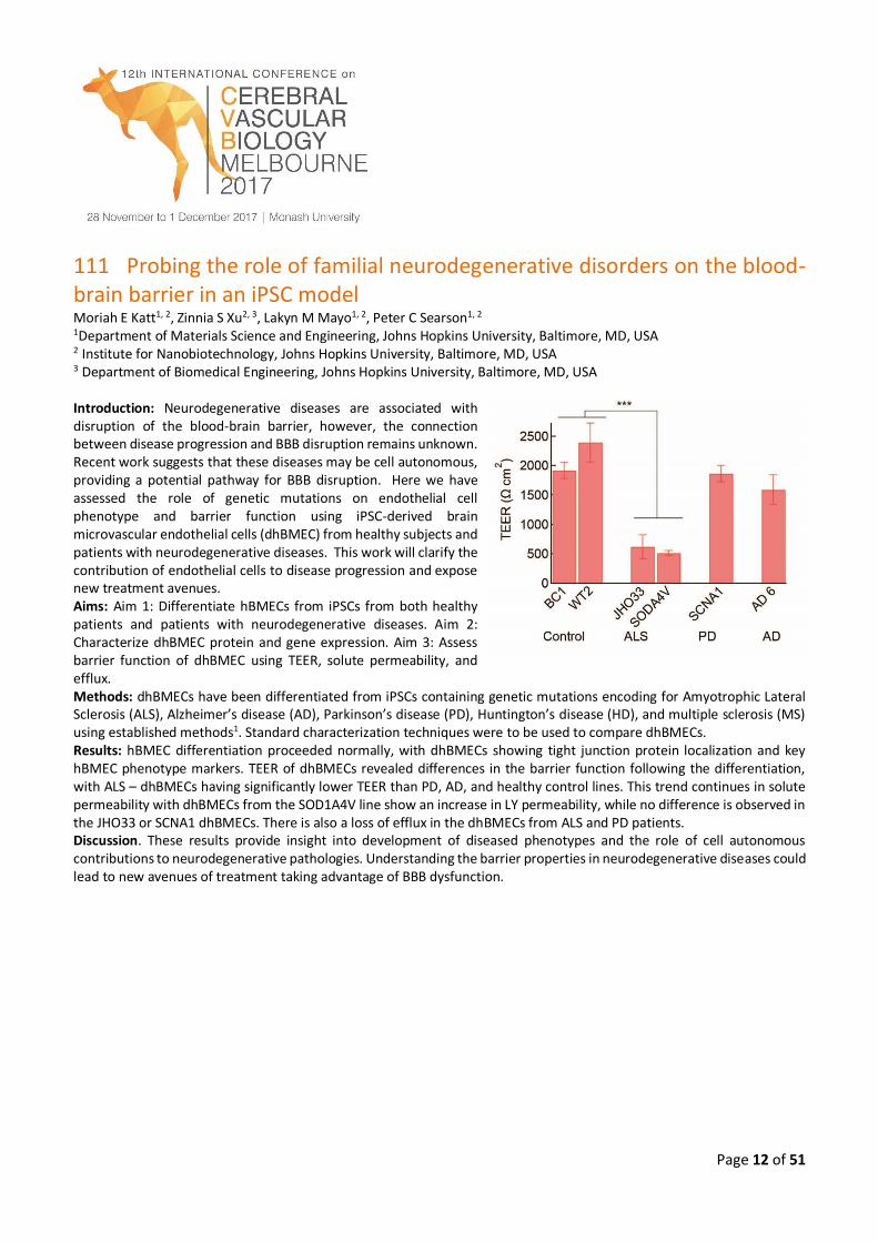
111 Probing the role of familial neurodegenerative disorders on the blood-brain barrier in an iPSC model Moriah E Katt1, 2, Zinnia S Xu2, 3, Lakyn M Mayo1, 2, Peter C Searson1, 2 1Department of Materials Science and Engineering, Johns Hopkins University, Baltimore, MD, USA 2 Institute for Nanobiotechnology, Johns Hopkins University, Baltimore, MD, USA 3 Department of Biomedical Engineering, Johns Hopkins University, Baltimore, MD, USA Introduction: Neurodegenerative diseases are associated with disruption of the blood-brain barrier, however, the connection between disease progression and BBB disruption remains unknown. Recent work suggests that these diseases may be cell autonomous, providing a potential pathway for BBB disruption. Here we have assessed the role of genetic mutations on endothelial cell phenotype and barrier function using iPSC-derived brain microvascular endothelial cells (dhBMEC) from healthy subjects and patients with neurodegenerative diseases. This work will clarify the contribution of endothelial cells to disease progression and expose new treatment avenues. Aims: Aim 1: Differentiate hBMECs from iPSCs from both healthy patients and patients with neurodegenerative diseases. Aim 2: Characterize dhBMEC protein and gene expression. Aim 3: Assess barrier function of dhBMEC using TEER, solute permeability, and efflux. Methods: dhBMECs have been differentiated from iPSCs containing genetic mutations encoding for Amyotrophic Lateral Sclerosis (ALS), Alzheimer’s disease (AD), Parkinson’s disease (PD), Huntington’s disease (HD), and multiple sclerosis (MS) using established methods1. Standard characterization techniques were to be used to compare dhBMECs. Results: hBMEC differentiation proceeded normally, with dhBMECs showing tight junction protein localization and key hBMEC phenotype markers. TEER of dhBMECs revealed differences in the barrier function following the differentiation, with ALS – dhBMECs having significantly lower TEER than PD, AD, and healthy control lines. This trend continues in solute permeability with dhBMECs from the SOD1A4V line show an increase in LY permeability, while no difference is observed in the JHO33 or SCNA1 dhBMECs. There is also a loss of efflux in the dhBMECs from ALS and PD patients. Discussion. These results provide insight into development of diseased phenotypes and the role of cell autonomous contributions to neurodegenerative pathologies. Understanding the barrier properties in neurodegenerative diseases could lead to new avenues of treatment taking advantage of BBB dysfunction.
Page 13 of 51
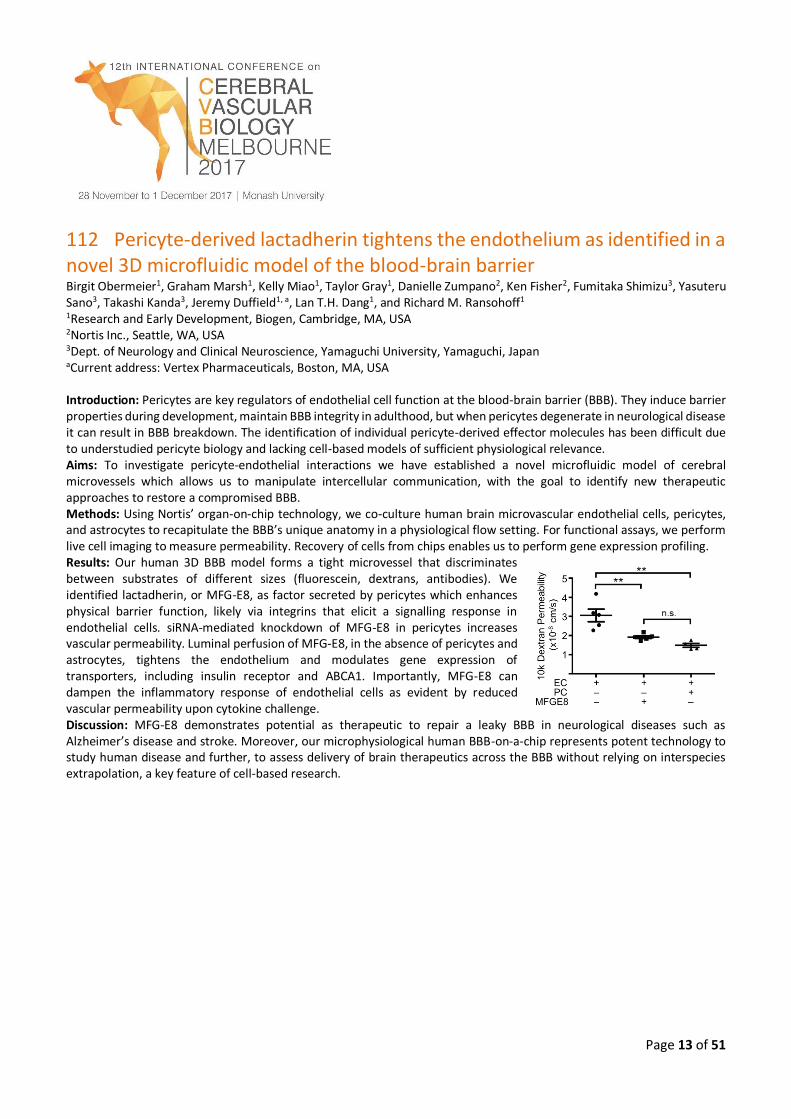
112 Pericyte-derived lactadherin tightens the endothelium as identified in a novel 3D microfluidic model of the blood-brain barrier Birgit Obermeier1, Graham Marsh1, Kelly Miao1, Taylor Gray1, Danielle Zumpano2, Ken Fisher2, Fumitaka Shimizu3, Yasuteru Sano3, Takashi Kanda3, Jeremy Duffield1, a, Lan T.H. Dang1, and Richard M. Ransohoff1 1Research and Early Development, Biogen, Cambridge, MA, USA 2Nortis Inc., Seattle, WA, USA 3Dept. of Neurology and Clinical Neuroscience, Yamaguchi University, Yamaguchi, Japan aCurrent address: Vertex Pharmaceuticals, Boston, MA, USA Introduction: Pericytes are key regulators of endothelial cell function at the blood-brain barrier (BBB). They induce barrier properties during development, maintain BBB integrity in adulthood, but when pericytes degenerate in neurological disease it can result in BBB breakdown. The identification of individual pericyte-derived effector molecules has been difficult due to understudied pericyte biology and lacking cell-based models of sufficient physiological relevance. Aims: To investigate pericyte-endothelial interactions we have established a novel microfluidic model of cerebral microvessels which allows us to manipulate intercellular communication, with the goal to identify new therapeutic approaches to restore a compromised BBB. Methods: Using Nortis’ organ-on-chip technology, we co-culture human brain microvascular endothelial cells, pericytes, and astrocytes to recapitulate the BBB’s unique anatomy in a physiological flow setting. For functional assays, we perform live cell imaging to measure permeability. Recovery of cells from chips enables us to perform gene expression profiling. Results: Our human 3D BBB model forms a tight microvessel that discriminates between substrates of different sizes (fluorescein, dextrans, antibodies). We identified lactadherin, or MFG-E8, as factor secreted by pericytes which enhances physical barrier function, likely via integrins that elicit a signalling response in endothelial cells. siRNA-mediated knockdown of MFG-E8 in pericytes increases vascular permeability. Luminal perfusion of MFG-E8, in the absence of pericytes and astrocytes, tightens the endothelium and modulates gene expression of transporters, including insulin receptor and ABCA1. Importantly, MFG-E8 can dampen the inflammatory response of endothelial cells as evident by reduced vascular permeability upon cytokine challenge. Discussion: MFG-E8 demonstrates potential as therapeutic to repair a leaky BBB in neurological diseases such as Alzheimer’s disease and stroke. Moreover, our microphysiological human BBB-on-a-chip represents potent technology to study human disease and further, to assess delivery of brain therapeutics across the BBB without relying on interspecies extrapolation, a key feature of cell-based research.
Page 14 of 51
114 Does the blood-brain barrier contribute to the variability of the response to lithium in bipolar disorder patients? Xavier Declèves1 1Faculty of Pharmacy, University Paris Descartes and Inserm UMRS-1144, Paris, France Introduction: Lithium (Li+) is proposed since a long time in the treatment of bipolar disorder (BP). Unfortunately 40% of patients do not respond to Li+ even with appropriate Li+ serum concentrations. Key factors of Li+ PK-PD relationships might involve its brain entry through the blood-brain barrier (BBB). Aims: In our research unit, our project aims at (i) describing the serum and brain PK, its regional brain distribution (ii) studying BBB permeability of Li+ using both in vivo and in vitro BBB models (iii) identifying sodium transporters that could be involved in Li+ exchanges through the BBB (iiii) studying the effect of Li+ on BBB biomarkers such as drug transporters. Methods: Serum and brain PK of Li+ in the rat was performed using both Inductively-coupled plasma mass spectrometry (ICP-MS) and Li-7 magnetic resonance imaging (7Li-MRI). BBB permeability of Li+ in the rat was performed using in situ brain perfusion technique (ISBP). In vitro BBB permeability of Li+ was also evaluated using two human BBB models: hCMEC/D31 cells for Li+ uptake experiments and human brain like endothelial cells from hematopoietic stem cells2 for cell monolayer Li+ permeability experiments. mRNA levels of sodium transporters were quantified using qRT-PCR in both in vitro models. We studied the effect of Li+ on the expression of ABC and SLC transporters, and sodium transporters in in vitro models. Results: Brain Li+ distribution was uneven but symmetrical, with consistently lower concentrations in the metencephalon and higher ones in the cortex. The average brain-to-plasma Li+ ratio was 0.34 ± 0.07. BBB Li+ permeability evaluated by ISBP was moderate as compared to other psychotropic drugs. Absence of sodium or presence of inhibitors of sodium transporters in the transport buffer used in in vitro experiments greatly affected Li+ uptake and permeability. Finally, Li+ has poor effect on modulating ABC and SLC transporters expression in BBB models. Discussion: Some new insights on the brain PK of Li+ and its effect on BBB biomarkers will be reviewed during the oral conference and discussed together with our recent published and unpublished data.
Page 15 of 51
115 Targeting blood-brain barrier transporters for CNS drug delivery Patrick T. Ronaldson, PhD1 1Department of Pharmacology, College of Medicine, University of Arizona, Tucson, AZ, United States Introduction: Drug delivery to the central nervous system (CNS) is limited by the blood-brain barrier (BBB). Therefore, the BBB has rendered treatment of brain diseases, such as those with a hypoxia/reoxygenation (H/R) component (i.e., ischemic stroke), challenging. Targeting endogenous BBB transporters is a strategy that can improve CNS drug delivery of drugs. Aims: Our goal is to demonstrate utility of targeting organic anion transporting polypeptide 1a4 (Oatp), an endogenous BBB uptake transporter, for CNS delivery of drugs. Particular emphasis is placed on regulation of Oatp1a4 by transforming
growth factor- (TGF-) signalling, required knowledge to advance this transporter as a target for CNS drug delivery. Methods: Female Sprague-Dawley rats were subjected to normoxia (Nx, 21% O2, 60 min), hypoxia (Hx, 6% O2, 60 min) or
H/R (6% O2, 60 min followed by 21% O2, various time points). TGF- signaling studies involved administration of bone morphogenetic protein (BMP)-9, an ALK1 agonist, or SB431542, an ALK5 antagonist. Protein expression and localization were measured using western blot analysis and immunofluorescence microscopy, respectively. Oatp1a4 transport activity was measured by in situ brain perfusion using established transport substrates (i.e., [3H]taurocholate, [3H]atorvastatin). Results: We observed expression and localization of Oatp1a4 in brain microvessels and blood-to-brain transport of Oatp1a4
substrates (i.e., taurocholate, atorvastatin) that is modulated by H/R. We have also shown that targeting TGF- signalling mediated via the ALK1 or ALK5 transmembrane receptors represents an opportunity to control Oatp1a4 functional expression. Discussion: Taken together, our studies have demonstrated that Oatp1a4 is a transporter target that has utility in mediated CNS delivery of drugs. Ongoing studies are aimed at performing detailed kinetic and molecular studies to advance this transporter for treatment of diseases such as ischemic stroke. Our studies also consider biological variables (i.e., age, sex) that affect Oatp1a4 transport properties and, ultimately, CNS drug delivery.

Page 16 of 51
116 Folate transport at the blood-brain barrier: A potential novel strategy for enhanced folate delivery into the brain Camille Alam1, Md. Tozammel Hoque1, I. David Goldman2 Reina Bendayan1 1Department of Pharmaceutical Sciences, Leslie Dan Faculty of Pharmacy, University of Toronto, Toronto, ON, Canada 2Department of Molecular Pharmacology, Albert Einstein College of Medicine, Bronx, New York, USA Introduction: Folates are essential for brain development and function. Folate transport in mammalian tissues is mediated by three major transport systems, i.e., reduced folate carrier (RFC), proton-coupled folate transporter (PCFT) and folate receptor alpha (FRα), known to be regulated by ligand-activated nuclear receptors such as vitamin D receptor (VDR). Folate uptake at the choroid plexus is critical to cerebral folate delivery. This occurs through the concerted actions of FRα and PCFT. Inactivating FRα or PCFT mutations cause cerebral folate deficiency, resulting in early childhood neurodegeneration. Thus, identifying alternative routes for brain folate delivery could lead to therapeutic benefits. Aims: We investigated, in vitro, the role of RFC in folate uptake at the blood-brain barrier (BBB) and its potential regulation by VDR. Methods: qPCR and immunoblotting were used to assess RFC expression in human (hCMEC/D3, hBMEC) and rodent (RBE4) cell cultures representative of BBB, and in isolated rodent brain capillaries. Uptake of 3H-methotrexate, a known RFC substrate, by hCMEC/D3 cells was measured in the presence of RFC inhibitors (pemetrexed, PT523). RFC functional expression was also examined in hCMEC/D3 cells and rodent brain capillaries treated with the VDR ligand, calcitriol (1,25(OH)2D3). Results: Robust expression of RFC was detected in the BBB systems. 3H-methotrexate uptake by hCMEC/D3 cells was linear for two minutes (Km = 16.5 μM) and significantly inhibited by pemetrexed (70%) and PT523 (50%). RFC transport activity was also increased in hCMEC/D3 cells and rodent brain capillaries exposed to calcitriol. Discussion: Detection of RFC functional expression in various BBB models along with its upregulation by VDR activation suggest a potential role for this transporter in folate brain permeability, especially when the predominant route of folate uptake at the choroid plexus is impaired. Together, these data suggest that augmenting RFC expression could constitute a novel strategy for enhancing brain folate delivery for the treatment of neurometabolic disorders caused by loss of FRα and PCFT function. (Supported by NSERC)
Page 17 of 51
117 Efficacy of targeting MDM2-P53 in glioblastoma: A story of tight junctions and transporters Minjee Kim1, Janice Laramy1, Daniel J. Ma2, Shuangling Zhang1, Katrina K. Bakken2, Brett L. Carlson2, David Calligaris3, Gautham Gampa1, Nathalie Agar3, Jann N. Sarkaria2 and William F. Elmquist1 1 Department of Pharmaceutics, Brain Barrier Research Centre, University of Minnesota 2 Mayo Clinic, Rochester, Minnesota 3 Harvard Medical School, Boston, Massachusetts Introduction: SAR405838, a recently developed and optimized inhibitor targeting the MDM2-p53 interaction, has been shown to have significant anticancer activity in solid tumours, however, its efficacy against tumours located in the brain has not been assessed. Given the highly heterogeneous disruption of the BBB by brain tumours, the penetration of a compound across an intact BBB is likely a critical factor for efficacy in primary and metastatic brain tumours, especially in invasive glioblastoma (GBM). Aims: The objective was to examine the brain distributional kinetics of the molecularly-targeted agent SAR405838, and correlate its CNS delivery with efficacy in GBM patient-derived xenograft models. Tumours were selected by genomic and proteomic profiling of MDM2 and P53. This highly translational approach combines targeted drug delivery with precision medicine to inform more effective treatment paradigms for brain tumours. Methods: A VEGFA-overexpressing GBM cell line (GBM108: high MDM2, wild-type P53) was generated using lentiviral transduction. In vivo efficacy studies were performed with intracranial or flank xenografts of patient-derived (PDX) GBM108 cells. Mice were treated with placebo or SAR405838 (50 mg/kg/d) and Texas Red (3kD) images were obtained in tumour-bearing mice to determine BBB permeability. The plasma and brain samples were sampled in wild-type (WT) and transgenic FVB mice; including Mdr1a/b-/- (PKO), Bcrp1-/- (BKO), and Mdr1a/b-/-Bcrp1-/- (TKO). Protein binding of SAR405838 was assessed by equilibrium dialysis. Steady-state samples were harvested after a 48-hour continuous infusion by osmotic pump. Either vehicle or elacridar (5mg/kg) was dosed 1 hour prior to SAR405838 oral dosing. SAR405838 concentrations in the samples and brain slices were analysed using LC-MS/MS and MALDI-Mass Spectrometry Imaging (MSI). Results: The distribution of SAR405838 was greater and more homogeneous in G108-VEGFA tumours. An efficacy study using orthotopic tumour placement showed a significant survival benefit in G108-VEGFA tumour bearing mice over the empty vector control group, even though the VEGFA tumours were more virulent. Texas Red images showed that the integrity of BBB was markedly disrupted in G108-VEGFA tumours. The partition coefficients of SAR405838 in the brain (calculated by AUCbrain over AUCplasma) and brain-to-plasma ratios acquired from the steady-state experiment showed that the accumulation of SAR405838, both Kp and Kp,uu, in the brain was much higher in PKO and TKO mice compared to WT and BKO mice. Concentration of SAR405838 in the brain was increased almost 9-fold by co-administration of elacridar in wild-type FVB mice. Discussion: Brain delivery of SAR405838 is significantly limited due to active efflux, likely p-glycoprotein mediated, at the BBB. The survival studies conducted in orthotopic models show that SAR405838 has greater efficacy in GBM tumours that secrete VEGF, leading to a disrupted BBB, allowing bypass of active efflux processes. Even though this “bypass” is related to the loss of endothelial tight junctions, the passive “intrinsic” BBB permeability of SAR405838 may be great enough for efficacious delivery, since co-administration of an efflux transporter inhibitor, elacridar, can increase the brain delivery of SAR405838 significantly (9-fold) and the brain distribution advantage of a P-gp deficient mouse was 70-fold greater than a transporter intact wild-type mouse. These mechanisms, that exclude drug from the tumour, especially in the intact areas of a heterogeneously leaky BBB, strongly indicate we need methods to overcome this barrier to employ targeted agents for selected tumour types. The promise of precision medicine in the treatment of glioblastoma will not be realized until we solve the delivery problem, which in this case may involve reducing the affinity for p-glycoprotein while retaining target selectivity.
Page 18 of 51
118 Pericyte remodelling and perivascular inflammation in neurological diseases Nicola Marchi Institute of Functional Genomics, CNRS-INSERM, Montpellier, France The pericyte-endothelial interface is essential for blood-brain barrier maintenance. Pericyte remodeling occurs in a number of CNS disorders, potentially modifying vascular permeability. We now provide evidence of a pro-inflammatory parenchymal response impacting NG2DsRed pericyte morphology and function. Multi-cellular reactivity occurring at the perivascular space is discussed.
119 Blood–brain and blood-cerebrospinal fluid-barrier impairments in Huntington’s disease: Potential implications for its pathophysiology Francesca Cicchetti, PhD Universite Laval, Canada Huntington’s disease (HD) is caused by a genetically encoded pathological protein (mutant huntingtin (mHtt)), which is thought to exert its effects in a cell-autonomous manner. However in recent years, there has been a shift in our understanding of the pathological mechanisms underlying this disease. This relates, in part, to work that we and others have done showing that despite its strictly genetic nature, HD involves non-cell autonomous phenomena that are biologically relevant. Our post-mortem analyses in HD patients who received fetal neural grafts revealed the presence of mHtt within the genetically normal transplants. In parallel, we have reported that fibroblasts or induced pluripotent stem cells (iPSC) derived from a juvenile HD patient [carrying 143 CAG repeats] and injected into the lateral ventricles of neonatal wild-type mice induce both motor and cognitive impairments in grafted animals over time. Finally, we have observed that mHtt is found in perivascular macrophages, and that both HD animals and patients have blood-brain barrier (BBB) leakage in addition to a more permeable blood-cerebrospinal fluid-barrier (BCSFB). Our work now focuses on the contribution of breaches in the BBB and the BCSFB to the dissemination of mHtt from the periphery to the central nervous system, or vice-versa. Not only may this phenomenon be an important player in the pathophysiology of HD it but may be common to other disorders, serving as a therapeutic target for a number of disorders of the central nervous system.
Page 19 of 51
120 Role of chemokines in regulating macrophage trafficking in the injured brain Maria Cristina Morganti-Kossmann1, 2, Bridgette Semple3 1Department of Epidemiology and Preventive Medicine, Monash University, Melbourne, Vic, Australia 2Barrow Neurological Institute, Department of Child Health, University of Arizona, Phoenix, AZ, USA 3Department of Medicine, Royal Melbourne Hospital, The University of Melbourne, Vic, Australia Introduction: Traumatic brain injury (TBI) is a devastating pathology lacking successful therapeutics. The opening of the blood-brain barrier (BBB) after TBI is a transient phenomenon. Yet the transmigration of blood leukocytes continues beyond the peak of BBB permeability. The accumulation of leukocytes and glial cells to the injured site is a regulated event requiring the secretion of chemokines, which activate and promote cell migration through the BBB. Sustained cell accumulation is associated with secondary brain damage. Aims: Chemokines are upregulated in human and rodent brain following TBI. Using knockout (KO) mice, we investigated whether deletion of chemokines/chemokine receptors improves morphological and neurological damage after focal brain injury. Methods: Cerebrospinal fluid (CSF) of TBI patients was collected for the analysis of CCL2/CXC2. Closed head injury (CHI) was applied to wild type (WT), CCL2- and CXCR2 knockout mice. Chemokines were measured by ELISA and cell accumulation by immunohistochemistry. 10-point Neurological Severity Score was used for sensory motor function. Results: In the CSF of TBI patients, CCL2 and CXC2 are elevated for several days and CXC2 concentrations correlate with BBB dysfunction. Both chemokines were upregulated at mRNA and protein level in post-mortem human brain and associated with infiltration of CD68+macrophages and GFAP+astrocytes. In the CHI model, CCL2, CXC2, MIP-1, RANTES, and KC increased in the cortex at 4-12 hours preceding neutrophil and macrophage infiltration. CCL2-KO mice displayed reduced lesion volume, neuronal loss and macrophage accumulation and faster neurological recovery. In Cxcr2−/− mice, neutrophil infiltration was reduced by 80% at 12 hours after TBI and coincided with reduced lesion and neuronal loss. Discussion: Our results support the impact of CCL2 and CXC2/CXCR2 in secondary brain damage by mediating the accumulation of leukocytes around the lesion. This data corroborates the hypothesis that chemokines may serve as therapeutic targets to attenuate secondary brain damage and improve TBI-related deficits.
Page 20 of 51
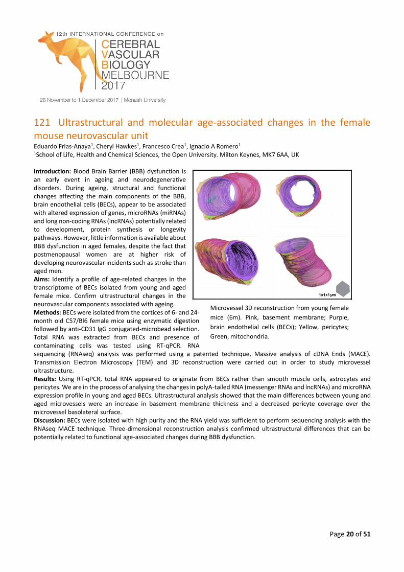
121 Ultrastructural and molecular age-associated changes in the female mouse neurovascular unit Eduardo Frias-Anaya1, Cheryl Hawkes1, Francesco Crea1, Ignacio A Romero1 1School of Life, Health and Chemical Sciences, the Open University. Milton Keynes, MK7 6AA, UK Introduction: Blood Brain Barrier (BBB) dysfunction is an early event in ageing and neurodegenerative disorders. During ageing, structural and functional changes affecting the main components of the BBB, brain endothelial cells (BECs), appear to be associated with altered expression of genes, microRNAs (miRNAs) and long non-coding RNAs (lncRNAs) potentially related to development, protein synthesis or longevity pathways. However, little information is available about BBB dysfunction in aged females, despite the fact that postmenopausal women are at higher risk of developing neurovascular incidents such as stroke than aged men. Aims: Identify a profile of age-related changes in the transcriptome of BECs isolated from young and aged female mice. Confirm ultrastructural changes in the neurovascular components associated with ageing. Methods: BECs were isolated from the cortices of 6- and 24-month old C57/Bl6 female mice using enzymatic digestion followed by anti-CD31 IgG conjugated-microbead selection. Total RNA was extracted from BECs and presence of contaminating cells was tested using RT-qPCR. RNA sequencing (RNAseq) analysis was performed using a patented technique, Massive analysis of cDNA Ends (MACE). Transmission Electron Microscopy (TEM) and 3D reconstruction were carried out in order to study microvessel ultrastructure. Results: Using RT-qPCR, total RNA appeared to originate from BECs rather than smooth muscle cells, astrocytes and pericytes. We are in the process of analysing the changes in polyA-tailed RNA (messenger RNAs and lncRNAs) and microRNA expression profile in young and aged BECs. Ultrastructural analysis showed that the main differences between young and aged microvessels were an increase in basement membrane thickness and a decreased pericyte coverage over the microvessel basolateral surface. Discussion: BECs were isolated with high purity and the RNA yield was sufficient to perform sequencing analysis with the RNAseq MACE technique. Three-dimensional reconstruction analysis confirmed ultrastructural differences that can be potentially related to functional age-associated changes during BBB dysfunction.
Microvessel 3D reconstruction from young female
mice (6m). Pink, basement membrane; Purple,
brain endothelial cells (BECs); Yellow, pericytes;
Green, mitochondria.
Page 21 of 51
122 ATN-161 stabilizes the blood-brain barrier and is neuroprotective after experimental ischemic stroke through inhibition of integrin α5β1 Danielle Edwards1, Katie Salmeron1, Justin F Fraser2, and Gregory J Bix3
1Department of Neuroscience, University of Kentucky, Lexington, KY, USA 2Department of Neurosurgery, University of Kentucky, Lexington, KY, USA 3Department of Neurology, University of Kentucky, Lexington, KY, USA Introduction: Stroke is a leading cause of death and disability with limited therapeutic options. The β1 integrin extracellular matrix receptor family has been linked to changes in BBB permeability via changes to tight junction (TJ) protein expression and function. We hypothesized that one particular β1 integrin subtype, α5β1, a pro-angiogenic fibronectin receptor that is expressed in developing brain vasculature but downregulated in the adult brain, contributes to BBB breakdown via effects on the localization and expression of the TJ protein claudin-5. Aims: Our aim was to determine the spatiotemporal expression of α5β1 integrin as well as the potential therapeutic and BBB stabilizing effect of inhibiting it with the small peptide ATN-161 following experimental stroke. Methods: In vivo: Male wildtype mice underwent transient middle cerebral artery occlusion for 1 hour. Mice were treated via intravenous and intraperitoneal injection with ATN-161 (1 mg/kg) upon reperfusion, 24 and 48 hours after reperfusion. Stroke volume was assessed on post-stroke day (PSD) 3 using cresyl violet. Immunohistochemical analysis of α5β1, claudin-5, NeuN, GFAP, and IgG expression was performed. In vitro: Mouse brain endothelial cell monolayers underwent oxygen-glucose deprivation or TNFα treatment followed by ATN-161 treatment. Permeability was assessed using FITC-dextran migration. Claudin-5 and α5β1 expression was also analyzed by immunocytochemistry. Results: α5β1 was acutely (PSD2) upregulated away from the vascular extracellular matrix basement membrane. ATN-161 decreased infarct volume and reduced BBB breakdown. In vitro, ATN-161 stabilized the endothelial cell monolayer permeability following OGD or TNFα and re-localized claudin-5 to the extracellular surface. Discussion: Endothelial cell α5β1 integrin expression is increased acutely after stroke, is expressed luminally (suggesting a non-basement membrane novel interaction and availability to intravascular therapeutics) upon upregulation, and may contribute to BBB breakdown and worsening brain injury. Furthermore, blocking α5β1 with ATN-161 could represent a promising novel therapeutic approach for ischemic stroke.
Page 22 of 51
123 Inflammation and immune mechanisms of brain damage after stroke Christopher G Sobey Department of Pharmacology, Anatomy and Microbiology, School of Life Sciences, La Trobe University, Bundoora, VIC, Australia Stroke accounts for more than 10% of deaths worldwide, and over a third of survivors are left with major neurological impairment. The need for new and effective therapies for stroke is therefore clear and urgent. While some advances have been made toward understanding its mechanisms, still only one pharmacological intervention has been found to reduce brain injury following clinical stroke – the ‘clot-buster’ recombinant tissue plasminogen activator. Unfortunately however, with a short time window of only 4.5 h, this therapy is available to less than 10% of stroke patients. For further advances in the clinical treatment of ischemic stroke, the complex mechanisms of cellular injury following cerebral ischemia must be elucidated to provide novel targets for future therapies. The initial ischemic insult is now known to be followed by induction of cytokines and chemokines, which attract numerous inflammatory cell types to the damaged brain region and ultimately contribute to secondary brain injury. Neutrophils, monocytes, T and B lymphocytes may each become activated, infiltrate the brain and modulate the severity of stroke outcome. This presentation will describe some of our recent work targeting various immune cell mechanisms for novel therapies in experimental acute stroke.
124 Movers and shapers in immune privilege of the CNS
Britta Engelhardt Theodor Kocher Institute, University of Bern, Bern, Switzerland Introduction: Discoveries leading to an improved understanding of immune surveillance of the central nervous system (CNS) have repeatedly provoked dismissal of the existence of immune privilege of the CNS. Recent rediscoveries of lymphatic vessels within the dura mater surrounding the brain by modern live cell imaging technologies have revived this discussion. Aims: We emphasize that understanding immune privilege of the CNS requires intimate knowledge of its unique anatomy. Methods: In vivo and in vitro studies on immune cell migration into the CNS during immune surveillance and neuroinflammation using live cell imaging. Results: I will summarize our current knowledge on the anatomical routes and cellular and molecular mechanisms involved in immune cell trafficking to the CNS and take into account differences in rodent and human CNS anatomy. Discussion: Data from us and others underscore that endothelial, epithelial and glial brain barriers establish compartments within the CNS that differ strikingly with regard to their accessibility to immune cell subsets.
Page 23 of 51
125 Brain endothelium: Both a target and an active player in neuroimmunology Georges E. R. GRAU Vascular Immunology Unit, Department of Pathology, Sydney Medical School & Marie Bashir Institute The University of Sydney, Camperdown, New South Wales, Australia Brain endothelial cells play a crucial role in health and integrity of the surrounding parenchyma. We are studying the pathophysiology of brain endothelium in various infectious settings. In the case of cerebral malaria, the microcirculation, and in particular the endothelium, appears to be pivotal in the interplay of Plasmodium-infected erythrocytes, host immune cells and host tissues. Our data lead us to propose a role for brain endothelium as a target as much as an effector in the immunopathology underpinning cerebral malaria. In the experimental model for cerebral malaria, we have shown that brain endothelial cells are affected by locally sequestered cells, which include infected erythrocytes, leucocytes and platelets. Intravital microscopy revealed that a critical number of CD8+ T effectors is required to induce disease and monocyte adherence to the brain vasculature. Depletion of monocytes at the onset of the neurological syndrome results in decreased lymphocyte accumulation, suggesting reciprocal effects of monocytes and T cells on their sequestration within brain microvessels. In addition to direct cell-cell contacts, brain endothelial cells are profoundly modified by membrane microparticles – also called microvesicles - released by these sequestered cells. Both adhesiveness and permeability of brain endothelial cells are dramatically enhanced by microparticles/microvesicles, notably those produced by platelets. Infected erythrocytes, in turn, release their own microvesicles, which have potent pro-inflammatory properties, as assessed by their enhancement of TNF production and CD40 expression on monocytes. On the other hand, brain endothelial cells can act as players because they release high numbers of microparticles upon stimulation by cytokines, which are found in high levels during cerebral malaria, both in patients and in the experimental model. These endothelial microparticles, in turn, amplify the local inflammatory responses that are characteristic of CM. Aside from their effects on immune cells, brain endothelial cells can increase the proliferation of Plasmodium falciparum through production of soluble, low molecular weight, yet unidentified growth factors. Interestingly, we showed that brain endothelial cells also play an important role in the determinism of the neurological syndrome, as demonstrated by the relation between their reactivity to cytokines and susceptibility to cerebral malaria. Indeed we demonstrated, both in the murine model and in patients that brain endothelial cells purified from individuals who developed cerebral malaria upregulate more adhesion molecules and release more microparticles than their counterparts isolated from normal subjects. Brain endothelial cells can pick up antigens from infected erythrocytes, present antigens and amplify T cell proliferation, as they express a set of relevant co-stimulatory molecules. Transfer of malarial antigens is evidenced by confocal microscopy, suggesting trogocytosis between infected erythrocytes and brain endothelial cells. We also found that not only brain endothelial cells but also the microparticles they release are able to activate T cells. Therefore, we analysed the cargo of microparticles released during experimental cerebral malaria, in terms of proteins and nucleic acids. Proteomics and microarray analyses identified several proteins and miRNAs associated with the development of the neurological syndrome, notably miRNAs capable of controlling endothelial functions. Recently we identified diannexin as a potent down-modulating agent of microparticle release by cytokine-activated brain endothelial cells. Our results suggest that diannexin inhibits endothelial vesiculation by binding the phosphatidylserine present either at the cell surface or at the level of the inner leaflet of the plasma membrane, shedding some light on microparticle production mechanisms. A better understanding of brain endothelial cell pathophysiology will allow for a modulation of their function. This should be useful in cerebral malaria as well as in other immune-mediated complications of infectious diseases that affect the brain
Page 24 of 51
126 The blood brain barrier and cerebral Malaria Midrelle Nandjou, William Okech and Monique Stins Molecular Microbiology and Immunology, Johns Hopkins School of Public Health, Baltimore, MD, USA Introduction: Cerebral malaria (CM) is the most severe form of Plasmodium falciparum malaria with mortality rates up to 30%. It affects mostly children in sub-Saharan Africa. Neurological signs and symptoms of CM include seizures, coma and brain edema. Upon efficient chemotherapy targeting the parasite, CM patients can recover but a subset of the CM survivors suffer permanent neurologic sequelae. Sequestration of late trophozoite and schizont stages of Plasmodium falciparum-infected red blood cells (PRBC) inside the brain’s micro-vasculature is considered a hallmark of CM. PRBC bind via the Plasmodium falciparum-encoded Erythrocyte Membrane Protein-1(Pf-EMP-1) to host adhesion proteins such as ICAM-1 and EPCR on the luminal surface of brain endothelial cells. Although PRBC sequestration occurs globally in the brain, there are striking differences in white matter (WM) versus gray matter (GM) pathology. Aims: To understand differences in vascular properties leading to PRBC sequestration and differential vascular pathologies in CM. Methods: Micro-vessels from different brain areas, including corpus callosum (CC), basal ganglia (BG), frontal cortex white matter (FWM), frontal cortex gray matter (FGM) were isolated. Microvessels were then characterized using endothelial marker CD31, glucose transporter type 1 (GLUT-1), endothelial protein C receptor (EPCR) and alkaline phosphatase (AP) by microscopy and quantified. Results: Microvessels greatly differed in their expression of EPCR, GLUT-1 and AP, not only along the vascular tree but also between the different brain areas. This may explain the observed striking differential neuropathology in the GM versus WM in CM patients. Discussion: As EPCR is a receptor for Pf-EMP1 and involved in intravascular sequestration of PRBC, this is relevant for CM neuro-pathogenesis. Microvessels in certain brain areas will be more susceptible to sequestration in CM than other brain areas. This may explain the observed differential neuropathologies in the GM versus WM in CM patients. Besides importance of these results for CM pathogenesis, this will likely also be of relevance for other neurological diseases involving vascular pathologies.
Page 25 of 51
127 HIV Antiretroviral therapy increases Leukocyte adhesion to the BBB Dionna W. Williams1, 2, Mike Veenstra2, Joan W. Berman 2, 3 1Department of Molecular and Comparative Pathobiology, Johns Hopkins University, Baltimore, MD, USA 2Department Pathology, Albert Einstein College of Medicine, Bronx, NY, USA 3Department of Microbiology and Immunology, Albert Einstein College of Medicine, Bronx, NY, USA Introduction: While antiretroviral therapy (ART) reduces plasma viral loads to near undetectable levels, it does not always quell virus to the same extent in other areas of the body. This is particularly true for the brain, which is separated from the periphery by the BBB. As a result, ongoing HIV replication occurs within the brain, establishing it as a viral reservoir. A more complete understanding of the effects of ART on BBB function will be integral to the elimination of the brain as a viral reservoir and HIV-associated neurologic disease. Aims: To assess the effects of ART on BBB permeability, junctional proteins, and leukocyte-endothelial interactions. Methods: Our in vitro model of the BBB is comprised of primary human brain microvascular endothelial cells cocultured with primary human astrocytes on opposing sides of a transwell tissue culture insert. Five ART drugs were added to the apical side of the BBB model for 24 hours, after which time permeability, junctional proteins (ALCAM, PECAM-1, ICAM-1, and JAM-A), and leukocyte adhesion were assessed. Results: Efavirenz and Tenofovir significantly increased all of the examined junctional proteins on the BBB, while Lamivudine increased only ALCAM. There was a functional consequence to this increase in junctional proteins, as Efavirenz, Tenofovir, and Lamivudine promoted the adhesion of CD3+ T cells, CD14+ monocytes, and CD14+CD16+ monocytes to the BBB. In contrast, Emtricitabine and Zidovudine had no effects on junctional protein expression or on leukocyte adhesion to the BBB. None of the ART drugs caused BBB permeability. Discussion: ART may facilitate the recruitment of HIV target cells into the brain by increasing the junctional molecules integral for leukocyte-endothelial cell interactions and transmigration, contributing to CNS HIV infection and the maintenance of the brain as a viral reservoir.
Page 26 of 51
128 Spatiotemporal regulation of transport dynamics in CSF: From Choroid Plexus via the CiLoN to the brain. Regina J Faubel1, 2 1 C.W.Lo, Department of Developmental Biology, Medical school University of Pittsburgh, Pittsburgh, PA, USA 2 G.Eichele, Department Genes and Behavior, Max-Planck-Institute for Biophysical Chemistry, Göttingen, Germany Introduction: Traditional barriers consist of cells sealed by tight junctions. Recent work showed that CSF-flow generated by ependymal cilia orchestrates distribution of CSF-components along the ventricular wall. Depending on the site of release, secretions are delivered coherently along flow-channels, prevented from entry into certain areas, or diluted in the ventricular volume. Thus, the ciliary logistic network (CiLoN) constitutes a CSF-barrier additional to the ChPx. Aims: We assess whether the circadian clock regulates the CSF-barriers and this way transport via the CSF-route. Methods: Explant-culture, luciferase-assay, transcriptome-profiling, global cilia-beating-analysis, flow-analysis. Results: ChPx and ependyma both harbour an intrinsic circadian clock. Transcriptome-analysis revealed circadian regulation of ChPx functions: Temporal clustering was observed for certain transport, production and perception of hormones and cytokines, degradation and detoxification. The CiLoN exhibits circadian rhythm with alternating presence of either straight flow-channels or spiral flow that leads to vigorous, local mixing and stagnating flow. This change is based on re-arrangement of cilia-beating direction. Further work revealed that the flow pattern is determined by local rather than CSF-derived chemical or mechanical cues. Discussion: In consideration of the complex architecture and precise embedding of the BBB, it appears reasonable that transport via the ChPx-CSF-route is not chaotic but fast, site-directed and efficiently regulated. Spatial compartmentation by the CiLoN might be necessary to orchestrate the multiplicity of functions exerted via the CSF. The circadian clock designates functional clusters to specific time windows and seems to synchronize ChPx and CiLoN to further minimize interference between diverse CSF-functions: There is a time for enrichment of CSF with neuroprotective secretions co-occuring with stagnating flow that promotes enrichment. And there is a time for flushing the brain with increased CSF-production, detoxification and straight flow-through. Our findings reveal the importance of chronopharmacological considerations in drug-targeting the brain via the CSF-route.
Page 27 of 51
129 Macromolecule distribution within brain extracellular and perivascular spaces in health and disease Robert G. Thorne1, 2, 3, 4, 5 1Pharmaceutical Sciences Division, University of Wisconsin-Madison School of Pharmacy 2Neuroscience Training Program, University of Wisconsin-Madison 3Institute for Clinical and Translational Research, University of Wisconsin-Madison 4Clinical Neuroengineering Training Program, University of Wisconsin-Madison 5Cellular and Molecular Pathology Graduate Training Program, University of Wisconsin-Madison Madison, Wisconsin, U.S.A. Introduction: The precise mechanisms governing the central distribution of macromolecules remain poorly understood, despite their importance for several ongoing clinical trials as well as physiological processes such as antibody trafficking for central immune surveillance. Much evidence exists to suggest that the short-range local distribution of macromolecules in the brain microenvironment is governed by diffusion through the narrow, tortuous extracellular space (ECS) of the neuropil. Long-range distribution within the central compartment can occur through convection or bulk flow with the cerebrospinal fluid and cerebral perivascular spaces and perhaps also within white matter. Aims: My laboratory aims to improve the prospects for effective central nervous system drug delivery of biologics by illuminating the key physiologic and anatomical determinants of macromolecule entry into and movement within the central compartment: (i) local transport processes governing distribution after injection into the brain parenchyma, (ii) whole-brain distribution and clearance mechanisms following intrathecal infusion, and (iii) nasal pathways for brain targeting after intranasal administration. Methods: Our past work has explored the diffusion of a variety of macromolecules after their injection into the neocortex (Thorne et al. PNAS, 2006 & 2008) or following intrathecal infusion, with the goal of using diffusion data to better understand complex CNS distribution patterns (Wolak et al. Journal of Controlled Release, 2015). A variety of different methods are used for the work, including fluorescence imaging and MRI, electron microscopy, and modelling (e.g. Kumar et al. Scientific Reports, 2016; Lochhead et al. JCBFM, 2015; Wolak & Thorne. Molecular Pharmaceutics, 2013; Lochhead & Thorne. Advanced Drug Delivery Reviews, 2012). Results: Our recent work suggests an emerging key mechanism underlying widespread brain delivery is the degree to which intrathecally or intranasally applied biologics gain access to and distribute within the perivascular spaces of cerebral blood vessels. Our current studies are investigating what governs the balance between extracellular diffusion and perivascular transport by employing a variety of different labeled tracer macromolecules, including antibodies, antibody fragments and oligonucleotides. Discussion: Examples will be presented to reveal how diffusive transport, binding, and perivascular flow affect the central delivery and distribution of macromolecules as well as suggest new ways in which to enhance delivery.
Page 28 of 51
130 Impairment of glymphatic function in the aging brain and Alzheimer’s disease Jeffrey J Iliff1, 2 1Department of Anesthesiology and Perioperative Medicine, 2Knight Cardiovascular Institute, Oregon Health & Science University, Portland, Oregon, USA While aging is the strongest risk factor for the development of Alzheimer’s disease, disruption of normal sleep patterns has long been associated with aging and more recently has been associated with the development of Alzheimer’s pathology. Recently, a brain-wide perivascular network termed the ‘glymphatic’ system, has been characterized that facilitates the clearance of interstitial solutes including amyloid beta and tau from the brain. Interestingly, this function was active primarily in the sleeping brain, and is impaired in both the aging and the post-traumatic brain, suggesting one possible basis for the link between aging, sleep disruption and neurodegenerative processes. New data from human clinical subjects suggests that changes in the in elements of the glymphatic system, including the astroglial water channel aquaporin-4 (AQP4) are associated with Alzheimer’s status and pathology, and neurocognitive decline. These findings suggest that glymphatic insufficiency may be one feature of the aging brain that renders it vulnerable to protein mis-aggregation in neurodegenerative conditions such as Alzheimer’s.
Page 29 of 51
131 Quantitative mass spectrometry imaging of drug distribution in the central nervous system Nathalie YR Agar1, 2, 3 1Department of Neurosurgery, Harvard Medical School, Brigham and Women’s Hospital, Boston, MA, USA 2Department of Radiology, Harvard Medical School, Brigham and Women’s Hospital, Boston, MA, USA 3Department of Cancer Biology, Dana-Farber Cancer Institute, Boston, MA, USA Introduction: Mass Spectrometry Imaging (MSI) and Stimulated Raman Imaging (SRI) provide complementary label-free molecular imaging options to guide the development of drugs targeting tumors of the central nervous system (CNS). Drug transit through the blood-brain barrier (BBB) is a limiting factor and present significant challenges in the development of brain tumor therapeutics. Aims: The use of a multimodal imaging approach combining MSI and SRI with correlation to MRI to track targeted therapeutics through the BBB could help in optimizing treatment conditions to overcome related limitations in treating brain cancers. Methods: Mice with established intracranial xenografts were treated with small molecule inhibitors under acute and chronic dosage regimens, euthanized, and brains were flash frozen and stored at -80°C until analysis. Tissue sections were prepared and collected onto ITO-coated microscopic slides for MALDI MSI and on optical slides for stimulated Raman scattering (SRSI), and standard histopathology evaluation. Samples were coated MALDI matrices and mass spectrometry images acquired using MALDI FTICR and MALDI TOF mass spectrometers. Stimulated Raman scattering (SRS) microscopy images were acquired with fields of view (FOV) of ~350×350 μm2 at a spatial resolution of ~500 nm. Unstained tissue sections from treated xenograft models were imaged at 2940 cm-1 (mainly attributed to CH3 in proteins), 2854 cm-1 (mainly attributed to CH2 in lipids), and 2800 cm-1 (absorption from red blood cells). This multicolor data allows to characterize both tissue architecture and biochemistry of the cancer tissue and the drug. Results: Drugs and their metabolites were identified on the basis of accurate mass measurement (better than 1 ppm) and/or tandem mass spectrometry analyses and mapped across each section following the signal of the drug and heme. The permeability of drugs through the brain vasculature was visualized by re-creating three-dimensional volumes and the use of a tissue mimetic model allowed to directly quantify the drug amount in each sample analyzed by mass spectrometry. SRS images were correlated to drug images. Discussion: The presented data have the potential to provide a valuable advance for the development and implementation of novel and more effective therapeutic approaches.
Page 30 of 51
132 Modelling the clearance of soluble metabolites from the brain. Implications for Alzheimer’s disease Roxana Aldea 1, 2, Roxana O. Carare3, Giles Richardson2 1 Institute for Complex Systems Simulations, University of Southampton, Southampton, United Kingdom 2 School of Mathematics, University of Southampton, Southampton, United Kingdom 3 Clinical Neurosciences, Southampton General Hospital, Southampton, United Kingdom Introduction: Inefficient clearance of the soluble metabolite amyloid-beta (Aβ) from the brain plays a key role in the development of cerebral amyloid angiopathy (CAA), a major pathological factor in Alzheimer’s disease [1]. Aims: This study aims to answer the unresolved questions regarding the removal of Aβ from the brain along the wall of cerebral arteries, i.e. the intramural peri-arterial drainage (IPAD) process [2]. Age-driven failure of this clearance process leads to onset of CAA [3]. We propose a novel hypothesis for the force generator behind perivascular drainage of Aβ along IPAD, with an emphasis on the vasomotion of cerebral arteries. We also highlight future avenues of modelling and imaging in cerebrovascular research that could help bridging the gap between vascular and neurodegenerative disorders such as Alzheimer’s disease. Methods: By employing physiological-driven mathematical and computational models, the fluxes of solutes out of the brain along IPAD are determined. Results: The cerebral vasomotion yields perivascular flow rates that are up to three orders of magnitude higher than those generated by the arterial pulsations alone [4]. Discussion: The proposed clearance mechanism of Aβ emphasizes the importance of cerebral vascular function, not only in cerebral blood supply, but also in the clearance of brain metabolites. These results challenge the dominant hypothesis of arterial pulse-driven perivascular drainage of Aβ from the brain, leading to a shift in perspective regarding the motive force of the IPAD process. These findings have implications for the development of new imaging techniques and targeted treatments in Alzheimer’s disease focused on cerebral vasomotion.
Page 31 of 51
133 Modulation of CSF inflammation and oxidative stress during perinatal injuries. A role for choroid plexuses? Jean-François Ghersi-Egea1 1Fluid Team and BIP facility, Lyon Neurosciences Research Center, INSERM U1028, CNRS UMR5292, Université Claude Bernard Lyon-1, Lyon, France The central nervous system develops in a defined fluid environment that needs to be preserved for normal neural network formation and cerebral maturation. Perinatal injuries induced by inflammatory, infectious, or hypoxic events or following exposure to toxic compounds, can induce central oxidative stress and inflammation ultimately leading to death or to dramatic neurological impairments. The CSF fills a large part of the cranial cavity volume at the perinatal period, and the choroid plexuses (CPs) mature early during development, especially with respect to neuroprotective genes. This makes the CPs-CSF system worth of investigation during this critical period of life. The blood-CSF barrier represents a main gate of access to the developing brain for both immune cells and reactive, pro-oxidant endo/xenobiotics. We used a combination of in vivo approaches and ex vivo and cellular models of the blood-CSF barrier, to explore the role of the CP-CSF system in promoting or preventing central consequences of perinatal injuries. We showed that the antioxidant and detoxification capacities of CPs are high at a time when astrocytic protection of neurons is only partial. Toll-like receptor 2-mediated responses to bacterial components include the migration of leukocytes, including polynuclear neutrophils, through the choroid plexus into the CSF by a mechanism that involves chemoattractive factors secreted by the choroidal epithelium. Neutrophil migration and elevated levels of proinflammatory molecules into the CSF was prevented by the administration of the glutathione precursor and antioxidant molecule N-acetylcysteine. The latter did not affect the choroidal secretion of chemoattractants induced by Toll-like receptor 2 activation. The existence of a direct link between oxidative stress regulation at the blood-CSF barrier and neutrophil migration into the CSF remains therefore to be clarified in the context of perinatal infection.
Page 32 of 51
134 The choroid plexus as an important player in neuroinflammation Roosmarijn E. Vandenbroucke1, 2 1Department of Biomedical Molecular Biology, Ghent University, Gent, Belgium 2VIB-UGent Center for Inflammation Research, VIB, Gent, Belgium Introduction: The blood-cerebrospinal fluid barrier (CSF) forms a unique interface between blood and brain. It consists of a single cell layer, called choroid plexus epithelium, situated at the interface of blood and CSF. The choroid plexus epithelium has different important functions: it forms a barrier to protect the brain from fluctuations in the blood thereby assuring brain homeostasis, produces CSF and is responsible for the active removal of toxic molecules from the brain. In recent years, the choroid plexus epithelium has gained increasing attention, especially its role in different pathologies. Aims: Our aim is to study the impact of inflammation on both barrier and secretory function of the choroid plexus epithelium and identify novel therapeutic strategies to prevent neuroinflammation. Methods: To study the impact of inflammation on the choroid plexus epithelium in vivo, different mouse models are used. To mimic systemic inflammation, we use mouse models of sepsis, namely endotoxemia (intraperitoneal injection of lipopolysaccharide (LPS)) and cecal ligation and puncture (CLP). Additionally, central inflammation associated with Alzheimer’s disease, is mimic by intracerebroventricular (icv) injection of amyloid beta oligomers (AβO) in wild type mice and by using the transgenic APP/PS1 mice. In parallel, also in vitro primary choroid plexus epithelium cultures are used. Barrier function is studied in vivo using 4 kDa FITC-dextran leakage analysis and analysis of tight junctions and in vitro using transepithelial electrical resistance (TEER) and electric cell-substrate impedance sensor (ECIS) measurements. Secretory function mainly focusses on the production and secretion of extracellular vesicles (EVs) and this is studied using qRT-PCR, electron microscopy, immunostainings, Nanoparticle Tracking Analysis (NTA). Results: Our studies show that both systemic1 and central inflammation2 induce increased blood-CSF barrier leakage. Additionally, peripheral inflammatory triggers also induce an increase in EV release by the choroid plexus epithelium into the CSF3. Strikingly, these choroid plexus-derived EVs were able to enter the brain parenchyma, were taken up by astrocytes and microglia and transferred a pro-inflammatory message to the brain3. Similarly, also AβO resulted in an increase in EV production, which plays a role in the AβO-induced cognitive decline4. Discussion: In conclusion, we found that both peripheral and central inflammatory triggers affect barrier and secretory activity of the choroid plexus epithelium and these results open up new therapeutic strategies to treat neuroinflammatory diseases.
135 Cerebrospinal fluid-based signalling in the developing brain
Maria K. Lehtinen1, 2 1Robertson Investigator, New York Stem Cell Foundation 2Department of Pathology, Boston Children’s Hospital, Harvard Medical School, Boston, MA, USA The earliest born neural stem cells in the developing brain are located along cerebrospinal fluid (CSF)-filled ventricles. From the time of neurulation, signals delivered by the CSF provide instructive cues to neural stem cells, and these favorable effects of CSF are age-dependent. While some factors in the CSF originate from adjacent neural progenitors, the choroid plexus, a sheet of epithelial cells located in each ventricle in the brain is considered to be the principal source of CSF for the brain. Choroid plexus-secretion of factors into the CSF varies across ventricles and with age. Current work in the lab investigates mechanisms regulating choroid plexus CSF production and composition.
Page 33 of 51
136 Oat1/3 and Oatp1a4 are acting as avid elimination pathway of organic anion in the CSF at the blood-arachnoid barrier (BAB) Masanori Tachikawa1, Yuka Yaguchi1, Zhengyu Zhang1, Yasuo Uchida1 and Tetsuya Terasaki1 1Division of Membrane Transport and Drug Targeting, Graduate School of Pharmaceutical Sciences, Tohoku University, Sendai, Miyagi, Japan Introduction: Previous concept has demonstrated that the choroid plexus and arachnoid membrane together act as blood-CSF interfaces and the arachnoid is generally impermeable to water-soluble substances1. However, we have found that the transporters-mediated choroid plexus uptake plus the CSF bulk flow can explain the total CSF elimination of prostaglandin D2 by only 27.9%2. We thus hypothesized that the arachnoid-forming blood-arachnoid barrier (BAB) transporters actively regulate the CSF clearance of water-soluble substances. Aims: The purpose of the present study was to clarify the protein profiles of organic anion transporters and their functional roles in the CSF clearance of organic anions at the BAB. Methods: Transporter protein expressions were determined by quantitative targeted absolute proteomics (QTAP) and immunohistochemistry. Para-aminohippuric acid (PAH) and fluorescent sulforhodamine-101 (SR-101) were used as model substrates of organic anion transporters Oat and Oatp, respectively. Each compound was injected with or without inhibitors into cisterna magna of rats, which could minimize the contribution of choroid plexus. Results: QTAP revealed the predominant level of Oat1 in the rat arachnoid mater and the concomitant expression of Oat3 in the arachnoid mater and choroid plexus. Immunostaining confirmed the localization of Oatp1a4 in the arachnoid mater. CSF elimination clearances of PAH and SR-101 were greater than that of inulin, a bulk flow marker. Inhibitors for oat1/3 and oatp1a4 significantly inhibited the elimination of PAH and SR-101, respectively. The distribution of SR-101 fluorescent was restricted in the arachnoid mater whereas the intensity was increased in the parenchyma of spinal cord by the co-injection of taurocholate, an Oatp1a4 specific inhibitor. Discussion: This is the first study demonstrating that Oat1/3 and Oatp1a4 at the BAB acts as avid clearance pathway of organic anions in the CSF.
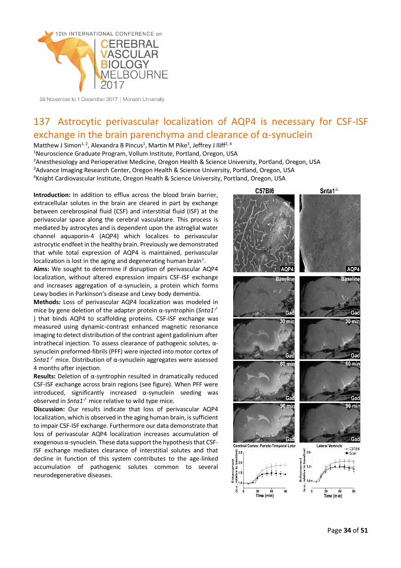
Page 34 of 51
137 Astrocytic perivascular localization of AQP4 is necessary for CSF-ISF exchange in the brain parenchyma and clearance of α-synuclein Matthew J Simon1, 2, Alexandra B Pincus1, Martin M Pike3, Jeffrey J Iliff2, 4 1Neuroscience Graduate Program, Vollum Institute, Portland, Oregon, USA 2Anesthesiology and Perioperative Medicine, Oregon Health & Science University, Portland, Oregon, USA 3Advance Imaging Research Center, Oregon Health & Science University, Portland, Oregon, USA 4Knight Cardiovascular Institute, Oregon Health & Science University, Portland, Oregon, USA Introduction: In addition to efflux across the blood brain barrier, extracellular solutes in the brain are cleared in part by exchange between cerebrospinal fluid (CSF) and interstitial fluid (ISF) at the perivascular space along the cerebral vasculature. This process is mediated by astrocytes and is dependent upon the astroglial water channel aquaporin-4 (AQP4) which localizes to perivascular astrocytic endfeet in the healthy brain. Previously we demonstrated that while total expression of AQP4 is maintained, perivascular localization is lost in the aging and degenerating human brain1. Aims: We sought to determine if disruption of perivascular AQP4 localization, without altered expression impairs CSF-ISF exchange and increases aggregation of α-synuclein, a protein which forms Lewy bodies in Parkinson’s disease and Lewy body dementia. Methods: Loss of perivascular AQP4 localization was modeled in mice by gene deletion of the adapter protein α-syntrophin (Snta1-/-
) that binds AQP4 to scaffolding proteins. CSF-ISF exchange was measured using dynamic-contrast enhanced magnetic resonance imaging to detect distribution of the contrast agent gadolinium after intrathecal injection. To assess clearance of pathogenic solutes, α-synuclein preformed-fibrils (PFF) were injected into motor cortex of Snta1-/- mice. Distribution of α-synuclein aggregates were assessed 4 months after injection. Results: Deletion of α-syntrophin resulted in dramatically reduced CSF-ISF exchange across brain regions (see figure). When PFF were introduced, significantly increased α-synuclein seeding was observed in Snta1-/- mice relative to wild type mice. Discussion: Our results indicate that loss of perivascular AQP4 localization, which is observed in the aging human brain, is sufficient to impair CSF-ISF exchange. Furthermore our data demonstrate that loss of perivascular AQP4 localization increases accumulation of exogenous α-synuclein. These data support the hypothesis that CSF-ISF exchange mediates clearance of interstitial solutes and that decline in function of this system contributes to the age-linked accumulation of pathogenic solutes common to several neurodegenerative diseases.
Page 35 of 51
138 Interactive forum 2: Transporters, nanoparticles and brain targeting: How close are we to a clinical solution? Danica Stanimirovic1 and William Elmquist2 1National Research Council, University of Ottawa, Ottawa, Canada 2Dept. of Pharmaceutics, Brain Barriers Research Centre, University of Minnesota, Minneapolis, Minnesota, USA Great strides have been made in recent years in determining the mechanisms, both anatomical and biochemical, that can limit drug delivery across the BBB. Based on this knowledge, there have been several techniques employed to improve the CNS delivery of vastly different compounds; from targeted small molecules to antibodies. These techniques can include: 1) the disruption of BBB tight junctions (e.g., osmotic, cytokine, ultrasound), 2) employing ‘molecular Trojan horses’ to help shuttle therapeutics across the BBB (e.g., fusion proteins, conjugates, nanoparticles), 3) structural modifications and adjuvant therapies that limit BBB delivery liabilities (e.g., medicinal chemistry approaches, inhibition of active efflux), and 4) direct delivery to the brain, bypassing the BBB (e.g., CED, IVC, implants). Yet, in spite of the wealth of mechanistic knowledge about the barrier, and specific ways to overcome it, there remain significant hurdles to employ these measures in clinical practice, including the clinical translation of preclinical findings, a follow-up from the first forum of the conference. This second interactive forum will discuss what these hurdles are, and how they may be overcome in future clinical trials.
139 Brain barriers and functional interfaces with sequential appearance of ABC efflux transporters during human development Kjeld Møllgård, Department of Cellular and Molecular Medicine, Faculty of Health and Medical Sciences University of Copenhagen, Blegdamsvej 3, DK-2200 Copenhagen, Denmark Adult brain is protected from entry of many drugs and toxins by specific mechanisms such as ABC efflux transporters. Little is known when these appear in human brain during its development. Cellular distribution of three main ABC transporters (ABCC1, ABCG2, and ABCB1) was determined at blood brain barriers and interfaces in human embryos and fetuses in the first half of gestation. Antibodies against claudin-5 and -11 together with antibodies to α-fetoprotein were used to describe morphological and functional aspects of brain barriers. First exchange interfaces to be established at 4-5 weeks post conception (wpc) are between brain and embryonic CSF and between outer surface of brain anlage and primary meninx. They already exclude α-fetoprotein, are immunopositive for both claudins as well as for ABCC1 and ABCG2. ABCB1 is detectable within a week of blood vessels first penetrating into brain parenchyma (6-7 wpc). ABCC1, ABCB1 and ABCG2 are present at blood-CSF barrier in all choroid plexuses from first appearance (7 wpc). Outer CSF-brain interfaces are established between 9-11 wpc exhibiting immunoreactivity for all three transporters. Results provide evidence for sequential establishment of brain exchange interfaces and spatial and temporal timetable for three main ABC transporters in early human brain.
Page 36 of 51
140 Age-dependent changes in the cellular and molecular responses to spinal cord injury: Importance on axonal regeneration and functional recovery W. Dalton Dietrich1, Norman Saunders2 1The Miami Project to Cure Paralysis, Departments of Neurological Surgery, University of Miami Miller School of Medicine, Miami FL USA 2Department of Pharmacology & Therapeutics, The University of Melbourne, Victoria Australia Spinal cord injury (SCI) is a significant international health problem with millions of people today living with chronic paralysis and serious quality of life issues. Currently there are no proven therapeutic strategies to promote functional recovery after severe SCI. Experimental and clinical studies therefore continue to clarify the pathophysiology of SCI with the goal of determining new treatments that may be administered in the early or late phases to protect vulnerable structures and enhance axonal regeneration. The mature spinal cord has very limited capacity to regenerate in contrast to the immature spinal cord which shows a remarkable regenerative ability in many species. Working with Professor Norman Saunders and colleagues over the last decade, our laboratories have successfully collaborated on clarifying the cellular and molecular mechanisms of SCI using a developmental animal model and cutting-edge research tools. For these studies the short tailed opossum Monodelphis domestica has been employed because of the ability to reproducibly transect the spinal cord in the neonatal state and the capacity for axon growth across the region and the regeneration of damage axons. However, this ability to regrow axons across the lesion is extremely age-dependent and declines after two weeks and disappears within one month. Thus, the model is extremely useful for addressing local or remote alterations that may play a significant role on promoting or inhibiting successful regeneration after SCI. New concepts regarding age-dependent changes in the damaged microenvironment including changes in inflammatory and growth promoting genes and proteins that would be expected to alter the potential for successful regeneration have been advanced. With the recent sequencing of the opossum genome and the advent of high throughput RNA sequencing, overall changes in gene expression in response to focal injury are now being evaluated throughout the neuroaxis. Together these findings stress a complexity of the molecular consequences of SCI and also emphasize a high degree of coordination within sites close to the injury or in distant locations such as the cell bodies of axotomized axons. This ongoing research program will be discussed with emphasis on age-dependent responses to injury important for successful axonal regeneration and functional recovery following SCI.

Page 37 of 51
141 Bipedal marsupials for studies of spinal cord development, injury and regeneration Mark D Habgood Department of Pharmacology & Therapeutics, University of Melbourne, Parkville, VIC, Australia Introduction: Decades of research efforts studying spinal cord injury (SCI) in quadrupedal animals have generated substantial information about the response to trauma and mechanisms behind axonal regenerative failure. However, translation of promising experimental results into clinical outcomes for patients has been very limited. This is partly because of difficulties in independently replicating promising results (Steward et al 2012), but mostly because of the lack of an accessible animal model in which to study therapies aimed at improving bipedal locomotion, which is characteristic for humans. Macropod marsupials (e.g. wallabies and kangaroos) have unique advantages as an animal model for studies of recovery from SCI. They are bipedal with patterns of postural and hind limb motor coordination that better reflect those of humans. In addition they are born at very early stages of central nervous system development, which allows experimental investigations and manipulations to be conducted at ages that would be inaccessible in utero in rodents and higher mammals. Aim: To determine the suitability of the tammar wallaby as an accessible bipedal model for studies of SCI. Methods: Spinal cord morphological repair and recovery of locomotor function was characterised in tammar wallabies after SCI at different stages of postnatal development. Results: Animals injured in the first three postnatal weeks exhibited remarkable recovery of bipedal overground locomotor function that was almost indistinguishable from uninjured animals and substantial morphological repair of the injury site. In contrast, animals injured at older ages (>6 weeks) did not re-grow supraspinal innervation and were not able to support themselves in a standing posture when tested at around 5 months. Discussion: The finding that there is a developmental stage common to all mammals when the injured spinal cord is naturally able to repair itself is likely to be important for identifying the factors expressed at later stages of development that prevent regenerative repair. We have shown that in the tammar wallaby, the switch to a non-regenerative phenotype occurs between 3 and 6 weeks of pouch life whereas in rodents and higher mammals it occurs in utero. The tammar young have an additional advantage for studies of SCI because of the length of postnatal life they spend carried in the pouch (around 180 days) where they have unlimited access to food and warmth. Thus potential treatments can be assessed over extended periods of time without being affected by mobility-related difficulties in feeding and care.
Page 38 of 51
142 What does the choroid plexus do? Shane A Liddelow1
1Department of Neurobiology, Stanford University, Stanford, California, USA The choroid plexuses in the lateral, third, and fourth ventricles of the brain are the principle sources of production and secretion of the cerebrospinal fluid (CSF). The choroid plexus and CSF (along with the blood-brain barrier proper (BBB) at the level of the blood vasculature) provide robust protection for the brain. This multi-faceted defence involves a “physical” barrier to impede entrance of toxic metabolites; a “biochemical” barrier that facilitates removal of moieties that circumvent the physical barrier; and buoyant physical protection by CSF itself to protect against trauma. Specific roles of the choroid plexuses make them integral for normal brain development and central nervous system (CNS) homeostasis, and these roles are considerably different to that in the adult brain under normal physiological conditions. Recent studies have revealed new, active roles for this dynamic system in the regulation of the CSF milieu during critical periods of development and in the healthy nervous system. The choroid plexus-CSF system is also important for repair after disease and trauma. Most notably, it has been suggested to provide a stem-cell like repository for neuronal and astrocyte glial cell progenitors. In recent years, the Saunders’ lab has made several key findings that outline how choroid plexus epithelial cells are born, how they are able to transport specific plasma proteins and lipid-soluble macromolecules, and describe the drug-efflux capacity of the structure as a whole. Additionally, genome-wide RNA sequencing experiments have provided insights into other possible roles of the choroid plexus in normal brain functioning and following injury/disease. One example is the role the plexus may play in immune surveillance in the brain. In contrast to the BBB, the choroid plexus interacts more dynamically with cells of the peripheral immune system. This includes mechanisms to support specific responses to disease and injury. The plexus epithelium is a key conduit for molecules and immune cells into the CSF/CNS, and they appear to be a major player in the immune response due to their high expression of adhesion molecules, as well as antigen presentation and chemokine secretion. This close homeostatic control of CNS, including the CSF, glia, and neurons, plays a vital role in normal and abnormal brain function. Dysfunctions of the choroid plexus and the BBB often correlate with and likely contribute to pathology of neurological conditions. However, more work is required to fully appreciate the influence of the choroid plexuses on the development of CNS immune and disease/injury responses. Proper understanding of these mechanisms will likely provide opportunities to develop therapeutic interventions that control inflammation and repair processes in the CNS. As such, proper understanding of the choroid plexus will likely prove important for the production of drug delivery and therapies to help ameliorate a wide range of neurological diseases
Page 39 of 51
143 Prediction of in vivo drug concentrations in the brain and cerebrospinal fluid based on in vitro parameters Yuichi Sugiyama Sugiyama Laboratory, RIKEN Innovation Center, RIKEN, Yokohama, Japan
Purpose: The pharmacological effects/adverse reactions in the central nervous system depends on the unbound drug concentration in the brain (Cu,brain). Because of the difficulty in direct measurement of Cu,brain, the concentration in the cerebrospinal fluid (CSF) (CCSF) is often used as a surrogate for Cu,brain in humans. Active efflux by P-gp and/or Bcrp at the blood-brain barrier (BBB) keeps the Cu,brain lower than the unbound concentration in the plasma. We performed a quantitative prediction of the concentration in the brain and CSF using the in vitro passive permeability and transport activities by P-gp and Bcrp. Methods: A three-compartment model which consisted of blood, brain and CSF compartments was used. A non-linear least squares method was performed to fit the equations obtained under steady-state to observed data, the unbound brain-and CSF-to-unbound plasma ratio (Kp,uu,brain or Kp,uu,CSF) of 22 compounds in rats, to obtain the kinetic parameters and scaling factors. Passive permeability was determined using PAMPA assay Transport activities by P-gp and Bcrp were evaluated in vitro by measuring the transcellular transport across the stable transfectants. Results: The predicted Kp,uu,brain and Kp,uu,CSF values of 19 compounds were within three-fold of the observed values. . Kp,uu,CSF/brain values of 21 compounds were within 3-fold of the observed values except for risperidone1). Conclusion: We have developed the model that could reasonably explain the CCSF and Cu,brain of drugs using in vitro parameters, the passive permeabilities, and the transport activities by P-gp and Bcrp. In my presentation, I want to refer further to our most recent studies to predict the time profiles of receptor occupancy in the human brain based on in vitro studies 2),3)
Page 40 of 51
144 Regional drug distribution: Implication for pharmacotherapy Irena Loryan, Sofia Gustafsson, Erik Melander, Margareta Hammarlund-Udenaes Translational PKPD, Department of Pharmaceutical Biosciences, Uppsala University, Sweden Introduction: Pharmacotherapy of various comorbidities, among them psychosis, in patients with Alzheimer’s disease (AD) is poorly investigated. According to current paradigm, an antipsychotic should achieve optimal receptor occupancy ranges for a clinical response with minimal extrapyramidal side effects. Hence, the evaluation of target-site concentration in brain regions of interest (ROIs) is essential to understand the exposure-response relationship in AD patients. Aims: The goal was to investigate brain regional drug distribution of paliperidone in i) healthy rats and ii) in post-mortem brain regional tissue from AD patients and healthy donors followed by iii) estimation of unbound target-site concentration in ROIs. Methods: Rats (n=7) were given an intravenous four hour infusion of paliperidone. Blood was sampled at 0, 150, 210 and 240 min. Hypothalamus, frontal cortex, cerebellum, striatum, hippocampus, and brainstem were dissected. The extent of blood-brain barrier (BBB) transport and intra-brain distribution were assessed using the Combinatory Mapping Approach (CMA-ROI). Human brain regional autopsy materials from control donors (n=6) and AD patients (n=6) were obtained from the Karolinska Institute Brain Bank. The fraction of unbound drug in plasma (fu,plasma) and in discrete rat and human brain regions (fu,ROI) were assessed by equilibrium dialysis. Results: The most dramatic 2.9-fold difference in spatial BBB drug transport assessed by Kp,uu,ROI was observed between frontal cortex (0.124±0.030) and cerebellum (0.043±0.011). The differences in BBB transport were important contributors to spatial occupancy of striatal D2 and cortical 5-HT2A receptors. A simulation study was performed for assessment of receptor occupancies in AD patients using fu,ROI values. Discussion: In AD patients, a contribution of BBB transport of paliperidone to spatial receptor occupancy is still unclear and this is an important avenue for future investigation.
Page 41 of 51
145 Prediction of human CSF and ISF concentrations based on rodent model exposure Elizabeth CM de Lange, Translational Pharmacology, Leiden Academic Centre for Drug Research, Leiden University, Gorlaeus Laboratories, Einsteinweg 55, 233 CC, Leiden, the Netherlands Despite enormous advances in brain research, central nervous system (CNS) diseases remain the world's leading cause of disability and account for more hospitalizations and prolonged care than almost all other diseases combined. This indicates a high unmet need for good brain drugs and drug therapies. For the development and optimization of therapies for CNS diseases it is important to obtain knowledge on the free concentration-time profile of drugs at their target site as this drives the time course of their effects. Measuring drug concentrations in the human CNS is highly restricted while also it is often difficult to quantify human brain drug effects. This indicates that we need indirect approaches by which we can predict human CNS target site concentrations and effects. Microdialysis can be used to obtain temporal information on free concentrations of compounds in the extracellular space of the CNS. Such data can be used to quantify transport of drugs across the blood-brain barriers and distribution of drugs within the brain, in terms of both rate and extent. This lecture will deal with translation approaches for predicting human CNS drug concentrations and effects, on the basis of preclinical experiments and advanced mathematical modelling. This will illustrate the added value of the in vivo microdialysis technique in the development of translational models.
Page 42 of 51
146 The quest for a brain penetrant MEK inhibitor: E6201 Gautham Gampa1, Minjee Kim1, Janice Laramy1, Nicholas Cook-Rostie1, Linda Paradiso2, Louis Depalatis2, William F Elmquist1
1Dept. of Pharmaceutics, Brain Barriers Research Centre, University of Minnesota, Minneapolis, Minnesota, USA 2Strategia Therapeutics Inc., Spring, Texas, USA Introduction: Melanoma has a propensity to metastasize to the brain and is associated with a median survival of less than 6 months. MAPK pathway is frequently deregulated in melanoma, and MEK is recognized as an important target. A key limitation in the treatment of brain metastases is restricted drug delivery, often due to active efflux at the BBB. E6201 is a potent MEK1 inhibitor with structural attributes that may result in reduced efflux, and thereby lead to enhanced brain penetration.
Aims: The purpose of this study was to evaluate the BBB penetration of E6201 and examine the role of efflux transporters on CNS exposure of unbound E6201. Methods: Brain distribution studies were performed in wild-type, Mdr1a/b‐/‐, Bcrp1-/-, and Mdr1a/b-/-Bcrp1-/-FVB mice following intravenous administration of 40 mg/kg E6201. In-vitro rapid equilibrium dialysis (RED) experiments were conducted to assess unbound fractions in plasma and brain. Samples were analysed using LC-MS/MS. Results: The brain concentrations at all measured time points were higher than plasma concentrations in all four genotypes. Brain-to-plasma AUC ratios in wild-type, Mdr1a/b‐/‐, Bcrp1-/-, and Mdr1a/b-/-Bcrp1-/-mice were 2.7, 4.4, 3.7 and 5.4, respectively. Brain distribution was only modestly improved in Mdr1a/b‐/‐, Bcrp1-/-, and Mdr1a/b-/-Bcrp1-/-mice. The fraction unbound (fu%) of E6201 was 3.4 ± 1.5% in plasma and 0.14 ± 0.02% in brain. The Kp,uu in wild-type, Mdr1a/b‐/‐, Bcrp1-/-, and Mdr1a/b-/-Bcrp1-/-mice were found to be 0.11, 0.18, 0.15 and 0.22, respectively; significantly higher than the approved MEK inhibitors.
Discussion: The superior brain distribution profile of E6201 makes it attractive for treatment of melanoma brain metastases, as well as other CNS tumours that may benefit from MEK inhibition. These studies indicate a minimal involvement of P-gp and Bcrp in limiting brain distribution of E6201, an attribute likely related to its novel design.
Page 43 of 51
147 Small molecule modulation of transporters for biologics at the blood-
brain Barrier: Lysosomal enzymes as an example
William A Banks Geriatric Research Education and Clinical Center, Veterans Affairs Puget Sound Health Care Center, Seattle, WA USA Division of Gerontology and Geriatric Medicine, Department of Medicine, University of Washington School of Medicine, Seattle WA USA Recent years has seen a remarkable increase in the number of new strategies directed towards CNS drug delivery. Many of these strategies take advantage of properties of the blood-brain barrier (BBB), including features that may be unique to BBB/pharmaceutical/disease interactions. One such property is the modulation of transporters for biologics by small molecules. Examples include modification of peptide transport system-1 by D-leucine, serotonin, and morphine; insulin transport by dexamethasone, and leptin transport by epinephrine and glucose. Small molecules can modulate influx or efflux transporters and can increase or decrease transport rates and have the potential in some circumstances of delivering an endogenous therapeutic to the brain without themselves needing to cross the BBB. Another example is the transport of some lysosomal enzymes, including beta glucuronidase (GUS) whose absence results in mucopolysaccharidosis type VII (MPS VII). GUS is transported across the BBB during the neonatal period by the mannose 6-phosphate receptor (M6PR), but BBB transport activity disappears with maturation, resulting in the loss of the ability of peripherally administered GUS to treat the CNS manifestations of MPS VII. To determine whether M6PR-dependent transport could be reinstated at the adult BBB, we treated adult mice with several agents known to modulate the BBB, including retinoic acid, lipoploysaccharide, insulin, and epinephrine. The most dramatic result was with epinephrine which returned GUS BBB transport rates to those seen in neonates. Transport was shown to be dependent on the M6PR and occurred in the absence of BBB disruption. Stimulation of both the alpha1 and alpha2 adrenergic receptor was required and high doses of the single agent clonidine, which is a potent alpha2 and a weak alpha1 agonist, could induce GUS transport across the BBB. These results show that small molecules can be used to enhance the delivery of biologicals to the brain.
Page 44 of 51
148 Regulation of the blood-brain barrier in health and disease Richard Daneman University of California, San Diego Vascular endothelial cells in the central nervous system (CNS) form a barrier that restricts the movement of molecules and ions between the blood and the brain. This blood-brain barrier (BBB) is crucial to ensure proper neuronal function and protect the CNS from injury and disease. Although the properties of the BBB are manifested in the endothelial cells, transplantation studies have demonstrated that the BBB is not intrinsic to the endothelial cells, but is induced by interactions with the neural cells. Here we use a genomic, genetic and molecular approach to elucidate the cellular and molecular mechanisms that regulate the formation and function of the BBB. We have identified a critical role for pericytes in regulating the permeability of CNS vessels by inhibiting the properties that make endothelial cells leaky. In particular pericytes limit the rate of transcytosis through endothelial cells as well as the expression of leukocyte adhesion molecules in CNS endothelial cells, which limits CNS immune infiltration. Furthermore, we have developed methods to highly purify and gene profile endothelial cells from different tissues, and by comparing the transcriptional profile of brain endothelial cells with those purified from the liver and lung, we have generated a comprehensive resource of transcripts that are specific to the BBB forming endothelial cells of the brain. We have further examined the profile of CNS endothelial cells following injury and disease and have identified molecular mechanisms by which pericytes control BBB formation, which are then disrupted during neurological disease leading to BBB dysfunction.
149 Targeted interrogation of the human blood-brain barrier phenotype
Ethan S Lippmann1, 2 1Department of Chemical and Biomolecular Engineering, Vanderbilt University, Nashville, TN, USA 2Department of Biomedical Engineering, Vanderbilt University, Nashville, TN, USA Dysfunction of the blood-brain barrier (BBB) is increasingly recognized as a key contributor to neurological disease and neurodegeneration. Though the signalling pathways that govern BBB development and maintenance are becoming increasingly well-characterized, only a few BBB-specific proteins have been identified that strongly impart barrier properties relative to peripheral endothelial cells. This lack of knowledge limits efforts to directly target the BBB and reverse its dysfunction as a therapeutic route for limiting neurovascular damage after disease onset. Conversely, high throughput screens could be conducted to identify compounds that prevent BBB disruption without prior knowledge of molecular targets, but this approach requires a high-fidelity source of BBB endothelium and a suitable readout of barrier malfunction. With these concerns in mind, this talk will describe our preliminary efforts to: 1) identify and validate novel contributors to human BBB function using induced pluripotent stem cells (iPSCs) and genome editing; 2) develop image-based screens based on BBB function that could be used to identify new chemical probes that reverse BBB deficits.
Page 45 of 51
150 Gene therapy at the blood-brain barrier as a strategy to enable protein secretion into the brain parenchyma Annette Burkhart1, Christine Bodelund Christiansen1, Eva Hede Olsen1, Thomas Lars Andresen2, Achim Aigner3, Louiza
Bohn Thomsen1, Torben Moos1 1 Laboratory of Neurobiology, Biomedicine, Department of Health Science and Technology, Aalborg University, Denmark 2 DTU Nanotech, Technical University of Denmark, Denmark 3 Rudolf-Boehm-Institute for Pharmacology and Toxicology, Clinical Pharmacology, University of Leipzig, Germany
Introduction: Treatment of chronic disorders affecting the central nervous system (CNS) is complicated by the inability of
many therapeutics to cross the blood-brain barrier (BBB). Non-viral gene therapy applied to brain capillary endothelial cells
(BCECs) denotes a novel approach to overcome the restraints in this passage, as turning BCECs into recombinant protein
factories by transfection could result in protein secretion further into the brain. Aims. The present study aims to investigate
the possibility of transfecting primary rat BECs for recombinant protein synthesis and secretion of the neuroprotective
protein erythropoietin (EPO).
Results: We previously showed that 4 % of RBECs with BBB properties could be transfected without disrupting the BBB
integrity in vitro1, but it can be questioned whether this is sufficient to enable protein secretion at therapeutic levels. The
present study, therefore, examined various transfection vectors, with regard to increasing the transfection efficiency
without disrupting the BBB integrity, and we were able to double the transfection efficiency. The increased transfection
efficiency resulted in therapeutic levels of recombinant EPO being secreted from the genetically modified RBECs into the
cell culture medium. Using an in vitro BBB co-culture model, the direction of secretion were found to be both luminal and
abluminal, and despite lower concentration of recombinant EPO reaching the abluminal chamber, the amount was
sufficient to evolve a biological effect on astrocytes cultured at the abluminal side in terms of upregulated gene expression
of brain-derived neurotrophic factor (BDNF)2.
Discussion: Non-viral gene therapy to RBECs leads to a bidirectional protein secretion, which signifies a method for
delivering therapeutic proteins across the BBB to target cells inside the CNS otherwise omitted due to the BBB2. We are
currently in the process of exploiting this strategy for the treatment of lysosomal storage diseases, where protein
replacement strategies hold therapeutic potential, but without relieving cerebral symptoms
Page 46 of 51
151 Challenging traditional views on amyloid transport at the BBB: A potential involvement of LRP1 in brain uptake of peripheral amyloid Steffen E. Storck1, Erik Hameister1, Franziska Röhr1, Julius Nahrath1, Thorsten Pflanzner1, Ingrid Prikulis2, Carsten Korth2, and Claus U. Pietrzik1 1Institute for Pathobiochemistry, University Medical Center of the Johannes Gutenberg-University, Mainz, Germany 2Department of Neuropathology, Heinrich Heine University Duesseldorf, Germany Introduction: Brain accumulation of amyloid β-peptide (Aβ) contributes to pathogenesis in Alzheimer disease (AD). Generally, receptor-mediated transport of Aβ across the blood-brain barrier (BBB) is believed to be facilitated by low-density receptor-related protein 1 (LRP1) mediating Aβ efflux from brain and receptor for advanced glycation end-products (RAGE) involved in brain uptake of peripheral Aβ from the blood. Aims: As LRP1 has widely been used as a target receptor for drug delivery into the brain, we studied a potential involvement of LRP1 in Aβ brain uptake from the periphery. Methods: To test this, we generated LRP1-specific antibodies blocking the binding of Aβ to the receptor, as well as mice that allow for depletion of LRP1 selectively in brain endothelium (LRP1BE
-/-, Storck et al., 2016). Results: Our data suggests that LRP1 is involved in reverse transport of Aβ from blood back into the brain. Application of LRP1-specifc antibodies revealed that LRP1 has two distinct Aβ binding sides that independently bind Aβ. Aβ1-42 transcytosis from blood-to-brain can be blocked by up to 50% with LRP1-antibodies. This inhibition correlates with brain-to blood clearance studies in LRP1BE
-/- mice (Storck et al., 2016) suggesting full blockage of Aβ-binding to LRP1. In an in vivo setting, these antibodies will now reveal the therapeutic strength of LRP1-blockage at the BBB preventing Aβ entry into the brain. Discussion: Or studies challenge the traditional view on Aβ BBB transport and show that the actions of receptors and/or transporters at the BBB might be more complex than unidirectional transport from either brain-to-blood or blood-to-brain. Therefore, exact BBB transport mechanisms are far from understood. In AD, most treatment strategies target brain Aβ by preventing Aβ entry or accelerating its clearance from brain. In order to develop appropriate treatment strategies, we therefore need to complement our prevailing understanding of molecular actions of receptors and transporters at the BBB.
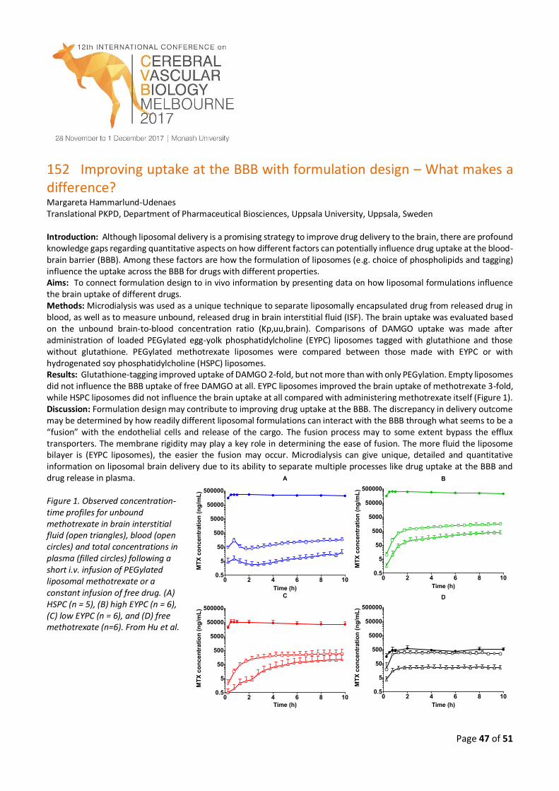
Page 47 of 51
152 Improving uptake at the BBB with formulation design – What makes a difference? Margareta Hammarlund-Udenaes Translational PKPD, Department of Pharmaceutical Biosciences, Uppsala University, Uppsala, Sweden Introduction: Although liposomal delivery is a promising strategy to improve drug delivery to the brain, there are profound knowledge gaps regarding quantitative aspects on how different factors can potentially influence drug uptake at the blood-brain barrier (BBB). Among these factors are how the formulation of liposomes (e.g. choice of phospholipids and tagging) influence the uptake across the BBB for drugs with different properties. Aims: To connect formulation design to in vivo information by presenting data on how liposomal formulations influence the brain uptake of different drugs. Methods: Microdialysis was used as a unique technique to separate liposomally encapsulated drug from released drug in blood, as well as to measure unbound, released drug in brain interstitial fluid (ISF). The brain uptake was evaluated based on the unbound brain-to-blood concentration ratio (Kp,uu,brain). Comparisons of DAMGO uptake was made after administration of loaded PEGylated egg-yolk phosphatidylcholine (EYPC) liposomes tagged with glutathione and those without glutathione. PEGylated methotrexate liposomes were compared between those made with EYPC or with hydrogenated soy phosphatidylcholine (HSPC) liposomes. Results: Glutathione-tagging improved uptake of DAMGO 2-fold, but not more than with only PEGylation. Empty liposomes did not influence the BBB uptake of free DAMGO at all. EYPC liposomes improved the brain uptake of methotrexate 3-fold, while HSPC liposomes did not influence the brain uptake at all compared with administering methotrexate itself (Figure 1). Discussion: Formulation design may contribute to improving drug uptake at the BBB. The discrepancy in delivery outcome may be determined by how readily different liposomal formulations can interact with the BBB through what seems to be a “fusion” with the endothelial cells and release of the cargo. The fusion process may to some extent bypass the efflux transporters. The membrane rigidity may play a key role in determining the ease of fusion. The more fluid the liposome bilayer is (EYPC liposomes), the easier the fusion may occur. Microdialysis can give unique, detailed and quantitative information on liposomal brain delivery due to its ability to separate multiple processes like drug uptake at the BBB and drug release in plasma. Figure 1. Observed concentration-time profiles for unbound methotrexate in brain interstitial fluid (open triangles), blood (open circles) and total concentrations in plasma (filled circles) following a short i.v. infusion of PEGylated liposomal methotrexate or a constant infusion of free drug. (A) HSPC (n = 5), (B) high EYPC (n = 6), (C) low EYPC (n = 6), and (D) free methotrexate (n=6). From Hu et al.
0 2 4 6 8 100.5
5
50
500
5000
50000
500000
Time (h)
MT
X c
on
cen
trati
on
(n
g/m
L)
A
0 2 4 6 8 100.5
5
50
500
5000
50000
500000
Time (h)
MT
X c
on
cen
trati
on
(n
g/m
L)
C
0 2 4 6 8 100.5
5
50
500
5000
50000
500000
B
Time (h)
MT
X c
on
cen
trati
on
(n
g/m
L)
0 2 4 6 8 100.5
5
50
500
5000
50000
500000
D
Time (h)
MT
X c
on
cen
trati
on
(n
g/m
L)
Page 48 of 51
153 Roche delivery platforms to overcome the blood-brain barrier
Eduard Urich1 1 Roche Pharmaceutical Research and Early Development, Roche Innovation Center Basel, Switzerland Although biotherapeutics have been successful in multiple disease areas such as oncology and inflammation and have vast potential for treating various brain disorders, their use has been limited due to low CNS exposure. This presentation will provide an overview of our recent achievements on the identification and refinement of novel antibody engineering platforms that are utilizing receptor-mediated transcytosis to cross the blood brain barrier (BBB). In particular, the Brain Shuttle technology platform will be described, which is designed to be engineered into a standard therapeutic antibody and other types of biologics for successful BBB transport. This system is based on a monovalent receptor binding mode to preserve the natural sorting of the receptor across the BBB. This will be illustrated with recent experimental data related to BBB biology, receptor trafficking and key properties of an efficient transport vector. The presentation will also cover the in vivo identification of transport peptides for large molecule delivery into the CNS using an animal model which is also applicable for the assessment of targeted delivery of biologics into the cerebrospinal fluid. These findings and the increased commitment to the BBB field we predict will have major implications for the development of biologics-based treatment of brain disorders in the near future.
Page 49 of 51
155 Translational scaling for intracerebroventricularly-administered BMN 250: predicting clinically effective dosing from animal models for the treatment of Sanfilippo Type B Anita Grover1, Emma McCullagh1, Mika Aoyagi-Scharber1, Stephen Maricich1, Jill CM Wait1, Andrew Melton1, Huiyu Zhou1, Roger Lawrence1, Heather Prill1, Brett Crawford1, Joshua Henshaw1 1BioMarin Pharmaceutical Inc., Novato, CA, United States Introduction: BMN 250 is being developed as an enzyme replacement therapy for the treatment of patients with Sanfilippo Type B (Mucopolysaccharidosis IIIB), a lysosomal storage disease in which patients lack alpha-N-acetylglucosaminidase (NAGLU) enzyme activity. BMN 250 is a fusion protein comprised of recombinant human NAGLU fused with truncated human insulin-like growth factor 2 via an amino acid linker to promote lysosomal delivery. Given the rarity of the disease and patient population, limited dose optimization can be studied during clinical trials. Therefore, Naglu-/- mouse and dog models are leveraged for support in determining a clinically effective dosing regimen for Sanfilippo Type B patients. Similarly, given the neurological disease pathophysiology, successful treatment will likely require replacement of enzyme activity in affected cells within the central nervous system via intracerebroventricular (ICV) administration directly into the cerebrospinal fluid (CSF). Aims: In this analysis, we assess various techniques for scaling between animal models of disease, incorporating the specific limitations of conducting studies in each animal model with the unique ICV administration route. Methods: Techniques assessed to predict a clinically effective dose include: allometric scaling of CSF pharmacokinetics (PK), dose scaling based on animal brain mass, and frequency scaling based on durability of pharmacodynamic (PD) response. Inter-individual variability was also considered in the analyses. Concordance of clinical predictions between the mouse and dog models was used to weigh the appropriateness of the candidate scaling methods, along with preliminary PK/PD data from an ongoing Phase 1/2 study of BMN 250 administered via the ICV route to Sanfillipo Type B patients. Results: Results suggest that clinical doses in the range of 300 mg weekly saturate the pharmacodynamic response to BMN 250 in patients. Discussion: Results from this work define a framework for translational scaling that can be leveraged to support dosing of future therapies administered through this increasingly applied route of administration.
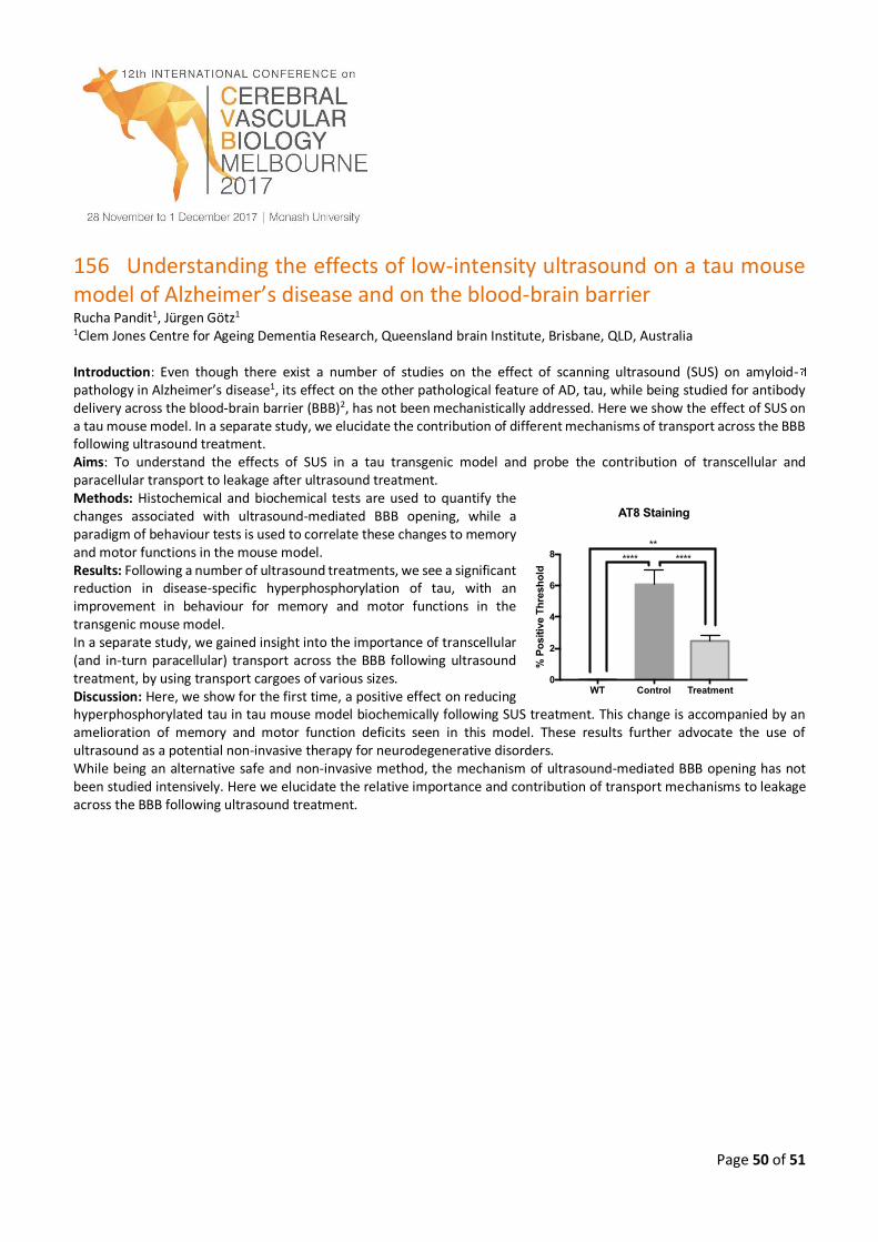
Page 50 of 51
156 Understanding the effects of low-intensity ultrasound on a tau mouse model of Alzheimer’s disease and on the blood-brain barrier Rucha Pandit1, Jürgen Götz1 1Clem Jones Centre for Ageing Dementia Research, Queensland brain Institute, Brisbane, QLD, Australia Introduction: Even though there exist a number of studies on the effect of scanning ultrasound (SUS) on amyloid- pathology in Alzheimer’s disease1, its effect on the other pathological feature of AD, tau, while being studied for antibody delivery across the blood-brain barrier (BBB)2, has not been mechanistically addressed. Here we show the effect of SUS on a tau mouse model. In a separate study, we elucidate the contribution of different mechanisms of transport across the BBB following ultrasound treatment. Aims: To understand the effects of SUS in a tau transgenic model and probe the contribution of transcellular and paracellular transport to leakage after ultrasound treatment. Methods: Histochemical and biochemical tests are used to quantify the changes associated with ultrasound-mediated BBB opening, while a paradigm of behaviour tests is used to correlate these changes to memory and motor functions in the mouse model. Results: Following a number of ultrasound treatments, we see a significant reduction in disease-specific hyperphosphorylation of tau, with an improvement in behaviour for memory and motor functions in the transgenic mouse model. In a separate study, we gained insight into the importance of transcellular (and in-turn paracellular) transport across the BBB following ultrasound treatment, by using transport cargoes of various sizes. Discussion: Here, we show for the first time, a positive effect on reducing hyperphosphorylated tau in tau mouse model biochemically following SUS treatment. This change is accompanied by an amelioration of memory and motor function deficits seen in this model. These results further advocate the use of ultrasound as a potential non-invasive therapy for neurodegenerative disorders. While being an alternative safe and non-invasive method, the mechanism of ultrasound-mediated BBB opening has not been studied intensively. Here we elucidate the relative importance and contribution of transport mechanisms to leakage across the BBB following ultrasound treatment.
WT Control Treatment0
2
4
6
8
% P
osit
ive T
hre
sh
old
AT8 Staining
**
**** ****
Page 51 of 51
157 Clearance of beta-amyloid is facilitated by apolipoprotein E and 1 circulating high-density lipoproteins in bioengineered human vessels Cheryl Wellington University of British Columbia, Canada Amyloid plaques, consisting of deposited beta-amyloid (Aβ), are a neuropathological hallmark of Alzheimer’s Disease (AD). Cerebral vessels play a major role in AD, as Aβ is cleared from the brain by pathways involving the cerebrovasculature, most AD patients have cerebrovascular amyloid (cerebral amyloid angiopathy (CAA), and cardiovascular risk factors increase dementia risk. Here we present a notable advance in vascular tissue engineering by generating the first functional 3-dimensioinal model of CAA in bioengineered human vessels. We show that lipoproteins including brain (apoE) and circulating (high-density lipoprotein) synergize to facilitate Aβ transport across bioengineered human cerebral vessels. These lipoproteins facilitate Aβ42 transport more efficiently than Aβ40, consistent with Aβ40 being the primary species that accumulates in CAA. Moreover, apoE4 is less effective than apoE2 in promoting Aβ transport, also consistent with the well-established role of apoE4 in Aβ deposition in AD.