10/8/2018 - uni.edu
TRANSCRIPT

10/8/2018
1
• The “typical” neuron is designed to receive neurotransmitter messages from other neurons.
• Sensory receptors, on the other hand, are specialized to receive sensory input from the outside world.
Vision The Eye and the Visual Receptors
Stimulus which activates visual receptors: light waves in the visible spectrum
Light waves are a small range of wavelengths
(~350-750 nanometers) of electromagnetic energy.
(Billionths of a meter)
(Book Fig. 6.7)
Of Macula
(book Fig. 6.1)
Transparent Cornea• Curve of cornea helps to focus
light waves on retina
• “Astigmatism” causes 2 focal points instead of 1
Green Muscular Iris & Black Pupil Hole• Muscles of iris under ANS control-
• Symp: dilate pupil
• Parasymp: constrict

10/8/2018
2
Of Macula
Lens becomes less
flexible later in life –
need reading glasses
Light travels
in straight lines
Looking Into Left Eye: Optic Disk or “Blind Spot”-axons exit eye to form optic nerve
You can
locate your
own blind
spot with
the demo
on p. 156.
Center (Macula) of Retina Needed for Detail Vision (Fovea especially)
Macular Degeneration
Loss of the critical
central region of retina
Smoking is #1
preventable cause
What you might see
Book Fig. 6.5 & 6.8
Rods vs Cones
• ~120 million/eye
• more in periphery
• very sensitive (low threshold)
• ~100 rods share same optic nerve fiber to brain
• night vision (scotopic vision)
• ~6 million/eye
• most in center, especially in the fovea
• Need bright light to reach threshold (photopic vision)
• have more private lines to brain- good for detail vision or “acuity”
• color vision
10/8/2018
3
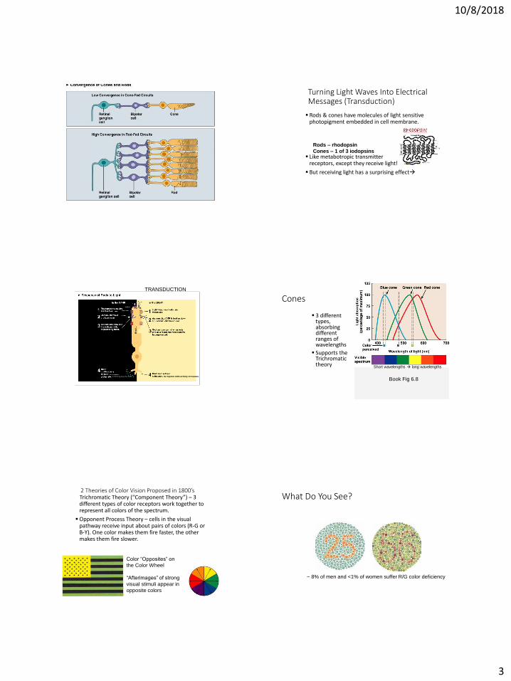
Turning Light Waves Into Electrical Messages (Transduction)
Rods & cones have molecules of light sensitive photopigment embedded in cell membrane.
Like metabotropic transmitter receptors, except they receive light!
But receiving light has a surprising effect
Rods – rhodopsin
Cones – 1 of 3 iodopsins
TRANSDUCTION
So bipolar cells activity increases.inhibitng bipolar cells
Cones
3 different types, absorbing different ranges of wavelengths
Supports the Trichromatic theory
Book Fig 6.8
Short wavelengths long wavelengths
2 Theories of Color Vision Proposed in 1800’sTrichromatic Theory (“Component Theory”) – 3 different types of color receptors work together to represent all colors of the spectrum.
Opponent Process Theory – cells in the visual pathway receive input about pairs of colors (R-G or B-Y). One color makes them fire faster, the other makes them fire slower.
Color “Opposites” on
the Color Wheel
“Afterimages” of strong
visual stimuli appear in
opposite colors
What Do You See?
~ 8% of men and <1% of women suffer R/G color deficiency
10/8/2018
4
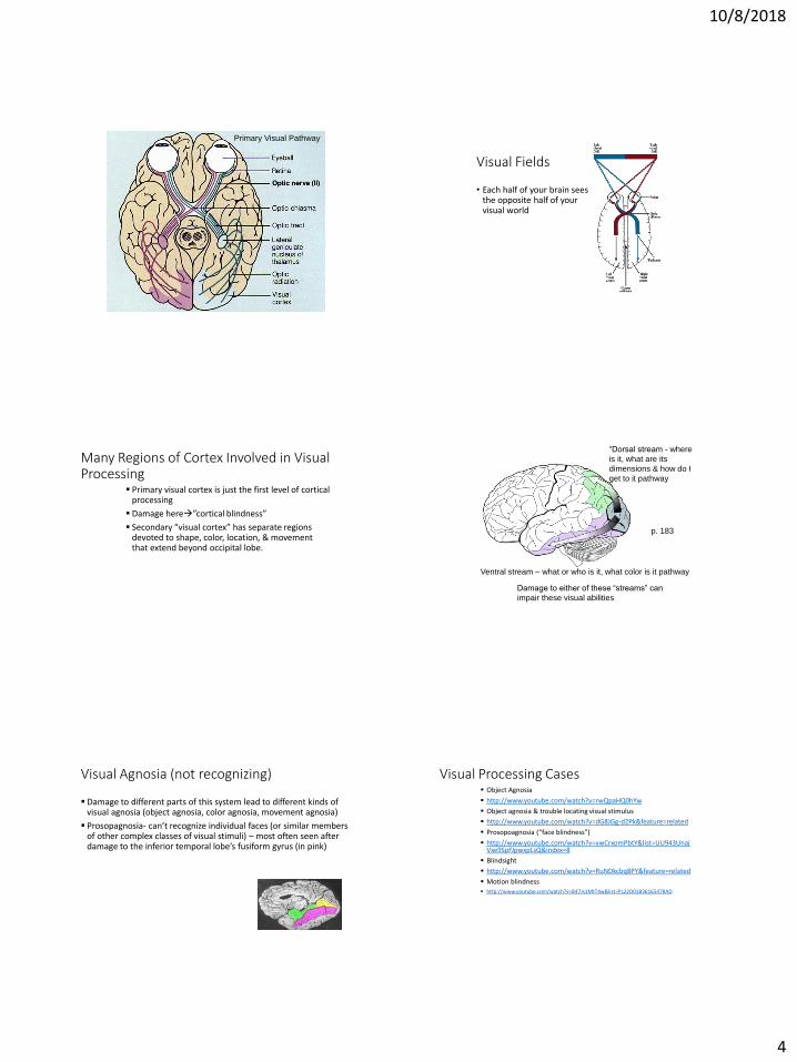
Primary Visual Pathway
Visual Fields
• Each half of your brain sees the opposite half of your visual world
Many Regions of Cortex Involved in Visual Processing
Primary visual cortex is just the first level of cortical processing
Damage here”cortical blindness”
Secondary “visual cortex” has separate regions devoted to shape, color, location, & movement that extend beyond occipital lobe.
“Dorsal stream - where
is it, what are its
dimensions & how do I
get to it pathway
Ventral stream – what or who is it, what color is it pathway
Damage to either of these “streams” can
impair these visual abilities
p. 183
Visual Agnosia (not recognizing)
Damage to different parts of this system lead to different kinds of visual agnosia (object agnosia, color agnosia, movement agnosia)
Prosopagnosia- can’t recognize individual faces (or similar members of other complex classes of visual stimuli) – most often seen after damage to the inferior temporal lobe’s fusiform gyrus (in pink)
Visual Processing Cases Object Agnosia
http://www.youtube.com/watch?v=rwQpaHQ0hYw
Object agnosia & trouble locating visual stimulus
http://www.youtube.com/watch?v=dG8JGg-d2Pk&feature=related
Prosopoagnosia (“face blindness”)
http://www.youtube.com/watch?v=vwCrxomPbtY&list=UU943UnajVxe9SpFJpwxpLsQ&index=8
Blindsight
http://www.youtube.com/watch?v=RuNDkcbq8PY&feature=related
Motion blindness
http://www.youtube.com/watch?v=B47Js1MtT4w&list=PL22D01B36165478AD

10/8/2018
5
• Hair cells in the auditory and vestibular systems mechanically open ion channels.
Sound Waves
Characteristics of Sound WavesBook Fig. 7.3
The Outer, Middle & Inner EarBook Fig. 7.2
Sound Triggered Movements in Ear
“Ossicles”
Outer ear
Middle ear
Inner earAuditory
Book Fig. 7.2 http://www.youtube.com/watch?v=r-
c5GpoD8wI
http://www.youtube.com/watch?v=P5gyQf2gVvA
Cross section of Cochlea
Basilar MembraneAuditory Nerve fibers
Book Fig. 7.2
http://www.youtube.com/watch?v=8wgfowbbTz0

10/8/2018
6
Organ of Cortihttp://www.youtube.com/watch?v=8wgfowbbTz0
Basilar Membrane
Tectorial Membrane
Book Fig. 7.2 Fluid Waves Traveling Thru Cochlea Cause Basilar Membrane Movement
• Where wave peaks varies with pitch & determines which hair cells will be stimulated.
Georg von Bekesy – 1961 Nobel Prize for his research on the traveling
waves in the cochlea.
http://www.youtube.com/watch?v=dyenMluFaUw&feature=related
http://www.youtube.com/watch?v=WO84KJyH5k8&feature=related
“Tonotopic” Relationship Between Place in Cochlea and Pitch
If our inner ear is working
perfectly we can hear
frequencies between
20-20,000 cps
“Place theory” best explains
pitch perception for the upper
80% of our hearing range &
explains freq specific hearing
losses
20-100 – frequency theory
100-4000 – volley theoryNear middle ear
Friction on tips of hair cells opens mechanically-gated K+ ion channels
K+ enters hair cells causing depolarization & transmitter release!
(fluid in cochlea has a different ion balance – disruption of that
balance can lead to hearing abnormal sounds (tinnitus))
Normal & “Trampled” Hair CellsExposed to Loud Sounds
• http://www.youtube.com/watch?v=Xo9bwQuYrRo (dancing hair cell)
• http://www.youtube.com/watch?v=ulAISCEQzRo
• (stereocilia)
Sound Localization• Brain processes time &
intensity & phase differences in what the right & left ears hear.
• Sound from right arrives sooner and louder in the right ear.

10/8/2018
7
Note: Input from each
ear goes to both sides of
brain but more strongly
to contralateral side.
Brainstem areas
involved in quick sound
localization and
auditory reflexes.
VIIIVIII
“Tonotopic” Map
in cortex &
cochlea
Book Fig. 7.6
Primary auditory cortex surrounded by higher level processing areas, analyzing more
complex sequences or combinations of sounds. Seems to be separate regions devoted
to what the stimulus is & where the stimulus came from.
Types of Deafness• ~250 million with hearing impairments; only a fraction are
completely deaf
• Conductive or Middle Ear Deafness – auditory stimulus does not pass normally through middle ear to cochlea
• Nerve/Neural or Inner Ear Deafness – due to damage to inner ear hair cells or auditory nerve due to:
• Genetics• Perinatal problems (illness during pregnancy, hypoxia during
birth, FAS)• Illness (meningitis, MS, Meniere’s)• Ototoxic drugs (quinine, some antibiotics, high doses of
NSAIDS, nicotine),• Loud sounds
• http://www.youtube.com/watch?v=9g0yThhJcxY
• Mosquito tones
Cochlear Implant can take the place of missing or damaged hair cells as long as auditory nerve fibers still run from cochlea to brain.