11. heterotopic ossification and venous thromboembolism post … · 2020-02-12 · heterotopic...
TRANSCRIPT
erabi.ca Version 13.0
11. Heterotopic Ossification and Venous
Thromboembolism Post Acquired Brain Injury
Robert Teasell MD FRCPC Shannon Janzen MSc
Daniel McCarthy MD (cand) Caitlin Cassidy MD
Magdalena Mirkowski MSc MScOT OT Reg.(Ont.) Shawn Marshall MSc MD FRCPC
Nora Cullen MSc MD FRCPC
erabi.ca Version 13.0
Key Points
Etidronate Disodium may prevent the development of heterotopic ossification in individuals with ABI.
Forceful joint manipulation may prevent bony ankylosis post ABI and may increase range of motion in joints affected by heterotopic ossification.
Radiotherapy and shock wave therapy may be effective for the treatment of pain and/or range of motion associated with heterotopic ossification in ABI populations.
Surgical excision of heterotopic ossification may improve range of motion and functional ability. Earlier surgical excision may not increase the risk of further heterotopic ossification, and may decrease surgical complications and increase functional outcomes. Sequential compression devices may not reduce the risk of developing deep vein thrombosis or pulmonary embolism post ABI. Intermittent compression devices do not aggravate intracranial hemodynamics in patients with severe ABI. Intermittent pneumatic compression devices and low molecular weight heparin may have a similar effect in terms of the prevention of deep vein thrombosis post ABI when compared to each other. Administration of pharmacological thromboembolic prophylaxis within the first 72 hours post ABI may be effective for reducing the risk of developing venous thromboembolism. Enoxaparin is effective for the prevention of venous thromboembolism development after elective neurosurgery and has not been found to cause excessive bleeding.
erabi.ca Version 13.0
Table of Contents
11.0 Introduction .................................................................................................................................... 1
11.1 Heterotopic Ossification ................................................................................................................. 1
11.1.1 Formation of Heterotopic Ossification Post Head Injury ......................................................... 1
11.1.2 Clinical Presentation of Heterotopic Ossification .................................................................... 2
11.1.3 Diagnostic Testing for Heterotopic Ossification ...................................................................... 2
11.1.4 Prevention of Heterotopic Ossification ................................................................................... 3
11.1.4.1 Continuous Passive Range of Motion ................................................................................. 3 11.1.4.2 Nonsteroidal Anti-Inflammatory Drugs .............................................................................. 3 11.1.4.3 Etidronate Disodium .......................................................................................................... 3
11.1.5 Treatment of Heterotopic Ossification .................................................................................... 4
11.1.5.1 Physiotherapy and Range of Motion Exercises ................................................................... 4 11.1.5.2 Radiotherapy and Extracorporeal Shock Wave Therapy ..................................................... 6 11.1.5.3 Surgical Interventions ........................................................................................................ 7
11.2 Venous Thromboembolism ........................................................................................................... 13
11.2.1 Incidence of Venous Thromboembolism Post-Head Injury ................................................... 13
11.2.2 Risk Factors for Venous Thromboembolism .......................................................................... 13
11.2.3 Clinical Presentation of Deep Vein Thrombosis and Pulmonary Embolism ........................... 14
11.2.4 Diagnostic Testing for Deep Vein Thrombosis ....................................................................... 14
11.2.4.1 Venous Ultrasound .......................................................................................................... 14 11.2.4.2 Venography ..................................................................................................................... 14 11.2.4.3 D-dimer Assay.................................................................................................................. 14
11.2.5 Diagnostic Testing for Pulmonary Embolism ......................................................................... 15
11.2.5.1 Ventilation/Perfusion Scanning ........................................................................................ 15 11.2.5.2 Computed Tomography Pulmonary Angiogram ................................................................ 16
11.2.6 Prevention of Venous Thromboembolism Post ABI .............................................................. 16
11.2.6.1 Non-Pharmacological Prophylaxis .................................................................................... 16 11.2.6.2 Pharmacological Prophylaxis ........................................................................................... 18
11.3 Conclusions ................................................................................................................................... 26
11.4 Summary ....................................................................................................................................... 27
11.5 References .................................................................................................................................... 28

erabi.ca Version 13.0
This review has been prepared based on the scientific and professional information available up to December 2018. The ERABI information is provided for informational and educational purposes only. If you have or suspect you have a health problem, you should consult your health care provider. The ERABI contributors shall not be liable for any damages, claims, liabilities, costs, or obligations arising from the use or misuse of this material.
Teasell R, Janzen S, McCarthy D, Cassidy C, Mirkowski M, Marshall S, Cullen N. (2019). Heterotopic Ossification and Venous Thromboembolism Post Acquired Brain Injury. In Teasell R, Cullen N, Marshall S, Janzen S, Faltynek P, Bayley M, editors. Evidence-Based Review of Moderate to Severe Acquired Brain Injury. Version 13.0: p1-36.
erabi.ca i Version 13.0
Abbreviations
ABI Acquired Brain Injury CTPA Computed Tomography Pulmonary Angiogram DVT Deep Venous Thrombosis EHDP Etidronate Disodium (ethane-1-hydroxy-1, 1-diphosphonate) GCS Glasgow Coma Scale HO Heterotopic Ossification LMWH Low-Molecular-Weight Heparin PCT Prospective Controlled Trial PE Pulmonary Embolism SCD Sequential Compression Devices TBI Traumatic Brain Injury V/Q Ventilation/Perfusion VTE Venous Thromboembolism
Heterotopic Ossification 1
Heterotopic Ossification and Venous Thromboembolism Post Acquired Brain
Injury
11.0 Introduction This module aims to provide information regarding what is currently known about the etiology and treatment of heterotopic ossification (HO) and venous thromboembolism (VTE) following acquired brain injury (ABI). Additionally, it is worth noting that the majority of interventions discussed for VTE are preventative measures. Information on diagnostic tools is also provided for both complications.
11.1 Heterotopic Ossification HO is the formation of pathologic bone within soft tissues, often muscle tissues, where bone formation does not usually occur (Watanabe & Sant, 2001). The incidence of HO in patients with traumatic brain injury (TBI) has been reported to range from 11% to 77% but is clinically relevant in 10-20% (Dizdar et al., 2013; Garland et al., 1980; Rogers, 1988; Sarafis et al., 1999; Sazbon et al., 1981; Simonsen et al., 2007; Zychowicz, 2013). Risk factors include skeletal trauma, spasticity, diffuse axonal injury, mechanical ventilation, prolonged immobilization, and injury severity (Huang et al., 2018; Moreta & de los Mozos, 2014). HO is often quite painful and limits joint mobility; the restricted joint range of motion may exacerbate disability and impede progress towards desired rehabilitation goals.
11.1.1 Formation of Heterotopic Ossification Post Head Injury The pathophysiology of HO is not fully understood. Mesenchymal stem cells are pleuripotent cells that can differentiate into cells capable of generating cartilage, bone, muscle, tendons, ligaments, or fat (Pape et al., 2004). It is thought that they play a pivotal role in the development of HO (Williams et al., 1999). HO development begins with the formation of osteoid periarticularly and intramuscularly and progresses to full calcification within a matter of weeks (Pape et al., 2001). Over the next few months, the calcified osteoid remodels into well-organized trabecular bone at which point it is considered to have matured (Pape et al., 2001). Several months after the initial trauma, patients with HO begin to experience restricted range of motion, pain, and potentially ankylosis (Banovac & Gonzalez, 1997; Garland et al., 1980). The bony lesion has been found to have a high metabolic rate, with a rate of bone formation more than three times greater than that of normal bone, and an osteoclastic density of more than twice the density found in normal bone (Puzas et al., 1987). There are neurogenic factors contributing to HO, although the mechanisms are not entirely clear yet (Brady et al., 2018; Hurvitz et al., 1992; Moreta & de los Mozos, 2014; Pape et al., 2001; Pape et al., 2004). It has also been noted that circulating factors promoting HO may be present in patients with head injuries (Pape et al., 2004). Many studies have shown enhanced osteogenesis in patients sustaining TBI (Trentz et al., 2005). The presence of certain hormonal factors early post injury influences the stimulation of osteoprogenitors within skeletal muscles (Ivanhoe et al., 2012). Further, tissue hypoxia, sympathetic changes, immobilization, remobilization, and spasticity are additional risk factors (Ivanhoe et al., 2012). Accelerated fracture healing and HO are well documented phenomena in these patients (Bidner et al., 1990; Keret et al., 1990).
Heterotopic Ossification 2
11.1.2 Clinical Presentation of Heterotopic Ossification Among individuals with TBI, the most common sites of HO are the soft tissues around the hip, elbow, shoulder, and knee (Garland, 1991; Garland et al., 1980; van Kampen et al., 2011; Vanden Bossche & Vanderstraeten, 2005). The hip is the most frequent site of ossification (Dizdar et al., 2013; Vanden Bossche & Vanderstraeten, 2005), with total ankylosis of the joint occurring in 5-16% of affected hips (Stover et al., 1991). HO of the shoulder has been found to affect 5% of individuals with a brain injury (Cipriano et al., 2009), while the knee is a less common site for HO following a head injury (Sarafis et al., 1999). When HO is present in the knee, it is usually seen medially (Hosalkar et al., 2013). The distribution of HO around the elbow occurs most commonly either anteriorly in the flexor muscles or posteriorly in the extensors (Sarafis et al., 1999). Of the joints affected by HO after head injury, ankylosis is most likely to occur in the posterior elbow (Garland et al., 1980). The onset of HO has been reported to vary between two and three weeks post injury (Watanabe & Sant, 2001). More recently, clinical signs and symptoms have been reported to develop 3-12 weeks post injury (Vanden Bossche & Vanderstraeten, 2005; Zychowicz, 2013). Pape and colleagues (2004) noted that clinical examination in the setting of HO may reveal a swollen, warm, painful joint which is often associated with decreased range of motion. The earliest sign is typically a loss of range of motion in the involved joint (Watanabe & Sant, 2001). Other indicators include erythema, palpation of a periarticular mass, and fever (Varghese, 1992). Because of the association with fever, it is sometimes difficult to differentiate HO from infection (Citta-Pietrolungo et al., 1992; Garland, 1991; Garland et al., 1980). Moreover, the clinical picture may be confused with deep vein thrombosis (DVT), local trauma, or fracture (Buschbacher, 1992; Jensen et al., 1987). HO will then progress from these initial symptoms into a mass with stiffness and induration (Zychowicz, 2013). Potential complications of HO include compression of blood vessels and nerves, breakdown of associated tissue, restricted motion, and loss of function (Zychowicz, 2013).
11.1.3 Diagnostic Testing for Heterotopic Ossification During the initial presentation, plain radiographs may be negative and will usually remain normal until ossification begins at approximately 4-6 weeks post injury. Serum levels of alkaline phosphatase, a glycoprotein in the plasma membrane of osteoblasts, and the erythrocyte sedimentation rate may become elevated early on but are non-specific. The triple phase technicium-99 bone scan remains the diagnostic gold standard. The test is positive if there is increased uptake during the first and second phases of the study. It typically becomes positive when clinical features appear (i.e., before an x-ray would be positive). In the HO literature there are several classification systems, with the Brooker classification system being one of the most widely used. The Brooker Classification system is typically used to classify ectopic-bone formation after total hip replacement. The system is based on anteroposterior radiographs of the pelvis and the categorization of the progression of HO into classes (Brooker et al., 1973). Brooker et al. (1973) define Class I as islands of bone within soft tissues about the hip; Class II as bone spurs from the pelvis or proximal end of the femur with at least 1cm between opposing bone surfaces; Class III as bone spurs from pelvis or proximal end of the femur, reducing space between opposing bone surfaces to less than 1cm; and Class IV as apparent bony ankylosis of hip. This classification system has been criticized for high inter-observer variability and poorly addressing information relevant to cases of neurogenic HO (Della Valle et al., 2002; Mavrogenis et al., 2012; Toom et al., 2005).
Heterotopic Ossification 3
11.1.4 Prevention of Heterotopic Ossification The development of HO in patients with TBI is hard to predict, making preventative treatment challenging. Prophylactic treatment options for HO include range of motion exercises, nonsteroidal anti-inflammatory medications, low-dose radiation, and etidronate disodium (EHDP) (Watanabe & Sant, 2001). Research on the efficacy of these treatments in patients with TBI is limited, and generally focused on secondary prevention after treatment of established HO. Further investigation is needed to develop more effective preventative treatments (Brady et al., 2018).
11.1.4.1 Continuous Passive Range of Motion Continuous passive motion devices have shown promising results in maintaining range of motion following total knee replacement (Nadler et al., 1993; Salter, 1996). Animal data shows that continuous passive motion does not increase the progression of HO (van Susante et al., 1996). Moreover, there is little human research evidence that HO is worsened by passive range of motion (Linan et al., 2001). Continuous passive range of motion for HO in patients with TBI has only been evaluated in combination with surgical excision, which is discussed later. Further research is needed in the ABI population.
11.1.4.2 Nonsteroidal Anti-Inflammatory Drugs The evidence for nonsteroidal anti-inflammatory medications as prophylactic treatment for HO stems mostly from the use of indomethacin or ibuprofen in patients following total hip arthroplasty (Fransen & Neal, 2004; Kjaersgaard-Andersen & Schmidt, 1986; Ritter & Sieber, 1985). In patients with spinal cord injuries, preliminary studies of indomethacin or selective Rofecoxib (a selective COX-2 inhibitor) have shown decreased incidence of HO (Banovac et al., 2004; Banovac et al., 2001). Although it has been reported that the prophylactic use of these medications significantly decreases HO formation following total hip arthroplasty, it is not known if they have the same effect in the ABI population.
11.1.4.3 Etidronate Disodium The use of EHDP, a bisphosphate, in the prophylaxis and treatment of HO is not well studied in the ABI population. EHDP works by preventing the aggregation, growth, and mineralization of calcium hydroxyapatite crystals which are essential for bone formation. EHDP may potentially delay fracture healing, as long-term use has been associated with osteomalacia.
Heterotopic Ossification 4
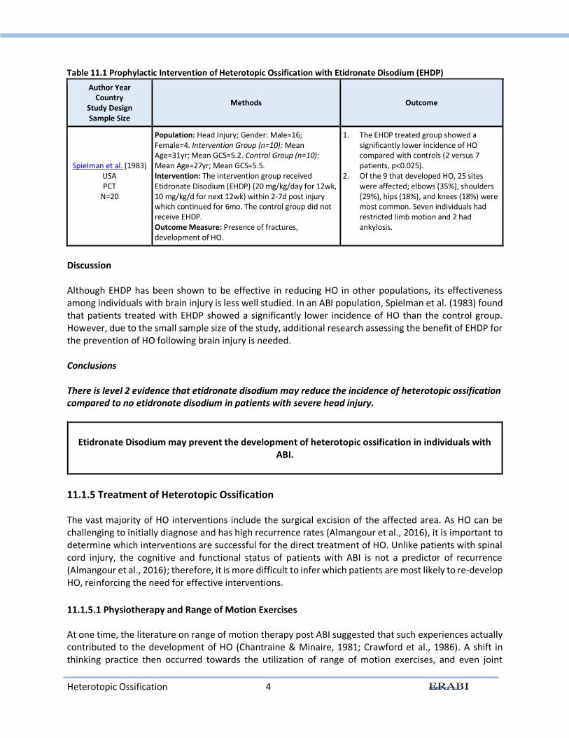
Table 11.1 Prophylactic Intervention of Heterotopic Ossification with Etidronate Disodium (EHDP)
Author Year Country
Study Design Sample Size
Methods Outcome
Spielman et al. (1983) USA PCT
N=20
Population: Head Injury; Gender: Male=16; Female=4. Intervention Group (n=10): Mean Age=31yr; Mean GCS=5.2. Control Group (n=10): Mean Age=27yr; Mean GCS=5.5. Intervention: The intervention group received Etidronate Disodium (EHDP) (20 mg/kg/day for 12wk, 10 mg/kg/d for next 12wk) within 2-7d post injury which continued for 6mo. The control group did not receive EHDP. Outcome Measure: Presence of fractures, development of HO.
1. The EHDP treated group showed a significantly lower incidence of HO compared with controls (2 versus 7 patients, p<0.025).
2. Of the 9 that developed HO, 25 sites were affected; elbows (35%), shoulders (29%), hips (18%), and knees (18%) were most common. Seven individuals had restricted limb motion and 2 had ankylosis.
Discussion Although EHDP has been shown to be effective in reducing HO in other populations, its effectiveness among individuals with brain injury is less well studied. In an ABI population, Spielman et al. (1983) found that patients treated with EHDP showed a significantly lower incidence of HO than the control group. However, due to the small sample size of the study, additional research assessing the benefit of EHDP for the prevention of HO following brain injury is needed. Conclusions There is level 2 evidence that etidronate disodium may reduce the incidence of heterotopic ossification compared to no etidronate disodium in patients with severe head injury.
Etidronate Disodium may prevent the development of heterotopic ossification in individuals with
ABI.
11.1.5 Treatment of Heterotopic Ossification The vast majority of HO interventions include the surgical excision of the affected area. As HO can be challenging to initially diagnose and has high recurrence rates (Almangour et al., 2016), it is important to determine which interventions are successful for the direct treatment of HO. Unlike patients with spinal cord injury, the cognitive and functional status of patients with ABI is not a predictor of recurrence (Almangour et al., 2016); therefore, it is more difficult to infer which patients are most likely to re-develop HO, reinforcing the need for effective interventions.
11.1.5.1 Physiotherapy and Range of Motion Exercises At one time, the literature on range of motion therapy post ABI suggested that such experiences actually contributed to the development of HO (Chantraine & Minaire, 1981; Crawford et al., 1986). A shift in thinking practice then occurred towards the utilization of range of motion exercises, and even joint
Heterotopic Ossification 5
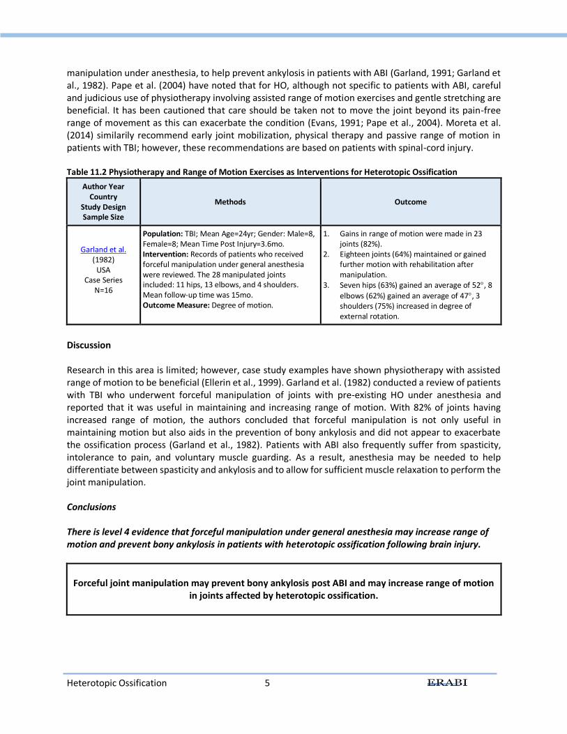
manipulation under anesthesia, to help prevent ankylosis in patients with ABI (Garland, 1991; Garland et al., 1982). Pape et al. (2004) have noted that for HO, although not specific to patients with ABI, careful and judicious use of physiotherapy involving assisted range of motion exercises and gentle stretching are beneficial. It has been cautioned that care should be taken not to move the joint beyond its pain-free range of movement as this can exacerbate the condition (Evans, 1991; Pape et al., 2004). Moreta et al. (2014) similarily recommend early joint mobilization, physical therapy and passive range of motion in patients with TBI; however, these recommendations are based on patients with spinal-cord injury. Table 11.2 Physiotherapy and Range of Motion Exercises as Interventions for Heterotopic Ossification
Author Year Country
Study Design Sample Size
Methods Outcome
Garland et al. (1982)
USA Case Series
N=16
Population: TBI; Mean Age=24yr; Gender: Male=8, Female=8; Mean Time Post Injury=3.6mo. Intervention: Records of patients who received forceful manipulation under general anesthesia were reviewed. The 28 manipulated joints included: 11 hips, 13 elbows, and 4 shoulders. Mean follow-up time was 15mo. Outcome Measure: Degree of motion.
1. Gains in range of motion were made in 23 joints (82%).
2. Eighteen joints (64%) maintained or gained further motion with rehabilitation after manipulation.
3. Seven hips (63%) gained an average of 52, 8
elbows (62%) gained an average of 47, 3 shoulders (75%) increased in degree of external rotation.
Discussion Research in this area is limited; however, case study examples have shown physiotherapy with assisted range of motion to be beneficial (Ellerin et al., 1999). Garland et al. (1982) conducted a review of patients with TBI who underwent forceful manipulation of joints with pre-existing HO under anesthesia and reported that it was useful in maintaining and increasing range of motion. With 82% of joints having increased range of motion, the authors concluded that forceful manipulation is not only useful in maintaining motion but also aids in the prevention of bony ankylosis and did not appear to exacerbate the ossification process (Garland et al., 1982). Patients with ABI also frequently suffer from spasticity, intolerance to pain, and voluntary muscle guarding. As a result, anesthesia may be needed to help differentiate between spasticity and ankylosis and to allow for sufficient muscle relaxation to perform the joint manipulation. Conclusions There is level 4 evidence that forceful manipulation under general anesthesia may increase range of motion and prevent bony ankylosis in patients with heterotopic ossification following brain injury.
Forceful joint manipulation may prevent bony ankylosis post ABI and may increase range of motion
in joints affected by heterotopic ossification.
Heterotopic Ossification 6
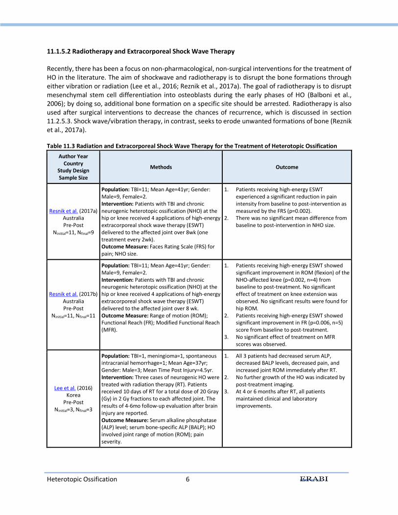
11.1.5.2 Radiotherapy and Extracorporeal Shock Wave Therapy Recently, there has been a focus on non-pharmacological, non-surgical interventions for the treatment of HO in the literature. The aim of shockwave and radiotherapy is to disrupt the bone formations through either vibration or radiation (Lee et al., 2016; Reznik et al., 2017a). The goal of radiotherapy is to disrupt mesenchymal stem cell differentiation into osteoblasts during the early phases of HO (Balboni et al., 2006); by doing so, additional bone formation on a specific site should be arrested. Radiotherapy is also used after surgical interventions to decrease the chances of recurrence, which is discussed in section 11.2.5.3. Shock wave/vibration therapy, in contrast, seeks to erode unwanted formations of bone (Reznik et al., 2017a). Table 11.3 Radiation and Extracorporeal Shock Wave Therapy for the Treatment of Heterotopic Ossification
Author Year Country
Study Design Sample Size
Methods Outcome
Resnik et al. (2017a) Australia Pre-Post
Ninitial=11, Nfinal=9
Population: TBI=11; Mean Age=41yr; Gender: Male=9, Female=2. Intervention: Patients with TBI and chronic neurogenic heterotopic ossification (NHO) at the hip or knee received 4 applications of high-energy extracorporeal shock wave therapy (ESWT) delivered to the affected joint over 8wk (one treatment every 2wk). Outcome Measure: Faces Rating Scale (FRS) for pain; NHO size.
1. Patients receiving high-energy ESWT experienced a significant reduction in pain intensity from baseline to post-intervention as measured by the FRS (p=0.002).
2. There was no significant mean difference from baseline to post-intervention in NHO size.
Resnik et al. (2017b) Australia Pre-Post
Ninitial=11, Nfinal=11
Population: TBI=11; Mean Age=41yr; Gender: Male=9, Female=2. Intervention: Patients with TBI and chronic neurogenic heterotopic ossification (NHO) at the hip or knee received 4 applications of high-energy extracorporeal shock wave therapy (ESWT) delivered to the affected joint over 8 wk. Outcome Measure: Range of motion (ROM); Functional Reach (FR); Modified Functional Reach (MFR).
1. Patients receiving high-energy ESWT showed significant improvement in ROM (flexion) of the NHO-affected knee (p=0.002, n=4) from baseline to post-treatment. No significant effect of treatment on knee extension was observed. No significant results were found for hip ROM.
2. Patients receiving high-energy ESWT showed significant improvement in FR (p=0.006, n=5) score from baseline to post-treatment.
3. No significant effect of treatment on MFR scores was observed.
Lee et al. (2016) Korea
Pre-Post Ninitial=3, Nfinal=3
Population: TBI=1, meningioma=1, spontaneous intracranial hemorrhage=1; Mean Age=37yr; Gender: Male=3; Mean Time Post Injury=4.5yr. Intervention: Three cases of neurogenic HO were treated with radiation therapy (RT). Patients received 10 days of RT for a total dose of 20 Gray (Gy) in 2 Gy fractions to each affected joint. The results of 4-6mo follow-up evaluation after brain injury are reported. Outcome Measure: Serum alkaline phosphatase (ALP) level; serum bone-specific ALP (BALP); HO involved joint range of motion (ROM); pain severity.
1. All 3 patients had decreased serum ALP, decreased BALP levels, decreased pain, and increased joint ROM immediately after RT.
2. No further growth of the HO was indicated by post-treatment imaging.
3. At 4 or 6 months after RT, all patients maintained clinical and laboratory improvements.
Heterotopic Ossification 7
Discussion Recent studies have shown that there are potential alternatives for the treatment of HO other than surgery, which is invasive and can be inefficient in terms of recurrence rates (Lee et al., 2016). A study by Resnik et al. (2017a) demonstrated that extracorporeal shock wave therapy was successful in reducing pain associated with HO. Patients experienced a significant improvement in the flexion range of motion of affected knees, however, no significant effects were seen on knee extension or hip range of motions (Reznik et al., 2017b). A study examining the effects of radiation therapy on HO also suggested positive results in terms of reduced pain and blood plasma levels of HO markers (Lee et al., 2016). Furthermore, results demonstrated immediate beneficial effects of radiation therapy on range of motion, in addition to the cessation of HO formation post treatment. Conclusions There is level 4 evidence that radiation therapy may prevent further formation of heterotopic ossification in ABI populations. There is level 4 evidence that radiation therapy may improve range of motion in joints affected by heterotopic ossification in ABI populations.
There is level 4 evidence that extracorporeal shock wave therapy may reduce pain associated with heterotopic ossification in ABI populations.
Radiotherapy and shock wave therapy may be effective for the treatment of pain and/or range of
motion associated with heterotopic ossification in ABI populations.
11.1.5.3 Surgical Interventions Surgical excision of the heterotopic bone is the treatment of choice for those in whom HO has generated marked functional impairment or ulcers in the skin due to deformity (Brady et al., 2018; Moreta & de los Mozos, 2014; Watanabe & Sant, 2001). A recent systematic review found that as high as 55% of those diagnosed with neurogenic HO required surgery (Almangour et al., 2016). It had been recommended, based on expert opinion, that surgical intervention be considered only 12 to 18 months after HO initiation to ensure that the bone tissue has matured, and to reduce the likelihood of HO recurrence (Garland, 1991; Sazbon et al., 1981); however, earlier surgical intervention has shown decreased osteopenia and ankylosis, without increased rates of recurrence (Almangour et al., 2016; Genet et al., 2012; Moreta & de los Mozos, 2014). There is some indication that EHDP and nonsteroidal anti-inflammatory medications may be useful in preventing HO recurrence following surgical excision (Watanabe & Sant, 2001). Further studies are needed to corroborate this.
Heterotopic Ossification 8
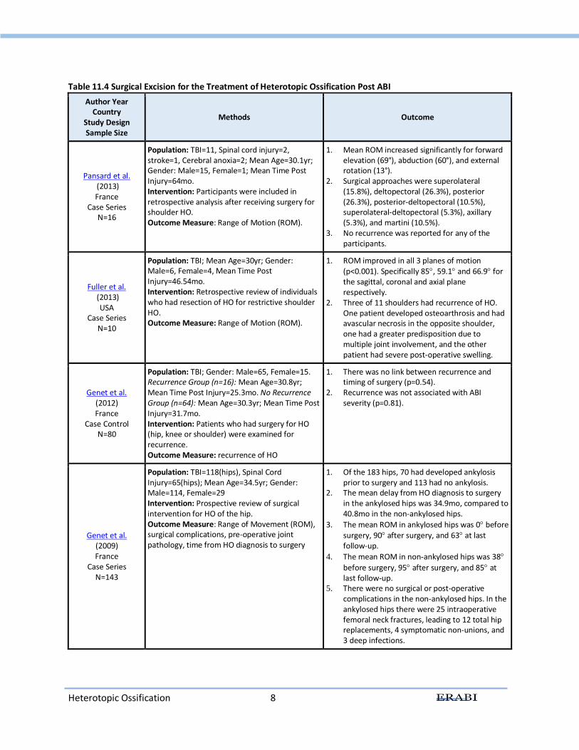
Table 11.4 Surgical Excision for the Treatment of Heterotopic Ossification Post ABI
Author Year Country
Study Design Sample Size
Methods Outcome
Pansard et al. (2013) France
Case Series N=16
Population: TBI=11, Spinal cord injury=2, stroke=1, Cerebral anoxia=2; Mean Age=30.1yr; Gender: Male=15, Female=1; Mean Time Post Injury=64mo. Intervention: Participants were included in retrospective analysis after receiving surgery for shoulder HO. Outcome Measure: Range of Motion (ROM).
1. Mean ROM increased significantly for forward elevation (69°), abduction (60°), and external rotation (13°).
2. Surgical approaches were superolateral (15.8%), deltopectoral (26.3%), posterior (26.3%), posterior-deltopectoral (10.5%), superolateral-deltopectoral (5.3%), axillary (5.3%), and martini (10.5%).
3. No recurrence was reported for any of the participants.
Fuller et al. (2013)
USA Case Series
N=10
Population: TBI; Mean Age=30yr; Gender: Male=6, Female=4, Mean Time Post Injury=46.54mo. Intervention: Retrospective review of individuals who had resection of HO for restrictive shoulder HO. Outcome Measure: Range of Motion (ROM).
1. ROM improved in all 3 planes of motion (p<0.001). Specifically 85, 59.1 and 66.9 for the sagittal, coronal and axial plane respectively.
2. Three of 11 shoulders had recurrence of HO. One patient developed osteoarthrosis and had avascular necrosis in the opposite shoulder, one had a greater predisposition due to multiple joint involvement, and the other patient had severe post-operative swelling.
Genet et al. (2012) France
Case Control N=80
Population: TBI; Gender: Male=65, Female=15. Recurrence Group (n=16): Mean Age=30.8yr; Mean Time Post Injury=25.3mo. No Recurrence Group (n=64): Mean Age=30.3yr; Mean Time Post Injury=31.7mo. Intervention: Patients who had surgery for HO (hip, knee or shoulder) were examined for recurrence. Outcome Measure: recurrence of HO
1. There was no link between recurrence and timing of surgery (p=0.54).
2. Recurrence was not associated with ABI severity (p=0.81).
Genet et al. (2009) France
Case Series N=143
Population: TBI=118(hips), Spinal Cord Injury=65(hips); Mean Age=34.5yr; Gender: Male=114, Female=29 Intervention: Prospective review of surgical intervention for HO of the hip. Outcome Measure: Range of Movement (ROM), surgical complications, pre-operative joint pathology, time from HO diagnosis to surgery
1. Of the 183 hips, 70 had developed ankylosis prior to surgery and 113 had no ankylosis.
2. The mean delay from HO diagnosis to surgery in the ankylosed hips was 34.9mo, compared to 40.8mo in the non-ankylosed hips.
3. The mean ROM in ankylosed hips was 0 before surgery, 90 after surgery, and 63 at last follow-up.
4. The mean ROM in non-ankylosed hips was 38
before surgery, 95 after surgery, and 85 at last follow-up.
5. There were no surgical or post-operative complications in the non-ankylosed hips. In the ankylosed hips there were 25 intraoperative femoral neck fractures, leading to 12 total hip replacements, 4 symptomatic non-unions, and 3 deep infections.
Heterotopic Ossification 9
Author Year Country
Study Design Sample Size
Methods Outcome
Fuller et al. (2005)
USA Case Series
N=17
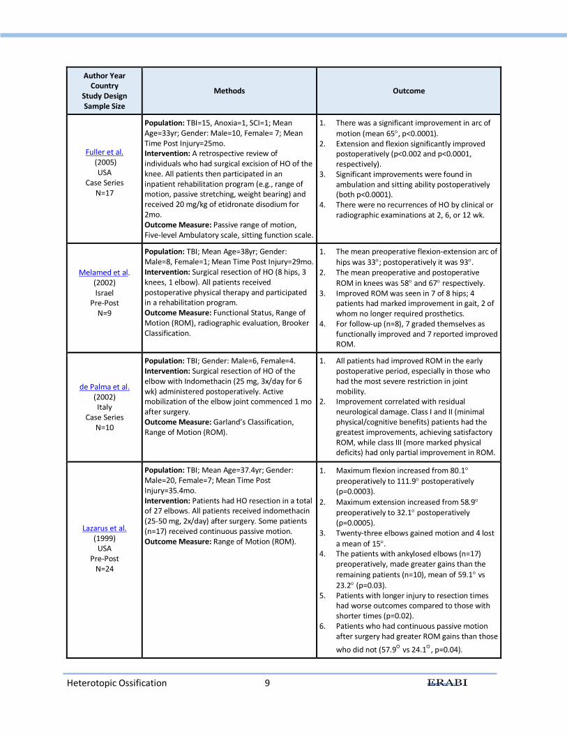
Population: TBI=15, Anoxia=1, SCI=1; Mean Age=33yr; Gender: Male=10, Female= 7; Mean Time Post Injury=25mo. Intervention: A retrospective review of individuals who had surgical excision of HO of the knee. All patients then participated in an inpatient rehabilitation program (e.g., range of motion, passive stretching, weight bearing) and received 20 mg/kg of etidronate disodium for 2mo. Outcome Measure: Passive range of motion, Five-level Ambulatory scale, sitting function scale.
1. There was a significant improvement in arc of
motion (mean 65, p<0.0001). 2. Extension and flexion significantly improved
postoperatively (p<0.002 and p<0.0001, respectively).
3. Significant improvements were found in ambulation and sitting ability postoperatively (both p<0.0001).
4. There were no recurrences of HO by clinical or radiographic examinations at 2, 6, or 12 wk.
Melamed et al. (2002) Israel
Pre-Post N=9
Population: TBI; Mean Age=38yr; Gender: Male=8, Female=1; Mean Time Post Injury=29mo. Intervention: Surgical resection of HO (8 hips, 3 knees, 1 elbow). All patients received postoperative physical therapy and participated in a rehabilitation program. Outcome Measure: Functional Status, Range of Motion (ROM), radiographic evaluation, Brooker Classification.
1. The mean preoperative flexion-extension arc of
hips was 33; postoperatively it was 93. 2. The mean preoperative and postoperative
ROM in knees was 58 and 67 respectively. 3. Improved ROM was seen in 7 of 8 hips; 4
patients had marked improvement in gait, 2 of whom no longer required prosthetics.
4. For follow-up (n=8), 7 graded themselves as functionally improved and 7 reported improved ROM.
de Palma et al. (2002) Italy
Case Series N=10
Population: TBI; Gender: Male=6, Female=4. Intervention: Surgical resection of HO of the elbow with Indomethacin (25 mg, 3x/day for 6 wk) administered postoperatively. Active mobilization of the elbow joint commenced 1 mo after surgery. Outcome Measure: Garland’s Classification, Range of Motion (ROM).
1. All patients had improved ROM in the early postoperative period, especially in those who had the most severe restriction in joint mobility.
2. Improvement correlated with residual neurological damage. Class I and II (minimal physical/cognitive benefits) patients had the greatest improvements, achieving satisfactory ROM, while class III (more marked physical deficits) had only partial improvement in ROM.
Lazarus et al. (1999)
USA Pre-Post
N=24
Population: TBI; Mean Age=37.4yr; Gender: Male=20, Female=7; Mean Time Post Injury=35.4mo. Intervention: Patients had HO resection in a total of 27 elbows. All patients received indomethacin (25-50 mg, 2x/day) after surgery. Some patients (n=17) received continuous passive motion. Outcome Measure: Range of Motion (ROM).
1. Maximum flexion increased from 80.1
preoperatively to 111.9 postoperatively (p=0.0003).
2. Maximum extension increased from 58.9 preoperatively to 32.1 postoperatively (p=0.0005).
3. Twenty-three elbows gained motion and 4 lost
a mean of 15. 4. The patients with ankylosed elbows (n=17)
preoperatively, made greater gains than the
remaining patients (n=10), mean of 59.1 vs
23.2 (p=0.03). 5. Patients with longer injury to resection times
had worse outcomes compared to those with shorter times (p=0.02).
6. Patients who had continuous passive motion after surgery had greater ROM gains than those
who did not (57.9 vs 24.1, p=0.04).

Heterotopic Ossification 10
Author Year Country
Study Design Sample Size
Methods Outcome
Ippolito et al. (1999a)
Italy Case Series
N=12
Population: TBI; Mean Age=29yr; Gender: Male=9, Female=3. Intervention: Surgical resection of hip HO (total of 13 hips). As an antibiotic prophylaxis, each patient received Cefazolin (800 mg, 3x/day for 2 wk). Indomethacin (50 mg, 2x/day for 6 wk) was also given after the operation. Outcome Measure: Walking capacity, hip range of motion (ROM).
1. All patients showed satisfactory ROM following the surgery.
2. Radiographs revealed remnants of HO following surgery; these remnants did not interfere with ROM.
3. At final follow up (mean 38 mo post-operative), 8 hips maintained initial gains in ROM, 2 had decreased ROM with no evidence of HO recurrence and 3 decreased ROM with partial or full recurrence of HO.
4. All patients who had a painful hip prior to operation (n=5) were pain free after.
5. Nine of 12 patients were non-ambulatory prior to surgery; post-operatively, 10/12 were able to ambulate (5 with braces or crutches).
Ippolito et al. (1999b)
Italy Case Series
N=14
Population: TBI; Mean Age=30.8yr; Gender: Male=10, Female=4. Intervention: Surgical resection of HO in 16 elbows. Immediately after the surgery, a continuous passive motion machine was applied and was gradually increased until the joint regained the whole arc of motion (6wk). Patients assigned to one of two groups. Group 1: elbows
ankylosed in position (ranged from 0-100; n=11
elbows) or group 2: elbows in which 10–25 of flexion was available (n=5 elbows). Outcome Measure: Arc of elbow range of motion (ROM).
1. At the end of surgery, the arc of flexion attained ranged from 90-145 in group 1 and
115-140 in group 2. 2. At follow up (mean 30.7 mo), the arc of flexion
(both active and passive) attained ranged from 30-135 in group 1 and 80-145 in group 2.
3. 9 joints lost ROM, 3 joints gained ROM and 4 joints retained the same ROM at follow-up, relative to post operation.
4. Partial recurrence was observed in 3 elbows. 5. The average arc of flexion for those who had
surgery <18 mo (n=11) or >18mo (n=5) after
coma, was 105 and 92, respectively.
Ippolito et al. (1999cc)
Italy Case Series
N=5
Population: TBI; Mean Age=26yr; Gender: Male=3, Female=2. Intervention: Patients had surgical resection of HO in 7 knees. Post-surgery, a continuous passive motion machine was applied and used daily until the joint had regained the whole arc of motion that was seen at time of operation (approximately 6 wk). Outcome Measure: Arc of knee motion, recurrence of HO.
1. At baseline all knees were in a fixed flexed position (10-40) with a painful arc of motion
(20-70). 2. At follow up (mean 34 mo) the arc of motion
had improved in all of the knees (0–130 in 3 knees, 0–120 in 3 knees, and 10–120 in 1 knee).
3. At follow up, arc of flexion was 10-100 for 2
patients, 0-120 for another 2 patients, and 0-
90, 5-110, and 0-130 for the remaining 3 patients.
4. None of the patients could walk before the operation; however, at follow up, all patients could walk and all knees were pain free.
5. Ossification did not recur in any of the knees.
Charnley et al. (1996) France
Case Series N=5
Population: TBI; Mean Age=28.4yr; Gender: Male=5, Female=0. Intervention: Patients underwent surgical excision of HO around the knee (total of 7 knees). Postoperatively, all patients underwent early
1. At follow up (mean 18mo) there was no delayed wound healing or recurrence of HO around the knee.
2. All patients had significant pain relief and improved ROM.
Heterotopic Ossification 11
Author Year Country
Study Design Sample Size
Methods Outcome
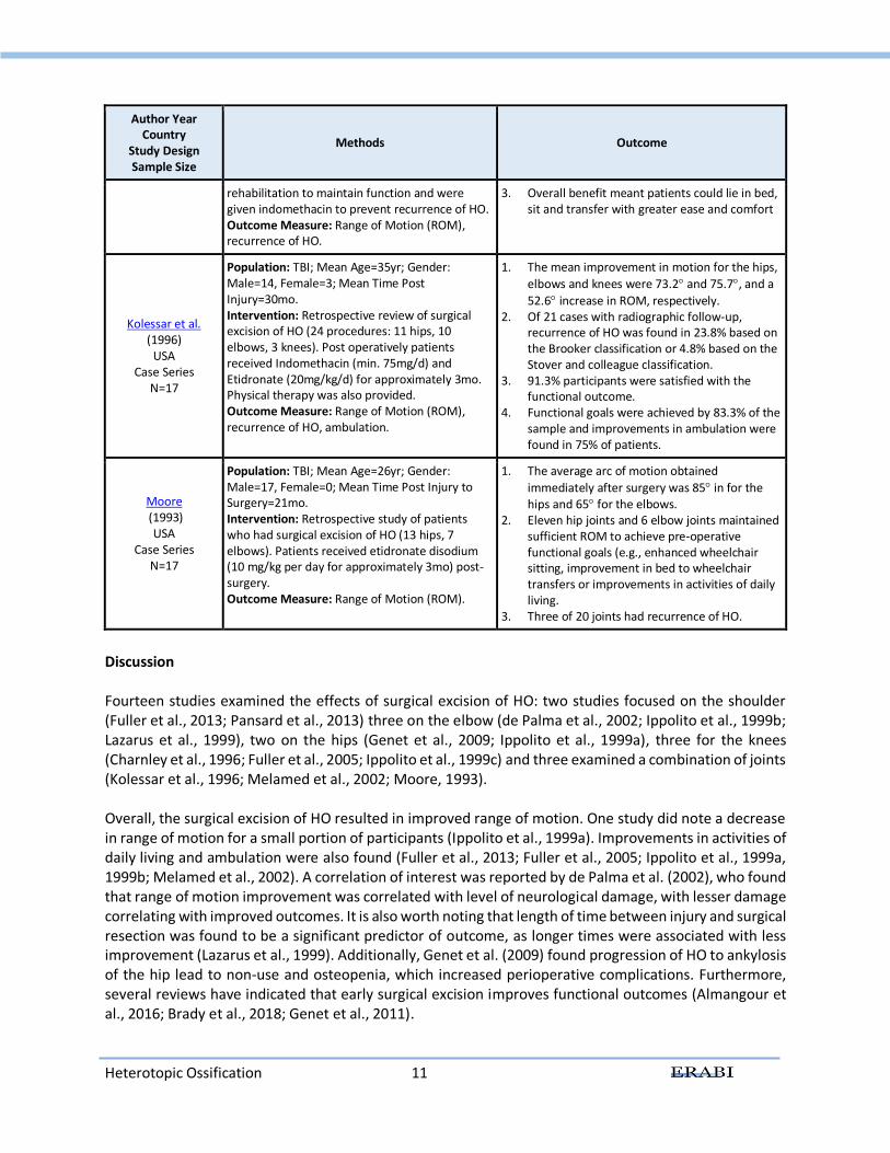
rehabilitation to maintain function and were given indomethacin to prevent recurrence of HO. Outcome Measure: Range of Motion (ROM), recurrence of HO.
3. Overall benefit meant patients could lie in bed, sit and transfer with greater ease and comfort
Kolessar et al. (1996)
USA Case Series
N=17
Population: TBI; Mean Age=35yr; Gender: Male=14, Female=3; Mean Time Post Injury=30mo. Intervention: Retrospective review of surgical excision of HO (24 procedures: 11 hips, 10 elbows, 3 knees). Post operatively patients received Indomethacin (min. 75mg/d) and Etidronate (20mg/kg/d) for approximately 3mo. Physical therapy was also provided. Outcome Measure: Range of Motion (ROM), recurrence of HO, ambulation.
1. The mean improvement in motion for the hips,
elbows and knees were 73.2 and 75.7, and a
52.6 increase in ROM, respectively. 2. Of 21 cases with radiographic follow-up,
recurrence of HO was found in 23.8% based on the Brooker classification or 4.8% based on the Stover and colleague classification.
3. 91.3% participants were satisfied with the functional outcome.
4. Functional goals were achieved by 83.3% of the sample and improvements in ambulation were found in 75% of patients.
Moore (1993)
USA Case Series
N=17
Population: TBI; Mean Age=26yr; Gender: Male=17, Female=0; Mean Time Post Injury to Surgery=21mo. Intervention: Retrospective study of patients who had surgical excision of HO (13 hips, 7 elbows). Patients received etidronate disodium (10 mg/kg per day for approximately 3mo) post-surgery. Outcome Measure: Range of Motion (ROM).
1. The average arc of motion obtained
immediately after surgery was 85 in for the hips and 65 for the elbows.
2. Eleven hip joints and 6 elbow joints maintained sufficient ROM to achieve pre-operative functional goals (e.g., enhanced wheelchair sitting, improvement in bed to wheelchair transfers or improvements in activities of daily living.
3. Three of 20 joints had recurrence of HO.
Discussion Fourteen studies examined the effects of surgical excision of HO: two studies focused on the shoulder (Fuller et al., 2013; Pansard et al., 2013) three on the elbow (de Palma et al., 2002; Ippolito et al., 1999b; Lazarus et al., 1999), two on the hips (Genet et al., 2009; Ippolito et al., 1999a), three for the knees (Charnley et al., 1996; Fuller et al., 2005; Ippolito et al., 1999c) and three examined a combination of joints (Kolessar et al., 1996; Melamed et al., 2002; Moore, 1993). Overall, the surgical excision of HO resulted in improved range of motion. One study did note a decrease in range of motion for a small portion of participants (Ippolito et al., 1999a). Improvements in activities of daily living and ambulation were also found (Fuller et al., 2013; Fuller et al., 2005; Ippolito et al., 1999a, 1999b; Melamed et al., 2002). A correlation of interest was reported by de Palma et al. (2002), who found that range of motion improvement was correlated with level of neurological damage, with lesser damage correlating with improved outcomes. It is also worth noting that length of time between injury and surgical resection was found to be a significant predictor of outcome, as longer times were associated with less improvement (Lazarus et al., 1999). Additionally, Genet et al. (2009) found progression of HO to ankylosis of the hip lead to non-use and osteopenia, which increased perioperative complications. Furthermore, several reviews have indicated that early surgical excision improves functional outcomes (Almangour et al., 2016; Brady et al., 2018; Genet et al., 2011).
Heterotopic Ossification 12
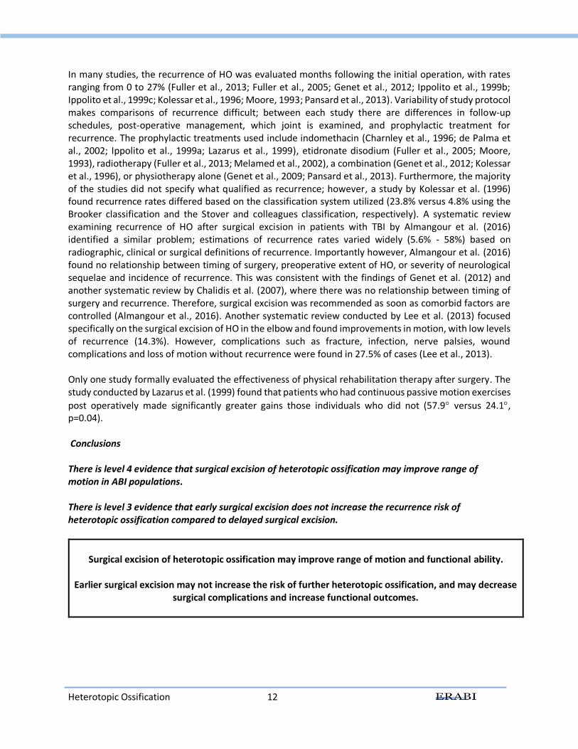
In many studies, the recurrence of HO was evaluated months following the initial operation, with rates ranging from 0 to 27% (Fuller et al., 2013; Fuller et al., 2005; Genet et al., 2012; Ippolito et al., 1999b; Ippolito et al., 1999c; Kolessar et al., 1996; Moore, 1993; Pansard et al., 2013). Variability of study protocol makes comparisons of recurrence difficult; between each study there are differences in follow-up schedules, post-operative management, which joint is examined, and prophylactic treatment for recurrence. The prophylactic treatments used include indomethacin (Charnley et al., 1996; de Palma et al., 2002; Ippolito et al., 1999a; Lazarus et al., 1999), etidronate disodium (Fuller et al., 2005; Moore, 1993), radiotherapy (Fuller et al., 2013; Melamed et al., 2002), a combination (Genet et al., 2012; Kolessar et al., 1996), or physiotherapy alone (Genet et al., 2009; Pansard et al., 2013). Furthermore, the majority of the studies did not specify what qualified as recurrence; however, a study by Kolessar et al. (1996) found recurrence rates differed based on the classification system utilized (23.8% versus 4.8% using the Brooker classification and the Stover and colleagues classification, respectively). A systematic review examining recurrence of HO after surgical excision in patients with TBI by Almangour et al. (2016) identified a similar problem; estimations of recurrence rates varied widely (5.6% - 58%) based on radiographic, clinical or surgical definitions of recurrence. Importantly however, Almangour et al. (2016) found no relationship between timing of surgery, preoperative extent of HO, or severity of neurological sequelae and incidence of recurrence. This was consistent with the findings of Genet et al. (2012) and another systematic review by Chalidis et al. (2007), where there was no relationship between timing of surgery and recurrence. Therefore, surgical excision was recommended as soon as comorbid factors are controlled (Almangour et al., 2016). Another systematic review conducted by Lee et al. (2013) focused specifically on the surgical excision of HO in the elbow and found improvements in motion, with low levels of recurrence (14.3%). However, complications such as fracture, infection, nerve palsies, wound complications and loss of motion without recurrence were found in 27.5% of cases (Lee et al., 2013). Only one study formally evaluated the effectiveness of physical rehabilitation therapy after surgery. The study conducted by Lazarus et al. (1999) found that patients who had continuous passive motion exercises
post operatively made significantly greater gains those individuals who did not (57.9 versus 24.1, p=0.04). Conclusions There is level 4 evidence that surgical excision of heterotopic ossification may improve range of motion in ABI populations.
There is level 3 evidence that early surgical excision does not increase the recurrence risk of heterotopic ossification compared to delayed surgical excision.
Surgical excision of heterotopic ossification may improve range of motion and functional ability.
Earlier surgical excision may not increase the risk of further heterotopic ossification, and may decrease
surgical complications and increase functional outcomes.
Heterotopic Ossification 13
11.2 Venous Thromboembolism Venous thromboembolism (VTE) is a blood clot that forms within a vein. The most common place for a blood clot to form is a deep vein, which is called a deep venous thrombosis or DVT. If the clot breaks off and travels to the lungs, causing partial or full occlusion, it is called a pulmonary embolism (PE) (Office of the Surgeon et al., 2008). Together, DVT and PE are referred to as VTE. VTE remains a common complication in patients who have sustained an ABI (Raslan et al., 2010; Scudday et al., 2011); however the scientific literature specific to ABI is quite limited. The following section presents ABI specific research regarding the prevention and treatment of VTE. Additional information on clinical presentation and testing practices is presented, however, it should be noted that not all in-text citations refer to research that meets the specific ERABI ABI inclusion criteria (mixed populations, age, mixed ABI severity, etc.) and therefore should be interpreted with caution when considering the application of any tests or indicators of VTE to an ABI population.
11.2.1 Incidence of Venous Thromboembolism Post-Head Injury In a large sample study consisting of 38,984 individuals with TBI, the incidence of VTE at the time of admission was 1.31% (Olufajo et al., 2016). At one-month post injury, the incidence for VTE increased to 1.87% and by one year it was 2.83% (Olufajo et al., 2016). The reported incidence of DVT among patients with TBI ranges from 11% to 54% (Carlile et al., 2010; Cifu et al., 1996; Denson et al., 2007; Geerts et al., 1994). The risk of developing a DVT or PE, in the absence of prophylaxis, is estimated to be approximately 20% post TBI (Haddad & Arabi, 2012). Severity of injury is found to be associated with incidence of VTE in isolated patients with TBI (Van Gent et al., 2014). Decisions on how to treat, and when, are often made on a case by case basis (Tang & Lobel, 2009). Experts recommend beginning pharmacological prophylaxis as early as 48 to 72 hours post injury (INESSS-ONF, 2017; Norwood et al., 2001). Unless contraindicated, mechanical thromboprophlaxis and low-molecular-weight heparin (LMWH) are recommened in the acute phase of recovery (Haddad & Arabi, 2012).
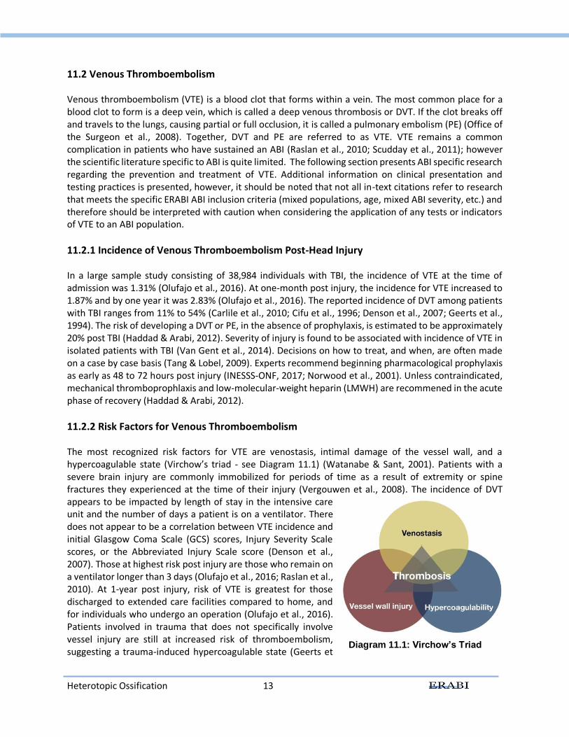
11.2.2 Risk Factors for Venous Thromboembolism The most recognized risk factors for VTE are venostasis, intimal damage of the vessel wall, and a hypercoagulable state (Virchow’s triad - see Diagram 11.1) (Watanabe & Sant, 2001). Patients with a severe brain injury are commonly immobilized for periods of time as a result of extremity or spine fractures they experienced at the time of their injury (Vergouwen et al., 2008). The incidence of DVT appears to be impacted by length of stay in the intensive care unit and the number of days a patient is on a ventilator. There does not appear to be a correlation between VTE incidence and initial Glasgow Coma Scale (GCS) scores, Injury Severity Scale scores, or the Abbreviated Injury Scale score (Denson et al., 2007). Those at highest risk post injury are those who remain on a ventilator longer than 3 days (Olufajo et al., 2016; Raslan et al., 2010). At 1-year post injury, risk of VTE is greatest for those discharged to extended care facilities compared to home, and for individuals who undergo an operation (Olufajo et al., 2016). Patients involved in trauma that does not specifically involve vessel injury are still at increased risk of thromboembolism, suggesting a trauma-induced hypercoagulable state (Geerts et
Diagram 11.1: Virchow’s Triad
Heterotopic Ossification 14
al., 1994; Geerts et al., 1996). Therefore, persons who have sustained a TBI appear to be at increased risk of developing VTE for multiple reasons.
11.2.3 Clinical Presentation of Deep Vein Thrombosis and Pulmonary Embolism A study found that up to 91% of thrombi form below the iliac level (De Maeseneer et al., 2016). The most common symptoms reported when a DVT is present are pain, swelling of the legs, and discoloration of the region (Collins, 2009). The clinical presentation of PE is challenging. Many cases are clinically silent (66%) with only 30% having the clinical features of a DVT (Garcia-Fuster et al., 2014). Asymptomatic PE is discovered in 70% of patients with confirmed clinically symptomatic DVT (Browse, 1974; Corrigan et al., 1974; Hull & Hirsh, 1983). Clinically, PE presents with tachycardia, tachypnea, hemoptysis, pleuritic chest pain and fever. Radiographic findings might include signs of consolidation or pleural effusion (Worku et al., 2014). Massive PE may cause right heart failure, which can progress to cardiovascular collapse, coma, and death.
11.2.4 Diagnostic Testing for Deep Vein Thrombosis A positive diagnosis of DVT can only be made if a venogram is positive or there is a positive venous ultrasound at two or more sites of the proximal veins. The diagnosis of DVT can be ruled out if there is a negative venogram, a negative D-dimer test or a normal venous ultrasound in patients with low clinical suspicion of DVT (Carlile et al., 2006).
11.2.4.1 Venous Ultrasound Venous ultrasound is often used to diagnose a DVT. There are several types of venous ultrasonography. They include compression ultrasound, duplex ultrasound, and color Doppler imaging. Although these types of venous ultrasonography are sometimes used interchangeably, their sensitivities and specificities for detecting acute DVT vary (Zierler, 2004). The sensitivity and specificity of compression ultrasonography for detecting DVTs is 43% and 85%, respectively (Girard et al., 2005). The weighted mean sensitivity and specificity of venous ultrasonography for the diagnosis of symptomatic proximal DVT are 97% and 94%, respectively; the sensitivity falls to 73% for distal DVT (Kearon et al., 1998; Zierler, 2004). Importantly, distal DVTs do not confer the same risk of extension to PE as do proximal DVTs. Typically, if a distal clot is going to extend proximally, this occurs within one week of its development. Consequently, serial ultrasound could be used in symptomatic patients in whom the test is initially negative as the test would become positive with the clot extension.
11.2.4.2 Venography Venography is considered a definitive test for DVT but it is an older invasive test whereby contrast dye is injected into the leg veins. Diagnosis of DVT is made if an intraluminal-filling defect is noted.
11.2.4.3 D-dimer Assay D-dimer assay is a rapid, non-invasive, and inexpensive test. Fibrin is the main component of thrombus formation and fibrin degradation products include D-dimers (Gill & Nahum, 2000). A positive D-dimer test is highly sensitive for the presence of a thrombus but lacks specificity since D-dimers are found in other disease states, including cancer, congestive heart failure, and inflammatory conditions (Raimondi et al.,
Heterotopic Ossification 15
1993). As a result, D-dimer assays have a high negative predictive value but a poor positive predictive value. To illustrate, Akman et al. (2004) reported that the sensitivity and negative predictive values of the D-dimer test were high, at 95.2% and 96.2% respectively, in a group of 68 rehabilitation patients (stroke, spinal cord injury, TBI, hip arthroplasty). The specificity and positive predictive values were low at 55.3% and 48.7%, respectively.
11.2.5 Diagnostic Testing for Pulmonary Embolism The diagnostic work-up for a suspected PE is a step-wise decision algorithm consisting of clinical likelihood and D-dimer testing (Di Nisio et al., 2016; Moore et al., 2018). Patients with low clinical suspicion of PE have D-dimer testing. If the D-dimer is negative, PE is ruled out in patients with low clinical suspicion of PE; if positive, patients move to imaging for PE. Patients with high clinical suspicion of PE do not have D-dimer testing and move straight to imaging. Patients with ABI are most often considered high-risk for PE. Computed Tomography Pulmonary Angiogram (CTPA) is the preferred imaging modality for diagnosis of PE (Di Nisio et al., 2016; Moore et al., 2018). Ventilation/Perfusion Scanning (V/Q Scan) can be used when CTPA is contraindicated. Combining imaging with pre-test clinical decision rules increases the predictive power in the diagnosis of PE.
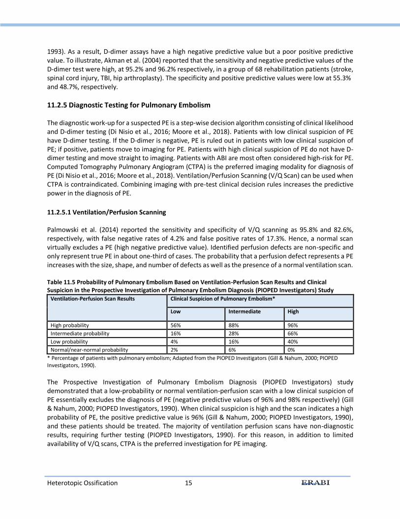
11.2.5.1 Ventilation/Perfusion Scanning Palmowski et al. (2014) reported the sensitivity and specificity of V/Q scanning as 95.8% and 82.6%, respectively, with false negative rates of 4.2% and false positive rates of 17.3%. Hence, a normal scan virtually excludes a PE (high negative predictive value). Identified perfusion defects are non-specific and only represent true PE in about one-third of cases. The probability that a perfusion defect represents a PE increases with the size, shape, and number of defects as well as the presence of a normal ventilation scan. Table 11.5 Probability of Pulmonary Embolism Based on Ventilation-Perfusion Scan Results and Clinical Suspicion in the Prospective Investigation of Pulmonary Embolism Diagnosis (PIOPED Investigators) Study
Ventilation-Perfusion Scan Results Clinical Suspicion of Pulmonary Embolism*
Low Intermediate High
High probability 56% 88% 96%
Intermediate probability 16% 28% 66%
Low probability 4% 16% 40%
Normal/near-normal probability 2% 6% 0%
* Percentage of patients with pulmonary embolism; Adapted from the PIOPED Investigators (Gill & Nahum, 2000; PIOPED Investigators, 1990).
The Prospective Investigation of Pulmonary Embolism Diagnosis (PIOPED Investigators) study demonstrated that a low-probability or normal ventilation-perfusion scan with a low clinical suspicion of PE essentially excludes the diagnosis of PE (negative predictive values of 96% and 98% respectively) (Gill & Nahum, 2000; PIOPED Investigators, 1990). When clinical suspicion is high and the scan indicates a high probability of PE, the positive predictive value is 96% (Gill & Nahum, 2000; PIOPED Investigators, 1990), and these patients should be treated. The majority of ventilation perfusion scans have non-diagnostic results, requiring further testing (PIOPED Investigators, 1990). For this reason, in addition to limited availability of V/Q scans, CTPA is the preferred investigation for PE imaging.
Heterotopic Ossification 16
11.2.5.2 Computed Tomography Pulmonary Angiogram CTPA is the preferred imaging modality for diagnosing PE (Di Nisio et al., 2016; Moore et al., 2018). It has become first line at most centers because it is fast, highly sensitive and specific, and can detect other causes of chest pain such as pneumonia, musculoskeletal injuries or pericardial abnormalities (Di Nisio et al., 2016). Combined with clinical probability rules there is a very high positive predictive value (Gottschalk et al., 2002; Stein et al., 2006). CTPA carries risks associated with radiation exposure, bleeding, adverse reaction to contrast medium, and is contraindicated in renal insufficiency. V/Q scan is used for investigating potential PE when CTPA is contraindicated (Di Nisio et al., 2016).
11.2.6 Prevention of Venous Thromboembolism Post ABI Several interventions have been examined for the prevention of DVT after an ABI, including mechanical therapy, pharmaceuticals, or a combination of both. In a systematic review, Hachem et al. (2018) found rates of VTE in patients with severe TBI not receiving anticoagulation prophylaxis were near 30%, compared to 5-10% of patients with prophylaxis. However, there is no agreement on the administration of these medications in terms of timing, dose, and/or which anticoagulation medication.
11.2.6.1 Non-Pharmacological Prophylaxis Non-pharmacological, mechanical interventions used to prevent the development of DVT post ABI include: the insertion of inferior vena cava filters, thromboembolism deterrent stockings, and intermittent pneumatic compression devices including arteriovenous foot pumps and sequential compression devices (SCDs). These devices operate primarily through two distinct mechanisms of action. The first is mechanical, in which the device increases the velocity of venous return to decrease venous stasis, thus reducing the opportunity for clot formation. The second, and perhaps more important mechanism, involves the systemic activation of the fibrinolytic system which, during compression, leads to the breakdown of fibrin clots associated with thromboembolism (Macatangay et al., 2008). The exception is vena cava filters, which operate by another method of mechanical VTE prevention (Watanabe & Sant, 2001). These filters are inserted into the inferior vena cava to prevent the passage of distal emboli into the lungs. Some reports have demonstrated success rates as high as 96% in the prevention of pulmonary emboli (Greenfield & Michna, 1988). However, the use of vena cava filters carries some associated risks. They can become blocked or dislodged which can increase the risk of an embolism. Some have also reportedly increased risks for repeated DVT compared with patients without such devices (Decousus et al., 1998).
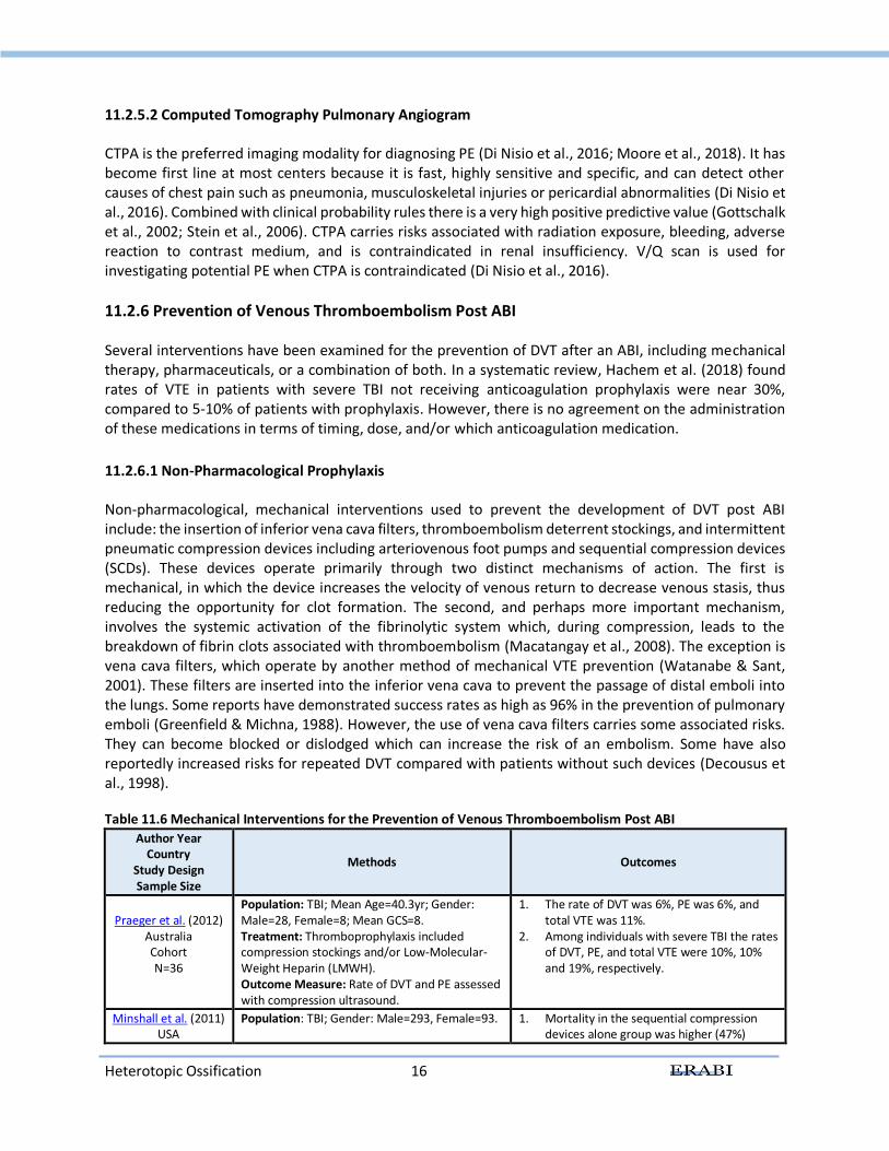
Table 11.6 Mechanical Interventions for the Prevention of Venous Thromboembolism Post ABI
Author Year Country
Study Design Sample Size
Methods Outcomes
Praeger et al. (2012) Australia Cohort N=36
Population: TBI; Mean Age=40.3yr; Gender: Male=28, Female=8; Mean GCS=8. Treatment: Thromboprophylaxis included compression stockings and/or Low-Molecular-Weight Heparin (LMWH). Outcome Measure: Rate of DVT and PE assessed with compression ultrasound.
1. The rate of DVT was 6%, PE was 6%, and total VTE was 11%.
2. Among individuals with severe TBI the rates of DVT, PE, and total VTE were 10%, 10% and 19%, respectively.
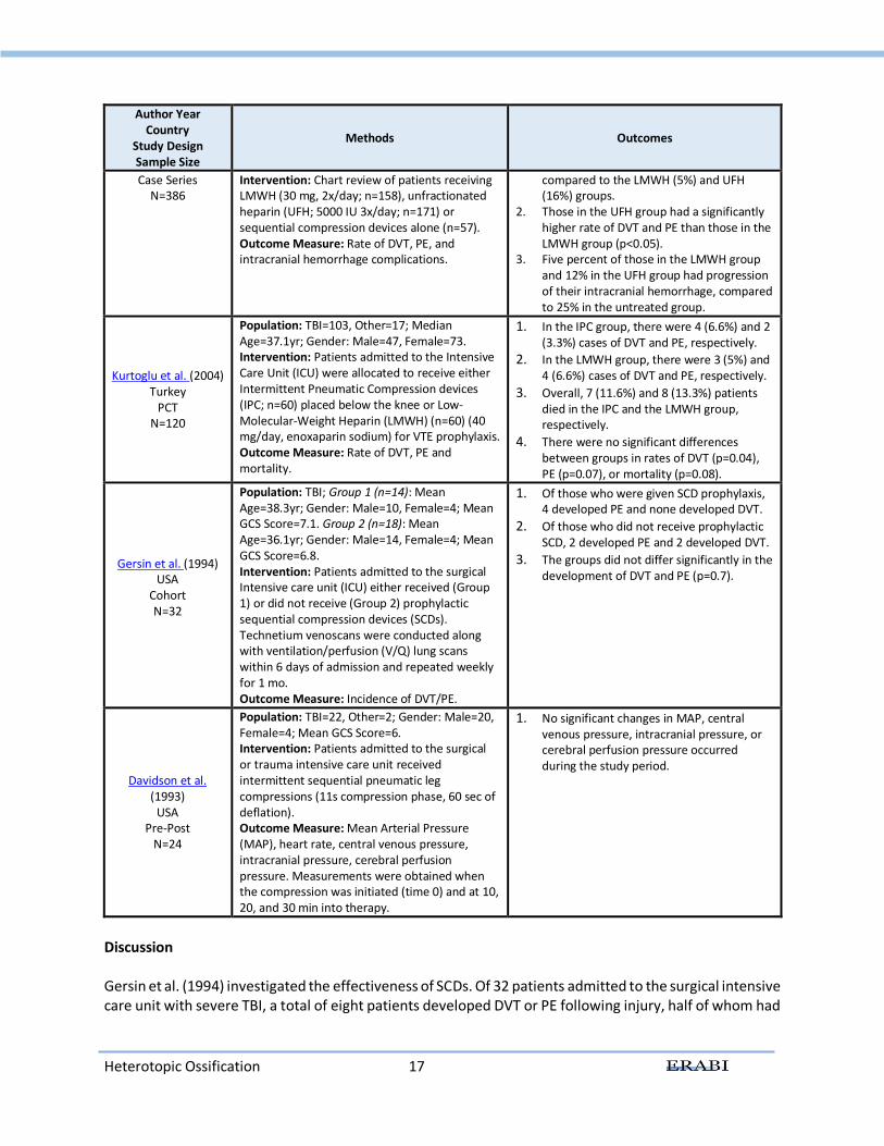
Minshall et al. (2011) USA
Population: TBI; Gender: Male=293, Female=93. 1. Mortality in the sequential compression devices alone group was higher (47%)
Heterotopic Ossification 17
Author Year Country
Study Design Sample Size
Methods Outcomes
Case Series N=386
Intervention: Chart review of patients receiving LMWH (30 mg, 2x/day; n=158), unfractionated heparin (UFH; 5000 IU 3x/day; n=171) or sequential compression devices alone (n=57). Outcome Measure: Rate of DVT, PE, and intracranial hemorrhage complications.
compared to the LMWH (5%) and UFH (16%) groups.
2. Those in the UFH group had a significantly higher rate of DVT and PE than those in the LMWH group (p<0.05).
3. Five percent of those in the LMWH group and 12% in the UFH group had progression of their intracranial hemorrhage, compared to 25% in the untreated group.
Kurtoglu et al. (2004) Turkey
PCT N=120
Population: TBI=103, Other=17; Median Age=37.1yr; Gender: Male=47, Female=73. Intervention: Patients admitted to the Intensive Care Unit (ICU) were allocated to receive either Intermittent Pneumatic Compression devices (IPC; n=60) placed below the knee or Low-Molecular-Weight Heparin (LMWH) (n=60) (40 mg/day, enoxaparin sodium) for VTE prophylaxis. Outcome Measure: Rate of DVT, PE and mortality.
1. In the IPC group, there were 4 (6.6%) and 2 (3.3%) cases of DVT and PE, respectively.
2. In the LMWH group, there were 3 (5%) and 4 (6.6%) cases of DVT and PE, respectively.
3. Overall, 7 (11.6%) and 8 (13.3%) patients died in the IPC and the LMWH group, respectively.
4. There were no significant differences between groups in rates of DVT (p=0.04), PE (p=0.07), or mortality (p=0.08).
Gersin et al. (1994) USA
Cohort N=32
Population: TBI; Group 1 (n=14): Mean Age=38.3yr; Gender: Male=10, Female=4; Mean GCS Score=7.1. Group 2 (n=18): Mean Age=36.1yr; Gender: Male=14, Female=4; Mean GCS Score=6.8. Intervention: Patients admitted to the surgical Intensive care unit (ICU) either received (Group 1) or did not receive (Group 2) prophylactic sequential compression devices (SCDs). Technetium venoscans were conducted along with ventilation/perfusion (V/Q) lung scans within 6 days of admission and repeated weekly for 1 mo. Outcome Measure: Incidence of DVT/PE.
1. Of those who were given SCD prophylaxis, 4 developed PE and none developed DVT.
2. Of those who did not receive prophylactic SCD, 2 developed PE and 2 developed DVT.
3. The groups did not differ significantly in the development of DVT and PE (p=0.7).
Davidson et al. (1993)
USA Pre-Post
N=24
Population: TBI=22, Other=2; Gender: Male=20, Female=4; Mean GCS Score=6. Intervention: Patients admitted to the surgical or trauma intensive care unit received intermittent sequential pneumatic leg compressions (11s compression phase, 60 sec of deflation). Outcome Measure: Mean Arterial Pressure (MAP), heart rate, central venous pressure, intracranial pressure, cerebral perfusion pressure. Measurements were obtained when the compression was initiated (time 0) and at 10, 20, and 30 min into therapy.
1. No significant changes in MAP, central venous pressure, intracranial pressure, or cerebral perfusion pressure occurred during the study period.
Discussion Gersin et al. (1994) investigated the effectiveness of SCDs. Of 32 patients admitted to the surgical intensive care unit with severe TBI, a total of eight patients developed DVT or PE following injury, half of whom had
Heterotopic Ossification 18
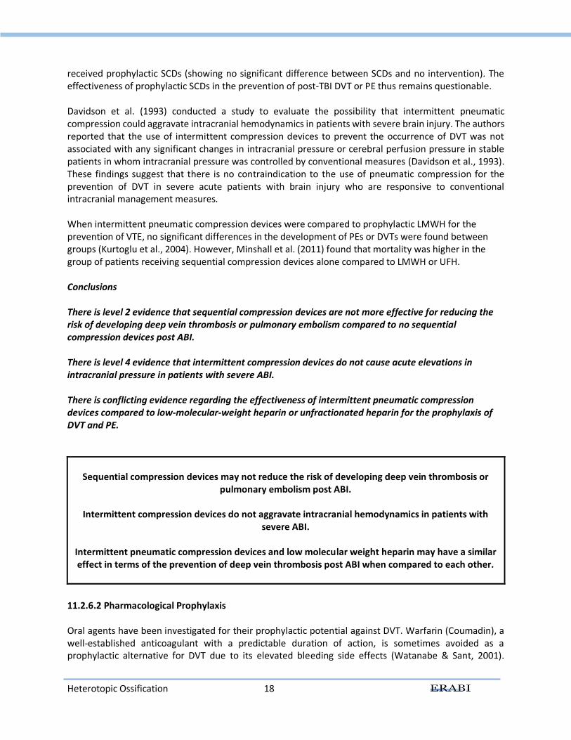
received prophylactic SCDs (showing no significant difference between SCDs and no intervention). The effectiveness of prophylactic SCDs in the prevention of post-TBI DVT or PE thus remains questionable. Davidson et al. (1993) conducted a study to evaluate the possibility that intermittent pneumatic compression could aggravate intracranial hemodynamics in patients with severe brain injury. The authors reported that the use of intermittent compression devices to prevent the occurrence of DVT was not associated with any significant changes in intracranial pressure or cerebral perfusion pressure in stable patients in whom intracranial pressure was controlled by conventional measures (Davidson et al., 1993). These findings suggest that there is no contraindication to the use of pneumatic compression for the prevention of DVT in severe acute patients with brain injury who are responsive to conventional intracranial management measures. When intermittent pneumatic compression devices were compared to prophylactic LMWH for the prevention of VTE, no significant differences in the development of PEs or DVTs were found between groups (Kurtoglu et al., 2004). However, Minshall et al. (2011) found that mortality was higher in the group of patients receiving sequential compression devices alone compared to LMWH or UFH. Conclusions There is level 2 evidence that sequential compression devices are not more effective for reducing the risk of developing deep vein thrombosis or pulmonary embolism compared to no sequential compression devices post ABI. There is level 4 evidence that intermittent compression devices do not cause acute elevations in intracranial pressure in patients with severe ABI. There is conflicting evidence regarding the effectiveness of intermittent pneumatic compression devices compared to low-molecular-weight heparin or unfractionated heparin for the prophylaxis of DVT and PE.
Sequential compression devices may not reduce the risk of developing deep vein thrombosis or
pulmonary embolism post ABI.
Intermittent compression devices do not aggravate intracranial hemodynamics in patients with severe ABI.
Intermittent pneumatic compression devices and low molecular weight heparin may have a similar effect in terms of the prevention of deep vein thrombosis post ABI when compared to each other.
11.2.6.2 Pharmacological Prophylaxis Oral agents have been investigated for their prophylactic potential against DVT. Warfarin (Coumadin), a well-established anticoagulant with a predictable duration of action, is sometimes avoided as a prophylactic alternative for DVT due to its elevated bleeding side effects (Watanabe & Sant, 2001).
Heterotopic Ossification 19
Albrecht and colleagues (2014) report that warfarin use is associated with lower rates of DVT and PE, but comes at the cost of the risk of increased hemorrhagic bleeding. However, some experts felt the use of warfarin was advisable, especially for high risk patients due to its benefit in treating undetected thrombosis; the therapeutic dose range for prophylaxis and treatment of thromboembolism are the same (Hirsh et al., 1992; Hyers et al., 1992; Landefeld & Goldman, 1989). In a multicenter observational study of DVT prophylaxis with a mixed TBI population sample of 932 patients treated with anticoagulation drugs, 71% were given LMWH, 23% unfractionated heparin, 1% Coumadin, and 3% were given both LMWH and Low-dose unfractionated heparin, none of which were associated with increased intracranial or systemic hemorrhage (Carlile et al., 2010). The Institut national d'excellence en santé et en services sociaux (INESSS) and Ontario Neurotrauma Foundation clinical practice guidelines for the rehabilitation of moderate to severe TBI recommend initiating thromboprophylaxis as soon as medically appropriate (level B evidence), and physical methods of thromboprophylaxis (ie. compression stockings) should be used when pharmacological prophylaxis is delayed or contraindicated (level B evidence)(INESSS-ONF, 2017). There is also evidence from a meta-analysis that aspirin has positive effects in the reduction of both DVT and PE, by 40% and 60% respectively ("Collaborative overview of randomised trials of antiplatelet therapy - III: Reduction in venous thrombosis and pulmonary embolism by antiplatelet prophylaxis among surgical and medical patients," 1994). A systematic review on anticoagulation in patients with severe TBI found 30% rate of VTE among patients not receiving anticoagulation compared to 5-10% in patients receiving anticoagulation therapy (Hachem et al., 2018). Overall, there is a lack of persuasive evidence to guide decisions about when to administer anticoagulant prophylaxis in those who sustain traumatic intracranial hemorrhage. Clinicians often make decisions based on their own assessments of the risks and benefits (Scales et al., 2010). To date no national standard of care exists for the administration of the pharmacological prophylaxis treatment of DVT post TBI (Phelan et al., 2012a). Differences in medications used for pharmacological thromboprophylaxis of patients with ABI is another important consideration. Subcutaneous heparin in low doses has been reported to be both safe and effective as prophylaxis against DVT development post ABI (Watanabe & Sant, 2001). The route of delivery may also affect the efficacy of anticoagulant prophylaxis (Watanabe & Sant, 2001). For this reason, intravenously delivered heparin may be more effective in the prevention of thromboembolism compared with subcutaneous administration, although this method of delivery might increase the risk of bleeding (Green et al., 1988). LMWH, which is injected subcutaneously, has gained popularity due to the ease of administration and dosage adjustment. Of note, low-molecular weight variants of unfractionated heparin are more expensive but the advantages are such that they have become the standard of care. Carlile et al. (2006) found that 15 of the 16 rehabilitation centers surveyed reported routinely initiating treatment with either LMWH or Low-dose unfractionated heparin. In a study with a mixed trauma population, low-dose unfractionated heparin was compared to enoxaparin (LMWH) for the treatment of DVT (Geerts et al., 1996). Of those receiving low-dose unfractionated heparin, 44% suffered a DVT compared to 31% of patients receiving enoxaparin (p=0.014) (Geerts et al., 1996). These results are consistent with Byrne et al. (2017) matched analysis in patients with isolated severe TBI, where patients who received LMWH had an adjusted odds ratio of 0.49 (95% CI=0.29-0.82) of developing a PE compared to patients who were treated with unfractionated heparin. The INESSS and Ontario Neurotrauma Foundation clinical practice guidelines for the rehabilitation of moderate to severe TBI recommends LMWH over unfractionated heparin after TBI (level C evidence), although these guidelines are mostly based on evidence in general trauma patients, and not TBI specifically (INESSS-ONF, 2017).
Heterotopic Ossification 20
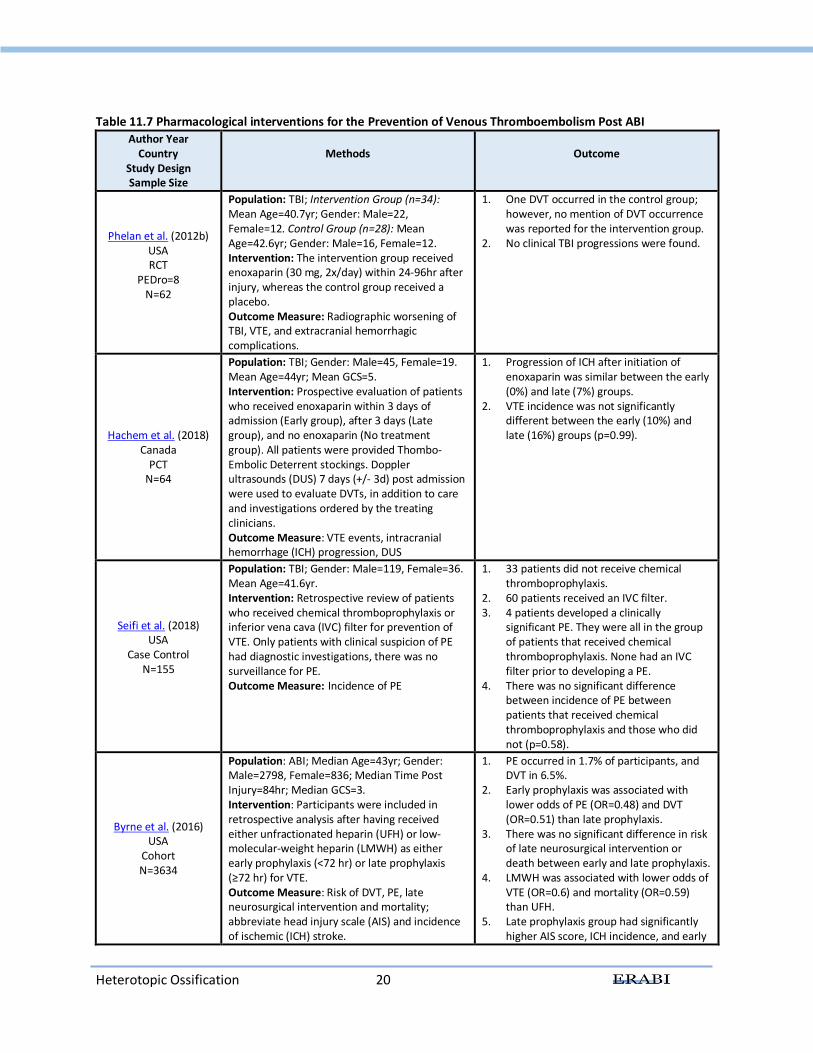
Table 11.7 Pharmacological interventions for the Prevention of Venous Thromboembolism Post ABI
Author Year Country
Study Design Sample Size
Methods
Outcome
Phelan et al. (2012b) USA RCT
PEDro=8 N=62
Population: TBI; Intervention Group (n=34): Mean Age=40.7yr; Gender: Male=22, Female=12. Control Group (n=28): Mean Age=42.6yr; Gender: Male=16, Female=12. Intervention: The intervention group received enoxaparin (30 mg, 2x/day) within 24-96hr after injury, whereas the control group received a placebo. Outcome Measure: Radiographic worsening of TBI, VTE, and extracranial hemorrhagic complications.
1. One DVT occurred in the control group; however, no mention of DVT occurrence was reported for the intervention group.
2. No clinical TBI progressions were found.
Hachem et al. (2018) Canada
PCT N=64
Population: TBI; Gender: Male=45, Female=19. Mean Age=44yr; Mean GCS=5. Intervention: Prospective evaluation of patients who received enoxaparin within 3 days of admission (Early group), after 3 days (Late group), and no enoxaparin (No treatment group). All patients were provided Thombo-Embolic Deterrent stockings. Doppler ultrasounds (DUS) 7 days (+/- 3d) post admission were used to evaluate DVTs, in addition to care and investigations ordered by the treating clinicians. Outcome Measure: VTE events, intracranial hemorrhage (ICH) progression, DUS
1. Progression of ICH after initiation of enoxaparin was similar between the early (0%) and late (7%) groups.
2. VTE incidence was not significantly different between the early (10%) and late (16%) groups (p=0.99).
Seifi et al. (2018) USA
Case Control N=155
Population: TBI; Gender: Male=119, Female=36. Mean Age=41.6yr. Intervention: Retrospective review of patients who received chemical thromboprophylaxis or inferior vena cava (IVC) filter for prevention of VTE. Only patients with clinical suspicion of PE had diagnostic investigations, there was no surveillance for PE. Outcome Measure: Incidence of PE
1. 33 patients did not receive chemical thromboprophylaxis.
2. 60 patients received an IVC filter. 3. 4 patients developed a clinically
significant PE. They were all in the group of patients that received chemical thromboprophylaxis. None had an IVC filter prior to developing a PE.
4. There was no significant difference between incidence of PE between patients that received chemical thromboprophylaxis and those who did not (p=0.58).
Byrne et al. (2016) USA
Cohort N=3634
Population: ABI; Median Age=43yr; Gender: Male=2798, Female=836; Median Time Post Injury=84hr; Median GCS=3. Intervention: Participants were included in retrospective analysis after having received either unfractionated heparin (UFH) or low-molecular-weight heparin (LMWH) as either early prophylaxis (<72 hr) or late prophylaxis (≥72 hr) for VTE. Outcome Measure: Risk of DVT, PE, late neurosurgical intervention and mortality; abbreviate head injury scale (AIS) and incidence of ischemic (ICH) stroke.
1. PE occurred in 1.7% of participants, and DVT in 6.5%.
2. Early prophylaxis was associated with lower odds of PE (OR=0.48) and DVT (OR=0.51) than late prophylaxis.
3. There was no significant difference in risk of late neurosurgical intervention or death between early and late prophylaxis.
4. LMWH was associated with lower odds of VTE (OR=0.6) and mortality (OR=0.59) than UFH.
5. Late prophylaxis group had significantly higher AIS score, ICH incidence, and early
Heterotopic Ossification 21
neurosurgical intervention rate than early prophylaxis group.
6. The late group most commonly received LMWH and early group most commonly received UFH.
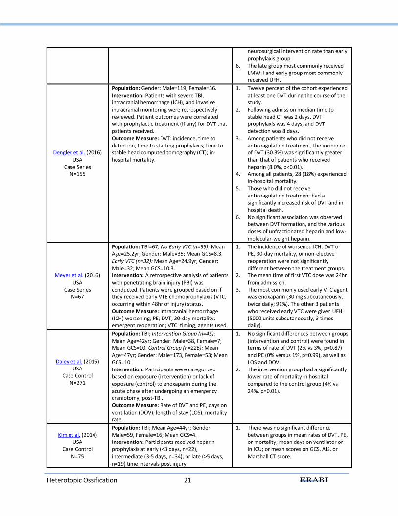
Dengler et al. (2016) USA
Case Series N=155
Population: Gender: Male=119, Female=36. Intervention: Patients with severe TBI, intracranial hemorrhage (ICH), and invasive intracranial monitoring were retrospectively reviewed. Patient outcomes were correlated with prophylactic treatment (if any) for DVT that patients received. Outcome Measure: DVT: incidence, time to detection, time to starting prophylaxis; time to stable head computed tomography (CT); in-hospital mortality.
1. Twelve percent of the cohort experienced at least one DVT during the course of the study.
2. Following admission median time to stable head CT was 2 days, DVT prophylaxis was 4 days, and DVT detection was 8 days.
3. Among patients who did not receive anticoagulation treatment, the incidence of DVT (30.3%) was significantly greater than that of patients who received heparin (8.0%, p<0.01).
4. Among all patients, 28 (18%) experienced in-hospital mortality.
5. Those who did not receive anticoagulation treatment had a significantly increased risk of DVT and in-hospital death.
6. No significant association was observed between DVT formation, and the various doses of unfractionated heparin and low-molecular-weight heparin.
Meyer et al. (2016) USA
Case Series N=67
Population: TBI=67; No Early VTC (n=35): Mean Age=25.2yr; Gender: Male=35; Mean GCS=8.3. Early VTC (n=32): Mean Age=24.9yr; Gender: Male=32; Mean GCS=10.3. Intervention: A retrospective analysis of patients with penetrating brain injury (PBI) was conducted. Patients were grouped based on if they received early VTE chemoprophylaxis (VTC, occurring within 48hr of injury) status. Outcome Measure: Intracranial hemorrhage (ICH) worsening; PE; DVT; 30-day mortality; emergent reoperation; VTC: timing, agents used.
1. The incidence of worsened ICH, DVT or PE, 30-day mortality, or non-elective reoperation were not significantly different between the treatment groups.
2. The mean time of first VTC dose was 24hr from admission.
3. The most commonly used early VTC agent was enoxaparin (30 mg subcutaneously, twice daily; 91%). The other 3 patients who received early VTC were given UFH (5000 units subcutaneously, 3 times daily).
Daley et al. (2015) USA
Case Control N=271
Population: TBI; Intervention Group (n=45): Mean Age=42yr; Gender: Male=38, Female=7; Mean GCS=10. Control Group (n=226): Mean Age=47yr; Gender: Male=173, Female=53; Mean GCS=10. Intervention: Participants were categorized based on exposure (intervention) or lack of exposure (control) to enoxaparin during the acute phase after undergoing an emergency craniotomy, post-TBI. Outcome Measure: Rate of DVT and PE, days on ventilation (DOV), length of stay (LOS), mortality rate.
1. No significant differences between groups (intervention and control) were found in terms of rate of DVT (2% vs 3%, p=0.87) and PE (0% versus 1%, p=0.99), as well as LOS and DOV.
2. The intervention group had a significantly lower rate of mortality in hospital compared to the control group (4% vs 24%, p=0.01).
Kim et al. (2014) USA
Case Control N=75
Population: TBI; Mean Age=44yr; Gender: Male=59, Female=16; Mean GCS=4. Intervention: Participants received heparin prophylaxis at early (<3 days, n=22), intermediate (3-5 days, n=34), or late (>5 days, n=19) time intervals post injury.
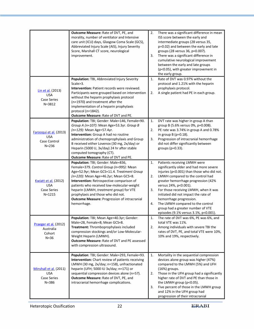
1. There was no significant difference between groups in mean rates of DVT, PE, or mortality; mean days on ventilator or in ICU; or mean scores on GCS, AIS, or Marshall CT score.
Heterotopic Ossification 22
Outcome Measure: Rate of DVT, PE, and morality, number of ventilator and Intensive care unit (ICU) days, Glasgow Coma Scale (GCS), Abbreviated Injury Scale (AIS), Injury Severity Score, Marshall CT score, neurological improvement.
2. There was a significant difference in mean ISS score between the early and intermediate groups (28 versus 35, p=0.02) and between the early and late groups (28 versus 36, p=0.007).
3. There was a significant difference in cumulative neurological improvement between the early and late groups (p<0.05), with greater improvement in the early group.
Lin et al. (2013) USA
Case Series N=3812
Population: TBI, Abbreviated Injury Severity Scale>3. Intervention: Patient records were reviewed. Participants were grouped based on intervention without the heparin prophylaxis protocol (n=1970) and treatment after the implementation of a heparin prophylaxis protocol (n=1842). Outcome Measure: Rate of DVT and PE.
1. Rate of DVT was 0.97% without the protocol and 1.21% with the heparin prophylaxis protocol.
2. A single patient had PE in each group.
Farooqui et al. (2013) USA
Case Control N=236
Population: TBI; Gender: Male=146, Female=90. Group A (n=107): Mean Age=53.3yr. Group B (n=129): Mean Age=57.4yr. Intervention: Group A had no routine administration of chemoprophylaxis and Group B received either Lovenox (30 mg, 2x/day) or Heparin (5000 U, 3x/day) 24 hr after stable computed tomography (CT). Outcome Measure: Rate of DVT and PE.
1. DVT rate was higher in group A than group B (5.6% versus 0%, p=0.008).
2. PE rate was 3.74% in group A and 0.78% in group B (p=0.18).
3. Progression of intracranial hemorrhage did not differ significantly between groups (p=0.33).
Kwiatt et al. (2012) USA
Case Series N=1215
Population: TBI; Gender: Male=836, Female=379. Control Group (n=995): Mean Age=52.9yr; Mean GCS=11.4. Treatment Group (n=220): Mean Age=46.2yr; Mean GCS=8. Intervention: Retrospective comparison of patients who received low-molecular-weight heparin (LMWH, treatment group) for VTE prophylaxis and those who did not. Outcome Measure: Progression of intracranial hemorrhage.
1. Patients receiving LMWH were significantly older and had more severe injuries (p<0.001) than those who did not.
2. LMWH compared to the control had greater hemorrhage progression (42% versus 24%, p<0.001).
3. For those receiving LMWH, when it was initiated did not impact the rate of hemorrhage progression.
4. The LMWH compared to the control group had a greater number of VTE episodes (9.1% versus 3.1%, p<0.001).
Praeger et al. (2012) Australia Cohort N=36
Population: TBI; Mean Age=40.3yr; Gender: Male=28, Female=8; Mean GCS=8. Treatment: Thromboprophylaxis included compression stockings and/or Low-Molecular-Weight Heparin (LMWH). Outcome Measure: Rate of DVT and PE assessed with compression ultrasound.
1. The rate of DVT was 6%, PE was 6%, and total VTE was 11%.
2. Among individuals with severe TBI the rates of DVT, PE, and total VTE were 10%, 10% and 19%, respectively.
Minshall et al. (2011) USA
Case Series N=386
Population: TBI; Gender: Male=293, Female=93. Intervention: Chart review of patients receiving LMWH (30 mg, 2x/day; n=158), unfractionated heparin (UFH; 5000 IU 3x/day; n=171) or sequential compression devices alone (n=57). Outcome Measure: Rate of DVT, PE, and intracranial hemorrhage complications.
1. Mortality in the sequential compression devices alone group was higher (47%) compared to the LMWH (5%) and UFH (16%) groups.
2. Those in the UFH group had a significantly higher rate of DVT and PE than those in the LMWH group (p<0.05).
3. Five percent of those in the LMWH group and 12% in the UFH group had progression of their intracranial
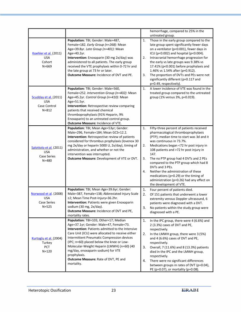
Heterotopic Ossification 23
hemorrhage, compared to 25% in the untreated group.
Koehler et al. (2011) USA
Cohort N=669
Population: TBI; Gender: Male=487, Female=182. Early Group (n=268): Mean Age=39.8yr. Late Group (n=401): Mean Age=40.2yr. Intervention: Enoxaparin (30 mg 2x/day) was administered to all patients. The early group received the VTE prophylaxis within 0-72 hr and the late group at 73 hr or later. Outcome Measure: Incidence of DVT and PE.
1. Those in the early group compared to the late group spent significantly fewer days on a ventilator (p<0.001), fewer days in ICU (p<0.002) and hospital (p<0.004).
2. Intracranial hemorrhage progression for the early vs late groups was 9.38% vs 17.41% (p<0.001) before prophylaxis and 1.46% vs 1.54% after (p=0.912).
3. The proportion of DVTs and PEs were not significantly different (p=0.117 and p=0.49, respectively).
Scudday et al. (2011) USA
Case Control N=812
Population: TBI; Gender: Male=560, Female=252. Intervention Group (n=402): Mean Age=45.2yr. Control Group (n=410): Mean Age=51.5yr. Intervention: Retrospective review comparing patients that received chemical thromboprophylaxis (91% Heparin, 9% Enoxaparin) to an untreated control group. Outcome Measure: Incidence of VTE.
1. A lower incidence of VTE was found in the treated group compared to the untreated group (1% versus 3%, p=0.019).
Salottolo et al. (2011) USA
Case Series N=480
Population: TBI; Mean Age=53yr; Gender: Male=296, Female=184; Mean GCS=12.2. Intervention: Retrospective review of patients considered for thrombus prophylaxis (lovenox 30 mg 2x/day or heparin 5000 U, 2x/day), timing of administration, and whether or not the intervention was interrupted. Outcome Measure: Development of VTE or DVT.
1. Fifty-three percent of patients received pharmacological thromboprophylaxis (PTP); median time to start was 3d and it was continuous in 73.7%.
2. Medications began <72 hr post injury in 108 patients and >72 hr post injury in 147.
3. The no PTP group had 4 DVTs and 2 PEs compared to the PTP group which had 8 DVTs and 3 PEs.
4. Neither the administration of these medications (p=0.29) or the timing of administration (p=0.26) had any effect on the development of VTE.
Norwood et al. (2008) USA
Case Series N=525
Population: TBI; Mean Age=39.6yr; Gender: Male=387, Female=138; Abbreviated Injury Scale ≥2; Mean Time Post-Injury=36.2hr. Intervention: Patients were given Enoxaparin sodium (30 mg, 2x/day). Outcome Measure: Incidence of DVT and PE, mortality rates.
1. Four percent of patients died.
2. Of 151 patients that underwent a lower extremity venous Doppler ultrasound, 6 patients were diagnosed with a DVT.
3. No patients within the study group were diagnosed with a PE.
Kurtoglu et al. (2004) Turkey
PCT N=120
Population: TBI=103, Other=17; Median Age=37.1yr; Gender: Male=47, Female=73. Intervention: Patients admitted to the Intensive Care Unit (ICU) were allocated to receive either Intermittent Pneumatic Compression devices (IPC; n=60) placed below the knee or Low-Molecular-Weight Heparin (LMWH) (n=60) (40 mg/day, enoxaparin sodium) for VTE prophylaxis. Outcome Measure: Rate of DVT, PE and mortality.
1. In the IPC group, there were 4 (6.6%) and 2 (3.3%) cases of DVT and PE, respectively.
2. In the LMWH group, there were 3 (5%) and 4 (6.6%) cases of DVT and PE, respectively.
3. Overall, 7 (11.6%) and 8 (13.3%) patients died in the IPC and the LMWH group, respectively.
4. There were no significant differences between groups in rates of DVT (p=0.04), PE (p=0.07), or mortality (p=0.08).
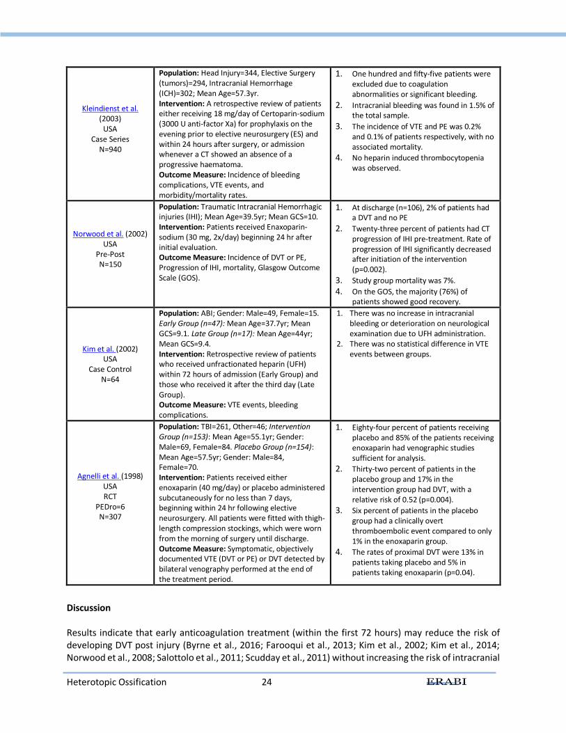
Heterotopic Ossification 24
Kleindienst et al. (2003)
USA Case Series
N=940
Population: Head Injury=344, Elective Surgery (tumors)=294, Intracranial Hemorrhage (ICH)=302; Mean Age=57.3yr. Intervention: A retrospective review of patients either receiving 18 mg/day of Certoparin-sodium (3000 U anti-factor Xa) for prophylaxis on the evening prior to elective neurosurgery (ES) and within 24 hours after surgery, or admission whenever a CT showed an absence of a progressive haematoma. Outcome Measure: Incidence of bleeding complications, VTE events, and morbidity/mortality rates.
1. One hundred and fifty-five patients were excluded due to coagulation abnormalities or significant bleeding.
2. Intracranial bleeding was found in 1.5% of the total sample.
3. The incidence of VTE and PE was 0.2% and 0.1% of patients respectively, with no associated mortality.
4. No heparin induced thrombocytopenia was observed.
Norwood et al. (2002) USA
Pre-Post N=150
Population: Traumatic Intracranial Hemorrhagic injuries (IHI); Mean Age=39.5yr; Mean GCS=10. Intervention: Patients received Enaxoparin-sodium (30 mg, 2x/day) beginning 24 hr after initial evaluation. Outcome Measure: Incidence of DVT or PE, Progression of IHI, mortality, Glasgow Outcome Scale (GOS).
1. At discharge (n=106), 2% of patients had a DVT and no PE
2. Twenty-three percent of patients had CT progression of IHI pre-treatment. Rate of progression of IHI significantly decreased after initiation of the intervention (p=0.002).
3. Study group mortality was 7%.
4. On the GOS, the majority (76%) of patients showed good recovery.
Kim et al. (2002) USA
Case Control N=64
Population: ABI; Gender: Male=49, Female=15. Early Group (n=47): Mean Age=37.7yr; Mean GCS=9.1. Late Group (n=17): Mean Age=44yr; Mean GCS=9.4. Intervention: Retrospective review of patients who received unfractionated heparin (UFH) within 72 hours of admission (Early Group) and those who received it after the third day (Late Group). Outcome Measure: VTE events, bleeding complications.
1. There was no increase in intracranial bleeding or deterioration on neurological examination due to UFH administration.
2. There was no statistical difference in VTE events between groups.
Agnelli et al. (1998) USA RCT
PEDro=6 N=307
Population: TBI=261, Other=46; Intervention Group (n=153): Mean Age=55.1yr; Gender: Male=69, Female=84. Placebo Group (n=154): Mean Age=57.5yr; Gender: Male=84, Female=70. Intervention: Patients received either enoxaparin (40 mg/day) or placebo administered subcutaneously for no less than 7 days, beginning within 24 hr following elective neurosurgery. All patients were fitted with thigh-length compression stockings, which were worn from the morning of surgery until discharge. Outcome Measure: Symptomatic, objectively documented VTE (DVT or PE) or DVT detected by bilateral venography performed at the end of the treatment period.
1. Eighty-four percent of patients receiving placebo and 85% of the patients receiving enoxaparin had venographic studies sufficient for analysis.
2. Thirty-two percent of patients in the placebo group and 17% in the intervention group had DVT, with a relative risk of 0.52 (p=0.004).
3. Six percent of patients in the placebo group had a clinically overt thromboembolic event compared to only 1% in the enoxaparin group.
4. The rates of proximal DVT were 13% in patients taking placebo and 5% in patients taking enoxaparin (p=0.04).
Discussion Results indicate that early anticoagulation treatment (within the first 72 hours) may reduce the risk of developing DVT post injury (Byrne et al., 2016; Farooqui et al., 2013; Kim et al., 2002; Kim et al., 2014; Norwood et al., 2008; Salottolo et al., 2011; Scudday et al., 2011) without increasing the risk of intracranial
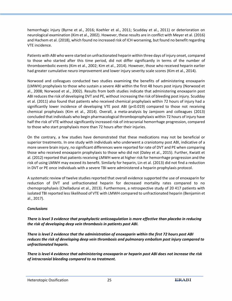
Heterotopic Ossification 25
hemorrhagic injury (Byrne et al., 2016; Koehler et al., 2011; Scudday et al., 2011) or deterioration on neurological examination (Kim et al., 2002). However, these results are in conflict with Meyer et al. (2016) and Hachem et al. (2018), which found no increased risk of ICH worsening, but found no benefit regarding VTE incidence. Patients with ABI who were started on unfractionated heparin within three days of injury onset, compared to those who started after this time period, did not differ significantly in terms of the number of thromboembolic events (Kim et al., 2002; Kim et al., 2014). However, those who received heparin earlier had greater cumulative neuro improvement and lower injury severity scale scores (Kim et al., 2014). Norwood and colleagues conducted two studies examining the benefits of administering enoxaparin (LMWH) prophylaxis to those who sustain a severe ABI within the first 48 hours post injury (Norwood et al., 2008; Norwood et al., 2002). Results from both studies indicate that administering enoxaparin post ABI reduces the risk of developing DVT and PE, without increasing the risk of bleeding post injury. Scudday et al. (2011) also found that patients who received chemical prophylaxis within 72 hours of injury had a significantly lower incidence of developing VTE post ABI (p<0.019) compared to those not receiving chemical prophylaxis (Kim et al., 2014). Overall, a meta-analysis by Jamjoom and colleagues (2013) concluded that individuals who begin pharmacological thromboprophylaxis within 72 hours of injury have half the risk of VTE without significantly increased risk of intracranial hemorrhage progression, compared to those who start prophylaxis more than 72 hours after their injuries. On the contrary, a few studies have demonstrated that these medications may not be beneficial or superior treatments. In one study with individuals who underwent a craniotomy post ABI, indicative of a more severe brain injury, no significant differences were reported for rate of DVT and PE when comparing those who received enoxaparin prophylaxis to those who did not (Daley et al., 2015). Further, Kwiatt et al. (2012) reported that patients receiving LMWH were at higher risk for hemorrhage progression and the risk of using LMWH may exceed its benefit. Similarly for heparin, Lin et al. (2013) did not find a reduction in DVT or PE once individuals with a severe TBI were administered a heparin prophylaxis protocol. A systematic review of twelve studies reported that overall evidence supported the use of enoxaparin for reduction of DVT and unfractionated heparin for decreased mortality rates compared to no chemoprophylaxis (Chelladurai et al., 2013). Furthermore, a retrospective study of 20 417 patients with isolated TBI reported less likelihood of VTE with LMWH compared to unfractionated heparin (Benjamin et al., 2017). Conclusions There is level 3 evidence that prophylactic anticoagulation is more effective than placebo in reducing the risk of developing deep vein thrombosis in patients post ABI. There is level 2 evidence that the administration of enoxaparin within the first 72 hours post ABI reduces the risk of developing deep vein thrombosis and pulmonary embolism post injury compared to unfractionated heparin.
There is level 4 evidence that administering enoxaparin or heparin post ABI does not increase the risk of intracranial bleeding compared to no treatment.
Heterotopic Ossification 26
Administration of pharmacological thromboembolic prophylaxis within the first 72 hours post ABI
may be effective for reducing the risk of developing venous thromboembolism.
Enoxaparin is effective for the prevention of venous thromboembolism development after elective neurosurgery and has not been found to cause excessive bleeding.
11.3 Conclusions Neurogenic HO is common but the actual etiology is not completely understood and there is a need to research effective prophylactic interventions. The most effective intervention for the treatment of HO overall is surgical excision. The most well studied intervention for the prevention of VTE is pharmacological thromboprophylaxis. There is moderate evidence that pharmacological prophylaxis with heparin, particularly LMWH helps reduce the risk of developing a VTE post ABI, without increasing the risk of intracranial bleeding in patients with TBI. Compression stockings are not more effective than LMWH. Unfortunately, the evidence in ABI specific populations is limited; the timing, agent and methods of anticoagulation therapy would benefit from more research.
Heterotopic Ossification 27
11.4 Summary There is level 2 evidence that etidronate disodium may reduce the incidence of heterotopic ossification compared to no etidronate disodium in patients with severe head injury. There is level 4 evidence that forceful manipulation under general anesthesia may increase range of motion and prevent bony ankylosis in patients with heterotopic ossification following brain injury. There is level 4 evidence that radiation therapy may prevent further formation of heterotopic ossification in ABI populations. There is level 4 evidence that radiation therapy may improve range of motion in joints affected by heterotopic ossification in ABI populations.
There is level 4 evidence that extracorporeal shock wave therapy may reduce pain associated with heterotopic ossification in ABI populations. There is level 4 evidence that surgical excision of heterotopic ossification may improve range of motion in ABI populations.
There is level 3 evidence that early surgical excision does not increase the recurrence risk of heterotopic ossification compared to delayed surgical excision. There is level 2 evidence that sequential compression devices are not more effective for reducing the risk of developing deep vein thrombosis or pulmonary embolism compared to no sequential compression devices post ABI. There is level 4 evidence that intermittent compression devices do not cause acute elevations in intracranial pressure in patients with severe ABI. There is conflicting evidence regarding the effectiveness of intermittent pneumatic compression devices compared to low-molecular-weight heparin or unfractionated heparin for the prophylaxis of DVT and PE. There is level 3 evidence that prophylactic anticoagulation is more effective than placebo in reducing the risk of developing deep vein thrombosis in patients post ABI. There is level 2 evidence that the administration of enoxaparin within the first 72 hours post ABI reduces the risk of developing deep vein thrombosis and pulmonary embolism post injury compared to unfractionated heparin.
There is level 4 evidence that administering enoxaparin or heparin post ABI does not increase the risk of intracranial bleeding compared to no treatment.
Heterotopic Ossification 28
11.5 References Agnelli, G., Piovella, F., Buoncristiani, P., Severi, P., Pini, M., D'Angelo, A., Beltrametti, C., Damiani, M.,
Andrioli, G. C., Pugliese, R., Iorio, A., & Brambilla, G. (1998). Enoxaparin plus compression stockings compared with compression stockings alone in the prevention of venous thromboembolism after elective neurosurgery. New England Journal of Medicine, 339(2), 80-85.
Akman, M. N., Cetin, N., Bayramoglu, M., Isiklar, I., & Kilinc, S. (2004). Value of the D-dimer test in diagnosing deep vein thrombosis in rehabilitation inpatients. Archives of Physical Medicine and Rehabilitation, 85(7), 1091-1094.
Albrecht, J. S., Liu, X., Baumgarten, M., Langenberg, P., Rattinger, G. B., Smith, G. S., Gambert, S. R., Gottlieb, S. S., & Zuckerman, I. H. (2014). Benefits and risks of anticoagulation resumption following traumatic brain injury. Journal of the American Medical Association: Internal Medicine, 174(8), 1244-1251.
Almangour, W., Schnitzler, A., Salga, M., Debaud, C., Denormandie, P., & Genet, F. (2016). Recurrence of heterotopic ossification after removal in patients with traumatic brain injury: A systematic review. Annals of Physical Rehabilitation Medicine, 59(4), 263-269.
Balboni, T. A., Gobezie, R., & Mamon, H. J. (2006). Heterotopic ossification: Pathophysiology, clinical features, and the role of radiotherapy for prophylaxis. International Journal of Radiation Oncology*Biology*Physics, 65(5), 1289-1299.
Banovac, K., & Gonzalez, F. (1997). Evaluation and management of heterotopic ossification in patients with spinal cord injury. Spinal Cord, 35(3), 158-162.
Banovac, K., Sherman, A. L., Estores, I. M., & Banovac, F. (2004). Prevention and treatment of heterotopic ossification after spinal cord injury. Journal of Spinal Cord Medicine, 27(4), 376-382.
Banovac, K., Williams, J. M., Patrick, L. D., & Haniff, Y. M. (2001). Prevention of heterotopic ossification after spinal cord injury with indomethacin. Spinal Cord, 39(7), 370-374.
Benjamin, E., Recinos, G., Aiolfi, A., Inaba, K., & Demetriades, D. (2017). Pharmacological Thromboembolic Prophylaxis in Traumatic Brain Injuries: Low Molecular Weight Heparin Is Superior to Unfractionated Heparin. Annals of Surgery, 266(3), 463-469.
Bidner, S. M., Rubins, I. M., Desjardins, J. V., Zukor, D. J., & Goltzman, D. (1990). Evidence for a humoral mechanism for enhanced osteogenesis after head injury. Journal of Bone and Joint Surgery - Series A, 72(8), 1144-1149.
Brady, R. D., Shultz, S. R., McDonald, S. J., & O'Brien, T. J. (2018). Neurological heterotopic ossification: Current understanding and future directions. Bone, 109, 35-42.
Brooker, A. F., Bowerman, J. W., Robinson, R. A., & Riley, L. H., Jr. (1973). Ectopic ossification following total hip replacement. Incidence and a method of classification. Journal of Bone and Joint Surgery, 55(8), 1629-1632.
Browse, N. L. (1974). Current thoughts on venous thromboembolism. Surgical Clinics of North America, 54(1), 229-238.
Buschbacher, R. (1992). Heterotopic ossification: a review. Clinical Review of Physical Medicine & Rehabilitation, 4, 199-213.
Byrne, J. P., Geerts, W., Mason, S. A., Gomez, D., Hoeft, C., Murphy, R., Neal, M., & Nathens, A. B. (2017). Effectiveness of low-molecular-weight heparin versus unfractionated heparin to prevent pulmonary embolism following major trauma: A propensity-matched analysis. Journal of Trauma and Acute Care Surgery, 82(2), 252-262.
Byrne, J. P., Mason, S. A., Gomez, D., Hoeft, C., Subacius, H., Xiong, W., Neal, M., Pirouzmand, F., & Nathens, A. B. (2016). Timing of Pharmacologic Venous Thromboembolism Prophylaxis in Severe Traumatic Brain Injury: A Propensity-Matched Cohort Study. Journal of the American College of Surgeons, 223(4), 621-631.
Heterotopic Ossification 29
Carlile, M., Nicewander, D., Yablon, S. A., Brown, A., Brunner, R., Burke, D., Chae, H., Englander, J., Flanagan, S., Hammond, F., Khademi, A., Lombard, L. A., Meythaler, J. M., Mysiw, W. J., Zafonte, R., & Diaz-Arrastia, R. (2010). Prophylaxis for venous thromboembolism during rehabilitation for traumatic brain injury: A multicenter observational study. Journal of Trauma - Injury, Infection and Critical Care, 68(4), 916-923.
Carlile, M. C., Yablon, S. A., Mysiw, W. J., Frol, A. B., Lo, D., & Diaz-Arrastia, R. (2006). Deep venous thrombosis management following traumatic brain injury: a practice survey of the traumatic brain injury model systems. Journal of Head Trauma & Rehabilitation, 21(6), 483-490.
Chalidis, B., Stengel, D., & Giannoudis, P. V. (2007). Early excision and late excision of heterotopic ossification after traumatic brain injury are equivalent: a systematic review of the literature. Journal of Neurotrauma, 24(11), 1675-1686.
Chantraine, A., & Minaire, P. (1981). Para-osteo-arthropathies. A new theory and mode of treatment. Scandinavian Journal of Rehabilitation Medicine, 13(1), 31-37.
Charnley, G., Judet, T., Garreau de Loubresse, C., & Mollaret, O. (1996). Excision of heterotopic ossification around the knee following brain injury. Injury, 27(2), 125-128.
Chelladurai, Y., Stevens, K. A., Haut, E. R., Brotman, D. J., Sharma, R., Shermock, K. M., Kebede, S., Singh, S., & Segal, J. B. (2013). Venous thromboembolism prophylaxis in patients with traumatic brain injury: A systematic review. F1000Research, 2.
Cifu, D. X., Kaelin, D. L., & Wall, B. E. (1996). Deep venous thrombosis: incidence on admission to a brain injury rehabilitation program. Archives of Physical Medicine & Rehabilitation, 77(11), 1182-1185.
Cipriano, C. A., Pill, S. G., & Keenan, M. A. (2009). Heterotopic ossification following traumatic brain injury and spinal cord injury. Journal of the American Academy of Orthopaedic Surgeons, 17(11), 689-697.
Citta-Pietrolungo, T. J., Alexander, M. A., & Steg, N. L. (1992). Early detection of heterotopic ossification in young patients with traumatic brain injury. Archives of Physical Medicine & Rehabilitation, 73(3), 258-262.
Collaborative overview of randomised trials of antiplatelet therapy - III: Reduction in venous thrombosis and pulmonary embolism by antiplatelet prophylaxis among surgical and medical patients. (1994). British Medical Journal, 308(6923), 235-246.
Collins, S. (2009). Deep vein thrombosis - an overview. Practice Nurse, 37(9), 23-25,27. Corrigan, T. P., Fossard, D. P., Spindler, J., Armstrong, P., Strachan, C. J., Johnston, K. W., & Kakkar, V. V.
(1974). Phlebography in the management of pulmonary embolism. British Journal of Surgery, 61(6), 484-488.
Crawford, C. M., Varghese, G., Mani, M. M., & Neff, J. R. (1986). Heterotopic ossification: Are range of motion exercises contraindicated? Journal of Burn Care and Rehabilitation, 7(4), 323-327.
Daley, M. J., Ali, S., & Brown, C. V. (2015). Late venous thromboembolism prophylaxis after craniotomy in acute traumatic brain injury. The American Surgeons, 81(2), 207-211.
Davidson, J. E., Willms, D. C., & Hoffman, M. S. (1993). Effect of intermittent pneumatic leg compression on intracranial pressure in brain-injured patients. Critical Care Medicine, 21(2), 224-227.
De Maeseneer, M. G., Bochanen, N., van Rooijen, G., & Neglen, P. (2016). Analysis of 1,338 Patients with Acute Lower Limb Deep Venous Thrombosis (DVT) Supports the Inadequacy of the Term "Proximal DVT". European Journal of Vascualr and Endovascular Surgery, 51(3), 415-420.
de Palma, L., Rapali, S., Paladini, P., & Ventura, A. (2002). Elbow heterotopic ossification in head-trauma patients: Diagnosis and treatment. Orthopedics, 25(6), 665-668.
Decousus, H., Leizorovicz, A., Parent, F., Page, Y., Tardy, B., Girard, P., Laporte, S., Faivre, R., Charbonnier, B., Barral, F. G., Huet, Y., & Simonneau, G. (1998). A clinical trial of vena caval filters in the prevention of pulmonary embolism in patients with proximal deep-vein thrombosis. New England Journal of Medicine, 338(7), 409-415.
Heterotopic Ossification 30
Della Valle, A. G., Ruzo, P. S., Pavone, V., Tolo, E., Mintz, D. N., & Salvati, E. A. (2002). Heterotopic ossification after total hip arthroplasty: a critical analysis of the Brooker classification and proposal of a simplified rating system. Journal of Arthroplasty, 17(7), 870-875.
Dengler, B. A., Mendez-Gomez, P., Chavez, A., Avila, L., Michalek, J., Hernandez, B., Grandhi, R., & Seifi, A. (2016). Safety of Chemical DVT Prophylaxis in Severe Traumatic Brain Injury with Invasive Monitoring Devices. Neurocritical Care, 25(2), 215-223.
Denson, K., Morgan, D., Cunningham, R., Nigliazzo, A., Brackett, D., Lane, M., Smith, B., & Albrecht, R. (2007). Incidence of venous thromboembolism in patients with traumatic brain injury. American Journal of Surgery, 193(3), 380-383; Discussion 383-384.
Di Nisio, M., van Es, N., & Buller, H. R. (2016). Deep vein thrombosis and pulmonary embolism. Lancet, 388(10063), 3060-3073.
Dizdar, D., Tiftik, T., Kara, M., Tunc, H., Ersoz, M., & Akkus, S. (2013). Risk factors for developing heterotopic ossification in patients with traumatic brain injury. Brain Inuryj, 27(7-8), 807-811.
Ellerin, B. E., Helfet, D., Parikh, S., Hotchkiss, R. N., Levin, N., Nisce, L., Nori, D., & Moni, J. (1999). Current therapy in the management of heterotopic ossification of the elbow: A review with case studies. American Journal of Physical Medicine and Rehabilitation, 78(3), 259-271.
Evans, E. B. (1991). Heterotopic bone formation in thermal burns. Clinical Orthopedics and Related Research (263), 94-101.
Farooqui, A., Hiser, B., Barnes, S. L., & Litofsky, N. S. (2013). Safety and efficacy of early thromboembolism chemoprophylaxis after intracranial hemorrhage from traumatic brain injury. Journal of Neurosurgery, 119(6), 1576-1582.
Fransen, M., & Neal, B. (2004). Non‐steroidal anti‐inflammatory drugs for preventing heterotopic bone formation after hip arthroplasty. Cochrane Database of Systematic Reviews(3).
Fuller, D. A., Mani, U. S., & Keenan, M. A. (2013). Heterotopic ossification of the shoulder in patients with traumatic brain injury. Journal of Shoulder and Elbow Surgery, 22(1), 52-56.
Fuller, D. A., Mark, A., & Keenan, M. A. E. (2005). Excision of heterotopic ossification from the knee: A functional outcome study. Clinical Orthopedics and Related Research(438), 197-203.
Garcia-Fuster, M. J., Fabia, M. J., Furio, E., Pichler, G., Redon, J., Forner, M. J., & Martinez, F. (2014). Should we look for silent pulmonary embolism in patients with deep venous thrombosis? BMC Cardiovascular Disorders, 14, 178.
Garland, D. E. (1991). A clinical perspective on common forms of acquired heterotopic ossification. Clinical Orthopedics and Related Research (263), 13-29.
Garland, D. E., Blum, C. E., & Waters, R. L. (1980). Periarticular heterotopic ossification in head-injured adults. Incidence and location. Journal of Bone and Joint Surgery - Series A, 62(7), 1143-1146.
Garland, D. E., Razza, B. E., & Waters, R. L. (1982). Forceful joint manipulation in head-injured adults with heterotopic ossification. Clinical Orthopedics and Related Research, No. 169, 133-138.
Geerts, W. H., Code, K. I., Jay, R. M., Chen, E., & Szalai, J. P. (1994). A prospective study of venous thromboembolism after major trauma. New England Journal of Medicine, 331(24), 1601-1606.
Geerts, W. H., Jay, R. M., Code, K. I., Chen, E., Szalai, J. P., Saibil, E. A., & Hamilton, P. A. (1996). A comparison of low-dose heparin with low-molecular-weight heparin as prophylaxis against venous thromboembolism after major trauma. New England Journal of Medicine, 335(10), 701-707.
Genet, F., Chehensse, C., Jourdan, C., Lautridou, C., Denormandie, P., & Schnitzler, A. (2012). Impact of the operative delay and the degree of neurologic sequelae on recurrence of excised heterotopic ossification in patients with traumatic brain injury. Journal of Head Trauma Rehabilitation, 27(6), 443-448.
Heterotopic Ossification 31
Genet, F., Jourdan, C., Schnitzler, A., Lautridou, C., Guillemot, D., Judet, T., Poiraudeau, S., & Denormandie, P. (2011). Troublesome heterotopic ossification after central nervous system damage: a survey of 570 surgeries. PLoS One, 6(1), e16632.
Genet, F., Marmorat, J. L., Lautridou, C., Schnitzler, A., Mailhan, L., & Denormandie, P. (2009). Impact of late surgical intervention on heterotopic ossification of the hip after traumatic neurological injury. Journal of Bone Joint Surgery, 91(11), 1493-1498.
Gersin, K., Grindlinger, G. A., Lee, V., Dennis, R. C., Wedel, S. K., & Cachecho, R. (1994). The efficacy of sequential compression devices in multiple trauma patients with severe head injury. Journal of Trauma, 37(2), 205-208.
Gill, P., & Nahum, A. (2000). Improving detection of venous thromboembolism. New technology holds promise for early, precise diagnosis. Postgraduate Medicine, 108(4), 24-26, 29-32, 38-40.
Girard, P., Sanchez, O., Leroyer, C., Musset, D., Meyer, G., Stern, J. B., & Parent, F. (2005). Deep venous thrombosis in patients with acute pulmonary embolism: prevalence, risk factors, and clinical significance. Chest, 128(3), 1593-1600.
Gottschalk, A., Stein, P. D., Goodman, L. R., & Sostman, H. D. (2002). Overview of Prospective Investigation of Pulmonary Embolism Diagnosis II. Seminal Nuclear Medicine, 32(3), 173-182.
Green, D., Lee, M. Y., Ito, V. Y., Cohn, T., Press, J., Filbrandt, P. R., VandenBerg, W. C., Yarkony, G. M., & Meyer, P. R., Jr. (1988). Fixed- vs adjusted-dose heparin in the prophylaxis of thromboembolism in spinal cord injury. Joural of the American Medical Association, 260(9), 1255-1258.
Greenfield, L. J., & Michna, B. A. (1988). Twelve-year clinical experience with the Greenfield vena caval filter. Surgery, 104(4), 706-712.
Hachem, L. D., Mansouri, A., Scales, D. C., Geerts, W., & Pirouzmand, F. (2018). Anticoagulant prophylaxis against venous thromboembolism following severe traumatic brain injury: A prospective observational study and systematic review of the literature. Clinical Neurology and Neurosurgery, 175, 68-73.
Haddad, S. H., & Arabi, Y. M. (2012). Critical care management of severe traumatic brain injury in adults. Scandinavian Journal of Trauma, Resuscitation and Emergency Medicine, 20, 12.
Hirsh, J., Dalen, J. E., Deykin, D., Poller, L., & Bussey, H. (1992). Oral anticoagulants. Mechanism of action, clinical effectiveness, and optimal therapeutic range. Chest, 108(4 Suppl), 231s-246s.
Hosalkar, H., Pandya, N. K., Baldwin, K., & Keenan, M. A. (2013). Neuro-Orthopedics. In N. D. Zasler, D. I. Katz, & R. D. Zafonte (Eds.), Brain Injury Medicine. New York, NY: Demos Medical Publishing.
Huang, H., Cheng, W. X., Hu, Y. P., Chen, J. H., Zheng, Z. T., & Zhang, P. (2018). Relationship between heterotopic ossification and traumatic brain injury: Why severe traumatic brain injury increases the risk of heterotopic ossification. Journal of Orthopaedic Translation, 12, 16-25.
Hull, R., & Hirsh, J. (1983). Long-term anticoagulant therapy in patients with venous thrombosis. Archives of Internal Medicine, 143(11), 2061-2063.
Hurvitz, E. A., Mandac, B. R., Davidoff, G., Johnson, J. H., & Nelson, V. S. (1992). Risk factors for heterotopic ossification in children and adolescents with severe traumatic brain injury. Archives of Physical Medicine and Rehabilitation, 73(5), 459-462.
Hyers, T. M., Hull, R. D., & Weg, J. G. (1992). Antithrombotic therapy for venous thromboembolic disease. Chest, 108(4 Suppl), 335s-351s.
INESSS-ONF. (2017). INESSS-ONF clinical practice guidelines for the rehabilitation of adults having sustained a moderate-to-severe TBI: The Institut national d'excellence en santé et en services sociaux and Ontario Neurotrauma Foundation.
Ippolito, E., Formisano, R., Caterini, R., Farsetti, P., & Penta, F. (1999a). Operative treatment of heterotopic hip ossification in patients with coma after brain injury. Clinical Orthopedics and Related Research (365), 130-138.
Heterotopic Ossification 32
Ippolito, E., Formisano, R., Caterini, R., Farsetti, P., & Penta, F. (1999b). Resection of elbow ossification and continuous passive motion in postcomatose patients. Journal of Hand Surgery America, 24(3), 546-553.
Ippolito, E., Formisano, R., Farsetti, P., Caterini, R., & Penta, F. (1999c). Excision for the treatment of periarticular ossification of the knee in patients who have a traumatic brain injury. Journal Bone and Joint Surgery America, 81(6), 783-789.
Ivanhoe, C. B., Durand-Sanchez, A., & Spier, E. T. (2012). Acute Rehabilitation. In N. Zasler, D. Katz, R. Zafonte, & D. Arciniegas (Eds.), Brain Injury Medicine, 2nd Edition: Principles and Practice: Demos Medical Publishing.
Jamjoom, A. A. B., & Jamjoom, A. B. (2013). Safety and efficacy of early pharmacological thromboprophylaxis in traumatic brain injury: Systematic review and meta-analysis. Journal of Neurotrauma, 30(7), 503-511.
Jensen, L. L., Halar, E., Little, J. W., & Brooke, M. M. (1987). Neurogenic heterotopic ossification. American Journal of Physical Medicine, 66(6), 351-363.
Kearon, C., Ginsberg, J. S., & Hirsh, J. (1998). The role of venous ultrasonography in the diagnosis of suspected deep venous thrombosis and pulmonary embolism. Annals of Internal Medicine, 129(12), 1044-1049.
Keret, D., Harcke, H. T., Mendez, A. A., & Bowen, J. R. (1990). Heterotopic ossification in central nervous system-injured patients following closed nailing of femoral fractures. Clinical Orthopedics and Related Research (256), 254-259.
Kim, J., Gearhart, M. M., Zurick, A., Zuccarello, M., James, L., & Luchette, F. A. (2002). Preliminary report on the safety of heparin for deep venous thrombosis prophylaxis after severe head injury. Journal of Trauma, 53(1), 38-42; Discussion 43.
Kim, L., Schuster, J., Holena, D., Sims, C., Levine, J., & Pascual, J. (2014). Early initiation of prophylactic heparin in severe traumatic brain injury is associated with accelerated improvement on brain imaging. Journal of Emergencies, Trauma and Shock, 7(3), 141-148.
Kjaersgaard-Andersen, P., & Schmidt, S. A. (1986). Indomethacin for prevention of ectopic ossification after hip arthroplasty. Acta Orthopaedica Scandinivica, 57(1), 12-14.
Kleindienst, A., Harvey, H. B., Mater, E., Bronst, J., Flack, J., Herenz, K., Haupt, W. F., & Schon, R. (2003). Early antithrombotic prophylaxis with low molecular weight heparin in neurosurgery. Acta Neurochiururgica (Wien), 145(12), 1085-1090; Discussion 1090-1081.
Koehler, D. M., Shipman, J., Davidson, M. A., & Guillamondegui, O. (2011). Is early venous thromboembolism prophylaxis safe in trauma patients with intracranial hemorrhage. J Trauma, 70(2), 324-329.
Kolessar, D. J., Katz, S. D., & Keenan, M. A. (1996). Functional outcome following surgical resection of heterotopic ossification in patients with brain injury. Journal of Head Trauma Rehabilitation, 11(4), 78-87.
Kurtoglu, M., Yanar, H., Bilsel, Y., Guloglu, R., Kizilirmak, S., Buyukkurt, D., & Granit, V. (2004). Venous thromboembolism prophylaxis after head and spinal trauma: Intermittent pneumatic compression devices versus low molecular weight heparin. World Journal of Surgery, 28(8), 807-811.
Kwiatt, M. E., Patel, M. S., Ross, S. E., Lachant, M. T., MacNew, H. G., Ochsner, M. G., Norwood, S. H., Speier, L., Kozar, R., Gerber, J. A., Rowell, S., Krishnakumar, S., Livingston, D. H., Manis, G., & Haan, J. M. (2012). Is low-molecular-weight heparin safe for venous thromboembolism prophylaxis in patients with traumatic brain injury? A Western Trauma Association multicenter study. Journal of Trauma and Acute Care Surgery, 73(3), 625-628.
Heterotopic Ossification 33
Landefeld, C. S., & Goldman, L. (1989). Major bleeding in outpatients treated with warfarin: incidence and prediction by factors known at the start of outpatient therapy. American Journal of Medicine, 87(2), 144-152.
Lazarus, M. D., Guttmann, D., Rich, C. E., & Keenan, M. A. E. (1999). Heterotopic ossification resection about the elbow. NeuroRehabilitation, 12(2), 145-153.
Lee, C. H., Shim, S. J., Kim, H. J., Yang, H., & Kang, Y. J. (2016). Effects of Radiation Therapy on Established Neurogenic Heterotopic Ossification. Annals of Rehabilitation Medicine, 40(6), 1135-1139.
Lee, E. K., Namdari, S., Hosalkar, H. S., Keenan, M. A., & Baldwin, K. D. (2013). Clinical results of the excision of heterotopic bone around the elbow: a systematic review. Journal of Shoulder and Elbow Surgery, 22(5), 716-722.
Lin, M., Davis, J. V., & Wong, D. T. (2013). Evaluation of heparin prophylaxis protocol on deep venous thrombosis and pulmonary embolism in traumatic brain injury. American Surgeon, 79(10), 1050-1053.
Linan, E., O'Dell, M. W., & Pierce, J. M. (2001). Continuous passive motion in the management of heterotopic ossification in a brain injured patient. American Journal Physical Medicine and Rehabilitation, 80(8), 614-617.
Macatangay, C., Todd, S. R., & Tyroch, A. H. (2008). Thromboembolic prophylaxis with intermittent pneumatic compression devices in trauma patients: a false sense of security? Journal of Trauma Nursing, 15(1), 12-15.
Mavrogenis, A. F., Guerra, G., Staals, E. L., Bianchi, G., & Ruggieri, P. (2012). A classification method for neurogenic heterotopic ossification of the hip. Journal of Orthopedic Traumatology, 13(2), 69-78.
Melamed, E., Robinson, D., Halperin, N., Wallach, N., Keren, O., & Groswasser, Z. (2002). Brain injury-related heterotopic bone formation: treatment strategy and results. American Journal of Physical Medicine and Rehabilitation, 81(9), 670-674.
Meyer, R. M., Larkin, M., Szuflita, N., Neal, C., Tomlin, J., Armonda, R., Bailey, J., & Bell, R. (2016). Early venous thromboembolism chemoprophylaxis in combat related penetrating brain injury. Journal of Neurosurgery, 124(4), A1176.
Minshall, C. T., Eriksson, E. A., Leon, S. M., Doben, A. R., McKinzie, B. P., & Fakhry, S. M. (2011). Safety and efficacy of heparin or enoxaparin prophylaxis in blunt trauma patients with a head abbreviated injury severity score >2. Journal of Trauma, 71(2), 396-399; discussion 399-400.
Moore, A. J. E., Wachsmann, J., Chamarthy, M. R., Panjikaran, L., Tanabe, Y., & Rajiah, P. (2018). Imaging of acute pulmonary embolism: an update. Cardiovascular Diagnostic Therapies, 8(3), 225-243.
Moore, T. J. (1993). Functional outcome following surgical excision of heterotopic ossification in patients with traumatic brain injury. Journal of Orthopedic Trauma, 7(1), 11-14.
Moreta, J., & de los Mozos, J. L. M. (2014). Heterotopic Ossification after Traumatic Brain Injury. In F. Sadaka & T. Quinn (Eds.), Traumatic Brain Injury: InTech.
Nadler, S. F., Malanga, G. A., & Zimmerman, J. R. (1993). Continuous passive motion in the rehabilitation setting. A retrospective study. American Journal of Physical Medicine and Rehabilitation, 72(3), 162-165.
Norwood, S. H., Berne, J. D., Rowe, S. A., Villarreal, D. H., & Ledlie, J. T. (2008). Early venous thromboembolism prophylaxis with enoxaparin in patients with blunt traumatic brain injury. Journal of Trauma, 65(5), 1021-1026; discussion 1026-1027.
Norwood, S. H., McAuley, C. E., Berne, J. D., Vallina, V. L., Kerns, D. B., Grahm, T. W., & McLarty, J. W. (2001). A potentially expanded role for enoxaparin in preventing venous thromboembolism in high risk blunt trauma patients. Journal of the American College Surgery, 192(2), 161-167.
Norwood, S. H., McAuley, C. E., Berne, J. D., Vallina, V. L., Kerns, D. B., Grahm, T. W., Short, K., & McLarty, J. W. (2002). Prospective evaluation of the safety of enoxaparin prophylaxis for venous
Heterotopic Ossification 34
thromboembolism in patients with intracranial hemorrhagic injuries. Archives of Surgery, 137(6), 696-701; discussion 701-692.
Office of the Surgeon General. (2008). Publications and Reports of the Surgeon General The Surgeon General's Call to Action to Prevent Deep Vein Thrombosis and Pulmonary Embolism. Rockville (MD): Office of the Surgeon General (US).
Olufajo, O. A., Yorkgitis, B. K., Cooper, Z., Rios-Diaz, A., Metcalfe, D., Havens, J. M., Kelly, E., Haider, A. H., Gates, J. D., & Salim, A. (2016). How long should we fear? Long-term risk of venous thromboembolism in patients with traumatic brain injury. Journal of Trauma and Acute Care Surgery, 81(1), 71-77.
Palmowski, K., Oltmanns, U., Kreuter, M., Mottaghy, F. M., Palmowski, M., & Behrendt, F. F. (2014). Diagnosis of pulmonary embolism: conventional ventilation/perfusion SPECT is superior to the combination of perfusion SPECT and nonenhanced CT. Respiration, 88(4), 291-297.
Pansard, E., Schnitzler, A., Lautridou, C., Judet, T., Denormandie, P., & Genet, F. (2013). Heterotopic ossification of the shoulder after central nervous system lesion: indications for surgery and results. Journal of Shoulder and Elbow Surgery, 22(6), 767-774.
Pape, H. C., Lehmann, U., van Griensven, M., Gansslen, A., von Glinski, S., & Krettek, C. (2001). Heterotopic ossifications in patients after severe blunt trauma with and without head trauma: incidence and patterns of distribution. Journal of Orthopedic Trauma, 15(4), 229-237.
Pape, H. C., Marsh, S., Morley, J. R., Krettek, C., & Giannoudis, P. V. (2004). Current concepts in the development of heterotopic ossification. Journal of Bone and Joint Surgery, 86(6), 783-787.
Phelan, H. A., Eastman, A. L., Madden, C. J., Aldy, K., Berne, J. D., Norwood, S. H., Scott, W. W., Bernstein, I. H., Pruitt, J., Butler, G., Rogers, L., & Minei, J. P. (2012a). TBI risk stratification at presentation: a prospective study of the incidence and timing of radiographic worsening in the Parkland Protocol. Journal of Trauma and Acute Care Surgery, 73(2 Suppl 1), S122-127.
Phelan, H. A., Wolf, S. E., Norwood, S. H., Aldy, K., Brakenridge, S. C., Eastman, A. L., Madden, C. J., Nakonezny, P. A., Yang, L., Chason, D. P., Arbique, G. M., Berne, J., & Minei, J. P. (2012b). A randomized, double-blinded, placebo-controlled pilot trial of anticoagulation in low-risk traumatic brain injury: The Delayed Versus Early Enoxaparin Prophylaxis i (DEEP I) study. Journal of Trauma and Acute Care Surgery, 73(6), 1434-1441.
PIOPED Investigators. (1990). Value of the ventilation/perfusion scan in acute pulmonary embolism. Results of the prospective investigation of pulmonary embolism diagnosis (PIOPED). Journal of the American Medical Association, 263(20), 2753-2759.
Praeger, A. J., Westbrook, A. J., Nichol, A. D., Wijemunige, R., Davies, A. R., Lyon, S. M., Wills, J. L., Bailey, M., Rosenfeld, J. V., & Cooper, D. J. (2012). Deep vein thrombosis and pulmonary embolus in patients with traumatic brain injury: a prospective observational study. Critical care and resuscitation : journal of the Australasian Academy of Critical Care Medicine, 14(1), 10-13.
Puzas, J. E., Evarts, C. M., & Brand, J. S. (1987). The stimulus for bone formation. In B. RA. (Ed.), Hip (1987/01/01 ed., pp. 25-38). St. Louis: CV Mosby.
Raimondi, P., Bongard, O., De Moerloose, P., Reber, G., Waldvogel, F., & Bounameaux, H. (1993). D-Dimer plasma concentration in various clinical conditions: Implication for the use of this test in the diagnostic approach of venous thromboembolism. Thrombosis Research, 69(1), 125-130.
Raslan, A. M., Fields, J. D., & Bhardwaj, A. (2010). Prophylaxis for venous thrombo-embolism in neurocritical care: a critical appraisal. Neurocritical Care, 12(2), 297-309.
Reznik, J. E., Biros, E., Lamont, A. C., Sacher, Y., Kibrik, O., Milanese, S., Gordon, S., & Galea, M. P. (2017a). A preliminary investigation on the effect of extracorporeal shock wave therapy as a treatment for neurogenic heterotopic ossification following traumatic brain injury. Part I: Effects on pain. Brain Injury, 31(4), 526-532.
Heterotopic Ossification 35
Reznik, J. E., Biros, E., Sacher, Y., Kibrik, O., Milanese, S., Gordon, S., & Galea, M. P. (2017b). A preliminary investigation on the effect of extracorporeal shock wave therapy as a treatment for neurogenic heterotopic ossification following traumatic brain injury. Part II: Effects on function. Brain Injury, 31(4), 533-541.
Ritter, M. A., & Sieber, J. M. (1985). Prophylactic indomethacin for the prevention of heterotopic bone formation following total hip arthroplasty. Clin Orthop Relat Res(196), 217-225.
Rogers, R. C. (1988). Heterotopic calcification in severe head injury: a preventive programme. Brain Injury, 2(2), 169-173.
Salottolo, K., Offner, P., Levy, A. S., Mains, C. W., Slone, D. S., & Bar-Or, D. (2011). Interrupted pharmocologic thromboprophylaxis increases venous thromboembolism in traumatic brain injury. Journal of Trauma, 70(1), 19-24; discussion 25-16.
Salter, R. B. (1996). History of rest and motion and the scientific basis for early continuous passive motion. Hand Clinics, 12(1), 1-11.
Sarafis, K. A., Karatzas, G. D., & Yotis, C. L. (1999). Ankylosed hips caused by heterotopic ossification after traumatic brain injury: a difficult problem. Journal of Trauma, 46(1), 104-109.
Sazbon, L., Najenson, T., Tartakovsky, M., Becker, E., & Grosswasser, Z. (1981). Widespread periarticular new-bone formation in long-term comatose patients. Journal of Bone and Joint Surgery, 63-b(1), 120-125.
Scales, D. C., Riva-Cambrin, J., Wells, D., Athaide, V., Granton, J. T., & Detsky, A. S. (2010). Prophylactic anticoagulation to prevent venous thromboembolism in traumatic intracranial hemorrhage: a decision analysis. Critical Care, 14(2), R72.
Scudday, T., Brasel, K., Webb, T., Codner, P., Somberg, L., Weigelt, J., Herrmann, D., & Peppard, W. (2011). Safety and efficacy of prophylactic anticoagulation in patients with traumatic brain injury. Journal of the American College of Surgeons, 213(1), 148-153; discussion 153-144.
Seifi, A., Dengler, B., Martinez, P., & Godoy, D. A. (2018). Pulmonary embolism in severe traumatic brain injury. Journal of Clinical Neuroscience, 57, 46-50.
Simonsen, L. L., Sonne-Holm, S., Krasheninnikoff, M., & Engberg, A. W. (2007). Symptomatic heterotopic ossification after very severe traumatic brain injury in 114 patients: incidence and risk factors. Injury, 38(10), 1146-1150.
Spielman, G., Gennarelli, T. A., & Rogers, C. R. (1983). Disodium etidronate: its role in preventing heterotopic ossification in severe head injury. Archive of Physical Medicine and Rehabilitation, 64(11), 539-542.
Stein, P. D., Fowler, S. E., Goodman, L. R., Gottschalk, A., Hales, C. A., Hull, R. D., Leeper, K. V., Jr., Popovich, J., Jr., Quinn, D. A., Sos, T. A., Sostman, H. D., Tapson, V. F., Wakefield, T. W., Weg, J. G., & Woodard, P. K. (2006). Multidetector computed tomography for acute pulmonary embolism. New England Journal of Medicine, 354(22), 2317-2327.
Stover, S. L., Niemann, K. M., & Tulloss, J. R. (1991). Experience with surgical resection of heterotopic bone in spinal cord injury patients. Clinical Orthopedics and Related Research(263), 71-77.
Tang, M. E., & Lobel, D. A. (2009). Severe traumatic brain injury: maximizing outcomes. Mount Sinai Journal of Medicine, 76(2), 119-128.
Toom, A., Fischer, K., Martson, A., Rips, L., & Haviko, T. (2005). Inter-observer reliability in the assessment of heterotopic ossification: proposal of a combined classification. International Orthopedics, 29(3), 156-159.
Trentz, O. A., Handschin, A. E., Bestmann, L., Hoerstrup, S. P., Trentz, O. L., & Platz, A. (2005). Influence of brain injury on early posttraumatic bone metabolism. Critical Care Medicine, 33(2), 399-406.
Turpie, A. G. G., Hirsh, J., Gent, M., Julian, D., & Johnson, J. (1989). Prevention of deep vein thrombosis in potential neurosurgical patients. A randomized trial comparing graduated compression
Heterotopic Ossification 36
stockings alone or graduated compression stockings plus intermittent pneumatic compression with control. Archives of Internal Medicine, 149(3), 679-681.
Van Gent, J. M., Bandle, J., Calvo, R. Y., Zander, A. L., Olson, E. J., Shackford, S. R., Peck, K. A., Sise, C. B., & Sise, M. J. (2014). Isolated traumatic brain injury and venous thromboembolism. Journal of Trauma and Acute Care Surgery, 77(2), 238-242.
van Kampen, P. J., Martina, J. D., Vos, P. E., Hoedemaekers, C. W., & Hendricks, H. T. (2011). Potential risk factors for developing heterotopic ossification in patients with severe traumatic brain injury. Journal of Head Trauma and Rehabilitation, 26(5), 384-391.
van Susante, J. L., Buma, P., Kirn, H. K., & Salter, R. B. (1996). Traumatic heterotopic bone formation in the quadriceps muscle. No progression by continuous passive motion in rabbits. Acta Orthopaedica Scandinivica, 67(5), 450-454.
Vanden Bossche, L., & Vanderstraeten, G. (2005). Heterotopic ossification: a review. Journal of Rehabilitation Medicine, 37(3), 129-136.
Varghese, G. (1992). Traumatic brain injury: heterotopic ossification. Physical Medicine Rehabilitation Clinics, 3, 407-415.
Vergouwen, M. D., Roos, Y. B., & Kamphuisen, P. W. (2008). Venous thromboembolism prophylaxis and treatment in patients with acute stroke and traumatic brain injury. Current Opinion Critical Care, 14(2), 149-155.
Watanabe, & Sant. (2001). Common medical complications of traumatic brain injury. . Physical Medicine and Rehabilitation:State of the Art Reviews, 15, 283-299.
Williams, J. T., Southerland, S. S., Souza, J., Calcutt, A. F., & Cartledge, R. G. (1999). Cells isolated from adult human skeletal muscle capable of differentiating into multiple mesodermal phenotypes. American Surgeon, 65(1), 22-26.
Worku, B., Gulkarov, I., Girardi, L. N., & Salemi, A. (2014). Pulmonary Embolectomy in the Treatment of Submassive and Massive Pulmonary Embolism. Cardiology, 129(2), 106-110.
Zierler, B. K. (2004). Ultrasonography and diagnosis of venous thromboembolism. Circulation, 109(12 Suppl 1), I9-14.
Zychowicz, M. E. (2013). Pathophysiology of heterotopic ossification. Orthopaedic Nursing, 32(3), 173-177.