11th annual amygdala, stress and ptsd conference: … annual amygdala, stress and ptsd conference:...
TRANSCRIPT
1
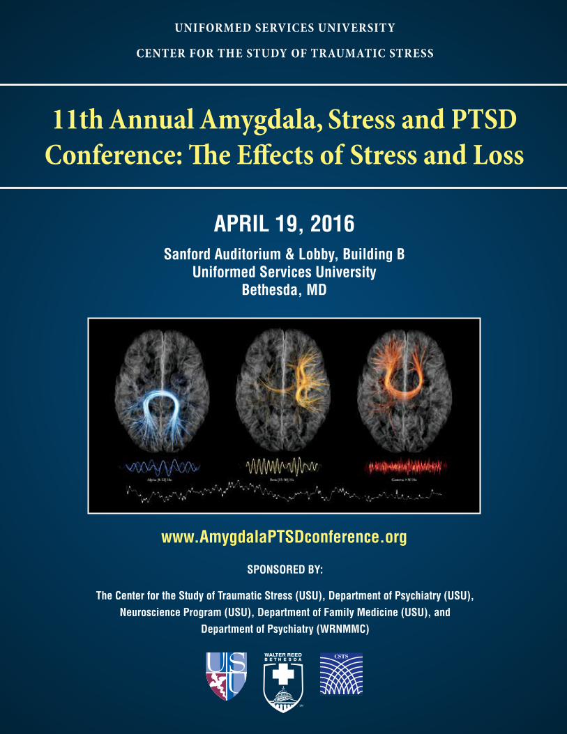
11th Annual Amygdala, Stress and PTSD Conference: The Effects of Stress and Loss
11th Annual Amygdala, Stress and PTSD Conference: The Effects of Stress and Loss
UNIFORMED SERVICES UNIVERSITY
CENTER FOR THE STUDY OF TRAUMATIC STRESS
APRIL 19, 2016 Sanford Auditorium & Lobby, Building B
Uniformed Services UniversityBethesda, MD
SPONSORED BY:
The Center for the Study of Traumatic Stress (USU), Department of Psychiatry (USU), Neuroscience Program (USU), Department of Family Medicine (USU), and
Department of Psychiatry (WRNMMC)
www.AmygdalaPTSDconference.org

2
11th Annual Amygdala, Stress and PTSD Conference: The Effects of Stress and Loss
3
11th Annual Amygdala, Stress and PTSD Conference: The Effects of Stress and Loss
11th Annual Amygdala, Stress and PTSD Conference: The Effects of Stress and Loss
UNIFORMED SERVICES UNIVERSITY
CENTER FOR THE STUDY OF TRAUMATIC STRESS
APRIL 19, 2016 Sanford Auditorium & Lobby, Building B
Uniformed Services UniversityBethesda, MD
SPONSORED BY:
The Center for the Study of Traumatic Stress (USU), Department of Psychiatry (USU), Neuroscience Program (USU), Department of Family Medicine (USU), and
Department of Psychiatry (WRNMMC)
www.AmygdalaPTSDconference.org
4
11th Annual Amygdala, Stress and PTSD Conference: The Effects of Stress and Loss
Background
The Amygdala Conference series is sponsored by the Center for the Study of Traumatic Stress under the direction of Robert J. Ursano, MD, the USU Graduate Neuroscience Program under the direction of Sharon L. Juliano, PhD, and also by the Department of Family Medicine under the direction of Mark Stephens, MD.
1
11th Annual Amygdala, Stress and PTSD Conference: The Effects of Stress and Loss
Table of Contents
Small Table Discussion Groups ................................................................................... 2
Agenda ............................................................................................................................ 3
Conference Speakers ..................................................................................................... 4
Moderators ..................................................................................................................... 7
Conference Leadership ................................................................................................. 8
Conference Committee .............................................................................................. 10
Conference Posters ...................................................................................................... 11
Continuing Education Credit .................................................................................... 26
2
11th Annual Amygdala, Stress and PTSD Conference: The Effects of Stress and Loss
Small Table Discussion GroupsUSU Cafeteria 0800–0850
Table 1: Mar Sanchez, PhDDevelopmental Impact of Early Life Stress on Primate Emotional and Stress Regulation: Prefrontal-Amygdala Circuits
Table 2: Patrizia Casaccia, MD, PhDThe Stress of Aging and Isolation on Epigenetic Regulation of Myelination
Table 3: Naomi M. Simon, MDUnderstanding the Diagnosis and Treatment of Complicated Grief: A Post-Loss Stress Disorder
Please join our small group discussions from 0800–0850 in the USU cafeteria, Building B. Each of the conference speakers will be available for more personalized and direct interaction with conference attendees. Separate registration for the discussion sessions is not necessary. Everyone registered for the conference is welcome and encouraged to attend.
Speakers
Table 4: Michael S. Fanselow, PhDNeural Mechanisms of Post Traumatic Stress Disorder as Seen Through Stress-Enhanced Fear Learning
Table 5: David S. Krantz, PhDStress, Anger, and Coronary Heart Disease: Downstream Effects of PTSD
3
11th Annual Amygdala, Stress and PTSD Conference: The Effects of Stress and Loss
AGENDA
0800-0900 Registration, Small Group Discussions and Poster Review
0900-0905 Conference Announcements — Gary H. Wynn, MD
0905-0915 Welcome and Introduction — Robert J. Ursano, MD
0915-1000 Mar Sanchez, PhD — Developmental Impact of Early Life Stress on Primate Emotional and Stress Regulation: Prefrontal-Amygdala Circuits
1000-1045 Patrizia Casaccia, MD, PhD — The Stress of Aging and Isolation on Epigenetic Regulation of Myelination
1045-1115 Coffee Break and Poster Review in Lobby
1115-1145 Discussion Panel 1 — Moderator, William P. Nash, MD
1145-1245 Lunch
1245-1330 Naomi M. Simon, MD — Understanding the Diagnosis and Treatment of Complicated Grief: A Post-Loss Stress Disorder
1330-1415 Michael S. Fanselow, PhD — Neural Mechanisms of Post-Traumatic Stress Disorder as seen through Stress-Enhanced Fear Learning
1415-1445 Coffee Break and Poster Review in Lobby
1445-1530 David S. Krantz, PhD — Stress, Anger, and Coronary Heart Disease: Downstream Effects of PTSD
1530-1600 Discussion Panel 2 — Moderator, Patcho N. Santiago, MD, MPH
1600-1615 Robert J. Ursano, MD — Closing Remarks
4
11th Annual Amygdala, Stress and PTSD Conference: The Effects of Stress and Loss
Conference Speakers
Patrizia Casaccia, MD, PhD
Patrizia Casaccia, MD, PhD is an internationally recognized expert in the field of myelin-ation and axonal damage in demyelinating disorders. She is a Professor of Neuroscience, Genomics and Multiscale Biology, and Neurology at the Icahn School of Medicine at Mount Sinai in New York. She received her medical degree
in Rome, where she was also a resident in Neurology. Dr. Casaccia then moved to the United States where she obtained a PhD in Neurobiology. After her post-doctor-al work at Weil Cornell Medical Center in New York, she moved to the Skirball Institute for Biomolecular Medi-cine at NYU and then to Robert Wood Johnson Medical School (Rutgers Medical School). In 2008 she moved to Mount Sinai, where she directs the Center of Excellence on Myelin Disorders at the Friedman Brain Institute. Her Research has a translational focus by investigating molecular and cellular mechanisms underlying myelin formation and neuroprotection.
Dr. Casaccia’s research is funded by grants from the National Institute for Neurological Disorders and Stroke, by the Department of Defense and by the Na-tional Multiple Sclerosis Society. She has more than 110 publications in top peer-reviewed journals, including Nature, Genes and Development, Nature Neuroscience, and Neuron. She has also received many honors and awards including the New Jersey Cancer Commission Award for Scientific Excellence (2001) and Recogni-tion Award Rutgers Women in Neuroscience. (2005). Dr. Casaccia is on the editorial board of multiple peer-re-viewed journals and serves on grant advisory panels for the National Institutes of Health, the Department of the Army and for the National Multiple Sclerosis Society.
Michael S. Fanselow, PhD
Dr. Michael Fanselow is Distinguished Professor, Psychology and Psychiatry Departments at the University of Los Angeles, CA. He also holds the Staglin Family Chair in Psychology and is Director of the Staglin Music Festival Center for Brain and Behav-ioral Health.
Dr. Fanselow received his MS degree from Brooklyn College in 1976, and his PhD from the University of Washington in 1980. Following his graduate work he held Assistant/Associate Professorships at Rennselaer Polytechnic Institute and Dartmouth College.
Dr. Fanselow’s laboratory has a long-standing interest in the nature and function of fear. “We want to understand the adaptive function of normal fear and how those systems go awry to produce anxiety disorders such as PTSD.” A series of questions of particular in-terest is how fear is learned and how fear memories are stored in the brain. That research concentrates its efforts on forebrain regions such as the amygdala, hippocam-pus, and neocortex. “In terms of neurotransmitter/mod-ulator systems, we have been concentrating our effort on glutamate, GABA, acetycholine, and opioids. More than simply tracing the circuits, we are trying to determine the specific contributions that different components of the circuit contribute to the complete experience of an emotional memory.” Another important question is how fear memories are translated into specific behavior patterns. That work primarily focuses on the midbrain periaqueductal gray. The laboratory uses rat and mouse models featuring site-specific pharmacological, opto-genetic and chemogenetic manipulations. Much of the current work examines behavior in genetically modified mice. “Our mission is to use every technique available to derive a complete understanding of fear-motivated be-havior and how to return the pathologically frightened brain to a state where fear and anxiety again serve the adaptive healthy defensive function they evolved for.”
5
11th Annual Amygdala, Stress and PTSD Conference: The Effects of Stress and Loss
David S. Krantz, PhD
David S. Krantz, PhD is Professor and former Chair in the Department of Medical and Clinical Psychology at Uniformed Services Univer-sity (USU), and a member of the Scientific Advisory Board of the Center for the Study of Traumatic Stress (CSTS). He received his BS from the
CCNY and PhD in Psychology from the University of Texas at Austin. Dr. Krantz has authored more than 180 scientific and professional publications on the effects of psychosocial stress on physical health, with a particular emphasis on psychosocial and biobehavioral factors in cardiovascular disorders. Dr. Krantz was the recipient of the American Psychological Association’s (APA) Early Career Scientific Award in 1982, and Annual Awards from the APA Division of Health Psychology in 1981 and 2000. He is a former Editor-in-Chief of the journal Health Psychology, Past-President of the APA Division of Health Psychology, and a Past President of the Acade-my of Behavioral Medicine Research.
For more than 20 years, the primary focus of Dr. Krantz’s work has been on the effects of acute stress as a trigger of clinical coronary heart disease events such as myocardial ischemia and malignant arrhythmias. In this research, he has collaborated with a multi-disciplinary team of cardiologists and psychologists to examine the prevalence, mechanisms, and prognostic significance of mental stress-induced ischemia. This work was later extended to investigating the effects of mental stress on markers of vulnerability to malignant arrhythmias. Cur-rently, he is Principal Investigator of the BETRHEART Study, which investigates the role of biobehavioral factors in the progression of heart failure, and is also a part of a team examining psychosocial influences on coronary heart disease in women (the NHLBI WISE study). Most recently, he has begun collaboration with Dr. Vincent Ho and other colleagues at USU on a “brain-heart initiative” to study the study the complex relationships between PTSD, mTBI, and coronary artery disease among combat veterans.
Mar Sanchez, PhD
Dr. Sanchez is an Associate Pro-fessor in the Department of Psy-chiatry & Behavioral Sciences, Emory University School of Medicine and Affiliate Scientist in the Division of Developmen-tal and Cognitive Neuroscience, Yerkes National Primate Re-search Center, Emory University.
Dr. Sanchez received her PhD in Neurobiology and Cell Biology from the Com-plutense University of Madrid, Spain and has expertise in stress neurobiology and behavioral neuroscience. Her lab studies the neurobiology of stress and emotion-al regulation during development, using nonhuman primate models. The main focus of her research pro-gram is to understand how early life stress (in particular, alterations in maternal care) affects the development of brain circuits that control emotional and stress reactivi-ty from infancy through adolescence, leading to psycho-pathology and pathophysiology characteristic of anxiety and mood disorders. These brain systems include pre-frontal-amygdala circuits. She uses a multidisciplinary approach, including analysis of neuroendocrine stress systems, social and emotional behavior and brain devel-opment using in vivo neuroimaging techniques (MRI, DTI, PET and resting state fMRI) and in vitro assays of brain neuropeptide and corticosteroid systems. She has established a successful research program, supported by NIH and NSF funding and by awards from private foundations (e.g. NARSAD, ASF and KTGF). Current awards include R01 grants from NIH/NICHD and NIH/NIDA and is co-PI in a project part of the Emory Autism Center of Excellence. Dr. Sanchez’s multidis-ciplinary and translational research bridges different disciplines and strong collaborations with clinical and basic researchers that study human developmental psychobiology, psychopathology, behavioral genetics/epigenetics and psychoneuroimmunology at Emory and other universities (Minnesota, Madison-Wisconsin, Maryland, OHSU, Pittsburgh, NYU, UNC) and the NIH. She is a member of the NIH CSR Biobehavioral Regula-tion, Learning and Ethology (BRLE) Study Section and serves in the editorial board of several journals, includ-ing Psychoneuroendoendocrinology and Development and Psychopathology.
6
11th Annual Amygdala, Stress and PTSD Conference: The Effects of Stress and Loss
Naomi M. Simon, MD
Dr. Naomi M. Simon is Pro-fessor of Psychiatry, Harvard Medical School, Director of the Center for Anxiety and Traumatic Stress Disorders, Director of the Complicat-ed Grief Program at MGH, and Chief Medical Officer of the Red Sox Foundation and
Massachusetts General Hospital Home Base Program. Dr. Simon’s major clinical and research interests include optimizing initial and next step psychotherapy and medication treatments for anxiety and stress related disorders, and understanding the presentation and the biological impact of trauma, loss, and anxiety disorders. She has served as a principal investigator or co-inves-tigator on numerous studies aimed at improving our understanding and treatment of post-traumatic stress disorder, panic disorder, generalized anxiety disorder,
social anxiety disorder, and the syndrome of compli-cated grief. As Chief Medical Officer at Home Base, she guided development of a novel clinical care model and the Home Base Training Institute through a public private partnership with MGH and the Red Sox Foun-dation to address the unmet needs of returning veter-ans and their families impacted by deployment related stress, PTSD, and traumatic brain injury.
Dr. Simon is Vice Chair of the Scientific Council of the Anxiety Disorders Association of America, a Distin-guished Fellow of the American Psychiatric Association, and a Member of the American College of Neuropsy-chopharmacology. She received an MD from Harvard Medical School, and completed a medical internship and residency in psychiatry at Columbia Presbyterian Hospital/New York State Psychiatric Institute. In addi-tion, she completed fellowship training in consultation psychiatry at MGH and has a Masters in Epidemiology from the Harvard School of Public Health.
7
11th Annual Amygdala, Stress and PTSD Conference: The Effects of Stress and Loss
Moderators
William P. Nash, MD
Before becoming the Director of Psychological Health for the U.S. Marine Corps in May 2015, Bill Nash was a psy-chiatric researcher, educator, and consultant in posttrau-matic stress and moral injury prevention and treatment for DoD and the VA. While on active duty in the Navy, CAPT
Nash provided far-forward psychological health services to the 1st Marine Division during the Second Battle of Fallujah, for which he was awarded a Bronze Star, and he led the development of the current Navy and Ma-rine Corps doctrine for Combat and Operational Stress Control, including its acknowledgment of moral injury and loss as important mechanisms of psychological injury. Dr. Nash has led or collaborated on research on risk and protective factors for combat-related PTSD, new measures for early recognition of traumatic stress injuries, and new cognitive-behavioral treatments for combat-related PTSD. He holds academic appointments at the University of California, San Diego, and Virginia Commonwealth University. Dr. Nash served as a mem-ber of the Institute of Medicine’s Committee for Assess-ment of the Effectiveness of PTSD Treatment in DoD and the VA, and he co-chaired the recently completed Chairman of the Joint Chiefs’ High-Risk Behavior Working Group.
Patcho Santiago, MD, MPHCDR, MC, USN
Dr. Santiago is the Program Director of the National Cap-ital Consortium Psychiatry Residency at the Walter Reed National Military Medical Center in Bethesda, Maryland. He is a board certified psychi-atrist and Associate Professor, Department of Psychiatry at the Uniformed Services Uni-
versity (USU), Bethesda, MD. As billeted faculty at USU, Dr. Santiago served as the USU Program Manager for the Army Study to Assess Risk and Resilience in Service members (Army STARRS) as well as the Associate Pro-gram Director for the Disaster and Preventive Psychia-try Fellowship.
Dr. Santiago graduated from Yale University with a Bachelor of Arts in economics and earned his medical degree from Jefferson Medical College under the Navy’s Health Professions Scholarship Program. After complet-ing his psychiatry residency at the Naval Medical Center San Diego, his first duty assignment was at the Naval Hospital Camp Pendleton, California. From there, he deployed to the Expeditionary Medical Facility at Camp Arifjan, Kuwait in support of Operation Iraqi Freedom. Dr. Santiago subsequently completed a Disaster and Preventive Psychiatry Fellowship at USU, earning his Masters in Public Health, and ultimately joining the faculty at USU, having research interests in psychiatric epidemiology and mental health promotion. From USU, Dr. Santiago deployed to Operation Enduring Freedom as the Officer-in-Charge of the Navy Mobile Care Team, based out of Bagram Air Field, Afghanistan.
8
11th Annual Amygdala, Stress and PTSD Conference: The Effects of Stress and Loss
Conference Leadership
Robert J. Ursano, MD
Dr. Ursano is Professor of Psychiatry and Neuroscience and Chairman of the De-partment of Psychiatry at the Uniformed Services University of the Health Sciences, Bethes-da, Maryland. He is founding Director of the Center for the Study of Traumatic Stress. In addition, Dr. Ursano is Editor
of Psychiatry, the distinguished journal of interperson-al and biological processes, founded by Harry Stack Sullivan. Dr Ursano completed twenty years service in USAF medical corps and retired as Colonel in 1991. He was educated at the University of Notre Dame and Yale University School of Medicine and did his psychiatric training at Wilford Hall USAF Medical Center and Yale University.
Dr. Ursano served as the Department of Defense representative to the National Advisory Mental Health Council of the National Institutes of Mental Health and is a past member of the Veterans Affairs Mental Health Study Section and the National Institute of Mental Health Rapid Trauma and Disaster Grant Review Sec-tion. He is a Distinguished Life Fellow in the American Psychiatric Association and a Fellow of the American College of Psychiatrists. Dr. Ursano was the first Chair-man of the American Psychiatric Association’s Com-mittee on Psychiatric Dimensions of Disaster. This work greatly aided the integration of psychiatry and public health in times of disaster and terrorism. Dr. Ursano was an invited participant to the White House Mental Health Conference in 1999. He has received the De-partment of Defense Humanitarian Service Award and the highest award of the International Traumatic Stress Society, The Lifetime Achievement Award, for “outstand-ing and fundamental contributions to understanding traumatic stress.” He is the recipient of the William C. Porter Award from the Association of Military Surgeons
Gary H. Wynn, MDLTC, MC, USA
Dr. Wynn is Assistant Chair and Associate Professor, Department of Psychiatry, Uniformed Ser-vices University and Scientist, Center for the Study of Traumat-ic Stress. After graduating from West Point in 1996, Dr. Wynn received his medical degree from the Uniformed Services University (USU) in Bethesda,
MD. Dr. Wynn completed a residency in Psychiatry and Internal Medicine at Walter Reed Army Medical Center. After completing his residency, he spent a year as the Division Psychiatrist for the 2nd Infantry Division at Camp Casey, Korea. Dr. Wynn spent the next three years as the Assistant Chief of Inpatient Psychiatry at Walter Reed Army Medical Center where he worked with service members returning from the conflicts in Iraq and Afghanistan. From 2009 through 2013, Dr. Wynn worked as a research psychiatrist in the Military Psychiatry Branch of the Center for Military Psychiatry and Neuroscience at the Walter Reed Army Institute of Research in Silver Spring, MD. In July, 2013, Dr. Wynn joined the USU Department of Psychiatry. He has published textbooks on the topics of drug interaction principles for medical practice and the clinical manage-ment of post traumatic stress disorder (PTSD) as well as fourteen book chapters and seventeen journal articles. His presentations at national and local conferences have covered topics ranging from drug interactions to PTSD and mild traumatic brain injury.
Continued
9
11th Annual Amygdala, Stress and PTSD Conference: The Effects of Stress and Loss
of the United States, the William Menninger Award of the American College of Physicians and the James Leonard Award of the Uniformed Services University. He is a frequent advisor on issues surrounding psycho-logical response to trauma to the highest levels of the US Government and specifically to the Department of Defense leadership.
Dr. Ursano has served as a frequent member of the National Academies of Science, Institute of Med-icine Committees and working groups including the Committee on Psychological Responses to Terrorism, Committee on PTSD, the Committee on Compensation for PTSD in Veterans and the Committee on Nuclear Preparedness; and the National Institute of Mental Health Task Force on Mental Health Surveillance After Terrorist Attack. In addition, he has served as a member of scientific advisory boards to the Secretary of Health
Robert J. Ursano, MD, Continued
and Human Services for disaster mental health and the Centers for Disease Control for preparedness and terrorism. Dr. Ursano is co-principal investigator of the largest NIMH grant ever given for the study of Suicide in the U.S. Army. In collaboration with his co-principal investigators at Harvard University, the University of Michigan and Columbia University the Army- STARRS grant will be the Framingham Study of suicidal behav-ior, and address a national as well as DoD mental health need. In 2014, Dr. Ursano and Dr. Matthew Friedman of the VA National Center for PTSD co-founded the Friedman-Leahy Brain Bank supported through Senator Patrick Leahy (D-VT). It is the first human brain bank dedicated to PTSD. This joint effort of many people was a 12 year project developing concepts, pilot data and support. Dr. Ursano has over 300 publications. He is co-author or editor of eight books.
10
11th Annual Amygdala, Stress and PTSD Conference: The Effects of Stress and Loss
Conference CommitteeRobert J. Ursano, MDProfessor and ChairDepartment of PsychiatryDirectorCenter for the Study of Traumatic StressUniformed Services University
Gary H. Wynn, MD, 2016 Chairman Assistant Chair and Associate ProfessorDepartment of PsychiatryScientistCenter for the Study of Traumatic StressUniformed Services University
David Mears, PhD, 2016 Co-Chairman Associate Professor Department of Anatomy, Physiology and GeneticsUniformed Services University
Mary Lee Dichtel, RN Senior Medical Editor Program Coordinator Educational Support Management Services, LLC
Ronald J. Whalen, PhD LTC, MSC, USA Assistant Professor, Counseling ServicesDepartment of Family MedicineUniformed Services University
K. Nikki Benevides, MA Assistant to the Director and Scientific Program
Coordinator Center for the Study of Traumatic StressDepartment of PsychiatryUniformed Services University
Kwang Choi, PhD Department of Psychiatry and Program in
NeuroscienceCenter for the Study of Traumatic StressUniformed Services University
F. Julian Lantry, BA Research AssistantDepartment of Psychiatry Center for the Study of Traumatic StressUniformed Services University
Jorge M. Hastings,TSgt, USAF, CADC NCOIC, Department of Psychiatry Center for the Study of Traumatic StressUniformed Services University
Syrus Henderson, MSgt, USAF Senior Enlisted Advisor, AF Element Flight Chief, Department of PsychiatryAdditional Duty First SergeantUnit Reserve CoordinatorUniformed Services University
Gwendolyn Morris Program Support Specialist Department of PsychiatryUniformed Services University
We extend special thanks to:Robyn Hulvey, CMP, CGMPAssistant Director of Meetings Office of Education and MeetingsHenry M. Jackson Foundation for the
Advancement of Military Medicine
Sebastien DeryCover PhotographMcGill University (Creative Commons)
11
11th Annual Amygdala, Stress and PTSD Conference: The Effects of Stress and Loss
Conference PostersPresented in the Lobby
Use of Ecological Momentary Assessment Methodology in the Study of Posttraumatic Stress Symptoms in U.S. Military Service Members .................................................. 12
Longitudinal Assessment of White Matter Microstructure Integrity Following Combat Deployment Relates to Posttraumatic Stress Disorder Symptom Severity in Service Members with a History of Traumatic Brain Injury: A Pilot Study ........................ 13
A Comparison of Two Sub-Anesthetic Intravenous Ketamine Doses in Rat: A Pilot Study................................................................................................................................. 14
Examination of the Gateway Hypothesis in Rats ............................................................. 15
Riluzole for PTSD: Efficacy of a Glutamatergic Modulator as Augmentation Treatment for Posttraumatic Stress Disorder ................................................................................ 16
Cerebrovascular Reactivity and Neuropsychological Function in Chronic TBI .......... 17
The Amygdala as a Brain Network Hub in Health and Disease: Findings from Graph Theoretical Studies ......................................................................................................... 18
In Vivo Brain Regional Glucose Uptake Following Intravenous Morphine Self-administration in Rats: 18FDG-PET Study ................................................................. 19
The Behavioral and Physiological Effects of Maternal Separation on Spraque-Dawley Dams and Pups ............................................................................................................... 20
Organization of Cortico-amygdala Afferents Provide a Scaffold for Age Dependent Retrieval of Contextual Fear ......................................................................................... 21
Effects of Non-Combative Military Humanitarian Aid and Disaster Relief on Mental Health of U.S. Service Members ................................................................................... 22
Calbrain is Reduced in PTSD Orbitofrontal Cortex in Parallel with Reductions in Dendritic Spine Density ................................................................................................ 23
Genetic Risk Mechanisms of Post-Traumatic Stress Disorder in Human Brain ........... 24
Transcriptional Effects of Radiation and GT3 Radioprotection on the Rhesus Macaque Brain, and Anatomical Subregions Associated with the Fronto-Limbic System .... 25
12
11th Annual Amygdala, Stress and PTSD Conference: The Effects of Stress and Loss
Use of Ecological Momentary Assessment Methodology in the Study of Posttraumatic Stress Symptoms in U.S.
Military Service Members
AuthorsQuinn M. Biggs, PhD1, Carol S. Fullerton, PhD1, Deborah Probe, MA1, Russell B. Carr, MD1,2, Nicole Dacuyan, BS1, Mackenzie Franz, BA1, Maegan Paxton, BS1, Leming Wang, MS1, Robert J. Ursano, MD1
Affiliations1. Center for the Study of Traumatic Stress,
Department of Psychiatry, Uniformed Services University of the Health Sciences, Bethesda, MD, USA
2. Walter Reed National Military Medical Center, Bethesda, MD, USA
ABSTRACTBackground: Rates of trauma-related distress are high in U.S. military service members. Assessments of post traumatic stress symptoms (PTSS) and as-sociated disorders (e.g., PTSD and depression) have traditionally been done retrospectively with paper and pencil assessments. Retrospective assessment methods are susceptible to biases in memory, errors in reporting, and do not account for daily symptom changes. Ecological momentary assessment (EMA) methods involve repeated sampling of current symp-toms and experiences while respondents are engaged in normal daily routines; as such, EMA methods may provide more detailed, reliable, and up-to-date assessments compared to retrospective assessments.
Method: Sixty-nine current or former U.S. military service members completed paper and pencil or electronic questionnaire assessments of PTSS using EMA methods. Assessments were administered four times daily (fixed interval, four hours apart) for 15 days. Data collection is ongoing.
Results: Preliminary analyses indicate that partic-ipants in both paper and electronic questionnaire groups report that daily assessments increase aware-ness of their mental and physical states. Participants who completed electronic questionnaires were significantly more likely than those who completed paper questionnaires to report that they would use daily assessments on their own if the questionnaires were made available. We will present data on ques-tionnaire completion rates and helpfulness of daily assessments.
Conclusion: Repeated symptom assessment by paper or electronic questionnaire may have intrinsic benefits for those who complete the assessments and, when shared with a behavioral health care provider, may be a rich resource to inform decisions about treatment.
13
11th Annual Amygdala, Stress and PTSD Conference: The Effects of Stress and Loss
Longitudinal Assessment of White Matter Microstructure Integrity Following Combat Deployment Relates to Posttraumatic Stress
Disorder Symptom Severity in Service Members with a History of Traumatic Brain Injury: A Pilot Study
AuthorsMichelle E. Costanzo, PhD1,2,3, Suzanne Leaman, PhD1, Dzung Pham, PhD3, Krista B. Highland, PhD1,2,3, David Keyser, PhD4, Dominic E. Nathan, PhD2,4, Paul Rapp, PhD4, Michael J. Roy, MD1,3
Affiliations1. Uniformed Services University of the Health
Sciences, Department of Medicine, Bethesda, MD, USA
2. The Henry M. Jackson Foundation, Bethesda, MD, USA
3. Center for Neuroscience and Regenerative Medicine, Bethesda, MD, USA
4. Uniformed Services University of the Health Sciences, Traumatic Injury Research Program, Department of Military and Emergency Medicine, Bethesda, MD, USA
ABSTRACTBackground: Diffusion tensor imaging (DTI) holds promise for distinguishing posttraumatic stress dis-order (PTSD) from the underlying physical damage associated with mild traumatic brain injury (mTBI). This is important given the significant symptom overlap between PTSD and mTBI. Moreover, exam-ining service members (SMs) immediately following combat deployment may be important for under-standing the interaction between psychological health and brain injury recovery.
Objective: The goal of this study was to examine a cohort of combat veterans with a history of mTBI who do not have the severity of symptoms to have
a clinical diagnosis of PTSD, within 2 months of return from deployment followed by follow-up eval-uations 3-6 months later.
Method: Using the Clinician-Administered PTSD Scale (CAPS) to assess PTSD symptoms and DTI fractional anisotropy to assess the white matter microstructure, we examined 14 service members (7 with a history of combat TBI and 7 deployed con-trols) to explore if psychological distress influences brain structure and behavioral symptoms overtime.
Results: Our results revealed that white matter microstructure of the uncinate fasciculus (UF) and cingulum was not significantly correlated with CAPS scores at baseline or follow-up in the control group. For those with a history of TBI, re-experienc-ing symptoms at baseline was positively correlated with left (r=.87, p<.05) and with right (r=.77, p<.05) cingulum at follow up and hyperarousal symptoms at baseline positively correlated right (r=.79, p<.05) cingulum at follow up. Avoidance symptoms (r=-.80, p<.05) and total CAPS score (r=-.86, p<.05) at baseline were negatively correlated with left UF at baseline. No significant correlations were observed between follow up CAPS scores and white matter microstructure.
Conclusions: This suggests recent proximity to com-bat stress may influence the relationship between symptom severity and white matter microstructure both immediately following deployment and over time in those in those with a history of head injury.
14
11th Annual Amygdala, Stress and PTSD Conference: The Effects of Stress and Loss
A Comparison of Two Sub-Anesthetic Intravenous Ketamine Doses in Rat: A Pilot Study
AuthorsKennett Radford, MS, CRNA1, Thomas Park, BS2, Sean Moran, PhD4, Lisa Osborne, PhD, CRNA1, Kwang Choi, PhD1,2,3
Affiliations1. Daniel K. Inouye Graduate School of Nursing,
Uniformed Services University of the Health Sciences, Bethesda, MD, USA.
2. Department of Psychiatry, Uniformed Services University of the Health Sciences, Bethesda, MD, USA.
3. Center for Study of Traumatic Stress, Uniformed Services University of the Health Sciences, Bethesda, MD, USA.
4. Department of Biochemistry and Molecular Biology, Uniformed Services University of the Health Sciences, Bethesda, MD, USA.
ABSTRACTBackground: Ketamine is a short-acting dissociative anesthetic that is used to treat chronic pain, depres-sion, and treatment refractory post-traumatic stress disorder. Intravenous (IV) ketamine infusions are used in clinical practice, however, researchers pri-marily use intraperitoneal (IP) injections in rodent studies. Pharmacokinetic differences between the two routes lead to difficulty in translating rodent models to human clinical practice. Our primary aim was to compare the effects of two sub-anesthetic IV ketamine doses on locomotion, acoustic startle reflex (ASR)/pre-pulse inhibition (PPI), and hotplate latency in rats.
Method: Jugular vein cannulated male Sprague-Dawley rats (300±50g) were given IV ket-amine doses (0, 2, & 5 mg/kg) in a counter-balanced design. Locomotor activity (over 15 min), ASR/PPI (at 20 min), and hotplate latency (20 to 50 min) were measured. Serum ketamine/norketamine (NKT) levels were measured using liquid chromatography/mass spectrometry in a separate group of animals. Locomotor and hotplate latency were analyzed with two-way ANOVA. ASR/PPI and ketamine/NKT serum levels were analyzed with one-way ANOVA.
Results: Ketamine 2mg/kg increased locomotor activity (0–3 min), had no change in ASR or PPI, and produced antinociception (vs saline) through 40 minutes. Ketamine 5mg/kg induced rapid dis-sociative symptoms (through 4 min), increased locomotion (4–9 min) after dosing, no effect on ASR but impaired PPI at 100dB (20 min), and produced significant antinociception (vs saline and 2mg/kg) through 50 minutes. Serum ketamine rapidly peak-ed and decreased while NKT gradually increased during 50 minutes after injection. Conclusion: IV ketamine dose-dependently in-creased locomotor activity, disrupted PPI, and produced significant antinociception. Disrupted PPI with 5mg/kg IV is similar to previous ASR/PPI studies using larger IP ketamine doses. There-fore, sub-anesthetic IV ketamine is a valid route for rodent behavior models. This pilot-study is the first step in a program of research exploring the impact of anesthetic agents following traumatic injury on development of stress related disorders.
15
11th Annual Amygdala, Stress and PTSD Conference: The Effects of Stress and Loss
Examination of the Gateway Hypothesis in Rats
AuthorsKathryn E. Eklund, BS1, Kevin Nishida, BS2, Erin B. Barry, MS3, Kwang H. Choi, PhD2, Neil E. Grunberg, PhD1,3
Affiliations1. Department of Medical & Clinical Psychology,
Uniformed Services University, Bethesda, MD, USA
2. Department of Psychiatry, Uniformed Services University, Bethesda, MD, USA
3. Department of Military & Emergency Medicine, Uniformed Services University, Bethesda, MD, USA
ABSTRACTBackground: Kandel (1977) proposed the “Gateway Hypothesis” that use of a “lower or softer” drug (e.g., cigarettes) increases the likelihood of subsequent use of a “higher or harder” drug (e.g., opiate). Whether this sequence involves causality, accessibility, cultural norms, or other factors is not clear because the hy-pothesis is based on epidemiological data. Yet, public policies, drug prevention programs, and treatments have inferred a causal relationship from use of softer to harder drugs.
Method: The experiment evaluated the Gateway Hy-pothesis in a rat model. Nicotine was administered to rats via a paradigm (SC osmotic minipumps)
that models effects of ½-1 pack human daily ciga-rette use. The nicotine paradigm was followed by IV morphine self-administration. Male and female rats (N=32) were exposed to nicotine or saline, followed by the opportunity to self-administer morphine for 10 days.
Results: Rats pre-exposed to nicotine self-adminis-tered significantly less morphine compared to rats pre-exposed to saline. This finding was significant for male and female rats.
Conclusion: The results contradict a causal mech-anistic interpretation of the Gateway Hypothesis, at least with regard to adult rats. It may be that this animal model does not parallel the human condi-tion, but this interpretation is unlikely because the nicotine and opiate paradigms that were combined in the present experiment have produced findings consistent with human studies of cigarette and opiate use. Alternatively, the age of the animals may matter because the Gateway Hypothesis has been based on initial soft drug exposure during childhood and adolescence. Another possibility is that re-verse-drug tolerance may be occurring which would be reflected by the present findings, yet could still be consistent with the Gateway Hypothesis in humans if people increase the likelihood of using a second drug but without actually using more of it.
16
11th Annual Amygdala, Stress and PTSD Conference: The Effects of Stress and Loss
Riluzole for PTSD: Efficacy of a Glutamatergic Modulator as Augmentation Treatment for Posttraumatic Stress Disorder
AuthorsPatricia T. Spangler, PhD1,2, James C. West, MD1,2, Brian Andrews-Shigaki, PhD2, Kyle Possemato, PhD3, Shannon McKenzie, BA3, Maegan Paxton, BS1, Tung Tu, BS1, Krista Engle, BA1, David M. Benedek, MD1,4
Affiliations1. Center for the Study of Traumatic Stress2. Uniformed Services University of the Health
Sciences3. Syracuse Veterans Administration Medical
Center4. Center for Forensic Behavioral Science at Walter
Reed National Military Medical Center
AbstractBackground: Because current pharmacological treatments for PTSD, and particularly combat-relat-ed PTSD, are suboptimal, there is an urgent need to develop novel treatments that rapidly and robustly improve symptoms. Drugs that reduce glutamate activity may help reverse loss of neuronal integrity and focal atrophy in brain regions implicated in PTSD pathophysiology. Riluzole is a glutamater-gic modulator that inhibits glutamate release and enhances AMPA trafficking and clearance of exces-sive synaptic glutamate resulting in neuroprotective properties. The objective of this study is to evaluate the efficacy of acute riluzole augmentation treatment in active duty and combat veterans with PTSD, with or without mild TBI.
Method: Up to 100 OEF and OIF veterans at Walter Reed National Military Medical Center (WRN-MMC) and the Syracuse VA Medical Center with suboptimal response to SSRI/SNRI treatment for PTSD are currently being enrolled in this 8-week RCT. Participants continue on their current med-ication and are randomized to augmentation with either riluzole or placebo. A subgroup of eligible par-ticipants from WRNMMC are undergoing 1H MRS scans before and after treatment to evaluate changes in N-acetyl asparate (NAA) levels in the amygdala and ACC. Results: Preliminary results from WRNMMC sam-ple (n=10) indicate that participants randomized to riluzole had increases in NAA in both amygdala and ACC, whereas participants randomized to placebo had NAA concentration decreases in both regions. On psychometric measures (n=15), there was a trend toward significant difference in depression change scores, such that participants randomized to riluzole had greater decreases in depression than did those randomized to placebo.
Conclusions: Given the very small sample size for these analyses, results should be viewed with caution and should not as yet be taken as evidence of rilu-zole’s efficacy. The results do show, however, that we have been able to successfully collect psychometric and neuroimaging data for this medication trial.
17
11th Annual Amygdala, Stress and PTSD Conference: The Effects of Stress and Loss
Cerebrovascular Reactivity and Neuropsychological Function in Chronic TBI
AuthorsAaron Wolfgang, BA1, Kimbra Kenney, MD1,2, Franck Amyot, PhD2, L. Turtzo, MD, PhD3,4, Carol Moore, MA3, Erika Silverman, BA2, Christian Shenouda, MD3, Emily Spessert, BA2, Leah Harburg, BA2, Eric Wassermann, MD3,4, Ramon Diaz-Arrastia, MD, PhD1,2
Affiliations1. Uniformed Services University of the Health
Sciences, Bethesda, MD 2. Center for Neuroscience and Regenerative
Medicine, Rockville, MD 3. National Institute of Neurological Disorders and
Stroke, Bethesda, MD4. National Institutes of Health, Bethesda, MD
ABSTRACTBackground: MRI blood oxygen level dependent (BOLD) with hypercapnia challenge reliably mea-sures cerebrovascular reactivity (CVR) and can assess traumatic vascular injury. Understanding relationships among CVR and neuropsychologi-cal function may be informative for the design of therapies directed at post-traumatic cerebrovascular dysfunction.
Objective: To investigate relationships between CVR in the whole brain (WB), gray matter (GM), and white matter (WM), and neuropsychological func-tion after traumatic brain injury (TBI).
Methods: 55 gender and age matched adults were enrolled: healthy controls (HC) (n=20), moderate/severe TBI (n=35). The subjects were 79% male, mean age 39±9 years, mean 16±3 years of educa-tion, and mean 32 months after injury. HC (n=16) and TBI (n=32) subjects underwent baseline fMRI, clinical and neuropsychological evaluation (TMT-A, TMT-B, WAIS-Processing Speed Index (PSI), CVLT, WRAT) and completed symptom questionnaires (RPQ-13, BSI) for post-concussive symptoms (PCS).
Results: Mean CVR measures are lower in TBI patients than HC. HC: WB CVR 0.226±0.022, GM CVR 0.269±0.034, WM CVR 0.152±0.020; and TBI: WB CVR 0.182±0.059, GM CVR 0.214±0.0699, WM CVR 0.127±0.046. All CVR measures distinguished TBI from healthy controls (Cohen’s d = 0.988, 1.001 and 0.705 for WB, GM and WM CVR; p = 0.006, 0.006, and 0.048, respectively). Receiver Operator Characteristic analysis yielded Area Under Curve discriminating TBI from HC of 0.744 (p = 0.007) for WB, 0.748 for GM (p = 0.007), and 0.682 for WM (p = 0.043) CVR. Among TBI subjects, the WAIS-PSI correlated with GM CVR (r = 0.369, p = 0.045). There was a trend towards significance for correla-tion between GM CVR and CVLT (r = 0.332, p = 0.07). There was no relationship between PCS and CVR measures.
Conclusion: All CVR measures distinguish TBI from HC, with GM CVR showing the largest effect size. GM CVR correlates weakly with neuropsycho-logical function, but not with PCS.
18
11th Annual Amygdala, Stress and PTSD Conference: The Effects of Stress and Loss
The Amygdala as a Brain Network Hub in Health and Disease: Findings from Graph Theoretical Studies
AuthorsDavid Mears, PhD1 and Harvey B. Pollard, MD, PhD1,2
Affiliations: 1. Department of Anatomy, Physiology & Genetics,
Uniformed Services University of the Health Sciences, Bethesda, MD, USA
2. Collaborative Health Initiative Research Program (CHIRP), Uniformed Services University of the Health Sciences, Bethesda, MD, USA
ABSTRACTBackground/Objectives: In recent years, the con-cepts of graph theory have been applied with in-creasing regularity to study the features of complex brain networks. In these studies, graph theory is typically used to calculate simplified metrics for comparing the global features of brain connec-tivity networks. However, graph theory also pro-vides unique connectivity measures for individual elements (“nodes”) within large networks, which yield novel insights into the importance of specific structures for information processing within specif-ic networks. Our objective was to use these recent studies to examine the role of the amygdala as a hub in normal and pathological brain networks.
Method: We reviewed published studies in which graph theory was applied to structural or functional brain networks, to identify works that specifically reported on the nodal properties of the amygdala. We then summarized what has been found to date about the role of the amygdala as a hub in different brain networks. Results: We found that a small subset of the hun-dreds of graph theory studies of the brain have reported specific metrics for the amygdala. The nodal properties of the amygdala were variable in these works. Nevertheless, a consistent finding was observed regarding measures of the centrality of the amygdala, where centrality refers to the importance of a node within a network. Specifically, the central-ity of the amygdala was found to be increased in networks for normal emotional processing. A similar finding was reported in several studies of resting brain networks in depressed patients.
Conclusions: We believe that the continued appli-cation of graph theory to brain networks will yield important new insights into the role of the amyg-dala in normal brain function and the pathogenesis of psychiatric disorders. Graph theory also offers promise as a potential diagnosis aid and as a tool for predicting and evaluating therapeutic outcomes.

19
11th Annual Amygdala, Stress and PTSD Conference: The Effects of Stress and Loss
In Vivo Brain Regional Glucose Uptake Following Intravenous Morphine Self-administration in Rats: 18FDG-PET Study
AuthorsThomas Park, BA1,2, Kevin Nishida, MS1,2, Colin M. Wilson, MA4, Shalini Jaiswal, MS3,4, Andrew R. Hoy, PhD3,4, Robert J. Ursano, MD1,2, Bernard J. Dardzins-ki, PhD3,4, Kwang H. Choi, PhD1,2
Affiliations1. Department of Psychiatry, Uniformed Services
University of the Health Sciences, Bethesda, MD, USA.
2. Center for Study of Traumatic Stress, Uniformed Services University of the Health Sciences, Bethesda, MD, USA.
3. Department of Radiology and Radiological Sciences, Uniformed Services University of the Health Sciences, Bethesda, MD, USA.
4. Center for Neuroscience and Regenerative Medicine, Uniformed Services University of the Health Sciences, Bethesda, MD, USA.
ABSTRACTBackground: Opioid abuse and the development of psychiatric disorders are major public health problems in the U.S. Abnormal brain energy metab-olism following chronic opioid use may contribute to this problem. However, detailed mechanisms by which opioid drugs impair brain energy metabolism remain unclear. Using in vivo 18fluorodeoxyglucose positron emission tomography/computed tomogra-phy (18F-FDG PET/CT), and intravenous morphine self-administration (MSA), we investigated the effects of two different 18F-FDG uptake conditions (awake vs. anesthetized) and voluntary opioid intake on regional glucose metabolism in laboratory rats.
Method: Jugular vein cannulated male Sprague- Dawley rats were allowed to self-administer intra-venous saline or morphine (0.5 mg/kg/infusion, 4hrs/day) for 12 days. The animals were scanned at a baseline (pre-MSA), and after 2 and 7 days of withdrawal from the self-administration. PET/CT images were acquired using an Inveon multimodal-ity preclinical scanner (Siemens Medical Solutions, Erlangen, Germany) in the small-animal Transla-tional Imaging Facility at USUHS. PET/CT data were analyzed using Vivoquant software (inviCRO, LLC Boston, MA) for volume of interest (VOI) and SPM12 for voxel-based analysis (VBA). Results: The animals readily acquired MSA and exhibited locomotor hyperactivity, constipation and less weight gain as compared with the saline con-trols. Iso-flurane anesthesia reduced glucose uptake in cortex, olfactory, basal ganglia, and thalamus while increasing glucose uptake in hypothalamus, hippocampus, midbrain and cerebellum as com-pared to the awake 18F-FDG uptake. Chronic MSA increased glucose uptake in the striatum in anesthe-tized, but not in the awake, 18F-FDG uptake condi-tion.
Conclusion: Baseline regional glucose uptake pat-tern is different depending on whether the animals were anesthetized or awake during the 18FDG uptake period. Increased glucose uptake in the striatum following MSA may be a biomarker for opiate addiction. This study is the first step in a program of research exploring the impact of opioid drug use on addiction and stress-related disorders following trauma.
20
11th Annual Amygdala, Stress and PTSD Conference: The Effects of Stress and Loss
The Behavioral and Physiological Effects of Maternal Separation on Spraque-Dawley Dams and Pups
AuthorsSusan Weiner1, Ryan Healy,1, Christina McKittrick, PhD1
Affiliations1. Drew University, Madison, NJ, USA
ABSTRACTBackground: Stress serves an adaptive function by causing the body to react to perceived threats to homeostasis. While acute stress is largely beneficial, chronic stress can have deleterious effects through over-activation of the hypothalamic-pituitary-adre-nal (HPA) axis. Previous research has demonstrated that chronic stress associated with early life traumas can have effects lasting into adulthood. This study investigated the behavioral and physiological effects of chronic maternal separation on Spraque-Dawley pups and dams.
Method: Four litters of Sprague-Dawley pups were used in this experiment, each with eight pups and counterbalanced for sex. From postnatal day 7 to 14, stress pups were separated from dams once daily for 3 hours, along with the rest of their littermates. The effect of this chronic stressor on depression-like behaviors was assessed in the forced swim test (FST), whiles its effects on spatial learning and memory
were tested in the Morris water maze (MWM). Anxi-ety-like behaviors were assessed on the elevated zero maze (EZM). An ELISA assay was used to measure corticosterone levels from tail blood in response to a single episode of restraint stress, which was com-pared across treatment groups.
Results: Separated pups showed significantly more depression like behaviors on the forced swim test, spending more time immobile and having shorter latencies to immobility that the control rats. Separat-ed pups also showed significant deficits during the learning trials of the Morris water maze, having lon-ger latencies to platform. Body weights of stressed pups also stayed significantly lower than those of control pups over time. There were no significant results of the corticosterone ELISA or the memory trial of the Morris water maze. Dams of the sepa-rated litters showed significant deficits on both the forced swim test and the elevated zero maze.
Conclusion: Maternal separation increased depres-sion like behaviors in the force swim test, but did not have a significant effect on learning and memory in the Morris water maze or on anxiety behaviors in the elevated zero maze. There was a trend for the dams of the separated pups to show behavioral changes as well.
21
11th Annual Amygdala, Stress and PTSD Conference: The Effects of Stress and Loss
Organization of Cortico-amygdala Afferents Provide a Scaffold for Age Dependent Retrieval of Contextual Fear
AuthorsAnthony J. Santarelli, BS1, Rogger F. Gutierrez1, Andrew M. Poulos, PhD1
Affiliations1. State University of New York, University at
Albany, Albany, NY, USA
ABSTRACTBackground: Mammalian species evolved a precise defensive system capable of associating environ-mental stimuli with aversive events. The basolateral amygdala complex (BLA) is the primary region involved in both the learning and retrieval of fear memories. During retrieval, cortical afferents of the BLA signal the presence or absence of associations that lead to defensive freezing. The cortico-BLA system is subject to structural and functional change across development, which produces age dependent responding to aversive associated environments. Here we aim to describe the connectional structure of the cortico-BLA system and its activity during retrieval of fear memories across juvenile, pre-ado-lescent, adolescent, and adult rodents.
Method: Long Evans rats at post-natal day 19, 24, 35, or 90 were infused with the retrograde tract tracer
fluorogold into the BLA to label cortical afferents. Following infusion, rats were exposed to a single tri-al contextual fear conditioning procedure consisting of either an immediate or delayed shock, and tested for retrieval 24 hours later. Brains were then har-vested and sliced for immunohistochemical analysis. Prelimbic (PL), infralimbic (IL), anterior cingulate (ACC), and entorhinal (EC) cortices were assessed for co-labeling of fluorogold and the immediate early gene c-fos.
Results: We observed an emergence of freezing be-havior at P24 with increasing duration progressing into adulthood. We also observed reduced cortical afferents in P19 and P24 rats throughout the PL, IL, and EC that did not reach full expression until P90. During retrieval, c-fos labeled BLA afferents emerged in the PL and EC at P24, albeit at reduced expression from P90 rats. Double labeling of EC neurons was only observed in rats capable of retriev-ing the contextual fear association.
Conclusions: Our results suggest the emergence of an age dependent contextual retrieval circuit that centers on the EC and BLA. Future studies will dis-sect this circuit in vivo to elucidate its function.
22
11th Annual Amygdala, Stress and PTSD Conference: The Effects of Stress and Loss
Effects of Non-Combative Military Humanitarian Aid and Disaster Relief on Mental Health of U.S. Service Members
AuthorsYeong Kim, MHAP1 and Patrick Richard, PhD2
Affiliations1. Center for Global Health Engagement, Rockville,
MD2. Uniformed Services University of the Health
Sciences, Bethesda, MD
ABSTRACTBackground/Objectives: There is a scarcity of research establishing a relationship between men-tal illness and U.S. military service members who participate in the field of military humanitarian assistance/disaster relief (HA/DR). One of the few studies on this subject showed that mental illness such as depression were not associated with military HA/DR; however, this study was limited by sample size. We examined the association between predis-posing, enabling, behavioral, and military-related factors and mental health outcomes by modifying the frameworks of Aday/Andersen’s Behavioral Model and Grossman’s Demand for Health Model. This study hypothesized that enabling factors are the primary channels through which HA/DR relates to mental health outcomes.
Method: We analyzed the 2011 Health Related Be-havior survey to examine: 1) factors associated with participation in HA/DR; 2) the relationship between HA/DR and mental illness; and 3) the relationship between HA/DR and mental health treatments. The analytic sample consisted of (n=573) adults who participated in recent HA/DR compared to (n=399) those who participated in older HA/DR prior to 2010. Results: Marginal effects from logistic regression models showed that junior enlisted members were more likely to report symptoms of PTSD (0.21, p<0.01) and anxiety (0.24, p<0.01) than senior officers. Conversely, subjects with at least an associ-ate’s degree were less likely to be affected by military related stress (-0.13, p<0.05) than those with only high school diplomas. Marines were more likely to report anxiety (0.27, p<0.01) and seek treatment (0.19, p<0.05) than Coastguard members.
Conclusion: There is a need to further investigate the impact of enabling factors, such as education, in preparation of U.S. service members for future global engagements.
23
11th Annual Amygdala, Stress and PTSD Conference: The Effects of Stress and Loss
Calbrain is Reduced in PTSD Orbitofrontal Cortex in Parallel with Reductions in Dendritic Spine Density
AuthorsKeith A. Young, PhD1, Lynn D. Selemon, PhD2,
Dianne A. Cruz, PhD3, Douglas E. Williamson, PhD3
Affiliations1. VA Center of Excellence for Research on
Returning War Veterans at Central Texas Veterans Heath Care System and Texas A&M Health Science Center, Temple, TX, USA.
2. Department of Neurobiology, Yale University School of Medicine, New Haven, CT, USA.
3. Duke University School of Medicine and Durham VA Medical Center, Durham, NC, USA.
ABSTRACTBackground: The orbitofrontal cortex (OFC, BA11) plays an important role in the management of emotional learning and for linking this learning with visceromotor responding. As outlined by Ongur and Price (2000), the OFC integrates information from the cortex, amygdala and mediodorsal thalamus and provides output to the striatum and brainstem/hy-pothalamus, modulating the autonomic nervous sys-tem and the HPA axis. We previously examined the relationship between dendritic OFC spine densities and FKBP5 gene expression in PTSD and normal controls (n=8/8). ANCOVA revealed that PTSD cases had a significantly elevated density of stubby spines and a trend for a reduction in mushroom spine density, with an inverse correlation between FKBP5 gene expression and mushroom/overall spine density (Young et al., 2015). In the present
study, we investigated gene expression of calbrain (CABP1), a neuron-specific calcium channel syn-aptic protein. Specifically, calbrain is a calmodulin “antagonist” at the C-terminal domain of L-type calcium channels like CACNA1C.
Method: FKBP5 and calbrain gene expression and modified Golgi dendritic spine counts in PTSD fro-zen brain from OFC were performed in 8 PTSD and 8 normal controls.
Results: Calbrain was significantly reduced in PTSD (P<0.05) and was directly correlated with mushroom spine density (r=0.56, P<0.05).
Conclusion: The data suggest that mushroom spine density reduction in PTSD is accompanied by a reduction in Calbrain gene expression. Calbrain may represent a class of synaptic elements found in mature mushroom-type spines. Elevated levels of FKBP5 could signify an up-regulation of FKBP5 in areas of neurons preserved in PTSD. Alternatively, since FKBP5 is expressed in astrocytes as well as neurons, the signal could represent an astrocytic response to neuronal stress and loss of dendritic spines. The data are consistent with observations of stress effects on cortical synaptic structure in animal models, observations of premature senescence in pe-ripheral organs in PTSD, and activation of intracel-lular pathways shared with neuroimmune responses in the CNS.
24
11th Annual Amygdala, Stress and PTSD Conference: The Effects of Stress and Loss
Genetic Risk Mechanisms of Post-Traumatic Stress Disorder in Human Brain
AuthorsRahul Bharadwaj, PhD1, Andrew Jaffe, PhD1, Qiang Chen, PhD1, Amy Deep, MS, BS1, Aaron Goldman, MS, BS1, Michelle Mighdoll, MS, BS1, John Cotoia, BS1, JooHeon Shin, PhD1, Anand Mattay, MD, PhD1, Daniel Weinberger, MD, PhD1, Joel Kleinman, MD, PhD1.
Affiliations1. Lieber Institute for Brain Development, Johns
Hopkins Medical Campus, Baltimore, MD, USA.
ABSTRACTBackground: There is a limited understanding of the molecular and functional biology associated with PTSD in human brain. Efforts thus far to identify differences in postmortem human brains between controls and PTSD subjects may be confounded by ante mortem substance abuse and/or treatment effect. Recent findings of genetic variants associat-ed with PTSD risk by many groups, including the Psychiatric Genomics Consortium Posttraumatic Stress Disorder Workgroup, have opened research avenues to identify the underlying biological mech-anisms for PTSD in the human brain. We present preliminary findings of multiple phenotype associa-tions including gene expression, DNA-methylation, and functional-neuroimaging for a ‘candidate’ PTSD risk SNP rs363276 (P=2.1X10⁻⁵), in normal human brains. These findings are currently being evaluated in PTSD brains in the next part of our approach to dissect mechanisms of genetic risk for PTSD.
Methods: We performed and analyzed DNA and RNA sequencing and DNA-methylation-microarray datasets to derive eQTL and meQTL associations in normal dorsolateral prefrontal cortex (N=320). We analyzed amygdala RNA sequencing data from the GTEx (Genotype to Tissue Expression) Consortium (N=67). We employed an fMRI paradigm to test the association between SNP genotype and amyg-dala response to fearful stimuli in normal controls (N=195).
Results: We report significant eQTL associa-tions between PTSD risk SNP rs363276 and two genes: SLC18A2 (P= 7.13X10⁻¹¹)* and PDZD8 (P=6.32X10⁻²⁴)*, with the PTSD risk allele ‘T’ asso-ciated with significantly lower levels of gene expres-sion for both. These associations were independently confirmed in the amygdala from the GTEX dataset. Rs363276 risk allele carriers also showed a signifi-cantly increased response in the amygdala (pFWE = .029) during an emotional face-matching task in healthy volunteers and were additionally associat-ed with DNA-methylation levels in the SLC18A2-PDZD8 genetic locus.
Conclusion: Our preliminary findings are the first steps of an approach to identify transcript mecha-nisms by which genetic variants potentially increase risk for PTSD.
* Linear regression, where the p-value is based on the Wald (T) statistic, which is slope of the genotype effect on expression over its standard error.
25
11th Annual Amygdala, Stress and PTSD Conference: The Effects of Stress and Loss
Transcriptional Effects of Radiation and GT3 Radioprotection on the Rhesus Macaque Brain, and Anatomical Subregions
Associated with the Fronto-Limbic System AuthorsJerez A. Te, PhD1,2, Matthew D. Wilkerson, PhD1,2, Kadharbatcha S. Saleem, PhD3, Vijay K. Singh, PhD4, Clifton L. Dalgard, PhD1,2,5, Gauthaman Sukumar, MS1,2, David M. Jacobowitz, PhD5, Harvey B. Pollard, MD, PhD1,2,5
Affiliations1. The American Genome Center, Uniformed
Services University of the Health Sciences, Bethesda, MD, USA
2. Collaborative Health Initiative Research Program (CHIRP), Uniformed Services University of the Health Sciences, Bethesda, MD, USA
3. National Institute of Mental Health, National Institutes of Health, Bethesda, MD, USA
4. Department of Pharmacology and Molecular Therapeutics, Armed Forces Radiobiological Institute (AFRRI), Uniformed Services University of the Health Sciences, Bethesda, MD, USA
5. Department of Anatomy, Physiology and Genetics, Uniformed Services University of the Health Sciences, Bethesda, MD, USA
ABSTRACTBackground/Objective: Developing biomedical strategies to prevent, mitigate, treat, and assess health consequences from exposure to ionizing radiation is important for countermeasures against radiation. Gamma-tocotrienol (GT3) is a candidate radiopro-tectant that counteracts radiation-induced injury. mRNA expression profiling of healthy, irradiated, and radioprotected irradiated nonhuman primate brains may identify biomarkers of exposure and identify dys-regulated molecular pathways.
Method: Method: Five Rhesus macaque were treated with GT3; 5 Rhesus received a vehicle. Ten Rhe-
sus were irradiated with at least 5.8 Gy. Seven brain regions (Amygdala, Orbital Frontal Cortex, Anterior Cingulate Cortex, Area 25, Insular Cortex, Hippo-campus, and Entorhinal Cortex) were harvested per Rhesus (n = 70). mRNA was extracted and sequenced using Illumina GAIIx. Published mRNA sequencing from unirradiated Rhesus brains were added to this cohort (Dalgard et al. 2015 Brain Research). mRNA sequencing data were computationally processed to produce Rhesus genomic alignments and to quantify gene expression. Differentially expressed genes were calculated using SAM and unsupervised clusters were generated through ConsensusClusterPlus (Wilkerson et al. 2010 Bioinformatics).
Results: The effect of radiation upon the Rhesus brain was evident by the number of genes that were both up-regulated (8,090, including RTN4R) and down-regulated (617, including COX7AI). Among vehicle-treated and irradiated Rhesus, different regions had dys-regulated genes associated with pathways relating to brain function and diseases. GT3 radio-protection had a large transcriptional effect including overexpression of defense, immune, and inflamma-tory response pathways and genes such as GABRB1. Analyzing our data for region-specific transcriptional responses to radioprotection, we found the anteri-or cingulate cortex had an appreciable number of uniquely expressed genes.
Conclusions: We have identified a diverse array of in-dividual gene and pathway modulations in the Rhesus brain that are a consequence of radiation exposure and that are modulated by a promising radiopro-tectant, GT3. These results provide a foundation for exposure-biomarker and therapeutic target discovery directly relevant to man.
26
11th Annual Amygdala, Stress and PTSD Conference: The Effects of Stress and Loss
Continuing Education Credit
PhysiciansThis live activity, Amygdala, Stress and PTSD Con-ference on April 19, 2016 has been reviewed and is acceptable for up to 5.25 prescribed credits by the American Academy of Family Physicians. Physicians should claim only the credit commensurate with the extent of their participation in the activity.
PsychologistsThe Department of Psychology, Walter Reed Na-tional Military Medical Center, is approved by the American Psychological Association to sponsor continuing education for psychologists. The Depart-ment of Psychology, Walter Reed National Military Medical Center, maintains responsibility for this program and its contents. 5.0 hours of CE credit will be given for full attendance at the conference. 2.0 hours of CE credit will be given for the morning session and 3.0 hours of CE credit will be given for the afternoon session. No credit will be provided for partial session attendance. On the day of the conference, participants must submit an evaluation sheet for each session to receive CE credit and doc-ument their attendance for each session via sign-in and sign-out rosters.
Social WorkersThe Department of Social Work, Walter Reed Na-tional Military Medical Center, maintains responsi-bility for this program and has been granted auto-matic authorization from the Maryland Board of Social Work Examiners to offer 5.0 category I CEU credits for this professional training activity. 2.0 hours of CEU credit will be given for the morning session and 3.0 hours of CEU credit will be given for the afternoon session. No credit will be provided for partial session attendance. On the day of the conference, participants must submit an evaluation sheet for each session to receive CE credit and doc-ument their attendance for each session via sign-in and sign-out rosters.
NursesWe regret we are unable to offer CEUs for nurses in 2016.
Psychologists and Social Workers attending the conference will be required to complete an evaluation form in order to receive
continuing education credit.
Questions? Please visit the Continuing Education table located in the lobby.
27
11th Annual Amygdala, Stress and PTSD Conference: The Effects of Stress and Loss
Notes

28
11th Annual Amygdala, Stress and PTSD Conference: The Effects of Stress and Loss
Notes
29
11th Annual Amygdala, Stress and PTSD Conference: The Effects of Stress and Loss
Notes
30
11th Annual Amygdala, Stress and PTSD Conference: The Effects of Stress and Loss
Notes
31
11th Annual Amygdala, Stress and PTSD Conference: The Effects of Stress and Loss
Notes
Please visit us at www.CSTSonline.orgFollow us on Twitter and like us on Facebook and receive notices when we add content.
www.twitter.com/CSTS_USU | www.facebook.com/USU.CSTS
32
11th Annual Amygdala, Stress and PTSD Conference: The Effects of Stress and Loss
Center for the Study of Traumatic StressDepartment of Psychiatry
Uniformed Services University4301 Jones Bridge Road, Bethesda, MD 20814
www.facebook.com/USU.CSTSwww.twitter.com/CSTS_USU