1,2,† , jin-ha choi 3,4,† , joungpyo lim 3 , sang-nam lee 3,
TRANSCRIPT

cancers
Review
Microfluidic Chip-Based Cancer Diagnosis and Prediction ofRelapse by Detecting Circulating Tumor Cells and CirculatingCancer Stem Cells
Hyeon-Yeol Cho 1,2,† , Jin-Ha Choi 3,4,† , Joungpyo Lim 3 , Sang-Nam Lee 5,* and Jeong-Woo Choi 3,*
�����������������
Citation: Cho, H.-Y.; Choi, J.-H.; Lim,
J.; Lee, S.-N.; Choi, J.-W. Microfluidic
Chip-Based Cancer Diagnosis and
Prediction of Relapse by Detecting
Circulating Tumor Cells and
Circulating Cancer Stem Cells.
Cancers 2021, 13, 1385. https://
doi.org/10.3390/cancers13061385
Academic Editor:
Kogularamanan Suntharalingam
Received: 31 January 2021
Accepted: 16 March 2021
Published: 18 March 2021
Publisher’s Note: MDPI stays neutral
with regard to jurisdictional claims in
published maps and institutional affil-
iations.
Copyright: © 2021 by the authors.
Licensee MDPI, Basel, Switzerland.
This article is an open access article
distributed under the terms and
conditions of the Creative Commons
Attribution (CC BY) license (https://
creativecommons.org/licenses/by/
4.0/).
1 Department of Bio & Fermentation Convergence Technology, Kookmin University, Seoul 02707, Korea;[email protected]
2 Interdisciplinary Program for Bio-health Convergence, Kookmin University, Seoul 02707, Korea3 Department of Chemical and Biomolecular Engineering, Sogang University, Seoul 04107, Korea;
[email protected] (J.-H.C.); [email protected] (J.L.)4 School of Chemical Engineering, Jeonbuk National University, Jeonju 54896, Korea5 Uniance Gene Inc., 1107 Teilhard Hall, 35 Baekbeom-Ro, Mapo-Gu, Seoul 04107, Korea* Correspondence: [email protected] (S.-N.L.); [email protected] (J.-W.C.);
Tel.: +(82)-2-705-8480 (J.-W.C.); Fax: +(82)-2-3274-0358 (J.-W.C.)† These authors contributed equally to this work.
Simple Summary: Metastasis is the main cause of cancer-related death. Circulating cancer stemcells have recently attracted attention because they have higher tumorigenicity than non-stem-likecirculating tumor cells. Despite the strong scientific evidence for circulating cancer stem cells andsecondary tumor formation, the exact mechanisms behind the generation and characteristics ofcirculating cancer stem cells are not yet fully understood because of their extreme scarcity. Thisreview aims to introduce the recent advances in the detection and analysis of circulating tumor cellsand circulating cancer stem cells.
Abstract: Detecting circulating tumor cells (CTCs) has been considered one of the best biomarkersin liquid biopsy for early diagnosis and prognosis monitoring in cancer. A major challenge ofusing CTCs is detecting extremely low-concentrated targets in the presence of high noise factorssuch as serum and hematopoietic cells. This review provides a selective overview of the recentprogress in the design of microfluidic devices with optical sensing tools and their application inthe detection and analysis of CTCs and their small malignant subset, circulating cancer stem cells(CCSCs). Moreover, discussion of novel strategies to analyze the differentiation of circulating cancerstem cells will contribute to an understanding of metastatic cancer, which can help clinicians to makea better assessment. We believe that the topic discussed in this review can provide brief guidelinefor the development of microfluidic-based optical biosensors in cancer prognosis monitoring andclinical applications.
Keywords: liquid biopsy; microfluidic platform; circulating tumor cells; circulating cancer stem cells;optical sensing; early diagnosis
1. Introduction
Over the past few decades, the development of anticancer drugs has successfullyadvanced through an ever more systematic understanding of cancer biology afforded byemerging research insights into the details of cancer genomics, metabolism, microenviron-ment, and other factors that affect the outcomes of the disease [1,2]. While the expectedlife span of cancer patients has been extended with new therapeutic strategies, cancerhas remained in the upper ranks when it comes to overall mortality from diseases [3].Over 90 percent of cancer deaths are due to distant metastasis from the primary tumorsite [4,5]. Therefore, it is essential to develop a novel strategy that can identify the presence
Cancers 2021, 13, 1385. https://doi.org/10.3390/cancers13061385 https://www.mdpi.com/journal/cancers

Cancers 2021, 13, 1385 2 of 17
of metastatic cancer early and analyze its characteristics. To this end, an analysis methodusing “liquid biopsy” has emerged.
As cancer progresses, information about cancer circulates in various forms in the bloodand in various tissues throughout the body, at the DNA, protein, exosome, and cellularlevels [6]. This information can be collected in a blood sample and analyzed to characterizecancer, a method referred to as “liquid biopsy” [7]. Compared to existing biopsy methods,liquid biopsy offers advantages in monitoring metastatic cancer because it not only mini-mizes the pain burden of the patient but also allows repeated sample collection. However,this method has only recently been used in clinical practice [8]. A major challenge of usingcirculating cancer biomarkers is detecting extremely low concentrations of targets amidstthe high noise introduced by the presence of serum and hematopoietic cells [9]. Variousmethods have been developed to overcome these limitations; among them, the microfluidictechnique is considered the most suitable method for selective isolation, enrichment, andtarget-specific analysis of circulating cancer biomarkers [10].
Here, our group applies its combined expertise in bioengineering and chemistry toprovide a comprehensive summary of the contributions of microfluidics and optical sensingmethods, two major detection techniques for circulating tumor cells (CTCs) and circulatingcancer stem cells (CCSCs) specifically, to liquid biopsy technology and applications.
2. Circulating Tumor Cells (CTCs) and Circulating Cancer Stem Cells (CCSCs)2.1. Circulating Tumor Cells (CTCs)
CTCs are floating tumor cells that traverse the body through the blood vessels andlymph nodes from the primary tumor or its metastases into the bloodstream, and theyhave been distinguished as epithelial cancer cells [7,11–14]. There is increasing interest inclinical applications for liquid biopsy, particularly with respect to CTCs, such as identifyingbiomarkers for the early detection of cancers, prediction of prognosis and metastasis, andmonitoring of drug efficacy against cancers [7,8,15,16]. CTCs are closely related to distantmetastases and are utilized as biomarkers of minimal residual disease (MRD) [17–19].Biomarkers have been established for diagnosing and monitoring metastatic cancers anddrug response in patients based on cancer type or based on the presence and quantity oftumor-specific markers on the cancer cells. Analysis of CTCs provides an easily repeatedand minimally invasive method to regularly monitor changes in tumor cells that have thepotential to launch and proliferate on new metastatic sites. Therefore, CTCs have beenutilized as precise predictive and prognostic material in patients to examine localized,circulating, metastatic, and recurring diseases. However, the tiny traces and heterogeneityof CTCs in body fluids such as blood, urine, and saliva place significant limitations on theisolation and detection of CTCs. For example, several studies have found fewer than tenCTCs, compared with nearly 10 million white blood cells (WBCs) and 5 billion red bloodcells (RBCs), in 1 mL of whole blood [20–22]. To overcome this limitation, diverse sensi-tive analytical methods with appropriate separation strategies into microfluidic deviceshave been developed. The separate problem of the heterogeneity of CTCs is reflected indifferences in protein expression on their surface membranes and variability in the ratiosof cellular contents, such as mRNA, miRNA, and other small molecules, depending uponwhich specific CTC is released from any given tumor [23–25]. Thus, the isolation andanalysis of various CTCs can provide detailed and specific information about tumor type,progression, metastasis, and response to drug treatment. In addition, this characterizationof CTCs holds promise for guiding personalized therapies and discovering novel drugswith better medicinal effects, especially when targeting the metastatic process.
2.2. Circulating Cancer Stem Cells (CCSCs)
CCSCs are subpopulations of CTCs which express stem cell markers, including CD24,CD44, CD133, CD166, and aldehyde dehydrogenase isoform 1 (ALDH1) [26–28]. CCSCs,which have the capability of the generation of new metastatic tumors, usually occur at arate of less than 5% of CTCs. Due to this capacity for generating new metastases, they are re-

Cancers 2021, 13, 1385 3 of 17
ferred to as tumor-initiating cells (TICs). Tumors are composed largely of non-tumorigeniccells and a few tumorigenic cells, CCSCs, or TICs. The relatively rare CTCs representa minute percentage of the total blood cells in circulation, ranging from only 1 to 100CTCs/mL of blood, among 4 × 10⁹ blood cells. CCSCs are expected to be present incirculation at a proportion of 0.01–2% of bulk CTCs [29,30]. Therefore, the total occurrenceof CCSCs is extremely rare. They can self-renew, extensively proliferate, and generatedifferentiated descendants, similarly to typical stem cells [31]. These cells show distincttumorigenic activity in xenograft transplantation models such as immunodeficient mice,which verifies their crucial role in cancerization [32]. Therefore, CSCs are regarded asthe root of a tumor. The CCSC is typically resistant to diverse cancer treatments such aschemotherapy, hypoxia, and radiotherapy [33]. Reliable identification of CCSCs is thus nec-essary, and therapies targeted to CCSCs have considerable potential in the management ofmetastatic cancer. However, the detection CCSCs is limited by a lack of clear understandingof their molecular characteristics, such as the precise surface markers that identify subsetsof CCSCs according to their aggressiveness or drug susceptibility. Precise measurement ofCCSCs is also technically challenging, due to their rare occurrence in the CTC populationrelative to a much larger background of blood cells. Therefore, an entirely novel in vitrodiagnostic platform is required to detect these extremely low concentrations of CCSCs.
3. Enrichment of Cancer-Related Circulating Cells3.1. Nanoparticle-Assisted Enrichment Strategy
Isolation and enrichment of CTCs and CCSCs from body fluids in liquid biopsies,with minimally invasive methods, hold prodigious potential for early cancer diagnosisand evaluation of therapeutic efficacy. To date, diverse platforms have been developedto efficiently separate CTCs and CCSCs. Among them, immunomagnetic separation byspecific antibody-functionalized magnetic nanoparticles is the most frequently used strat-egy [34]. This technique offers several advantages, owing to the fast magnetic response,high surface area, and good biocompatibility of magnetic nanoparticles. Nie et al. devel-oped folic acid (FA)-functionalized magnetic iron oxide nanoparticles for direct ovariancancer CTC separation from the blood of clinical ovarian cancer patients [35]. In thissystem, a two-step binding mechanism was used to increase the number of nanoparti-cles attached to the CTCs, thereby improving their capture efficiency. FA-functionalizedmagnetic nanoparticles (MNPs) and streptavidin-MNPs were attached to the surfaces ofCTCs simultaneously, using biotin–bovine serum albumin (BSA)–FA. Using this two-stepbinding system, CTCs were successfully separated from patients’ blood samples withsignificant isolation efficiency in the absence of prior pretreatment. Chang et al. furthershowed that two fluorescent magnetic mesoporous silica nanoparticles (M-MSNs) with rod-and sphere-shaped forms could be used to isolate CTCs [36]. To attach to the CTCs, theanti-epithelial cell adhesion molecule (anti-EpCAM) antibody was functionalized on thedifferent shapes of M-MSNs. Rod-shaped M-MSNs exhibited faster enrichment of CTCsin spiked cells and real samples than the sphere-shaped M-MSNs. These results verifiedthat the shape of M-MSNs could affect their interaction with CTCs and their separationefficiency. Meng et al. also developed RBC membrane-coated magnetic nanoparticleswith the anti-EpCAM modification [37]. RBC membrane-coated particles were preventedfrom adsorbing non-specific biomolecular interactions in protein-enriched plasma, suchas blood. Using spiked blood samples, they found that the isolation of PC-3 cells usingRBC-magnetic nanoparticles was superior to the non-functionalized magnetic nanoparticle,increasing efficiency from 60.22% to 95.71%. Wu et al. demonstrated the superparam-agnetic positively charged nanoparticle (SPPCN)-based isolation of CTCs from the realblood samples of 25 colorectal cancer patients [38]. Due to the negative surface chargeof CTCs, serum protein-coated, positively charged magnetic nanoparticles can trap dif-ferent types of CTCs according to their surface protein expression. In this study, CTCswere separated and identified in 1 mL of blood samples from all 25 colorectal cancer pa-tients. For the isolation of CTCs, nanoparticle-integrated microfluidic devices have been

Cancers 2021, 13, 1385 4 of 17
employed to maximize efficiency by integrating magnetic force. Zhao et al. presenteda laminar-flow microfluidic ferrohydrodynamic cell separation (FCS) device which wasable to enrich rare CTCs. It could separate the CTCs with high throughput (6 mL h−1),high purity of low concentrations (11.7% purity in ~100 cells mL−1), and a high rate ofrecovery (92.9%), in a biocompatible manner [39]. This microfluidic system took advantageof the magnetic buoyancy force to sort magnetic nanoparticle-functionalized CTCs accord-ing to their size while maintaining their viability and the surface expression of specificproteins. Shi et al. used a wavy-herringbone-structured microfluidic device to separaterare CTCs using anti-EpCAM functionalized magnetic nanoparticles (Figure 1a) [40]. CTC-capturing magnetic nanoparticles were trapped over the periodic U-shaped site in thewavy-herringbone on the polydimethylsiloxane (PDMS) surface by an external magneticfield and were released by removing the magnetic force. The capture efficiency from wholeblood averaged 81.5 ± 12.0% in a low concentration, as low as 100 mL−1 of the HCT-116cells. Abate et al. developed a simple and portable microfluidic device that enabled CTCcollection with a highly sensitive (single-cell resolution) visual quantitative detection mod-ule (Figure 1b) [41]. An aptamer-functionalized magnetic nanoparticle was tagged onto theCTCs and separated by magnetic force. After sorting, CTCs were detected in a volumetricbar-chart chip by colorimetry, using a platinum nanoparticle, hydrogen peroxide, and ink.The authors claimed that this microfluidic platform was sensitive enough to quantify theCTCs, even at the level of a single CTC cell, by a change in distance moved by the ink.
Cancers 2021, 13, x FOR PEER REVIEW 4 of 18
magnetic nanoparticles was superior to the non-functionalized magnetic nanoparticle, in-creasing efficiency from 60.22% to 95.71%. Wu et al. demonstrated the superparamagnetic positively charged nanoparticle (SPPCN)-based isolation of CTCs from the real blood samples of 25 colorectal cancer patients [38]. Due to the negative surface charge of CTCs, serum protein-coated, positively charged magnetic nanoparticles can trap different types of CTCs according to their surface protein expression. In this study, CTCs were separated and identified in 1 mL of blood samples from all 25 colorectal cancer patients. For the isolation of CTCs, nanoparticle-integrated microfluidic devices have been employed to maximize efficiency by integrating magnetic force. Zhao et al. presented a laminar-flow microfluidic ferrohydrodynamic cell separation (FCS) device which was able to enrich rare CTCs. It could separate the CTCs with high throughput (6 mL h−1), high purity of low concentrations (11.7% purity in ~100 cells mL−1), and a high rate of recovery (92.9%), in a biocompatible manner [39]. This microfluidic system took advantage of the magnetic buoyancy force to sort magnetic nanoparticle-functionalized CTCs according to their size while maintaining their viability and the surface expression of specific proteins. Shi et al. used a wavy-herringbone-structured microfluidic device to separate rare CTCs using anti-EpCAM functionalized magnetic nanoparticles (Figure 1a) [40]. CTC-capturing magnetic nanoparticles were trapped over the periodic U-shaped site in the wavy-herringbone on the polydimethylsiloxane (PDMS) surface by an external magnetic field and were released by removing the magnetic force. The capture efficiency from whole blood averaged 81.5 ± 12.0% in a low concentration, as low as 100 mL−1 of the HCT-116 cells. Abate et al. devel-oped a simple and portable microfluidic device that enabled CTC collection with a highly sensitive (single-cell resolution) visual quantitative detection module (Figure 1b) [41]. An aptamer-functionalized magnetic nanoparticle was tagged onto the CTCs and separated by magnetic force. After sorting, CTCs were detected in a volumetric bar-chart chip by colorimetry, using a platinum nanoparticle, hydrogen peroxide, and ink. The authors claimed that this microfluidic platform was sensitive enough to quantify the CTCs, even at the level of a single CTC cell, by a change in distance moved by the ink.
In the aforementioned studies, the immunomagnetic nanoparticle is utilized to label the epithelial marker expressed cells for the isolation of CTCs. On the other hand, it also has been used to eliminate the potential contaminants, WBCs, from the blood sample. Even though this requires additional steps, the commercialized CTC enumeration plat-forms, such as CellSearch and IsoFlux, involve both positive and negative selection pro-cesses with magnetic nanoparticles to improve the detection reliability of sorted cells.
Figure 1. (a) Schematic images of capture, the release of circulating tumor cells (CTCs), and capture efficiency of CTCsusing wavy-herringbone-structured microfluidic devices. This figure is reproduced from [40] (© 2017 The Royal Societyof Chemistry); (b) Quantitative detection of CTCs using volumetric bar-chart chip by analysis of distance moved by ink,proportional to CTC concentration. This figure is reproduced from [41] (© 2019 John Wiley & Sons).
In the aforementioned studies, the immunomagnetic nanoparticle is utilized to labelthe epithelial marker expressed cells for the isolation of CTCs. On the other hand, it alsohas been used to eliminate the potential contaminants, WBCs, from the blood sample. Eventhough this requires additional steps, the commercialized CTC enumeration platforms,such as CellSearch and IsoFlux, involve both positive and negative selection processes withmagnetic nanoparticles to improve the detection reliability of sorted cells.
Cancers 2021, 13, 1385 5 of 17
3.2. Direct Capturing on the Microfluidic Device with Nano- and Microstructures
Besides magnetic nanoparticle-assisted isolation of cells for liquid biopsy, a directcapturing strategy was also developed in a microfluidic device. In this analytical method,biomolecules such as antibodies and aptamers conjugated to the surface proteins of CTCsand CCSCs are immobilized on a specific section of the microfluidic device. Kim et al.developed a graphene oxide (GO)-functionalized microfluidic device for the isolation ofCTCs with particular channel geometry for uniform flow distribution [42]. Control of flowdistribution improved the isolation purity of CTCs, and multiple analyses were possiblein one microfluidic device. In this device, GO played an important role in widening thesurface area for the separation of CTCs. Using this platform, metastatic breast cancer(MBC) patient-derived CTCs were successfully isolated from a one-milliliter blood sample.Isolated exosomes were analyzed by immunofluorescence methods and qRT-PCR wasused for CTC expression analysis. This strategy revealed interpatient heterogeneity ofoncogenic signatures, such as epithelial-to-mesenchymal transition (EMT) and apoptotic-resistant mechanisms. Zeinali et al. described a system of two-connected CTC carpetchips, composed of micro-sized posts, with antibody functionalization [43]. This dualcapture strategy using anti-EpCAM and anti-CD133 facilitated the isolation of distinct,heterogeneous CTC populations (epithelial CTCs and EMT cells) simultaneously frompancreatic cancer patient samples with over 97% recovery and 76% purity. CollectedCTCs using this sequential microfluidic device could be further analyzed for specificgene expression related to metastasis and prognosis. The authors claimed that targetinggenes integral to the EMT process and personalized therapy could reduce metastasis andincrease the survival rate of cancer patients. Loeian et al. produced a nanotube-CTC-chip, which consisted of film-typed carbon nanotubes and electrodes, with microarraybatch manufacturing techniques [44]. This 76-element microarray was used to enrichCTCs based on the excellent adherent property of the carbon nanotube. Compared tocollagen adhesion matrix (CAM) scaffolding, carbon nanotube scaffolding showed over90% adherence and 100% tracking efficiency. Remarkably, this device could be used toidentify single CTCs exhibiting multiple phenotypes in the early (CK8/18, EGFR) andadvanced stages (Her2, EGFR) of breast cancer. Chen et al. developed a 3D-printedmicrofluidic device with anti-EpCAM functionalization for the isolation of CTCs from thebodily fluid (Figure 2a) [45]. The 3D printing strategy increased the surface area drasticallyby permitting manipulated fluid flow patterns. In this manner, 3D-printed objects wereintegrated into the microfluidic device layer-by-layer, and anti-EpCAM immobilizationwas employed. This process yielded capture efficiencies of the CTCs from different cancercells of up to 92.42 ± 2.00% (MCF-7), 87.74 ± 1.22% (SW480), and 89.35 ± 1.21% (PC3).Similarly, Varillas et al. immobilized antibodies against a CSC biomarker, CD133, on thesurface of a microfluidic platform for the treatment monitoring of patients by tracking theCTCs and CCSCs in the patient’s blood (Figure 2b) [46]. The microfluidic channel wasdesigned with a herringbone structure to enhance the mixing of the microfluidic fluid.This method revealed that the majority (84.4%) of patient blood samples were positive forCTCs, and 70.8% of samples were positive for CSCs. Further, their numbers decreasedwith clinical treatment.
3.3. Density-Based Isolation on the Microfluidic Device
Due to the mass and density of the CTCs and CCSCs in body fluid, they can beseparated or discriminated from the residual biomaterials using dynamic fluidic flow inmicrofluidic devices and analyzed for cancer-related information. Several attempts at CTCisolation have been made by manipulating microfluidic flow. Chiu et al. developed acell manipulating microfluidic system integrated with optically induced dielectrophoresis(ODEP) for the isolation of CTCs according to size [47]. ODEP-based techniques offer asimple, non-contact method of cell manipulation. ODEP generates a non-uniform electricfield by light illumination, which can then be used to manipulate the electrically polar-ized CTCs. Using this device, it is possible to isolate integral CTC clusters with high cell

Cancers 2021, 13, 1385 6 of 17
purity (91.5 ± 5.6%), at a high recovery rate (70.5 ± 5.2%). Antfolk et al. presented anovel integrated microfluidic device that enabled acoustofluidic label-free isolation, directdielectrophoretic trapping, and observation of single live cells [48]. The combined microflu-idic system executed the separation, concentration, and trapping of single live CTCs forautomated analysis without sample transfer. This method reduced the analysis time ofthe isolated and trapped CTCs and did not require labeling methods such as antibodies orother affinity-based molecules. Xue et al. demonstrated the continuous-flow separation ofCTCs in the dynamic Halbach array magnet-integrated microfluidic device [49]. The dy-namic movement of the Halbach array magnet created a continuous magnetic force towardthe inside of the microfluidic channel and induced movement of magnetic bead-labeledWBCs, thereby segregating them from the CTCs. This device efficiently captured CTCsfrom whole blood with high throughput (6 mL h−1) and yielded a high average capturerate of more than 90.0% at an optimal condition (flow rate, 100 mL min−1; concentrationof CD45-labeled immunomagnetic beads, a ratio of 20:1). Zhou et al. reported a simplemicrofluidic chip system, enabling the on-chip separation, capture, and immunofluores-cence assay of CTCs simultaneously (Figure 2c) [50]. This microfluidic device consisted ofan upstream channel for cell isolation by size and a downstream chamber for cell trappingby a micropost array and culture system. Isolated cells could easily be cultured by addinga growth medium at the inlet ports every 12 h, which was possible without a pumpingsystem. The authors successfully separated Hep G2 cells from blood samples and culturedthem in situ on-chip for more than 10 days without the need for a bulky pumping systemand obtained a 68% survival rate.
Cancers 2021, 13, x FOR PEER REVIEW 6 of 18
(ODEP) for the isolation of CTCs according to size [47]. ODEP-based techniques offer a simple, non-contact method of cell manipulation. ODEP generates a non-uniform electric field by light illumination, which can then be used to manipulate the electrically polarized CTCs. Using this device, it is possible to isolate integral CTC clusters with high cell purity (91.5 ± 5.6%), at a high recovery rate (70.5 ± 5.2%). Antfolk et al. presented a novel inte-grated microfluidic device that enabled acoustofluidic label-free isolation, direct dielec-trophoretic trapping, and observation of single live cells [48]. The combined microfluidic system executed the separation, concentration, and trapping of single live CTCs for auto-mated analysis without sample transfer. This method reduced the analysis time of the isolated and trapped CTCs and did not require labeling methods such as antibodies or other affinity-based molecules. Xue et al. demonstrated the continuous-flow separation of CTCs in the dynamic Halbach array magnet-integrated microfluidic device [49]. The dy-namic movement of the Halbach array magnet created a continuous magnetic force to-ward the inside of the microfluidic channel and induced movement of magnetic bead-labeled WBCs, thereby segregating them from the CTCs. This device efficiently captured CTCs from whole blood with high throughput (6 mL h−1) and yielded a high average cap-ture rate of more than 90.0% at an optimal condition (flow rate, 100 mL min−1; concentra-tion of CD45-labeled immunomagnetic beads, a ratio of 20:1). Zhou et al. reported a simple microfluidic chip system, enabling the on-chip separation, capture, and immunofluores-cence assay of CTCs simultaneously (Figure 2c) [50]. This microfluidic device consisted of an upstream channel for cell isolation by size and a downstream chamber for cell trapping by a micropost array and culture system. Isolated cells could easily be cultured by adding a growth medium at the inlet ports every 12 h, which was possible without a pumping system. The authors successfully separated Hep G2 cells from blood samples and cultured them in situ on-chip for more than 10 days without the need for a bulky pumping system and obtained a 68% survival rate.
Figure 2. (a) Schematic images of the fabrication process of 3D-printed microfluidic device for CTC isolation; capture efficiency of CTCs under different conditions is quantified. This figure is reproduced from [45] (© 2019 Elsevier B.V.); (b) Schematic image of the geometrically enhanced mixing microfluidic chip for CTC and circulating cancer stem cell (CCSC) counting. This figure is reproduced from [46] (© 2019 Ivyspring International Publisher); (c) Schematic images of size-based CTC separation using microfluidic chip and growth of separated CTCs from blood on the microfluidic chip. This figure is reproduced from [50] (© 2020 The Royal Society of Chemistry).
Figure 2. (a) Schematic images of the fabrication process of 3D-printed microfluidic device for CTC isolation; captureefficiency of CTCs under different conditions is quantified. This figure is reproduced from [45] (© 2019 Elsevier B.V.);(b) Schematic image of the geometrically enhanced mixing microfluidic chip for CTC and circulating cancer stem cell (CCSC)counting. This figure is reproduced from [46] (© 2019 Ivyspring International Publisher); (c) Schematic images of size-basedCTC separation using microfluidic chip and growth of separated CTCs from blood on the microfluidic chip. This figure isreproduced from [50] (© 2020 The Royal Society of Chemistry).
In summary, for CTC isolation, several biological materials, such as RBCs and WBCs,in the biological complex could interrupt and decrease the isolation efficiency. Therefore,it is essential to show the CTC isolation efficiency using a real sample. In order to verifythe efficient isolation of CTCs in each of the studies, a whole-blood sample or WBC-mixed
Cancers 2021, 13, 1385 7 of 17
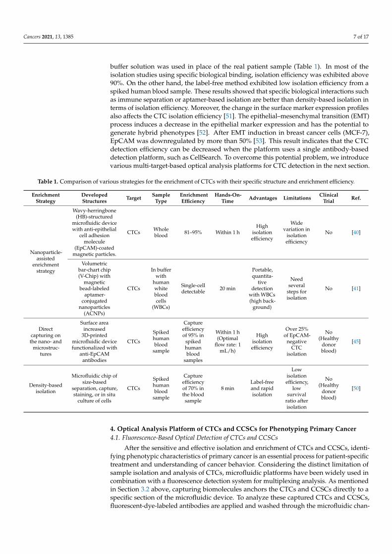
buffer solution was used in place of the real patient sample (Table 1). In most of theisolation studies using specific biological binding, isolation efficiency was exhibited above90%. On the other hand, the label-free method exhibited low isolation efficiency from aspiked human blood sample. These results showed that specific biological interactions suchas immune separation or aptamer-based isolation are better than density-based isolation interms of isolation efficiency. Moreover, the change in the surface marker expression profilesalso affects the CTC isolation efficiency [51]. The epithelial–mesenchymal transition (EMT)process induces a decrease in the epithelial marker expression and has the potential togenerate hybrid phenotypes [52]. After EMT induction in breast cancer cells (MCF-7),EpCAM was downregulated by more than 50% [53]. This result indicates that the CTCdetection efficiency can be decreased when the platform uses a single antibody-baseddetection platform, such as CellSearch. To overcome this potential problem, we introducevarious multi-target-based optical analysis platforms for CTC detection in the next section.
Table 1. Comparison of various strategies for the enrichment of CTCs with their specific structure and enrichment efficiency.
EnrichmentStrategy
DevelopedStructures Target Sample
TypeEnrichmentEfficiency
Hands-On-Time Advantages Limitations Clinical
Trial Ref.
Nanoparticle-assisted
enrichmentstrategy
Wavy-herringbone(HB)-structured
microfluidic devicewith anti-epithelial
cell adhesionmolecule
(EpCAM)-coatedmagnetic particles.
CTCs Wholeblood 81–95% Within 1 h
Highisolationefficiency
Widevariation in
isolationefficiency
No [40]
Volumetricbar-chart chip(V-Chip) with
magneticbead-labeled
aptamer-conjugated
nanoparticles(ACNPs)
CTCs
In bufferwith
humanwhitebloodcells
(WBCs)
Single-celldetectable 20 min
Portable,quantita-
tivedetection
with WBCs(high back-
ground)
Needseveral
steps forisolation
No [41]
Directcapturing onthe nano- and
microstruc-tures
Surface areaincreased
3D-printedmicrofluidic devicefunctionalized with
anti-EpCAMantibodies
CTCs
Spikedhumanblood
sample
Captureefficiencyof 95% in
spikedhumanblood
samples
Within 1 h(Optimal
flow rate: 1mL/h)
Highisolationefficiency
Over 25%of EpCAM-
negativeCTC
isolation
No(Healthy
donorblood)
[45]
Density-basedisolation
Microfluidic chip ofsize-based
separation, capture,staining, or in situ
culture of cells
CTCs
Spikedhumanblood
sample
Captureefficiencyof 70% inthe blood
sample
8 minLabel-freeand rapidisolation
Lowisolationefficiency,
lowsurvival
ratio afterisolation
No(Healthy
donorblood)
[50]
4. Optical Analysis Platform of CTCs and CCSCs for Phenotyping Primary Cancer4.1. Fluorescence-Based Optical Detection of CTCs and CCSCs
After the sensitive and effective isolation and enrichment of CTCs and CCSCs, identi-fying phenotypic characteristics of primary cancer is an essential process for patient-specifictreatment and understanding of cancer behavior. Considering the distinct limitation ofsample isolation and analysis of CTCs, microfluidic platforms have been widely used incombination with a fluorescence detection system for multiplexing analysis. As mentionedin Section 3.2 above, capturing biomolecules anchors the CTCs and CCSCs directly to aspecific section of the microfluidic device. To analyze these captured CTCs and CCSCs,fluorescent-dye-labeled antibodies are applied and washed through the microfluidic chan-

Cancers 2021, 13, 1385 8 of 17
nel. To identify the CTCs and CCSCs, a broad spectrum of cancer-related cell surfacemarkers has been used (Table 2) [54–73].
Table 2. Common cell surface markers to identify CTCs and CCSCs.
Origin of Cancer CTC Markers Ref. CCSC Markers Ref.
In general EpCAM+ or Cytokeratin+, CD45− [54]Brain cancer
(glioblastoma)EGFR+, Ki67+ or EB1+
MCAM+ or MCSP+[62][63] SSEA1+, CD133+ [55]
Breast cancer EpCAM+, HER2+, EGFR+ [64] CD44+/CD24low/−, CD133+ [70]Lung cancer Folate receptor+ [65] CD133+ [60]Liver cancer ASGPR, GPC3, and EpCAM [66] CD133+/CD44+, CD90+ [68]
Gastric cancer HER2+ [69] CD133+/CD44+ [59]
Colorectal cancer CK20+, CEA+ [67] CD133+/CD44+/ESAhigh, CD166+,CD26+ [58]
Pancreatic cancer CA19-9+ [71] CD44+/CD24+/ESA+, CD133+ [57]Prostate cancer PSMA+ [72] CD44+ [56]Ovarian cancer CA124+, HE4+ [73] ALDH1+/CD44+ or CD133+ [61]
After the first report by Nagrath et al. of microfluidic device-based CTC detection byimmobilizing anti-EpCAM antibodies on micropillars in the microfluidic channel, manysimilar approaches have been published [54]. Ahmed et al. developed a size-dictatedimmunocapture chip with a triangular microarray structure that can selectively enhance theinteraction of CTC by deterministic lateral displacement (Figure 3a) [74]. The anti-EpCAMantibody-coated micropillars successfully captured more than 90% of CTCs (92.2 ± 6.4%).Even though WBCs were well distinguished from CTCs by multiplex immunofluorescencestaining on the chip, staining requires several steps for each antibody, including washes,and captured CTCs may be lost at each of these steps. To overcome this issue, Lee et al.introduced a hybrid fluorescence nanoparticle-based CTC capture and analysis system(Figure 3b) [64]. The hybrid fluorescence nanoparticle (HNP) is composed of a quantumdot, antibody, and biotinylated DNA, which constitute a signaling element, CTC labelingelement, and capturing element, respectively. Streptavidin was used to coat the micropillarsin a microfluidic channel to hold the biotinylated DNA of HNPs, which resulted in thesuccessful capture of the target CTCs. This methodology allowed visual discrimination ofthe captured CTCs, which were labeled in different colors depending on surface markerexpression due to the ratio of different HNPs which individually recognized the antibodyand quantum dots. Moreover, the DNA of HNP served as a cleavable linker, whereby arestriction enzyme was used to recover captured CTCs by mild cleavage of DNA. Suchrecovery of CTCs from the microfluidic device enables further analysis of the capturedCTCs, which in turn better informs clinical decisions. Recently, Armbrecht et al. introducedanother tool to analyze cytokine secretion from captured CTCs within a microfluidicsystem (Figure 3c) [75]. Using this system, the secretion level of granulocyte colony-stimulating factor (G-CSF), which indicates acute inflammation, was directly quantified.These advanced microfluidic sensing systems have granted researchers access to directproteomic profiling of CTCs, permitting a better understanding of the molecular pathwaysand signals involved in the metastatic process.
The intensity of the fluorescence signal of immune-labeled CTCs and CCSCs correlatesdirectly with surfacer marker expression. To enhance the fluorescence signal of capturedCTCs, Zhang et al. introduced a magnetic “squashing” technique on the plasmonic gold(pGOLD) chip in a microfluidic device (Figure 3d) [76]. After CTCs were magnetically cap-tured on the pGOLD chip, near-infrared (NIR) fluorescence enhancement (≈50–120-fold)was used to interrogate the squashed/flattened morphology of CTCs by magnetic forces.Due to the proximity of NIR labels on CTCs to the plasmonic gold chip, the fluorescencesignal was enhanced by surface plasmon resonance. This research holds potential forCCSC detection. Interestingly, CCSCs showed greater cytoskeletal and nucleoskeletaldeformability and motility compared to CTCs [77]. By monitoring their deformation andmotion within the microfluidic device, Zhang et al. were able to distinguish the more

Cancers 2021, 13, 1385 9 of 17
highly tumorigenic cells, CCSCs, from surrounding CTCs. Although the effect of squashingon cell viability was not described in this study, mechanical modulation of the cell wassuccessfully executed for fluorescence detection of circulating cells.
Cancers 2021, 13, x FOR PEER REVIEW 9 of 18
fluorescence signal was enhanced by surface plasmon resonance. This research holds po-tential for CCSC detection. Interestingly, CCSCs showed greater cytoskeletal and nucleo-skeletal deformability and motility compared to CTCs [77]. By monitoring their defor-mation and motion within the microfluidic device, Zhang et al. were able to distinguish the more highly tumorigenic cells, CCSCs, from surrounding CTCs. Although the effect of squashing on cell viability was not described in this study, mechanical modulation of the cell was successfully executed for fluorescence detection of circulating cells.
Figure 3. (a) Schematic images of size-dictated immunocapture chip (SDI-Chip) and analysis of surface marker of captured CTCs. This figure is reproduced from [74] (© 2017 John Wiley & Sons); (b) Schematic images of hybrid nanoparticle-based CTC capture using microfluidic chip and selective recovery of captured CTCs from the microfluidic chip. This figure is reproduced from [64] (© 2013 Elsevier B.V.); (c) Microfluidic chip design and operation for CTC capture and protein se-cretion analysis. This figure is reproduced from [75] (© 2020 John Wiley & Sons); (d) Schematic images of a plasmonic gold chip used in the microfluidic immunomagnetic method and screening of CTCs in cancer patients. This figure is reproduced from [76] (© 2018 John Wiley & Sons).
4.2. Raman Spectroscopy-Based Optical Detection of CTCs and CCSCs Although fluorescence and surface plasmon resonance (SPR) detection methods are
the most well-established optical sensing tools for a microfluidic device, there is still a need to improve the multiplexity of these methods. The wide variety of required sets of antibodies for each primary organ is one challenge associated with the identification of CTCs using surface marker expression analysis. To this end, surface-enhanced Raman spectroscopy (SERS) has been introduced as a new tool for optical sensing platforms. Ra-man spectroscopy has significant advantages over fluorescence imaging, including mini-mizing the background noise from the blood that results from autofluorescence signals. Distinctive non-overlapping peaks are detected from a large pool of chemical dyes for multiplex imaging, allowing for more precise signal characterization.
Figure 3. (a) Schematic images of size-dictated immunocapture chip (SDI-Chip) and analysis of surface marker of capturedCTCs. This figure is reproduced from [74] (© 2017 John Wiley & Sons); (b) Schematic images of hybrid nanoparticle-basedCTC capture using microfluidic chip and selective recovery of captured CTCs from the microfluidic chip. This figureis reproduced from [64] (© 2013 Elsevier B.V.); (c) Microfluidic chip design and operation for CTC capture and proteinsecretion analysis. This figure is reproduced from [75] (© 2020 John Wiley & Sons); (d) Schematic images of a plasmonic goldchip used in the microfluidic immunomagnetic method and screening of CTCs in cancer patients. This figure is reproducedfrom [76] (© 2018 John Wiley & Sons).
4.2. Raman Spectroscopy-Based Optical Detection of CTCs and CCSCs
Although fluorescence and surface plasmon resonance (SPR) detection methods arethe most well-established optical sensing tools for a microfluidic device, there is still aneed to improve the multiplexity of these methods. The wide variety of required sets ofantibodies for each primary organ is one challenge associated with the identification ofCTCs using surface marker expression analysis. To this end, surface-enhanced Ramanspectroscopy (SERS) has been introduced as a new tool for optical sensing platforms.Raman spectroscopy has significant advantages over fluorescence imaging, includingminimizing the background noise from the blood that results from autofluorescence signals.Distinctive non-overlapping peaks are detected from a large pool of chemical dyes formultiplex imaging, allowing for more precise signal characterization.
To sense CTCs and CCSCs with SERS, metal nanoparticles are typically used, as theyoffer specific advantages for isolating and enriching targets found in the blood. However,the high mobility of CTCs and CCSCs makes it difficult to find the focal point of the laser onthe SERS-tagged cells. To overcome this technical limitation, magnetic nanoparticles have

Cancers 2021, 13, 1385 10 of 17
become a popular element in SERS probe design. To control the binding of cells with SERSprobes magnetically in a microfluidic channel, Xiong et al. developed magnetic nanochains(Magchains) (Figure 4a) [78]. Similar to Zong’s approach, an antibody-conjugated Magchainand a gold nanorod-based SERS probe are mixed in the mixing chamber of the device andform sandwich immune complexes when the target is present. Sandwich complexes arethen guided into the small-sized chamber in a microfluidic device by magnetic force. Theuse of such gathered complexes successfully enhanced the SERS signal of cancer biomarkersin this study. In an alternative approach, Cho et al. designed a new class of SERS probecomprising five families distinguished by unique sets of antibodies, Raman dye, and adouble-stranded DNA linker (Figure 4b) [70]. One of these SERS probes was conjugatedwith an anti-CD133 antibody for isolating circulating cancer stem cells (CCSCs). Here,CCSCs were successfully isolated from a mixed population of CTCs and hematopoieticstem cells on the microfluidic device. Mapping results clearly showed distinctive signaldifferences according to surface marker expression. Willner et al. developed SERS dropletmicrofluidics for single-cell analysis of CTCs (Figure 4c) [79]. A single prostate cancer cellwas trapped with SERS nanoprobes in the microfluidic device and isolated droplets werekept stationary during SERS interrogation. One drawback of this method is that mappingthe SERS signal over a large area within the microfluidic device is a time-consuming processand negatively affects cell viability. Pallaoro et al. developed an integrated microfluidicSERS system that can identify and count cancer cells from a mixed population of cellsflowing through a microfluidic channel (Figure 4d) [80]. In their study, CTCs were labeledwith silver nanoparticle dimers conjugated with a Raman-active reporter molecule andpassed through a flow-focused microfluidic channel, which forces the cells into a singleline. Each cancer cell was correctly identified among a proportionally larger number ofnormal cells by their Raman spectra.
4.3. Colorimetry-Based Optical Detection of Circulating Cancer Biomarkers
Similarly, many other studies have taken advantage of nanoparticles to detect CTCs(Figure 5a) [81,82]. The simplest and fastest of these methods is to conjugate the particlesto nucleic acids, such as DNA and aptamer, that bind selectively with the overexpressedproteins on CTC membranes or nucleic acids in the target CTCs. As the nucleic acidsselectively bind to the CTCs, the nanoparticles aggregate, forming a larger structure andinducing a color change in the CTC-containing solution. The higher the concentration ofCTCs, the more nanoparticles are aggregated by the nucleic acids that selectively bind to theCTCs, resulting in color changes that vary according to the resulting change in absorbance.Lu et al. reported a multifunctional, oval-shaped, gold nanoparticle-based, selectivebreast cancer cell detection system [83]. In this study, the surfaces of oval-shaped goldnanoparticles were modified with an S6 RNA aptamer and an anti-HER2/c-erb-2 antibodyto achieve high selectivity and sensitivity for a target cancer cell. This strategy made itpossible not only to optically confirm the number of cancer cells in solution by the nakedeye, but also to resolve the signal using a two-photon scattering assay in solutions withlow concentrations of cancer cells. For more efficient discrimination of target cells amongthe various types of cells typically present in biological samples, Liu et al. developedmicrofluidic channels that permit aptamer-specific capture of target cells [84]. Targetcells were captured inside of the microfluidic channel, then subjected to a flow of goldnanoparticle-conjugated aptamer. This microfluidic channel configuration detected targetcells in short timescales and with relatively large volumes of samples. Li et el. introducedmicrofluidic devices that employ a lateral flow assay for quantitative and rapid point-of-caring tests (Figure 5b) [85]. This device used antibody-conjugated platinum nanoparticlesto capture prostate-specific antigens. Platinum nanoparticle-catalyzed oxygen generatedby H2O2 solution forces ink through the microfluidic device. This distance-based readoutsystem provides rapid quantitation, eliminating the need for complex analytical equipment.

Cancers 2021, 13, 1385 11 of 17Cancers 2021, 13, x FOR PEER REVIEW 11 of 18
Figure 4. (a) Schematic illustration of the Magchain integrated microfluidic chip. This figure is reproduced from [78] (© BY-NC 4.0, 2018); (b) Illustration of Raman-active nanoprobe-based circulating cancer stem cell analysis. This figure is reproduced from [70] (© 2018 Elsevier B.V.); (c) Illustration of a single-cell encapsulation event within the microfluidic device. This figure is reproduced from [79] (© 2018 American Chemical Society); (d) Graphical depiction of device layout and flow dynamics. This figure is reproduced from [80] (© 2015 American Chemical Society).
4.3. Colorimetry-Based Optical Detection of Circulating Cancer Biomarkers Similarly, many other studies have taken advantage of nanoparticles to detect CTCs
(Figure 5a) [81,82]. The simplest and fastest of these methods is to conjugate the particles to nucleic acids, such as DNA and aptamer, that bind selectively with the overexpressed proteins on CTC membranes or nucleic acids in the target CTCs. As the nucleic acids se-lectively bind to the CTCs, the nanoparticles aggregate, forming a larger structure and inducing a color change in the CTC-containing solution. The higher the concentration of CTCs, the more nanoparticles are aggregated by the nucleic acids that selectively bind to the CTCs, resulting in color changes that vary according to the resulting change in absorb-ance. Lu et al. reported a multifunctional, oval-shaped, gold nanoparticle-based, selective breast cancer cell detection system [83]. In this study, the surfaces of oval-shaped gold nanoparticles were modified with an S6 RNA aptamer and an anti-HER2/c-erb-2 antibody to achieve high selectivity and sensitivity for a target cancer cell. This strategy made it possible not only to optically confirm the number of cancer cells in solution by the naked eye, but also to resolve the signal using a two-photon scattering assay in solutions with low concentrations of cancer cells. For more efficient discrimination of target cells among the various types of cells typically present in biological samples, Liu et al. developed mi-crofluidic channels that permit aptamer-specific capture of target cells [84]. Target cells were captured inside of the microfluidic channel, then subjected to a flow of gold nano-particle-conjugated aptamer. This microfluidic channel configuration detected target cells
Figure 4. (a) Schematic illustration of the Magchain integrated microfluidic chip. This figure is repro-duced from [78] (© BY-NC 4.0, 2018); (b) Illustration of Raman-active nanoprobe-based circulatingcancer stem cell analysis. This figure is reproduced from [70] (© 2018 Elsevier B.V.); (c) Illustration ofa single-cell encapsulation event within the microfluidic device. This figure is reproduced from [79](© 2018 American Chemical Society); (d) Graphical depiction of device layout and flow dynamics.This figure is reproduced from [80] (© 2015 American Chemical Society).
Cancers 2021, 13, x FOR PEER REVIEW 12 of 18
in short timescales and with relatively large volumes of samples. Li et el. introduced mi-crofluidic devices that employ a lateral flow assay for quantitative and rapid point-of-caring tests (Figure 5b) [85]. This device used antibody-conjugated platinum nanoparticles to capture prostate-specific antigens. Platinum nanoparticle-catalyzed oxygen generated by H2O2 solution forces ink through the microfluidic device. This distance-based readout system provides rapid quantitation, eliminating the need for complex analytical equip-ment.
Figure 5. (a) Schematic images of aptamer-conjugated gold nanoparticle-based assay system and colorimetric analysis of target cancer cells. This figure is reproduced from [82] (© 2018 American Chemical Society); (b) Schematic images of lateral flow assay-based microfluidic device and verification process of the target molecule by distance traveled by ink. This figure is reproduced from [85] (© 2019 Royal Society of Chemical).
5. Outlook In this review, we summarized recent strategies of isolation and analysis platforms
for CTCs and CCSCs. Since CTCs need to be detected from blood samples in the presence of a tremendous number of RBCs and WBCs, the microfluidic platform is the most suitable system. Moreover, multi-probe-based optical analysis platforms are required not only to identify the origin and subtype of primary cancer but also to improve the detection relia-bility from heterogeneous phenotypes. Even though recent advances in optical analysis-based microfluidic devices have shown great success with CTC enumeration, they cannot quickly provide information about the entire sample because they can analyze only a frac-tion of the injected sample at a time. Thus, these optical analysis-based microfluidics ap-proaches to analyzing various kinds of cancer biomarkers are low-throughput, despite their high sensitivity. Therefore, a more effective method for analyzing separated CTCs on microfluidic devices is needed to achieve clinical relevance.
For this reason, a variety of analytical processes were introduced into microfluidic devices, including electrochemical, fluorescence, and chemiluminescence techniques [64,86]. Among these methodologies, the electrochemical technique, which measures the
Figure 5. (a) Schematic images of aptamer-conjugated gold nanoparticle-based assay system andcolorimetric analysis of target cancer cells. This figure is reproduced from [82] (© 2018 AmericanChemical Society); (b) Schematic images of lateral flow assay-based microfluidic device and verifica-tion process of the target molecule by distance traveled by ink. This figure is reproduced from [85](© 2019 Royal Society of Chemical).

Cancers 2021, 13, 1385 12 of 17
5. Outlook
In this review, we summarized recent strategies of isolation and analysis platforms forCTCs and CCSCs. Since CTCs need to be detected from blood samples in the presence ofa tremendous number of RBCs and WBCs, the microfluidic platform is the most suitablesystem. Moreover, multi-probe-based optical analysis platforms are required not onlyto identify the origin and subtype of primary cancer but also to improve the detectionreliability from heterogeneous phenotypes. Even though recent advances in optical analysis-based microfluidic devices have shown great success with CTC enumeration, they cannotquickly provide information about the entire sample because they can analyze only afraction of the injected sample at a time. Thus, these optical analysis-based microfluidicsapproaches to analyzing various kinds of cancer biomarkers are low-throughput, despitetheir high sensitivity. Therefore, a more effective method for analyzing separated CTCs onmicrofluidic devices is needed to achieve clinical relevance.
For this reason, a variety of analytical processes were introduced into microfluidic de-vices, including electrochemical, fluorescence, and chemiluminescence techniques [64,86].Among these methodologies, the electrochemical technique, which measures the electri-cal signal generated by the varying distribution of electrons that results from chemicalreactions, has been widely adopted due to its uniquely high sensitivity, selectivity, andthroughput [87,88]. Gurudatt et al. developed a microfluidic device for CTC separationby size variation and electrochemical distinction of their origin (Figure 6a) [72]. With thissystem, cancer patients’ samples were analyzed to validate the reliability of the microfluidicchannel in a clinical application. Wu et al. developed a paper-based microfluidic immun-odevice that employed electrochemical- and fluorescent-mediated signal amplification forCTC detection (Figure 6b) [73]. Under optimal conditions, the detection limit of this novelimmunodevice was as low as 10 cells/mL.
On the other hand, the rarity of CTCs and CCSCs is the leading limiting factor forclinical commercialization. To overcome this issue, additional detection of other circu-lating cancer biomarkers such as exosomes and circulating tumor DNA (ctDNA) can beconsidered. Exosomes play a critical role in a communication system between cells, car-rying several biomolecules from one cell to another [89–91]. In cancer conditions, cancercell-derived exosomes are secreted into the bodily fluids with high stability and in higheramounts than the normal cells [92]. Furthermore, the basic concept of exosome detectionis the same as CTC detection. For the development of exosome-based early diagnosisand analysis of cancers, tetraspanins (such as CD9, CD63, CD81, and CD82) are typicallyutilized as the capturing molecules for the detection of cancer-associated exosomes. Forcancer-related antigens on the lipid bilayer of exosomes, many different proteins can beutilized as biomarkers, depending on the host cancer cells, including HER2, CEA, EpCAM,IGFR, PSMA, etc., which are used as diagnostic and therapeutic markers. Therefore, thedetection method of exosomes is very similar to that of CTCs and CCSCs, so the samedevice can be applied for both CTC/CCSCs and exosomes. Consequently, by detectingboth circulating cancer cells and exosomes together, the rarity issue will be mitigated.

Cancers 2021, 13, 1385 13 of 17
Cancers 2021, 13, x FOR PEER REVIEW 13 of 18
electrical signal generated by the varying distribution of electrons that results from chem-ical reactions, has been widely adopted due to its uniquely high sensitivity, selectivity, and throughput [87,88]. Gurudatt et al. developed a microfluidic device for CTC separa-tion by size variation and electrochemical distinction of their origin (Figure 6a) [72]. With this system, cancer patients’ samples were analyzed to validate the reliability of the mi-crofluidic channel in a clinical application. Wu et al. developed a paper-based microfluidic immunodevice that employed electrochemical- and fluorescent-mediated signal amplifi-cation for CTC detection (Figure 6b) [73]. Under optimal conditions, the detection limit of this novel immunodevice was as low as 10 cells/mL.
On the other hand, the rarity of CTCs and CCSCs is the leading limiting factor for clinical commercialization. To overcome this issue, additional detection of other circulat-ing cancer biomarkers such as exosomes and circulating tumor DNA (ctDNA) can be con-sidered. Exosomes play a critical role in a communication system between cells, carrying several biomolecules from one cell to another [89–91]. In cancer conditions, cancer cell-de-rived exosomes are secreted into the bodily fluids with high stability and in higher amounts than the normal cells [92]. Furthermore, the basic concept of exosome detection is the same as CTC detection. For the development of exosome-based early diagnosis and analysis of cancers, tetraspanins (such as CD9, CD63, CD81, and CD82) are typically utilized as the capturing molecules for the detection of cancer-associated exosomes. For cancer-re-lated antigens on the lipid bilayer of exosomes, many different proteins can be utilized as biomarkers, depending on the host cancer cells, including HER2, CEA, EpCAM, IGFR, PSMA, etc., which are used as diagnostic and therapeutic markers. Therefore, the detection method of exosomes is very similar to that of CTCs and CCSCs, so the same device can be applied for both CTC/CCSCs and exosomes. Consequently, by detecting both circulating cancer cells and exosomes together, the rarity issue will be mitigated.
Figure 6. (a) Schematic images detailing the process of CTC capture using a functional channel wall and separation of target CTCs in mixed cancer cell samples. This figure is reproduced from [93] (© 2019 Elsevier B.V.); (b) Schematic image showing the fabrication and capture process of a paper-based microfluidic immunodevice and electrochemical signals obtained from CTCs detected in various concentrations. This figure is reproduced from [94] (© 2014 John Wiley & Sons).
6. Conclusions In conclusion, we review recent efforts made to integrate nanotechnology-based op-
tical biosensors with microfluidic systems for the detection of CTCs and CCSCs. Each method has unique properties and optimal target conditions for successful detection. For
Figure 6. (a) Schematic images detailing the process of CTC capture using a functional channel walland separation of target CTCs in mixed cancer cell samples. This figure is reproduced from [93](© 2019 Elsevier B.V.); (b) Schematic image showing the fabrication and capture process of a paper-based microfluidic immunodevice and electrochemical signals obtained from CTCs detected invarious concentrations. This figure is reproduced from [94] (© 2014 John Wiley & Sons).
6. Conclusions
In conclusion, we review recent efforts made to integrate nanotechnology-basedoptical biosensors with microfluidic systems for the detection of CTCs and CCSCs. Eachmethod has unique properties and optimal target conditions for successful detection.For this purpose, isolation, enrichment, capture, and post-sensing steps should take intoconsideration the type of CTCs or CCSCs to be detected. With CTC/CCSC enumeration,it can be directly applied to assist in identifying early cancer treatment response andprognosis [70,95]. In particular, a recent study with human patients showed that the numberof CCSCs is more critical than CTCs in the overall survival periods, respectively [95]. Sincethe differentiation condition of CSC in vivo and in vitro is difficult to exactly match, itis limited in predicting the exact cancer phenotype of differentiated CSCs. However,we found that differentiated CSCs’ surface marker profiles were similar to the tissuesamples of the secondary tumor in vivo [70]. Regarding this result, we believe that thedetection and analysis of CCSCs has great potential to contribute to clinical applicationsfor cancer treatment.
Author Contributions: H.-Y.C., J.-H.C., and J.-W.C. organized the structure of the manuscript. H.-Y.C., J.-H.C., J.L., S.-N.L., and J.-W.C. collaboratively wrote the manuscript. All authors have readand agreed to the published version of the manuscript.
Funding: This research was supported by the Basic Science Research Program through the Na-tional Research Foundation of Korea (NRF), funded by the Ministry of Education (No. NRF-2019R1I1A1A01058888); the National Research Foundation of Korea (NRF) grant funded by theKorean government (MSIT) (No. 2019R1A2C3002300), and the Basic Science Research Programthrough the National Research Foundation of Korea (NRF), funded by the Ministry of Education(No. 2016R1A6A1A03012845).
Conflicts of Interest: The authors declare no conflict of interest. The funders had no role in the designof the study; in the collection, analyses, or interpretation of data; in the writing of the manuscript, orin the decision to publish the results.
References1. Jin, M.-Z.; Jin, W.-L. The updated landscape of tumor microenvironment and drug repurposing. Signal Transduct. Target. Ther.
2020, 5, 1–16. [CrossRef]2. Navya, P.; Kaphle, A.; Srinivas, S.; Bhargava, S.K.; Rotello, V.M.; Daima, H.K. Current trends and challenges in cancer management
and therapy using designer nanomaterials. Nano Converg. 2019, 6, 1–30. [CrossRef] [PubMed]

Cancers 2021, 13, 1385 14 of 17
3. Zhang, Y.-B.; Pan, X.-F.; Chen, J.; Cao, A.; Zhang, Y.-G.; Xia, L.; Wang, J.; Li, H.; Liu, G.; Pan, A. Combined lifestyle factors,incident cancer, and cancer mortality: A systematic review and meta-analysis of prospective cohort studies. Br. J. Cancer 2020,122, 1085–1093. [CrossRef] [PubMed]
4. Riggio, A.I.; Varley, K.E.; Welm, A.L. The lingering mysteries of metastatic recurrence in breast cancer. Br. J. Cancer 2021, 124,13–26. [CrossRef] [PubMed]
5. Chaffer, C.L.; Weinberg, R.A. A perspective on cancer cell metastasis. Science 2011, 331, 1559–1564. [CrossRef]6. Gold, B.; Cankovic, M.; Furtado, L.V.; Meier, F.; Gocke, C.D. Do circulating tumor cells, exosomes, and circulating tumor nucleic
acids have clinical utility?: A report of the association for molecular pathology. J. Mol. Diagn. 2015, 17, 209–224. [CrossRef]7. Crowley, E.H.; Di Nicolantonio, F.; Loupakis, F.; Bardelli, A. Liquid biopsy: Monitoring cancer-genetics in the blood. Nat. Rev.
Clin. Oncol. 2013, 10, 472–484. [CrossRef]8. Wang, J.; Chang, S.; Li, G.; Sun, Y. Application of liquid biopsy in precision medicine: Opportunities and challenges. Front. Med.
2017, 11, 522–527. [CrossRef]9. Tayoun, T.; Faugeroux, V.; Oulhen, M.; Aberlenc, A.; Pawlikowska, P.; Farace, F. CTC-Derived Models: A Window into the
Seeding Capacity of Circulating Tumor Cells (CTCs). Cells 2019, 8, 1145. [CrossRef]10. Kulasinghe, A.; Wu, H.; Punyadeera, C.; Warkiani, M.E. The Use of microfluidic technology for cancer applications and liquid
biopsy. Micromachines 2018, 9, 397. [CrossRef]11. Alix-Panabières, C.; Pantel, K. Circulating tumor cells: Liquid biopsy of cancer. Clin. Chem. 2013, 59, 110–118. [CrossRef]12. Alix-Panabières, C.; Pantel, K. Clinical Applications of Circulating Tumor Cells and Circulating Tumor DNA as Liquid Biopsy.
Cancer Discov. 2016, 6, 479–491. [CrossRef] [PubMed]13. van de Stolpe, A.; Pantel, K.; Sleijfer, S.; Terstappen, L.W.; Den Toonder, J.M. Circulating tumor cell isolation and diagnostics:
Toward routine clinical use. AACR 2011. [CrossRef]14. Bu, J.; Shim, J.-E.; Lee, T.H.; Cho, Y.-H. Multi-modal liquid biopsy platform for cancer screening: Screening both cancer-associated
rare cells and cancer cell-derived vesicles on the fabric filters for a reliable liquid biopsy analysis. Nano Converg. 2019, 6, 1–8.[CrossRef]
15. Heitzer, E.; Ulz, P.; Geigl, J.B. Circulating Tumor DNA as a liquid biopsy for cancer. Clin. Chem. 2015, 61, 112–123. [CrossRef][PubMed]
16. Mastoraki, S.; Strati, A.; Tzanikou, E.; Chimonidou, M.; Politaki, E.; Voutsina, A.; Psyrri, A.; Georgoulias, V.; Lianidou, E.S.ESR1 methylation: A liquid biopsy–based epigenetic assay for the follow-up of patients with metastatic breast cancer receivingendocrine treatment. Clin. Cancer Res. 2017, 24, 1500–1510. [CrossRef] [PubMed]
17. Tie, J.; Wang, Y.; Tomasetti, C.; Li, L.; Springer, S.; Kinde, I.; Silliman, N.; Tacey, M.; Wong, H.-L.; Christie, M.; et al. Circulatingtumor DNA analysis detects minimal residual disease and predicts recurrence in patients with stage II colon cancer. Sci. Transl.Med. 2016, 8, 346ra92. [CrossRef]
18. Krishnamurthy, S.; Cristofanilli, M.; Singh, B.; Reuben, J.; Gao, H.; Cohen, E.N.; Andreopoulou, E.; Hall, C.S.; Lodhi, A.; Jackson,S.; et al. Detection of minimal residual disease in blood and bone marrow in early stage breast cancer. Cancer 2010, 116, 3330–3337.[CrossRef] [PubMed]
19. Pantel, K.; Alix-Panabières, C. Liquid biopsy and minimal residual disease—Latest advances and implications for cure. Nat. Rev.Clin. Oncol. 2019, 16, 409–424. [CrossRef]
20. Ozkumur, E.; Shah, A.M.; Ciciliano, J.C.; Emmink, B.L.; Miyamoto, D.T.; Brachtel, E.; Yu, M.; Chen, P.-I.; Morgan, B.; Trautwein, J.;et al. Inertial Focusing for Tumor Antigen-Dependent and -Independent Sorting of Rare Circulating Tumor Cells. Sci. Transl. Med.2013, 5, 179ra47. [CrossRef]
21. Danova, M.; Torchio, M.; Mazzini, G. Isolation of rare circulating tumor cells in cancer patients: Technical aspects and clinicalimplications. Expert Rev. Mol. Diagn. 2011, 11, 473–485. [CrossRef]
22. Fachin, F.; Spuhler, P.; Martel-Foley, J.M.; Edd, J.F.; Barber, T.A.; Walsh, J.; Karabacak, M.; Pai, V.; Yu, M.; Smith, K.; et al. Monolithicchip for high-throughput blood cell depletion to sort rare circulating tumor cells. Sci. Rep. 2017, 7, 1–11. [CrossRef]
23. Brouwer, A.; De Laere, B.; Peeters, D.; Peeters, M.; Salgado, R.; Dirix, L.; Van Laere, S. Evaluation and consequences ofheterogeneity in the circulating tumor cell compartment. Oncotarget 2016, 7, 48625–48643. [CrossRef] [PubMed]
24. Müller, V.; Stahmann, N.; Riethdorf, S.; Rau, T.; Zabel, T.; Goetz, A.; Jänicke, F.; Pantel, K. Circulating tumor cells in breast cancer:Correlation to bone marrow micrometastases, heterogeneous response to systemic therapy and low proliferative activity. Clin.Cancer Res. 2005, 11, 3678–3685. [CrossRef] [PubMed]
25. Powell, A.A.; Talasaz, A.H.; Zhang, H.; Coram, M.A.; Reddy, A.; Deng, G.; Telli, M.L.; Advani, R.H.; Carlson, R.W.; Mollick, J.A.;et al. Single Cell Profiling of Circulating Tumor Cells: Transcriptional Heterogeneity and Diversity from Breast Cancer Cell Lines.PLoS ONE 2012, 7, e33788. [CrossRef]
26. Yang, M.-H.; Imrali, A.; Heeschen, C. Circulating cancer stem cells: The importance to select. Chin. J. Cancer Res. 2015, 27, 437–449.27. Liao, W.-T.; Ye, Y.-P.; Deng, Y.-J.; Bian, X.-W.; Ding, Y.-Q. Metastatic cancer stem cells: From the concept to therapeutics. Am. J.
Stem Cells 2014, 3, 46–62. [PubMed]28. Dalerba, P.; Cho, R.W.; Clarke, M.F. Cancer Stem Cells: Models and Concepts. Annu. Rev. Med. 2007, 58, 267–284. [CrossRef]
[PubMed]29. Galanzha, E.I.; Kim, J.-W.; Zharov, V.P. Nanotechnology-based molecular photoacoustic and photothermal flow cytometry
platform forin-vivodetection and killing of circulating cancer stem cells. J. Biophotonics 2009, 2, 725–735. [CrossRef]

Cancers 2021, 13, 1385 15 of 17
30. Kantara, C.; O’Connell, M.R.; Luthra, G.; Gajjar, A.; Sarkar, S.; Ullrich, R.L.; Singh, P. Methods for detecting circulating cancer stemcells (CCSCs) as a novel approach for diagnosis of colon cancer relapse/metastasis. Lab. Investig. 2015, 95, 100–112. [CrossRef][PubMed]
31. Fan, S.T.; Yang, Z.F.; Ho, D.W.; Ng, M.N.; Yu, W.C.; Wong, J. Prediction of Posthepatectomy Recurrence of HepatocellularCarcinoma by Circulating Cancer Stem Cells. Ann. Surg. 2011, 254, 569–576. [CrossRef] [PubMed]
32. Baccelli, I.; Schneeweiss, A.; Riethdorf, S.; Stenzinger, A.; Schillert, A.; Vogel, V.; Klein, C.; Saini, M.; Bäuerle, T.; Wallwiener,M.; et al. Identification of a population of blood circulating tumor cells from breast cancer patients that initiates metastasis in axenograft assay. Nat. Biotechnol. 2013, 31, 539–544. [CrossRef] [PubMed]
33. Vinogradov, S.; Wei, X. Cancer stem cells and drug resistance: The potential of nanomedicine. Nanomedicine 2012, 7, 597–615.[CrossRef] [PubMed]
34. Kim, K.-J.; Cho, H.-Y.; Lee, W.-J.; Choi, J.-W. Subtyping of Magnetically isolated breast cancer cells using magnetic force microscopy.Biotechnol. J. 2018, 13, e1700625. [CrossRef]
35. Nie, L.; Li, F.; Huang, X.; Aguilar, Z.P.; Wang, Y.A.; Xiong, Y.; Fu, F.; Xu, H. Folic Acid Targeting for Efficient Isolation andDetection of Ovarian Cancer CTCs from Human Whole Blood Based on Two-Step Binding Strategy. ACS Appl. Mater. Interfaces2018, 10, 14055–14062. [CrossRef] [PubMed]
36. Chang, Z.-M.; Wang, Z.; Shao, D.; Yue, J.; Xing, H.; Li, L.; Ge, M.; Li, M.; Yan, H.; Hu, H.; et al. Shape engineering boosts magneticmesoporous silica nanoparticle-based isolation and detection of circulating tumor cells. ACS Appl. Mater. Interfaces 2018, 10,10656–10663. [CrossRef] [PubMed]
37. Meng, Q.-F.; Cheng, Y.-X.; Huang, Q.; Zan, M.; Xie, W.; Sun, Y.; Li, R.; Wei, X.; Guo, S.-S.; Zhao, X.-Z.; et al. Biomimeticimmunomagnetic nanoparticles with minimal nonspecific biomolecule adsorption for enhanced isolation of circulating tumorcells. ACS Appl. Mater. Interfaces 2019, 11, 28732–28739. [CrossRef] [PubMed]
38. Wu, S.; Gu, L.; Qin, J.; Zhang, L.; Sun, F.; Liu, Z.; Wang, Y.; Shi, D. Rapid label-free isolation of circulating tumor cells from patients’peripheral blood using electrically charged Fe3O4 nanoparticles. ACS Appl. Mater. Interfaces 2020, 12, 4193–4203. [CrossRef]
39. Zhao, W.; Cheng, R.; Jenkins, B.D.; Zhu, T.; Okonkwo, N.E.; Jones, C.E.; Davis, M.B.; Kavuri, S.K.; Hao, Z.; Schroeder, C.; et al.Label-free ferrohydrodynamic cell separation of circulating tumor cells. Lab Chip 2017, 17, 3097–3111. [CrossRef]
40. Shi, W.; Wang, S.; Maarouf, A.; Uhl, C.G.; He, R.; Yunus, D.; Liu, Y. Magnetic particles assisted capture and release of rarecirculating tumor cells using wavy-herringbone structured microfluidic devices. Lab Chip 2017, 17, 3291–3299. [CrossRef]
41. Abate, M.F.; Jia, S.; Ahmed, M.G.; Li, X.; Lin, L.; Chen, X.; Zhu, Z.; Yang, C. Visual quantitative detection of circulating tumor cellswith single-cell sensitivity using a portable microfluidic device. Small 2019, 15, e1804890. [CrossRef] [PubMed]
42. Kim, T.H.; Yoon, H.J.; Fouladdel, S.; Wang, Y.; Kozminsky, M.; Burness, M.L.; Paoletti, C.; Zhao, L.; Azizi, E.; Wicha, M.S.; et al.Characterizing circulating tumor cells isolated from metastatic breast cancer patients using graphene oxide based microfluidicAssay. Adv. Biosyst. 2019, 3, e1800278. [CrossRef] [PubMed]
43. Zeinali, M.; Murlidhar, V.; Fouladdel, S.; Shao, S.M.; Zhao, L.L.; Cameron, H.; Bankhead, A.; Shi, J.Q.; Cuneo, K.C.; Sahai, V.; et al.Profiling Heterogeneous Circulating Tumor Cells (CTC) populations in pancreatic cancer using a serial microfluidic ctc carpetchip. Adv. Biosyst. 2018, 2, 1800228. [CrossRef]
44. Loeian, M.S.; Aghaei, S.M.; Farhadi, F.; Rai, V.; Yang, H.W.; Johnson, M.D.; Aqil, F.; Mandadi, M.; Rai, S.N.; Panchapakesan, B.Liquid biopsy using the nanotube-CTC-chip: Capture of invasive CTCs with high purity using preferential adherence in breastcancer patients. Lab Chip 2019, 19, 1899–1915. [CrossRef]
45. Chen, J.; Liu, C.-Y.; Wang, X.; Sweet, E.; Liu, N.; Gong, X.; Lin, L. 3D printed microfluidic devices for circulating tumor cells(CTCs) isolation. Biosens. Bioelectron. 2020, 150, 111900. [CrossRef]
46. Varillas, J.I.; Zhang, J.; Chen, K.; Barnes, I.I.; Liu, C.; George, T.J.; Fan, Z.H. Microfluidic isolation of circulating tumor cells andcancer stem-like cells from patients with pancreatic ductal adenocarcinoma. Theranostics 2019, 9, 1417–1425. [CrossRef]
47. Chiu, T.-K.; Chao, A.-C.; Chou, W.-P.; Liao, C.-J.; Wang, H.-M.; Chang, J.-H.; Chen, P.-H.; Wu, M.-H. Optically-induced-dielectrophoresis (ODEP)-based cell manipulation in a microfluidic system for high-purity isolation of integral circulating tumorcell (CTC) clusters based on their size characteristics. Sens. Actuators B Chem. 2018, 258, 1161–1173. [CrossRef]
48. Antfolk, M.; Kim, S.H.; Koizumi, S.; Fujii, T.; Laurell, T. Label-free single-cell separation and imaging of cancer cells using anintegrated microfluidic system. Sci. Rep. 2017, 7, 46507. [CrossRef]
49. Xue, M.; Xiang, A.; Guo, Y.; Wang, L.; Wang, R.; Wang, W.; Ji, G.; Lu, Z. Dynamic Halbach array magnet integrated microfluidicsystem for the continuous-flow separation of rare tumor cells. RSC Adv. 2019, 9, 38496–38504. [CrossRef]
50. Zhou, J.; Tu, C.; Liang, Y.; Huang, B.; Fang, Y.; Liang, X.; Ye, X. The label-free separation and culture of tumor cells in a microfluidicbiochip. Analyst 2020, 145, 1706–1715. [CrossRef]
51. Cho, H.-Y.; Choi, J.-H.; Kim, K.-J.; Shin, M.; Choi, J.-W. Microfluidic system to analyze the effects of interleukin 6 on lymphaticbreast cancer metastasis. Front. Bioeng. Biotechnol. 2021, 8, 611802. [CrossRef]
52. Jie, X.-X.; Zhang, X.-Y.; Xu, C.-J. Epithelial-to-mesenchymal transition, circulating tumor cells and cancer metastasis: Mechanismsand clinical applications. Oncotarget 2017, 8, 81558. [CrossRef]
53. Hyun, K.-A.; Koo, G.-B.; Han, H.; Sohn, J.; Choi, W.; Kim, S.-I.; Jung, H.-I.; Kim, Y.-S. Epithelial-to-mesenchymal transition leadsto loss of EpCAM and different physical properties in circulating tumor cells from metastatic breast cancer. Oncotarget 2016, 7,24677–24687. [CrossRef]
Cancers 2021, 13, 1385 16 of 17
54. Nagrath, S.; Sequist, L.V.; Maheswaran, S.; Bell, D.W.; Irimia, D.; Ulkus, L.E.; Smith, M.R.; Kwak, E.L.; Digumarthy, S.R.;Muzikansky, A.; et al. Isolation of rare circulating tumour cells in cancer patients by microchip technology. Nature 2007, 450,1235–1239. [CrossRef]
55. Singh, S.K.; Hawkins, C.; Clarke, I.D.; Squire, J.A.; Bayani, J.; Hide, T.; Henkelman, R.M.; Cusimano, M.D.; Dirks, P.B. Identificationof human brain tumour initiating cells. Nature 2004, 432, 396–401. [CrossRef]
56. Patrawala, L.; Calhoun, T.; Schneiderbroussard, R.; Li, H.; Bhatia, B.; Tang, S.; Reilly, J.; Chandra, D.; Zhou, J.; Claypool, K.; et al.Highly purified CD44+ prostate cancer cells from xenograft human tumors are enriched in tumorigenic and metastatic progenitorcells. Oncogene 2006, 25, 1696–1708. [CrossRef]
57. Lee, C.J.; Dosch, J.; Simeone, D.M. Pancreatic Cancer Stem Cells. J. Clin. Oncol. 2008, 26, 2806–2812. [CrossRef] [PubMed]58. Vermeulen, L.; Todaro, M.; Melo, F.D.S.E.; Sprick, M.R.; Kemper, K.; Alea, M.P.; Richel, D.J.; Stassi, G.; Medema, J.P. Single-cell
cloning of colon cancer stem cells reveals a multi-lineage differentiation capacity. Proc. Natl. Acad. Sci. USA 2008, 105, 13427–13432.[CrossRef]
59. Takaishi, S.; Okumura, T.; Tu, S.; Wang, S.S.W.; Shibata, W.; Vigneshwaran, R.; Gordon, S.A.K.; Shimada, Y.; Wang, T.C.Identification of gastric cancer stem cells using the cell surface marker CD44. Stem Cells 2009, 27, 1006–1020. [CrossRef] [PubMed]
60. Tirino, V.; Camerlingo, R.; Franco, R.; Malanga, D.; La Rocca, A.; Viglietto, G.; Rocco, G.; Pirozzi, G. The role of CD133 in theidentification and characterisation of tumour-initiating cells in non-small-cell lung cancer. Eur. J. Cardio Thorac. Surg. 2009, 36,446–453. [CrossRef] [PubMed]
61. Wang, Y.-C.; Yo, Y.-T.; Lee, H.-Y.; Liao, Y.-P.; Chao, T.-K.; Su, P.-H.; Lai, H.-C. ALDH1-bright epithelial ovarian cancer cells areassociated with CD44 expression, drug resistance, and poor clinical outcome. Am. J. Pathol. 2012, 180, 1159–1169. [CrossRef][PubMed]
62. Krol, I.; Castro-Giner, F.; Maurer, M.; Gkountela, S.; Szczerba, B.M.; Scherrer, R.; Coleman, N.; Carreira, S.; Bachmann, F.; Anderson,S.; et al. Detection of circulating tumour cell clusters in human glioblastoma. Br. J. Cancer 2018, 119, 487–491. [CrossRef] [PubMed]
63. Lynch, D.; Powter, B.; Po, J.W.; Cooper, A.; Garrett, C.; Koh, E.-S.; Sheridan, M.; Van Gelder, J.; Darwish, B.; McKechnie, S.; et al.Isolation of circulating tumor cells from glioblastoma patients by direct immunomagnetic targeting. Appl. Sci. 2020, 10, 3338.[CrossRef]
64. Lee, H.J.; Cho, H.-Y.; Oh, J.H.; Namkoong, K.; Lee, J.G.; Park, J.-M.; Lee, S.S.; Huh, N.; Choi, J.-W. Simultaneous capture and insitu analysis of circulating tumor cells using multiple hybrid nanoparticles. Biosens. Bioelectron. 2013, 47, 508–514. [CrossRef][PubMed]
65. Gallo, M.; De Luca, A.; Maiello, M.R.; D’Alessio, A.; Esposito, C.; Chicchinelli, N.; Forgione, L.; Piccirillo, M.C.; Rocco, G.;Morabito, A.; et al. Clinical utility of circulating tumor cells in patients with non-small-cell lung cancer. Transl. Lung Cancer Res.2017, 6, 486–498. [CrossRef]
66. Court, C.M.; Hou, S.; Winograd, P.; Segel, N.H.; Li, Q.W.; Zhu, Y.; Sadeghi, S.; Finn, R.S.; Ganapathy, E.; Song, M.; et al. A novelmultimarker assay for the phenotypic profiling of circulating tumor cells in hepatocellular carcinoma. Liver Transplant. 2018, 24,946–960. [CrossRef]
67. Cohen, S.; Punt, C.; Iannotti, N.; Saidman, B.; Sabbath, K.; Gabrail, N.; Picus, J.; Morse, M.; Mitchell, E.; Miller, M. Prognosticsignificance of circulating tumor cells in patients with metastatic colorectal cancer. Ann. Oncol. 2009, 20, 1223–1229. [CrossRef]
68. Cao, L.; Zhou, Y.; Zhai, B.; Liao, J.; Xu, W.; Zhang, R.; Li, J.; Zhang, Y.; Chen, L.; Qian, H. Sphere-forming cell subpopulations withcancer stem cell properties in human hepatoma cell lines. BMC Gastroenterol. 2011, 11, 1–11. [CrossRef]
69. Iwatsuki, M.; Toyoshima, K.; Watanabe, M.; Hayashi, N.; Ishimoto, T.; Eto, K.; Iwagami, S.; Baba, Y.; Yoshida, N.; Hayashi, A.;et al. Frequency of HER2 expression of circulating tumour cells in patients with metastatic or recurrent gastrointestinal cancer. Br.J. Cancer 2013, 109, 2829–2832. [CrossRef]
70. Cho, H.-Y.; Hossain, K.; Lee, J.-H.; Han, J.; Lee, H.J.; Kim, K.-J.; Kim, J.-H.; Lee, K.-B.; Choi, J.-W. Selective isolation andnoninvasive analysis of circulating cancer stem cells through Raman imaging. Biosens. Bioelectron. 2018, 102, 372–382. [CrossRef]
71. Zhang, X.; Shi, S.; Zhang, B.; Ni, Q.; Yu, X.; Xu, J. Circulating biomarkers for early diagnosis of pancreatic cancer: Facts and hopes.Am. J. Cancer Res. 2018, 8, 332–353.
72. Pantel, K.; Hille, C.; Scher, H.I. Circulating tumor cells in prostate cancer: From discovery to clinical utility. Clin. Chem. 2019, 65,87–99. [CrossRef] [PubMed]
73. Guo, Y.-X.; Neoh, K.H.; Chang, X.-H.; Sun, Y.; Cheng, H.-Y.; Ye, X.; Ma, R.-Q.; Han, R.P.; Cui, H. Diagnostic value of HE4+circulating tumor cells in patients with suspicious ovarian cancer. Oncotarget 2018, 9, 7522–7533. [CrossRef] [PubMed]
74. Ahmed, M.G.; Abate, M.F.; Song, Y.; Zhu, Z.; Yan, F.; Xu, Y.; Wang, X.; Li, Q.; Yang, C. Isolation, detection, and antigen-basedprofiling of circulating tumor cells using a size-dictated immunocapture chip. Angew. Chem. Int. Ed. 2017, 56, 10681–10685.[CrossRef]
75. Armbrecht, L.; Rutschmann, O.; Szczerba, B.M.; Nikoloff, J.; Aceto, N.; Dittrich, P.S. Quantification of protein secretion fromcirculating tumor cells in microfluidic chambers. Adv. Sci. 2020, 7, 1903237. [CrossRef]
76. Zhang, R.; Le, B.; Xu, W.; Guo, K.; Sun, X.; Su, H.; Huang, L.; Huang, J.; Shen, T.; Liao, T.; et al. Magnetic “Squashing” ofCirculating tumor cells on plasmonic substrates for ultrasensitive nir fluorescence detection. Small Methods 2019, 3, 1800474.[CrossRef]

Cancers 2021, 13, 1385 17 of 17
77. Zhang, W.; Kai, K.; Choi, D.S.; Iwamoto, T.; Nguyen, Y.H.; Wong, H.; Landis, M.D.; Ueno, N.T.; Chang, J.; Qin, L. Microfluidicsseparation reveals the stem-cell-like deformability of tumor-initiating cells. Proc. Natl. Acad. Sci. USA 2012, 109, 18707–18712.[CrossRef] [PubMed]
78. Xiong, Q.; Lim, C.Y.; Ren, J.; Zhou, J.; Pu, K.; Chan-Park, M.B.; Mao, H.; Lam, Y.C.; Duan, H. Magnetic nanochain integratedmicrofluidic biochips. Nat. Commun. 2018, 9, 1–11. [CrossRef] [PubMed]
79. Willner, M.R.; McMillan, K.S.; Graham, D.; Vikesland, P.J.; Zagnoni, M. Surface-enhanced raman scattering based microfluidicsfor single-cell analysis. Anal. Chem. 2018, 90, 12004–12010. [CrossRef]
80. Pallaoro, A.; Hoonejani, M.R.; Braun, G.B.; Meinhart, C.D.; Moskovits, M. Rapid identification by surface-enhanced ramanspectroscopy of cancer cells at low concentrations flowing in a microfluidic channel. ACS Nano 2015, 9, 4328–4336. [CrossRef]
81. Borghei, Y.-S.; Hosseini, M.; Dadmehr, M.; Hosseinkhani, S.; Ganjali, M.R.; Sheikhnejad, R. Visual detection of cancer cells bycolorimetric aptasensor based on aggregation of gold nanoparticles induced by DNA hybridization. Anal. Chim. Acta 2016, 904,92–97. [CrossRef]
82. Medley, C.D.; Smith, J.E.; Tang, Z.; Wu, Y.; Bamrungsap, S.; Tan, W. Gold nanoparticle-based colorimetric assay for the directdetection of cancerous cells. Anal. Chem. 2008, 80, 1067–1072. [CrossRef]
83. Lu, W.; Arumugam, S.R.; Senapati, D.; Singh, A.K.; Arbneshi, T.; Khan, S.A.; Yu, H.; Ray, P.C.; Yu, S.A.K.H. Multifunctionaloval-shaped gold-nanoparticle-based selective detection of breast cancer cells using simple colorimetric and highly sensitivetwo-photon scattering assay. ACS Nano 2010, 4, 1739–1749. [CrossRef]
84. Liu, W.; Wei, H.; Lin, Z.; Mao, S.; Lin, J.-M. Rare cell chemiluminescence detection based on aptamer-specific capture inmicrofluidic channels. Biosens. Bioelectron. 2011, 28, 438–442. [CrossRef] [PubMed]
85. Li, Z.; Chen, H.; Wang, P. Lateral flow assay ruler for quantitative and rapid point-of-care testing. Analyst 2019, 144, 3314–3322.[CrossRef] [PubMed]
86. Sierra, J.; Marrugo-Ramírez, J.; Rodriguez-Trujillo, R.; Mir, M.; Samitier, J. Sensor-integrated microfluidic approaches for liquidbiopsies applications in early detection of cancer. Sensors 2020, 20, 1317. [CrossRef] [PubMed]
87. Cho, Y.-W.; Park, J.-H.; Lee, K.-H.; Lee, T.; Luo, Z.; Kim, T.-H. Recent advances in nanomaterial-modified electrical platforms forthe detection of dopamine in living cells. Nano Converg. 2020, 7, 1–14. [CrossRef]
88. Nunna, B.B.; Mandal, D.; Lee, J.U.; Singh, H.; Zhuang, S.; Misra, D.; Bhuyian, N.U.; Lee, E.S. Detection of cancer antigens (CA-125)using gold nano particles on interdigitated electrode-based microfluidic biosensor. Nano Converg. 2019, 6, 1–12. [CrossRef][PubMed]
89. Simons, M.; Raposo, G. Exosomes—Vesicular carriers for intercellular communication. Curr. Opin. Cell Biol. 2009, 21, 575–581.[CrossRef] [PubMed]
90. Théry, C.; Zitvogel, L.; Amigorena, S. Exosomes: Composition, biogenesis and function. Nat. Rev. Immunol. 2002, 2, 569–579.[CrossRef]
91. Mathivanan, S.; Ji, H.; Simpson, R.J. Exosomes: Extracellular organelles important in intercellular communication. J. Proteom.2010, 73, 1907–1920. [CrossRef] [PubMed]
92. Azmi, A.S.; Bao, B.; Sarkar, F.H. Exosomes in cancer development, metastasis, and drug resistance: A comprehensive review.Cancer Metastasis Rev. 2013, 32, 623–642. [CrossRef] [PubMed]
93. Gurudatt, N.; Chung, S.; Kim, J.-M.; Kim, M.-H.; Jung, D.-K.; Han, J.-Y.; Shim, Y.-B. Separation detection of different circulatingtumor cells in the blood using an electrochemical microfluidic channel modified with a lipid-bonded conducting polymer. Biosens.Bioelectron. 2019, 146, 111746. [CrossRef]
94. Wu, Y.; Xue, P.; Hui, K.M.; Kang, Y. Electrochemical- and fluorescent-mediated signal amplifications for rapid detection of low-abundance circulating tumor cells on a paper-based microfluidic immunodevice. ChemElectroChem 2014, 1, 722–727. [CrossRef]
95. Lee, C.-H.; Hsieh, J.C.-H.; Wu, T.M.-H.; Yeh, T.-S.; Wang, H.-M.; Lin, Y.-C.; Chen, J.-S.; Lee, C.-L.; Huang, W.-K.; Hung, T.-M.;et al. Baseline circulating stem-like cells predict survival in patients with metastatic breast Cancer. BMC Cancer 2019, 19, 1–10.[CrossRef]