12 lead ecgs: ischemia, injury & infarction terry white, rn, emt-p
TRANSCRIPT
12 Lead ECGs:Ischemia, Injury &
Infarction
Terry White, RN, EMT-P

Ischemia, Injury & Infarction
DefinitionsInjury/Infarct RecognitionLocalization & EvolutionReciprocal ChangesThe High Acuity Patient

The Three I’s
Ischemia lack of oxygenationST segment depression or T wave inversion
Injuryprolonged ischemiaST segment elevation
Infarctdeath of tissuemay or may not show a Q wave
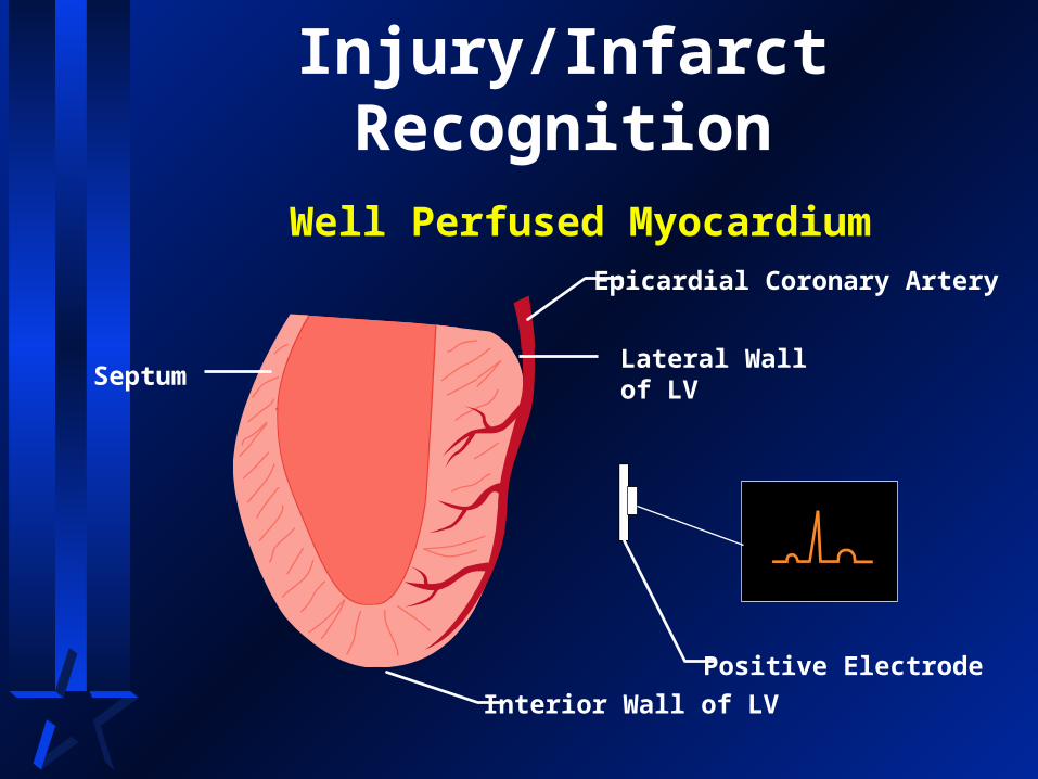
Injury/Infarct Recognition
Epicardial Coronary Artery
Lateral Wall of LV
Positive Electrode
Septum
Interior Wall of LV
Well Perfused Myocardium
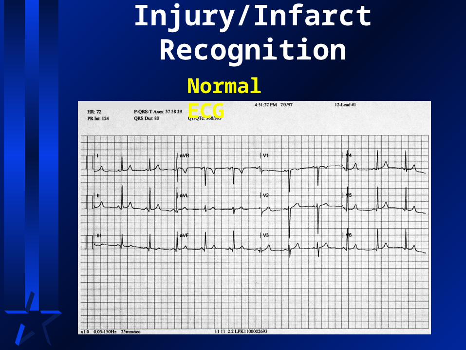
Injury/Infarct Recognition
Normal ECG

Injury/Infarct Recognition
Epicardial Coronary Artery
Lateral Wall of LVSeptum
Interior Wall of LV
Ischemia
Positive Electrode
Left Ventricular
Cavity
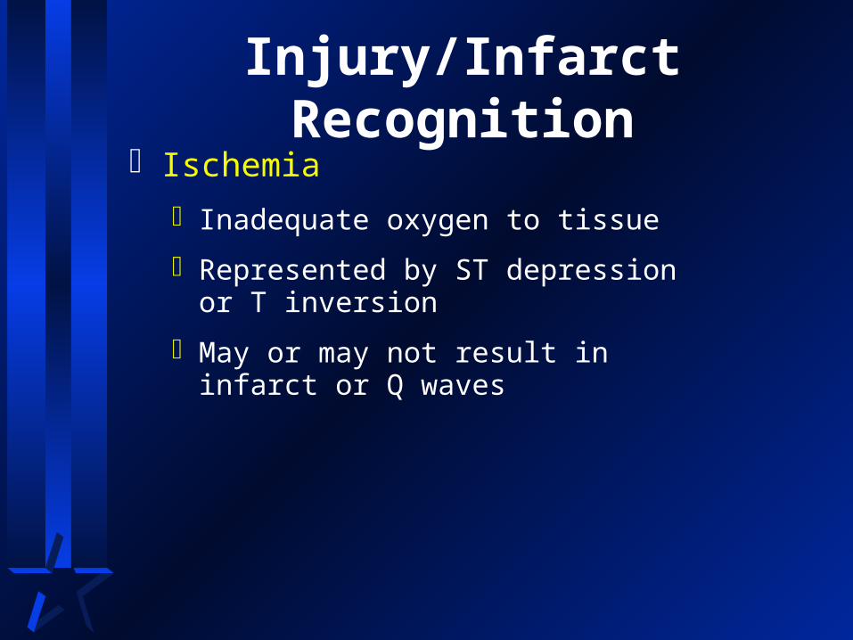
Injury/Infarct RecognitionIschemia
Inadequate oxygen to tissue
Represented by ST depression or T inversion
May or may not result in infarct or Q waves

Injury/Infarct RecognitionST Segment Depression
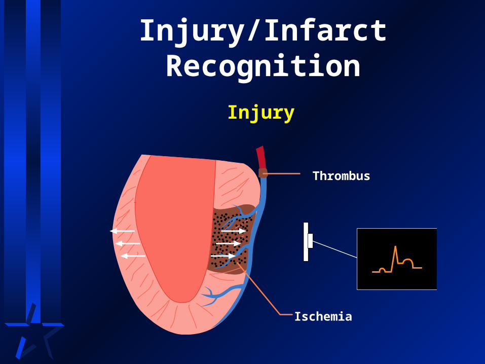
Injury/Infarct Recognition
Thrombus
Ischemia
InjuryInjury
Injury/Infarct Recognition
Injury
Prolonged ischemia
Represented by ST elevation
referred to as an “injury pattern”
Usually results in infarct
may or may not develop Q wave

Injury/Infarct RecognitionST Segment Elevation

Injury/Infarct Recognition
Infarcted AreaElectrically Silent
Depolarization
Infarct

Injury/Infarct Recognition
Infarct
Death of tissue
Represented by Q wave
Not all infarcts develop Q waves
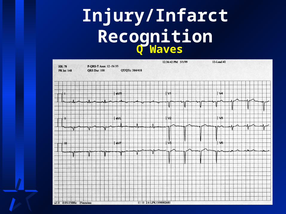
Injury/Infarct RecognitionQ Waves

Injury/Infarct Recognition
Infarcted Area Electrically Silent
Thrombus
Depolarization
Ischemia
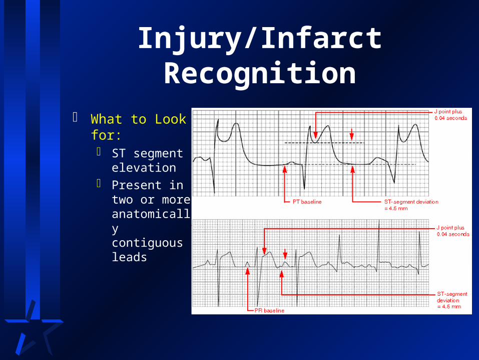
Injury/Infarct Recognition
What to Look for: ST segment
elevation Present in two
or more anatomically contiguous leads

Injury/Infarct Recognition: Practice
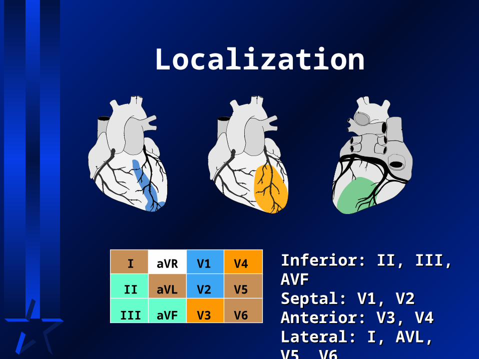
Localization
Inferior: II, III, AVFInferior: II, III, AVFSeptal: V1, V2Septal: V1, V2Anterior: V3, V4Anterior: V3, V4Lateral: I, AVL, V5, V6Lateral: I, AVL, V5, V6
I
II
III
aVR
aVL
aVF
V1
V2
V3
V4
V5
V6
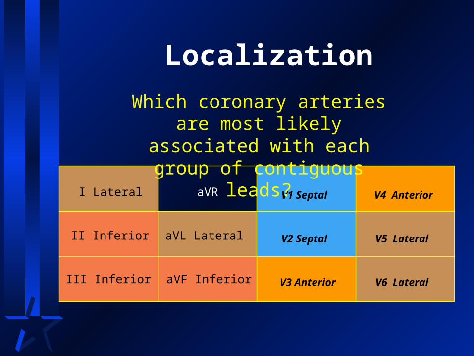
Localization
I Lateral
II Inferior
III Inferior
aVR
aVL Lateral
V1 Septal
aVF Inferior
V2 Septal
V3 Anterior
V4 Anterior
V5 Lateral
V6 Lateral
Which coronary arteries are most likely associated with each group of
contiguous leads?
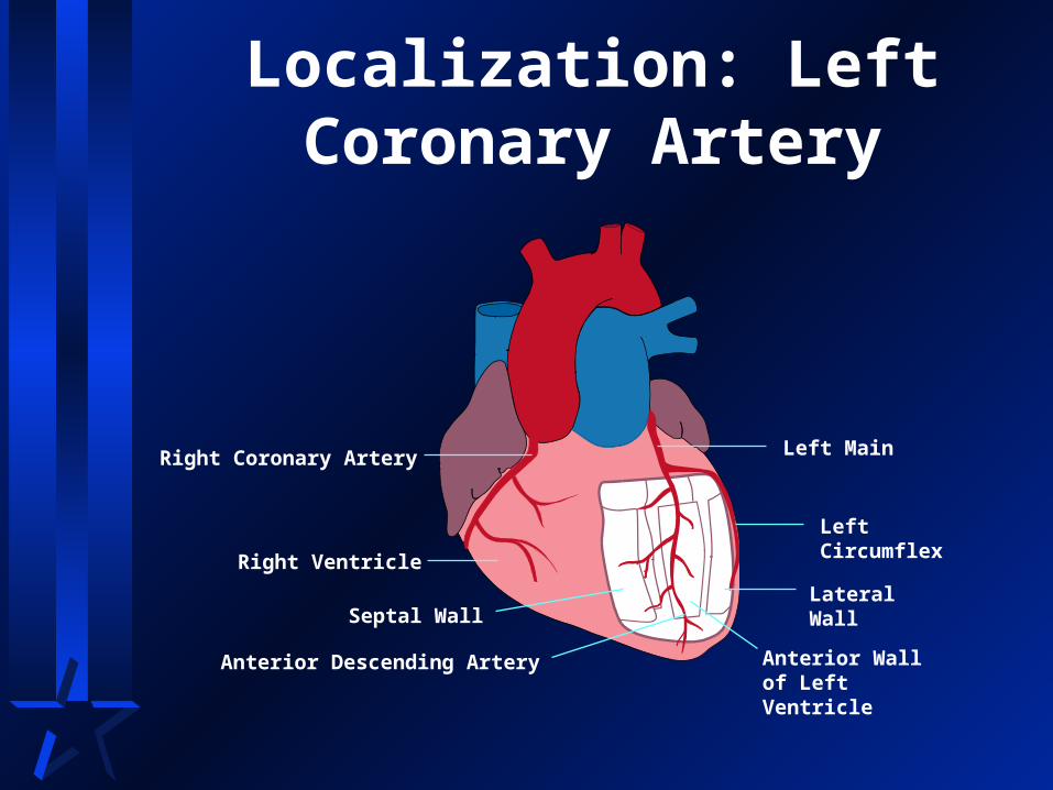
Localization: Left Coronary Artery
Left Main
Left Circumflex
Lateral Wall
Anterior Wall of Left Ventricle
Septal Wall
Right Ventricle
Right Coronary Artery
Anterior Descending Artery
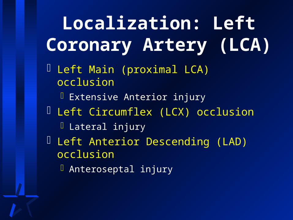
Localization: Left Coronary Artery (LCA)
Left Main (proximal LCA) occlusionExtensive Anterior injury
Left Circumflex (LCX) occlusionLateral injury
Left Anterior Descending (LAD) occlusionAnteroseptal injury

Localization Practice ECG

Localization Practice ECG

Localization Practice ECG

Localization: Extensive Anterior MI
Evidence in septal, anterior, and lateral leads
Often from proximal LCA lesion
“Widow Maker”
Complications commonLeft ventricular failure
CHF / Pulmonary Edema
Cardiogenic Shock

Localization: Definitive Therapy for Extensive AWMI
Normal blood pressure Thrombolysis may be indicated
Signs of shockPTCACABG

Localization: LCA Occlusions
Other considerationsBundle branches supplied by LCASerious infranodal heart block may
occur

Localization: Right Coronary Artery
Right Coronary Artery
Posterior Descending Artery
Inferior Wall of left ventricle
Posterior Wall
Lateral Wall
Left Ventricle
Left Coronary Artery

Localization: Right Coronary Artery (RCA)
Proximal RCA occlusionRight Ventricle injuredPosterior wall of left ventricle injured Inferior wall of left ventricle injured
Posterior descending artery (PDA) occlusion Inferior wall of right ventricle injured

Localization Practice ECG

Localization: Proximal RCA Occlusion
Right Ventricular Infarct (RVI)12-lead ECG does not view right ventricleUse additional leads
V3R - V6R V4R
Right precordial leads same anatomical landmarks as on left for V3 -
V6 but placed on the right side
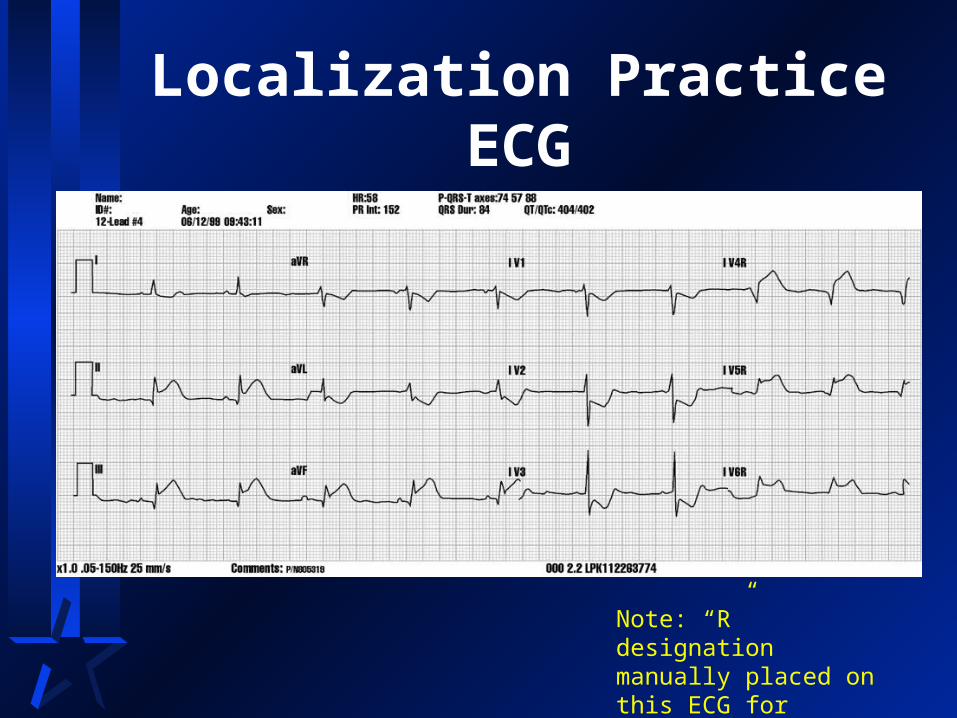
Localization Practice ECG
Note: “R” designation manually placed on this ECG for teaching purposes
Localization: ECG Evidence of RVI
Inferior MI (always suspect RVI)
Look for ST elevation in right-sided V leads (V3-V6)

Localization: Physical Evidence of RVI
Dyspnea with clear lungs
Jugular vein distension
HypotensionRelative or absolute

Localization: Treatment for RVI
Use caution with vasodilatorsSmall incremental doses of MSNTG by drip
Treat hypotension with fluidOne to two liters may be requiredLarge bore IV lines
Localization: Posterior Wall MI (PWMI)
Usually extension of an inferior or lateral MIPosterior wall receives blood from RCA & LCA
Common with proximal RCA occlusions
Occurs with LCX occlusions
Identified by reciprocal changes in V1-V4 May also use Posterior leads to identify
V7: posterior axillary line level with V6 V8: mid-scapular line level with V6 V9: left para-vertebral level with V6

Localization Practice ECG
Localization: Left Coronary Dominance
Approximately 10% of populationLCX connects to posterior descending artery
and dominates inferior wall perfusion
In these cases when LCX is occluded, lateral and inferior walls infarct Inferolateral MI

Localization Practice ECG
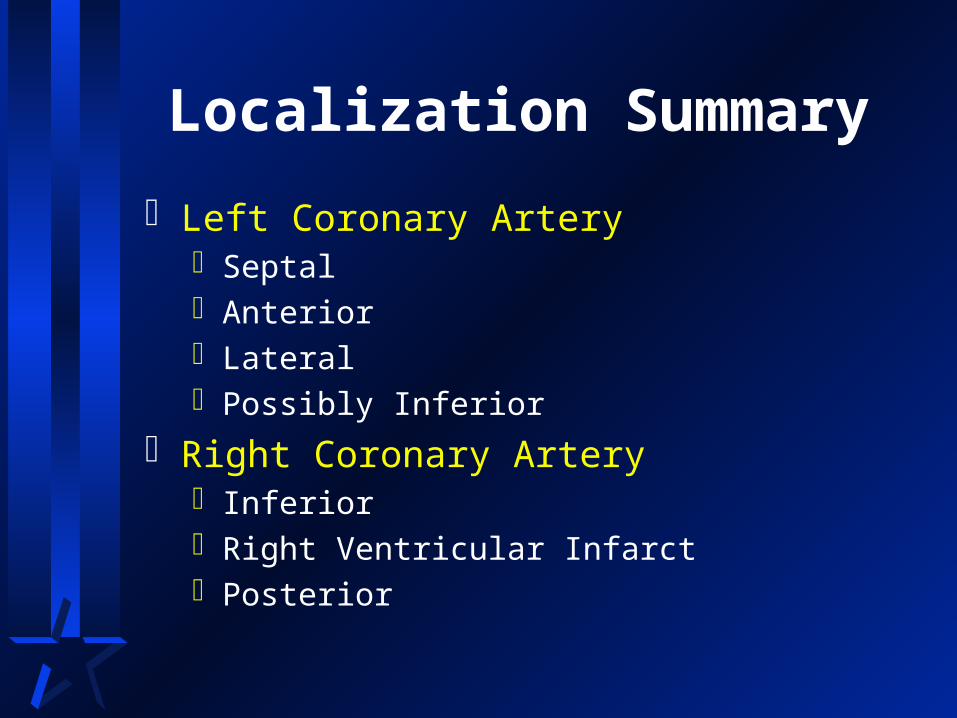
Localization Summary
Left Coronary ArterySeptalAnteriorLateralPossibly Inferior
Right Coronary Artery InferiorRight Ventricular InfarctPosterior

Evolution of AMI
Hyperacute Early change suggestive
of AMI Tall & Peaked May precede clinical
symptoms Only seen in leads
looking at infarcting area Not used as a diagnostic
finding

Evolution of AMI
Acute ST segment elevation Implies myocardial
injury occurring Elevated ST segment
presumed acute rather than old
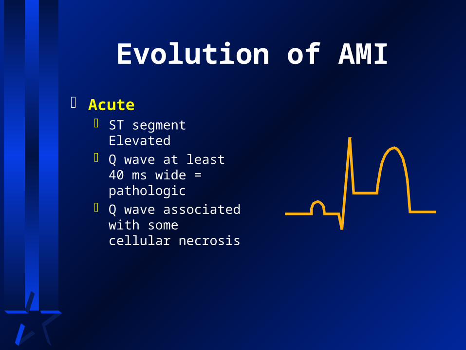
Evolution of AMI
Acute ST segment Elevated Q wave at least 40 ms
wide = pathologic Q wave associated
with some cellular necrosis
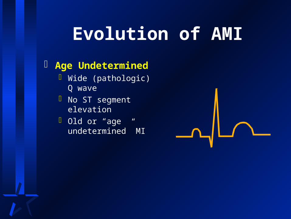
Evolution of AMI
Age Undetermined Wide (pathologic) Q
wave No ST segment
elevation Old or “age
undetermined” MI

AMI Recognition
A normal 12-lead ECG DOES NOT
mean the patient is not having acute
ischemia, injury or infarction!!!
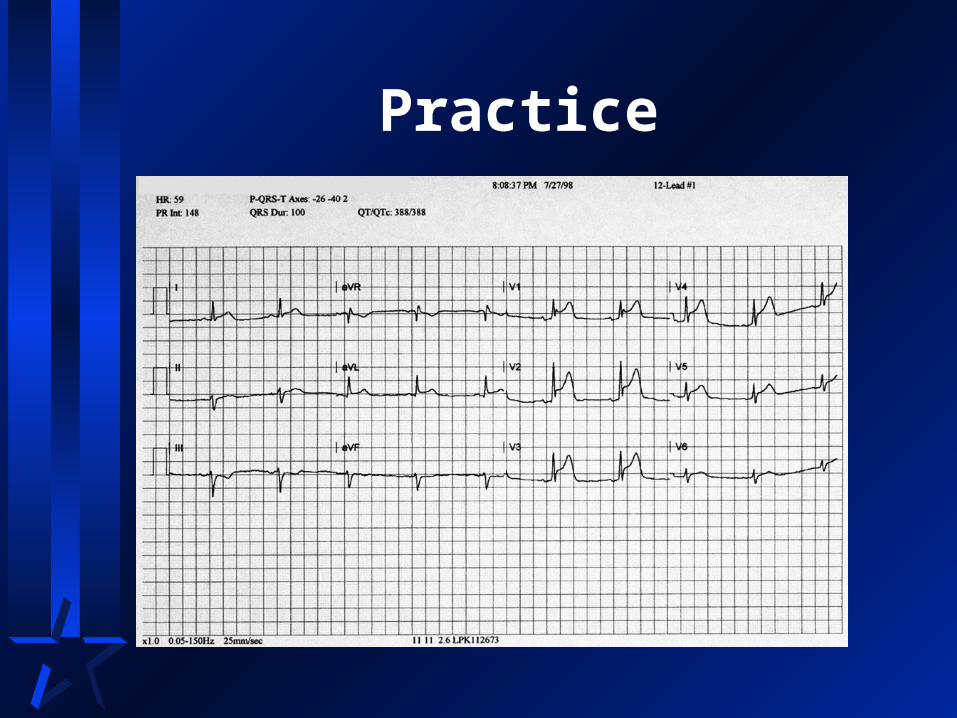
Practice

Practice

Practice
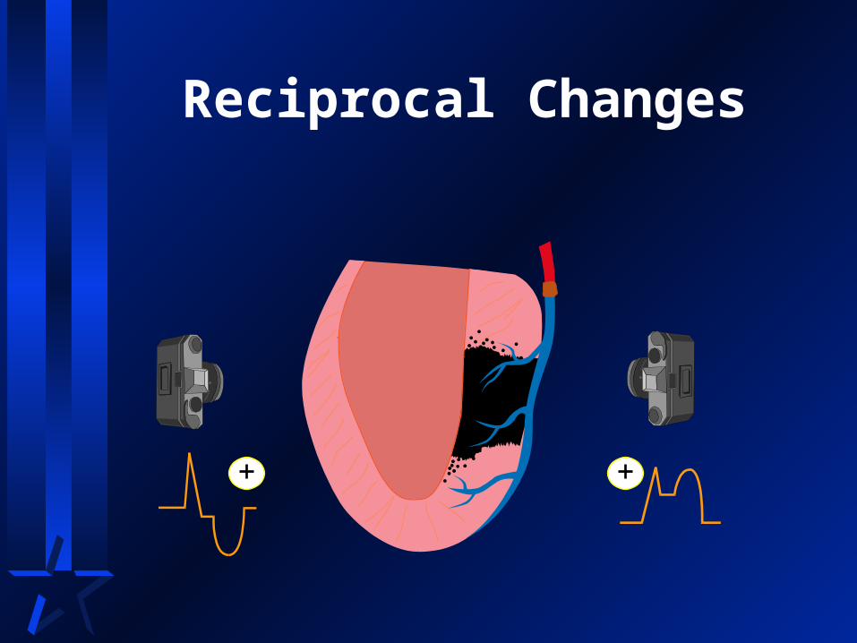
Reciprocal Changes
Reciprocal Changes
II, III, aVFII, III, aVF I, aVL, V leadsI, aVL, V leads
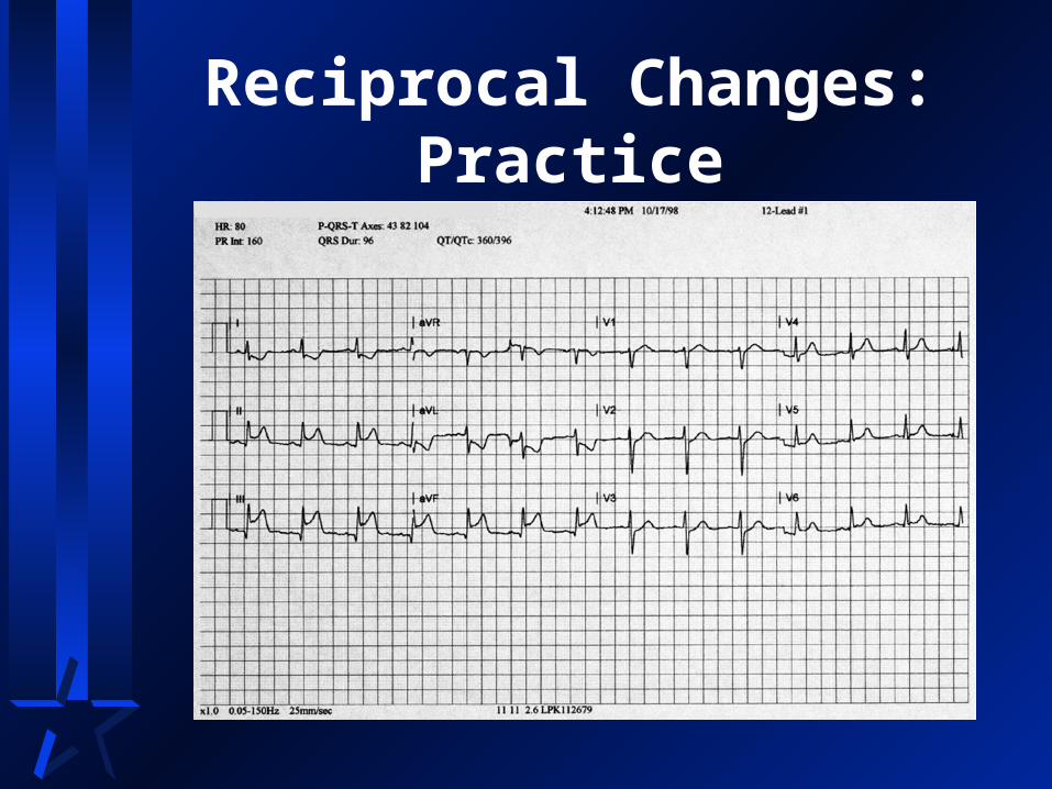
Reciprocal Changes: Practice

Reciprocal Changes: Practice

AMI Recognition
Reciprocal changesNot necessary to presume infarctionStrong confirming evidence when
presentNot all AMIs result in reciprocal
changes

Summary
ST segment elevation is presumptive evidence for AMI
Other conditions may also cause ST elevation
Known as Imposters
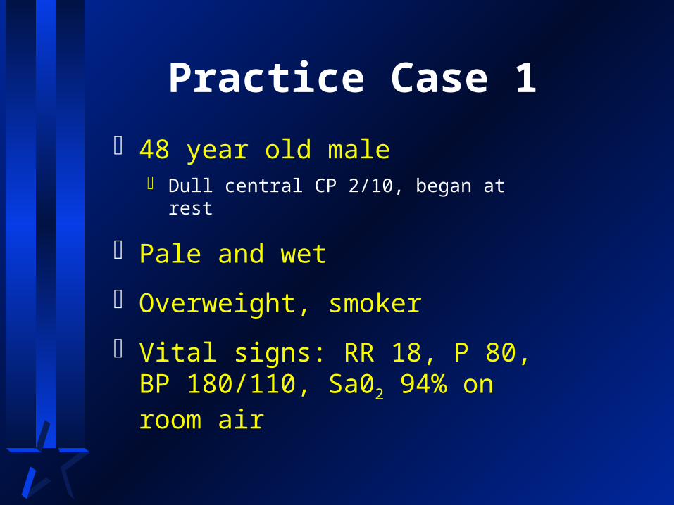
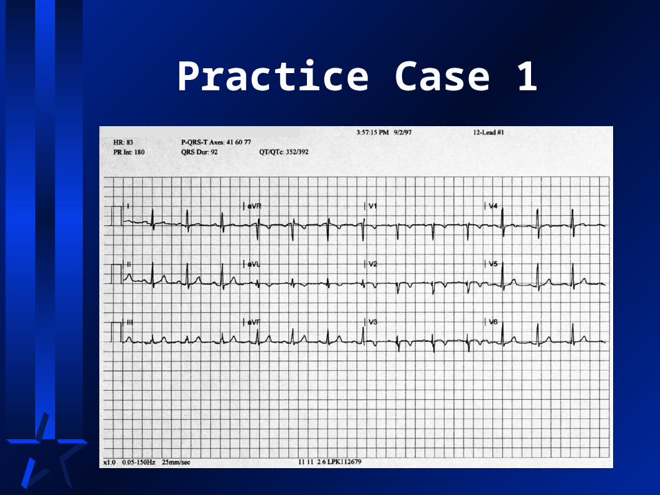
Practice Case 1
48 year old male Dull central CP 2/10, began at rest
Pale and wet
Overweight, smoker
Vital signs: RR 18, P 80, BP 180/110, Sa02 94% on room air
Practice Case 1
Practice Case 268 year old female
Sudden onset of anxiety and restlessness, States she “can’t catch her breath” Denies chest pain or other discomfort
History of IDDM and hypertension
RR 22, P 110, BP 190/90, Sa02 88% on NC at 4 lpm

Practice Case 2

Practice Case Summary
Must take into Account
Story
Risk factors
ECG
Treatment