18 - buteyko cliniccontrol+of+tongue+movement+with... · little is known of hypoglossal motoneuron...
TRANSCRIPT
http://cro.sagepub.com/Critical Reviews in Oral Biology & Medicine
http://cro.sagepub.com/content/12/1/18The online version of this article can be found at:
DOI: 10.1177/10454411010120010101
2001 12: 18CROBMA. Sawczuk and K.M. Mosier
Neural Control of Tongue Movement With Respect To Respiration and Swallowing
Published by:
http://www.sagepublications.com
On behalf of:
International and American Associations for Dental Research
can be found at:Critical Reviews in Oral Biology & MedicineAdditional services and information for
http://cro.sagepub.com/cgi/alertsEmail Alerts:
http://cro.sagepub.com/subscriptionsSubscriptions:
http://www.sagepub.com/journalsReprints.navReprints:
http://www.sagepub.com/journalsPermissions.navPermissions:
What is This?
- Jan 1, 2001Version of Record >>
by guest on September 11, 2013 For personal use only. No other uses without permission.cro.sagepub.comDownloaded from
NEURAL CONTROL OF TONGUE MOVEMENTWITH RESPECT TO RESPIRATION AND SWALLOWINGA. Sawczukl*K.M. Mosier2Departments of 1,20ral Pathology, Biology, and Diagnostic Sciences, 2Radiology, and INeuroscience, University of Medicine and Dentistry of New Jersey, 110 Bergen St., Newark, NJ 07103-2400;*corresponding author, [email protected]
ABSTRACT: The tongue must move with remarkable speed and precision between multiple orofacial motor behaviors that are executedvirtually simultaneously. Our present understanding of these highly integrated relationships has been limited by their complexity. Recentresearch indicates that the tongue's contribution to complex orofacial movements is much greater than previously thought. The purposeof this paper is to review the neural control of tongue movement and relate it to complex orofacial behaviors. Particular attention willbe given to the interaction of tongue movement with respiration and swallowing, because the morbidity and mortality associated withthese relationships make this a primary focus of many current investigations. This review will begin with a discussion of peripheraltongue muscle and nerve physiology that will include new data on tongue contractile properties. Other relevant peripheral oral cavityand oropharyngeal neurophysiology will also be discussed. Much of the review will focus on brainstem control of tongue movement andmodulation by neurons that control swallowing and respiration, because it is in the brainstem that orofacial motor behaviors sort them-selves out from their common peripheral structures. There is abundant evidence indicating that the neural control of protrusive tonguemovement by motoneurons in the ventral hypoglossal nucleus is modulated by respiratory neurons that control inspiratory drive. Yet,little is known of hypoglossal motoneuron modulation by neurons controlling swallowing or other complex movements. There is evi-dence, however, suggesting that functional segregation of respiration and swallowing within the brainstem is reflected in somatotopywithin the hypoglossal nucleus. Also, subtle changes in the neural control of tongue movement may signal the transition between res-piration and swallowing. The final section of this review will focus on the cortical integration of tongue movement with complex orofa-cial movements. This section will conclude with a discussion of the functional and clinical significance of cortical control with respect torecent advances in our understanding of the peripheral and brainstem physiology of tongue movement.
Keywords. Hypoglossal, complex movement, brainstem, motor cortex, muscle properties.
(I) IntroductionThe tongue is a mass of uncompartmentalized interdigitat-
ing muscles that rapidly and accurately changes the direc-tion of its movement in response to the demands of multiplecomplex orofacial behaviors. These behaviors include respira-tion, swallowing, mastication, speech, licking, gaping, cough-ing, gagging, and vomiting. Five of these processes (respira-tion, swallowing, mastication, licking, and gaping) are con-trolled by central pattern generators that are located in themedulla and pons of the brainstem and that transformascending and descending signals into rhythmic and pat-terned behaviors (Dellow and Lund, 1971; Wiesenfeld et al.,1977; Jean, 1984a,b; Dinardo and Travers, 1994; Nakamura andKatakura, 1995; Westberg et al., 1998; Travers et at., 2000). Thespeed and precision with which the tongue must movebetween these essentially concurrent behaviors are governedby its contractile properties, the intrinsic properties ofhypoglossal motoneurons, generator-produced rhythmicmodulation of hypoglossal motoneuron activity, and descend-ing influences from cortical and subcortical nuclei.Investigations of the neural control of tongue movement havecontributed significantly to our present limited understandingof the neural control of complex orofacial behaviors. Futurestudies may offer insight into the neural substrate underlyingthe rapid switching that occurs among complex behaviors
such as respiration, swallowing, and tongue movement.Knowledge of tongue neurophysiology and its modulation by
neurons that control complex movements has been retarded byobstacles such as investigators' inability to measure tongue con-tractile properties and by the lack of substantive information aboutneural circuitry at the brainstem and cortical levels of the neuraxis.Some of these problems have been overcome by recent innovationsin study techniques that have enabled us to investigate the rela-tionships between tongue movement and other complex orofacialbehaviors more completely. In this review, we will discuss theresults of studies published during the past decade that haveshaped our current knowledge of the neural control and contractileproperties of the tongue. Additionally, control of the oropharynxand larynx will be reviewed as it relates either directly or indirectlyto the modulation of tongue movement. At the brainstem level,somatotopy within the hypoglossal nucleus (NXII), selected aspectsof hypoglossal motoneuron membrane properties, and hypoglossalafferents relevant to respiration and swallowing will be examined.Neural networks controlling complex orofacial movements such asrespiration and swallowing will also be reviewed as they relate tomodulation of hypoglossal motoneurons. We will conclude ourreview process with a discussion of the cortical control of tonguemovement and the potential for plasticity within this system, par-ticularly with regard to dysfunction and clinical relevance.
Although recent investigations have made substantial contribu-tions to our understanding of the complexities surrounding tonguemovement, much remains to be done, especially with regard to clin-
18 Crit Rev Oral Biol Med 12(l):18-37 (2001)18 Crit Rev Oral Biol Med 12(l):18-37 (2001) by guest on September 11, 2013 For personal use only. No other uses without permission.cro.sagepub.comDownloaded from
ical application. Disruption of the neural control oftongue movement can seriously impede the functionof rhythmic behaviors. Of those physiological process-es involving the tongue, respiration and swallowingare associated with the highest morbidity and mortal-ity. In fact, hypoglossal motoneuron dysfunction hasbeen proposed to be a primary contributor to respira-tory disorders such as sleep apnea and Sudden InfantDeath Syndrome (SIDS) (Mitra et al, 1986, Haddad andDonnelly, 1990; O'Kusky and Norman, 1992, Kubin etal, 1996; Urban et al, 1996). Furthermore, swallowingdisorders (i.e, dysphagia) associated with hypoglossalmotoneuron dysfunction can lead to aspiration pneu-monia and its consequences. The contribution oftongue movement disruption to the dysfunction ofother complex behaviors has not yet been appreciat-ed, because so little is known about their interactions.For this reason, our review of the relationship oftongue movement to complex orofacial behaviors will Afocus on respiration and swallowing.
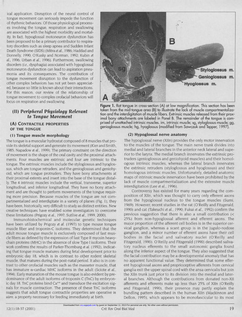
Figure 1. R
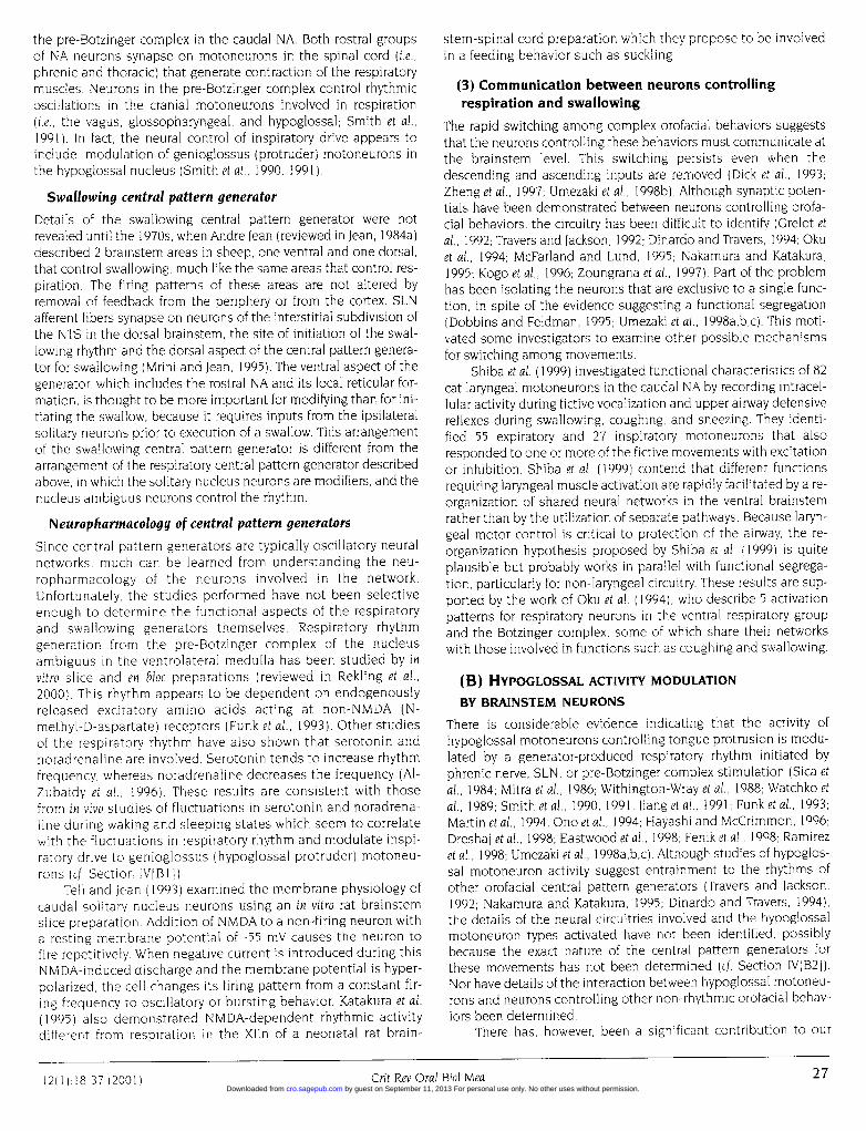
(11) Peripheral Physiology Relevant taken from(II)tion and thito Tongue Movement imal bony(A) CONTRACTILE PROPERTIES prised of urOF THE TONGUE genioglossi(I) Tongue muscle morphology
The tongue is a muscular hydrostat composed of 8 muscles that pro-vide its skeletal support and generate its movement (Kier and Smith,1985; Napadow et al, 1999). The primary constraint on the directionof movement is the shape of the oral cavity and the proximal attach-ments Four muscles are extrinsic and four are intrinsic to thetongue. The extrinsic muscles include the styloglossus and hyoglos-sus, which are tongue retruders, and the genioglossus and geniohy-oid, which are tongue protruders. They have bony attachments attheir proximal extents and insert into the base of the tongue distal-ly The 4 intrinsic muscles include the vertical, transverse, superiorlongitudinal, and inferior longitudinal They have no bony attach-ment and are thought to perform movements of the tongue requir-ing more precision. Because muscles of the tongue are not com-partmentalized and interdigitate in a variety of planes (Fig. 1), theyhave been, historically, very difficult to study as distinct entities Newcreative techniques have enabled some investigators to overcomethese limitations (Prigozy et al., 1997; Sutlive et al., 1999, 2000).
Immunohistochemical and molecular genetic techniqueshave been utilized by Prigozy et al. (1997) to type mouse tonguemuscle fiber and troponin-C isoforms. They determined that theadult mouse tongue muscle is exclusively composed of fast mus-cle fibers as defined by the expression of fast Type 11 myosin heavy-chain proteins (MHC) in the absence of slow Type I isoforms. Theirwork confirms the results of Parker-Thornburg et al. ( 1992), indicat-ing that tongue muscle matures during fetal development prior toembryonic day 18, which is in contrast to other rodent skeletalmuscle, that matures during the post-natal period. It also is in con-trast to other orofacial muscles such as the masseter muscle, thathas immature a-cardiac MHC isoforms in the adult (Sciote et al,1994). Early maturation of the mouse tongue is also evident by pre-dominance of the adult isoforms of troponin C (TnC) by embryon-ic day 18. TnC proteins bind Call and transduce the excitation sig-nals for muscle contraction. The presence of these TnC isoformssuggests that adult tongue contractile properties are operative inutero, a property necessary for feeding immediately at birth.
,,?-i;Rat tongue in cross-section (A) at low magnification. This section has beenthe mid-tongue area (B) to illustrate the lack of muscle compartmentaliza-e interdigitation of muscle fibers. Extrinsic muscles released from their prox-attachments are labeled in Panel B. The remainder of the tongue is com-iattached intrinsic muscles. im, intrinsic muscle; sg, styloglossus muscle; gg,is muscle; hg, hyoglossus (modified from Sawczuk and Tepper, 1 997).
(2) Hypoglossal nerve anatomyThe hypoglossal nerve (Xlln) provides the only motor innervationto the muscles of the tongue The main nerve trunk divides intomedial and lateral branches in the anterior neck lateral and supe-rior to the larynx The medial branch innervates the extrinsic pro-truders (genioglossus and geniohyoid muscles) and their homol-ogous intrinsic muscles, whereas the lateral branch innervatesthe extrinsic retruders (styloglossus and hyoglossus) and theirhomologous intrinsic muscles. Unfortunately, detailed anatomicmaps of intrinsic muscle innervation have been prohibited by thelack of muscle compartmentalization and the extensive muscleinterdigitation (Lee et al., 1996).
Controversy has existed for many years regarding the com-position of Xlln, which was thought to carry only efferent axonsfrom the hypoglossal nucleus to the tongue muscles (Sumi,1969). However, recent studies in the rat (O'Reilly and Fitzgerald,1990) and cat (Takeuchi et al, 1990; Fukui et al., 1992) confirm theprevious suggestion that there is also a small contribution (<25%) from non-hypoglossal afferent and efferent axons. Themajor proportion of the afferent cell bodies is in the superior cer-vical ganglion, whereas a scant group is in the jugulo-nodoseganglion, and a minor number of efferent axons have their cellbodies in the facial and salivatory nuclei (O'Reilly andFitzgerald, 1990). O'Reilly and Fitzgerald (1990) described saliva-tory nucleus efferents to the small autonomic ganglia foundalong the inferior aspect of the tongue. They also suggested thatthe facial contribution may be a developmental anomaly that hasno apparent functional value. They determined that some effer-ent hypoglossal axons and proprioceptive afferents from cervicalganglia exit the upper spinal cord with the ansa cervicalis but jointhe XIln trunk just prior to its division into the medial and later-al branches Although the contributions from donated non-XIIafferents and efferents make up less than 25% of Xlln (O'Reillyand Fitzgerald, 1990), their presence may partly explain thebundling pattern described in the human XIIn (Mackinnon andDellon, 1995), which appears to be monofascicular to its most
12)1) 1837)2001)Oral Biol Med
1 2( 1) 18-37 (2001 ) Crit Rev Oral Biol Med by guest on September 11, 2013 For personal use only. No other uses without permission.cro.sagepub.comDownloaded from

"0IFigure 2. The rat hypoglossal nerve bundle contains between 3000 ancthat are primarily (> 2250 to 2625) hypoglossal efferents. These nervesmovement by responding to signals from neurons that control orofacial bEbrainstem. (Modified from Kalantarian et al., 1998)
distal aspect, where it branches into 5 fascicules. The remainderof this discussion will focus on hypoglossal efferents which arethe major contributors to Xlln I> 75%).
The remarkably large mean number of myelinated axons (9200+ 2182) reported by Mackinnon and Dellon (1995) suggests that,even after adjustment for donated afferents and efferents, morethan 7000 axons comprise the efferent portion of the human Xllnthat is devoted to motor control of the human tongue. The esti-mated number of axons in the rat Xlln is between 3300 and 3700(Lewis et al., 1971; Lodge et al., 1973; Kalantarian et al., 1998; Fig. 2).Results of electron microscopic imaging suggest that < 1% of theseaxons are unmyelinated (Lodge et al., 1973). If corrected for donat-ed axons, the number of axons in the peripheral nerve is a reason-able match with the estimated number of motoneurons in the rathypoglossal nucleus (NXII), which is between 2500 and 3500depending on the technique used to stain and count the neurons(Lewis et al., 1971; McClung and Goldberg, 1999)
Regardless of the exact number of neurons innervatingtongue muscles, it is much higher than the number of motoneu-rons devoted to groups of limb muscles (e.q., about 1000motoneurons innervate the posterior compartment of the catlimb, which includes soleus, plantaris, and medial and lateralgastrocnemius) but is consistent with other- muscle groups, suchas those of the digits and inner ear, that are required to executefinely tuned movements. In other motor systems, precision isfacilitated by a decrease in the number of muscle fibers con-trolled by a single nerve (i e., an increased innervation ratio). Asan example, each neuron innervates more than 600 muscle fibersin the cat gastrocnemius muscle (Burke and Tsairis, 1973), where-as the human digit has an innervation ratio of I to 10, and themuscles of the inner ear have a -to- I innervation ratio (reviewed
in Binder, 1989). The innervation ratio of individ-ual tongue muscles has not been determined,but Sutlive et al. (1999, 2000) estimate the rat
< styloglossus muscle to have a ratio of 1 22 andthe genioglossus muscle to have a ratio of 20.The ratio for intrinsic tongue muscles is expect-ed to be much closer to that of the digits,because these are the muscles devoted to thenumore precise movements required for speechand mastication. The work of Mu and Sanders(1999) provides further reinforcement for thelarge innervation ratio of intrinsic tongue mus-
cles. Using Sihler's stain to examine innervationof the canine tongue muscles by Xlln, they esti-mated approximately 50 primary branches fromthe lateral and medial nerve trunks, of which atleast 32 project to muscle compartments of theintrinsic tongue muscles.
(3) Tongue muscle contractile propertiesTongue muscle interdigitation, absence of com-partmentalization, and the lack of discrete attach-ment sites have impeded the determination oftongue contractile properties and severely retard-ed studies of tongue physiology. Recently,Goldberg and his colleagues (Gilliam and
1 3500 axons Goldberg, 1995; Sutlive et al., 1999, 2000) cre-
direct tongue atively circumvented some of these problems innhaviors in the their electrophysiologic studies of rat tongue con-
tractile properties By attaching the tongue to asmall strain gauge, Gilliam and Goldberg (1995)
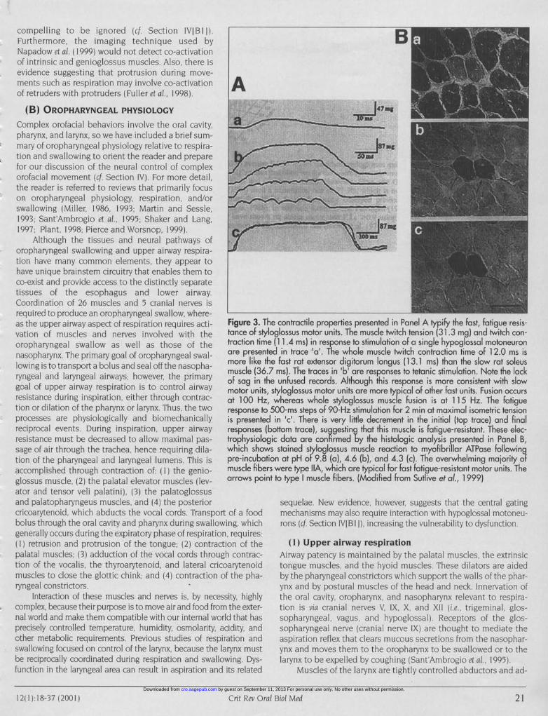
were reliably able to measure speed, force, and fatigue propertiesof retrusive and protrusive movements in response to peripheralstimulation of the hypoglossal whole nerve and its branches andto central stimulation within the hypoglossal nucleus. Their resultssuggest that the tongue retruders and protruders are primarilycomposed of fast, fatigue-resistant muscle groups. Sutlive et al.(1999, 2000) initiated their investigations of individual muscles bycarefully dissecting the styloglossus muscle from its attachments tothe whole tongue and the genioglossus from its proximal attach-ment to the mandible. Using electrophysiologic techniques forstudying contractile properties and combining the results with his-tochemical data, they were able to demonstrate the predominanceof fast-fatigue-resistant motor units (> 90%) (Fig. 3) Most recently,Sokoloff (2000) identified the intrinsic tongue motor units as fast-fatigue-resistant and primarily retrusive These results are consis-tent with the demands placed upon tongue muscles to move withremarkable speed and precision between the various complex oro-facial behaviors.
The functional designation of extrinsic tongue muscles asretruders or protruders distinct from intrinsic muscle influencehas been brought into question by Napadow et al. (1999) who per-formed MR image analysis of human tongue movement and con-cluded that the orthogonal intrinsic muscles were the primaryprotruders, with no contribution from the genioglossus muscle.They suggest that control of protrusion by core tongue muscles isconsistent with protrusive movements by other muscularhydrostats (e.g., the elephant trunk, Kier and Smith, 1985)Although there is probably a contribution to protrusion fromintrinsic muscles, the data supporting the genioglossus muscle asa primary protruder during respiration and Sokoloff's (2000) resultsindicating that intrinsic motor units are retruders are much too
20 Grit Rev Oral Biol Med12)1) 18
37120011
Crit Rev Oral Biot Med 1 2( 1):I 8-37 (2001 )20 by guest on September 11, 2013 For personal use only. No other uses without permission.cro.sagepub.comDownloaded from
compelling to be ignored (cf Section IVIBI I).Furthermore, the imaging technique used byNapadow etal. (1999) would not detect co-activationof intrinsic and genioglossus muscles. Also, there isevidence suggesting that protrusion during move-ments such as respiration may involve co-activation Aof retruders with protruders (Fuller et al., 1998).
(B) OROPHARYNGEAL PHYSIOLOGY
Complex orofacial behaviors involve the oral cavity,pharynx, and larynx, so we have included a brief sum-mary of oropharyngeal physiology relative to respira-tion and swallowing to orient the reader and preparefor our discussion of the neural control of complexorofacial movement (cf. Section IV). For more detail,the reader is referred to reviews that primarily focuson oropharyngeal physiology, respiration, and/orswallowing (Miller, 1986, 1993; Martin and Sessle,1993; Sant'Ambrogio et al, 1995; Shaker and Lang,1997; Plant, 1998; Pierce and Worsnop, 1999). W
Although the tissues and neural pathways oforopharyngeal swallowing and upper airway respira-tion have many common elements, they appear tohave unique brainstem circuitry that enables them toco-exist and provide access to the distinctly separatetissues of the esophagus and lower airway.Coordination of 26 muscles and 5 cranial nerves isrequired to produce an oropharyngeal swallow, where-as the upper airway aspect of respiration requires acti- Figure 3.vation of muscles and nerves involved with the tance of st)oropharyngeal swallow as well as those of the traction tinnasopharynx. The primary goal of oropharyngeal swal- are preserlowing is to transport a bolus and seal off the nasopha- morue lieryngeal and laryngeal airways; however, the primary omfussag 3goal of upper airway respiration is to control airway motor unit!resistance during inspiration, either through contrac- at 1 00 H:tion or dilation of the pharynx or larynx Thus, the two response tcprocesses are physiologically and biomechanically is presentEreciprocal events. During inspiration, upper airway responsesresistance must be decreased to allow maximal pas- trophysiolcsage of air through the trachea, hence requiring dila- which shoation of the pharyngeal and laryngeal lumens This is pre-incubcaccomplished through contraction of (I) the genio- muscle fibEglossus muscle, (2) the palatal elevator muscles (lev- arrows poator and tensor veli palatini), (3) the palatoglossusand palatopharyngeus muscles, and (4) the posteriorcricoarytenoid, which abducts the vocal cords. Transport of a foodbolus through the oral cavity and pharynx during swallowing, whichgenerally occurs during the expiratory phase of respiration, requires(II retrusion and protrusion of the tongue; (2) contraction of thepalatal muscles; (3) adduction of the vocal cords through contrac-tion of the vocalis, the thyroarytenoid, and lateral cricoarytenoidmuscles to close the glottic chink; and (4) contraction of the pha-ryngeal constrictors.
Interaction of these muscles and nerves is, by necessity, highlycomplex, because their purpose is to move air and food from the exter-nal world and make them compatible with our internal world that hasprecisely controlled temperature, humidity, osmolarity, acidity, andother metabolic requirements. Previous studies of respiration andswallowing focused on control of the larynx, because the larynx mustbe reciprocally coordinated during respiration and swallowing. Dys-function in the laryngeal area can result in aspiration and its related
[he contractile properties presented in Panel A typify the fast, fatigue resis-yloglossus motor units. The muscle twitch tension (31.3 mg) and twitch con-ie (111.4 ms) in response to stimulation of a single hypoglossal motoneuronited in trace 'a'. The whole muscle twitch contraction time of 12.0 ms isthe fast rat extensor digitorum longus (13.1 ms) than the slow rat soleus6.7 ms). The traces in 'b' are responses to tetanic stimulation. Note the lackthe unfused records. Although this response is more consistent with slowt, styloglossus motor units are more typical of other fast units. Fusion occursz, whereas whole styloglossus muscle fusion is at 115 Hz. The fatiguet 500-ms steps of 90-Hz stimulation for 2 min at maximal isometric tensionrd in 'c'. There is very little decrement in the initial (top trace) and final(bottom trace), suggesting that this muscle is fatigue-resistant. These elec-)gic data are confirmed by the histologic analysis presented in Panel B,ws stained styloglossus muscle reaction to myofibrillar ATPase followingition at pH of 9.8 (a), 4.6 (b), and 4.3 (c). The overwhelming majority ofers were type IIA, which are typical for fast fatigue-resistant motor units. Theint to type muscle fibers. (Modified from Sutlive et ao., 1999)
sequelae. New evidence, however, suggests that the central gatingmechanisms may also require interaction with hypoglossal motoneu-rons (cf Section IVIBI 1), increasing the vulnerability to dysfunction.
(1) Upper airway respirationAirway patency is maintained by the palatal muscles, the extrinsictongue muscles, and the hyoid muscles. These dilators are aidedby the pharyngeal constrictors which support the walls of the phar-ynx and by postural muscles of the head and neck. Innervation ofthe oral cavity, oropharynx, and nasopharynx relevant to respira-tion is via cranial nerves V, IX, X, and XII (i.e., trigeminal, glos-sopharyngeal, vagus, and hypoglossal). Receptors of the glos-sopharyngeal nerve (cranial nerve IX) are thought to mediate theaspiration reflex that clears mucous secretions from the nasophar-ynx and moves them to the oropharynx to be swallowed or to thelarynx to be expelled by coughing (Sant'Ambrogio et al., 1995).
Muscles of the larynx are tightly controlled abductors and ad-
Crit Rev Oral Biol Med 2 11 2( 1):I18-37 (2001 ) by guest on September 11, 2013 For personal use only. No other uses without permission.cro.sagepub.comDownloaded from
ductors that serve as the final bastions of airway protection, ad-mitting perfectly primed air and restricting foreign material fromthe lungs. Motor and sensory control of the larynx is from the su-perior laryngeal nerve (SLN). It modulates respiration by detectingupper airway collapse through laryngeal pressure-sensing mech-anoreceptors and by initiating reflex mechanisms to ensure pres-ervation of patency (reviewed in Sant'Ambrogio et al., 1995). Thesereflex mechanisms serve two purposes: (1) to increase activationof upper airway abductor muscles and (2) to decrease inspiratorydrive by changing the timing of respiration and decreasing thepressure generated by the thoracic pump muscles. The genioglos-sus muscle is one of the primary upper airway dilators affected bythis reflex (reviewed in Martin et al., 1994; Sant'Ambrogio et al.,1995; Fregosi and Fuller, 1997; Eastwood et al., 1998; Fenik et al.,1998; Pierce and Worsnop, 1999). Laryngeal muscles also havethermo- and chemoreceptors that protect the lower airway fromthe potentially harmful effects of cold and ensure that secretionsentering the lower airway are isosmolar. Additionally, these recep-tors detect increased levels of CO which have been shown toreduce ventilation and increase genioglossus activity(Sant'Ambrogio et al., 1995).
(2) Oropharyngeal swallowingOropharyngeal swallowing afferents include the trigeminal, glos-sopharyngeal, and vagal nerves (cranial nerves V, IX, and X).Touch and pressure receptors for the trigeminal nerve in thetongue determine the shape, texture, and stereoscopic aspectsof the bolus. In fact, it has been suggested that the oral cavityacts as a rheometer during formation of the bolus by sensing andaltering the shape and viscosity of food (Coster and Schwartz,1987). The oropharynx is primarily composed of striated musclehaving glossopharyngeal and vagal mechanoreceptors. The over-lying mucosa also contains chemoreceptors, thermoreceptors,and free nerve endings. Slowly adapting pressure receptors ofthe anterior and posterior tonsillar pillars and of the posteriorwall of the pharynx are responsible for initiation of the swallow-ing "reflex". This reflex is more like an automatic behavior than atrue reflex and, as such, can be modified by peripheral and cen-tral input to the central pattern generator for swallowing (cf.Section IV; reviewed in Miller, 1986; Martin and Sessle, 1993).
Swallowing efferents include the trigeminal, facial, glos-sopharyngeal, vagus, and hypoglossal nerves (cranial nerves V,VII, IX, X, XII) as well as cervical nerves 1 to 3. The musclesinvolved are those identified above for upper airway respiration.Like respiration, they provide an environment in which the foodis not simply propelled through a conduit by gravity but isprocessed and actively transported in a highly dynamic and plas-tic manner (Gay et al., 1994; Palmer, 1998). Airway protection fromthe food bolus occurs at the larynx by adduction of laryngeal mus-cles, epiglottic tilt, hyoid elevation, and laryngeal elevation thatpermits passive opening of the upper esophageal sphincter (UES)for food to enter the esophagus. This process is controlled by theSLN. In fact, electrical stimulation of the SLN is the preferredmethod of producing swallowing in experimental animals(Kessler and lean, 1985; Car and Amri, 1987; Amri et al., 1991).
(C) PERIPHERAL INTERACTIONOF TONGUE MOVEMENT, RESPIRATION, AND SWALLOWING
The relationship between the neural control of tongue movementand respiration has been well-established (cf. Section IV). Less isknown of the relationship between tongue movement and swal-lowing, perhaps because the peripheral structures and innerva-
tion have been difficult to isolate from those of respiration. Also,the adductive movements that produce tongue protrusion toensure airway patency during respiration are less complex thantongue movements for swallowing, which require a combinationof retrusion with protrusion.
The relationships between these movements in respirationand swallowing are influenced by several variables, includingposture, respiratory-related chemoreception, and bolus consis-tency. Swallowing occurs during inspiration in most animals andnon-human primates but during expiration in the adult human.McFarland et al. (1994) found that human swallows were com-pleted earlier in expiration (closer to inspiration) if performed ina position on all four limbs that is more typical of non-humananimals. It is likely that posture also affects the modulation oftongue movement during swallowing and respiration.
The modulation of XIIn activity by inspiratory drive has beenrecognized since the early 1980s (reviewed in Bartlett and St. John,1988). Bartlett et al. (1987) examined this relationship more closelyin decerebrate, paralyzed cats by studying peripheral nerve activityduring hypercapnia and hypoxia. They found that the XIIn responseto changes in ventilatory chemoreception was much greater thanphrenic activity and that it occurred earlier during inspiration. Sincethese early investigations, much attention has been given to themodulation of genioglossus (i.e., protruder) activity by inspiratorydrive, particularly with regard to hypoxia (Martin et al., 1994; Hayashiand McCrimmon, 1996; Eastwood et al., 1998; Feniket al., 1998). Mostrecently, Fuller et al. (1998) demonstrated co-activation of tongueprotruders and retruders in response to changes in respiratorychemoreceptor stimulation in the rat which resulted in more tongueretraction than protrusion. They hypothesize that co-activation mayimprove pharyngeal airway mechanics by stiffening the tongue,albeit at the expense of narrowing the airway. Analysis of theseresults suggests that we have much to learn about the modulationof tongue activity by respiration and, presumably, by swallowing.
The impact of bolus consistency and mode of delivery onswallowing have been appreciated for many years by researchersand utilized therapeutically by clinicians. Recently, Preiksaitis andMills (1996) investigated the effects of bolus consistency and pre-sentation on the coordination of respiration and swallowing inhealthy adults. They determined that swallow-related apnea,which lasts 1 or 2 sec and occurs during expiration, is maintainedover a variety of bolus volumes and consistencies when studied assingle-bolus swallows. During more complex eating and drinkingthat simulates the typical food-intake pattern, however, this apneais followed by inspiration. They suggest that such a pattern putsthe aspiration-prone individual at increased risk.
(111) Hypoglossal Nucleus Anatomyand Motoneuron Physiology
(A) FUNCTIONAL ORGANIZATIONOF THE HYPOGLOSSAL NUCLEUS
(1) Somatotopy within the hypoglossal nucleusHypoglossal motoneuron somata are located in the hypoglossalnucleus (NXII), which is positioned in the ventral medulla andcaudal pons near the midline. In the rat, this nucleus extendsabout 2.0 mm rostrocaudally x 1.5 mm mediolaterally from slight-ly caudal of the obex rostrally to the mid-fourth ventricle. Effortsto determine the precise location of motoneurons within thenucleus have been compromised, because interdigitation oftongue muscles has resulted in most anatomic studies being
22Ci e rlBo e 211-7(0122 Crit Rev Oral Biol Med 12(1l):18-37 (2001 )
by guest on September 11, 2013 For personal use only. No other uses without permission.cro.sagepub.comDownloaded from

plagued by contamination of tracers bleeding into adjacent mus-cles Investigators generally agree, however, that the motoneuronsare arranged somatotopically (Fig. 4; Odutola, 1976; Uemura-Sumi etcil., 1988; Sokoloff and Deacon, 1992, Altschuleretal, 1994;Guo et al., 1996). Motoneurons controlling tongue protrusion are inthe ventral part of the nucleus, whereas motoneurons controllingretrusion are in the lateral and dorsolateral parts of the nucleusMotoneurons controlling the intrinsic tongue muscles (primarilyimportant for speech and mastication) occupy the remainder ofthe nucleus. The ventral subdivision is in the caudal 2/3 of thenucleus, whereas the dorsal subdivision is in the rostral 2/3 of thenucleus The two subdivisions overlap in the middle third of thenucleus About 95% of the neurons in the hypoglossal nucleus aremotoneurons, whereas the remaining 5% are interneurons (Vianaet al, 1990) The interneurons are primarily restricted to the dorso-lateral, lateral, and ventral margins of the nucleus (Aldes, 1990;Altschuler et al., 1994).
(2) Functional organization of the hypoglossal nucleusThere is anatomical evidence of a functional organization to thehypoglossal nucleus and the brainstem networks that modulatethem (Dobbins and Feldman, 1995). Neuronal networks control-ling tongue retruders are proposed to be primarily in the area ofthe solitary nucleus of the dorsal brainstem, whereas protruderneurons are primarily in the ventrolateral brainstem near thenucleus ambiguus (cf Section IVIBI). The hypothesis of function-al segregation has far-reaching implications for studies of all oro-facial behaviors, including tongue movement, but substantiationrequires unambiguous identification of motoneurons in thenucleus. This means that confirmation of label in motoneuronsmust be verified by the demonstration of tracer injection siteslocalized to the individual tongue muscle being studied.Unfortunately, these data were not available for most previousanatomical studies of the hypoglossal nucleus because of thecompartmental and histologic limitations placed on tracer injec-tion into the tongue muscle (cf. Section illAl).
In their study of the hypoglossal complex in the monkey,Sokoloff and Deacon (1992) attempted to address the problem oftracers bleeding between muscles by meticulously relatingmotoneurons of the nucleus labeled with wheat-germ agglutininconjugated to horseradish peroxidase (WGA-HRP) to tracer injec-tions in the tongue. Their results were a more accurate confirma-tion of previous work in cats and rats that demonstrate a dorsalcompartment within the nucleus that controls retrusion and a ven-tral compartment that controls protrusion.
Several investigators have performed labeling studies oftongue protruder motoneurons (particularly the genioglossus)that were controlled by injection-site verification (Brozanski et al.,1989, Sokoloff and Deacon, 1992; Aldes, 1995). Their work is bestsummarized by Aldes (1995), who studied 72 rat motoneuronslabeled with horseradish peroxidase (HRP) applied to the wholenerve or injected into the muscle as a conjugate with cholera toxin(CTHRP) By using the overlapping tracer injection technique ofSokoloff and Deacon (1992), Aldes (1995) identified three sub-compartments within the ventral hypoglossal nucleus. Moto-neurons of the medial subcompartment (n = 300; D = 22.84 + 2 33pLm) innervate the verticalis and transversus intrinsic muscles,motoneurons of the lateral compartment (n = 200, D = 27.86 ± 2.8pLm) innervate the genioglossus muscle, and motoneurons of thelateral accessory compartment (outside the main nucleus; n = 100;D = 34.46 + 2.61 p.m) innervate the geniohyoid muscle.
McClung and Goldberg (1999) isolated rat styloglossus and hyo-
Cadi *AR:4 ' :
Figure 4. Somatotopy of the hypoglossal nucleus produced by retro-grade tracer (cholera toxin conjugated to horseradish peroxidase)applied to the hypoglossal nerve branches. Stain applied to the rightlateral branch labels the right dorsal motoneurons, and stain to the leftmedial branch labels the left ventral motoneurons. The rostrocaudal dif-ferences in the distribution of these motoneurons are also evident in thisFig. The lateral branch primarily projects to the rostral 2/3 of thenucleus, whereas the medial branch projects to the medial 2/3 of thenucleus. (Modified from McClung and Goldberg, 1999)
glossus muscles by careful dissection prior to injecting tracer(CTHRP) and thus were able to identify the motoneuron somata inNXII more precisely (Fig. 4). They counted about 100 hyoglossusmotoneurons with a mean somata diameter of 29.9 + 3.3 pLm and 50to 100 styloglossus motoneurons (probably closer to 100, see Sutliveet al., 1999) with a mean somata diameter of 30.6 + 3 4 pLm The samegroup of investigators also estimated the genioglossus motoneuronpool to have about 150 motoneurons (Sutlive et al, 2000) Aides(1995) estimated ventrolateral compartment motoneurons (consis-tent with genioglossus projection sites) to be 29 49 + 2 47 p.m in di-ameter and geniohyoid motoneurons in the lateral accessory com-
Crit Rev Oral Biol Med
'%.. "I
. I . .'? .
1 2( 1):I18-37 (2001 ) 23 by guest on September 11, 2013 For personal use only. No other uses without permission.cro.sagepub.comDownloaded from
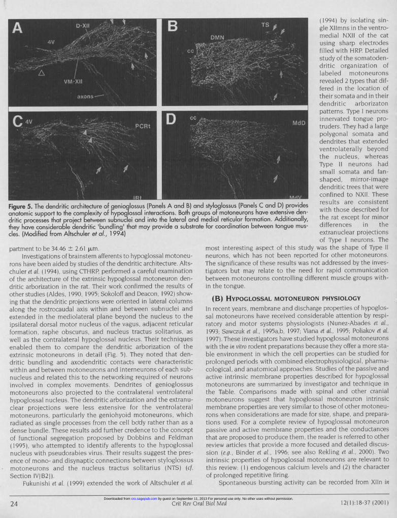
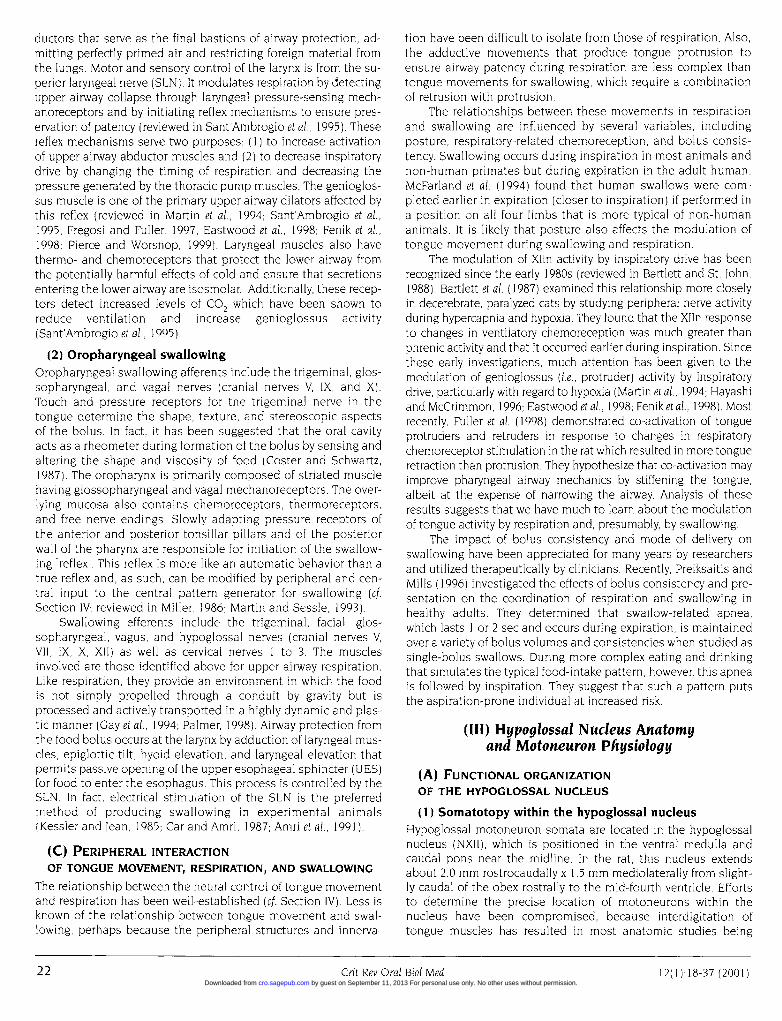
Figure 5. The dendritic architecture of genioglossus (Panels A and B) andanatomic support to the complexity of hypoglossal interactions. Both groudritic processes that project between subnuclei and into the lateral and nthey have considerable dendritic 'bundling' that may provide a substratecles. (Modified from Altschuler et al., 1 994)
partment to be 34.46 + 2.61 lm.Investigations of brainstem afferents to hypoglossal motoneu-
rons have been aided by studies of the dendritic architecture. Alts-chuler et cit. (1994), using CTHRP, performed a careful examinationof the architecture of the extrinsic hypoglossal motoneuron den-dritic arborization in the rat Their work confirmed the results ofother studies (Aldes, 1990, 1995; Sokoloff and Deacon, 1992) show-ing that the dendritic projections were oriented in lateral columnsalong the rostrocaudal axis within and between subnuclei andextended in the mediolateral plane beyond the nucleus to theipsilateral dorsal motor nucleus of the vagus, adjacent reticularformation, raphe obscurus, and nucleus tractus solitarius, aswell as the contralateral hypoglossal nucleus Their techniquesenabled them to compare the dendritic arborization of theextrinsic motoneurons in detail (Fig. 5). They noted that den-dritic bundling and axodendritic contacts were characteristicwithin and between motoneurons and interneurons of each sub-nucleus and related this to the networking required of neuronsinvolved in complex movements. Dendrites of genioglossusmotoneurons also projected to the contralateral ventrolateralhypoglossal nucleus. The dendritic arborization and the extranu-clear projections were less extensive for the ventrolateralmotoneurons, particularly the geniohyoid motoneurons, whichradiated as single processes from the cell bo-dy rather than as adense bundle These results add further credence to the conceptof functional segregation proposed by Dobbins and Feldman(1995), who attempted to identify afferents to the hypoglossalnucleus with pseudorabies virus, Their results suggest the pres-ence of mono- and disynaptic connections between styloglossusmotoneurons and the nucleus tractus solitarius (NTS) (cfSection IVI B2j).
Fukunishi et cil ( 1999) extended the work of Altschuler et al.
(1994) by isolating sin-gle Xllmns in the ventro-medial NXII of the catusing sharp electrodesfilled with HRP Detailedstudy of the somatoden-dritic organization of
r n labeled motoneuronsrevealed 2 types that dif-fered in the location oftheir somata and in theirdendritic arborizatonpatterns. Type neuronsinnervated tongue pro-truders They had a largepolygonal somata anddendrites that extendedventrolaterally beyondthe nucleus, whereasType 11 neurons hadsmall somata and fan-shaped, mirror-imagedendritic trees that wereconfined to NXII These
styloglossus (Panels C and D) provides results are consistentips of motoneurons have extensive den- with those described foriedial reticular formation. Additionally, the rat except for minorfor coordination between tongue mus- differences in the
extranuclear projectionsof Type neurons. The
most interesting aspect of this study was the shape of Type 11neurons, which has not been reported for other motoneurons.The significance of these results was not addressed by the inves-tigators but may relate to the need for rapid communicationbetween motoneurons controlling different muscle groups with-in the tongue.
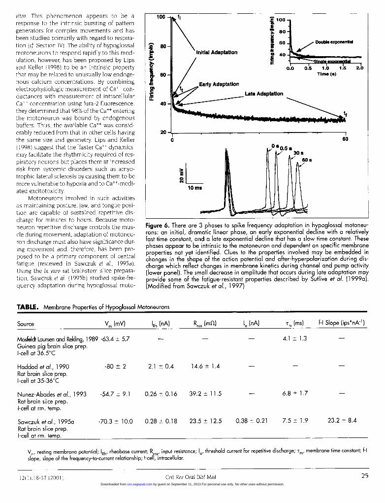
(B) HYPOGLOSSAL MOTONEURON PHYSIOLOGYIn recent years, membrane and discharge properties of hypoglos-sal motoneurons have received considerable attention by respi-ratory and motor systems physiologists (Nunez-Abades et al.,1993; Sawczuk et al., 1995a,b, 1997; Viana et cil, 1995; Poliakov et cil.1997). These investigators have studied hypoglossal motoneuronswith the in vitro rodent preparations because they offer a more sta-ble environment in which the cell properties can be studied forprolonged periods with combined electrophysiological, pharma-cological, and anatomical approaches. Studies of the passive andactive intrinsic membrane properties described for hypoglossalmotoneurons are summarized by investigator and technique inthe Table. Comparisons made with spinal and other cranialmotoneurons suggest that hypoglossal motoneuron intrinsicmembrane properties are very similar to those of other motoneu-rons when considerations are made for size, shape, and prepara-tions used. For a complete review of hypoglossal motoneuronpassive and active membrane properties and the conductancesthat are proposed to produce them, the reader is referred to otherreview articles that provide a more focused and detailed discus-sion (e.g., Binder et al., 1996, see also Rekling et al., 2000). Twointrinsic properties of hypoglossal motoneurons are relevant tothis review. (I) endogenous calcium levels and (2) the characterof prolonged repetitive firing.
Spontaneous bursting activity can be recorded from XIIn in
24 Grit Rev Orcil Biol Med12(11 18 3712001)
Crit Rev Oral Biol Med 1 2 II): 18-37 (200 124 by guest on September 11, 2013 For personal use only. No other uses without permission.cro.sagepub.comDownloaded from
vitro. This phenomenon appears to be aresponse to the intrinsic bursting of patterngenerators for complex movements and hasbeen studied primarily with regard to respira-tion (cf. Section IV). The ability of hypoglossalmotoneurons to respond rapidly to this mod-ulation, however, has been proposed by Lipsand Keller (1998) to be an intrinsic propertythat may be related to unusually low endoge-nous calcium concentrations. By combiningelectrophysiologic measurement of Ca++ con-ductances with measurement of intracellularCall concentration using fura-2 fluorescence,they determined that 98% of the Call enteringthe motoneuron was bound by endogenousbuffers. Thus, the available Call was consid-erably reduced from that in other cells havingthe same size and geometry. Lips and Keller(1998) suggest that the "faster Ca++ dynamics"may facilitate the rhythmicity required of res-piratory neurons but places them at increasedrisk from systemic disorders such as amyo-trophic lateral sclerosis by causing them to bemore vulnerable to hypoxia and to Ca++-medi-ated excitotoxicity.
Motoneurons involved in such activitiesas maintaining posture, jaw, and tongue posi-tion are capable of sustained repetitive dis-charge for minutes to hours. Because moto-neuron repetitive discharge controls the mus-cle during movement, adaptation of motoneu-ron discharge must also have significance dur-ing movement and, therefore, has been pro-posed to be a primary component of centralfatigue (reviewed in Sawczuk et al., 1995a).Using the in vitro rat brainstem slice prepara-tion, Sawczuk et al. (1995b) studied spike-fre-quency adaptation during hypoglossal moto-
100 - fl
80iVInitial Adaptation
II.cFM
100
2h 80
60 -4 Doubl exponentlal
c 40-C
0.0 0.5 1.0 1.5 2.0Time (a)
.Early Adaptation
60
E
°OI10 Ms
Figure 6. There are 3 phases to spike frequency adaptation in hypoglossal motoneu-rons: an initial, dramatic linear phase, an early exponential decline with a relativelyfast time constant, and a late exponential decline that has a slow time constant. Thesephases appear to be intrinsic to the motoneuron and dependent on specific membraneproperties not yet identified. Clues to the properties involved may be embedded inchanges in the shape of the action potential and after-hyperpolarization during dis-charge which reflect changes in membrane kinetics during channel and pump activity(lower panel). The small decrease in amplitude that occurs during late adaptation mayprovide some of the fatigue-resistant properties described by Sutlive et al. (1 999a).(Modified from Sawczuk et al., 1997)
TABLE. Membrane Properties of Hypoglossal Motoneurons
Source Vm (mV) 'Rh (nA) Rnss (mSl) lo (nA) Tm (ins) f-I Slope (ips*nAk1)
Mosfeldt Laursen and Rekling, 1989 -63.4 + 5.7 - 4.1 + 1.3Guinea pig brain slice prep.I-cell at 36.5°C
Haddad etal., 1990 -80 2 2.1 +0.4 14.6 1.4Rat brain slice prep.I-cell at 35-36°C
Nunez-Abades etal., 1993 -54.7 + 9.1 0.26 + 0.16 39.2 + 11.5 6.8 + 1.7Rat brain slice prep.I-cell at rm. temp.
Sawczuk et al., 1995a -70.3 + 10.0 0.28 + 0.18 23.5 + 12.5 0.38 + 0.21 7.5 + 1.9 23.2 + 8.4Rat brain slice prep.I-cell at rm. temp.
Vm, resting membrane potential; 'Rh, rheobase current; Rnss input resistance; lo/ threshold current for repetitive discharge; Tm/ membrane time constant; f-Islope, slope of the frequency-to-current relationship; I-cell, intracellular.
1211183710011Gri Re Ora Bil Md 2
25Crit Rev Oral Biol Med12( ):I 8-37 (2001 by guest on September 11, 2013 For personal use only. No other uses without permission.cro.sagepub.comDownloaded from
neuron repetitive discharge by injecting a series of long constant-current pulses into the hypoglossal motoneurons. They identifiedthree distinct phases (Fig. 6) to hypoglossal motoneuron adapta-tion which appear to be produced by different membrane mecha-nisms (Sawczuk et al., 1995b, 1997). The first phase is a dramatic lin-ear decrease in firing frequency that is complete within 1 to 4 inter-spike intervals (isi). Following this initial adaptation is an expo-nential decline in frequency lasting for the duration of firing, whichmay be several minutes. This decline is an intrinsic, non-varyingproperty of the motoneuron that is described by one of two possi-ble exponential functions, either a single function for late adapta-tion only or a double function that includes both early and latecomponents to adaptation.
The time-course of hypoglossal spike-frequency adaptation isso consistent over repeated trials that it appears to be a fingerprint forintrinsic cell activity (Sawczuk et al., 1995a). Furthermore, this con-sistency remains even when the discharge is interrupted by theintroduction of current pulses of various durations or by extendedperiods of 'white noise". Most importantly, the late phase of adap-tation has a small-magnitude change over several minutes(Sawczuk et al., 1995b), suggesting that fatigue resistance is a prop-erty of hypoglossal motoneurons. These results support the investi-gations of Sutlive et al. ( 1999, 2000) that demonstrated fast, fatigue-resistant contractile properties for the styloglossus muscle. Theyalso lend credence to the proposal that fatigue resistance is desir-able for tongue function during complex movements such as respi-ration, swallowing, and speech. Fatigue resistance may be particu-larly important for facilitating the constant, rapid switching betweenthese movements that must occur during a 24-hour period.
(IV) Neural Controlof Complex Orofacial Movements
(A) BRAINSTEM CONTROLOF SWALLOWING AND RESPIRATION
Earlier in this review, we noted that, although upper airway respi-ration and oropharyngeal swallowing have peripheral structures incommon, it is in the brainstem that they appear to segregate.Unfortunately, details of the circuitry have been difficult to studybecause of the shared peripheral innervation. Some insight hasbeen gained from neurophysiological investigations of lower air-way respiration, esophageal swallowing, and tongue movement.We will review selected aspects of brainstem anatomy and physiol-ogy related to swallowing and respiration prior to examining theirinvolvement in the control of tongue movement. For a more com-plete review of the neural control of swallowing and respiration, thereader is referred to other sources (Miller, 1986, 1993; Sessle andHenry, 1989; Bianchi et al., 1995; Plant, 1998).
(1) Brainstem anatomy relevantto respiration and swallowing
Primary vagal and glossopharyngeal afferents from the oral cavity,pharynx, and larynx enter the brainstem from the various cranialnerve ganglia that house their cell bodies and project to the solitarynucleus (NTS). The brainstem projection of the cervical vagusnerves and the superior laryngeal (SLN) and pharyngeal (PhN)nerves in the rat is confined to the interstitial nucleus of the NTS(Altschuler et al., 1989; Mrini and Jean, 1995; Furusawa et al., 1996;Sawczuk and Covell, 1999), which extends from slightly caudal to theobex in a rostral direction for a distance of approximately 600 to 800pim. Umezaki et al. (1998a) identified neurons that were exclusively
swallow-related in the cat interstitial nucleus lateral to the solitarytract. The intermediate nucleus in the mid-NTS is rostral to theinterstitial nucleus, receives afferent projections from the glos-sopharyngeal nerve, and relays sensory information from the palateand pharynx. The ventral and ventrolateral subdivisions of the NTSreceive pulmonary afferents (Kalia and Richter, 1985, 1988).
Kawai and Senba (1996) were able to maintain and study thelocal circuitry of the caudal solitary nucleus in a rat thin-slicepreparation. They describe 3 different cell groups: (1) local circuitneurons with axon collaterals restricted to the solitary nucleus butwith the main axon leaving the nucleus, (2) presumed interneuronswith axons restricted within the solitary nucleus, and (3) projectionneurons whose axons leave the nucleus. The projection neuronshave a somal surface area > 150 pLm2, whereas both the local cir-cuit neurons and the putative interneurons have smaller cell bod-ies, < 150 pLm2. Projection neurons have few, if any, axonal collat-erals, whereas both local circuit neurons and the putative interneu-rons have extensive arborization of their axonal collaterals.
Vagal and glossopharyngeal efferents to the oral cavity, pharynx,and larynx have their cell bodies in the nucleus ambiguus (NA).Furusawa et al. (1996) have shown that rat SLN efferents are localizedto a 1-mm segment of the NA between 1.5 and 2.5 mm rostral to theobex. Bieger and Hopkins (1987) localized the PhN motoneuron poolto the caudal aspect of the semicompact formation of the NA, where-as the SLN motoneuron pool is in the rostral aspect. Altschuler et al.(1991) used cholera toxin-HRP to characterize pharyngeal efferentneurons in the NA semicompact formation into 3 shapes: multipolar(20-30 x 25-35 Rrm), oval (15-20 x 20-25 Rrm), and fusiform (15 x 30Rm). Their functional significance is not clear.
(2) Central pattern generatorsComplex orofacial movements are either tonic, phasic, or episod-ic motor sequences characterized by rhythmic, patterned, orcyclic activity. These motor sequences appear to be controlled bycentral pattern generators (cpg) in the brainstem. Cpgs arethought to be small networks of neurons or, perhaps, a singleneuron that will produce an oscillatory discharge in response tostimulation and will continue to oscillate even when the stimula-tion has been removed (reviewed in Feldman and McCrimmon,1999; see also Stein et al., 1997, and Rekling et al., 2000). There are5 brainstem cpgs that have been proposed to control orofacialmovements for respiration, swallowing, mastication, licking, andrejection. We will limit this discussion to the generators for res-piration and swallowing. The reader is encouraged to refer toother recent resources for investigations of the cpgs for mastica-tion (reviewed in Lund et al., 1998), licking (Travers and Jackson,1992; Brozek et al., 1996; Travers et al., 2000), and rejection(Dinardo and Travers, 1994).
Respiratory central pattern generatorThe rhythmic activity of respiration is controlled in the brainstemby at least 2 central pattern generators: (1) the pneumotaxic cen-ter in the pons, which includes the nucleus parabrachialis medi-alis and the Kolliker-Fuse nucleus; and (2) the dorsal and ventralrespiratory groups of the dorsomedial medulla, which are associ-ated with the caudal NTS and NA, respectively. The pneumotaxiccenter may be important for switching between respiratory pha-ses, but its lesion does not abolish respiratory rhythm, whichappears to be centered in the NA (Smith et al., 1991). Inspiratory-phase activity is controlled by neurons in the middle and rostralaspects of the NA. Expiration is controlled by neurons of theBotzinger complex in the rostral NA and inspiration by neurons in
26CrtRvOaBilMd2()8-7(0126 Crit Rev Oral Biol Med 12(1):18-37 (2001) by guest on September 11, 2013 For personal use only. No other uses without permission.cro.sagepub.comDownloaded from
the pre-Botzinger complex in the caudal NA. Both rostral groupsof NA neurons synapse on motoneurons in the spinal cord (i.e.phrenic and thoracic) that generate contraction of the respiratorymuscles. Neurons in the pre-Botzinger complex control rhythmicoscillations in the cranial motoneurons involved in respiration(i.e., the vagus, glossopharyngeal, and hypoglossal; Smith et al.,1991). In fact, the neural control of inspiratory drive appears toinclude modulation of genioglossus (protruder) motoneurons inthe hypoglossal nucleus (Smith et al., 1990, 1991).
Swallowing central pattern generatorDetails of the swallowing central pattern generator were notrevealed until the 1970s, when Andre lean (reviewed in lean, 1984a)described 2 brainstem areas in sheep, one ventral and one dorsal,that control swallowing, much like the same areas that control res-piration. The firing patterns of these areas are not altered byremoval of feedback from the periphery or from the cortex. SLNafferent fibers synapse on neurons of the interstitial subdivision ofthe NTS in the dorsal brainstem, the site of initiation of the swal-lowing rhythm and the dorsal aspect of the central pattern genera-tor for swallowing (Mrini and lean, 1995). The ventral aspect of thegenerator, which includes the rostral NA and its local reticular for-mation, is thought to be more important for modifying than for ini-tiating the swallow, because it requires inputs from the ipsilateralsolitary neurons prior to execution of a swallow. This arrangementof the swallowing central pattern generator is different from thearrangement of the respiratory central pattern generator describedabove, in which the solitary nucleus neurons are modifiers, and thenucleus ambiguus neurons control the rhythm.
Neuropharmacology of central pattern generatorsSince central pattern generators are typically oscillatory neuralnetworks, much can be learned from understanding the neu-
ropharmacology of the neurons involved in the network.Unfortunately, the studies performed have not been selectiveenough to determine the functional aspects of the respiratoryand swallowing generators themselves. Respiratory rhythmgeneration from the pre-Botzinger complex of the nucleusambiguus in the ventrolateral medulla has been studied by invitro slice and en bloc preparations (reviewed in Rekling et al.,2000). This rhythm appears to be dependent on endogenouslyreleased excitatory amino acids acting at non-NMDA (N-methyl D-aspartate) receptors (Funk et al., 1993). Other studiesof the respiratory rhythm have also shown that serotonin andnoradrenaline are involved. Serotonin tends to increase rhythmfrequency, whereas noradrenaline decreases the frequency (Al-Zubaidy et al., 1996). These results are consistent with thosefrom in vivo studies of fluctuations in serotonin and noradrena-line during waking and sleeping states which seem to correlatewith the fluctuations in respiratory rhythm and modulate inspi-ratory drive to genioglossus (hypoglossal protruder) motoneu-rons (cf. Section IVI B Il).
Tell and lean (1993) examined the membrane physiology ofcaudal solitary nucleus neurons using an in vitro rat brainstemslice preparation. Addition of NMDA to a non-firing neuron witha resting membrane potential of -55 mV causes the neuron tofire repetitively. When negative current is introduced during thisNMDA-induced discharge and the membrane potential is hyper-polarized, the cell changes its firing pattern from a constant fir-ing frequency to oscillatory or bursting behavior. Katakura et al.(1995) also demonstrated NMDA-dependent rhythmic activitydifferent from respiration in the XIIn of a neonatal rat brain-
stem-spinal cord preparation which they propose to be involvedin a feeding behavior such as suckling.
(3) Communication between neurons controllingrespiration and swallowing
The rapid switching among complex orofacial behaviors suggeststhat the neurons controlling these behaviors must communicate atthe brainstem level. This switching persists even when thedescending and ascending inputs are removed (Dick et al., 1993;Zheng et al., 1997; Umezaki et al., 1998b). Although synaptic poten-tials have been demonstrated between neurons controlling orofa-cial behaviors, the circuitry has been difficult to identify (Grelot etal., 1992; Travers and Jackson, 1992; Dinardo and Travers, 1994; Okuet al., 1994; McFarland and Lund, 1995; Nakamura and Katakura,1995; Kogo et al., 1996; Zoungrana et al., 1997). Part of the problemhas been isolating the neurons that are exclusive to a single func-tion, in spite of the evidence suggesting a functional segregation(Dobbins and Feldman, 1995; Umezaki et al., 1998a,b,c). This moti-vated some investigators to examine other possible mechanismsfor switching among movements.
Shiba et al. (1999) investigated functional characteristics of 82cat laryngeal motoneurons in the caudal NA by recording intracel-lular activity during fictive vocalization and upper airway defensivereflexes during swallowing, coughing, and sneezing. They identi-fied 55 expiratory and 27 inspiratory motoneurons that alsoresponded to one or more of the fictive movements with excitationor inhibition. Shiba et al. (1999) contend that different functionsrequiring laryngeal muscle activation are rapidly facilitated by a re-
organization of shared neural networks in the ventral brainstemrather than by the utilization of separate pathways. Because laryn-geal motor control is critical to protection of the airway, the re-
organization hypothesis proposed by Shiba et al. (1999) is quiteplausible but probably works in parallel with functional segrega-tion, particularly for non-laryngeal circuitry. These results are sup-ported by the work of Oku et al. (1994), who describe 5 activationpatterns for respiratory neurons in the ventral respiratory groupand the Botzinger complex, some of which share their networkswith those involved in functions such as coughing and swallowing.
(B) HYPOGLOSSAL ACTIVITY MODULATION
BY BRAINSTEM NEURONS
There is considerable evidence indicating that the activity ofhypoglossal motoneurons controlling tongue protrusion is modu-lated by a generator-produced respiratory rhythm initiated byphrenic nerve, SLN, or pre-Botzinger complex stimulation (Sica et
al., 1984; Mitra et al., 1986; Withington-Wray et al., 1988; Watchko et
al., 1989; Smith et al., 1990, 1991; Jiang et al., 1991; Funk et al., 1993;Martin et al., 1994; Ono et al., 1994; Hayashi and McCrimmon, 1996;Dreshaj et al., 1998; Eastwood et al., 1998; Fenik et al., 1998; Ramirezet al., 1998; Umezaki et al., 1998a,b,c). Although studies of hypoglos-sal motoneuron activity suggest entrainment to the rhythms ofother orofacial central pattern generators (Travers and Jackson,1992; Nakamura and Katakura, 1995; Dinardo and Travers, 1994),the details of the neural circuitries involved and the hypoglossalmotoneuron types activated have not been identified, possiblybecause the exact nature of the central pattern generators forthese movements has not been determined (cf. Section IVIB21).Nor have details of the interaction between hypoglossal motoneu-rons and neurons controlling other non-rhythmic orofacial behav-iors been determined.
There has, however, been a significant contribution to our
2712(1.1837)2011GritRevOralBio MeCrit Rev Oral Biol Med1 2( 1):18-37 (2001 )
by guest on September 11, 2013 For personal use only. No other uses without permission.cro.sagepub.comDownloaded from

Figure 7. Rhythmic discharge suggestive of inspiratory drive has been rea 500-prm s ice of caudal rat brainstem. This rhythmic activity is also reflneous hypoglossal discharge recorded from the nerve rootlets at "B". Adprotruder motoneurons in thie ventral hypoglossal nucleus (not shown) arneurons in the pre-Botzinger complex that produce this respiratory rhythmSmith et al., 1 991)
understanding of the hypoglossal response to afferents from thework of Takata (1993), who measured post-synaptic potentials(psp's) in cat protruder and retruder motoneurons activated bylingual nerve stimulation. Using in vivo intracellular recordingtechniques, he found different activation patterns from eachmotoneuron type. Protruders responded with either an inhibito-ry post-synaptic potential (ipsp) alone (40% of 250 protrudermotoneurons), an ipsp-ipsp sequence (30%) having a shortstrychnine (glycine antagonist)-sensitive ipsp and a long picro-toxin-sensitive ipsp, or an excitatory psp (epsp)-ipsp sequence(30%). Retruders responded with either an epsp alone (7% of 200motoneurons), an epsp-ipsp sequence (68%), or an ipsp-ipspsequence with the same sensitivities to strychnine and picrotox-in as protruders (25%). The ipsp-ipsp sequence was produced bybilateral afferents for protruders, but the strychnine-sensitiveipsp was unilateral and the picrotoxin-sensitive ipsp was con-tralateral for retruders. Takata (1993) did not investigate the epsppharmacologically. Although these results are not directly relatedto swallowing or respiratory afferents, they lend further supportto the segregation of retruder and protruder motoneurons.
(1) Modulation of hypoglossal motoneuron activityby respiratory neurons
The relationship between extrinsic tongue muscles and airwaypatency has been recognized for many years (reviewed in Lowe,1981), as has the modulation of hypoglossal activity by inspiratorydrive (reviewed in Bartlett and St. John, 1988; Withington-Wray etal., 1988), but the interdependent nature of this relationship wasnot revealed until recently. Identification of the neural circuitry andtransmitters required for coupling between hypoglossal motoneu-ron activity and rhythmic oscillations of the respiratory central pat-tern generator has required the use of in vitro preparations (Smithet al., 1990, 1991; Funk et al., 1994) and has been instrumental in iso-lating the neurons responsible for respiratory rhythm. By progres-sively reducing the brainstem-spinal cord en bloc preparation to thedimensions of a thick slice (i.e., 500 pLm) containing the necessaryneural circuitry for generating respiratory rhythm, Smith et al. (I1990,1991) were able to isolate inspiratory rhythmic activity to the pre-Botzinger complex of the caudal NA (Fig. 7). This area also includ-ed the hypoglossal nucleus and nerve fragments that were used to
nephrine (NE) from the locus subcoeruleus (Aldes, 1990). TRHincreases the excitability of hypoglossal motoneurons by decreas-
28CrtRvOaBilMd1()l-7(01
monitor spontaneous rhythm generation.These studies clearly demonstrated the modu-lation of hypoglossal motoneuron discharge byinspiratory drive and have catalyzed a surge ofresearch activity that has unequivocallychanged our understanding of the relationship
U|of the neural control of tongue movement to
-60mV respiration and, perhaps, to other orofacialbehaviors.
In vitro techniques have been most valu-able in examining the pharmacologic nature ofthe circuitry (reviewed in Rekling et al., 2000).Several neurotransmitters have been pro-posed to be involved in modulation of
corded at "A" of hypoglossal motoneurons by inspiratory drivelected in sponta- (i.e., genioglossus motoneurons). The primaryIditionally, sinle neurotransmitter activating genioglossusre modulated by motoneurons from projection neurons of the(Adapted from pre-Botzinger complex is glutamate, which
acts at AMPA/kainate receptors (Funk et al.,1993). The activation appears to be mediatedby adenosine triphosphate (ATP), which is also
excitatory and is proposed to act at P2 hypoglossal motoneuronreceptors (reviewed in Funk et al., 1997). The functional signifi-cance of the purinergic mediation is not known.
Serotonin has also been widely implicated in the respiratorymodulation of hypoglossal motoneurons at the respiratory rhythmgenerator in the caudal NA and at genioglossus motoneurons inner-vated by caudal medullary raphe neurons (Aldes et al., 1989; Morin etal., 1992; Li et al., 1993; Manaker and Tischler, 1993). Serotonin mod-ulation of genioglossus activity has been the focus of studies inves-tigating changes in inspiratory drive during sleep, and the disruptionof this modulation has been postulated to contribute to sleep apneaand Sudden Infant Death Syndrome (Kubin et al., 1996; Radulovackiet al., 1998). In the in vitro neonatal rat slice preparation, serotoninincreases respiratory rhythm discharge and XIIn discharge (Morin etal., 1992). However, the effect of serotonin on hypoglossal motoneu-rons has been the source of some controversy, because it acts on avariety of receptors producing either excitation or inhibition(Monteau et al., 1990; Berger et al., 1992; Morin et al., 1992). There issome suggestion that this differential activation is related to a com-bination of receptor subtype and developmental stage. In general,activation of serotonin 5-HT2 post-synaptic receptors produces aninward, depolarizing current in hypoglossal motoneurons (Berger etal., 1992; Al-Zubaidy et al., 1996; Okabe and Kubin, 1996), whereas theinhibitory effect of serotonin has been isolated to pre-synaptic 5-HT B receptors in adults (Okabe and Kubin, 1996; Singer and Berger,1996) and post-synaptic 5-HTlA receptors in neonates. The neonatal5-HT receptors appear to mediate the inhibition of N- and P/0-typecurrents producing a decrease in the amplitude of the apamin-sen-sitive mid-portion of the after-hyperpolarization that follows theaction potential, an effect not found in adults (Bayliss et al., 1997).Okabe et al. (1997) detected mRNA for 5-HT receptors in thehypoglossal nucleus, which included the 5-HT receptors, 1 B, 2A, 2B,and, in lesser amounts, 3 and 7. The functional significance of theseresults has not yet been determined.
Other hypoglossal neuromediators implicated in sleep-relatedrespiratory disorders such as sleep apnea are the peptide, thy-rotropin-releasing hormone (TRH) from the caudal medullary rapheneurons (Manaker and Tischler, 1993), and the transmitter, norepi-
28 Crit Rev Oral Biol Med 12(l):18-37 (2001) by guest on September 11, 2013 For personal use only. No other uses without permission.cro.sagepub.comDownloaded from
ing the resting K+ conductance through a G-protein-mediatedmechanism and increasing an inward current (Rekling, 1992; Baylisset al., 1992, 1994, 1997). Bayliss et al. (1992) suggest that becauseboth genioglossus motoneurons and raphe neurons are less activeduring sleep, they increase the possibility of airway collapse andobstruction. Norepinephrine is excitatory to hypoglossal motoneu-rons (Parkis et al., 1995) but decreases respiratory rhythm (Al-Zubaidy et al., 1996) and is tonically released during waking states.The decrease in this tonic release during sleep has suggested tosome investigators that the removal of excitatory drive togenioglossus motoneurons could decrease protrusive tongue activ-ity and lead to airway block. Funk et al. (1994) studied the develop-ment of TRH and NE potentiation of mice hypoglossal motoneurondischarge. They found that NE potentiation is maximum approxi-mately 2 weeks earlier than TRH potentiation, which is maximum bypost-natal day 21 in rats (Bayliss et al., 1994). The advantage to dif-ferential development of these neuromodulators has not yet beendetermined but has potential for elucidating the underlying mech-anisms of such disorders as Sudden Infant Death Syndrome.
(2) Interaction of hypoglossal motoneuronswith swallowing neurons
Clinical and behavioral investigators have traditionally consideredtongue movement to begin the swallow and to constitute the onlyvoluntary phase of the swallow (reviewed in Logemann, 1983).Classically, the swallow is believed to proceed sequentially withthe pharyngeal phase initiated at the completion of the oralphase. This concept implies that the neural regulation of the tran-sition from the voluntary to involuntary (pharyngeal) phases ofswallowing at the level of the brainstem begins with signals fromhypoglossal motoneurons. Recent imaging and electromyograph-ic studies (Gay et al., 1994; Mosier, 1997) demonstrate that thetemporal, kinematic, and electromyographic parameters of oral,pharyngeal, and laryngeal events during swallowing are notsequential but consist of concurrent overlapping events, and thatall parts of the swallow are modifiable. Analysis of these datasuggests that although hypoglossal motoneurons may receivesignals (ascending or descending) that cause them to initiatebolus formation and transport, they may also become entrainedto a swallowing rhythm that was initiated by ascending or, per-haps more notably, by descending signals received by the swal-lowing generator itself.
Although all tongue muscles help form and transport thebolus, the tongue muscle group most likely to be activated firstby swallowing rhythm initiators in the solitary nucleus are retrud-er muscles. The somatotopy of NXII places the hypoglossal retru-der motoneurons closest to NTS neurons in the dorsolateralbrainstem which may facilitate this activation. Conversely, theprotruder muscle group must be activated to allow the bolus topass into the pharynx immediately following movements, such aslaryngeal closure and epiglottic tilt, that protect the airway. Theprotruder motoneurons are located in the ventral brainstem clo-ser to the ventral NA swallowing motoneurons and the respirato-ry central pattern generator. The work of Dobbins and Feldman(1995), who studied hypoglossal afferents with the pseudorabiesvirus, suggests a differential innervation of retruder and protrud-er motoneurons. They found that rat retruder motoneuronsreceive their primary innervation from pre-motoneurons in thedorsolateral medulla, and that protruder motoneurons are pri-marily innervated by the medullary pre-motoneurons ventrolat-eral to NXII Some motoneurons are commonly innervated bypre-motoneurons in the medial reticular formation. These results
motivated Dobbins and Feldman (1995) to propose that thebrainstem is functionally segregated with regard to modulationof hypoglossal activity.
Tomomune and Takata (1988) performed intracellular recor-dings from hypoglossal protruder (genioglossus) and retruder (sty-loglossus) motoneurons of the anesthetized cat during swallowingproduced by repetitive stimulation of the SLN and by water placedon the dorsum of the tongue. They describe excitation of stylo-glossus motoneurons and an excitation-suppression sequence ofgenioglossus motoneurons during the oropharyngeal swallowwhich is coupled to rhythmic stimulation of the SLN. These intra-cellular recordings were correlated with EMG activity of the sty-loglossus and genioglossus muscles. The suppression phase ofgenioglossus motoneuron activity correlated with styloglossusmuscle activation. Tomomune and Takata did not investigate theorigin of these signals but hypothesized that they were from theswallowing central pattern generator. Sumi's investigation (1969)of rhythmic activity of hypoglossal neurons in response to cortical-ly induced swallowing and chewing in rabbits also indicated a dif-ferential response pattern from the retruder and protrudermotoneurons for swallowing that was not produced for chewing.
This anatomy and physiology suggest that both retruder andprotruder hypoglossal motoneurons are modulated by the swal-lowing rhythm, and that the pattern of control includes modulationof retruder motoneurons by NTS neurons and modulation of pro-truder motoneurons by the NA neurons, which is also the site ofrespiratory rhythm generation. The difficult task has been to isolatethe brainstem circuits for swallowing so that they are accessible forinvestigation. The results of Dobbins and Feldman (1995) suggestthat the pathways between neurons of the solitary tract and retru-der motoneurons are either disynaptic or monosynaptic. Altschuleret al. (1994) showed that some retruder motoneurons have den-drites that extend into the solitary nucleus and adjacent reticularformation, suggesting that monosynaptic input from NTS neuronscould occur in the hypoglossal nucleus or adjacent reticular forma-tion by NTS projection neurons or in the solitary nucleus by local-circuit NTS neurons (Kawai and Senba, 1996; cf. Sections IIIJAI,IVIAII). Although Altschuler et al. (1994) also found protruder den-drites in the NTS area, they were significantly fewer.
Sawczuk et al. (2000; Fig. 8) have initiated studies examiningthe activation of motoneurons in the dorsolateral hypoglossalnucleus (i.e., putative retruder motoneurons) by stimulation ofNTS neurons in the rat brainstem slice preparation. XlImnsresponded to NTS stimulation with short-latency epsps and, insome motoneurons, ipsps that follow high-frequency stimulation,suggesting that the projection pathway is monosynaptic.Stimulation of other ventral areas of the brainstem slice previous-ly shown to be respiratory projection sites produced combina-tions of polysynaptic psps. The excitatory component of the NTSprojection appears to be mediated by glutamate, because it isblocked by the non-NMDA antagonist, DNQX, whereas theinhibitory component appears to be glycine-mediated, because itis blocked by strychnine. These results provide further support forthe functional segregation hypothesis proposed by Dobbins andFeldman (1995).
(3) Switching functions and modulationof hypoglossal activity
The tongue moves with remarkable speed and precision betweencomplex movements such as respiration and swallowing. Themechanisms are not known but have been the source of consider-able research energy and frustration. One likely scenario is that
12111~~~~~~~~1837201Gie OaiMd2Crit Rev Oral Biol Med 291 2( 1):18-37 (2001 )
by guest on September 11, 2013 For personal use only. No other uses without permission.cro.sagepub.comDownloaded from

Figure 8. Stimulation of the solitary tract in the rat brainstem slice preFtion produced short-latency epsps in this hypoglossal motoneuron thalowed high-frequency stimulation of 50 to 100 Hz (Panel A). When the nbrane potential was depolarized, a short-latency ipsp was unmasked Idie trace, Panel B). Bath application of DNQX eliminated the epsps (1trace, Panel B), suggesting that the excitatory neurotransmitter, glutamaacting at non-NMDA receptors. Bath application of strychnine eliminatelipsps (upper trace, Panel B), suggesting that the inhibitory neurotransris glycine. (Adapted from Sawczuk et al., 2000)
the central pattern generators of one movement suppress those ofthe other (McFarland and Lund, 1995; Ono et al., 1998a,b). Thisproposal would operate equally well whether the neurons arefunctionally segregated, activated by shared pathways (Dobbinsand Feldman, 1995), or activated by networks that re-organize(Shiba et al., 1999). Ono et al. (I1998a) have described several inspi-ratory reticular pre-motoneurons in the cat that project only tohypoglossal motoneurons or project to both pharyngeal andhypoglossal motoneurons. Their activation patterns suggest thatthe majority (76%) of single-projection neurons "switched"between inspiration and fictive ingestion, whereas none of thedual-projection neurons displayed this property. The mechanismfor this switching behavior was not addressed. These neuronstended to be positioned either ventrolaterally to the NTS or dor-somedially to the NA, although more of those near the NTS pro-
A4 mV1
-57 mV
B-30
~35|-8 DNQX + Strychi-35
-40
E 5|t Pre-DN!045
-50
-55 | Prer-DDN(
-60
nem- recording techniques to examine the tongue area of the(mid- primate Ml during tongue protrusion tasks as well as dur-ower ing swallowing behaviors. The primate tongue Ml occu-eths pies a relatively large field at the inferolateral portion of
fltter the precentral gyrus. Recordings of single neurons duringa tongue protrusion task and during a swallowing taskshowed significant changes in the firing rate associatedwith each task. Most notable, however, was the observa-tion that the firing rate was differentially modulated,
depending on whether the monkey performed the tongue protru-sion task or the swallowing task (Fig. 9).
One consideration is that the differential modulation of thesecortical neurons in the tongue M 1 may reflect spatially segregatedneuronal populations within this cortical area. However, Martin andher colleagues (1997) found that the neurons that responded pref-erentially to tongue protrusion tasks or to swallowing tasks weredistributed throughout the tongue M 1, and moreover, there was nospatial segregation between these neuronal populations (Fig. 10).
Despite a lack of evidence for spatial segregation among theneurons of the tongue MI, it should be emphasized that specif-ic neuronal populations within tongue MI respond differentiallyto different tasks involving the tongue. Thus, the primate tongueMI, while lacking a discrete functional homunculus, neverthe-less contains multiple "distinct efferent zones" that generate
30 Crit Rev Orcil Biol Med 12(1):18-37
0 20 40 60 80 100
Time (ms)
jected to both the hypoglossal and phrenic motoneurons(Ono et al., 1994). They suggest that the dual innervationprovides a means of synchronizing synergistic musclesduring respiration. Their results support the investiga-tions of Amri et al. (1990), who describe swallowinginterneurons in the ventral medulla that project tomotoneurons in both NXII and the trigeminal motornucleus. In an unrelated study, Ono et al. (1998b) studiedpre-motoneurons adjacent to the ventrolateral hypoglos-sal nucleus that were swallow-related and inhibitory toputative genioglossus motoneurons, and that appearedto suppress genioglossus muscle activity during swallow-ing. Sawczuk et al. (2000) stimulated the same group ofpre-motoneurons in the ventrolateral rat brainstem slicepreparation that Ono et al. (1998b) studied in the cat. Theyrecorded from putative retruder motoneurons in the dor-solateral NXII and studied primarily epsps, althoughsome psps also had an inhibitory component. This dualinnervation lends further support to the arguments of Onoetcal. (1998a,b).
(C) CORTICAL CONTROL
While the contributions from neuroanatomical, pharmaco-logical, and electrophysiological studies of brainstem net-works have significantly furthered our understanding of theneural control of tongue movement, the control of tonguemovement at a central level remains largely unknown.Primarily, the technical limitations of studying cortical net-works in awake, behaving animals or humans have ham-pered progress in this branch of tongue research.Nevertheless, several recent advances in techniques forstudying cortical processes have provided new insightsinto the central representation of tongue movement. Forexample, the role of the primary motor cortex (Ml) in con-trol of tongue movements has been investigated, mostextensively by Sessle and colleagues (Murray et al., 1991;Murray and Sessle, 1992a,b,c; Martin et al., 1997). Thesestudies used intracortical microstimulation (ICMS) and
30 Crit Rev Oral Biol Med 12(l):18-37 (2001) by guest on September 11, 2013 For personal use only. No other uses without permission.cro.sagepub.comDownloaded from
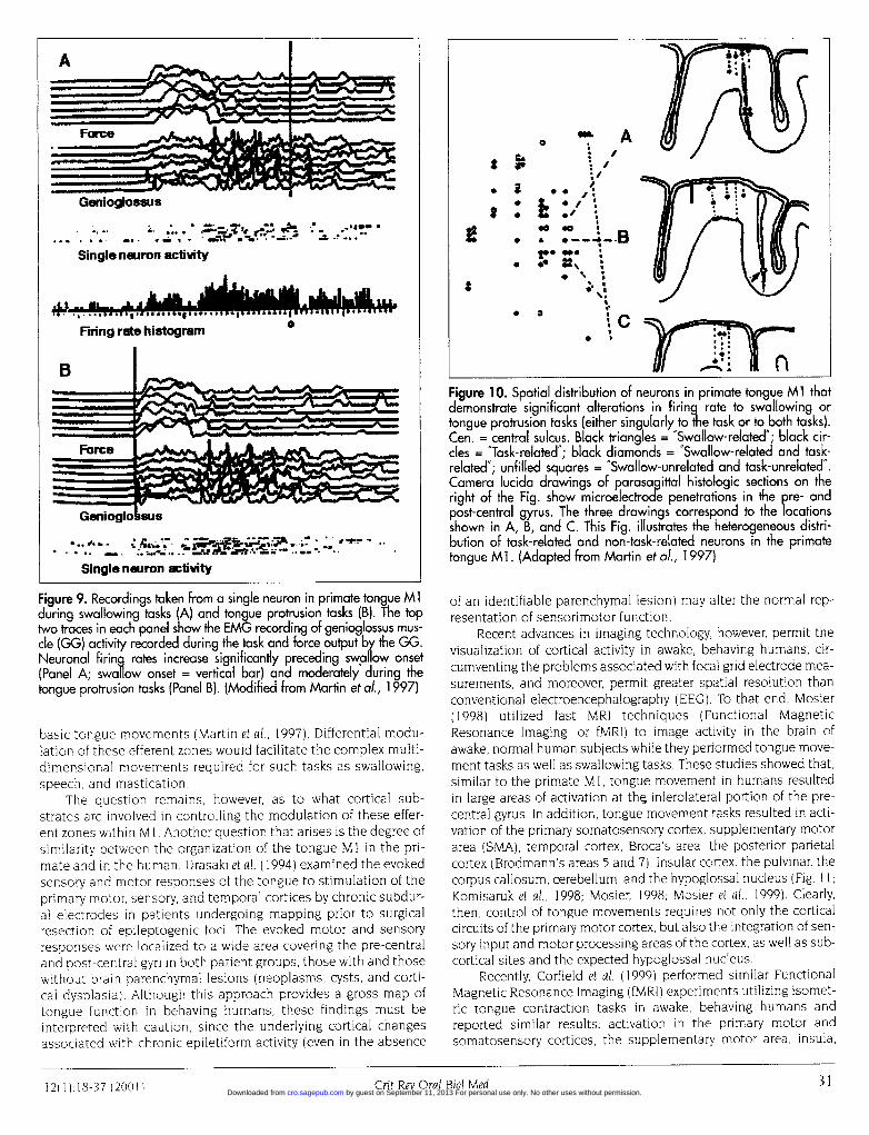
Figure 9. Recordings taken from a single neuron in primate tongue Mlduring swallowing tasks (A) and tongue protrusion tasks (B). The toptwo traces in each panel show the EMG recording of genioglossus mus-cle (GG) activity recorded during the task and force output by the GG.Neuronal firing rates increase significantly preceding swa ow onset(Panel A; swallow onset = vertical bar) and moderately during thetongue protrusion tasks (Panel B). (Modified from Martin et al., 1997)
basic tongue movements (Martin et al, 1997). Differential modu-lation of these efferent zones would facilitate the complex multi-dimensional movements required for such tasks as swallowing,speech, and mastication.
The question remains, however, as to what cortical sub-strates are involved in controlling the modulation of these effer-ent zones within M 1. Another question that arises is the degree ofsimilarity between the organization of the tongue Ml in the pri-mate and in the human. Urasaki et al. (1994) examined the evokedsensory and motor responses of the tongue to stimulation of theprimary motor, sensory, and temporal cortices by chronic subdur-al electrodes in patients undergoing mapping prior to surgicalresection of epileptogenic foci. The evoked motor and sensoryresponses were localized to a wide area covering the pre-centraland post-central gyri in both patient groups, those with and thosewithout brain parenchymal lesions (neoplasms, cysts, and corti-cal dysplasia). Although this approach provides a gross map oftongue function in behaving humans, these findings must beinterpreted with caution, since the underlying cortical changesassociated with chronic epiletiform activity (even in the absence
*w*.
e* ---.3
n,nFigure 10. Spatial distribution of neurons in primate tongue Ml thatdemonstrate significant alterations in firinq rate to swallowing ortongue protrusion tasks (either singularly to the task or to both tasks).Cen. = central sulcus. Black triangles = "Swallow-related"; black cir-cles = "Task-related'; black diamonds = 'Swallow-related and task-related'; unfilled squares = 'Swallow-unrelated and task-unrelated".Camera lucida drawings of parasag ittal histologic sections on theright of the Fig. show microelectrode penetrations in the pre- andpost-central gyrus. The three drawings correspond to the locationsshown in A, B, and C. This Fig. illustrates the heterogeneous distri-bution of task-related and non-task-related neurons in the primatetongue Ml. (Adapted from Martin et al., 1997)
of an identifiable parenchymal lesion) may alter the normal rep-resentation of sensorimotor function.
Recent advances in imaging technology, however, permit thevisualization of cortical activity in awake, behaving humans, cir-cumventing the problems associated with focal grid electrode mea-surements, and moreover, permit greater spatial resolution thanconventional electroencephalography (EEG). To that end, Mosier(1998) utilized fast MRI techniques (Functional MagneticResonance Imaging, or fMRI) to image activity in the brain ofawake, normal human subjects while they performed tongue move-ment tasks as well as swallowing tasks. These studies showed that,similar to the primate Ml, tongue movement in humans resultedin large areas of activation at the inferolateral portion of the pre-central gyrus. In addition, tongue movement tasks resulted in acti-vation of the primary somatosensory cortex, supplementary motorarea (SMA), temporal cortex, Broca's area, the posterior parietalcortex (Brodmann's areas 5 and 7), insular cortex, the pulvinar, thecorpus callosum, cerebellum, and the hypoglossal nucleus (Fig. 1 1;Komisaruk et al., 1998; Mosier, 1998; Mosier et al., 1999). Clearly,then, control of tongue movements requires not only the corticalcircuits of the primary motor cortex, but also the integration of sen-sory input and motor processing areas of the cortex, as well as sub-cortical sites and the expected hypoglossal nucleus.
Recently, Corfield et al. (1999) performed similar FunctionalMagnetic Resonance Imaging (fMRI) experiments utilizing isomet-ric tongue contraction tasks in awake, behaving humans andreported similar results: activation in the primary motor andsomatosensory cortices, the supplementary motor area, insula,
Grit Rev Oral Biol Med 3 1Crit Rev Oral Biol Med1 2( 1 ):1 8-37 (.200 1 ) by guest on September 11, 2013 For personal use only. No other uses without permission.cro.sagepub.comDownloaded from
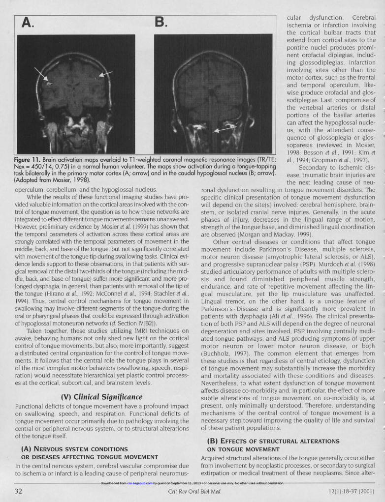
A. B.
Figure 11. Brain activation maps overlaid to Ti -weighted coronal magn(Nex = 450/14; 0.75) in a normal human volunteer. The maps show activtask bilaterally in the primary motor cortex (A; arrow) and in the caudal h(Adapted from Mosier, 1 998).operculum, cerebellum, and the hypoglossal nucleus.
While the results of these functional imaging studies have pro-vided valuable information on the cortical areas involved with the con-trol of tongue movement, the question as to how these networks areintegrated to effect different tongue movements remains unanswered.However, preliminary evidence by Mosier et al. (1999) has shown thatthe temporal parameters of activation across these cortical areas arestrongly correlated with the temporal parameters of movement in themiddle, back, and base of the tongue, but not significantly correlatedwith movement of the tongue tip during swallowing tasks. Clinical evi-dence lends support to these observations, in that patients with sur-gical removal of the distal two-thirds of the tongue (including the mid-dle, back, and base of tongue) suffer more significant and more pro-longed dysphagia, in general, than patients with removal of the tip ofthe tongue (Hirano et al., 1992; McConnel et 61., 1994; Stachler et al.,1994) Thus, central control mechanisms for tongue movement inswallowing may involve different segments of the tongue during theoral or pharyngeal phases that could be expressed through activationof hypoglossal motoneuron networks (cf. Section IVIB21).
Taken together, these studies utilizing fMRI techniques onawake, behaving humans not only shed new light on the corticalcontrol of tongue movements, but also, more importantly, suggesta distributed central organization for the control of tongue move-ments. It follows that the central role the tongue plays in severalof the most complex motor behaviors (swallowing, speech, respi-ration) would necessitate hierarchical yet plastic control process-es at the cortical, subcortical, and brainstem levels
(V) Clinical SignificanceFunctional deficits of tongue movement have a profound impacton swallowing, speech, and respiration. Functional deficits oftongue movement occur primarily due to pathology involving thecentral or peripheral nervous system, or to structural alterationsof the tongue itself
(A) NERVOUS SYSTEM CONDITIONSOR DISEASES AFFECTING TONGUE MOVEMENT
In the central nervous system, cerebral vascular compromise dueto ischemia or infarct is a leading cause of peripheral neuromus-
cular dysfunction. Cerebralischemia or infarction involvingthe cortical bulbar tracts thatextend from cortical sites to thepontine nuclei produces promi-nent orofacial diplegias, includ-ing glossodiplegias. Infarctioninvolving sites other than themotor cortex, such as the frontaland temporal operculum, like-wise produce orofacial and glos-sodiplegias. Last, compromise ofthe vertebral arteries or distalportions of the basilar arteriescan affect the hypoglossal nucle-us, with the attendant conse-quence of glossoplegia or glos-soparesis (reviewed in Mosier,1998, Besson et al., 1991; Kim et
etic resonance images (TR/TE; al., 1994; Gropman et al, 1997)ation during a tongue-tapping Secondary to ischemic dis-ypoglossal nucleus (B; arrow). ease, traumatic brain injuries are
the next leading cause of neu-ronal dysfunction resulting in tongue movement disorders. Thespecific clinical presentation of tongue movement dysfunctionwill depend on the site(s) involved cerebral hemisphere, brain-stem, or isolated cranial nerve injuries. Generally, in the acutephases of injury, decreases in the lingual range of motion,strength of the tongue base, and diminished lingual coordinationare observed (Morgan and Mackay, 1999)
Other central diseases or conditions that affect tonguemovement include Parkinson's Disease, multiple sclerosis,motor neuron disease (amyotrophic lateral sclerosis, or ALS),and progressive supranuclear palsy (PSP) Murdoch el al, ( 1998)studied articulatory performance of adults with multiple sclero-sis and found diminished peripheral muscle strength,endurance, and rate of repetitive movement affecting the lin-gual musculature, yet the lip musculature was unaffected.Lingual tremor, on the other hand, is a unique feature ofParkinson's Disease and is significantly more prevalent inpatients with dysphagia (Ali et al., 1996) The clinical presenta-tion of both PSP and ALS will depend on the degree of neuronaldegeneration and sites involved, PSP involving centrally medi-ated tongue pathways, and ALS producing symptoms of uppermotor neuron or lower motor neuron disease, or both(Buchholz, 1997) The common element that emerges fromthese studies is that regardless of central etiology, dysfunctionof tongue movement may substantially increase the morbidityand mortality associated with these conditions and diseases.Nevertheless, to what extent dysfunction of tongue movementaffects disease co-morbidity and, in particular, the effect of moresubtle alterations of tongue movement on co-morbidity is, atpresent, only minimally understood. Therefore, understandingmechanisms of the central control of tongue movement is anecessary step toward improving the quality of life and survivalof these patient populations.
(B) EFFECTS OF STRUCTURAL ALTERATIONSON TONGUE MOVEMENT
Acquired structural alterations of the tongue generally occur eitherfrom involvement by neoplastic processes, or secondary to surgicalextirpation or medical treatment of these neoplasms. Since alter-
32 Crii Re. 0r611 Bid Med32 Crit Rev Oral Biot Med 1 2(1 1H8-37 (2001 ) by guest on September 11, 2013 For personal use only. No other uses without permission.cro.sagepub.comDownloaded from
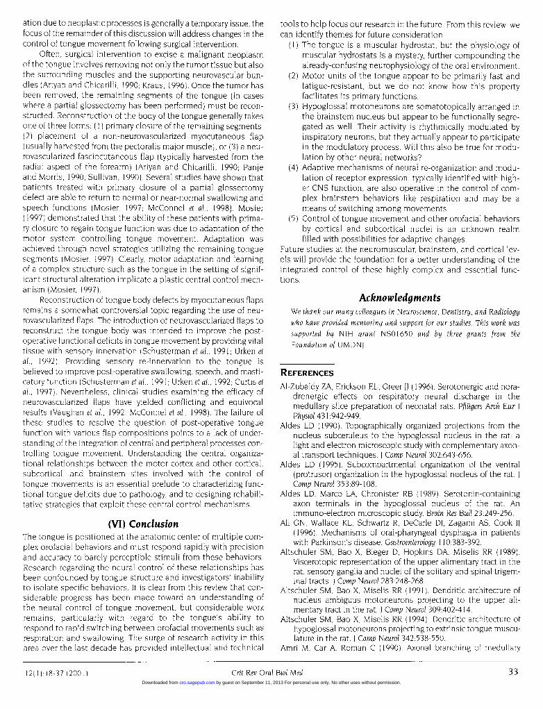
ation due to neoplastic processes is generally a temporary issue, thefocus of the remainder of this discussion will address changes in thecontrol of tongue movement following surgical intervention.
Often, surgical intervention to excise a malignant neoplasmof the tongue involves removing not only the tumor tissue but alsothe surrounding muscles and the supporting neurovascular bun-dles (Ariyan and Chicarilli, 1990; Kraus, 1996). Once the tumor hasbeen removed, the remaining segments of the tongue (in caseswhere a partial glossectomy has been performed) must be recon-structed. Reconstruction of the body of the tongue generally takesone of three forms: (I) primary closure of the remaining segments,(2) placement of a non-neurovascularized myocutaneous flap(usually harvested from the pectoralis major muscle), or (3) a neu-rovascularized fasciocutaneous flap (typically harvested from theradial aspect of the forearm) (Ariyan and Chicarilli, 1990; Panjeand Morris, 1990; Sullivan, 1990). Several studies have shown thatpatients treated with primary closure of a partial glossectomydefect are able to return to normal or near-normal swallowing andspeech functions (Mosier, 1997; McConnel et al., 1998). Mosier(1997) demonstrated that the ability of these patients with prima-ry closure to regain tongue function was due to adaptation of themotor system controlling tongue movement. Adaptation wasachieved through novel strategies utilizing the remaining tonguesegments (Mosier, 1997). Clearly, motor adaptation and learningof a complex structure such as the tongue in the setting of signif-icant structural alteration implicate a plastic central control mech-anism (Mosier, 1997).
Reconstruction of tongue body defects by myocutaneous flapsremains a somewhat controversial topic regarding the use of neu-rovascularized flaps. The introduction of neurovascularized flaps toreconstruct the tongue body was intended to improve the post-operative functional deficits in tongue movement by providing vitaltissue with sensory innervation (Schusterman et al., 1991; Urken etal., 1992). Providing sensory re-innervation to the tongue isbelieved to improve post-operative swallowing, speech, and masti-catory function (Schusterman et al., 1991; Urken et al., 1992; Curtis etal., 1997). Nevertheless, clinical studies examining the efficacy ofneurovascularized flaps have yielded conflicting and equivocalresults (Vaughan et al., 1992; McConnel et al., 1998). The failure ofthese studies to resolve the question of post-operative tonguefunction with various flap compositions points to a lack of under-standing of the integration of central and peripheral processes con-trolling tongue movement. Understanding the central organiza-tional relationships between the motor cortex and other cortical,subcortical, and brainstem sites involved with the control oftongue movements is an essential prelude to characterizing func-tional tongue deficits due to pathology, and to designing rehabili-tative strategies that exploit these central control mechanisms.
(VI) ConclusionThe tongue is positioned at the anatomic center of multiple com-plex orofacial behaviors and must respond rapidly with precisionand accuracy to barely perceptible stimuli from these behaviors.Research regarding the neural control of these relationships hasbeen confounded by tongue structure and investigators' inabilityto isolate specific behaviors. It is clear from this review that con-siderable progress has been made toward an understanding ofthe neural control of tongue movement, but considerable workremains, particularly with regard to the tongue's ability torespond to rapid switching between orofacial movements such asrespiration and swallowing. The surge of research activity in thisarea over the last decade has provided intellectual and technical
tools to help focus our research in the future. From this review, wecan identify themes for future consideration:
(1) The tongue is a muscular hydrostat, but the physiology ofmuscular hydrostats is a mystery, further compounding thealready-confusing neurophysiology of the oral environment.
(2) Motor units of the tongue appear to be primarily fast andfatigue-resistant, but we do not know how this propertyfacilitates its primary functions.
(3) Hypoglossal motoneurons are somatotopically arranged inthe brainstem nucleus but appear to be functionally segre-gated as well. Their activity is rhythmically modulated byinspiratory neurons, but they actually appear to participatein the modulatory process. Will this also be true for modu-lation by other neural networks?
(4) Adaptive mechanisms of neural re-organization and modu-lation of receptor expression, typically identified with high-er CNS function, are also operative in the control of com-plex brainstem behaviors like respiration and may be ameans of switching among movements.
(5) Control of tongue movement and other orofacial behaviorsby cortical and subcortical nuclei is an unknown realmfilled with possibilities for adaptive changes.
Future studies at the neuromuscular, brainstem, and cortical lev-els will provide the foundation for a better understanding of theintegrated control of these highly complex and essential func-tions.
AcknowledgmentsWe thank our many colleagues in Neuroscience, Dentistry, and Radiologywho have provided mentoring and support for our studies. This work wassupported by NIH grant NS01650 and by three grants from theFoundation of UMDNJ.
REFERENCESAl-Zubaidy ZA, Erickson EL, Greer JJ (1996). Serotonergic and nora-
drenergic effects on respiratory neural discharge in themedullary slice preparation of neonatal rats. Pflugers Arch Eur JPhysiol 431:942-949.
Aldes LD (1990). Topographically organized projections from thenucleus subceruleus to the hypoglossal nucleus in the rat: alight and electron microscopic study with complementary axon-al transport techniques. J Comp Neurol 302:643-656.
Aldes LD (1995). Subcompartmental organization of the ventral(protrusor) organization in the hypoglossal nucleus of the rat. JComp Neurol 353:89-108.
Aldes LD, Marco LA, Chronister RB (1989). Serotonin-containingaxon terminals in the hypoglossal nucleus of the rat. Animmuno-electron microscopic study. Brain Res Bull 23:249-256.
Ali GN, Wallace KL, Schwartz R, DeCarle Di, Zagami AS, Cook IJ(1996). Mechanisms of oral-pharyngeal dysphagia in patientswith Parkinson's disease. Gastroenterology 110:383-392.
Altschuler SM, Bao X, Bieger D, Hopkins DA, Miselis RR (1989).Viscerotopic representation of the upper alimentary tract in therat sensory ganglia and nuclei of the solitary and spinal trigem-inal tracts. J Comp Neurol 283:248-268.
Altschuler SM, Bao X, Miselis RR (1991). Dendritic architecture ofnucleus ambiguus motoneurons projecting to the upper ali-mentary tract in the rat. J Comp Neurol 309:402-414.
Altschuler SM, Bao X, Miselis RR (1994). Dendritic architecture ofhypoglossal motoneurons projecting to extrinsic tongue muscu-lature in the rat. I Comp Neurol 342:538-550.
Amri M, Car A, Roman C (1990). Axonal branching of medullary
12(1) 18 37(2001) Crit Rev Oral Biol Med 33Crit Rev Oral Biol Med 331 2( 1 ) :1 8-37 ( 200 1 )
by guest on September 11, 2013 For personal use only. No other uses without permission.cro.sagepub.comDownloaded from
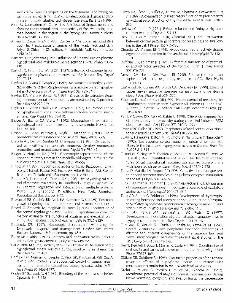
swallowing neurons projecting on the trigeminal and hypoglos-sal motor nuclei: demonstration by electrophysiological and flu-orescent double labeling techniques. Exp Brain Res 81:384-390.
Amri M, Lamkadem M, Car A (1991). Effects of lingual nerve andchewing cortex stimulation upon activity of the swallowing neu-rons located in the region of the hypoglossal motor nucleus.Brain Res 548:149-155.
Ariyan S, Chicarilli ZN (1990). Cancer of the upper aerodigestivetract. In: Plastic surgery: tumors of the head, neck and skin.Ariyan S, Chicarilli ZN, editors. Philadelphia: W.B. Saunders, pp.3450-3451.
Bartlett D, St. John WM (1988). Influence of lung volume on phrenic,hypoglossal and mylohyoid nerve activities. Resp Physiol 73:97-110.
Bartlett D, Knuth SL, Ward DK (1987). Influence of extreme hyper-capnia on respiratory motor nerve activity in cats. Resp Physiol70:173-181.
Bayliss DA, Viana F, Berger AJ (1992). Mechanisms underlying exci-tatory effects of thyrotropin-releasing hormone on rat hypoglos-sal motoneurons in vitro. I Neurophysol 68:1733-1745.
Bayliss DA, Viana F, Berger Al (1994). Effects of thyrotropin-releas-ing hormone on rat motoneurons are mediated by G proteins.Brain Res 668:220-229.
Bayliss DA, Viana F, Talley EM, Berger Al (1997). Neuromodulationof hypoglossal motoneurons: cellular and developmental mech-anisms. Resp Physiol 110: 139-150.
Berger Al, Bayliss DA, Viana F (1992). Modulation of neonatal rathypoglossal motoneuron excitability by serotonin. Neurosci Lett143:164-168.
Besson G, Bogousslavsky J, Regh F, Maeder P (1991). Acutepseudobulbar or suprabulbar palsy. Arch Neurol 48:501-507.
Bianchi AL, Denavit-Saubie M, Champagnat J (1995). Central con-trol of breathing in mammals: neuronal circuitry, membraneproperties, and neurotransmitters. Physiol Rev 75:1-45.
Bieger D, Hopkins DA (1987). Viscerotopic representation of theupper alimentary tract in the medulla oblongata in the rat: thenucleus ambiguus. J Comp Neurol 262:546-562.
Binder MD (1989). Properties of motor units. In: Textbook of physi-ology. 21 st ed. Patton HD, Fuchs AF, Hille B, Scher AM, SteinerR, editors. Philadelphia: Saunders, pp. 512-513.
Binder MD, Heckman CJ, Powers RK (1996). The physiological con-trol of motoneuron activity. In: Handbook of physiology. Section12. Exercise: regulation and integration of multiple systems.Rowell LB, Shepherd JT, editors. New York: AmericanPhysiological Society, pp. 3-53.
Brozanski BS, Guthrie RD, Volk EA, Cameron WE (1989). Postnatalgrowth of genioglossal motoneurons. Ped Pulmonol 7:133-139.
Brozek G, Zhuravin IA, Megirian D, Bures J (1996). Localization ofthe central rhythm generator involved in spontaneous consum-matory licking in rats: functional ablation and electrical brainstimulation studies. Proc Natl Acad Sci USA 93:3325-3329.
Buchholz DW (1997). Neurologic disorders of swallowing. In:Dysphagia: diagnosis and management. Groher ME, editor.Boston: Butterworth-Heinemann, pp. 48-51.
Burke RE, Tsairis P (1973). Anatomy and innervation ratios in motorunits of cat gastrocnemius. J Physiol 234:749-765.
Car A, Amri M (1987). Activity of neurons located in the region of thehypoglossal motor nucleus during swallowing in sheep. ExpBrain Res 69:175-182.
Corfield DR, Murphy K, Josephs 0, Fink GR, Frackowiak RSJ, Guz A,et al. (1999). Cortical and subcortical control of tongue move-ment in humans; a functional neuroimaging study using fMRI. IAppl Physiol 86:1468-1477.
Coster ST, Schwartz WH (1987). Rheology of the swallow-safe bolus.Dysphagia 1:1 13-1 18.
Curtis DA, Plesh 0, Miller Al, Curtis TA, Sharma A, Schweitzer R, etal. (1997). A comparison of masticatory function in patients withor without reconstruction of the mandible. Head & Neck 19:287-296.
Dellow PG, Lund JP (1971). Evidence for central timing of rhythmi-cal mastication. I Physiol 215:1-13.
Dick TE, Oku Y, Romaniuk JR, Cherniak NS (1993). Interactionbetween central pattern generators for breathing and swallow-ing in the cat. I Physiol 465:715-730.
Dinardo LA, Travers JB (1994). Hypoglossal neural activity duringingestion and rejection in the awake rat. I Neurophysiol 72:1181-1191.
Dobbins EG, Feldman JL (1995). Differential innervation of protrud-er and retractor muscles of the tongue in rat. J Comp Neurol357:376-394.
Dreshaj LA, Haxhiu MA, Martin RJ (1998). Role of the medullaryraphe nuclei in the respiratory response to CO2' Resp Physiol111:15-23.
Eastwood PR, Curran AK, Smith CA, Dempsey JA (1998). Effect ofupper airway negative pressure on inspiratory drive duringsleep. I Appl Physiol 84:1063-1075.
Feldman JL, McCrimmon DR (1999). Neural control of breathing. In:Fundamental neuroscience. Zigmond MJ, Bloom FE, Landis SC,Roberts JL, Squire LR, editors. San Diego: Academic Press, pp.1063-1090.
FenikV, Davies RO, Pack Al, Kubin L (1998). Differential suppressionof upper airway motor activity during carbachol-induced, REMsleep-like atonia. Am I Physiol 275:R1013-1024.
Fregosi RF, Fuller DD (1997). Respiratory-related control of extrinsictongue muscle activity. Resp Physiol 110:295-306.
Fukui Y, Hayakawa T, Itoh M, Fujimoto Y, Nishimura Y, Takeuchi Y(1992). The superior cervical ganglion: origin of sympatheticfibers in the facial and hypoglossal nerves in the cat. Brain ResBull 28:811-815.
Fukunishi Y, Nagase Y, Yoshida A, Moritani M, Honma S, YohsukeH, et al. (1999). Quantitative analysis of the dendritic architec-tures of cat hypoglossal motoneurons stained intracellularlywith horseradish peroxidase. J Comp Neurol 405:345-358.
Fuller D, Mateika JH, Fregosi RF (1 998). Co-activation of tongue pro-truder and retractor muscles during chemoreceptor stimulationin the rat. J Physiol 507:265-276.
Funk GD, Smith IC, Feldman IL (1993). Generation and transmissionof respiratory oscillations in medullary slices: role of excitatoryamino acids. J Neurophysiol 70:1497-1515.
Funk GD, Smith IC, Feldman JL (1994). Development of thyrotropin-releasing hormone and norepinephrine potentiation of inspira-tory-related hypoglossal motoneuron discharge in neonatal andjuvenile mice in vitro. J Neurophysiol 72:2538-2541.
Funk GD, Parkis MA, Selvaratnam SR, Walsh C (1997).Developmental modulation of glutamatergic inspiratory drive tohypoglossal motoneurons. Resp Physiol 110:125-137.
Furusawa K, Yasuda K, Okuda D, Tanaka M, Yamaoka M (1996).Central distribution and peripheral functional properties ofafferent and efferent components of the superior laryngealnerve: morphological and electrophysiological studies in therat. J Comp Neurol 375:147-156.
Gay T, Rendell J, Spiro I, Mosier K, Lurie A (1994). Coordination oforal cavity and laryngeal movements during swallowing. J ApplPhysiol 77:357-365.
Gilliam EE, Goldberg Sl (1995). Contractile properties of the tonguemuscles: effects of hypoglossal nerve and extracellularmotoneuron stimulation in rat. I Neurophysiol 74:547-555.
Grelot L, Milano S, Portillo F, Miller AD, Bianchi AL (1992).Membrane potential changes of phrenic motoneurons duringfictive vomiting, coughing, and swallowing in the decerebrate
34 Crit Rev Oral Bial Med 37(2001134 Crit Rev Oral Biol Med 12(l):18-37 (2001) by guest on September 11, 2013 For personal use only. No other uses without permission.cro.sagepub.comDownloaded from
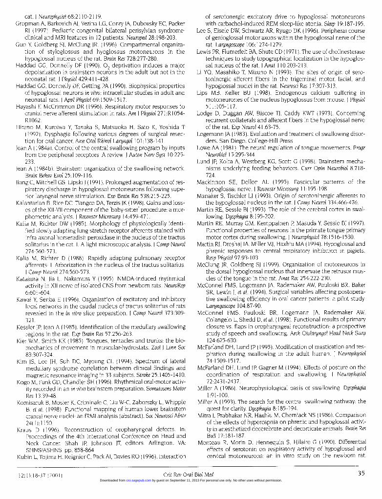
cat. I Neurophysiol 68:2110-2119.Gropman A, Barkovich Al, Vezina LG, Conry IA, Dubovsky EC, Packer
RI (1997). Pediatric congenital bilateral perisylvian syndrome:clinical and MRI features in 12 patients. Neuroped 28:198-203.
Guo Y, Goldberg SJ, McClung JR, (1996). Compartmental organiza-tion of styloglossus and hyoglossus motoneurons in thehypoglossal nucleus of the rat. Brain Res 728:277-280.
Haddad GG, Donnelly DF (1990). °2 deprivation induces a majordepolarization in brainstem neurons in the adult but not in theneonatal rat. Physiol 429:411-428.
Haddad GG, Donnelly DF, Getting PA (1990). Biophysical propertiesof hypoglossal neurons in vitro: intracellular studies in adult andneonatal rats. J Appl Physiol 69:1509-1517.
Hayashi F, McCrimmon DR (1996). Respiratory motor responses tocranial nerve afferent stimulation in rats. Am J Physiol 271 :R1054-R1062.
Hirano M, Kuroiwa Y, Tanaka S, Matsuoka H, Sato K, Yoshida T(1992). Dysphagia following various degrees of surgical resec-tion for oral cancer. Ann Otol Rhinol Laryngol 101: 138-141.
Jean A (1984a). Control of the central swallowing program by inputsfrom the peripheral receptors. A review. J Auton Nerv Syst 10:225-233.
Jean A (1984b). Brainstem organization of the swallowing network.Brain Behav Evol 25:109-116.
Jiang C, Mitchell GS, Lipski 1 (1991). Prolonged augmentation of res-piratory discharge in hypoglossal motoneurons following supe-rior laryngeal nerve stimulation. Exp Brain Res 538:215-225.
Kalantarian B, Rice DC, Tiangco DA, Terzis 1K (1998). Gains and loss-es of the XII-VII component of the "baby-sitter" procedure: a mor-phometric analysis. I Reconstr Microsurg 14:459-471.
Kalia M, Richter DW (1985). Morphology of physiologically identi-fied slowly adapting lung stretch receptor afferents stained withinfra-axonal horseradish peroxidase in the nucleus of the tractussolitarius in the cat. 1. A light microscopic analysis. J Comp Neurol274:560-573.
Kalia M, Richter D (1988). Rapidly adapting pulmonary receptorafferents: 1. Arborization in the nucleus of the tractus solitarius.J Comp Neurol 274:560-573.
Katakura N, hia L, Nakamura Y (1995). NMDA-induced rhythmicalactivity in XIl nerve of isolated CNS from newborn rats. NeuroRep6:601-604.
Kawai Y, Senba E (1996). Organization of excitatory and inhibitorylocal networks in the caudal nucleus of tractus solitarius of ratsrevealed in the in vitro slice preparation. J Comp Neurol 373:309-321.
Kessler JP, Jean A (1985). Identification of the medullary swallowingregions in the rat. Exp Brain Res 57:256-263.
Kier WM, Smith KK (1985). Tongues, tentacles and trunks: the bio-mechanics of movement in muscular-hydrostats. Zool J Linn Soc83:307-324.
Kim IS, Lee IH, Suh DC, Myoung CL (1994). Spectrum of lateralmedullary syndrome correlation between clinical findings andmagnetic resonance imaging in 33 subjects. Stroke 25:1405-1410.
Kogo M, Funk GD, Chandler SH (1996). Rhythmical oral-motor activ-ity recorded in an in vitro brainstem preparation. Somatosens MotorRes 13:39-48.
Komisaruk B, Mosier K, Criminale C, Liu W-C, Zaborszky L, WhippleB, et al. (1998). Functional mapping of human lower brainstemcranial nerve nuclei: an fRMI analysis (abstract). Soc NeurosciAbstr24(1):1150.
Kraus D (1996). Reconstruction of oropharyngeal defects. In:Proceedings of the 4th International Conference on Head andNeck Cancer. Shah JP, Johnson JT, editors. Arlington, VA:SHNS/ASHNS, pp. 858-864.
Kubin L, Tojima H, Reignier C, PackAl, Davies RO (1996). Interaction
of serotonergic excitatory drive to hypoglossal motoneuronswith carbachol-induced REM sleep-like atonia. Sleep 19:187-195.
Lee S, Eisele DW, Schwartz AR, Ryugo DK (1996). Peripheral courseof genioglossal motor axons within the hypoglossal nerve of therat. Laryngoscope 106:1274-1279.
Lewis PR, Flumerfelt BA, Shute CD (I1971). The use of cholinesterasetechniques to study topographical localization in the hypoglos-sal nucleus of the rat. I Anat 110:203-213.
Li YQ, Masahiko T, Mizuno N (1993). The sites of origin of sero-toninergic afferent fibers in the trigeminal motor, facial, andhypoglossal nuclei in the rat. Neurosci Res 17:307-313.
Lips MB, Keller BU (1998). Endogenous calcium buffering inmotoneurones of the nucleus hypoglossus from mouse. I Physiol511:105-117.
Lodge D, Duggan AW, Biscoe TJ, Caddy KWT (1973). Concerningrecurrent collaterals and afferent fibers in the hypoglossal nerveof the rat. Exp Neurol 41:63-75.
Logemann JA (1983). Evaluation and treatment of swallowing disor-ders. San Diego: College-Hill Press.
Lowe AA (1981). The neural regulation of tongue movements. ProgrNeurobiol 15:295-344.
Lund JP, Kolta A, Westberg KG, Scott G (1998). Brainstem mecha-nisms underlying feeding behaviors. Curr Opin Neurobiol 8:718-724.
Mackinnon SE, Dellon AL (1995). Fascicular patterns of thehypoglossal nerve. J Reconstr Microsurg 11: 195-198.
Manaker S, Tischler LJ (1993). Origin of serotoninergic afferents tothe hypoglossal nucleus in the rat. J Comp Neurol 334:466-476.
Martin RE, Sessle BJ (1993). The role of the cerebral cortex in swal-lowing. Dysphagia 8:195-202.
Martin RE, Murray GM, Kemppainen P, Masuda Y, Sessle BJ (1997).Functional properties of neurons in the primate tongue primarymotor cortex during swallowing. J Neurophysiol 78:1516-1530.
Martin RJ, Dreshaj IA, Miller MJ, Haxhiu MA (1994). Hypoglossal andphrenic responses to central respiratory inhibition in piglets.Resp Physiol 97:93-103.
McClung JR, Goldberg SJ (1999). Organization of motoneurons inthe dorsal hypoglossal nucleus that innervate the retrusor mus-cles of the tongue in the rat. Anat Rec 254:222-230.
McConnel FMS, Logemann JA, Rademaker AW, Pauloski BR, BakerSR, Lewin J, et al. (1994). Surgical variables affecting postopera-tive swallowing efficiency in oral cancer patients: a pilot study.Laryngoscope 104:87-90.
McConnel FMS, Pauloski BR, Logemann IA, Rademaker AW,Colangelo L, Shedd D, et al. (I1998). Functional results of primaryclosure vs. flaps in oropharyngeal reconstruction: a prospectivestudy of speech and swallowing. Arch Otolaryngol Head Neck Surg124:625-630.
McFarland DH, Lund JP (1995). Modification of mastication and res-piration during swallowing in the adult human. J Neurophysiol74:1509-1517.
McFarland DH, Lund JP, Gagner M (1994). Effects of posture on thecoordination of respiration and swallowing. I Neurophysiol72:2431-2437.
Miller A (1986). Neurophysiological basis of swallowing. Dysphagia1:91-100.
Miller A (1993). The search for the central swallowing pathway: thequest for clarity. Dysphagia 8:185-194.
Mitra J, Prabhakar NR, Haxhiu M, Cherniack NS (1986). Comparisonof the effects of hypercapnia on phrenic and hypoglossal activi-ty in anesthetized decerebrate and decorticate animals. Brain ResBull 17:181-187.
Monteau R, Morin D, Hennequin S, Hilaire G (1990). Differentialeffects of serotonin on respiratory activity of hypoglossal andcervical motoneurons: an in vitro study on the newborn rat.
1211) 18 37)2001) Grit Rev Oral Biol Med3535Crit Rev Oral Biol Med12(1 1H8-37 (2001)
by guest on September 11, 2013 For personal use only. No other uses without permission.cro.sagepub.comDownloaded from

Neurosci Lett 111:127-132.Morgan AS, Mackay LE (1999). Causes and complications associat-
ed with swallowing disorders in traumatic brain injury. J HeadTrauma Rehabil 14:454-461.
Morin D, Monteau R, Gilaire G (1992). Compared effects of sero-tonin on cervical and hypoglossal inspiratory activities: an invitro study in the newborn rat. I Physiol 451:605-629.
Mosfeldt Laursen A, Rekling JC (1989). Electrophysiological proper-ties of hypoglossal motoneurons of guinea-pigs studied in vitro.Neuroscience 30:619-637.
Mosier K (1997). The motor control of swallowing. Ann Arbor: UMIPublications, pp. 214-249.
Mosier K (1998). Functional imaging of swallowing. In: Medical radi-ology. Diagnostic imaging and radiation oncology: modern headand neck imaging. Mukherji SK, Castelijns JA, editors. Berlin:Springer-Verlag, pp. 79-86.
Mosier KM, Kalnin AJ, Komisaruk BR, Behin B, Liu WC, Criminale C(1999). Spatial-temporal properties of peripheral movementduring cortical activation in fMRI; a study of tongue movementduring different tasks (abstract). Proc Radiol Soc N Am 213:140.
Mrini A, lean A (1995). Synaptic organization of the interstitial sub-division of the nucleus tractus solitarii and of its laryngeal affer-ents in the rat. I Comp Neurol 355:221-236.
Mu L, Sanders 1 (1999). Neuromuscular organization of the caninetongue. Anat Rec 256:412-424.
Murdoch BE, Spencer TJ, Theodoros DG, Thompson EC (1998). Lipand tongue function in multiple sclerosis: a physiological analy-sis. Motor Control 2:148-160.
Murray GM, Sessle BJ (1992a). Functional properties of single neu-rons in the face primary motor cortex of the primate. I. Input andoutput features of tongue motor cortex. J Neurophysiol 67:747-758.
Murray GM, Sessle BI (1992b). Functional properties of single neu-rons in the face primary motor cortex of the primate. II.Relations with trained orofacial motor behavior. J Neurophysiol67:759-774.
Murray GM, Sessle BI (1992c). Functional properties of single neu-rons in the face primary motor cortex of the primate. III.Relations with different directions of trained tongue protrusion.J Neurophysiol 67:775-785.
Murray GM, Lin LD, Moustafa E, Sessle BJ (1991). Effects ofreversible inactivation by cooling of the primate face motor cor-tex on the performance of a trained tongue-protrusion task anda trained biting task. I Neurophysiol 65:511 -530.
Nakamura Y, Katakura N (1995). Generation of masticatory rhythmin the brainstem. Neurosci Res 23:1-19.
Napadow VI, Chen 0, Wedeen VI, Gilbert RJ (1999). Intramuralmechanics of the human tongue in association with physiolog-ical deformations. J Biomech 32:1-12.
Nunez-Abades PA, Spielman J, Barrionuevo G, Cameron WE (1993).In vitro electrophysiology of developing genioglossal motoneu-rons in the rat. J Neurophysiol 70:1401-1411.
O'Kusky JR, Norman MG (1992). Sudden infant death syndrome:postnatal changes in the numerical density and total number ofneurons in the hypoglossal nucleus. I Neuropathol Exp Neurol5:577-584.
O'Reilly PMR, Fitzgerald MJT (1990). Fibre composition of thehypoglossal nerve in the rat. I Anat 172:227-243.
Odutola AB (1976). Cell grouping and golgi architecture of thehypoglossal nucleus of the rat. Exp Neurol 52:356-371.
Okabe S, Kubin L (1996). Role of 5HT1 receptors in the control ofhypoglossal motoneurons in vivo. Sleep 19(l0):S150-S153.
Okabe S, Mackiewicz M, Kubin L (1997). Serotonin receptor mRNAexpression in the hypoglossal motor nucleus. Resp Physiol110:151-160.
Oku Y, Tanaka I, Ezure K (1994). Activity of bulbar respiratory neu-rons during fictive coughing and swallowing in the decerebratecat. I Physiol 480:309-324.
Ono T, Ishiwata Y, Inaba N, Kuroda T, Nakamura Y (1994).Hypoglossal premotor neurons with rhythmical inspiratory-related activity in the cat: localization and projection to thephrenic nucleus. Exp Brain Res 98:1-12.
Ono T, Ishiwata Y, Inaba N, Kuroda T, Nakamura Y (1998a).Modulation of the inspiratory-related activity of hypoglossalpremotor neurons during ingestion and rejection in the decere-brate cat. J Neurophysiol 80:48-58.
Ono T, Ishiwata Y, Kuroda T, Nakamura Y (1998b). Swallowing-relat-ed perihypoglossal neurons projecting to hypoglossalmotoneurons in the cat. I Dent Res 77:351-360.
Palmer JB (1998). Bolus aggregation in the oropharynx does notdepend on gravity. Arch Phys Med Rehabil 79:691-696.
Panje MIR, Morris MR (1990). Oral cavity and oropharyngeal recon-struction. In: Plastic surgery: tumors of the head, neck and skin.Vol. 5. Ariyan S, Chicarilli ZN, editors. Philadelphia: W.B.Saunders, pp. 1479-1497.
Parker-Thornburg J, Bauer B, Palermo J, Robbins J (1992). Structuraland developmental analysis of two linked myosin heavy chaingenes. Dev Biol 159:99-107.
Parkis MA, Bayliss DA, Berger AJ (1995). Actions of norepinephrineon rat hypoglossal motoneurons. J Neurophysiol 74:1911-1919.
Pierce RJ, Worsnop CJ (1999). Upper airway function and dysfunc-tion in respiration. Clin Exp Pharmacol Physiol 26: 1-10.
Plant RL (1998). Anatomy and physiology of swallowing in adultsand geriatrics. Otolaryngol Clin N Am 31:507-524.
Poliakov AV, Powers RK, Binder MD (1997). Functional identificationof the input-output transforms of motoneurones in the rat andcat. I Physiol 504:401-424.
Preiksaitis HG, Mills CA (1996). Coordination of breathing and swal-lowing: effects of bolus consistency and presentation in normaladults. J Appl Physiol 81:1707-1714.
Prigozy TI, Dalrymple K, Shuler C, Kedes L (1997). Differentialexpression of troponin C genes during tongue myogenesis. DevDyn 209:36-44.
Radulovacki M, Trbovic SM, Carley DW (1998). Serotonin 5-HT3-receptor antagonist GR38032F suppresses sleep apneas in rats.Sleep 21:131-136.
Ramirez JM, Quellmalz UJA, Wilken B, Richter DW (1998). Thehypoxic response of neurones within the in vitro mammalian res-piratory network. J Physiol 507:571-582.
Rekling JC (1992). Interaction between thyrotropin-releasing hor-mone (TRH) and NMDA-receptor-mediated responses inhypoglossal motoneurones. Brain Res 578:289-296.
Rekling JC, Funk GD, Bayliss DA, Dong X-W, Feldman IL (2000).Synaptic control of motoneuronal excitability. Physiol Rev 80:767-852.
Sant'Ambrogio G, Tsubone H, Sant'Ambrogio FB (1995). Sensoryinformation from the upper airway: role in the control of breath-ing. Resp Physiol 102:1-16.
SawczukA, Covell DA (1999). Wheat germ agglutinin conjugated toTRITC: a novel approach for labeling primary projection neuronsof peripheral afferent nerves. I Neurosci Meth 93:139-147.
Sawczuk A, Tepper JM (1997). Use of multiple fluorescent tracers toassist in hypoglossal motoneuron identification (abstract). SocNeurosci Abstr 23:2084.
Sawczuk A, Powers RK, Binder MD (1995a). Spike-frequency adap-tation studied in hypoglossal motoneurons of the rat. JNeurophysiol 73:1799-1810.
Sawczuk A, Powers RK, Binder MD (1995b). Intrinsic properties ofmotoneurons: implications for muscle fatigue. In: Fatigue, neur-al and muscular mechanisms. Gandevia SC, Enoka RM,
36 Crit Rev Oral Biol Med (2001)
36 Crit Rev Oral Biol Med 12(1):18-37 (2001) by guest on September 11, 2013 For personal use only. No other uses without permission.cro.sagepub.comDownloaded from
McComas Al, Stuart DG, Thomas CK, editors. New York: PlenumPress, pp. 123-134.
Sawczuk A, Powers RK, Binder MD (1997). Contribution of outwardcurrents to spike-frequency adaptation in hypoglossalmotoneurons of the rat. I Neurophysiol 78:2246-2253.
Sawczuk A, Kolta A, Lund IP (2000). An in vitro study of projectionsfrom the nucleus of the solitary tract to hypoglossal motoneu-rons (abstract). J Dent Res 79(Spec Iss):406.
Schusterman MA, Kroll SS, Weber RS, Byers RM, Gullamondegui 0,Goepfert H (1991) I ntraoral soft tissue reconstruction after can-cer ablation: a comparison of the pectoralis major flap and thefree radial forearm flap. Am J Surg 162:397-399.
Sciote IJ, Rowlerson AM, Hopper C, Hunt NP (1994). Fibre type clas-sification and myosin isoforms in the human masseter muscle.J Neurol Sci 126:15-24.
Sessle BI, Henry IL (1989). Neural mechanisms of swallowing: neu-rophysiological and neurochemical studies on brainstem neu-rons in the solitary tract region. Dysphagia 4:61-75.
Shaker R, Lang IM (1997). Reflex mediated airway protective mech-anisms against retrograde aspiration. Am J Med 103(5A):64S-73S.
Shiba 1, Satoh 1, Kobayashi N, Hayashi F (1999). Multifunctionallaryngeal motoneurons: an intracellular study in the cat. INeurosci 19:2717-2727.
Sica AL, Cohen MI, Donnelly DF, Zhang H (1984). Hypoglossalmotoneuron responses to pulmonary and superior laryngealafferent inputs. Resp Physiol 56:339-357.
Singer IH, Berger Al (1996). Presynaptic inhibition by serotonin: apossible mechanism for switching motor output of thehypoglossal nucleus. Sleep 19(10):S 146-S 149.
Smith IC, Greer JI, Liu G, Feldman IL 11990). Neural mechanismsgenerating respiratory pattern in mammalian brainstem-spinalcord in vitro. I Neurophysiol 64: 1149-1169.
Smith IC, Ellenberger HH, Ballanyi K, Richter DW, Feldman JL(1991). Pre-Botzinger complex: a brainstem region that maygenerate respiratory rhythm in mammals. Science 254:726-729.
Sokoloff Al (2000). Localization and contractile properties of intrin-sic longitudinal motor units of the rat tongue. I Neurophysiol84:827-835.
Sokoloff Al, Deacon TW (1992). Musculotopic organization of thehypoglossal nucleus in the cynomolgus monkey, Macaca fascicu-laris. I Comp Neurol 324:81-93.
Stachler RI, Hamlet SL, Mathog RH, Jones L, Heilbrun LK, Manov LJ,et al. (1994). Swallowing of bolus types by postsurgical head andneck cancer patients. Head & Neck 16:413-419.
Stein PSG, Grillner S, Selverston Al, Stuart DG, editors (1997).Neurons, networks, and motor behavior. Cambridge: The MITPress.
Sullivan MI (1990). Head and neck reconstruction by free tissuetransfer. In Plastic surgery: tumors of the head, neck and skin.Vol. 5. Ariyan S, Chicarilli ZN, editors. Philadelphia: W.B.Saunders, pp. 181-189.
Sumi T (1970). Activity in single hypoglossal fibers during corticallyinduced swallowing and chewing in rabbits. PflUgers Arch314:329-346.
Sutlive TG, McClung IR, Goldberg Si (1999). Whole-muscle andmotor-unit contractile properties of the styloglossus muscle inrat. J Neurophysiol 82:584-592.
Sutlive TG, Shall MS, McClung JR, Goldberg SI (2000). Contractileproperties of the tongue's genioglossus muscle and motor unitsin the rat. Muscle & Nerve 23:416-425.
Takata M (1993). Two types of inhibitory postsynaptic potentials inthe hypoglossal motoneurons. Progr Neurobiol 40:385-411.
Takeuchi Y, Hayakawa T, Ozaki HS, Kito J, Satoda T, Matsushima R
(1990). Afferent fibers in the hypoglossal nerve: a horseradishperoxidase study in the cat. Brain Res Bull 24:81-87.
Tell F, lean A (1993). Ionic basis for endogenous rhythmic patternsinduced by activation of N-methyl-D-aspartate receptors inneurons of the rat nucleus tractus solitarii. J Neurophysiol70:2379-2390.
Tomomune N, Takata M (1988). Excitatory and inhibitory post-synaptic potentials in cat hypoglossal motoneurons duringswallowing. Exp Brain Res 71:262-272.
Travers JB, Jackson LM (1992). Hypoglossal neural activity duringlicking and swallowing in the awake rat. I Neurophysiol 67:1171-1184.
Travers JB, DiNardo LA, Karimnamazi H (2000). Medullary reticularformation activity during ingestion and rejection in the awakerat. Exp Brain Res 71:262-272.
Uemura-Sumi M, Itoh M, Mizuno N (1988). The distribution ofhypoglossal motoneurons in the dog, rabbit, and rat. AnatEmbryol 177:389-394.
Umezaki T, Matsuse T, Shin T (1998a). Medullary swallowing-relat-ed neurons in the anesthetized cat. NeuroReport 9:1793-1798.
Umezaki T, Shiba K, Zheng Y, Miller AD (1998b). Upper airway motoroutputs during vomiting versus swallowing in the decerebratecat. Brain Res 781:25-36.
Umezaki T, Nakazawa K, Miller AD (1998c). Behaviors of hypoglos-sal hyoid motoneurons in laryngeal and vestibular reflexes andin deglutition emesis. Am I Physiol 274:8950-8955.
Urasaki E, Uematsu S, Gordon B, Lesser RP (1994). Cortical tongueareas studied by chronically implanted subdural electrodes-with special reference to parietal motor and frontal sensoryresponses. Brain 117:117-132.
Urban PP, Schlegel I, Ellrich J, Koehler J, Hopf HC (1996).Electrophysiological brainstem investigations in obstructivesleep apnoea syndrome. J Neurol 243:171-174.
Urken ML, Weinberg H, Vickery C, Aviv 1, Buchbinder D, Lawson W,et al. (1992). The combined sensate radial forearm and iliac crestfree flaps for reconstruction of significant glossectomy-mandibulectomy defects. Laryngoscope 102:543-558.
Vaughan ED, Bainton R, Martin IC (1992). Improvements in mor-bidity of mouth cancer using microvascular free flap reconstruc-tions. J Cranio-Maxillo-Fac Surg 20:132- 143.
Viana F, Gibbs L, Berger Al (1990). Double- and triple-labeling offunctionally characterized central neurons projecting to periph-eral targets studied in vitro. Neuroscience 38:829-841.
Viana F, Bayliss DA, Berger Al (1995). Repetitive firing properties ofdeveloping rat brainstem motoneurones. I Physiol 486:745-761.
Watchko JF, Klesh KW, O'Day TL, Weiss MG, Guthrie RD (1989).Genioglossal recruitment during acute hypoxia and hypercapniain kittens. Ped Pulmon 7:235-243.
Westberg KG, Clavelou P, Sandstrom G, Lund JP (1998). Evidencethat trigeminal brainstem interneurons form subpopulations toproduce different forms of mastication in the rabbit. J Neurosci18:6466-6479.
Wiesenfeld Z, Halpern BP, Tapper DN (1977). Licking behavior: evi-dence of hypoglossal oscillator. Science 196:1122-1124.
Withington-Wray DJ, Mifflin SW, Spyer KM (1988). Intracellularanalysis of respiratory-modulated hypoglossal motoneurons inthe cat. Neuroscience 25:1041-105 1.
Zheng Y, Umezaki T, Nakazawa K, Miller AD (1997). Role of pre-inspi-ratory neurons in vestibular and laryngeal reflexes in swallowingand vomiting. Neurosci Lett 225:161-164.
Zoungrana OR, Amri M, Car A, Roman C (1997). Intracellular activi-ty of motoneurons of the rostral nucleus ambiguus during swal-lowing in sheep. J Neurophysiol 77:909-922.
12)1)18-37~~~~~~(20)Cie rlBdMd337Crit Rev Oral Biol Med12(l):18-37 (2001 )
by guest on September 11, 2013 For personal use only. No other uses without permission.cro.sagepub.comDownloaded from