18mbo22c-u3 paper v - anatomy and embryology
TRANSCRIPT

18MBO22C-U3
PAPER – V - ANATOMY AND EMBRYOLOGY
Unit - 3
Anther Development & Microsprogenesis
Dr.K.Kalimuthu
Assistant Professor
PG and Research Department of Botany
Government Arts College (autonomous)
Coimbatore -18
Mobile No : 9843366622
.

Parts Of Flower

The Stamen
Stamen in a flower consists of two parts, the long narrow
stalk like filament and upper broader knob-like bi-lobed
anther (Fig. 2.3 A).
The proximal end of the filament is attached to the
thalamus or petal of the flower. The number and length of
stamens vary in different species.

T.S. OF Anther
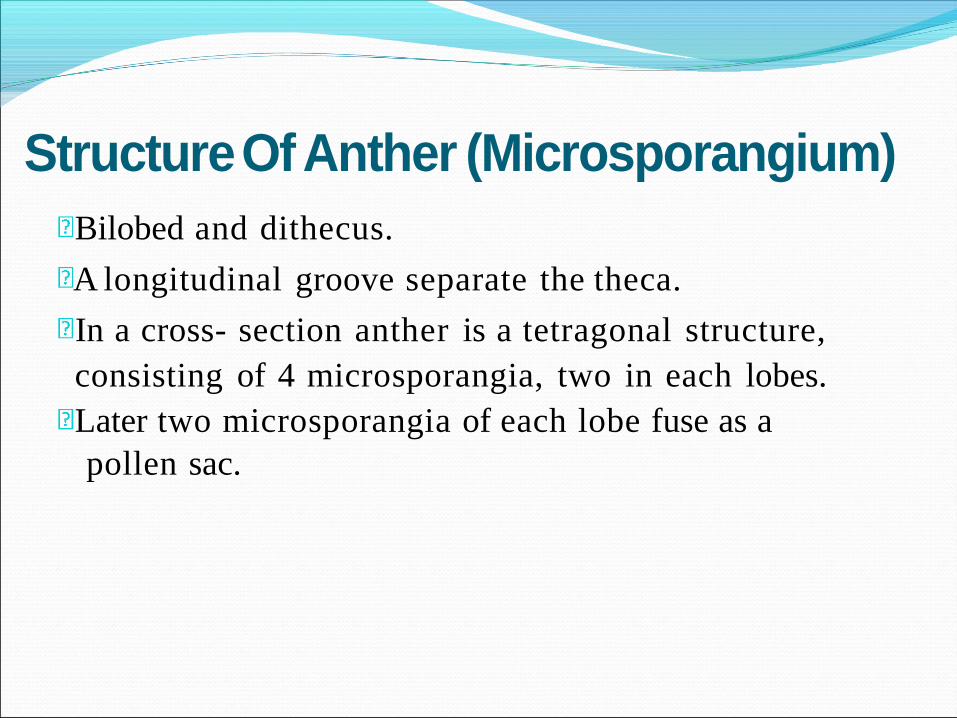
Structure Of Anther (Microsporangium)
Bilobed and dithecus.
A longitudinal groove separate the theca.
In a cross- section anther is a tetragonal structure,
consisting of 4 microsporangia, two in each lobes.
Later two microsporangia of each lobe fuse as a
pollen sac.

Structure of microsporangium (pollen sac)

A microsporangium is circular and surrounded by 4
layers.
These layers are -
- Epidermis,
- Endothecium,
- Middle layers
- Tapetum.
Outermost layers protect the pollen and help in
dehiscence of anther to releasepollen.
Structure Of Anther

Function of anther cell and tissue types


Anther first appears as a cylindrical structure composed of a
mass of meristematic cells.
Which consists of three “germ’ layers designated as L1, L2 and
L3, which gives rise to different anther tissues.
Thus ones specified the development fate of L1, L2, and L3 layer
derivatives is fixed


In most cases individual tissues and cell types are derived from
a single germ layer.
For instance L1 layer gives rise to the epidermis and stomium.
The former is greatly stretched and flattened in a mature anther.
The stomium is located between the two locules of each anther
lobe, and the cells in this region are thin walled and in the form
of a longitudinal slit.
The L2 layer gives rise to the archesporial cells, microspore
mother cells, endothecium, and middle wall layers that lie
between the epidermis and the tapetum.
The archesporial cells are hypodermal in origin and consist of
one to more vertical rows of large cells with dense cytoplasm
and deeply staining nuclei.
The cells divide periclinally into primary parietal cell toward
the periphery and sporogenous cell toward the inside

The parietal cells undergo a series of periclinal and anticlinal
divisions to form two to five concentric layers of anther wall
[endothecium, middle wall layers (three layered) and outer
tapetum].
The L3 layer gives rise to the connective, vascular bundle, and
circular cell cluster adjacent to the stomium.
Both the L2 and L3 layers contribute to tapetum formation.
Tapetal cells along the upper portion (inner) of the pollen sacs
are specified from the L3-derived connective tissue, whereas
those that line the lower portion (outer) of the pollen sacs are
specified from the L2- derived archesporial lineage.
Archesporial cells destined to differentiate into microsporangia
and surrounding tapetum and endothecium tissue, arise
simultaneously in each corner of the anther primordium,
while the vascular tissues differentiate within the centre of the
anther primordium and establish a connection with the filament.


Epidermis – The epidermis is a single layered
protective sheath of the anther. It divides
anticlinally and tries to keep space with the
enlarging internal tissues of the anther.
It provides the structural integrity to the anther,
assists in gaseous diffusion, prevents moisture loss,
and in the dehiscence of the anther lobes.
ANTHERWALL

The outer most layers of the descendants of the parietal
cell located immediately below the epidermis are called
the endothecium.
It attains the maximum development before the
dehiscence of the anther.
The cells are radially elongated and decorated with
fibrous bands
The endothecium is associated with high proportion of
α-cellulose and small amount of lignin at maturity.
The specialized nature of the endothecium together with
the stomium helps in the dehiscence of the anther.
ANTHERWALL Endothecium

Middle layers Next to endothecium are 1-3 middle layers.
The cells of the middle layer are usually ephemeral and
become flattened and crushed by early meiosis in the
pollen mother cell.
The layers persist in Ranunculus and Lilium, and the layer
adjacent to the endothecium may even develop fibrous
thickenings.
In few instances it also serves to store starch that is later
mobilized to the developing pollen

Tapetum- it is the innermost layer of anther wall and is usually
derived from the parietal layer
- It is composed of single layer of cell characters by dense
protoplasm and prominent nuclei.
- The tapetum surrounds the sporogenous tissue and attains
maximum development when the microspores are in the
tetrad stage, After which they go into decline that results in
the collapse of the cells.
The cells of the tapetum are characterized as:
a. They are distinctly enlarged and always ephemeral.
b. The cytoplasm is rich in ribosomes, mitochondria, E.R., many
vesicles and active organelles.
c. Cells may be multinucleate or polyploid and are comparatively rich
in DNA.
d. There is irregular mitotic divisions and nuclear fusion.
e. They are characterized by rapid and intense activity with
degeneration of their cytoplasm.

Tapetal cells undergo dynamic instabiliy during their short life
span.
The characteristic cytological feature of the tapetal cells,
irrespective of the type, is the increase in the content of their DNA’
which is initiated with meiosis in microsporocytes and extends
through the meiotic division.
DNA increase is not followed by regular mitotic division it results
in certain cytological abnormalities, like multinucleate cells,
endomitosis, polyploid nuclei, polyteny and endoreduplication.
Behaviour of the Nucleus in the Tapetal Cells

Endomitosis
It is a condition where chromosome duplication and chromatid separation
take place within the intact nuclear membrane and without the formation of
a spindle. The consequence is the formation of a large polyploid nucleus.
Multinucleate condition
It is a common feature of the tapetal cells, where the nuclear division is
synchronous in amoeboid type and asynchronous in secretory type of
tapetum and is not accompanied by cytokinesis. Based on the number of
nuclear divisions, cells may have 2, 4, 8, or 16 nuclei. In case of nuclear
fusion cells outside the expected series of nuclei number may appear.
Restitution nuclei
Mitosis is normal up to the early stage of anaphase, from then onward the
two chromosome sets are included within a common nuclear membrane,
thus forming a restitution nucleus.
Polyteny
It is a case of increase in chromonemata number per chromosome, thus there
is alternation in chromosome number per nucleus

- Depending upon behavior tapetum is of 2 type
1. Amoeboid or Invasive or Periplasmodial Tapetum- it
is of primitive type. later during the drying up process
of anther, periplasmodium hydrates and deposits as
tryphine on the wall of pollen grain.
1. Secretory tapetum-secretory tapetal cell remain attached
to middle layer till the development of pollen grains . It
is more common among angiosperm


It provide nourishment to the
developing pollen grain
It help in the formation of exine
It hepls in the transport of food material
to inside of the anther
Tapetum helps in the formation of
pollen wall
It helps in the secretion of the enzyme
callase (β-1,3- glucanase) to dissolve the
callosic wall of the tetrad and set them free.
Function of tapetum

The role of the tapetal cells in the secretion of sporopollenin
precursor
Onagraceae tapetal cells play a role in formation of fine flexible
threads, known as viscin threads, in continuation with the outer
layer of the exine
Asteraceae the tapetum forms an acetolysis resistant membrane
outside the sporogenous tissue
Formation of Ubisch bodies.(small acellular structure of
sporopollenin)
Formation of pollenkitt, and tryphine, which are deposited on the
pollen surface and helps to bind pollen grains together, and for
efficient insect pollination.
Formation of pollen wall during post- meiotic period
Secretion of polysaccharides into the locules d u ring the free
microspore stage, which are absorbed by microspores

Primary sporogenous tissue give rise to microspore
mother cell
Some of sporogenous cell remain non functional and
serve as the food material for the developing
microspore
MMC under goes meiosis to form microspore tetrad
which seprate out to form microspore or pollen grain
The process of formation of microspore from MMC is
called microsporogensis
Sporogenous tissue

The process of formation of microspores from a pollen
mother cell through meiosis is called
microsporogenesis.
The cells of sporogenous tissue undergo meiosis to
form microspore tetrad arranged in a cluster of 4
cells..
As each cell of sporogenous tissue has potential to
form tetrad, so each cell is a microspore mother cell
(PMC).
On maturation and dehydration of anther, the spores
dissociate and develop into pollengrains.
Pollen grains release with the dehiscence of anther.
Microsporogenesis


Pollen Grain (Male Gametoph y t e )
Pollen grains develop from the diploid microspore mother
cells in pollen sacs of anthers.
pollen grain is a haploid, unicellular body with a single
nucleus.
Pollen grains are generally spherical measuring about 25-30
micrometeres in diameter
Have two layered wall- outer hard exine layer and inner
thin intine.
Exine- made up of sporopolenin. Resistant to organic matter,
withstand high temperature, acids, alkalis and enzymes. It
has prominent apertures called germ pores, where
sporopolenin is absent.
Intine- It is thin, continuous layer, made of cellulose and
pectin.

Pollen Grain (Male Gametophyte) Pollen grain cytoplasm is surrounded byplasma membrane.
Mature pollen grain has 2 cells- (i) vegetative cell (ii) generative cell.
Vegetative cell- bigger, abundant food reserve, large irregular nucleus.
Generative cell- small, spindle shaped with dense cytoplasm and a nucleus, floats in vegetative cell cytoplasm.
In 60% species pollen grains are shed in 2 celled stage where as 40% species shed in 3 celled stage in which generative cell divides mitotically into 2 male gametes.

Kinds of microspore tetrads in Angiosperms


Palynology:
Pollen Morphology and Biology
By
Dr.K.Kalimuthu
Assistant Professor

Objectives
• Enumerate and identify important
palynological features of
angiosperms;
• Relate these features to plant
systematics; and
• Demonstrate taxonomic evidence in
palynology.

What is palynology?
• Palynology (palynos, dust) is the science of pollen grains and spores.
• Pollen grains are male gametophytes or reproductive cells of a flowering plant.

Pollen Biology

Pollen Biology
• Pollen grains consist of a hard outer wall (exine) and an inner softer wall (intine) which encloses the cytoplasm with its cells (nuclei) and organelles.

Pollen Biology
• A pollen grain contains the gamete of the angiosperm plant.
male
• Pollen has two functions-reproduction and reward of visitors.
• The outer layer of a pollen and spores often contains a special compound, sporopollenin, which degradation by various bacteria and fungi.
resists chemicals,

Pollen Biology
• The pollen wall is designated to protect the sperm nucleus from desiccation and irradiation during transport from the anther to the stigma.
Pollen Biology
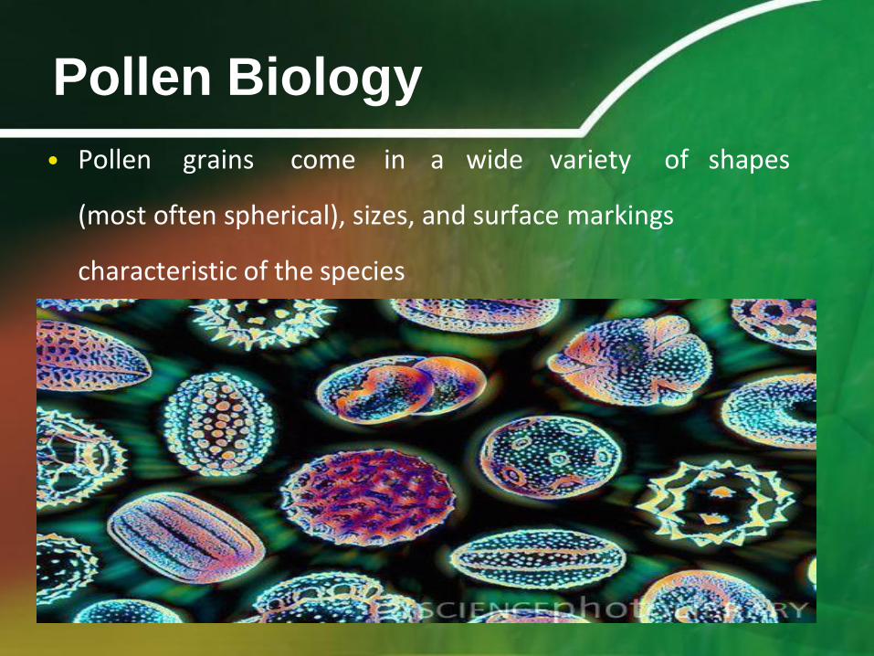
• Pollen grains come in a wide variety of shapes
(most often spherical), sizes, and surface markings
characteristic of the species

Palynology and Systematics
• Palynology is used in phylogenetic
analysis.
• It can be utilized in plant identification
• Extant plants
• Fossil plants
–Paleopalynology/paleobotany
– past plant communities, climate,
biogeography, migration

Palynology and Systematics
• The morphology of pollen grains forms the
basic criteria for their identification..
• The palynological features of a spore or
pollen grain can often be used to identify a
particular taxon.
• Pollen data provides information of
changes in vegetation, climate, and
human disturbance of terrestrial
ecosystems.

Pollen Analysis
• Sediments are collected
• Pollen grains are isolated
from the sediment matrix
via chemical treatments.
• Isolated pollen grains are
mounted onto a glass
slide, and they are
identified and quantified
under a microscope.

Palynological Features
Used in Plant Systematics
• Pollen Nucleus
Number
• Pollen Storage
Product
• Pollen Unit
• Pollen Polarity
• Pollen Aperture
• Pollen Size
• Pollen Shape
• Pollen Sculpturing
• Pollen Wall Structure

Pollen Nucleus No.
binucleate versus trinucleate

Binucleate Plant Families
A. Tradescantia
virginia
(Commelinaceae)
B. Smilacina
stellata
(Liliaceae)
C. Rosaceae
D.Chrysanthemum
(Asteraceae)
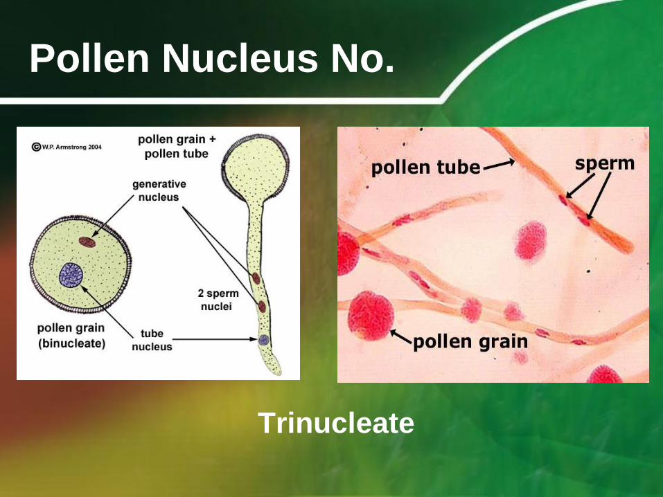
Pollen Nucleus No.
Trinucleate

Pollen Nucleus No.
Trinucleate
(Caryophyllaceae-Pollen of pink family)

Pollen Storage Product
• Pollen grains contain high-energy storage
reserves. These are composed of either
starch versus oil
(Poaceae & Rhizophoraceae)(Sonneratiaceae)
• This distribution can be phylogenetically
informative in angiosperms.
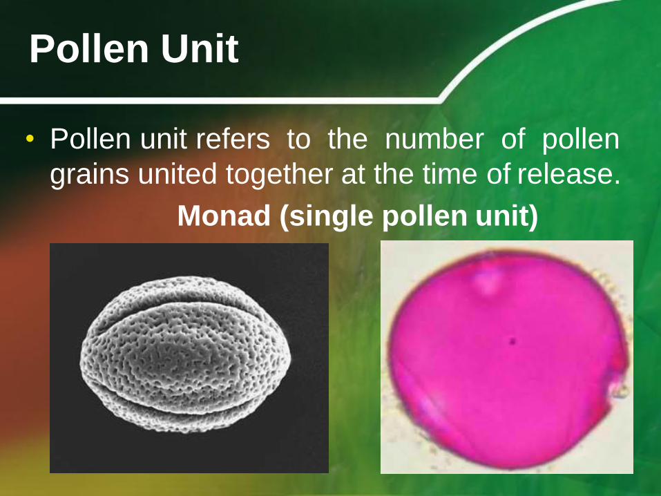
Pollen Unit
• Pollen unit refers to the number of pollen
grains united together at the time of release.
Monad (single pollen unit)

Monad
• Single and unfused pollen grain
• Examples (majority of angiosperms)
Hibiscus trionum, Malvaceae Nypa fruticans, Arecaceae
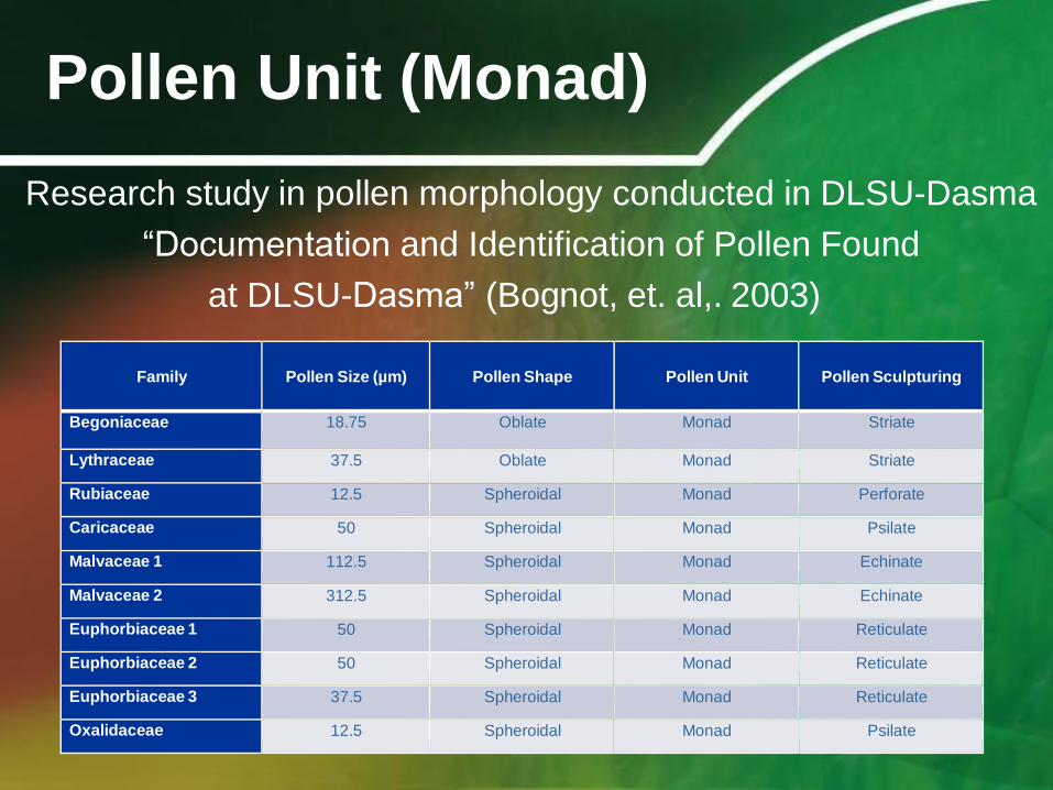
Pollen Unit (Monad)
Research study in pollen morphology conducted in DLSU-Dasma
“Documentation and Identification of Pollen Found
at DLSU-Dasma” (Bognot, et. al,. 2003)
Family
Pollen Size (µm)
Pollen Shape
Pollen Unit
Pollen Sculpturing
Begoniaceae 18.75 Oblate Monad Striate
Lythraceae 37.5 Oblate Monad Striate
Rubiaceae 12.5 Spheroidal Monad Perforate
Caricaceae 50 Spheroidal Monad Psilate
Malvaceae 1 112.5 Spheroidal Monad Echinate
Malvaceae 2 312.5 Spheroidal Monad Echinate
Euphorbiaceae 1 50 Spheroidal Monad Reticulate
Euphorbiaceae 2 50 Spheroidal Monad Reticulate
Euphorbiaceae 3 37.5 Spheroidal Monad Reticulate
Oxalidaceae 12.5 Spheroidal Monad Psilate

Pollen Unit
Dyad

Pollen Unit
Dyad
Ascarina philippinensis
(Chloranthaceae)

Pollen Unit
Tetrad
(four pollen grain fused together)
Tetrahedral Tetragonal

Example of tetrad
• Tetrahedral tetrad-Ericaceae (mint family)
• Tetragonal tetrad-Philydraceae and Fabaceae
Tetragonal tetrad
(Mimosa pudica)
Fabaceae

Pollen Unit (Tetrad)
Tetragonal tetrad
Philydraceae
(Pollen of mint family)
Tetragonal tetrad
Goodeniaceae

Pollen Unit
Decussate tetrad-pollen grains are in two
pairs arranged at right angles to one another
Lachnanthes, Haemodoraceae

Pollen Unit
Polyad (multiple of 8
fused grains)

Pollen Unit (Polyad)
• Research Studies on Pollen Unit and Systematics
“Pollen Morphology of Family Fabaceae
(Leguminosae) in DLSU-Dasma Campus”
(Guiao, et. al, 2003)
Common Name
Pollen Unit
Pollen Shape/Size
Aperture
Polarity
Acacia Polyad Prolate/Spheroidal Inaperturate Isopolar
Alibangbang Monad Oblate spheroidal Tricolpate Isopolar
Pine Tree Massulae polyad Prolate/Spheroid Isopolar
Dapdap Monad Oblate spheroidal Triporate Isopolar
Makahiya
Tetrahedral tetrad
Oblate
Syncolpate
Isopolar
Ipil-ipil Monad Prolate Tricolpate Isopolar

Polyad
Parkia speciosa
(Fabaceeae)

Pollen Unit (Polyad)
• Research Studies on Pollen Unit and Systematics
“Key to Pollen Identification in DLSU-Dasma
Campus”
(Aquino, et. al, 2003)
Family Name Dispersal Unit
Annonaceae Decussate and tetragonal tetrad
Moraceae Tetrad
Onagraceae Polyad/Tetrad
Portulaceae Tetrahedral tetrads
Sterculiaceae Polyad

Pollen Unit
Pollinium
(Apocynaceae and Orchidaceae)

Let’s remember!!!
Pollen Units • Monad
• Dyad
• Tetrad (tetrahedral, tetragonal,
decussate)
• Polyad
• Pollinium

Pollen Polarity
• It refers to the position of one or more
apertures relative to spatial reference.
• Observing a pollen grain from the
direction of a pole is known as polar
view; observing from the equatorial
direction is an equatorial view.
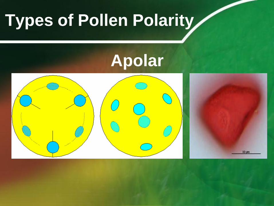
Types of Pollen Polarity
Apolar

Types of Pollen Polarity
Heteropolar

Types of Pollen Polarity
Isopolar
Cucurbitaceae

Let’s remember!!!
Pollen Polarity
• Apolar
• Heteropolar
• Isopolar

Pollen Aperture
Aperture - specially delimited region or an
opening in a pollen grain wall; colpus
(elongated) or pores (rounded)
*Colpi are regarded as more primitive than
pores. (Takhatajan)
Function:
-Point of pollen tube exitus, where pollen
tube grows out.
- Harmomegathy (resistant to decay)

Pollen Aperture
• Simple apertures are more primitive
than compound.
• Few apertures are more primitive
than several.

Pollen Aperture
• Colpus-elongated aperture or a sulcus-
slit or groove aperture occurring at the
distal pole
• Porus-circular aperture
• The number of apertures of any shape
can be designated by appending the
prefix mono-, di-, tri-, tetra-, penta-,
hexa- or poly- to the terms colpate or
porate.

Pollen Aperture
Monocolpate (Magnoliaceae)

Pollen Aperture
Monocolpate (Asteraceae)

Pollen Aperture
Monoporate (Poaceae)

Pollen Aperture
Tricolpate

Pollen Aperture (Tricolpate)
(Acanthaceae)

Pollen Aperture
triporate

Pollen Aperture (Triporate)
Cucurbitaceae

Pollen Aperture
Tricolporate (Goodenaceae)

Pollen Aperture
Polyporate
Amaranthaceae Convolvulaceae

Pollen Aperture
Pantoporate (pori occur globally on the
pollen grain surface)

Pollen Morphology and
Systematics
• Research Study
“Morphological Characteristics of Mangrove
Pollen”
The study revealed a crucial relationship
between pollen and mangrove families.
(refer pages 3-4 of the abstract)

Let’s remember!!!
Pollen Apertures
• Monocolpate
• Monoporate
• Tricolpate
• Tricolporate
• Polyporate
• Pantoporate

Pollen Size
• Pollen size can vary tremendously across
taxa. Size is typically measured in terms of
both the polar diameter and the equatorial
diameter.
• Typical pollen grains = 25-50 µm
• Pollen diameter ranges < 5 µm to > 200 µm
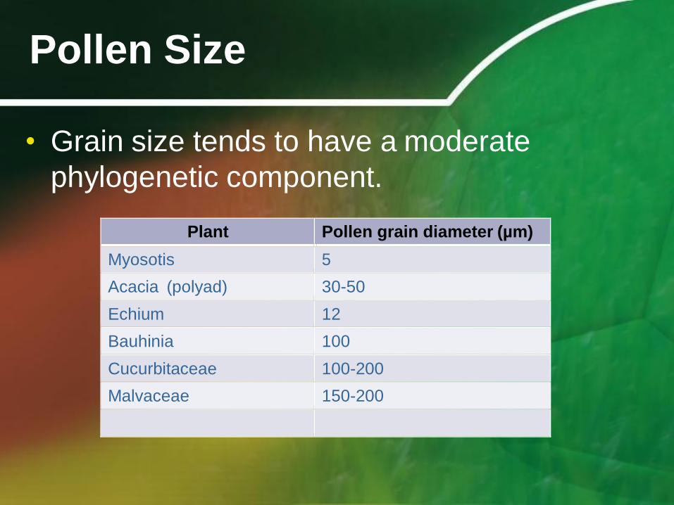
Pollen Size
• Grain size tends to have a moderate
phylogenetic component.
Plant Pollen grain diameter (µm)
Myosotis 5
Acacia (polyad) 30-50
Echium 12
Bauhinia 100
Cucurbitaceae 100-200
Malvaceae 150-200

Pollen Size
• It can be inferred from the table that borage
family has small pollen while mallow and
cucumber families have large pollen.

Pollen Size
• Plant Families in DLSU-Dasma with single pollen units
“Documentation and Identification of Pollen Found
at DLSU-Dasma” (Bognot, et. al,. 2003)
Family
Pollen Size (µm)
Pollen Shape
Pollen Unit
Pollen Sculpturing
Begoniaceae 18.75 Oblate Monad Striate
Lythraceae 37.5 Oblate Monad Striate
Rubiaceae 12.5 Spheroidal Monad Perforate
Caricaceae 50 Spheroidal Monad Psilate
Malvaceae 1 112.5 Spheroidal Monad Echinate
Malvaceae 2 312.5 Spheroidal Monad Echinate
Euphorbiaceae 1 50 Spheroidal Monad Reticulate
Euphorbiaceae 2 50 Spheroidal Monad Reticulate
Euphorbiaceae 3 37.5 Spheroidal Monad Reticulate
Oxalidaceae 12.5 Spheroidal Monad Psilate

Pollen Shape
Spheroidal
(globose or ball-shaped)

Pollen Shape
Spheroidal
Rubiaceae Cucurbitaceae

Pollen Shape
Oblate
(compressed along the polar axis like a
tangerine)

Pollen Shape
Oblate
Apocynaceae Euphorbiaceae

Pollen Shape
Prolate
(elongated along the polar axis like a
cucumber)

Pollen Shape
Prolate
Euphorbiaceae Rhizophoraceae Sonneratiaceae

Pollen Shape
• Plant Families in DLSU-Dasma with single pollen units
“Documentation and Identification of Pollen Found
at DLSU-Dasma” (Bognot, et. al,. 2003)
Family
Pollen Size (µm)
Pollen Shape
Pollen Unit
Pollen Sculpturing
Begoniaceae 18.75 Oblate Monad Striate
Lythraceae 37.5 Oblate Monad Striate
Rubiaceae 12.5 Spheroidal Monad Perforate
Caricaceae 50 Spheroidal Monad Psilate
Malvaceae 1 112.5 Spheroidal Monad Echinate
Malvaceae 2 312.5 Spheroidal Monad Echinate
Euphorbiaceae 1 50 Spheroidal Monad Reticulate
Euphorbiaceae 2 50 Spheroidal Monad Reticulate
Euphorbiaceae 3 37.5 Spheroidal Monad Reticulate
Oxalidaceae 12.5 Spheroidal Monad Psilate
• Research study on Mangrove Pollen (pages 3-4)

Let’s remember!!!
Pollen shape
• Spheroidal
• Oblate
• Prolate

Pollen Sculpturing
Echinate
(spinelike)
Verrucate
(wart-like)

Pollen Sculpturing Echinate
Asteraceae

Pollen Sculpturing Verrucate
Caryophyllaceae

Pollen Sculpturing
Rugulose
(brainlike)
Foveolate
(pitted surface)

Pollen Sculpturing Rugulate
Acanthaceae Fabaceae

Pollen Sculpturing
Reticulate (netlike)
Striate
(with stripes)

Pollen Wall Structure
tectate-columellate
(majority of the angiosperms)

Pollen Wall Structure
tectate-columellate
(roof and footlike layer)

Let’s summarize!!!
Palynological Features Used in
Plant Systematics
• Pollen Nucleus
Number
• Pollen Storage
Product
• Pollen Unit
• Pollen Polarity
• Pollen Aperture
• Pollen Size
• Pollen Shape
• Pollen Sculpturing
• Pollen Wall Structure
