©1993. used by permission of springer-verlag. 1. cells are fundamental units of life 2. cells use...
TRANSCRIPT

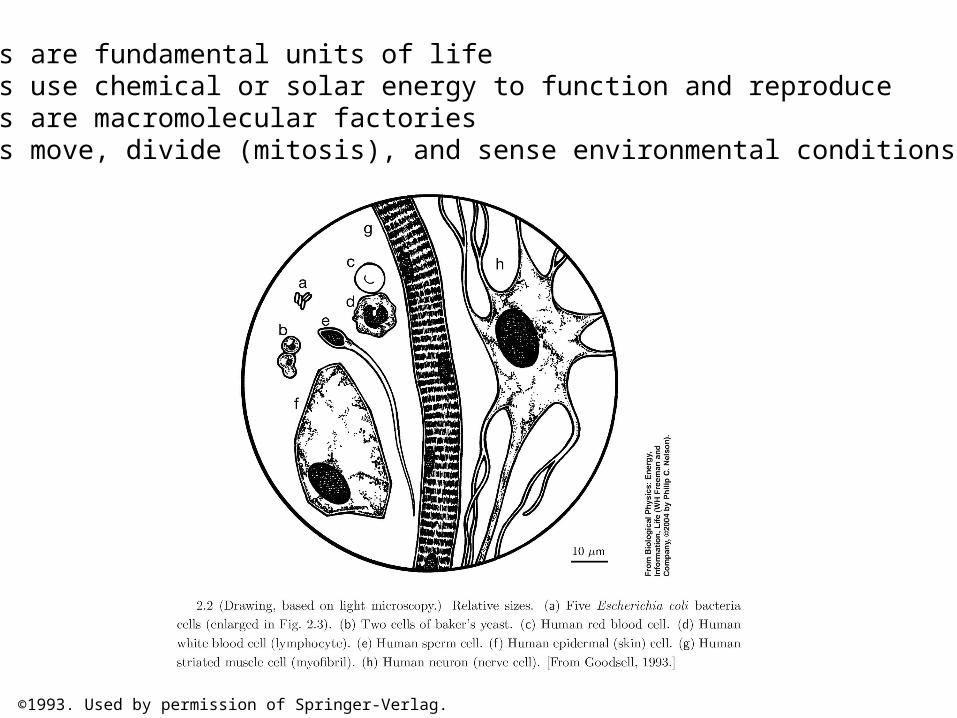
©1993. Used by permission of Springer-Verlag.
1. Cells are fundamental units of life2. Cells use chemical or solar energy to function and reproduce3. Cells are macromolecular factories4. Cells move, divide (mitosis), and sense environmental conditions
©1993. Used by permission of Springer-Verlag.
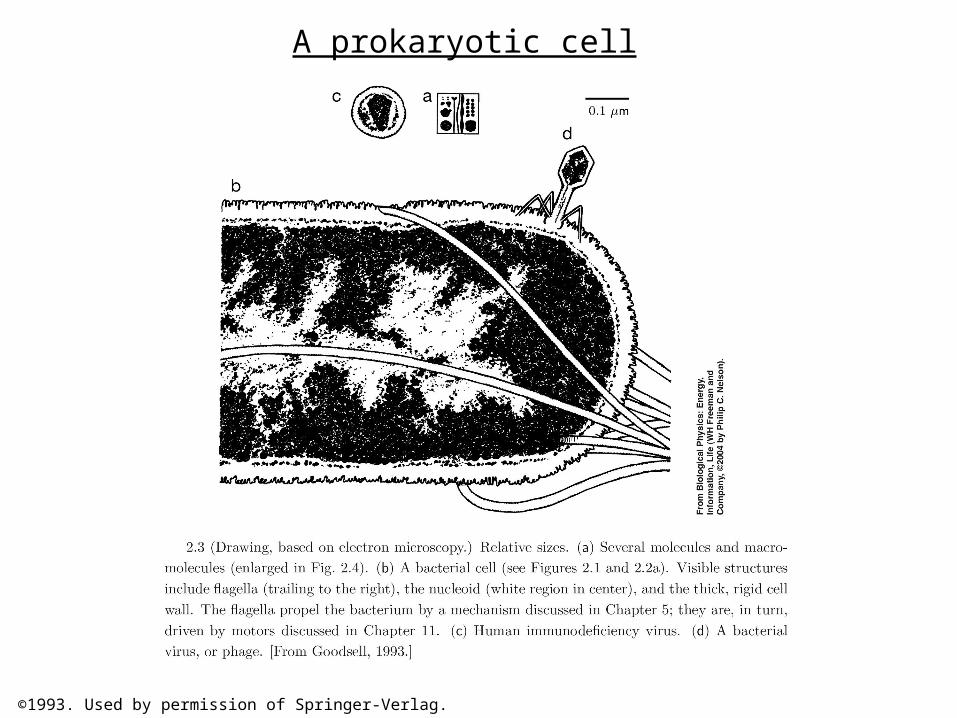
A prokaryotic cell
©1993. Used by permission of Springer-Verlag.
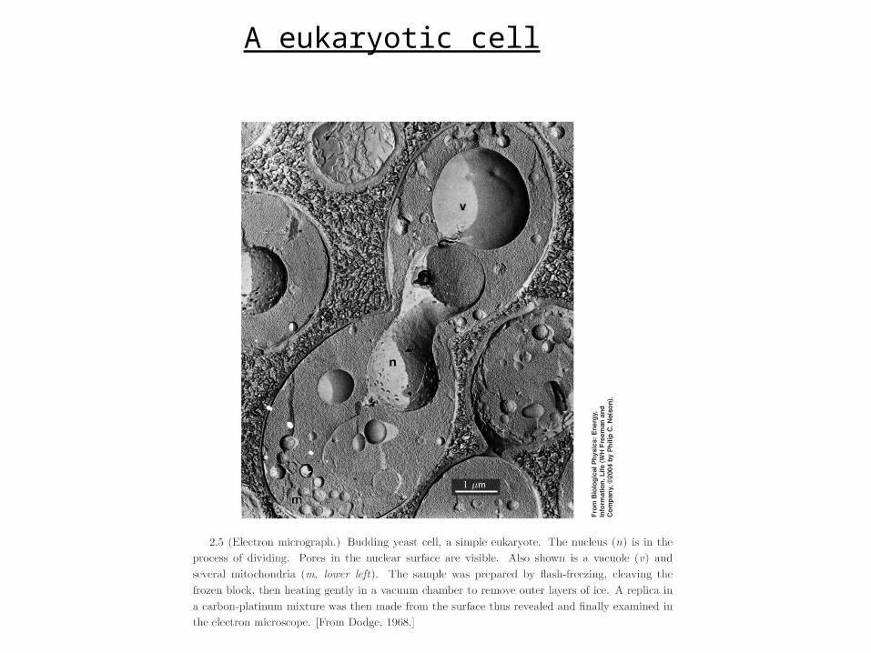
A eukaryotic cell

(b) ©1980. Used by permission of Elsevier Science.
Mitochondria are organelles that metabolize conversion of chemical energy from food into ATP.
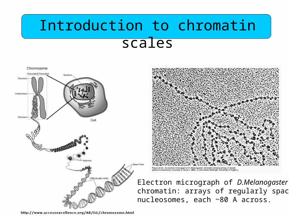
Introduction to chromatin scales
Electron micrograph of D.Melanogasterchromatin: arrays of regularly spacednucleosomes, each ~80 A across.

Courtesy of Dr. Julian Heath.
©1982. Used by permission of Jones and Bartlett Publishers, Sudbury MA.

Molecular composition of bacterial cells by weightMolecular composition of bacterial cells by weight:
Small molecules 74%water 70%amino acids, sugars, fatty acids, ions 4%
Macromolecules 26%proteins 15%RNA 6%DNA 1%lipids 2%polysaccharides 2%

©1991 Larry Gonick.

©1982, American Association for the Advancement of Science. Used by permission.

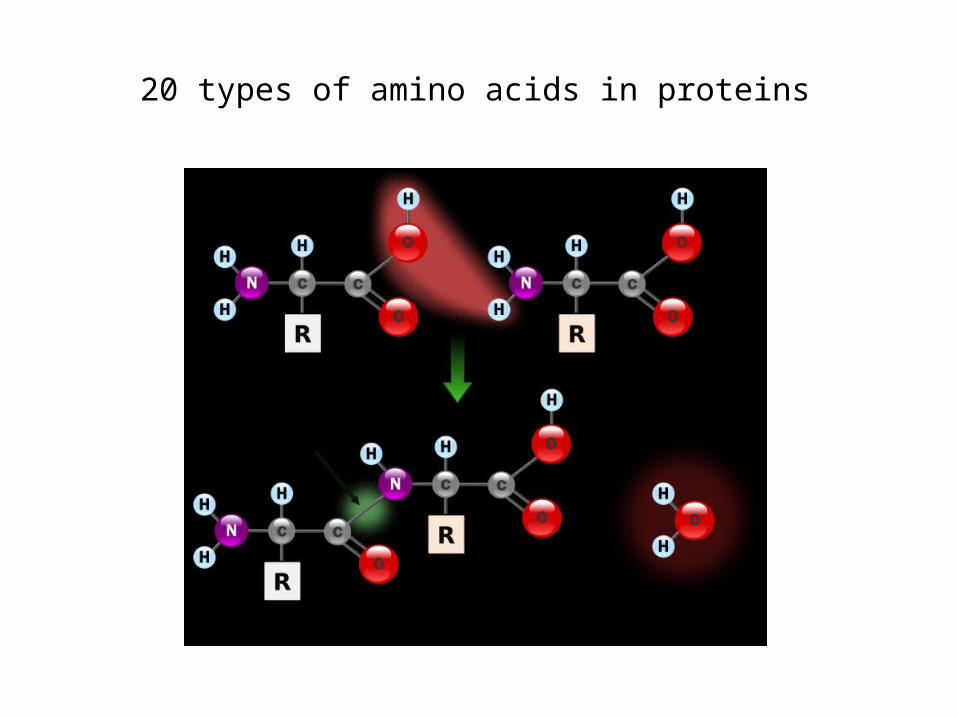
20 types of amino acids in proteins
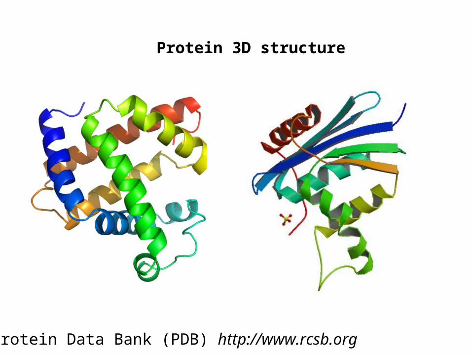
Protein 3D structure
Protein Data Bank (PDB) http://www.rcsb.org

Protein Data Base accession code 1VII ({C.J. McKnight, D.S. Doering, P.T. Matsudaira, P.S. Kim, J. Mol. Biol. 260 126 (1996)).

Elastic rod model
DNA looping induced by a Lac repressor tetramer
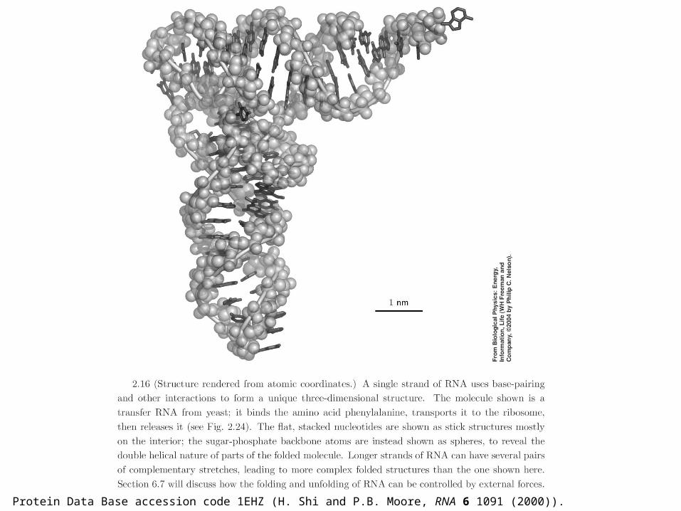
Protein Data Base accession code 1EHZ (H. Shi and P.B. Moore, RNA 6 1091 (2000)).

©1993. Used by permission of Springer-Verlag.
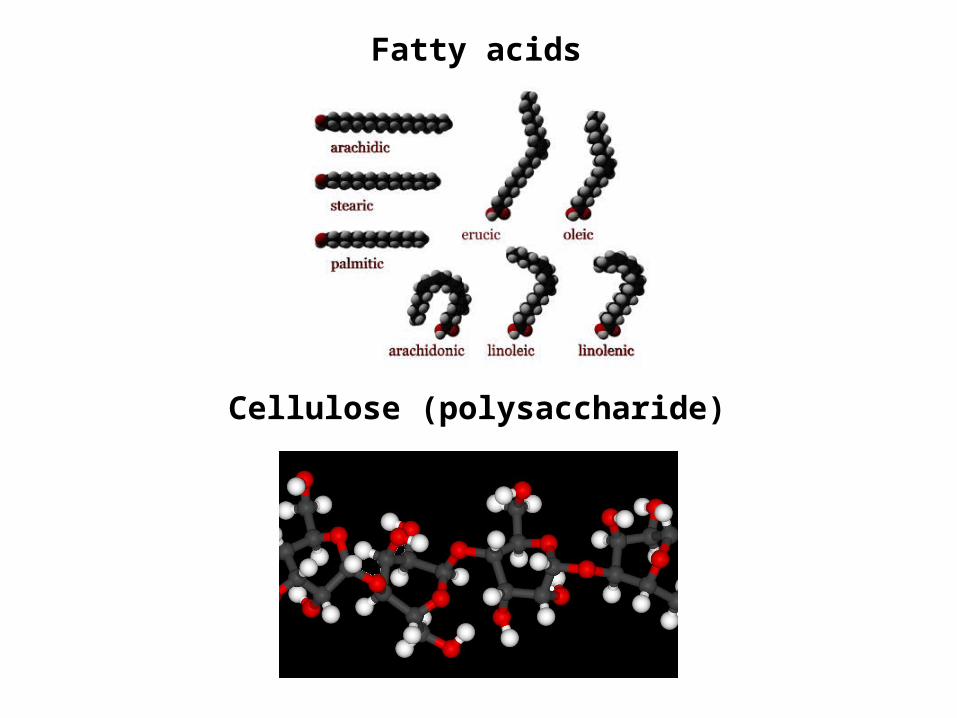
Fatty acids
Cellulose (polysaccharide)

A single peptide (protein building block)
A polypeptide chain

A tyrosine (TYR) amino acid (one of 20 naturallyoccurring amino acids)
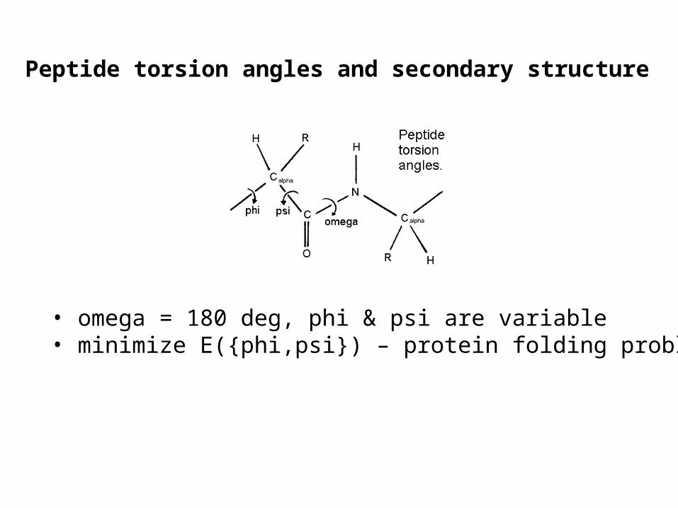
Peptide torsion angles and secondary structure
• omega = 180 deg, phi & psi are variable• minimize E({phi,psi}) – protein folding problem

Secondary structure elements: alpha & 3-10 helices
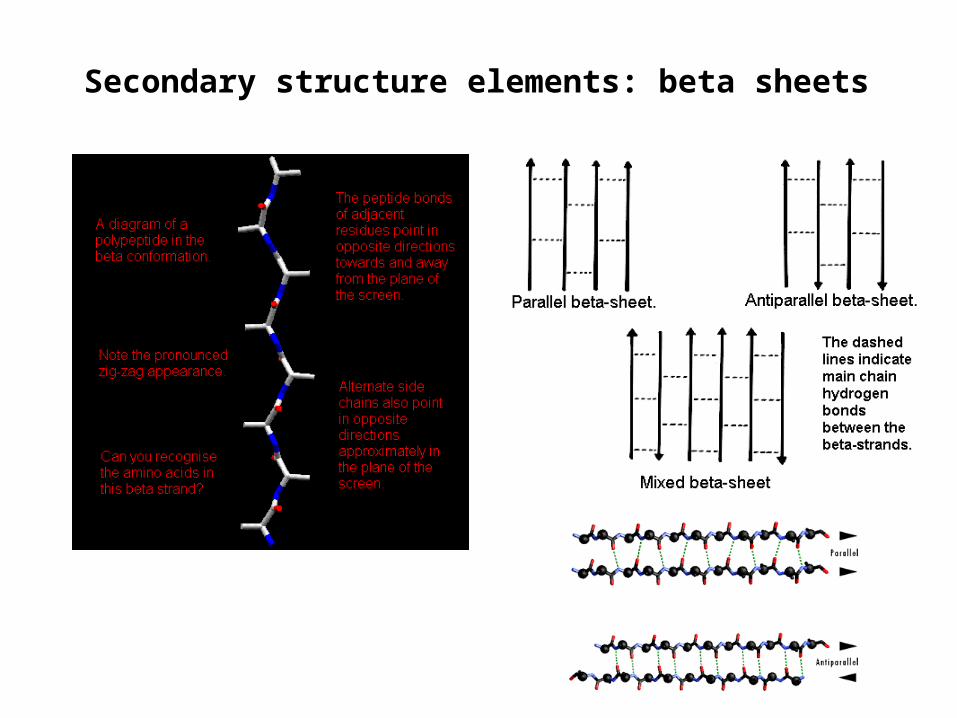
Secondary structure elements: beta sheets

The Ramachandran plot
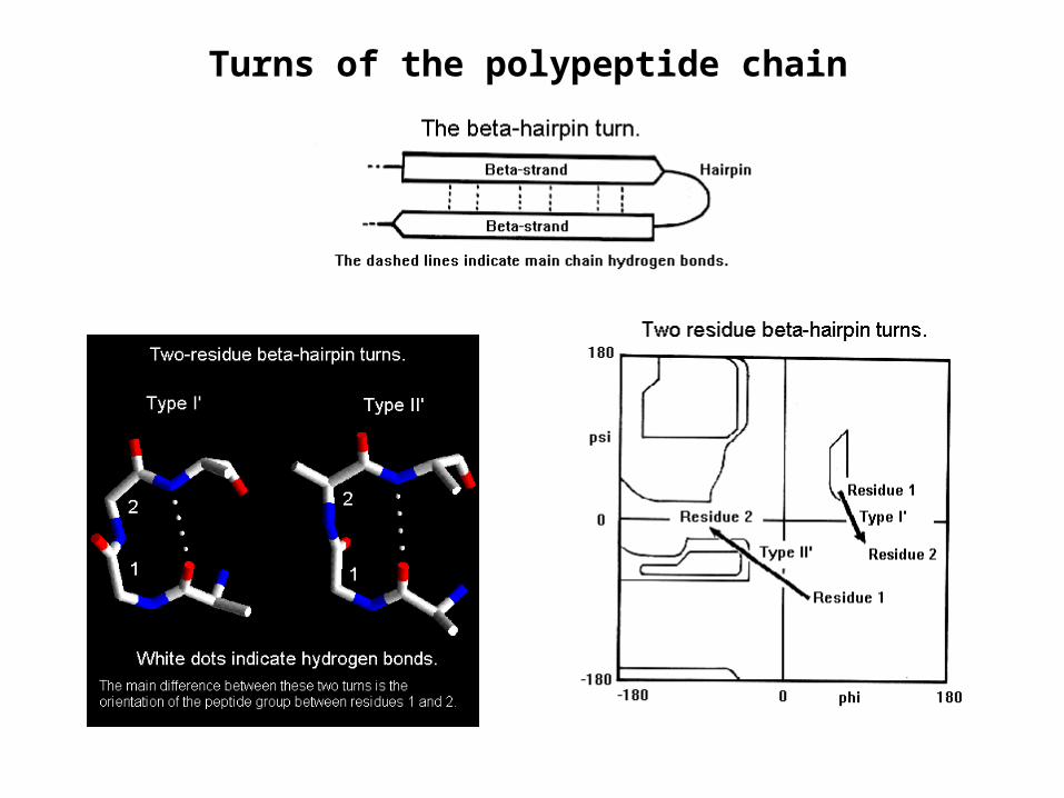
Turns of the polypeptide chain
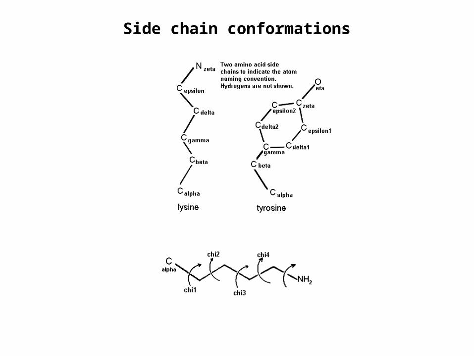
Side chain conformations
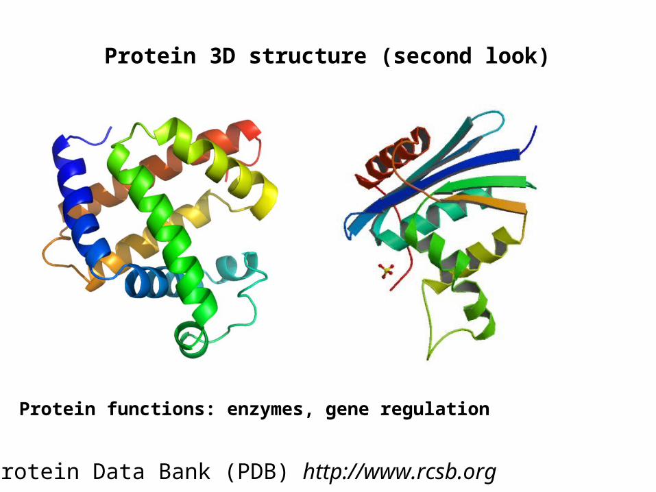
Protein 3D structure (second look)
Protein Data Bank (PDB) http://www.rcsb.org
Protein functions: enzymes, gene regulation

DNA & RNA

The genetic code

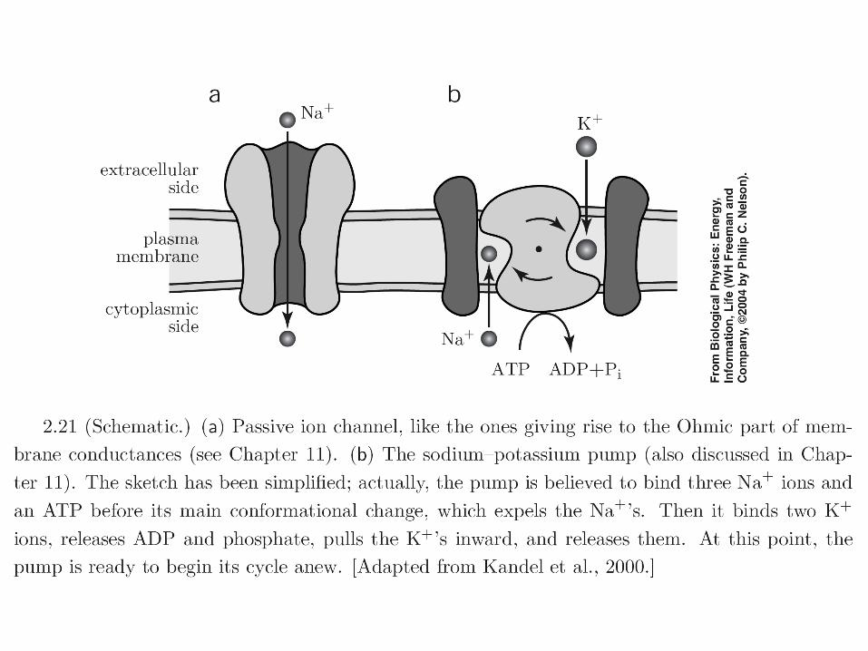
• Microtubules (25 nm): cytoskeleton• Actin filaments (F-actin; 7 nm): actin cortex

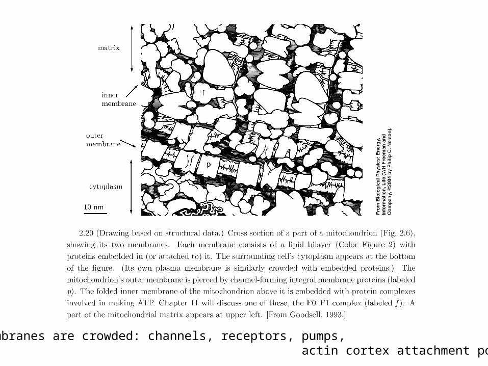
Cell membranes are crowded: channels, receptors, pumps, actin cortex attachment points


©1993. Used by permission of Springer-Verlag.
©1993. Used by permission of Springer-Verlag.