2 3 4 5 6 accepted -...
TRANSCRIPT
1
1
2
3
4
5
6
Phototrophic Fe(II) Oxidation Promotes Organic 7
Carbon Acquisition by Rhodobacter capsulatus SB1003 8
Nicky C. Caiazza1,3, Douglas P. Lies1,3 and Dianne K. Newman1,2,3* 9
1Division of Geological and Planetary Sciences and 2Division of Biology, 10
California Institute of Technology, Pasadena, CA 91125 and 3Howard Hughes 11
Medical Institute 12
13
14
15
*corresponding author: 16
Department of Biology, MIT 17
77 Massachussetts Ave., 68-380 18
Cambridge, MA 02139 19
Phone: (617) 324-2770 20
22
ACCEPTED
Copyright © 2007, American Society for Microbiology and/or the Listed Authors/Institutions. All Rights Reserved.Appl. Environ. Microbiol. doi:10.1128/AEM.02830-06 AEM Accepts, published online ahead of print on 10 August 2007
2
Abstract 1
Anoxygenic phototrophic Fe(II) oxidation is usually considered to be a 2
lithoautotrophic metabolism that contributes to primary production in Fe-based 3
ecosystems. In this study, we employed Rhodobacter capsulatus SB1003 as a 4
model organism to test the hypothesis that phototrophic Fe(II) oxidation can be 5
coupled to organic carbon acquisition. R. capsulatus SB1003 oxidized Fe(II) 6
under anoxic conditions in a light-dependent manner, but failed to grow 7
lithoautotrophically on soluble Fe(II). When provided with Fe(II)-citrate, however, 8
growth was observed that was dependent upon microbially catalyzed Fe(II) 9
oxidation, resulting in the formation of Fe(III)-citrate. Subsequent photochemical 10
breakdown of Fe(III)-citrate yielded acetoacetic acid, that supported growth in the 11
light but not the dark. Deletion of genes (RR00247-RR00248) that encode 12
homologs of atoA and atoD, required for acetoacetic acid utilization, severely 13
impaired the ability of R. capsulatus SB1003 to grow on Fe(II)-citrate. The 14
growth yield achieved by R. capsulatus SB1003 in the presence of citrate cannot 15
be explained by lithoautotrophic growth on Fe(II) enabled by indirect effects of 16
the ligand (such as altering the thermodynamics of Fe(II) oxidation or preventing 17
cell encrustation). Together, these results demonstrate that R. capsulatus 18
SB1003 grows photoheterotrophically on Fe(II)-citrate. Nitrilotriacetic acid (NTA) 19
also supported light-dependent growth on Fe(II), suggesting that Fe(II) oxidation 20
may be a general mechanism whereby some Fe(II)-oxidizing bacteria mine 21
otherwise inaccessible organic carbon sources. 22
23
ACCEPTED
3
Introduction 1
The discovery of anoxygenic phototrophic Fe(II) oxidizing bacteria was first 2
reported in the 1990’s (14, 21, 43) nearly 20 years after Garrels and Perry 3
postulated that these organisms could have been dominant players in ancient 4
Fe-based aquatic ecosystems (17). The photosynthetic reaction center of these 5
organisms uses light energy and electrons from Fe(II) to generate cellular energy 6
in the form of ATP and reducing equivalents in the form of NAD(P)H. The latter is 7
used to convert CO2 into biomass, thus Fe(II) oxidization can be used to support 8
photoautotrophic growth. Fe(II) oxidizing phototrophs do not always couple Fe(II) 9
oxidation to autotrophic growth, however: some strains oxidize Fe(II) while 10
growing photoheterotrophically on an organic substrate, such as acetate or 11
succinate (14, 22). In these cases, it is not clear whether Fe(II) oxidation benefits 12
the cell. 13
14
Recently, phototrophic Fe(II) oxidation has gained attention for the potential role 15
it might have played in the deposition of Archaean and early Proterozoic banded 16
iron formations (BIFs) (14, 20, 26, 43). This metabolism is also interesting from 17
the perspective of the evolution of photosynthesis. Of all reductants know to 18
support anoxygenic photosynthesis, ferrous iron [Fe(II)] has the highest midpoint 19
potential and may have been a transitional electron donor during the evolution of 20
H2O-accommodating reaction centers (31-33). 21
22
ACCEPTED

4
Interestingly, one aspect often neglected in studies or models pertaining to 1
biologically catalyzed Fe(II) oxidation is that Fe often exists in chelated forms. In 2
organic-rich freshwater systems and the surface waters of the oceans, the vast 3
majority of the dissolved Fe pool is strongly bound to organic ligands (18, 27, 34, 4
44). The presence of chelators can affect Fe(II) oxidation in multiple ways. One 5
possibility is that the Fe(II)-chelate complex can make Fe(II) more available to 6
the cells. This could be a factor when there is a propensity to form ferrous 7
minerals. Another possible role for a chelator is to keep the ferric iron [Fe(III)] that 8
is generated as a result of microbial Fe(II) oxidation in a soluble form and prevent 9
cell encrustation. Ferric minerals are often observed in association with the cell 10
surface of cultures growing phototrophically on Fe(II) (22, 25). The chelating 11
agent nitrilotriacetic acid (NTA) was shown to prevent the formation of crystalline 12
precipitates on the surface of Fe-grown Rhodomicrobium vannielii cultures in 13
comparison to those lacking NTA (22). 14
15
The presence of a chelator will also alter the thermodynamics of Fe(II) oxidation. 16
As predicted by the Nernst equation, altering the [Fe(III)]/[Fe(II)] ratio will change 17
the midpoint potential of the redox couple. When the midpoint potential 18
decreases, a more reducing environment favoring Fe(II) oxidation will result. 19
Thus, the presence of a chelator that has a higher affinity for Fe(III) than Fe(II) 20
can decrease the midpoint potential by preferentially depleting the pool of Fe(III) 21
relative to Fe(II). 22
23
ACCEPTED

5
In addition to these effects, a frequently overlooked impact that a chelator/ligand 1
may have on microbial Fe(II) oxidation stems from the potential photochemical 2
reactions that can occur between the ligand and Fe(III). Such reactions have 3
been well described in the biological oceanographic literature (4, 5, 30). For 4
example, one class of marine siderophores contains α-carboxylate moieties such 5
as citrate that are photochemically active in the presence of Fe(III). Examples 6
include aerobactin produced by Vibrio sp. DS40M5 (28), ochrobactins produced 7
by Ochrobactrum sp. SP18 (30), and synechobactins produced by 8
Synechococcus PCC 7002 (24). Photochemistry between the above 9
siderophores with Fe(III) has been documented and results in reduction of Fe(III) 10
and photolytic decarboxylation of the ligand (4, 5). One would expect such a 11
process to influence microbial Fe(II) oxidation in at least two ways: 1.) 12
Photochemical reactions would (re)generate Fe(II) that could then be used by 13
organisms capable of Fe-based phototrophic metabolisms, and 2.) The 14
photochemical breakdown of the ligand could produce organic material that could 15
be used for heterotrophic growth. 16
17
In this study, we provide data to support the hypothesis that anoxygenic 18
phototrophic Fe(II) oxidation can enable photoheterotrophic cell growth through 19
the use of a ferrated-ligand that is subject to photochemical degradation. We 20
choose citrate as a model ligand because it is a photochemically reactive moiety 21
found in siderophores and is not used as a carbon source by many phototrophs 22
(41). R. capsulatus SB1003 serves as our model organism because it does not 23
ACCEPTED

6
grow photoheterotrophically on citrate nor photoautotrophically on soluble Fe(II) 1
but does oxidize Fe(II) (10). Here, we show that phototrophic Fe(II) oxidation 2
promotes organic carbon acquisition by SB1003, and discuss the broader 3
implications of these findings. 4
ACCEPTED

7
Materials and methods 1
Bacterial strains, media, and chemicals. All bacterial strains, plasmids, and 2
primers used in this study are listed in Table 1. R. capsulatus SB1003 was grown 3
chemoheterotrophically at 30˚C in YP medium [0.3% yeast extract and 0.3% 4
Bacto peptone (Difco)]. FEM buffered at pH 6.8 with bicarbonate was used as the 5
base medium for phototrophic growth (11, 14). For photoheterotrophic growth 6
FEM was supplemented with either 10 mM acetate, 10 mM citrate plus 4-6 mM 7
FeCl2, 10 mM citrate plus 4-6 mM FeCl3, or 10 mM NTA plus 4-6 mM FeCl2. FEM 8
medium containing 4-6 mM Fe(II) was made as described previously (11). FEM 9
medium containing 4-6 mM Fe(III)-citrate was made by adding FeCl3-citrate from 10
an anoxic stock solution. The atmosphere for photoheterotrophic growth was 11
80% N2: 20% CO2. For photoautotrophic growth cells were grown in FEM with a 12
headspace of 80% H2: 20% CO2. Unless noted otherwise, phototrophically grown 13
cells were incubated at 30˚C under constant illumination 15 cm from a 34W 14
incandescent light source. Escherichia coli was cultured on Lysogeny Broth (LB) 15
(6) and strain WM3064 was supplemented with 0.3 mM diaminopimelic acid 16
(DAP). Sacchromyces cerevisiae was routinely cultured on YPD medium [1% 17
yeast extract, 2% Bacto peptone (Difco), 2% dextrose] at 30˚C or on SD Medium-18
URA (Q-BIOgene) for gap repair cloning. Gentamicin (Gm) was used at 1.25 19
µg/ml and 10 µg/ml and kanamycin (Kn) was used at 5 µg/ml and 10 µg/ml for R. 20
capsulatus SB1003 and E. coli strains, respectively. All enzymes used for DNA 21
manipulation were purchased from New England Biolabs (Ipswich, MA). 22
23
ACCEPTED
8
Analytical methods. (i) Ferrozine assay. A sterile syringe was used to withdraw 1
and transfer ~200 µl of sample from anaerobic cultures into the well of a 96 well 2
plate (Becton Dickinson, Franklin Lakes, NJ). Ten microliters of this aliquot was 3
immediately transferred, in triplicate, into wells containing 90 µl of 1N HCl. One 4
hundred microliters of 0.1% ferrozine in 50% ammonium acetate was added, 5
mixed and allowed to incubate at room temperature for 10 minutes. The 6
absorbance at 570 nm was measured. A standard curve for Fe(II) prepared over 7
the range of 0.5-4 mM was used to calculated the Fe(II) sample concentration. 8
(ii) Cell suspension assay. All cell suspension assays were prepared and 9
conducted at room temperature in an anaerobic chamber containing an 10
atmosphere of 5% H2:80% N2:15% CO2 and incubated in the presence or 11
absence of light as indicated. Cells were pre-grown in FEM + H2 until mid 12
exponential phase (OD660 of ~0.3) and 8 ml of culture was harvested and washed 13
in an equal volume of assay buffer [50 mM HEPES (N-2-hydroxyethylpiperazine-14
N`-2-ethanesulfonic acid) and 20 mM NaCl at pH 7], resuspended in assay buffer 15
containing 0.4-0.6 mM FeCl2 and 20 mM NaHCO3, and 100 µl aliquots were 16
dispensed in 96-well plates. The Fe(II) concentration was measured as a function 17
of time by adding 100 µl of ferrozine reagent to the samples and measuring the 18
absorbance at 570 nm. 19
(iii) Protein assay. A sterile syringe was used to withdraw 1 ml of sample from 20
anaerobic cultures and transfer it to a 2 ml microcentrifuge tube containing 800 µl 21
oxalate solution (28 g of ammonium oxalate and 15 g of oxalic acid per liter) and 22
100 µl of 100 mM ferrous ethylenediammonium sulfate. The contents were mixed 23
ACCEPTED
9
by vortexing for 30 seconds and then incubated at 37˚C for 30 minutes for 1
mineral dissolution. Total protein was precipitated by addition of 75 µl of 10 M 2
trichloroacetic acid (TCA), vortexing, and incubating on ice for 30 minutes. 3
Precipitated protein was collected by centrifugation, 16,000 X g for 15 minutes, at 4
4˚C. The pellet was resuspended in 500 µl of 0.1N NaOH and boiled for 5 5
minutes. In triplicate, 160 µl of the cooled sample was mixed with 40 µl of 6
Bradford reagent and the samples processed as described by the manufacturer 7
(Bio-Rad, Hercules, CA). 8
(iv) High-pressure liquid chromatography (HPLC). A syringe was used to 9
withdraw 0.5 ml of sample from anaerobic cultures and transfer it to a Spin-X 10
0.22 µm cellulose acetate filter (Costar, Corning, NY) and the cells were removed 11
by centrifugation at 16,000 X g for 10 minutes. Citrate, β-ketoglutarate, and 12
acetoacetic acid were analyzed using an Aminex HPX-87H column (Bio-Rad, 13
Hercules, CA) at 30˚C. Phosphoric acid (30 mM, pH 1.2) was used as the mobile 14
phase and the absorbance was monitored at 210 nm with a UV detector. 15
16
Molecular techniques. Cloning was carried out in E. coli UQ950 by standard 17
methods (2) and constructs were mated into R. capsulatus SB1003 using E. coli 18
WM3064. Annotated sequences of the R. capsulatus SB1003 genome were 19
obtained using Ergo Light (http://www.ergo-light.com/) from the R. capsulatus 20
SB1003 genome available through Integrated Genomics 21
(www.integratedgenomics.com). 22
ACCEPTED
10
(i) In-frame deletion of hupSL. A knockout construct for the R. capsulatus 1
SB1003 hupSL genes was generated by using yeast gap repair cloning to insert 2
~1 Kb of DNA flanking the 5'- and 3'-regions of hupSL into pMQ30 using the 3
primers listed in Table 1. The plasmid pMQ30 is a sacB-based allelic exchange 4
vector for gram-negative bacteria that cannot support the ColE1 origin of 5
replication and contains CEN6/ARSH4 DNA sequences to support replication in 6
yeast and a URA3 yeast selectable marker (35). Primers pnc001 and pnc002 7
were used to amplify the 5’-flanking DNA and primers pnc003 and pnc004 were 8
used to amplify the 3’-flanking DNA. The 5'- and 3'- flanking fragments were 9
cloned in vivo into pMQ30 using the S. cerevisiae uracil auxotrophic strain 10
InvSc1® (Invitrogen, Carlsbad, CA) as previously described (7, 15) resulting in 11
pNC001. Transformants that had undergone gap repair cloning and inserted the 12
DNA fragments into pMQ30 by homologous recombination were selected on a 13
medium lacking uracil and plasmids were isolated and transformed into E. coli 14
UQ950 as previously described (7, 23). 15
The native sacB promoter found on pMQ30 did not express in R. capsulatus 16
SB1003. Therefore primers pnc017 and pnc018 were used to amplify the hupSL 17
knockout fragment from pNC001 and the amplified fragment was digested using 18
engineered restriction sites (SacI and XbaI) and ligated into pZJD29a (J. Jiang 19
and C. E. Bauer, unpublished plasmid construction) digested with SacI and XbaI 20
resulting in pNC005. The plasmid was electroporated into E. coli WM3064 and 21
mated into R. capsulatus SB1003, selecting for Gm resistance. Resolution of the 22
ACCEPTED
11
integrated plasmid was performed by selection on 5% sucrose followed by PCR-1
based screening for the loss of the wild type gene. 2
(ii) Construction of a yeast-based allelic exchange vector for phototrophic 3
bacteria. Plasmid pNC006 is a sacB-based allelic exchange vector for gram-4
negative bacteria that cannot support the ColE1 origin of replication, but can be 5
maintained in S. cerevisiae and E. coli. Plasmid pNC006 was built by cloning a 6
copy of the sacB gene under control of a native R. capsulatus promoter into 7
pMQ87 using yeast gap repair cloning as described above. Plasmid pMQ87 is a 8
suicide vector for gram-negative bacteria and contains the CEN6/ARSH4 DNA 9
sequences for replication in yeast and a URA3 yeast selectable marker (35). It 10
was linearized with the restriction enzyme Xho I. Primers pnc021 and pnc022 11
were designed to amplify a 2 Kb fragment from pZJD29a containing the sacB 12
gene under control of the R. capsulatus pucAB promoter. The digested plasmid 13
(pMQ87) and the PCR fragment were co-transformed into S. cerevisiae InvSc1® 14
(Invitrogen) as described above. Yeast transformants that had inserted the PCR 15
fragment into pMQ87 by gap repair homologous recombination, resulting in 16
pNC006, were selected as described above. 17
(iii) In-frame deletion of RRC00247-RRC00248. Plasmid pNC007 was 18
constructed using yeast gap repair cloning as described above. Briefly, primers 19
pnc023 and pnc027 were used to amplify the 5’-flanking DNA fragment and 20
primer pnc026 and pnc028 were used to amplify the 3’-flanking DNA fragment. 21
The 5’- and 3’- flanking DNA fragments and pNC006 linearized with BamHI were 22
co-transformed into S. cerevisiae and homologous recombination events leading 23
ACCEPTED
12
to the formation of pNC007 were selected for as described above. The plasmids 1
were mated into R. capsulatus SB1003 and the resulting integrants and resolved 2
mutants were determined as described above. 3
(iv) Construction of RRC00247-RRC00248 complementation plasmid. Primers 4
pnc033 and pnc034 were designed ~500 bp upstream and downstream of the 5
RRC00247-RRC00248 operon. The primers were engineered to create a PCR 6
fragment that could be digested with SacI and XbaI and ligated into pMQ131 7
digested with those same enzymes. Plasmid pMQ131 is a broad host range 8
plasmid designed for cloning using the yeast gap repair system (R.M.Q. Shanks 9
and G.A. O’Toole, unpublished: www.dartmouth.edu/~gotoole/vectors.html). This 10
construct presumably carries the native RRC00247-RRC00248 promoter. 11
ACCEPTED

13
Results 1
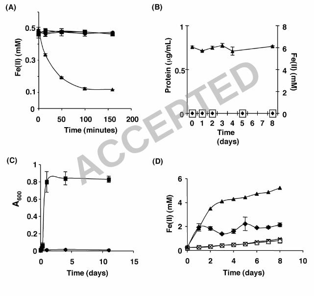
Anoxygenic phototrophic Fe(II) oxidation by R. capsulatus SB1003. Fig. 1A 2
and a previous report from our lab (10) show that whole cell suspensions of R. 3
capsulatus SB1003 possess light-dependent Fe(II) oxidation activity. In this 4
assay, cells from H2-grown, exponential phase cultures are washed and 5
resuspended in a medium containing soluble Fe(II) and the ability of the cell 6
suspension to oxidize Fe(II) is measured as a function of time using the ferrozine 7
assay. Total iron [Fe(II) + Fe(III)] remains constant through these experiments 8
(data not shown). Oxidation of Fe(II) is independent of cell growth due to the 9
short time scale of this assay (Fig. 1A, less than 3 hours) and the large number 10
of cells present in the suspension (see Materials and Methods section for 11
details). Thus this assay only measures the ability of an organism to oxidize 12
Fe(II) and is not a measure of an organism’s ability to grow via anoxygenic 13
phototrophic Fe(II) oxidation. 14
15
To test if R. capsulatus SB1003 could grow via anoxygenic phototrophic Fe(II) 16
oxidation, cells from H2-grown cultures were diluted in a medium containing ~6 17
mM Fe(II) as the sole electron source. Although R. capsulatus SB1003 did 18
possess light-dependent Fe(II) oxidation activity (Fig. 1A) it did not exhibit 19
photoautotrophic growth on Fe(II) (Fig. 1B). Over the course of 8 days, the 20
protein concentration of the Fe(II) cultures inoculated with R. capsulatus SB1003 21
was identical to that of the uninoculated media controls and remained below the 22
detection limit of the protein assay used (Fig. 1B). In addition, the Fe(II) 23
ACCEPTED

14
concentration (Fig. 1B) remained ~6 mM throughout the time course of the 1
experiment indicating that appreciable Fe(II) oxidation did not occur in the 2
presence of the small amount of cells that are incapable of growth on Fe(II). 3
4
Phototrophic growth of R. capsulatus SB1003 on Fe(III)-citrate. R. 5
capsulatus SB1003 is able to oxidize iron (Fig. 1A) but cannot grow 6
photoautotrophically when Fe(II) is provided as the sole electron source (Fig. 7
1B). This led us to speculate about alternative functions for Fe(II) oxidation in this 8
organism. One explanation for the Fe(II) oxidation activity is that it provides a 9
mechanism for organic carbon acquisition. In numerous environments, iron exists 10
in chelated forms and many Fe(III)-chelates are subject to light-induced photo-11
redox reactions that result in the reduction of Fe(III) and the degradation of the 12
chelator (4, 5). We hypothesized that microbially catalyzed Fe(II) oxidation 13
coupled with ferrated-ligand photochemistry could convert otherwise inaccesible 14
pools of carbon into labile substrates capable of supporting microbial growth. 15
16
To test this hypothesis, citrate was chosen as a model carbon source because it 17
has been reported not to be metabolized by R. capsulatus SB1003 under 18
anoxygenic phototrophic conditions (41) and is also subject to photochemistry in 19
the presence of Fe(III) (1). Our hypothesis predicts that microbially catalyzed 20
Fe(II) oxidation in the presence of citrate will lead to the formation of Fe(III)-21
citrate, and that the latter will undergo photochemical degradation to produce a 22
carbon source(s) that can sustain growth. We confirmed that R. capsulatus 23
ACCEPTED
15
SB1003 cannot use citrate as a carbon source for photoheterotrophic growth 1
under the culture conditions used in this study, as compared with 2
photoheterotrophic growth with acetate as carbon source (Fig. 1C). The above 3
hypothesis also requires Fe(III)-citrate photochemistry to occur under our culture 4
conditions. To test this, we incubated uninoculated medium containing Fe(III)-5
citrate in the presence or absence of light and measured the reduction of Fe(III) 6
using the ferrozine assay (Fig. 1D). The concentration of Fe(II) rapidly increased 7
(indicating Fe(III) reduction) to ~4 mM during the first 2 days of incubation for 8
uninoculated medium exposed to light and over the next 6 days slowly 9
approached ~6 mM. In contrast, the Fe(II) levels in the non-illuminated 10
uninoculated medium remained below 1 mM for the duration of the experiment. 11
These data indicated that Fe(III)-citrate photochemistry resulting in the 12
production of Fe(II) can occur in our experimental system. 13
14
The next prediction of our hypothesis is that R. capsulatus SB1003 should be 15
able to use Fe(III)-citrate as a carbon source in a light-dependent manner. To 16
test this, cells from H2-grown cultures were diluted in a medium containing Fe(III)-17
citrate and incubated in the presence or absence of light. Over the course of the 18
experiment, the protein concentration of the cultures exposed to light increased 19
from levels that were undetectable at day 0 to levels of ~139 µg/ml by day 8. 20
Protein concentration remained undetectable in cultures incubated in the dark. 21
This indicated that R. capsulatus SB1003 can grow on Fe(III)-citrate in a light-22
dependent manner. 23
ACCEPTED

16
1
In addition to protein level, the Fe(II) concentration of the illuminated and non-2
illuminated SB1003 cultures supplemented with Fe(III)-citrate were monitored as 3
a function of time (Fig. 1D). The concentration of Fe(II) increased slightly in non-4
illuminated cultures but was not significantly different from the non-illuminated 5
uninoculated medium indicating a low level of Fe reduction in the system that is 6
independent of the bacteria present. When Fe(III)-citrate-containing cultures 7
were incubated in the light, a rapid increase in the Fe(II) concentration was 8
observed within the first 24 h after which the concentration of Fe(II) stabilized. 9
Our assay conditions take into account intracellular Fe(II) as well as cell 10
associated Fe(II). Therefore, the different levels of Fe(II) achieved between 11
illuminated SB1003 cultures and uninoculated media incubated in the light is not 12
a result of cellular Fe(II) consumption. This suggests that the concentration of 13
Fe(II) in the illuminated cultures is mainly dependent on a combination of the rate 14
of abiotic, light-dependent Fe(III) reduction and the rate of bacterial Fe(II) 15
oxidation. 16
17
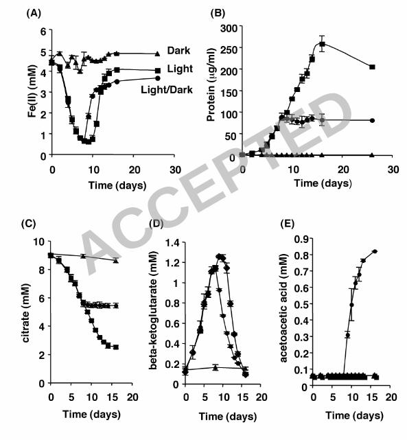
Phototrophic growth of R. capsulatus SB1003 on Fe(II)-citrate. Having 18
shown that R. capsulatus SB1003 can grow phototrophically on Fe(III)-citrate, 19
our next objective was to determine whether the light-dependent Fe(II) oxidation 20
activity of this organism observed in Fig. 1A could support growth through the 21
generation of Fe(III)-citrate from Fe(II)-citrate. Cells from H2-grown cultures were 22
diluted in a medium containing ~4-5 mM Fe(II) and 10 mM citrate and incubated 23
ACCEPTED
17
under light regimes consisting of constant illumination, constant darkness, or 1
constant illumination initially for 7 days followed by constant darkness for the 2
duration of the experiment (Fig. 2). Aliquots were removed from the cultures at 3
various time points and the Fe(II) concentration (Fig. 2A) and the protein 4
concentration (Fig. 2B) were measured. 5
6
In the absence of light, Fe(II) concentrations remained ~4.5 mM for the duration 7
of the experiment, indicating that little or no Fe(II) oxidation occurred in the dark. 8
In the presence of light, the initial rate of Fe oxidation that occurred over the first 9
2 days was slow and was followed by an increased rate of Fe oxidation that 10
lasted through day 8. During this time period the Fe(II) concentration in the 11
cultures decreased from ~4.4 mM to ~0.6 mM, demonstrating that Fe(II) 12
oxidation in the presence of citrate is light-dependent and consistent with the 13
findings seen in the cell suspension assay (Fig. 1A). For cultures kept in 14
constant light, the Fe(II) concentration remained ~0.6 mM from day 8 to day 10. 15
At this point the Fe(II) concentration began to increase and eventually leveled at 16
~4.0 mM by day 14. 17
18
To test if the Fe(III) reduction that occurred from day 10 to day 14 was light 19
dependent, we transferred phototrophically grown cultures to dark conditions 20
after the Fe(II) oxidation phase (day 7) and the concentration of Fe(II) was 21
measured as a function of time. While cultures left continuously in the light 22
maintained Fe(II) concentrations at ~0.6 mM through day 10, the cultures that 23
ACCEPTED

18
were moved to the dark showed immediate Fe(III) reduction and the Fe(II) 1
concentration increased to ~2.8 mM by day 10, and continued to increase until 2
eventually leveling off at ~3.5 mM. Thus, the Fe(III) reduction observed in 3
continuously illuminated cultures beyond day 7 cannot be solely accounted for by 4
photochemical reactions and could be catalyzed by a light-independent bacterial 5
process. In fact, strains of R. capsulatus previously have been shown to exhibit 6
Fe(III) reduction which is not coupled to growth, although the mechanism is 7
unknown (12). 8
9
Bacterial growth on Fe(II)-citrate was measured in terms of protein concentration 10
(µg/ml) and is depicted in Fig. 2B. No increase in biomass was observed in 11
cultures incubated in constant darkness. For phototrophic cultures, slow growth 12
was observed for the first 4 days before an increase in biomass occurred. In 13
cultures incubated under constant light, growth occurred during both the Fe(II) 14
oxidation phase (day 0 to day 9) as protein levels reached ~100 µg/ml, and 15
during the Fe(III) reduction phase (day 10 to day 14) as protein levels continued 16
to increase and reached ~250 µg/ml. This indicated that anaerobic growth on 17
Fe(II)-citrate is a phototrophic process during the Fe(II) oxidation phase. 18
19
The protein concentration of cultures incubated in the light during the Fe(II) 20
oxidation phase (day 0 to day 7) and then transferred to the dark during the Fe 21
reduction phase (day 8 to day 16) revealed that biomass increased (to ~100 22
µg/ml) only in the presence of light (day 0 to day 7) and growth ceased upon 23
ACCEPTED
19
transfer to the dark (day 8 to day 16). This indicated that the growth seen during 1
the Fe(III) reduction phase (day 8 to day 14) is light dependent and that the 2
Fe(III) reduction observed during the dark incubation cannot support anaerobic 3
growth of R. capsulatus SB1003 under these conditions. 4
5
Fe-citrate photochemistry and the formation of potential carbon sources 6
for photoheterotrophic growth. It is well documented that the initial 7
photochemistry occurring between Fe(III) and citrate leads to the reduction of 8
Fe(III) and the oxidation of citrate yielding Fe(II) and β-ketoglutarate (1, 16) 9
according to the following reaction: citrate + 2Fe3+ + hν � β-ketoglutarate + CO2 10
+ 2Fe2+ + 2H+. In an aqueous environment, β-ketoglutarate spontaneously 11
undergoes light-independent decarboxylation, producing acetoacetic acid (i.e. β-12
ketoglutarate � acetoacetate + CO2) (1, 16). Our hypothesis predicts that β-13
ketoglutarate and/or acetoacetic acid should be present in the supernatant of R. 14
capsulatus SB1003 cultures growing on Fe(II)-citrate and that one or both of 15
these compounds can support the growth of this organism under phototrophic 16
conditions. 17
18
To test this, in addition to measuring the Fe(II) and protein concentrations of 19
Fe(II)-citrate cultures grown under various light regimes (Fig. 2A and B), we also 20
measured the concentrations of the suspected carbon intermediates using HPLC 21
(citrate, Fig 2C; β-ketoglutarate, Fig. 2D; acetoacetic acid, Fig. 2E). In cultures 22
maintained in constant darkness the citrate concentration remained constant (~9 23
ACCEPTED
20
mM) for the duration of the experiment while β-ketoglutarate and acetoacetic acid 1
were not detected. In contrast, the citrate concentration steadily decreased from 2
~9 mM to ~2.5 mM in cultures subjected to light. In these cultures the β-3
ketoglutarate concentration steadily increased to ~1.25 mM by day 9 and then 4
steadily decreased to below detection by day 16. Acetoacetic acid was not 5
detected in the illuminated cultures. 6
7
In cultures initially subjected to constant light, the citrate concentration decreased 8
from ~9 mM to ~5.5 mM during the light phase (day 0 to day 7) and leveled off 9
upon transfer to the dark (day 8 to day 16). The β-ketoglutarate concentration of 10
these cultures steadily increased to ~1.15 mM during exposure to the light and 11
then steadily decreased to below the limit of detection upon transfer to constant 12
darkness. Acetoacetic acid was not detected in these cultures during exposure to 13
light and only accumulated when cultures were transferred to constant darkness, 14
steadily increasing to ~0.8 mM by the end of the experiment. 15
16
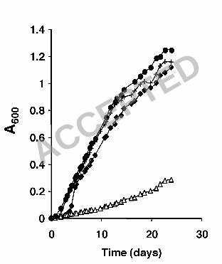
The genes RRC00247-RRC00248 are required for optimal growth on Fe(III)-17
citrate. Bacterial growth on Fe(II)-citrate only occurs in the presence of light (Fig. 18
2B). During this time period β-ketoglutarate accumulated (Fig. 2D) while 19
acetoacetic acid could not be detected (Fig. 2E). Acetoacetic acid concentrations 20
only build to detectable levels when cultures are shifted from the light to the dark, 21
which is a condition that leads to growth cessation (Fig. 2B and 2E). The fact that 22
acetoacetic acid only accumulated after growth ceased suggested that this 23
ACCEPTED
21
intermediate, and not β-ketoglutarate, was the labile carbon source capable of 1
supporting phototrophic growth of R. capsulatus SB1003 when grown on Fe(II)- 2
or Fe(III)-citrate. 3
A genetic approach was taken to test if acetoacetic acid was indeed the carbon 4
source used by R. capsulatus SB1003 under these conditions. An in-frame 5
deletion of the genes RRC00247 and RRC00248 was constructed on the 6
chromosome of R. capsulatus SB1003. The genes RRC00247 and RRC00248 7
encode proteins that are homologous to the E. coli proteins AtoD and AtoA, 8
respectively. atoD and RRC00247 are 42.3% identical and atoA and RRC00248 9
are 49.8% identical at the amino acid level. In E. coli, these proteins are involved 10
in the utilization of acetoacetic acid. The proteins AtoA and AtoD form the acetyl-11
CoA:acetoacetyl-CoA transferase, which catalyzes the conversion of acetoacetic 12
acid into acetoacetyl-CoA (36). The latter is converted to acetyl-CoA by the 13
acetyl-CoA acetyltransferase encoded in E. coli by the atoB gene (13). In 14
comparison to the wild-type strain, no growth defect was observed when the R. 15
capsulatus SB1003 ∆RRC00247-RRC00248 (encoding homologs of AtoA and 16
AtoD) mutant was grown under photoheterotrophic conditions on acetate or 17
under photoautotrophic conditions on H2 (data not shown). However, when the R. 18
capsulatus SB1003 ∆RRC00247-RRC00248 mutant was grown phototrophically 19
on Fe(III)-citrate and carried pMQ131 (empty vector control) a growth defect was 20
observed relative to the wild-type carrying pMQ131 (Fig. 3). The growth defect 21
could be complemented through the introduction of a plasmid containing 22
RRC00247 and RRC00248 (pNC008) into the R. capsulatus SB1003 23
ACCEPTED

22
∆RRC00247-RRC00248 mutant (Fig. 3). This plasmid (pNC008) did not 1
significantly alter the ability of the wild-type strain to grow on Fe(III)-citrate (Fig. 2
3). These data indicate that acetoacetic acid is the primary growth substrate for 3
R. capsulatus SB1003 when grown on Fe(III)-citrate or Fe(II)-citrate under 4
phototrophic conditions. 5
6
Other Fe(II) complexes tested for phototrophic growth of R. capsulatus 7
SB1003. To explore whether there were other examples of Fe(II) oxidation 8
serving as a mechanism for carbon acquisition, we tested other Fe chelators at 9
concentrations of 10-20 mM. Addition of EDTA to the Fe(II) medium did not lead 10
to cell growth and led to technical difficulties with Fe(II) detection because it 11
functioned as a better Fe(II) chelator than the ferrozine reagent. Addition of 12
oxalate to the Fe(II) medium caused precipitation of medium components making 13
its effects on Fe(II) oxidation and growth difficult to interpret. In the absence of 14
Fe(II), R. capsulatus SB1003 was able to grow photoheterotrophically on 15
glutamate; thus this chelator could not be used to test our hypothesis. However, 16
similarly to Fe(II)-citrate, light-dependent growth and light-dependent Fe(II) 17
oxidation were observed in the presence of Fe(II)-NTA. 18
19
The uptake hydrogenase does not function as the Fe(II) oxidoreductase. 20
Photoautotrophic growth on H2 requires an uptake hydrogenase. It has been 21
postulated that this enzyme could oxidize Fe(II) under more reducing conditions 22
such as in the presence of a chelator (45). Therefore we constructed an in-frame 23
ACCEPTED
23
deletion of the genes (hupSL) that encode the subunits of the uptake 1
hydrogenase in R. capsulatus SB1003 (9, 29). As expected the ∆hupSL mutant 2
was unable to grow photoautotrophically on H2 (data not shown), but in the 3
presence of NTA this mutant was able to oxidize Fe(II) and produced levels of 4
biomass indistinguishable from the wild type strain (data not shown) indicating 5
that the uptake hydrogenase of R. capsulatus SB1003 does not function as the 6
Fe(II) oxidoreductase. 7
ACCEPTED
24
Discussion 1
Microbially catalyzed Fe(II) oxidation, whether occurring in oxic or anoxic 2
environments, at neutral or acidic pH, or in the presence or absence of light, is 3
generally accepted as a lithoautotrophic mode of growth [for a recent review see 4
Weber et al. (42)]. In this report, we have taken the initial steps in characterizing 5
a new role for microbially catalyzed Fe(II) oxidation: one that supports organic 6
carbon acquisition. 7
8
Using R. capsulatus SB1003 as an example of what we suspect may be true for 9
many other Fe(II)-oxidizing bacteria, we have shown that it can oxidize soluble 10
Fe(II) but cannot use soluble Fe(II) to support a lithotrophic metabolism based on 11
photoautotrophic Fe(II) oxidation (Fig. 1A and 1B). Instead, R. capsulatus 12
SB1003 exploits Fe(II) oxidation to access organic carbon sources (e.g. citrate, 13
NTA) that it could not use otherwise. A model of this is shown in Fig. 4. R. 14
capsulatus SB1003 cannot grow on either soluble Fe(II) (Fig. 1B) or citrate (Fig. 15
1C) alone. However, it can grow phototrophically if both Fe(II) and citrate are 16
provided in the medium (Fig. 2B). Growth is dependent upon microbially 17
catalyzed Fe(II) oxidation (Fig. 2A) leading to the formation of Fe(III)-citrate, 18
which, through consecutive photochemical and spontaneous decarboxylation 19
reactions, is converted into acetoacetic acid (Fig. 2C-E). A mutant with a 20
chromosomal deletion of genes essential for acetoacetic acid utilization 21
(∆RRC00247-RRC00248) has a severe growth defect on Fe(III)-citrate indicating 22
that, during the breakdown of Fe(III)-citrate, carbon is primarily assimilated 23
ACCEPTED

25
through the incorporation of acetoacetic acid (Fig. 3). In this fashion, R. 1
capsulatus SB1003 is able to support photoheterotrophic growth on an otherwise 2
non-utilizable carbon source as a result of Fe(II) oxidation. To our knowledge, 3
this is the first example of an organism benefiting from Fe(II) oxidation in this 4
fashion. 5
6
Although it is possible that citrate may facilitate photoautotrophic growth by 7
altering the redox potential or by preventing cell encrustation, our data indicate 8
that Fe(II) oxidation is coupled to photoheterotrophic growth. This is supported by 9
the following logic. For a metabolism based on photoautotrophic Fe(II) oxidation, 10
the maximum theoretical yield of protein produced per mole of soluble Fe(II) 11
oxidized based on the stoichiometry of CO2 reduction coupled to Fe(II) oxidation 12
is ~3.5 g/mol (25) according to the following equation: 4 Fe2+ + HCO3– + 10 H2O 13
= <CH2O> + 7 H+ + 4 Fe(OH)3. When R. capsulatus SB1003 was grown on 14
Fe(II)-citrate, greater than 200 µg/ml of protein was produced at the expense of 15
~5 mM of Fe(II), corresponding to a molar growth yield (~20 g/mol) that is 5 16
times greater than the theoretical maximum for anoxygenic photoautotrophic 17
Fe(II) oxidation. Thus, the growth achieved under these conditions cannot be a 18
result of chelator-facilitated photoautotrophic Fe(II) oxidation and is best 19
explained by Fe(II) oxidation leading to citrate breakdown and eventual 20
photoheterotrophic growth on acetoacetic acid. In agreement with this, the R. 21
capsulatus SB1003 ∆RR00247-RR00248 mutant, which lacks homologs of 22
ACCEPTED
26
genes involved in acetoacetic acid utilization (atoAD), was severely defective for 1
growth on Fe(III)-citrate (Fig. 3). 2
3
We are further investigating additional organic ligands to see if they have the 4
same effect as citrate. Of particular interest is the fact that R. capsulatus SB1003 5
can also grow on Fe(II)-NTA. We propose that coupling the Fe(II) oxidation 6
activity of R. capsulatus SB1003 with Fe(III)-NTA photochemistry degrades 7
recalcitrant NTA into a labile carbon source, analogous to what occurs for Fe(III)-8
citrate. Fe(III)-NTA photochemistry is well documented in the literature and the 9
expected products are Fe(II), iminodiacetate (IDA), formaldehyde, and CO2 (37, 10
40). Based on the presence of a gene annotated as a glutathione-dependent 11
formaldehyde dehydrogenase (RRC01948) in the sequenced genome of R. 12
capsulatus SB1003, we predict that growth on Fe(II)-NTA occurs as a result of 13
Fe(II) oxidation and Fe(III)-NTA photochemistry leading to the production of 14
formaldehyde. Glutathione-dependent formaldehyde dehydrogenases, such as 15
FrmA in E. coli, generate NAD(P)H through the oxidation of a substrate (S-16
hydroxymethylglutathione) produced by the spontaneous reaction of 17
formaldehyde and glutathione (19). It remains to be shown whether this is also 18
the case for R. capsulatus SB1003. 19
20
Our data demonstrating that microbial Fe(II) oxidation can lead to carbon 21
acquisition supports the previous notion that microbial metal oxidation can be 22
used to break down pools of recalcitrant carbon. Sunda et al have shown that 23
ACCEPTED
27
Mn(IV) oxides degrade humic and fulvic substances into low molecular weight 1
organic compounds (pyruvate, acetone, acetaldehyde, and formaldehyde) (38) 2
and it has been suggested that Mn(IV) oxides are deposited on the bacterial cell 3
surface to lyse inaccessible organic material into potential growth substrates 4
(39). As observed with Mn(IV) oxides, it has been shown that biologically 5
catalyzed Fe(III) oxides also localize to the cell surface (22, 25) and based on our 6
findings, such localization could aid in the ability of cells to access extracellular 7
pools of organic carbon. Determining whether Fe(II) oxidation can facilitate the 8
utilization of humic and fulvic substances as carbon sources in a priority for future 9
research. 10
11
In addition to the Fe(II) oxidizing phototrophs, it will be interesting to examine 12
other Fe(II)-oxidizing bacteria (e.g. aerobic/anaerobic, neutrophilic, and 13
acidophilic) to determine whether Fe(II) oxidation is a widespread mechanism for 14
organic carbon acquisition in the photic zone. We are encouraged by reports in 15
the literature that other phototrophs may exhibit this behavior. For instance, R. 16
vannielii is another example of an organism that cannot grow 17
photoautotrophically on soluble Fe(II) but is able to simultaneously oxidize Fe(II) 18
and grow in the presence of organic acids and chelators such as NTA (22). It is 19
also noteworthy that the R. vannielii strain isolated in this study came from Fe(II) 20
enrichment cultures that also contained trace amounts of organic material 21
present in the inoculum. The authors concluded that, although R. vannielii cannot 22
grow photoautotrophically on Fe(II) alone, the ability of this organism to oxidize 23
ACCEPTED

28
Fe(II) may be a selective advantage in nature as long as organic material is 1
present (22). Based on our data, we speculate that Fe(II) oxidation in the 2
presence of organic material can be a selective advantage leading to the 3
photochemical breakdown of otherwise inaccessible organic compounds into 4
suitable growth substrates. Of course, strict chemolithoautotrophs (such as 5
certain aerobic acidophilic Fe(II) oxidizers) would not be expected to benefit from 6
this process. 7
8
Our findings, in conjunction with those regarding Fe(III)-ligand photochemistry in 9
the open ocean [reviewed by Barbeau et al. (3)], have implications for our 10
understanding of the iron and carbon cycles and the effect that these cycles may 11
have on one another in both past and present environments. Ancient Fe-based 12
ecosystems rely on Fe(II)-oxidizing phototrophs as the primary producers and 13
essential catalysts of Fe(II) oxidation. As discussed by Canfield et al., it is 14
believed that a significant fraction of Fe(II) may have entered such systems 15
though weathering and re-reduction of iron oxides resulting from phototrophic 16
Fe(II) oxidation by Fe(III) reducing bacteria (8). Another layer that can be added 17
to this model with respect to the re-reduction of Fe(III), is the photochemistry that 18
can occur between Fe(III) and organic ligands. This alternative mechanism could 19
promote iron cycling in the photic zone, the niche of Fe(II) oxidizing phototrophs. 20
Future work in both freshwater and marine environments will be necessary to test 21
these predictions. 22
23
ACCEPTED

29
Acknowledgments 1
We thank Yun Wang for expertise and technical assistance with HPLC. We also 2
thank Yongqin Jiao and anonymous reviewers for their constructive comments 3
on the manuscript. This work was supported by grants from the Howard Hughes 4
Medical Institute and Packard Foundation to D.K.N. 5
ACCEPTED
30
Reference 1
1. Abrahamson, H. B., A. B. Rezvani, and J. G. Brushmiller. 1994. 2
Photochemical and spectroscopic studies of complexes of iron(III) with 3
citric-acid and other carboxylic-acid. Inorg. Chim. Acta. 226:117-127. 4
2. Ausubel, F. M., R. Brent, R. E. Kingston, D. D. Moore, J. G. Seidman, J. 5
A. Smith, and K. Struhl. 1992. Current protocols in molecular biology. 6
Greene Publishing Associates and Wiley Interscience, New York. 7
3. Barbeau, K. 2006. Photochemistry of organic iron(III) complexing ligands 8
in oceanic systems. Photochem. Photobiol. 82:1505-1516. 9
4. Barbeau, K., E. L. Rue, K. W. Bruland, and A. Butler. 2001. Photochemical 10
cycling of iron in the surface ocean mediated by microbial iron(III)-binding 11
ligands. Nature 413:409-413. 12
5. Barbeau, K., G. Zhang, D. H. Live, and A. Butler. 2002. Petrobactin, a 13
photoreactive siderophore produced by the oil-degrading marine 14
bacterium Marinobacter hydrocarbonoclasticus. J. Am. Chem. Soc. 15
124:378-379. 16
6. Bertani, G. 2004. Lysogeny at mid-twentieth century: P1, P2, and other 17
experimental systems. J. Bacteriol. 186:595-600. 18
7. Burke, D., Dawson, D., & Stearns, T. 2000. Methods in yeast genetics: a 19
Cold Spring Harbor Laboratory course manual. Cold Spring Harbor 20
Laboratory Press, Plainview, New York. 21
8. Canfield, D. E., M. T. Rosing, and C. Bjerrum. 2006. Early anaerobic 22
metabolisms. Philos. Trans. R. Soc. Lond. B-Biol. Sci. 361:1819-1834. 23
ACCEPTED
31
9. Cauvin, B., A. Colbeau, and P. M. Vignais. 1991. The hydrogenase 1
structural operon in Rhodobacter capsulatus contains a third gene, hupM, 2
necessary for the formation of a physiologically competent hydrogenase. 3
Mol. Microbiol. 5:2519-2527. 4
10. Croal, L. R., Y. Q. Jiao, and D. K. Newman. 2007. The fox operon from 5
Rhodobacter strain SW2 promotes phototrophic Fe(II) oxidation in 6
Rhodobacter capsulatus SB1003. J. Bacteriol. 189:1774-1782. 7
11. Croal, L. R., C. M. Johnson, B. L. Beard, and D. K. Newman. 2004. Iron 8
isotope fractionation by Fe(II)-oxidizing photoautotrophic bacteria. 9
Geochim. Cosmochim. Acta. 68:1227-1242. 10
12. Dobbin, P. S., L. H. Warren, N. J. Cook, A. G. McEwan, A. K. Powell, and 11
D. J. Richardson. 1996. Dissimilatory iron(III) reduction by Rhodobacter 12
capsulatus. Microbiol. 142:765-774. 13
13. Duncombe, G. R., and F. E. Frerman. 1976. Molecular and catalytic 14
properties of the acetoacetyl-coenzyme A thiolase of Escherichia coli. 15
Arch. Biochem. Biophys. 176:159-170. 16
14. Ehrenreich, A., and F. Widdel. 1994. Anaerobic oxidation of ferrous iron by 17
purple bacteria, a new type of phototrophic metabolism. Appl. Environ. 18
Microbiol. 60:4517-4526. 19
15. Elble, R. 1992. A simple and efficient procedure for transformation of 20
yeasts. Biotechniques 13:18-20. 21
ACCEPTED
32
16. Frahn, J. L. 1958. The photochemical decomposition of the citrate-ferric 1
iron complex - a study of the reaction products by paper ionophoresis. 2
Aust. J. Chem. 11:399-405. 3
17. Garrels, R. M., and E. A. Perry Jr. 1974. Cycling of carbon, sulfur, and 4
oxygen through geologic time, p. 303-336. In E. D. Goldberg (ed.), The 5
sea, vol. 5. Wiley, New York. 6
18. Gledhill, M., and C. M. G. Vandenberg. 1994. Determination of 7
complexation of iron(III) with natural organic complexing ligands in 8
seawater using cathodic stripping voltammetry. Mar. Chem. 47:41-54. 9
19. Gutheil, W. G., E. Kasimoglu, and P. C. Nicholson. 1997. Induction of 10
glutathione-dependent formaldehyde dehydrogenase activity in 11
Escherichia coli and Hemophilus influenza. Biochem. Biophys. Res. 12
Commun. 238:693-696. 13
20. Hartman, H. 1984. The evolution pf photosynthesis and microbial mats: a 14
speculation on the banded iron formations, p. 449-453. In Y. Cohen, R. W. 15
Castenholz, and H. O. Halvorson (ed.), Microbial mats: stromatolites. Alan 16
R. Liss, Inc, New York. 17
21. Heising, S., L. Richter, W. Ludwig, and B. Schink. 1999. Chlorobium 18
ferrooxidans sp. nov., a phototrophic green sulfur bacterium that oxidizes 19
ferrous iron in coculture with a "Geospirillum" sp. strain. Arch. Microbiol. 20
172:116-124. 21
22. Heising, S., and B. Schink. 1998. Phototrophic oxidation of ferrous iron by 22
a Rhodomicrobium vannielii strain. Microbiol. 144:2263-2269. 23
ACCEPTED

33
23. Hoffman, C. S., and F. Winston. 1987. A ten-minute DNA preparation from 1
yeast efficiently releases autonomous plasmids for transformation of 2
Escherichia coli. Gene 57:267-272. 3
24. Ito, Y., and A. Butler. 2005. Structure of synechobactins, new 4
siderophores of the marine cyanobacterium Synechococcus sp PCC 5
7002. Limnol. Oceanogr. 50:1918-1923. 6
25. Jiao, Y. Y. Q., A. Kappler, L. R. Croal, and D. K. Newman. 2005. Isolation 7
and characterization of a genetically tractable photoautotrophic Fe(II)-8
oxidizing bacterium, Rhodopseudomonas palustris strain TIE-1. Appl. 9
Environ. Microbiol. 71:4487-4496. 10
26. Kappler, A., C. Pasquero, K. Konhauser, and D. K. Newman. 2005. 11
Deposition of banded iron formations by phototrophic Fe(II)-oxidizing 12
bacteria. Geology 33:865-868. 13
27. Kuma, K., J. Nishioka, and K. Matsunaga. 1996. Controls on iron(III) 14
hydroxide solubility in seawater: the influence of pH and natural organic 15
chelators. Limnol. Oceanogr. 41:396-407. 16
28. Kupper, F. C., C. J. Carrano, J. U. Kuhn, and A. Butler. 2006. 17
Photoreactivity of iron(III) - aerobactin: photoproduct structure and iron(III) 18
coordination. Inorg. Chem. 45:6028-6033. 19
29. Leclerc, M., A. Colbeau, B. Cauvin, and P. M. Vignais. 1988. Cloning and 20
sequencing of the genes encoding the large and the small subunits of the 21
H2 uptake hydrogenase (hup) of Rhodobacter capsulatus. Mol. Gen. 22
Genet. 214:97-107. 23
ACCEPTED
34
30. Martin, J. D., Y. Ito, V. V. Homann, M. G. Haygood, and A. Butler. 2006. 1
Structure and membrane affinity of new amphiphilic siderophores 2
produced by Ochrobactrum sp SP18. J. Biol. Inorg. Chem. 11:633-641. 3
31. Olson, J. M. 2001. 'Evolution of Photosynthesis' (1970), re-examined thirty 4
years later. Photosynth. Res. 68:95-112. 5
32. Olson, J. M. 1970. The evolution of photosynthesis. Science 168:438-446. 6
33. Olson, J. M., and R. E. Blankenship. 2004. Thinking about the evolution of 7
photosynthesis. Photosynth. Res. 80:373-386. 8
34. Rue, E. L., and K. W. Bruland. 1995. Complexation of iron(III) by natural 9
organic-ligands in the central North Pacific as determined by a new 10
competitive ligand equilibration adsorptive cathodic stripping voltammetric 11
method. Mar. Chem. 50:117-138. 12
35. Shanks, R. M., N. C. Caiazza, S. M. Hinsa, C. M. Toutain, and G. A. 13
O'Toole. 2006. Saccharomyces cerevisiae-based molecular tool kit for 14
manipulation of genes from gram-negative bacteria. Appl. Environ. 15
Microbiol. 72:5027-5036. 16
36. Sramek, S. J., and F. E. Frerman. 1975. Purification and properties of 17
Escherichia coli coenzyme A-transferase. Arch. Biochem. Biophys. 18
171:14-26. 19
37. Stolzberg, R. J., and D. N. Hume. 1975. Rapid formation of iminodiacetate 20
from photochemical degradation of Fe(III)nitrilotriacetate solutions. 21
Environ. Sci. Technol. 9:654-656. 22
ACCEPTED

35
38. Sunda, W. G., and D. J. Kieber. 1994. Oxidation of humic substances by 1
manganese oxides yields low-molecular-weight organic substrates. Nature 2
367:62-64. 3
39. Tebo, B. M., D. B. Edwards, W. G. Sunda, and D. J. Kieber. 1995. 4
Bacterial manganese oxidation leads to the degradation and utilization of 5
natural organic-matter. Abstr. Pap. Am. Chem. Soc. 209:77 (GEOC). 6
40. Trott, T., R. W. Henwood, and C. H. Langford. 1972. Sunlight 7
photochemistry of ferric nitrilotriacetate complexes. Environ. Sci. Technol. 8
6:367-368. 9
41. Trueper, H. G., and N. Pfennig. 1978. Taxonomy of the Rhodospirillales, 10
p. 19-27. In R. K. Clayton and W. R. Sistrom (ed.), The photosynthetic 11
bacteria. Plenum Press, New York. 12
42. Weber, K. A., L. A. Achenbach, and J. D. Coates. 2006. Microorganisms 13
pumping iron: anaerobic microbial iron oxidation and reduction. Nat. Rev. 14
Microbiol. 4:752-764. 15
43. Widdel, F., S. Schnell, H. S., A. Ehrenreich, B. Assmus, and B. Schink. 16
1993. Ferrous ion oxidation by anoxygenic phototrophic bacteria. Nature 17
362:834-835. 18
44. Wu, J., and G. W. Luther III. 1995. Complexation of Fe(III) by natural 19
organic ligands in the Northwest Atlantic Ocean by a competitive ligand 20
equilibrium method and a kinetic approach. Mar. Chem. 50:159-178. 21
ACCEPTED

36
45. Zadvorny, O. A., N. A. Zorin, and I. N. Gogotov. 2006. Transformation of 1
metals and metal ions by hydrogenases from phototrophic bacteria. Arch. 2
Microbiol. 184:279-285. 3
ACCEPTED

37
1
Figure Legends. 2
Figure 1. Anoxygenic phototrophic Fe(II) oxidation and phototrophic 3
growth on Fe(III)-citrate by R. capsulatus SB1003. (A) R. capsulatus SB1003 4
can perform phototrophic Fe(II) oxidation. A cell suspension assay was 5
performed in medium containing 0.5 mM Fe(II). σ, R. capsulatus SB1003 in the 6
light; �, R. capsulatus SB1003 in the dark; �, uninoculated control in the light. 7
(B) R. capsulatus SB1003 cannot grow by Fe(II) oxidation. Protein concentration 8
and Fe(II) concentration were measured over time for cells incubated in the 9
presence of 4-6 mM Fe(II). �, R. capsulatus SB1003 in the light; �, 10
uninoculated control in the light; σ, Fe(II) concentration of the R. capsulatus 11
SB1003 cultures. (C). R. capsulatus SB1003 cannot use citrate as a 12
phototrophic carbon source. Cells were incubated in medium containing 10 mM 13
citrate (�) or 10 mM acetate (�) and assayed for growth. (D) Phototrophic 14
growth of R. capsulatus SB1003 in the presence of Fe(II)-citrate. Cells were 15
incubated in medium containing 5 mM Fe(III) and 10 mM citrate. Uninoculated 16
controls were prepared and incubated similarly. Over time, aliquots were 17
removed and Fe(II) concentrations were measured. End point protein 18
concentrations were determined for inoculated samples: �, R. capsulatus 19
SB1003 in the light (final protein concentration ~139 µg/ml); X, R. capsulatus 20
SB1003 in the dark (final protein concentration below detection); σ, uninoculated 21
control in the light; �, uninoculated control in the dark. Error bars in A, B, C, and 22
D show the standard deviation of triplicate cultures. 23
ACCEPTED
38
1
Figure 2. Phototrophic growth of R. capsulatus SB1003 on Fe(II)-citrate. 2
Cells were incubated in medium containing 4-6 mM Fe(II) and 10 mM citrate in 3
the light (�), dark (σ), or in the light and shifted to the dark on day 7 (�). Over 4
time, aliquots were removed and Fe(II) (A), protein (B), citrate (C), β-5
ketoglutarate (D), and acetoacetic acid (E) concentrations were measured. Error 6
bars show the standard deviation of triplicate cultures. 7
8
Figure 3. Acetoacetic acid utilization is required for optimal growth of R. 9
capsulatus SB1003 on Fe(III)-citrate. Wild type R. capsulatus SB1003 and a 10
mutant with a deletion of two genes (RRC00247-RRC00248) involved in 11
acetoacetic acid utilization were tested for photoheterotrophic growth in the 12
presence of Fe(III)-citrate. Cultures were incubated in medium containing 5 mM 13
Fe(III) and 10 mM citrate in the light and the absorbance at 600 nm was 14
measured over time. �, wild-type/pMQ131 (vector control); +, wild-type 15
/pNC008 (+RRC00247-RRC00248); �, ∆RRC00247-RRC00248/pMQ131 16
(vector control); �, ∆ RRC00247-RRC00248/pNC008 (+RRC00247-RRC00248). 17
Data are representative of triplicate cultures. 18
19
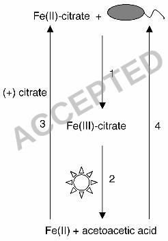
Figure 4. Model for Fe(II) oxidation leading to carbon acquisition. In the 20
presence of Fe(II)-citrate R. capsulatus SB1003 is able to oxidize Fe(II) leading 21
to the formation of Fe(III)-citrate (arrow 1). In the presence of light, Fe(III)-citrate 22
undergoes a photochemical reaction yielding Fe(II) and β-ketoglutarate and the 23
ACCEPTED

39
latter spontaneously decarboxylates into acetoacetic acid (arrow 2). As a result of 1
the photochemical reaction, the resulting Fe(II) can become bound by citrate 2
(arrow 3) and microbially re-oxidized. The acetoacetic acid that results from 3
these reactions can be used as a carbon source for growth (arrow 4). 4
ACCEPTED
40
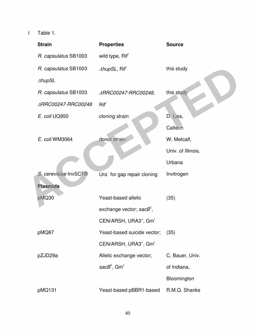
Table 1. 1
Strain Properties Source
R. capsulatus SB1003 wild type, Rifr
R. capsulatus SB1003
∆hupSL
∆hupSL, Rifr this study
R. capsulatus SB1003
∆RRC00247-RRC00248
∆RRC00247-RRC00248,
Rifr
this study
E. coli UQ950 cloning strain D. Lies,
Caltech
E. coli WM3064 donor strain W. Metcalf,
Univ. of Illinois.
Urbana
S. cerevisiae InvSC1® Ura− for gap repair cloning Invitrogen
Plasmids
pMQ30 Yeast-based allelic
exchange vector; sacB1,
CEN/ARSH, URA3+, Gmr
(35)
pMQ87 Yeast-based suicide vector;
CEN/ARSH, URA3+, Gmr
(35)
pZJD29a Allelic exchange vector;
sacB1, Gmr
C. Bauer, Univ.
of Indiana,
Bloomington
pMQ131 Yeast-based pBBR1-based R.M.Q. Shanks
ACCEPTED

41
shuttle vector; CEN/ARSH,
URA3+, Knr
and G.A.
O’Toole,
Dartmouth
University
(www.dartmout
h.edu/~gotoole
/vectors.html)
pNC001 hupSL deletion fragments
cloned into pMQ30
This study
pNC005 hupSL deletion fragments
cloned into pZJD29a
This study
pNC006 Yeast-based allelic
exchange vector; sacB 1,
CEN/ARSH, URA3+, Gmr
This study
pNC007 RRC00247-RRC00248
deletion fragment cloned
into pNC006
This study
pNC008 RRC00247-RRC00248
genes clones into pMQ131;
Knr
This study
Primers 5’ to 3’
pnc001 GTGGAATTGTGAGCGGATAACAATTTCACACAGGAAACAGCTCTGGGCGAGAACCTTTGG
ACCEPTED
42
pnc002 CCTTGACGGTGGTCAGGCAATTGTCCCTCCCTTGC
pnc003 GCAAGGGAGGGACAATTGCCTGACCACCGTCAAGG
pnc004 GGCAAATTCTGTTTTATC AGACCGCTTCTGCGTTCT GATCTCTTCCAGAAACGGCAGC
pnc017 GGCGGAGCTCCTGGGCGAGAACCTTTGG
pnc018 GGCGTCTAGACTCTTCCAGAAACGGCAGC
pnc021 ATGCCACGATCCTCGCCCTGCTGGCGAAGATCGACTCTAGCCACAGTCGATGAATCCAG
pnc022 ATCTCTAAGAAACCATTATTATCATGACATTAACCCGACAATTCGACCTGAAAATTCC
pnc023 GGCAAATTCTGTTTTATC AGACCGCTTCTGCGTTCT GATGAAGATCGACGAGGTTCTGG
pnc027 AGGTTCGAGATGATCCGGTTGAAACCCTCCCGTTACCTTG
pnc028 CAAGGTAACGGGAGGGTTTCAACCGGATCATCTCGAACCT
pnc026 TGGAATTGTGAGCGGATAACAATTTCACACAGGAAACAGCTCGAGACGGCT TTCCACAGG
pnc033 GGCGTCTAGAATGTGAAGCACAACAGCACCTACG
pnc034 GGCGGAGCTCATAATGGCGCAGGTTCTGCCAAAG
1
1The sacB in pMQ30 is under control of the native promoter and the sacB gene 2
in pZJD29a and pNC006 is under control of the R. capsulatus pucAB promoter. 3
4
ACCEPTED
ACCEPTED
ACCEPTED
ACCEPTED
ACCEPTED