2 5 7 8 9 10 11 brian jonesa , xiaotong pengb
TRANSCRIPT
1
1
2
3
4
5
6
7
Laminae development in opal-A precipitates associated with seasonal growth of the form-8
genus Calothrix (Cyanobacteria), Rehai geothermal area, Tengchong, Yunnan Province, 9
China 10
Brian JONESa , Xiaotong PENGb 11
a Department of Earth and Atmospheric Sciences, University of Alberta, Edmonton, Alberta, 12
T6G 2E3, Canada. 13
b Sanya Institute of Deep-Sea Science and Engineering, Chinese Academy of Science, Sanya, 14
572000, China. 15
Corresponding author. 16
E-mail address: [email protected] (B. Jones).17
2
ABSTRACT 18
The western discharge apron at Meinuquan (Rehai geothermal area, Yunnan Province, China), 19
which incorporates the upper terrace, terrace front, and lower terrace, is covered with laminated 20
opal-A precipitates that have formed from the spring waters that flow across its surface. 21
Laminae are formed of silicified Calothrix mats or featureless opal-A that contain no microbes, 22
scattered spherical and rod-shaped microbes, and/or rare Calothrix. Rapid silicification of the 23
Calothrix led to preservation of their basal heterocysts, vegetative cells, trichomes, tapering 24
filaments, and laminated and splayed sheaths. 25
The Calothrix mats grew during the dry season when there was maximum sunlight because 26
of low cloud cover. During this time, the mats grew under stable conditions because the water 27
that flowed across the discharge apron was sourced from the springs, and temperature and water 28
geochemistry was more or less constant. Growth of the Calothrix mats decreased during the wet 29
season (April to late September) when sunlight is reduced due to the extensive cloud cover 30
associated with the monsoonal rains. During the wet season, water flowing over the discharge 31
apron is a mixture of rainwater, runoff from the surrounding hillsides, and spring water. Such 32
variable flow conditions, water temperatures, and water geochemistry curtailed microbe growth 33
and impacted silica precipitation. 34
The precipitates at Meinuquan are like those associated with some Icelandic hot springs. 35
Although growth of Calothrix is controlled by sunlight in both settings, the periods of maximum 36
sunlight in China (October-March) and Iceland (June-August) are at different times of the year 37
because of their geographic locations. 38
Keywords: Opal-A, Calothrix, hot springs, microbe silicification, seasonal laminae.39
3
1. Introduction 40
The form-genus Calothrix, first described and defined by Agardh (1824), is a common 41
filamentous cyanobacterium found in modern spring systems throughout the world, including 42
those in Yellowstone National Park (Weed, 1889; Tilden, 1897, 1898; Copeland, 1936; Norris 43
and Castenholz, 2005), Iceland (Konhauser et al., 2001), New Zealand (Cassie, 1989), India 44
(Roy et al., 2014), and Bulgaria (Lukavský et al., 2011). Although some species of Calothrix 45
can survive in water temperatures up to 52-54°C (Castenholz, 1969, his Table 3; Colwell and 46
Fuentes, 1975, their Fig. 2), most thrive where the water temperatures are in the 20-40°C range 47
(Copeland, 1936; Nash, 1938; Walter, 1976; Cady and Farmer, 1996; Walter et al., 1996). Many 48
other environmental factors also influence the growth and development of Calothrix, including 49
UV radiation (Brenowitz and Castenholz, 1997; Dillon and Castenholz, 2003; Dillon et al., 2003; 50
Norris and Castenholz, 2005). Calothrix has commonly been used to assess microbe 51
silicification because naturally silicified specimens are abundant (Hugo et al., 2011) and this 52
cyanobacterium is susceptible to silicification under controlled laboratory conditions (Phoenix et 53
al., 2000, 2002; Yee et al., 2003; Benning et al., 2004, 2005). 54
This study focuses on laminated opal-A deposits that cover a hot-spring Meinuquan 55
(Beauty Pond) discharge apron that is located in the Rehai geothermal area, which is situated ~13 56
km southwest of Tengchong in the Yunnan Province of China (Fig. 1). The stratigraphic 57
architecture of these opal-A deposits is fundamentally control by the silicification of the 58
Calothrix mats that thrived on this discharge apron. Using these samples, this paper focuses on 59
(1) preservational aspects of Calothrix from different parts of the discharge apron, (2) the 60
significance of the pigmentation that is evident in the silicified sheaths of some of the Calothrix, 61
and (3) interpretation of the cyclic alternation between laminae formed of silicified Calothrix and 62

4
laminae devoid of Calothrix. Through careful examination of the textures in the siliceous 63
sinters, this research shows that the dry season, which is characterized by low rainfall and low 64
cloud cover but many hours of sunshine, encouraged growth of the Calothrix mats whereas the 65
onset of heavy rain and reduced hours of sunlight in the wet season led to the death of the 66
Calothrix mats. 67
2. General setting 68
2.1. Geological setting 69
The Rehai Geothermal Field (Fig. 1B), characterized by numerous active springs with 70
highly variable water temperatures, pH values, compositions (Table 1), and diverse arrays of 71
microbes (Lin et al., 2002, 2005; Guo et al., 2003; He et al., 2004; Chen et al., 2008; Ding et al., 72
2008; Jiang et al., 2009; Lu et al., 2009; Song et al., 2009, 2010; Han et al., 2010; Hong et al., 73
2010; Hedlund et al., 2012; Briggs et al., 2014), is centered on the Ruidian-Tengchong Fault. 74
The geothermal waters, which are probably of meteoric origin, are heated in the subsurface by 75
magma (Zhao et al., 1996, their Fig. 3; Du et al., 2005; Shangguan et al., 2005) or the 76
Yanshanian granite (Liao et al., 1991; Yan and Wan, 1998). The Tengchong volcanic field is 77
located at the east end of the Xizang (Tibet) – Yunnan geothermal zone (Tong and Zhang, 1989; 78
Kearey and Wei, 1993) near the border between China and Myanmar (Fig. 1A). Numerous 79
volcanoes and extensive faulting characterize this area (Jiang, 1998; Jiang et al., 1998; Du et al., 80
2005; Wang et al., 2006), which formed when the Burmese Block was thrust under the 81
Tengchong Microplate during the Cenozoic (Shangguan et al., 2005). Earthquakes are still 82
common in the area today. 83
5
2.1. Climate 84
Detailed climate data are not available specifically for the Rehai geothermal area. Thus, the 85
climate records for Tengchong county as provided by the China Meteorological Data Sharing 86
System (http://cdc.cma.gov.cn) are used in this study. 87
This part of the Yunnan Province enjoys a highland subtropical climate with an average 88
rainfall of 1480 mm/year and an average air temperature of 14.9°C (based on 1971-2000 period). 89
The climate records between 2000 and 2013 are characterized by the following annual patterns. 90
• Annual variations in temperature that range from 1 to 17°C in December-January to 17-91
24°C in July, August, and September (Fig. 2A). 92
• Monthly rainfall that varies form 0 mm in January to as high as 375 mm in July (Fig. 2B). 93
• The maximum hours of sunshine is in the dry season (250-280 hours/month from October 94
to April) when there is little cloud cover whereas the minimum hours of sunshine (less than 95
100 hours/month) is in the wet season (May to September) when there is maximum cloud 96
cover because of the monsoonal rains (Fig. 2C). 97
Collectively, the temperature, rainfall, and hours of sunshine divided each year into the dry 98
season (low T, low rainfall, high sunshine) and the wet season (high T, high rainfall, low 99
sunshine). The wet season typically lasts from May to late September with the dry season 100
extending from October to April. 101
3. Methods 102
Examination of the Meinuquan (Fig. 1C) complex took place in 2011 and 2013 when the 103
discharge apron and nearby springs were examined, described, photographed, and water 104
temperatures and pH measured. Samples of opal-A precipitates and water were collected in 105
April, 2013. Water samples were passed through a syringe filter with a 0.22 µm filtration 106
6
membrane before being stored in polypropylene bottles until analysis for major cations and 107
anions at the Saskatchewan Research Council (Canada), about 4 weeks after they had been 108
collected. The elements Ca, Mg, Na, K, Si and S were determined by Inductively Coupled 109
Plasma Atomic Emission Spectroscopy (ICP-AES) and alkalinity (including p alkalinity) was 110
determined by titration with sulphuric acid on an auto-titration system. The bicarbonate, 111
carbonate and hydroxides were calculated from the pH and alkalinity results. The chloride was 112
measured colorimetrically and fluoride was determined by ion selective electrode. 113
Samples of the precipitates were collected (with permission) where possible. Given that 114
this is a major tourist attraction, sampling was done carefully so that little or no visible damage 115
was done. In the Meinuquan complex, for example, sampling was restricted to the western 116
discharge apron (Fig. 3) where short (up to 2 cm long) cores (1.5 cm and 3.0 cm diameter) were 117
obtained from the upper terrace, the terrace front, and the lower terrace. Where possible, small 118
hand samples were extracted. 119
Thermal images of the surfaces on the discharge apron were taken using a Fluke Ti 100 120
Thermal Imager, which measures temperatures from -20 to 250°C with a measurement accuracy 121
of 2%. 122
Six large (3 x 2 cm) and two small (4.5 x 2 cm) thin sections, each impregnated with blue 123
epoxy, were made from the available samples so that the fabrics of these precipitates could be 124
established with particular emphasis being placed on the lamination styles. 125
Small fracture samples, broken from the cores and hand samples, were mounted on 126
scanning electron microscope (SEM) stubs using conductive glue and then sputter coated with 127
thin layer of carbon so that they could be examined on a JOEL 6400FE scanning electron 128
microscope. Imaging was done with an accelerating voltage of 5 kV whereas energy-dispersive 129
7
X-ray (EDX) analyses and back scattered electron imaging (BSEI) were done with an 130
accelerating voltage of 20 kV. The location and orientation of all samples was recorded so that 131
the different fabrics could be related to each other. The 436 SEM photomicrographs formed an 132
integral part of this study. 133
4. The Meinuquan complex 134
The Meinuquan (Beauty Pond) complex consists of a large, triangular shaped, varicoloured 135
discharge apron that is bounded by a wall (up to 6 m high) on its north side and footpaths along 136
its west and south margins (Fig. 1C). Most of the water that flows over this apron comes from 137
Yanjiangquan (Figs. 1C, 3A), Zhenhuquan (Figs. 1C, 3A), and Gumingquan (Figs. 1C, 3B) 138
springs, which are located on the north side of the footpath that is located on top of the wall that 139
defines the northern boundary of the discharge apron (Figs. 1C, 3A). All of these springs have, 140
to some extent, been anthropogenically modified. 141
The Meinuquan discharge apron is herein divided into the “eastern discharge apron” and 142
the “western discharge apron” (Figs. 1C, 3A). The eastern discharge apron is formed of a raised, 143
sloping bench that is bounded to the north by a vegetated area and the north wall and to the south 144
by a narrow terrace (Fig. 3B). Along the south edge of the terrace, there is a steep drop-off to the 145
pathway that is located below (Fig. 3B). Most of the water on this part of the system comes from 146
Gumingquan (Drum Beating Spring), which discharges water with a T of 87°C, pH of 8.8, and a 147
flow rate of 1.19 L/sec (Fig. 1C). 148
The “western discharge apron”, which is ~ 14 m long (parallel to flow direction), ~ 12 m 149
wide, and 5 m high, is divided into (1) the upper terrace that is centered around the top pool and 150
has a low downslope gradient, (2) the terrace front where there is steep drop-off from the upper 151
terrace, and (3) the lower terrace with a low downslope gradient that stretches from the base of 152
8
the terrace front to the pathway (Fig. 3A). The water that flows across this discharge apron 153
comes from Yanjiangquan and Zhenhuquan (Figs. 1C, 3A). Yanjiangquan (Sisters Spring) 154
comprises the Young Sister that discharges water at ~86°C, pH of 9.0, and a flow rate of 0.3 155
L/sec and the Old Sister that discharges water at 91°C, pH of 8.9, with a flow rate of 0.2 L/sec. 156
Zhenhuquan (Pearl Spring) discharges water with a T of 91°C, pH of 3.8, and a flow rate of 0.2 157
L/sec. The water from each of these springs discharges into small channels located on the north 158
side of the footpath that is located at top of the wall that forms the northern margin of the 159
Meinuquan complex. That water then flows through a pipe under the footpath and cascades 160
down the wall into a shallow pool at the foot of the wall (Fig. 3A). The water in that pool has a 161
temperature of 66° and pH of 9.1 (Fig. 1C). From there, the water disperses down the discharge 162
apron. By the time it has reaches the channel at the bottom of the apron, the water has a T of 163
34°C and pH of 9.5 (Fig. 1C). 164
4.1. Water flow on western discharge apron 165
Water flow from the top pool is generally low because of the low combined volume (~ 42 166
L/min) of water that comes from Yanjiangquan and Zhenhuquan. This flow is focused largely 167
into shallow, narrow channels that radiate downslope (Fig. 3A). Although the water in the 168
channels may be up to 5 mm deep, it is typically no more than a thin film. Areas between these 169
channels are dry or damp. The pattern of water dispersal across the discharge apron is 170
highlighted by the colorful microbial mats that preferentially develop in the channels and along 171
the margins of the channels (Fig. 3A). 172
The water in the top pool has a temperature of 66°C, whereas water in channels on the 173
lower terrace has a temperature of 34°C. Thermal imaging shows that the laterally and vertically 174
complex temperature gradients on the discharge apron are centered on the channels with flowing 175
9
water (Fig. 4A-F). Thus, areas on the terrace front covered with black microbial mats are usually 176
the “hot” areas with temperatures in the 40 to 50°C range, whereas areas devoid of microbial 177
mats are typically “cold” with temperatures in the 20 to 25°C range (Fig. 4A-F). Areas between 178
the “hot” and “cold” zones have transitional temperatures that are typically about 35°C (Fig. 4D). 179
In many of the “warm” and “hot” areas there is no obvious running water and it is only with 180
careful inspection that it becomes apparent that these areas are either damp or covered by a thin 181
film of flowing water. In these areas it is impossible to measure the water temperature with a 182
conventional thermometer. 183
On days when there is heavy rainfall, the entire surface of the discharge apron becomes 184
soaked as the rainwater flows downslope, and runoff from the steep slopes around Meinuquan 185
flows downslope. Mixing of the rainwater with the spring water leads to dilution of the spring 186
water and reduction in its temperature and pH. During periods of heavy rainfall, the combined 187
volume of rainwater and runoff may exceed the volume of spring water that is fed onto its 188
surface. During the dry season, conditions on the Meinuquan discharge apron are relatively 189
stable because virtually all of the water comes from the springs. During the wet season, 190
however, conditions are highly variable with water temperature and geochemistry varying as 191
rainwater and runoff mix with the spring discharge. 192
4.2. Surface deposits on the western discharge apron 193
The upper terrace is covered with white, laminated opal-A deposits that have a smooth 194
surface (Fig. 5A). As the gradient becomes steeper, shallow rimstone pools develop. The 195
terrace front is characterized by numerous microgours (Fig. 5B) that are morphologically akin to 196
microgours found on the terrace fronts on Waikite Geyser in New Zealand (Jones et al., 2011, 197
their Fig. 9E). These semi-circular microgours, with raised outer rims, are up to 3 cm long 198
10
(parallel to terrace front) and up to 1 cm wide (90° to terrace front). Neighbouring microgours 199
commonly merged to form larger structures (Fig. 3B). After heavy rain, water fills the small 200
pools that commonly appear to have minor amounts of sediment on their floors. 201
The wet parts of the terrace front, located around the narrow streams of flowing water, are 202
typically covered by green to black microbial mats that mask the underlying microgours (Fig. 203
5C-E). In areas with the highest water flow, filamentous microbes, up to 2 cm long, are 204
highlighted by their coating of white opal-A that contrasts sharply with the green to black 205
microbial mats in the background (Fig. 5D, E). 206
The surface of the discharge apron, especially in the marginal areas, is commonly covered 207
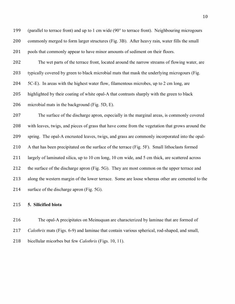
with leaves, twigs, and pieces of grass that have come from the vegetation that grows around the 208
spring. The opal-A encrusted leaves, twigs, and grass are commonly incorporated into the opal-209
A that has been precipitated on the surface of the terrace (Fig. 5F). Small lithoclasts formed 210
largely of laminated silica, up to 10 cm long, 10 cm wide, and 5 cm thick, are scattered across 211
the surface of the discharge apron (Fig. 5G). They are most common on the upper terrace and 212
along the western margin of the lower terrace. Some are loose whereas other are cemented to the 213
surface of the discharge apron (Fig. 5G). 214
5. Silicified biota 215
The opal-A precipitates on Meinuquan are characterized by laminae that are formed of 216
Calothrix mats (Figs. 6-9) and laminae that contain various spherical, rod-shaped, and small, 217
bicellular micorbes but few Calothrix (Figs. 10, 11). 218
11
5.1. Calothrix – upper terrace 219
On the upper terrace, Calothrix grow in tufts that are formed of numerous erect filaments 220
(Figs. 6A, 7A, B). No pigmentation (Fig. 6A, 7A-C) is associated with these filaments that are 221
(1) characterized by basal heterocysts that are 3.2 to 6.0 µm (average 4.3 µm) in diameter and 2.7 222
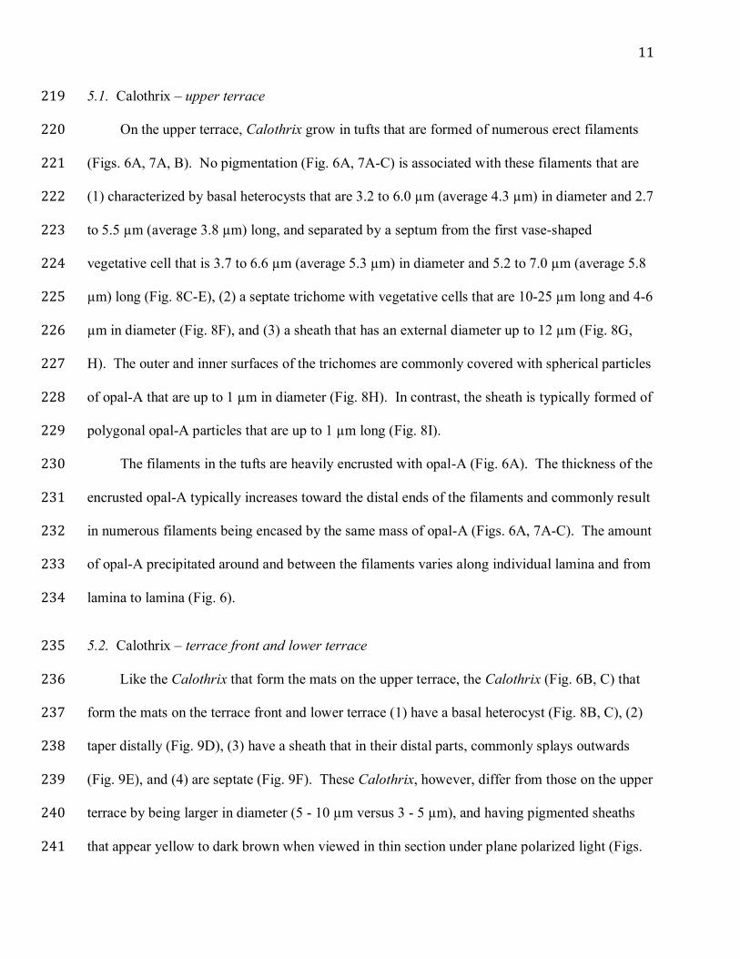
to 5.5 µm (average 3.8 µm) long, and separated by a septum from the first vase-shaped 223
vegetative cell that is 3.7 to 6.6 µm (average 5.3 µm) in diameter and 5.2 to 7.0 µm (average 5.8 224
µm) long (Fig. 8C-E), (2) a septate trichome with vegetative cells that are 10-25 µm long and 4-6 225
µm in diameter (Fig. 8F), and (3) a sheath that has an external diameter up to 12 µm (Fig. 8G, 226
H). The outer and inner surfaces of the trichomes are commonly covered with spherical particles 227
of opal-A that are up to 1 µm in diameter (Fig. 8H). In contrast, the sheath is typically formed of 228
polygonal opal-A particles that are up to 1 µm long (Fig. 8I). 229
The filaments in the tufts are heavily encrusted with opal-A (Fig. 6A). The thickness of the 230
encrusted opal-A typically increases toward the distal ends of the filaments and commonly result 231
in numerous filaments being encased by the same mass of opal-A (Figs. 6A, 7A-C). The amount 232
of opal-A precipitated around and between the filaments varies along individual lamina and from 233
lamina to lamina (Fig. 6). 234
5.2. Calothrix – terrace front and lower terrace 235
Like the Calothrix that form the mats on the upper terrace, the Calothrix (Fig. 6B, C) that 236
form the mats on the terrace front and lower terrace (1) have a basal heterocyst (Fig. 8B, C), (2) 237
taper distally (Fig. 9D), (3) have a sheath that in their distal parts, commonly splays outwards 238
(Fig. 9E), and (4) are septate (Fig. 9F). These Calothrix, however, differ from those on the upper 239
terrace by being larger in diameter (5 - 10 µm versus 3 - 5 µm), and having pigmented sheaths 240
that appear yellow to dark brown when viewed in thin section under plane polarized light (Figs. 241
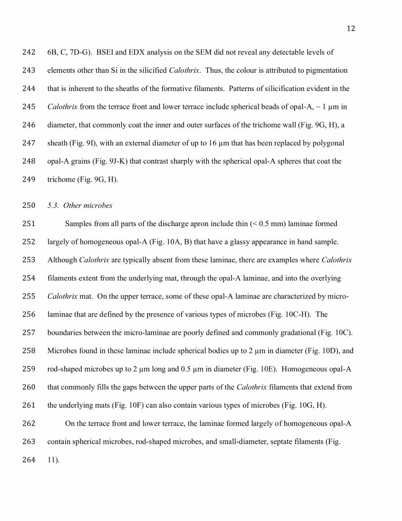
12
6B, C, 7D-G). BSEI and EDX analysis on the SEM did not reveal any detectable levels of 242
elements other than Si in the silicified Calothrix. Thus, the colour is attributed to pigmentation 243
that is inherent to the sheaths of the formative filaments. Patterns of silicification evident in the 244
Calothrix from the terrace front and lower terrace include spherical beads of opal-A, ~ 1 µm in 245
diameter, that commonly coat the inner and outer surfaces of the trichome wall (Fig. 9G, H), a 246
sheath (Fig. 9I), with an external diameter of up to 16 µm that has been replaced by polygonal 247
opal-A grains (Fig. 9J-K) that contrast sharply with the spherical opal-A spheres that coat the 248
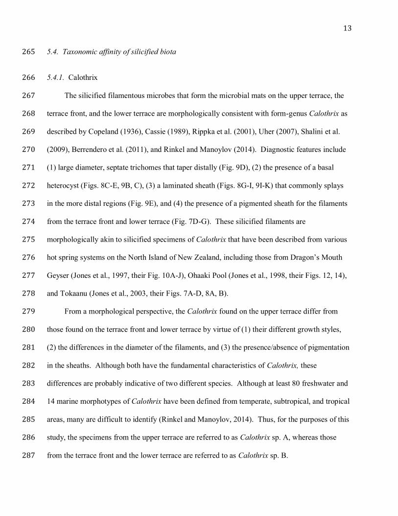
trichome (Fig. 9G, H). 249
5.3. Other microbes 250
Samples from all parts of the discharge apron include thin (< 0.5 mm) laminae formed 251
largely of homogeneous opal-A (Fig. 10A, B) that have a glassy appearance in hand sample. 252
Although Calothrix are typically absent from these laminae, there are examples where Calothrix 253
filaments extent from the underlying mat, through the opal-A laminae, and into the overlying 254
Calothrix mat. On the upper terrace, some of these opal-A laminae are characterized by micro-255
laminae that are defined by the presence of various types of microbes (Fig. 10C-H). The 256
boundaries between the micro-laminae are poorly defined and commonly gradational (Fig. 10C). 257
Microbes found in these laminae include spherical bodies up to 2 µm in diameter (Fig. 10D), and 258
rod-shaped microbes up to 2 µm long and 0.5 µm in diameter (Fig. 10E). Homogeneous opal-A 259
that commonly fills the gaps between the upper parts of the Calothrix filaments that extend from 260
the underlying mats (Fig. 10F) can also contain various types of microbes (Fig. 10G, H). 261
On the terrace front and lower terrace, the laminae formed largely of homogeneous opal-A 262
contain spherical microbes, rod-shaped microbes, and small-diameter, septate filaments (Fig. 263
11). 264
13
5.4. Taxonomic affinity of silicified biota 265
5.4.1. Calothrix 266
The silicified filamentous microbes that form the microbial mats on the upper terrace, the 267
terrace front, and the lower terrace are morphologically consistent with form-genus Calothrix as 268
described by Copeland (1936), Cassie (1989), Rippka et al. (2001), Uher (2007), Shalini et al. 269
(2009), Berrendero et al. (2011), and Rinkel and Manoylov (2014). Diagnostic features include 270
(1) large diameter, septate trichomes that taper distally (Fig. 9D), (2) the presence of a basal 271
heterocyst (Figs. 8C-E, 9B, C), (3) a laminated sheath (Figs. 8G-I, 9I-K) that commonly splays 272
in the more distal regions (Fig. 9E), and (4) the presence of a pigmented sheath for the filaments 273
from the terrace front and lower terrace (Fig. 7D-G). These silicified filaments are 274
morphologically akin to silicified specimens of Calothrix that have been described from various 275
hot spring systems on the North Island of New Zealand, including those from Dragon’s Mouth 276
Geyser (Jones et al., 1997, their Fig. 10A-J), Ohaaki Pool (Jones et al., 1998, their Figs. 12, 14), 277
and Tokaanu (Jones et al., 2003, their Figs. 7A-D, 8A, B). 278
From a morphological perspective, the Calothrix found on the upper terrace differ from 279
those found on the terrace front and lower terrace by virtue of (1) their different growth styles, 280
(2) the differences in the diameter of the filaments, and (3) the presence/absence of pigmentation 281
in the sheaths. Although both have the fundamental characteristics of Calothrix, these 282
differences are probably indicative of two different species. Although at least 80 freshwater and 283
14 marine morphotypes of Calothrix have been defined from temperate, subtropical, and tropical 284
areas, many are difficult to identify (Rinkel and Manoylov, 2014). Thus, for the purposes of this 285
study, the specimens from the upper terrace are referred to as Calothrix sp. A, whereas those 286
from the terrace front and the lower terrace are referred to as Calothrix sp. B. 287
14
5.4.2. Other microbes 288
The microbes found in the opal-A precipitates from Meinuquan can only be characterized 289
in terms of their shape, size, and for some specimens the presence of septa (Figs. 10, 11). The 290
lack of diagnostic morphological features precludes identification. 291
6. Laminations 292
All of the opal-A precipitates on the Meinuquan discharge apron are laminated, with the 293
laminae being highlighted by variations in colour, texture, and porosity (Figs. 6, 7D-G). The 294
laminae found on the upper terrace are subtly different from those found on the terrace front and 295
lower terrace. 296
Silicified Calothrix mats up to 4 mm thick dominate the precipitates that are found around 297
the pool on the upper terrace (Figs. 6A, 10A). These silicified mats are either stacked one on top 298
of the other or separated by laminae, typically < 1 mm thick, that are formed of dense, largely 299
featureless opal-A (Fig. 6A). The variable appearance of the silicified Calothrix mats in hand 300
sample and thin section is largely a function of the amount of opal-A that was precipitated 301
around and between the filaments. Thus, areas with little opal-A encrustation are far more 302
friable than those parts of the mats where opal-A encrustation around the filaments was extensive 303
(Fig. 6A). Sharp, well-defined bases but diffuse, irregular upper boundaries characterize all of 304
the laminae formed by the Calothrix mats (Fig. 9A). 305
On the terrace front and lower terrace, the opal-A precipitates are formed of alternating 306
Calothrix mats and layers of homogeneous, glass-like opal-A. The deposits on these parts of the 307
discharge apron are much harder than the precipitates found on the upper terrace. The silicified 308
Calothrix mats on the terrace front and lower terrace differ from those on the upper terrace 309
because (1) they are accentuated by the yellowish-brown pigmentation of the Calothrix sheaths 310
15
(Figs. 6B,C, 7D-G), (2) the growth patterns of the Calothrix are different, and (3) the patterns of 311
opal-A precipitation around those filaments are also different (Fig. 6). In contrast, the laminae 312
formed of homogeneous, glass-like opal-A with scattered non-filamentous microbes are the same 313
over the entire extent of the discharge apron (Figs. 10, 11). On the lower terrace, the uppermost 314
parts of these laminae, just beneath the base of the filamentous microbial mats, are commonly 315
characterized by small (< 0.15 mm long), subangular to angular grains that are formed of opal-A, 316
K-feldspar, and quartz (Fig. 6G). 317
7. Discussion 318
Laboratory experiments designed to examine the factors that control microbial silicification 319
commonly use Calothrix because of its apparent susceptibility to silicification (e.g., Phoenix et 320
al., 2000, 2002; Yee et al., 2003). Based on experiments involving Calothrix collected from 321
Krusivik hot spring (Iceland), Phoenix et al. (2000) showed that (1) filaments became covered 322
with a mineral crust, up to 5 µm thick, after only 12 days in a silica solution, (2) mineralization 323
was restricted to extracellular material such as the sheath, and (3) the sheath allowed the 324
microbes to survive because it provided sites for mineralization and acted as a filter against 325
colloidal silica. Later experiments with the same strain of Calothrix led to the conclusion that 326
this microbe was characterized by a highly reactive cell wall but a poorly reactive sheath 327
(Phoenix et al., 2002). Further experiments with the same strain of Calothrix led Yee et al. 328
(2003) to postulate that silica precipitation was largely abiogenic. Benning et al. (2005), 329
however, argued that the single-step batch experiments used by Phoenix et al. (2000, 2002) and 330
Yee et al. (2003) did not accurately reflect conditions in hot spring systems. They noted that 331
other experiments that used organosilicon solvents or inorganic silica concentrations showed that 332
microbial silicification depended on many different complex interactions (Ferris et al., 1988; 333
16
Westall et al., 1995; Konhauser et al., 2001; Toporski et al., 2002; Mountain et al., 2003). Hugo 334
et al. (2011), based on samples collected from springs in Yellowstone National Park, suggested 335
that early silicification of Calothrix was focused entirely in the sheath and argued that the 336
microbes were more actively involved with silica precipitation than previously thought. 337
Irrespective of the nuances involved, silicification must take place because the (1) microbes 338
cannot prevent it, or (2) silica coating is, in some way, advantageous to the organism (Phoenix et 339
al., 2000). 340
Silicified Calothrix have been reported from spring systems throughout the world, 341
including those in Yellowstone National Park, U.S.A. (Cady and Farmer, 1996; Hugo et al., 342
2011), New Zealand (Jones et al., 1997, 1998, 2001a, b, 2003; Jones and Renaut, 2003), and 343
Iceland (Konhauser et al., 2001). Rapid silicification seems to be the norm with Calothrix 344
filaments commonly being partly silicified while they are still alive (Jones et al., 1998, their Fig. 345
15). Silicified Calothrix from New Zealand, for example, are typically well preserved with 346
distally tapering septate filaments encased by laminated and splayed sheaths (e.g., Jones et al., 347
2001a, their Fig. 6G; 2003, their Fig. 7C). In addition to these features, silicified Calothrix from 348
Meinuquan also display well-preserved basal heterocysts (Figs. 8C-E, 9B, C), vegetative cells 349
(Figs. 8E, F, 9C, F), and trichomes (Figs. 8D, F, H, 9F, G, I). The fact that these soft-tissue 350
components show little evidence of shrinkage or desiccation implies that silicification was rapid 351
and took place before decay and distortion of the soft tissues started. Silicification of these 352
elements involved opal-A spheres that are up to 1 µm but more commonly < 500 nm in diameter 353
(Figs. 8E, H, 9G, H). The sheaths must have also undergone rapid silicification because laminae 354
(Figs. 8H-I, 9I, K, L) and splaying (Fig. 9D, E) are apparent in the sheaths, and pigmentation of 355
the sheath is still evident in Calothrix sp. B (Fig. 7D-G). Silicification of the sheaths, however, 356
17
involved the development of polygonal-shaped opal-A particles, up to 1 µm long (Figs. 8I, 9L) 357
that contrast sharply with the spherical opal-A particles evident in the silicified cells and 358
trichome walls (compare Fig. 9L with 9H). Such polygonal-shaped opal-A particles are not 359
unique to the Chinese specimens because they are also evident in silicified Calothrix from New 360
Zealand (Jones et al., 1997, their Fig. 10G, J; Jones and Renaut, 2003, their Fig. 6G). The reason 361
for this contrasting style of opal-A particles is not known. These inferences regarding the 362
rapidity of silicification of Calothrix are consistent with conclusions that Bartley (1996) 363
proposed based on the experimental silicification of various types of microbes. 364
The pigmentation of Calothrix sp. B on the Meinuquan discharge apron is similar to that 365
associated with pigmented sheaths of extant Calothrix, which is generally attributed to the 366
presence of scytonemin (Brenowitz and Castenholz, 1997; Dillon and Castenholz, 2003; Dillon 367
et al., 2003; Norris and Castenholz, 2005). Variations in the pigmentation colour depends on the 368
amount of scytonemin in the sheaths even among populations that are, according to their 16s 369
rDNA, closely related (Dillon and Castenholz, 2003; Dillon et al., 2003). Although the exact 370
cause of this variation is not known, it has generally been attributed to environmental factors 371
(Dillon and Castenholz, 2003; Norris and Castenholz, 2005). Scytonemin, which is a stable 372
molecule that is not actively degraded by cyanobacteria (Garcia-Pichel and Castenholz, 1991; 373
Norris and Castenholz, 2005), is important because it acts as a barrier against UV radiation 374
(Garcia-Pichel and Castenholz, 1991; Dillon and Castenholz, 1999, 2003; Dillon et al., 2003; 375
Norris and Castenholz, 2005). The pigmentation in the sheaths of Calothrix sp. B from 376
Meiuquan accentuates the laminae that are clearly evident in hand samples (Fig. 5H) and thin 377
section (Fig. 6D-G). In contrast to Calothrix sp. B, no pigmentation is evident in the sheaths of 378
Calothrix sp. A (Fig. 6A-C) from Meinuquan and there is less color differential between the 379
18
constituent laminae (Fig. 5G). The lack of pigmentation in the sheaths of Calothrix sp. A may be 380
due to scytonemin being absent or present only in very low concentrations. 381
Precipitates found on the discharge aprons of hot springs, irrespective of their composition, 382
are commonly characterized by layering that is highlighted by variations in colour, composition, 383
and/or fabric (e.g., Walter et al., 1972; Jones et al., 1997; Kano et al., 2003; Okumura et al., 384
2011, 2013). Many of these successions are characterized of recurring “couplets” (paired 385
laminae with different fabrics) that have typically been linked to cyclic variations in the local 386
climate that operate on diurnal, seasonal, and/or annual time scales (Symoens, 1957; Monty, 387
1967; Walter et al., 1972; Doemel and Brock, 1974, 1977; Monty, 1976; Park, 1976; Golubic 388
and Focke, 1978; Chafetz and Folk, 1984; Chafetz et al., 1991; Casanova, 1994; Freytet and Plet, 389
1996; Renaut et al., 1996; Jones et al., 1998, 1999; Konhauser et al., 2001; Kano et al., 2003; 390
Berelson et al., 2011; Petryshyn et al., 2012) and/or seasonal variations in the composition of the 391
microbial communities that inhabit these systems (Norris et al., 2002; Lacap et al., 2007; 392
MacKenzie et al., 2013; Briggs et al., 2014). In such complicated systems it is perhaps not 393
surprising that the linkage between laminae cyclicity and specific aspects of the depositional 394
environments is difficult to identify, even when careful monitoring is employed in modern, 395
active environments. Berelson et al. (2011), for example, showed that siliceous stromatolites 396
from Obsidian Pool in Yellowstone National Park included 80 couplets (light lamina formed of 397
erect filaments alternating with dark lamina formed of reclining silicified bacteria) that formed 398
over a period of 141 days for an average of 1.75 couplets per day. They argued that this average 399
number probably reflects the fact that there might have been days when the diurnal contrasts in 400
factors, such as temperature, were insufficient to trigger a change in the fabrics of the 401
precipitates. 402
19
Laminated precipitates found on the western discharge apron of the Meinuquan complex 403
primarily reflect the growth cycles of the Calothrix microbial mats, whereby conditions 404
favourable for their growth were periodically interrupted by periods when their growth ceased. 405
Calothrix is a common inhabitant of those parts of hot spring systems where the water 406
temperatures are in the 20-40°C range (Copeland, 1936; Nash, 1938; Walter, 1976; Cady and 407
Farmer, 1996; Walter et al., 1996). Sinters from Krisuvik hot spring in Iceland are characterized 408
by layers formed mainly of intact, vertically aligned silicified cyanobacteria (mostly Calothrix) 409
that have a sharp base and gradational top that alternate with layers of opal-A that are devoid of 410
microbes (Konhauser et al., 2001). Konhauser et al. (2001) argued that the alternating laminae 411
must reflect the growth and activity of the microbes because the spring waters that flow across 412
the discharge apron have a more or less constant temperature throughout the year. Thus, it was 413
suggested that maximum growth of the Calothrix took place during the spring and summer when 414
there is almost continuous daylight (~ 20 hours per day in June) given that Iceland lies close to 415
the Arctic Circle. In contrast, during the winter month, growth of the microbial mats ceased 416
because the number of hours of daylight is severely reduced (4 to 7 hours in January). Thus, 417
development of the Calothrix mats was linked directly to the hours of sunlight that varied 418
between different seasons. 419
The Meinuquan discharge apron, like Krisuvik, experiences seasonal variations in climate. 420
In the Tengchong area, low air temperatures characterize the dry winter months even though the 421
number of hours of sunlight is high because cloud cover is minimal (Fig. 2). During the wet 422
season, the air temperatures are higher but the number of hours of sunlight is low because of the 423
increased cloud cover associated with the monsoonal rains (Fig. 2). Under similar climate 424
conditions, Lacap et al. (2007) found that floating microbial mats in tropical geothermal spring 425
20
pools in the Philippines became established and grew thicker during the dry season between 426
January to April. With the onset of heavy rains in July those mats were physically damaged and 427
the biomass decreased. For the high temperature springs in the Rehai geothermal area, Briggs et 428
al. (2014) found that the spring waters had higher concentrations of K, Ca, ammonia, Na, N, 429
DOC, and 18O in June than they did in January. They argued that these changes were related to 430
differences in the run-off from the surrounding area and/or the shallow recharge of the area, both 431
of which are related to rainfall. Analyses of the high temperature springs (excluding Meinuquan) 432
showed that the microbial biotas sampled in June contained more non-thermophilic microbes that 433
samples collected in January (Briggs et al., 2014). 434
Growth of the Calothrix-dominated mats on the Meinuquan discharge apron is controlled 435
by the interaction between the spring waters that flow over its surface and seasonal variations in 436
the hours of sunshine and rainfall. During the dry season (October to April), rainfall is minimal 437
(Fig. 2) and water flow over the discharge apron is sourced mainly from the springs. During 438
those times, growth of the microbial mats and silica precipitation is controlled largely by water 439
temperature and the geochemistry of the spring waters. Given the low volumes of spring waters 440
that disperse across the discharge apron, growth of the Calothrix-dominated mats is patchy, 441
being limited to those areas where suitable temperature regimes exist in and around the channels 442
that funnel the spring water downslope (Fig. 4). These shallow channels are prone to frequent 443
temporal changes in direction as opal-A precipitation commonly leads to the formation of dams 444
across the channels that impeded downslope flow. During the wet season, two important 445
changes take place, namely: (1) the composition of the water flowing over the discharge apron 446
becomes more variable, ranging from just spring water on rain-free days to waters that are a 447
mixture of rain, run-off, and spring water on wet days, and (2) on wet days water will flow over 448
21
the entire surface of the discharge apron and will not be confined to the shallow channels that 449
funnel the spring water downslope on dry days. Given that Meinuquan is located on a steep 450
valley side, run-off can be high. Heavy rain and run-off leads to (1) the entire discharge apron 451
being kept wet, (2) considerable volumes of non-spring water flowing over the discharge apron, 452
and (3) cooling and dilution of the spring waters as they mix with the rainwater and runoff. Such 453
fluctuating conditions would probably be detrimental to growth of the Calothrix mats and 454
severely curtail precipitation of opal-A. 455
On Meinuquan, the cyclic alternation between silicified Calothrix mats and layers of opal-456
A with only a sparse microbially biota can be attributed to seasonal contrasts in the weather that 457
have a significant impact on the volume and geochemistry of the water that flows across the 458
discharge apron. Maximum growth of the Calothrix mats on the Meinuquan complex probably 459
takes place during the dry season when the number of hours of sunlight was at its maximum and 460
growth was associated with water that was sourced largely from the springs. Although this 461
conclusion is similar to that reached by Konhauser et al. (2001) for Krisuvik hot spring in 462
Iceland, it is important to note that the periods when sunlight is at a maximum is different in the 463
two areas. For Meinuquan, growth of the Calothrix mats took place during the dry season from 464
October to April when sunlight is at a maximum because of low cloud cover. In contrast, growth 465
of the Calothrix mats at Krisuvik takes place during the summer months (May to August) when 466
Iceland experiences almost continuous sunlight because of its proximity to the Arctic Circle. 467
Although sunlight is the environmental factor that promotes the growth of Calothrix in both 468
areas, the sunlight maxima in Tengchong and Krusivik occur at different times of the year 469
because they are related to different controlling factors. 470
8. Conclusions 471
22
Detailed examination of recent opal-A precipitates on the Meinuquan discharge apron has 472
led to the following important conclusions. 473
• The precipitates are formed of alternating silicified Calothrix mats and thin layers of opal-474
A that are generally devoid of Calothrix. 475
• Calothrix sp. A and sp. B are exceptionally well-preserved with basal heterocysts, distally 476
tapering filaments, laminated and splayed sheaths, silicified vegetative cells, and trichomes 477
being readily apparent. Such preservation indicates that rapid silicification took place 478
before the microbes underwent desiccation and decay. 479
• Pigmentation of the sheath, related to the presence of scytonemin, is evident in Calothrix 480
sp. B. This pigmentation, which provided Calothrix with UV protection, accentuates the 481
laminated appearance of the deposits. 482
• The laminae reflect seasonal climate controls with the total number of hours of sunlight 483
being the key factor. Sunlight irradiance is at a maximum during the dry season when 484
cloud cover is minimal. In contrast, during the wet season from April to September, 485
sunlight is reduced because cloud cover is extensive due to the monsoonal rains. 486
• During the dry season, the water that flows over the discharge apron is sourced largely 487
from the springs. During the wet season, water that flows over the discharge apron is more 488
variable because it is formed of rainwater, runoff, and spring waters. 489
• The alternating laminae in the opal-A deposits at Meinuquan are similar to those reported 490
from Krusivik hot spring in Iceland. Although the hours of sunlight seem to be responsible 491
in both settings, the actual timing differs for the two areas. On Iceland, maximum sunlight 492
occurs during the summer, whereas on Meinuquan, maximum sunlight occurs during the 493
winter dry season. 494

23
495
Acknowledgements 496
Samples used in this study were collected with the permission of Yunnan Tengchong Rehai 497
Tour Developing Co., Ltd., which is the administration section of the Rehai geothermal area. 498
Financial support for this research came from the Natural Sciences and Engineering Council of 499
Canada (to Jones), the National Natural Science Foundation of China (grants 41172309 and 500
41272370 to Peng) and the Frontier Project of the Chinese Academy of Science (SIDSSE1301 to 501
Peng). We are indebted to George Braybrook who took the SEM images used in this paper. 502
503
24
REFERENCES 504
Agardh, C.A., 1824. Systema Algarum. Litteris Berlingianis, Lund, Sweden. 505
Bartley, J.K., 1996. Actualistic taphonomy of cyanobacteria: implications for the Precambrian 506
fossil record. Palaios 11, 571-586. 507
Benning, L.G., Phoenix, V., Mountain, B.W., 2005. Biosilicification: the role of cyanobacteria in 508
silica sinter deposition. In: Gadd, M.G., Semple, T.K., Lappin-Scott, M.H. (Eds.), Micro-509
organisms and Earth Systems: Advances in Geomicrobiology. Society for General 510
Microbiology Symposium. Cambridge University Press, Cambridge, pp. 131-150. 511
Benning, L.G., Phoenix, V.R., Yee, N., Konhauser, K.O., 2004. The dynamics of cyanobacterial 512
silicification: an infrared micro-spectroscopic investigation. Geochemica et Cosmochemica 513
Acta 68, 743-757. 514
Berelson, W.M., Corsetti, F.A., Pepe-Ranney, C., Hammond, D.E., Beaumont, W., Spear, J.R., 515
2011. Hot spring siliceous stromatolites from Yellowstone National Park: assessing growth 516
rate and laminae formation. Geobiology 9, 411-424. 517
Berrendero, E., Perona, E., Mateo, P., 2011. Phenotypic variability and phylogenetic 518
relationships of the genera Tolypothrix and Calothrix (Nostocales, Cyanobacteria) from 519
running water. International Journal of Systematic and Evolutionary Microbiology 61, 3039-520
3051. 521
Brenowitz, S., Castenholz, R.W., 1997. Long-term effects of UV and visible irradiance on 522
natural populations of a scytonemin-containing cyanobacterium (Calothrix sp.). FEMS 523
Microbiology Ecology 24, 343-352. 524
Briggs, B.R., Brodie, E.L., Tom, L.M., Dong, H., Jiang, H., Huang, Q., Wang, S., Hou, W., Wu, 525
G., Huang, L., Hedlund, B.P., Zhang, C., Dijkstra, P., Hungate, B.A., 2014. Seasonal 526
25
patterns in microbial communities inhabiting the hot springs of Tengchong, Yunnan 527
Province, China. Environmental Microbiology 16, 1579-1591. 528
Cady, S.L., Farmer, J.D., 1996. Fossilization processes in siliceous thermal springs: trends in 529
preservation along thermal gradients. In: Bock, G.R., Goode, J.A. (Eds.), Evolution of 530
Hydrothermal Ecosystems on Earth (and Mars?). Ciba Foundation Symposium. Wiley, 531
Chichester, U.K., pp. 150-173. 532
Casanova, J., 1994. Stromatolites from the East Africa Rift: a synopsis. In: Bertrand-Safarti, J., 533
Monty, C. (Eds.), Phanerozoic Stromatolites. Kluwer, Dordrecht, The Netherlands, pp. 193-534
226. 535
Cassie, V., 1989. A taxonomic guide to thermally associated algae (excluding diatoms) in New 536
Zealand. Bibliotheca Phycologica 38, 161-255. 537
Castenholz, R.W., 1969. Thermophilic blue-green algae and the thermal environment. 538
Bacteriological Reviews 33, 476-504. 539
Chafetz, H.S., Folk, R.L., 1984. Travertines: depositional morphology and the bacterially 540
constructed constituents. Journal of Sedimentary Petrology 54, 289-316. 541
Chafetz, H.S., Utech, N.M., Fitzmaurice, S.P., 1991. Differences in the δ13O and δ13C signatures 542
of seasonal laminae comprising travertine stromatolites. Journal of Sedimentary Petrology 543
61, 1015-1028. 544
Chen, B., Wei, Y.-L., Jing, S.-R., Ji, X.-L., Lu, Y.-Q., Lin, L., 2008. Identification of a 545
thermoacidophilic Sulfolobus sp. isolated from a hot spring in Tengchong Rehai. 546
Microbiology 35, 1868-1872. 547
Colwell, R.K., Fuentes, E.R., 1975. Experimental studies of the niche. Annual Review of 548
Ecology and Systematics 6, 281-310. 549
26
Copeland, J.J., 1936. Yellowstone thermal Myxophyceae. Annals of the New York Academy of 550
Sciences 36, 1-229. 551
Dillon, J.G., Castenholz, R.W., 1999. Scytonemin, a cyanobacterial sheath pigment, protects 552
against UVC radiation: Implications for early photosynthetic life. Journal of Phycology 35, 553
673-681. 554
Dillon, J.G., Castenholz, R.W., 2003. The synthesis of the UV-screening pigment, scytonemin, 555
and photosynthetic performance in isolate from closely related natural populations of 556
cyanobacteria (Calothrix sp.). Envionmental Microbiology 5, 484-491. 557
Dillon, J.G., Miller, S.R., Castenholtz, R.W., 2003. UV-acclimation responses in natural 558
populations of cyanobacteria (Calothrix sp.). Environmental Microbiology 5, 473-483. 559
Ding, J.-N., He, H., Zhang, C.-G., Yu, Y.-Z., Qiu, G.-Z., 2008. Isolation and chacterization of 560
YNTC-1, a novel Alicyclobacillus sendiaensis strain. Journal of the Central South University 561
of Technology 15, 508-514. 562
Doemel, W.N., Brock, T.D., 1974. Bacterial stromatolites: origin of laminations. Science 184, 563
1083-1085. 564
Doemel, W.N., Brock, T.D., 1977. Structure, growth, and decomposition of laminated algal-565
bacterial mats in alkaline hot springs. Applied and Environmental Microbiology 34, 433-566
452. 567
Du, J., Liu, C., Fu, B., Ninomia, Y., Zhang, Y., Wang, C., Wang, H., Sun, Z., 2005. Variations of 568
geothermometry and chemical-isotope compositions of hot spring fluids in the Rehai 569
geothermal field, southwestern China. Journal of Volcanology and Geothermal Research 570
142, 243-261. 571
27
Ferris, F.G., Fyfe, W.S., Beverdige, T.J., 1988. Metallic ion binding by Bacillus subtilis: 572
Implications for the fossilization of microorganisms. Geology 16, 149-152. 573
Freytet, P., Plet, A., 1996. Modern freshwater microbial carbonates: the Phormidium 574
stromatolites (tufa-travertine) of southeastern Burgundy (Paris Basin, France). Facies 34, 575
219-238. 576
Garcia-Pichel, F., Castenholz, R.W., 1991. Characterization and biological implications of 577
scytonemin, a cyanobacterial sheath pigment. Journal of Phycology 27, 395-409. 578
Golubic, S., Focke, J.W., 1978. Phormidium hendersonii Howe: identity and significance of a 579
modern stromatolite building micro-organism. Journal of Sedimentary Petrology 48, 761-580
764. 581
Guo, G., Wang, T.W., Zhu, W., Zhang, D., Cui, X., Xu, L., Peng, Q., 2003. The phylotype of 582
Thermus from the Rehai geothermal area, Tengchong, China. The Journal of Microbiology 583
41, 152-156. 584
Han, J., Chen, B., Hong, W., Ji, X., Wei, Y., Lin, L., 2010. Diversity of thermoacidophilic 585
Solfolobus in hot springs in Tengchong of Yunnan, China. Chinese Journal of Applied 586
Environmental Biology 16, 692-696. 587
He, Z.-G., Zhong, H., Li, Y., 2004. Acidianus tengchongensis sp. nov., a new species of 588
acidothermophilic Archaeon isolated from an acidothermal spring. Current Microbiology 48, 589
159-163. 590
Hedlund, B.P., Cole, J.K., Williams, A.J., Hou, W., Zhou, E.M., Li, W., Dong, H., 2012. A 591
review of the microbiology of the Rehai geothermal field in Tengchong, Yunnan Province, 592
China. Geoscience Frontiers 3, 273-288. 593
28
Hong, W., Han, G., Dai, X., Ji, X., Wei, Y., Lin, L., 2010. Isolation and characterization of a 594
Thermus lytic bacteriphage from Tengchong Rehai hot spring. Acta Microbiologica Sinica 595
50, 322-327. 596
Hugo, R.C., Cady, S.L., Smythe, W., 2011. The role of extracellular polymeric substances in the 597
silification of Calothrix: Evidence from microbial mat communities in hot springs at 598
Yellowstone National Park, USA. Geomicrobiological Journal 28, 667-675. 599
Jiang, C., 1998. Period division of volcanic activities in the Cenozoic era of Tengchong. Journal 600
of Seismological Research 21, 320-329. 601
Jiang, C., Liu, Y., Liu, Y., Xu, G., Liu, S.-J., 2009. Isolation and characterization of ferrus- and 602
sulfur-oxidizing bacteria from Tengchong solfataric region, China. Journal of Environmental 603
Sciences 21, 1247-1252. 604
Jiang, C., Zhou, R., Yao, X., 1998. Fault structure of Tengchong volcano. Journal of 605
Seismological Research 21, 330-336. [in Chinese with English abstract]. 606
Jones, B., Renaut, R.W., 2003. Hot spring and geyser sinters: the integrated product of 607
precipitation, replacement, and deposition. Canadian Journal of Earth Sciences 40, 1549-608
1569. 609
Jones, B., Renaut, R.W., Owen, R.B., 2011. Life cycle of a geyser discharge apron: Evidence 610
from Waikite Geyser, Whakarewarewa geothermal area, North Island, New Zealand. 611
Sedimentary Geology 236, 77-94. 612
Jones, B., Renaut, R.W., Rosen, M.R., 1997. Biogenicity of silica precipitation around geysers 613
and hot-spring vents, North Island, New Zealand. Journal of Sedimentary Research 67, 88-614
104. 615
29
Jones, B., Renaut, R.W., Rosen, M.R., 1998. Microbial biofacies in hot-spring sinters: a model 616
based on Ohaaki Pool, North Island, New Zealand. Journal of Sedimentary Research 68, 617
413-434. 618
Jones, B., Renaut, R.W., Rosen, M.R., 1999. Actively growing siliceous oncoids in the Waiotapu 619
geothermal area, North Island, New Zealand. Journal of the Geological Society, London 620
156, 89-103. 621
Jones, B., Renaut, R.W., Rosen, M.R., 2001a. Microbial construction of siliceous stalactites at 622
geysers and hot springs: examples from the Whakarewarewa geothermal area, North Island, 623
New Zealand. Palaios 16, 73-94. 624
Jones, B., Renaut, R.W., Rosen, M.R., 2001b. Taphonomy of silicified filamentous microbes in 625
modern geothermal sinters–implications for identification. Palaios 16, 580-592. 626
Jones, B., Renaut, R.W., Rosen, M.R., 2003. Silicified microbes in a geyser mound: the enigma 627
of low-temperature cyanobacteria in a high-temperature setting. Palaios 18, 87-109. 628
Kano, A., Matsuoka, J., Kojo, T., Fujii, H., 2003. Origin of annual laminations in tufa deposits, 629
southwest Japan. Palaeogeography, Palaeoclimatology, Palaeoecology 191, 243-262. 630
Kearey, P., Wei, H., 1993. Geothermal fields of China. Journal of Volcanology and Geothermal 631
Research 56, 415-428. 632
Konhauser, K.O., Phoenix, V.R., Bottrell, S.H., Adams, D.G., Head, I.M., 2001. Microbial-silica 633
interactions in Icelandic hot spring sinter: possible analogues for some Precambrian siliceous 634
stromatolites. Sedimentology 48, 415-433. 635
Lacap, D.C., Barraquio, W., Pointing, S.B., 2007. Thermophilic microbial mats in a tropical 636
geothermal location display pronounced seasonal changes but appear resilient to stochastic 637
disturbance. Environmental Microbiology 9, 3065-3076. 638
30
Liao, Z., Minzi, S., Guoying, G., 1991. Characteristics of the reservoir of the Rehai geotehrmal 639
field in Tengchong, Yunnan Province, China. Acta Geologica Sinica 4, 307-320. 640
Lin, L., Chen, C., Peng, Q., Ben, K., Zhou, Z., 2002. Thermus rehai sp. nov. from Rehai of 641
Tengchong, Yunnan Province, China. Journal of Basic Microbiology 42, 337-344. 642
Lin, L., Zhang, J., Wei, Y., Chen, C., Peng, Q., 2005. Phylogenetic analysis of several Thermus 643
strains from Rehai of Tengchong, Yunnan, China. Canadian Journal of Microbiology 51, 644
881-886. 645
Lu, Y., Chen, B., Liu, X., Ji, X., Wei, Y., Lin, L., 2009. Isolation and identification of seven 646
thermophilic and anaerobic bacteria from hot springs in Tengchong Rehai. Acta 647
Microbiologica Sinica 49, 1234-1239. 648
Lukavský, J., Furnadzhieva, S., Pilarski, P., 2011. Cyanobacteria of the thermal springs at 649
Pancharevo, Sofia, Bularia. Acta Botanica Croatica 70, 191-208. 650
MacKenzie, R.M., Pedrós-Alió, C., Díez, B., 2013. Bacterial composition of microbial mats in 651
hot springs in Northern Patagonia: variations with seasons and temperature. Extremophiles 652
17, 123-136. 653
Monty, C.L.V., 1967. Distribution and structure of recent stromatolitic algal mats, eastern 654
Andros Island, Bahamas. Societé Géologique de Belgique, Annales 90, 55-100. 655
Monty, C.L.V., 1976. The origin and development of cryptalgal fabrics. In: Walter, M.R. (Ed.), 656
Stromatolites. Developments in Sedimentology. Elsevier, Amsterdam, pp. 193-250. 657
Mountain, B.W., Benning, L.G., Boerema, J.A., 2003. Experimental studies on New Zealand hot 658
spring sinters: rates of growth and textural development. Canadian Journal of Earth Sciences 659
40, 1643-1667. 660
31
Nash, A., 1938. The cyanophyceae of the thermal regions of Yellowstone National Park, U.S.A., 661
and or Rotorua and Whakarewarewa, New Zealand; with some ecological data. Ph.D. 662
Thesis, Minnesota. 663
Norris, T.B., Castenholz, R.W., 2005. Effects of environmental stressors on photsynthetic 664
microorganisms in geothermal springs of Yellowstone National Park. In: McDermott, T.R. 665
(Ed.), Geothermal Biology and Geochemistry in Yellowstone National Park: Proceedings of 666
the Thermal Biology Institute Workshop. Montana State University Publications, 667
Yellowstone National Park, pp. 221-233. 668
Norris, T.B., McDermott, T.R., Castenholtz, R.W., 2002. The long-term effect of UV exclusion 669
on the microbial composition and photsynthetic competence of bacteria in hot-spring 670
microbial mats. FEMS Microbiology Ecology 39, 193-209. 671
Okumura, T., Takashima, C., Shiraishi, F., Nishida, S., Kano, A., 2013. Processes forming daily 672
laminations in a microbe-rich travertine under low flow condition at the Nagano-yu hot 673
spring, southwestern Japan. Geomicrobiological Journal 30, 910-927. 674
Okumura, T., Takashima, C., Shiraishi, F., Nishida, S., Yukimura, K., Naganuma, T., Arp, G., 675
Kano, A., 2011. Microbial processes forming daily lamination in an aragonite travertine, 676
Nagano-yu Hot Spring, southwest Japan. Geomicrobiological Journal 28, 135-148. 677
Park, R., 1976. A note on the significance of lamination in stromatolites. Sedimentology 23, 379-678
393. 679
Petryshyn, V.A., Corsetti, F.A., Berelson, W.M., Beaumont, W., Lund, S.P., 2012. Stromatolite 680
lamination frequency, Walker Lake, Nevada: Implications for stromatolites as biosignatures. 681
Geology 40, 499-502. 682
32
Phoenix, V.R., Adams, D.G., Konhauser, K.O., 2000. Cyanobacterial viability during 683
hydrothermal biomineralisation. Chemical Geology 169, 329-338. 684
Phoenix, V.R., Martinez, R.E., Konhauser, K.O., Ferris, F.G., 2002. Characterization and 685
implications of the cell surface reactivity of Calothrix sp. strain KC97. Applied and 686
Environmental Microbiology 68, 4827-4834. 687
Renaut, R.W., Jones, B., Rosen, M.R., 1996. Primary silica oncoids from Orakeikorako hot 688
springs, North Island, New Zealand. Palaios 11, 446-458. 689
Rinkel, B.E., Manoylov, K.M., 2014. Calothrix – an evaluation of fresh water species in United 690
States rivers and streams, their distribution and preliminary ecological findings. Proceedings 691
of the Academy of Natural Sciences of Philadelphia 163, 43-59. 692
Rippka, R., Castenholz, R.W., Herdman, M., 2001. Form-genus 1. Calothrix Agardh 1824. In: 693
Boone, D.R., Castenholz, R.W. (Eds.), Bergey's Manual of Systematic Bacteriology. 1. The 694
Archaea and the Deeply Branching and Phototrophic Bacteria. Second Edition. Springer, 695
New York, pp. 582-585. 696
Roy, S., Debnath, M., Ray, S., 2014. Cyanobacterial flora of the geothermal spring at Panifala, 697
West Bengal, India. Phykos 44, 1-8. 698
Shalini, S., Dhar, D.W., Gupta, R.K., 2009. Morphological and physiochemical characterization 699
of Calothrix strains. Acta Botanica Hungarica 51, 195-216. 700
Shangguan, Z., Zhao, C., Li, H., Gao, Q., Sun, M., 2005. Evolution of hydrothermal explosions 701
at Rehai geothermal field, Tengchong volcanic region, China. Geothermics 34, 58-526. 702
Song, Z., Zhi, X., Li, W., Jiang, H., Zhang, C., Dong, H., 2009. Actinobacterial diversity in hot 703
springs in Tengchong (China), Kamchatka (Russia), and Nevada (USA). Geomicrobiology 704
Journal 26, 256-263. 705
33
Song, Z.Q., Chen, J.Q., Jiang, H.C., Zhou, E.M., Tang, S.K., Zhi, X.Y., Zhang, L.X., Zhang, 706
C.L.L., Li, W.J., 2010. Diversity of Crenarchaeota in terrestrial hot springs in Tengchong, 707
China. Extremophiles 2010, 287-296. 708
Symoens, J.J., 1957. Les eaux douces des Ardennes et des régions viosines: les milieux et leur 709
végétation algale. Bulletin de la Société Royale de Botanique de Belgique 89, 111-314. 710
Tilden, J.E., 1897. On some algal stalactites of the Yellowstone National Park. Botanical Gazette 711
24, 194-199. 712
Tilden, J.E., 1898. Observations on some west American thermal algae. Botanical Gazette 25, 713
89-105. 714
Tong, W., Zhang, M., 1989. Geothermics in Tengchong [in Chinese]. Science Press, Beijing, 65-715
75. 716
Toporski, J.K.W., Steele, A., Westall, F., Thomas-Keprta, K.L., McKay, D.S., 2002. The 717
simulated silicification of bacteria - new clues to the modes and timing of bacterial 718
preservation and implications for the search for extraterrestrial microfossils. Astrobiology 2, 719
1-26. 720
Uher, B., 2007. Morphological classification of three subaerial Calothrix species (Nostocales, 721
Cyanobacteria). Fottea, Olomouc 7, 33-38. 722
Walter, M.R., 1976. Hot-springs sediments in Yellowstone National Park. In: Walter, M.R. 723
(Ed.), Stromatolites. Developments in Sedimentology 35. Elsevier, Amsterdam, pp. 489-498. 724
Walter, M.R., Bauld, J., Brock, J.D., 1972. Siliceous algal and bacterial stromatolites in hot 725
spring and geyser effluents of Yellowstone National Park. Science 178, 402-405. 726
34
Walter, M.R., Desmarais, D., Farmer, J.D., Hinman, N.W., 1996. Lithofacies and biofacies of 727
Mid-Paleozoic thermal spring deposits in the Drummond Basin, Queensland, Australia. 728
Palaios 11, 497-518. 729
Wang, F., Peng, Z.C., Zhu, R.X., He, H.Y., Yang, L.K., 2006. Petrogenesis and magma 730
residence time of lavas from Tengchong volcanic field (China): evidence from U series 731
disequilibria and 40Ar/39Ar dating. Geochemistry, Geophysics, Geosystems 7. 732
Weed, W.H., 1889. The vegetation of hot springs. American Naturalist 23, 394-398. 733
Westall, F., Boni, L., Guerzoni, E., 1995. The experimental silicification of microorganisms. 734
Palaeontology 38, 495-528. 735
Yan, K., Wan, D., 1998. Studies on mechanism and chemical characteristics of hot spring 736
swarms in Tengchong area [in Chinese]. Journal of Seismological Research 21, 388-396. 737
Yee, N., Phoenix, V.R., Konhauser, K.O., Benning, L.G., Ferris, F.G., 2003. The effect of 738
cyanobacteria on silica precipitation at neutral pH: implications for bacterial silicification in 739
geothermal hot springs. Chemical Geology 199, 83-90. 740
Zhao, P., Liao, Z., Guo, G., Zhao, F., 1996. Steam quantitative analysis and its implications in 741
the Rehai geothermal field, Tengchong, China. Chinese Science Bulletin 41, 501-505. 742
743
35
FIGURE CAPTIONS 744
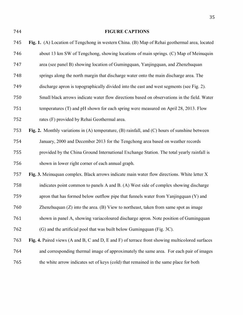
Fig. 1. (A) Location of Tengchong in western China. (B) Map of Rehai geothermal area, located 745
about 13 km SW of Tengchong, showing locations of main springs. (C) Map of Meinuquin 746
area (see panel B) showing location of Gumingquan, Yanjingquan, and Zhenzhuquan 747
springs along the north margin that discharge water onto the main discharge area. The 748
discharge apron is topographically divided into the east and west segments (see Fig. 2). 749
Small black arrows indicate water flow directions based on observations in the field. Water 750
temperatures (T) and pH shown for each spring were measured on April 28, 2013. Flow 751
rates (F) provided by Rehai Geothermal area. 752
Fig. 2. Monthly variations in (A) temperature, (B) rainfall, and (C) hours of sunshine between 753
January, 2000 and December 2013 for the Tengchong area based on weather records 754
provided by the China Ground International Exchange Station. The total yearly rainfall is 755
shown in lower right corner of each annual graph. 756
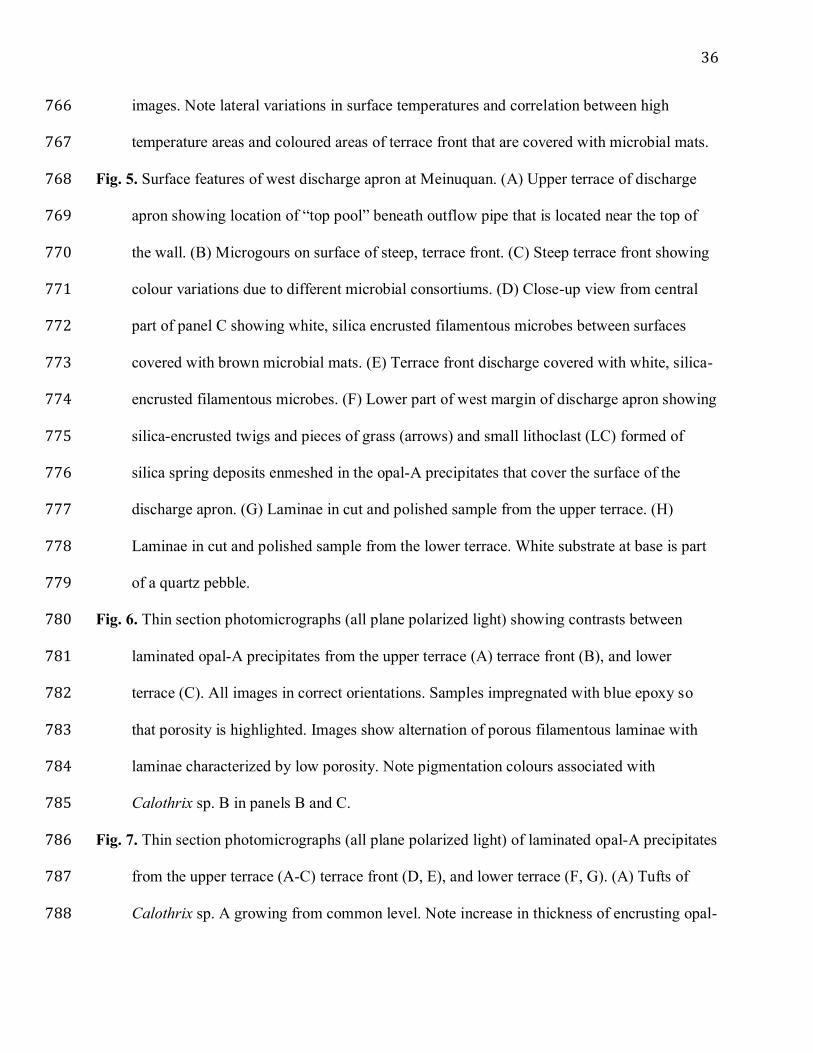
Fig. 3. Meinuquan complex. Black arrows indicate main water flow directions. White letter X 757
indicates point common to panels A and B. (A) West side of complex showing discharge 758
apron that has formed below outflow pipe that funnels water from Yanjingquan (Y) and 759
Zhenzhuquan (Z) into the area. (B) View to northeast, taken from same spot as image 760
shown in panel A, showing variacoloured discharge apron. Note position of Gumingquan 761
(G) and the artificial pool that was built below Gumingquan (Fig. 3C). 762
Fig. 4. Paired views (A and B, C and D, E and F) of terrace front showing multicolored surfaces 763
and corresponding thermal image of approximately the same area. For each pair of images 764
the white arrow indicates set of keys (cold) that remained in the same place for both 765
36
images. Note lateral variations in surface temperatures and correlation between high 766
temperature areas and coloured areas of terrace front that are covered with microbial mats. 767
Fig. 5. Surface features of west discharge apron at Meinuquan. (A) Upper terrace of discharge 768
apron showing location of “top pool” beneath outflow pipe that is located near the top of 769
the wall. (B) Microgours on surface of steep, terrace front. (C) Steep terrace front showing 770
colour variations due to different microbial consortiums. (D) Close-up view from central 771
part of panel C showing white, silica encrusted filamentous microbes between surfaces 772
covered with brown microbial mats. (E) Terrace front discharge covered with white, silica-773
encrusted filamentous microbes. (F) Lower part of west margin of discharge apron showing 774
silica-encrusted twigs and pieces of grass (arrows) and small lithoclast (LC) formed of 775
silica spring deposits enmeshed in the opal-A precipitates that cover the surface of the 776
discharge apron. (G) Laminae in cut and polished sample from the upper terrace. (H) 777
Laminae in cut and polished sample from the lower terrace. White substrate at base is part 778
of a quartz pebble. 779
Fig. 6. Thin section photomicrographs (all plane polarized light) showing contrasts between 780
laminated opal-A precipitates from the upper terrace (A) terrace front (B), and lower 781
terrace (C). All images in correct orientations. Samples impregnated with blue epoxy so 782
that porosity is highlighted. Images show alternation of porous filamentous laminae with 783
laminae characterized by low porosity. Note pigmentation colours associated with 784
Calothrix sp. B in panels B and C. 785
Fig. 7. Thin section photomicrographs (all plane polarized light) of laminated opal-A precipitates 786
from the upper terrace (A-C) terrace front (D, E), and lower terrace (F, G). (A) Tufts of 787
Calothrix sp. A growing from common level. Note increase in thickness of encrusting opal-788
37
A towards top of each filament. (B) Tuft of Calothrix sp. A with upper parts of filaments 789
encrusted by thick layers of opal-A. (C) Upper part of tuft showing intertwined filaments of 790
Calothrix sp. A (arrows) encrusted with thick layer of opal-A. (D, E) Sample from terrace 791
front showing open, porous (blue) laminae alternating with white, dense, opal-A laminae. 792
Yellowish-brown hue due to pigmentation associated with Calothrix sp. B. Note variations 793
in proportions of porous laminae and white, dense laminae evident in panels D and E. (F, 794
G) Sample from lower terrace showing recurring cycles formed of porous laminae (blue) 795
alternating with laminated formed of dense, opal-A with yellowish-brown pigmentation 796
associated with Calothrix sp. B. In panel G, note small opal-A lithoclasts evident in upper 797
part of dense, opal-A laminae (arrows). 798
Fig. 8. SEM photomicrographs of Calothrix sp. A from sample collected from upper terrace 799
near pool (same sample that is shown in Fig. 7A-C). (A) Vertical cross-section showing 800
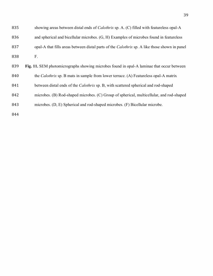
basal areas of filamentous microbial tufts growing from common level. Note numerous 801
filaments in each tuft and porous areas between the tufts. White letter B indicates tuft 802
shown in panel B. (B) Enlarged view of tuft showing numerous filaments encased in opal-803
A. (C, D) Calothrix sp. B with well-preserved basal heterocysts (H). (E) Basal heterocyst 804
(H) succeeded by vase-shaped vegetative cell. (F) Distal part of filamentous microbe 805
showing filament wall (W), septa (S), and silicified vegetative cells (SC). (G) Oblique 806
cross-sections through silicified Calothrix sp. B filament showing sheath (SH) around open 807
lumens (L). (H) Oblique longitudinal section showing trichome (T) encased by sheath 808
(SH). (I) Vaguely laminated sheath formed of polygonal opal-A grains. 809
Fig. 9. SEM photomicrographs of Calothrix sp. B forming mats on terrace front (Fig. 7D, E) and 810
lower terrace (Fig. 7F, G). (A) Mats formed of Calothrix sp. B, from terrace front. (B) 811
38
Basal heterocyst (H). (C) Basal part of filament showing basal heterocyst (H), vase-shaped 812
vegetative cell, and collapsed trichome encased by sheath. (D) Longitudinal section 813
through filament, with sheath, showing distal tapering. (E) Distal part of filament showing 814
splaying of sheath (arrows). (F) Silicified vegetative cell, septa, and trichome wall (W) in 815
middle part of Calothrix sp. B filament. (G) Outer surface of trichome covered with small 816
opal-A spheres. White letter H indicates position of panel H. (H) Enlarged view of opal-A 817
spheres with strands of mucus on outside of trichome. (I) Oblique transverse section 818
through Calothrix sp. B showing sheath around silicified trichome. (J) Longitudinal cross-819
section through silicified filaments of Calothrix sp. B showing open trichome (T), wall of 820
trichome (W), and sheath (Sh). (K) Enlarged view from panel J showing trichome wall (W) 821
and sheath (SH). Note polygonal shape of opal-A grains that form the sheath. (L) Outer 822
surface of sheath covered with polygonal opal-A grains. 823
Fig. 10. SEM photomicrographs of sample from upper terrace (same sample as shown in Figs. 824
7A-C, 8) showing non-filamentous laminae. (A) General view showing contrast between 825
filamentous mats (FM) and non-filamentous (NF) laminae. Contrast in appearance between 826
different mats reflects the amount of opal-A that was precipitated around and between the 827
filamentous microbes. Box labeled B indicates position of panel B. (B) Laminae formed of 828
opal-A sandwiched between two laminae that are formed of silicified Calothrix sp. A. C 829
indicates position of panel C. (C) Opal-A laminae divided into parts I (mainly spherical 830
microbes), II (mainly rod-shaped microbes), and III (rare to no microbes). Boundaries 831
indicated by white dashed lines. D and E indicate positions of panels D and E, respectively. 832
(D) Group of spherical microbes embedded in featureless opal-A. (E) Group of small rod-833
shaped microbes held in featureless opal-A. (F) Upper part of filamentous microbial mat 834
39
showing areas between distal ends of Calothrix sp. A. (C) filled with featureless opal-A 835
and spherical and bicellular microbes. (G, H) Examples of microbes found in featureless 836
opal-A that fills areas between distal parts of the Calothrix sp. A like those shown in panel 837
F. 838
Fig. 11. SEM photomicrographs showing microbes found in opal-A laminae that occur between 839
the Calothrix sp. B mats in sample from lower terrace. (A) Featureless opal-A matrix 840
between distal ends of the Calothrix sp. B, with scattered spherical and rod-shaped 841
microbes. (B) Rod-shaped microbes. (C) Group of spherical, multicellular, and rod-shaped 842
microbes. (D, E) Spherical and rod-shaped microbes. (F) Bicellular microbe. 843
844