20030701 177 - apps.dtic.mil · skpl, we performed a co-immunoprecipitation experiment by...
TRANSCRIPT
AD
Award Number: DAMD17-00-1-0135
TITLE: Characterization of a Novel Nuclear Hormone ReceptorCoactivator, Uba3, and its Function in Breast Cancer
PRINCIPAL INVESTIGATOR: Andrew P. DennisBert W. O'Malley, M.D.Zafar Nawaz, Ph.D.
CONTRACTING ORGANIZATION: Baylor College of Medicine
Houston, Texas 77030
REPORT DATE: March 2003
TYPE OF REPORT: Annual Summary
PREPARED FOR: U.S. Army Medical Research and Materiel CommandFort Detrick, Maryland 21702-5012
DISTRIBUTION STATEMENT: Approved for Public Release;Distribution Unlimited
The views, opinions and/or findings contained in this report arethose of the author(s) and should not be construed as an officialDepartment of the Army position, policy or decision unless sodesignated by other documentation.
20030701 177
Form ApprovedREPORT DOCUMENTATION PAGE OMB No. 074-0188
Public reporting burden for this collection of information is estimated to average 1 hour per response, Including the time for reviewing instructions, searching existing data sources, gathering and maintainingthe data needed, and completing and reviewing this collection of information. Send comrrents regarding this burden estimate or any other aspect of this collection of information, Including suggestions forreducing this burden to Washington Headquarters Services, Directorate for Information Operations and Reports, 1215 Jefferson Davis Highway, Suite 1204, Arlington, VA 22202-4302, and to the Office ofManagement and Budget, Paperwork Reduction Project (0704-0188), Washington, DC 20503
1. AGENCY USE ONLY (Leave blank) 2. REPORT DATE 3. REPORT TYPE AND DATES COVEREDMarch 2003 Annual Summary (1 Mar 00 - 28 Feb 03)
4. TITLE AND SUBTITLE 5. FUNDING NUMBERSCharacterization of a Novel Nuclear Hormone DAMD17-00-1-0135Receptor Coactivator, Uba3, and its Function inBreast Cancer6. AUTHOR(S):Andrew P. DennisBert W. O'Malley, M.D.Zafar Nawaz, Ph.D.7. PERFORMING ORGANIZATION NAME(S) AND ADDRESS(ES) 8. PERFORMING ORGANIZATION
REPORT NUMBERBaylor College of MedicineHouston, Texas 77030
E-Mail: andrew. [email protected]
9. SPONSORING / MONITORING AGENCY NAME(S) AND ADDRESS(ES) 10. SPONSORING / MONITORINGAGENCY REPORT NUMBER
U.S. Army Medical Research and Materiel CommandFort Detrick, Maryland 21702-5012
11. SUPPLEMENTARY NOTES
12a. DISTRIBUTION / AVAILABILITY STATEMENT 12b. DISTRIBUTION CODEApproved for Public Release; Distribution Unlimited
13. Abstract (Maximum 200 Words) (abstract should contain no proprietary or confidential information)We previously identified Uba3 as a nuclear hormone receptor coactivator that functions as part ofthe NEDD8 pathway to influence ER- and PR-mediated transcription. Although our previous modelfailed to demonstrate the connection between Uba3, the NEDD8 pathway, and transcription, we now havesufficient evidence to suggest the NEDD8 pathway is linked to ER- and PR-mediated transcription viathe SCF E3 ligase complex. Here we show that Skpl can associate with ERa by co-immunoprecipitation,and with the pS2 promoter by ChIP assay. We have investigated the mRNA expression of Uba3, PR, andERa in several common tumor cell lines. We were not able to detect abnormal expression of hUba3mRNA in the breast cancer cell lines. Furthermore, we were unable to correlate hUba3 mRNA
expression with PR and ERa expression across all cell lines. We also investigated in silico thepotential that Uba3 mRNA expression may be regulated by estrogen and progesterone. Althoughpossible binding sites for ER and PR were localized within the Uba3 promoter, we were not ableto detect induction or repression of Uba3 mPRNA by estradiol, and SERMs tamoxifen, and raloxifene, orprogesterone.
14. SUBJECT TERMS: 15. NUMBER OF PAGEScancer biology, steroid hormones/receptor coactivators, cell cycle 13proteins, tumor suppressor proteins 16. PRICE CODE
17. SECURITY CLASSIFICATION 18. SECURITY CLASSIFICATION 19. SECURITY CLASSIFICATION 20. LIMITATION OF ABSTRACTOF REPORT OF THIS PAGE OF ABSTRACT
Unclassified Unclassified Unclassified UnlimitedNSN 7540-01-280-5500 Standard Form 298 (Rev. 2-89)
Prescribed by ANSI Std. Z39-18298-102
TABLE OF CONTENTS
Cover ............................................................................................................................................................................ 1Form SF298 ................................................................................................................................................................. 2Table of Contents ........................................................................................................................................................ 3Introduction: ............................................................................................................................................................... 4Body ............................................................................................................................................................................. 4Key Research Accomplishm ents ................................................................................................................................ 6Reportable outcomes .................................................................................................................................................. 6Conclusions .................................................................................................................................................................. 6References: .................................................................................................................................................................. 8Appendix: .................................................................................................................................................................... 9
3
INTRODUCTION:
Estrogen and Progesterone regulate the growth and development of the mammary glandvia a signaling cascade that is initiated through binding to their cognate nuclear hormonereceptors (NHRs), the estrogen receptor c/13 (ER) and progesterone receptor-A/-B (PR),members of the ligand-inducible transcription factor superfamily (1,2). ER and PR-target genesare further regulated by recruitment of coregulator proteins, which positively affect(coactivators) or negatively affect (corepressors) ligand-activated transcription (3).
We have previously identified human ubiquitin-activating enzyme 3 (hUba3) as aprogesterone receptor (PR) interacting protein in a yeast 2-hybrid assay. Human Uba3 is amember of the NEDD8 (neural precursor cell-expressed developmentally down-regulated)ubiquitin-like protein modification pathway (4). The NEDD8 pathway functions through apathway-specific El enzyme (a heterodimer composed of hUba3 and APP-BP1 (amyloidprecursor protein binding-protein 1) and an E2 enzyme (Ubcl2 (ubiquitin-conjugating enzyme12)), but lacks a cognate E3 (and one may not exist) (4). We previously reported an intricaterelationship between the NEDD8 pathway and NHR coactivation, although we were not able todemonstrate how this pathway influences ER- and PR-dependent transcription.
The Cullin proteins (Cul-1, -2, -3, -4a, -4b, and -5) have been the only identified targetproteins of the NEDD8 modification pathway (5). The Cullin proteins are an integral componentof the SCF ubiquitin-protein (E3) ligase (a complex of Skpl, Cullin-x, and an F-box protein).Modification of the Cullin proteins by NEDD8 appears to control the ubiquitin ligase activity ofthe SCF complex by recruiting Cdc34, a ubiquitin-conjuagting (E2) enzyme used by thiscomplex (6). Since the E2-E3 components are juxtaposed to one another, it is thought that thisproximity contributes to the enhancement of SCF complex function. Thus, the NEDD8 pathwaymay contribute to degradation of components involved in transcription via the SCF complex(7,8).
Several lines of evidence support the possibility that degradation is necessary fortranscription to proceed (7,8). Using the ChIP assay as tool to understand how the NEDD8pathway influences ER-mediated transcription we have been able to localize a component of theSCF complex to the pS2 promoter in an estradiol dependent manner. We have also shownligand-dependent association between ERax and Skpl by immunoprecipitation. In agreementwith "Task 2" and "Task 3" in the original proposal, we investigated the expression of Uba3 inseveral tumor cell lines and compared its expression to that of ERac and PR. Finally we wishedto investigate the potential for Uba3 mRNA expression to be inducible by estradiol, tamoxifen,raloxifene, and progesterone.
BODY
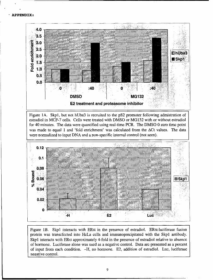
We have previously reported the characterization of hUba3 and APP-BP1 as nuclearreceptor cofactors capable of functioning as coactivators in transient transfection assays in HeLacells. Based on our experiements with hUba3 and Ubcl2, we concluded that the NEDD8pathway may play an important role in nuclear receptor-dependent transcription. Since we werenot able to identify any physical interactions between hUba3 and PR and ER in vitro and in vivo,it was thought that hUba3 may interact with other protein components involved in transcription.We therefore developed a chromatin immunoprecipitation (ChIP) assay system to characterizethe involvement of hUba3 during estrogen-dependent pS2 transcription in MCF-7 cells. Usingan antibody we generated in rabbit, we attempted to localize hUba3 to the pS2 promoter uponadministration of estradiol (Figure la). Unfortunately, we were not able to recruit hUba3 to thepS2 promoter whereas we were able to see recruitment of ERa (data not shown). We modifiedthe ChIP assay detergents in the immunoprecipitation step to optimally precipitate hUba3protein, but these alterations did not the change the results we observed in the ChIP assay. We
4
surmised that hUba3 and the NEDD8 pathway might be influencing ER-mediated transcriptionthrough the SCF E3 ligase complex. Using an antibody against one of the core proteins of theSCF complex, Skpl, we were able to see its recruitment to the pS2 promoter upon addition ofestradiol (figure la). The recruitment was more profound when MG132, a proteasome inhibitor,was used in the experiment. We interpreted this result to mean that the interaction of the SCF E3ligase complex with the pS2 promoter is likely transient and unstable due to the degradation-promoting (ubiquitylation) activity of this complex. To determine if ERoc could interact withSkpl, we performed a co-immunoprecipitation experiment by immunoprecipitating Skpl anddetecting ERa (figure lb). The ERa was fused to the N-terminus of luciferase, and thereforelight units were used to quantify the interaction between Skpl and ERa instead of a Westernblot. The results show that ERoa interacts with Skpl in the presence of estradiol and this resultagrees with the results we observed in the ChIP assay. Our results support a model where hUba3and the NEDD8 pathway influence ER-dependent transcription through the SCF complex.Consistent with our results, hUba3 is not present at the promoter, but rather stimulates covalentattachment of NEDD8 to the Cullin protein within the SCF complex. This covalent modificationactivates the E3 ligase activity of the SCF complex, degrading components of the transcriptionapparatus. We are not able to conclude what the function of the ligase activity is, but thepossibilities are intriguing. Ubiquitylation/degradation of transcription proteins may benecessary to permit promoter escape of RNA polymerase ]1. Ubiquitylation/degradation mayalso necessary to remove activated ligand-bound ERax from the promoter, thus attenuatingestrogen signaling. Importantly, both possibilities may be correct; that is, activation oftranscription and degradation are inherently coupled events. This way the cell has a mechanismto control the amount of transcription upon stimulation. Regardless of the correct downstreammodel, the data I have presented underscore the integration of the NEDD8 pathway in ER- andPR-dependent transcription.
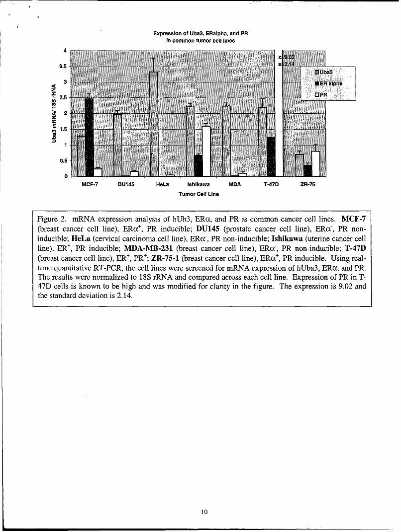
Illumination of hUba3 and the NEDD8 pathway in ER- and PR-mediated transcriptionand the importance of ER and PR in breast cancer development support the possibility thatdisrupted expression of NEDD8 pathway proteins may play an important role in breast cancer.To this end we to wished to study the expression of hUba3, ERoa, and PR in several breast cancercell lines. We chose to study hUba3 expression in 7 cell lines: 4 breast cancer cell lines and 3'non-breast' cancer cell lines. We included 3 'non-breast' cancer cell lines to infer expression ofhUba3 in other cancer cell lines. (Details of the cell lines are described in figure 2.) Using real-time Taqman quantitative RT-PCR, we assessed the mRNA expression of hUba3, ERoa, and PR(figure 2). The level of hUba3 mRNA was highest in HeLa cells and lowest in ZR-75 cells.Comparing hUba3 mRNA expression across all cancer cell lines, we were not able to observesignificant differences between the breast cancer cell lines and the other cancer cell lines (Figure2, compare MCF-7, T-47D, MDA, ZR-75 with that of DU145, lahikawa, and Hela cell lines).Perhaps more importantly, hUba3 mRNA expression did not correlate with ER and PR mRNAexpression in any of the cell lines tested (figure 2).
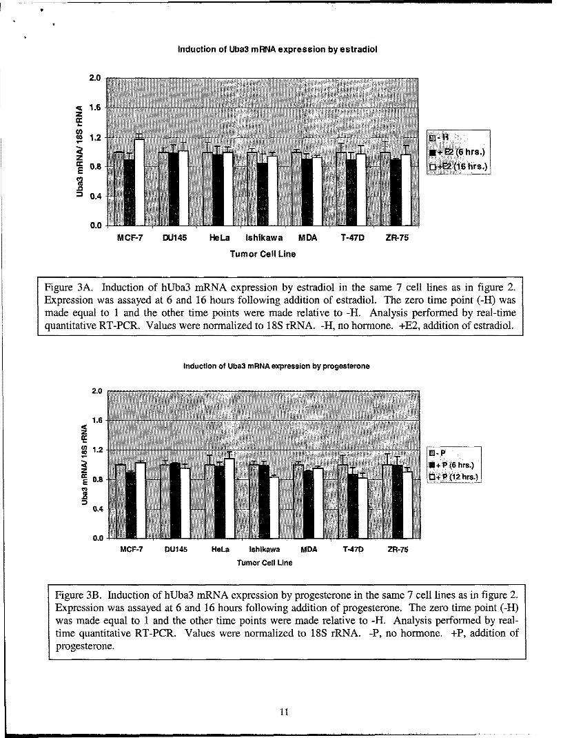
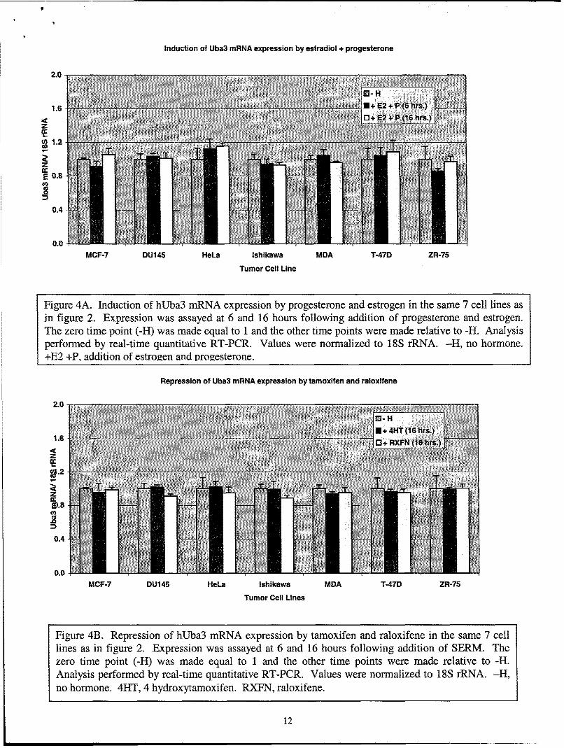
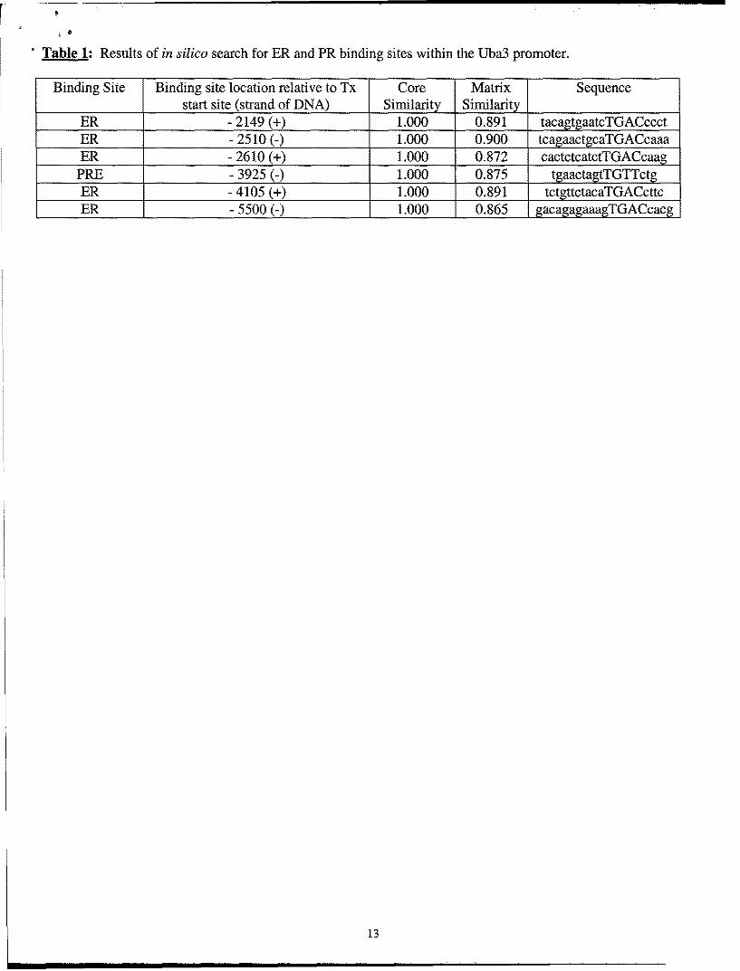
While these studies were ongoing, a group published a report that the overexpression ofhUba3 promotes degradation of the ERoc protein through the SCF E3 complex suggesting thatregulation of ERa by the NEDD8 pathway occurs at the protein level (9). It is possible thathUba3 can be regulated by ER and/or PR, providing a negative feedback on ER and/ or PRsignaling. To expedite this possibility we searched the hUba3 promoter (-5000bp to -lbp) forER and PR binding sites in silico using MatInspector v2.2 software with the Transfac Database(transcfac.gbd.de). The results of the in silico search are shown in Table 1. We were able to find5 ERE half sites and 1 PR half site, but we were not able to find any full palindromes. Welimited our stringency to half sites because several ER target genes, for example PR and c-Myc,are known to be regulated by ER half-sites. We investigated the induction of hUba3 mRNAexpression using real-time quantitative RT-PCR in the above mentioned cancer cell lines. Wedid not observe any induction of hUba3 mRNA by progesterone or estrogen, or co-stimulation
5
by both estrogen and progesterone (figure 3 and 4A). Rather, the expression of hUba3 mRNA isrefractory to progesterone and estrogen in all cell lines tested. We also examined the repressionof hUba3 mRNA expression by the SERMs, tamoxifen and raloxifene. Synonymous with theresults observed with estradiol and progesterone, repression of hUba3 is unaffected by tamoxifenor raloxifene (figure 4B).
KEY RESEARCH ACCOMPLISHMENTS
" We have successfully determined how hUba3 and the NEDD8 pathway influences ER-mediated transcription through additional experiments not outlined in "Task 1", butnecessary to complete "Task 1."
"* Through ChIP assay analysis, we have been able to show that Skpl is localized to thepS2 promoter with ERca following administration of estradiol.
"* By co-immunoprecipitation, we observed an estrogen-dependent interaction with Skpland ERcL.
"* We have completed most of "Task 2" by examining the mRNA expression of hUba3, ERcL,and PR in several breast cancer cell lines.
"* We have shown that hUba3 mRNA expression is widely expressed in breast cancer celllines and cell lines derived from other tissues.
"* hUba3 mRNA expression does not correlate with ER/ PR mRNA levels in the cancercell lines tested.
"* hUba3 mRNA expression is unaffected by ligands to ER and PR.
REPORTABLE OUTCOMES
None
CONCLUSIONS
We have made substantial progress in understanding how hUba3 and the NEDD8pathway influence ER- and PR-mediated transcription. From the experiments we outlined in"Task 1" we were able to conclude that hUba3 and APP-BP1 are unique in their own right, butwere not able to understand how this pathway is integrated with NHR function, an importantaspect of "Task 1". Because of this unforeseen situation, we added several importantexperiments that provided insight into the true function of the NEDD8 pathway in ER- and PR-mediated transcription. We now conclude that the SCF E3 ligase complex is recruited with EROupon addition of estradiol to the pS2 promoter. Therefore, hUba3 (at a site other than thepromoter) functions as a coactivator by stimulating 'neddylation' (NEDD8 conjugation) of theSCF E3 ligase complex (at the promoter), leading to enhanced ubiquitylation of unknownproteins within the transcription apparatus. It is not quite clear yet, but ubiquitylation and/ orproteasome-mediated degradation somehow aides the process of ER-mediated transcription.
6
We have made significant progress in characterizing the expression of hUba3mRNA in several cancer cell lines in agreement with "Task 2". Of the 7 cancer cell lines weinvestigated, we were able to observe differences in hUba3 mRNA expression, but were not ableto conclude that mRNA expression of hUba3 contributes to carcinogenesis. Furthermore, wewere not able to observe any significant correlation between hUba3 mRNA expression, and ERoxand PR mRNA expression. Based on a report that the NEDD8 pathway controls degradation ofERa protein, we hypothesized that ER and PR signaling may regulate hUba3, providing anegative feedback mechanism that would attenuate this signaling. We observed that hUba3mRNA expression is refractory to estrogen and progesterone signaling. Additionally, we wereunable to alter hUba3 expression with the SERMs tamoxifen and raloxifene. We conclude thatthe mRNA expression of hUba3 is constant and unaffected by ER and PR signals. It is possible,and even likely that most differences among ER, PR, and hUba3 will be observed at the proteinlevel. Comparison of hUba3 protein expression with ER and PR expression in tumor biopsysamples is the focus of "Task 3".
7
REFERENCES:
1. Sutherland RL, Prall OW, Watts CK, Musgrove EA. Estrogen and progestin regulation ofcell cycle progression.J Mammary Gland Biol Neoplasia. 1998 Jan;3(1):63-72.
2. Tsai MJ, O'Malley BW. Molecular mechanisms of action of steroid/thyroid receptorsuperfamily members.Annu Rev Biochem. 1994;63:451-86.
3. McKenna NJ, Xu J, Nawaz Z, Tsai SY, Tsai MJ, O'Malley BW. Nuclear receptorcoactivators: multiple enzymes, multiple complexes, multiple functions. J Steroid Biochem MolBiol. 1999 Apr-Jun;69(1-6):3-12.
4. Gong L, Yeh ET. Identification of the activating and conjugating enzymes of the NEDD8conjugation pathway.J Biol Chem. 1999 Apr 23;274(17):12036-42.
5. Hori T, Osaka F, Chiba T, Miyamoto C, Okabayashi K, Shimbara N, Kato S, Tanaka K.Covalent modification of all members of human cullin family proteins by NEDD8. Oncogene.1999 Nov 18;18(48):6829-34.
6. Kawakami T, Chiba T, Suzuki T, Iwai K, Yamanaka K, Minato N, Suzuki H, Shimbara N,Hidaka Y, Osaka F, Omata M, Tanaka K. NEDD8 recruits E2-ubiquitin to SCF E3 ligase.EMBO J. 2001 Aug 1;20(15):4003-12.
7. Lonard DM, Nawaz Z, Smith CL, O'Malley BW. The 26S proteasome is required forestrogen receptor-alpha and coactivator turnover and for efficient estrogen receptor-alphatransactivation. Mol Cell. 2000 Jun;5(6):939-48.
8. Muratani M, Tansey WP. How the ubiquitin-proteasome system controls transcription. NatRev Mol Cell Biol. 2003 Mar;4(3):192-201.
9. Fan M, Bigsby RM, Nephew KP. The NEDD8 pathway is required for proteasome-mediateddegradation of human estrogen receptor (ER)-alpha and essential for the antiproliferative activityof ICI 182,780 in ERalpha-positive breast cancer cells. Mol Endocrinol. 2003 Mar;17(3):356-65.
8
APPENDIX:
4.0-
30E 2.5
0 hUba3
01.5
IL 1.0
0 :40 040
DMSO AG132
E2 treatment and proteasome inhibitor
Figure 1A. Skpl, but not hUba3 is recruited to the pS2 promoter following admistration ofestradiol in MCF-7 cells. Cells were treated with DMSO or MG132 with or without estradiolfor 40 minutes. The data were quantified using real-time PCR. The DMSO 0 zero time pointwas made to equal 1 and 'fold enrichment' was calculated from the ACt values. The datawere normalized to input DNA and a non-specific internal control (not seen).
0.12
0.08 -Co 0.06 - E Skpl
0.04
0.02
.H E2 Luc
Figure lB. Skpl interacts with ERox in the presence of estradiol. ERa-luciferase fusionprotein was transcfected into HeLa cells and immunoprecipitated with the Skpl antibody.Skpl interacts with ERox approximately 4-fold in the presence of estradiol relative to absenceof hormone. Luciferase alone was used as a negative control. Data are presented as a percentof input from each condition. -H, no hormone. E2, addition of estradiol. Luc, luciferasenegative control.
9
Expression of Uba3, ERalpha, and PRin common tumor cell lines
4-
3.52
354 0 Uba3
3- NUER alphaZ
2M 0 PR
0.5
MCF-7 DU145 HleLa Ishikawa MDA T-47D ZR-75
Tumor Cell Line
Figure 2. mRNA expression analysis of hUb3, ERc•, and PR is common cancer cell lines. MCF-7(breast cancer cell line), ERu+, PR inducible; DU145 (prostate cancer cell line), ERaf, PR non-inducible; ileLa (cervical carcinoma cell line), ERc(, PR non-inducible; Ishik~awa (uterine cancer cellline), ER÷, PR inducible; MLDA-MB-231 (breast cancer cell line), ERc-, PR non-inducible; T-4"/D(breast cancer cell line), ER÷, PR÷; ZlR-75-1 (breast cancer cell line), ERoc÷, PR inducible. Using real-time quantitative RT-PCR, the cell lines were screened for mRNA expression of hUba3, ERca, and PR.The results were normalized to 18S rRNA and compared across each cell line. Expression of PR in T-47D cells is known to be high and was modified for clarity in the figure. The expression is 9.02 andthe standard deviation is 2.14.
10
K
Induction of Uba3 mRNA expression by estradiol
2.0 T
<• .6
Co~1.2H z I+•(6hrs.)
IC 0J+E2 (16 hrs.)
= 0.4
0.0
MCF-7 DU145 HeLa Ishikawa MDA T-47D ZR-75
Tumor Cell Line
Figure 3A. Induction of hUba3 mRNA expression by estradiol in the same 7 cell lines as in figure 2.Expression was assayed at 6 and 16 hours following addition of estradiol. The zero time point (-H) wasmade equal to 1 and the other time points were made relative to -H. Analysis performed by real-timequantitative RT-PCR. Values were normalized to 18S rRNA. -H, no hormone. +E2, addition of estradiol.
Induction of Uba3 mRNA expression by progesterone
2.0
1.6
z
J 2 2 + P (12 hrs.)E 0.8
0.4
0.0
MCF-7 DU145 HeLa Ishikawa MDA T-47D ZR-75
Tumor Cell Line
Figure 3B. Induction of hUba3 mRNA expression by progesterone in the same 7 cell lines as in figure 2.Expression was assayed at 6 and 16 hours following addition of progesterone. The zero time point (-H)was made equal to 1 and the other time points were made relative to -H. Analysis performed by real-time quantitative RT-PCR. Values were normalized to 18S rRNA. -P, no hormone. +P, addition ofprogesterone.
11
Induction of Uba3 mRNA expression by estradiol + progesterone
2.0
I£V) 1.2
2T
ccc og
0.4
0.0MCF-7 DU145 HeLa Ishikawa MDA T-47D ZR-75
Tumor Cell Line
Figure 4A. Induction of hUba3 mRNA expression by progesterone and estrogen in the same 7 cell lines asin figure 2. Expression was assayed at 6 and 16 hours following addition of progesterone and estrogen.The zero time point (-H) was made equal to 1 and the other time points were made relative to -H. Analysisperformed by real-time quantitative RT-PCR. Values were normalized to 18S rRNA. -H, no hormone.+E2 +P, addition of estrogen and progesterone.
Repression of Uba3 mRNA expression by tamoxifen and raloxifene
2.0
0+4T(16 hrs.)1.8 FN(16hrs.)
Z
'C
U, .2 -- ---
Z
0.4
0.0-
MCF-7 DU145 HeLa Ishikawa MDA T-47D ZR-75
Tumor Cell Lines
Figure 4B. Repression of hUba3 mRNA expression by tamoxifen and raloxifene in the same 7 celllines as in figure 2. Expression was assayed at 6 and 16 hours following addition of SERM. Thezero time point (-H) was made equal to 1 and the other time points were made relative to -H.Analysis performed by real-time quantitative RT-PCR. Values were normalized to 18S rRNA. -H,no hormone. 4HT, 4 hydroxytamoxifen. RXFN, raloxifene.
12
r
Table 1: Results of in silico search for ER and PR binding sites within the Uba3 promoter.
Binding Site Binding site location relative to Tx Core Matrix Sequencestart site (strand of DNA) Similarity Similarity
ER - 2149 (+) 1.000 0.891 tacagtgaatcTGACccctER - 2510 (-) 1.000 0.900 tcagaactgcaTGACcaaaER - 2610 (+) 1.000 0.872 cactctcatctTGACcaag
PRE - 3925 (-) 1.000 0.875 tgaacta TGTTctER - 4105 (+) 1.000 0.891 tctgttctacaTGACcttcER - 5500 (-) 1.000 0.865 gacagagaaagTGACcacg
13