2007 report of the international dry eye workshop (dews)
TRANSCRIPT

The Ocular Surface / april 2007, VOl. 5, NO. 2 / www.theocularsurface.com 59
2007 Report of the International Dry Eye
WorkShop (DEWS) Sponsored by the Tear Film & Ocular Surface Society
INTRODUCTION TO THE 2007 REPORT OF THEINTERNATIONAL DRY EYE WORKSHOP (DEWS)
THE DEFINITION AND CLASSIFICATION OF DRY EYE DISEASE
THE EPIDEMIOLOGY OF DRY EYE DISEASE
METHODOLOGIES TO DIAGNOSE AND MONITOR DRY EYE DISEASE
DESIGN AND CONDUCT OF CLINICAL TRIALS
MANAGEMENT AND THERAPY OF DRY EYE DISEASE
RESEARCH IN DRY EYE
www.theocularsurface.com
A peer-reviewed journal, indexed in MEDLINE/PubMed and EMBASE
SPECIAL ISSUE
Tear Film & Ocular Surface Society
April 2007Volume 5, Number 2
Ocular SurfaceTHE
A JOURNAL OF REVIEW LINKING LABORATORY SCIENCE, CLINICAL SCIENCE, AND CLINICAL PRACTICE
The Ocular Surface / april 2007, VOl. 5, NO. 2 / www.theocularsurface.com60
Article submission: Questions regarding manuscript prepa-ration not addressed in the Information for Authors may be addressed to the Managing Editor: Susan Erickson, 130 Winchester St., Brookline, MA 02446. Tel: 617-731-3415; Email: [email protected]. Information for Authors is available at www.theocularsurface.com.
Subscription rates Individual: $154 per year in the US and Canada; $199 per year rest of world (includes air mail deliv-ery). Institution:$249 per year in the US and Canada; $299 per year rest of world (includes air mail delivery). Internet-only subscription: $139 (US and rest of world). Print-only subscription: $139 (US and Canada); $139 (rest of world). Back issues are $45 per issue. Subscriptions may be ordered online at www.theocularsurface.com or at The Ocular Surface, 75 Maiden Lane, Suite 408, New York, NY 10038, USA. tel: 212-791-1440 fax: 212-791-4980. Claims for missing issues must be filed within 120 days of the issue date.
Indexing The Ocular Surface is indexed in MEDLINE/PubMed and EMBASE. The Ocular Surface is printed on acid-free paper that meets the requirements of ANSI/NISO Z39.48-1992 (Permanence of Paper).
Library reprints Contact the Copyright Clearance Center, Inc., 222 Rosewood Drive, Danvers, MA 01923 USA. tel: 978-646-2600; fax: 978-646-8600 e-mail: [email protected] online: www.copyright.com.
Advertising and bulk reprint inquiries should be addressed to LaVon Kellner, Ethis Communications, Inc., 75 Maiden Lane, Suite 408, New York, NY 10038. tel: 212-791-1440; fax: 212-791-4980; e-mail: [email protected]. Con-tact authors for single reprint copies.
The Ocular Surface (ISSN 1542-0124) is published quarterly by Ethis Communications, Inc., 75 Maiden Lane, Suite 408, New York, NY 10038, USA. Copyright 2006 Ethis Commu-nications, Inc. All rights reserved. Neither The Ocular Surface nor the publisher assume any responsibility for any injury and/or damage to persons or property as a matter of product liability, negligence or otherwise, or from any use or operation of any methods, products, instructions or ideas contained in this publication. No part of these contents may be reproduced without permission.
publisher,directorofglobalsales:laVon Marie Kellner
editorialdirector: David Kellner
artdirector: Deborahanne chingas Sandke
productiondirector:charlotte l. latham
circulationmanager: Simon Wang
Ocular SurfaceTHE
A JOURNAL OF REVIEW LINKING LABORATORY SCIENCE, CLINICAL SCIENCE, AND CLINICAL PRACTICE
A peer-reviewed journal

The Ocular Surface / april 2007, VOl. 5, NO. 2 / www.theocularsurface.com 61 61
editor-in-chiefGary N. Foulks, MD, FACS
Professor, Ophthalmology, University of Louisville,
Louisville, KY
founding editorMichael A. Lemp, MD
Washington, DC
section editors
laboratory scienceJames V. Jester, PhD
Professor, Ophthalmology, University of California, Irvine, CA
clinical science,innovative techniques and technology
Gary N. Foulks, MD
clinical practiceJohn Sutphin, Jr, MD
Professor and Chair, Ophthalmology, University of Kansas Medical Center, Kansas City, KS
features editors
Juan Murube, MD, PhDProfessor, Ophthalmology,
University of Alcala, Madrid, Spain
Gary D. Novack, PhDPresident, PharmaLogic
Development Inc., San Rafael, CA
managing editorSusan ericksonBrookline, MA
Mark B. Abelson, MD, Director, Ophthalmic Research Associates, North Andover, MA
Penny A. Asbell, MD, Professor, Ophthalmology, Mount Sinai Medical Center, New York, NY
Christophe Baudouin, MD, PhD, Professor and Director, Ophthalmology, Quinze-Vingts Hôpital, University of Paris, Paris, France
Roger W. Beuerman, PhD, Professor, Ophthalmology, Cell Biology, and Anatomy, Louisiana State University Eye Center, New Orleans, LA, and Singapore Eye Research Institute, Singapore
Stefano Bonini, MD, Professor and Chairman, Ophthalmology, University of Rome, Rome, Italy
Anthony Bron, FRCS, Professor Emeritus, Nuffield Laboratory of Ophthalmology, Oxford, UK
M. Reza Dana, MD, MPH, Senior Scientist, Schepens Eye Research Institute, Boston, MA
Darlene A. Dartt, PhD, Senior Scientist, Schepens Eye Research Institute, Boston, MA
Harminder S. Dua, MD, PhD, Professor, Ophthalmology and Vision Science, University of Nottingham, UK
Suzanne M. J. Fleiszig, OD, PhD, Associate Professor, Vision Science and Optometry, University of California Berkeley, Berkeley, CA
Desmond Fonn, Dip Optom, M Optom (NSW), FAAO, Professor, School of Optometry, University of Waterloo, Waterloo, Ontario, Canada
Ilene K. Gipson, PhD, Senior Scientist, Schepens Eye Research Institute, Boston, MA
W. Bruce Jackson, MD, Professor and Chairman, Ophthalmology, University of Ottawa, Ottawa, Ontario, Canada
Winston W.-Y. Kao, PhD, Director, Ophthalmic Research, University of Cincinnati, Cincinnati, OH
Shigeru Kinoshita, MD, Professor and Chairman, Ophthalmology, Kyoto Prefectural University of Medicine, Kyoto, Japan
Friedrich E. Kruse, MD, Professor and Chairman, University Eye Clinic, University of Erlangen-Nuremberg, Erlangen, Germany
Peter Laibson, MD, Co-director, Cornea Service, Wills Eye Hospital, Philadelphia, PA
Mark J. Mannis, MD, Professor, Ophthalmology, University of California, Davis, Davis, CA
William D. Mathers, MD, Professor, Casey Eye Institute, Oregon Health Sciences University, Portland, OR
James P. McCulley, MD, Professor and Chairman, Ophthalmology, University of Texas Southwestern Medical Center, Dallas, TX
Austin K. Mircheff, PhD, Professor, Physiology and Biophysics, and Ophthalmology, Doheny Eye Institute, University of Southern California, Los Angeles, CA
Teruo Nishida, MD, Professor and Chairman, Biomolecular Recognition and Ophthalmology, Yamaguchi University School of Medicine, Yamaguchi, Japan
Stephen C. Pflugfelder, MD, Professor, Ophthalmology, Baylor College of Medicine, Houston, TX
Kenneth A. Polse, OD, MS, Professor, Vision Science and Optometry, University of California, Berkeley, Berkeley, CA
Gullapalli N. Rao, MD, Director, LV Prasad Eye Institute, Hyderabad, India
Maurizio Rolando, MD, Professor, Neuroscience and Ophthalmology, University of Genoa, Genoa, Italy
Janine Smith, MD, Deputy Clinical Director, National Eye Institute, National Institutes of Health, Bethesda, MD
Michael E. Stern, PhD, Research Investigator, Allergan Inc., Irvine, CADavid A. Sullivan, PhD, Senior Scientist, Schepens Eye Research
Institute, Boston, MADeborah F. Sweeney, OD, PhD, Associate Professor, University of New
South Wales, Sydney, NSW, AustraliaDonald T. Tan, FRCS, Director, Singapore Eye Research Institute,
National University of Singapore, SingaporeTimo Tervo, MD, PhD, Professor, Applied Clinical Ophthalmology,
Helsinki University Eye Hospital, Helsinki, FinlandAlan Tomlinson, PhD, DSc, FCOptom, Professor and Head, Vision
Sciences, Glasgow Caledonian University, Glasgow, Scotland, UKScheffer Tseng, MD, PhD, President, Ocular Surface Center, Miami, FLKazuo Tsubota, MD, Professor and Head, Department of Ophthalmology, Keio University School of Medicine, Tokyo, JapanGraeme Wilson, PhD, Professor, Optometry, Indiana University,
Bloomington, IN
EDItorS
EDItorIAL BoArD
Ocular SurfaceTHE
A JOURNAL OF REVIEW LINKING LABORATORY SCIENCE, CLINICAL SCIENCE, AND CLINICAL PRACTICE
A peer-reviewed journal
Additional biographical information is available at www.theocularsurface.com

The Ocular Surface / april 2007, VOl. 5, NO. 2 / www.theocularsurface.com62

The Ocular Surface / april 2007, VOl. 5, NO. 2 / www.theocularsurface.com 63
Editorial
DEWS Report: A Mission Completed
his issue of The Ocular Surface is very unusual. As the official report of the Dry Eye Work-Shop (DEWS), it is an encyclopedic review of dry eye disease and, additionally, a guide to resources archived on the internet. It is the product of a team of international experts who
have labored over 3 years to compile an evidence-based review of the present state of knowledge for dry eye disease and the methods used to evaluate, diagnose, and manage the disorder. It summarizes the findings of current research and identifies future needs for a better understanding of the etiology, pathogenesis, and potential therapy of the disease.
The process of deliberation and discussion that underpins this arduous endeavor is described in the “Introduction” and in various chapters of the volume. Suffice it to say that an international community of clinicians and scientists with expertise in all aspects of dry eye disease collaborated to search the literature, collect and validate data, and incorporate it into reports. The process of commentary and adjudication of differing opinions was open, yet subject to several levels of validation. The product is a written document that serves as a guide to a vast amount of information that is archived both in this special issue and on a supporting website (www.tearfilm.org) that is accessible to all.
The chapter on Definition and Classification expands the characterization of dry eye disease and places it within the perspective of ocular surface disease. The chapter on Epidemiology provides commentary on the implications of the disease, as well as comparison of the methods available to evaluate symptoms and factors contributory to the disease. The Diagnostic Methodologies chapter not only provides valuable discussion of the parameters of dry eye disease, but also catalogs and validates a vast collection of clinical and research methods, including questionnaires, to monitor the disease. The Research chapter summarizes past and present findings, and identifies areas whose further study will contribute to the understanding of the etiopathogenesis and consequences of dry eye disease. The chapter on Clinical Trials provides recommendations with regard to both general and specific guidelines for clinical trials in dry eye disease and identifies the idiosyncrasies and confounding outcome variables for such trials. The chapter on Management and Therapy catalogs the options for therapy and recommends a contemporary strategy for management of dry eye disease.
As would be expected for a multifactorial disease that has many nuances in clinical and patho-logical expression, opinions differ even amongst the experts as to the most appropriate way to char-acterize and label some aspects of the disease. This proved true for the definition and classification of the disease. Some key concepts in the appreciation of dry eye were identified from the literature. One such concept was the characterization of the Lacrimal Functional Unit,1 which has highlighted the interdependence of components of the lacrimal system in maintaining the integrity of the ocular surface. Some new concepts were constructed in the deliberation process of the Subcommittee work, including a concept suggested by Dr. Christophe Baudoin—a Vicious Circle of dry eye disease, by which various risk factors may interact to precipitate and perpetuate the condition.2 The concept of the Ocular Surface System, developed by the Research Subcommittee, extends the scope of the ocular surface to a collection of contiguous tissues that share embryonal, innervational, histological, and hormonal background.
The time and effort necessary to compile and collate this project and the summary document was extraordinary. The endeavor could never have been completed without the sponsorship and commitment of The Tear Film & Ocular Surface Society and the officers and staff of that organization. The planning and execution of the organizational meetings, the coordination of the conferences for presentation of the collected information, the facilitation of the discussions of the DEWS participants, and the administrative direction of the publication process were achieved through the tireless efforts of Dr. David A., Rose M. and Amy G. Sullivan. The deliberations of the Steering Committee were essential to the completion of the task. Likewise, the leaders of the various Subcommittees were in-
T
The Ocular Surface / april 2007, VOl. 5, NO. 2 / www.theocularsurface.com 65

The Ocular Surface / april 2007, VOl. 5, NO. 2 / www.theocularsurface.com64
strumental in providing the building blocks for construction of the final product. A special congratulations and thank you is due Professor Anthony J. Bron, who devoted endless hours and energy to leading the writing team through multiple iterations of the text and the references to provide a harmonization of the various reports. The ultimate coordination and editing of the document was in the capable hands of Susan Erickson, for whom we are most appreciative. Particular appreciation is extended to Ethis Communications, Inc. for embracing the publication of this work, which should serve as a valuable reference for all those who investigate and man-age patients with dry eye disease. Last but far from least is a heartfelt thank you to the Corporate Sponsors of the Dry Eye WorkShop, who provided the financial resources and encourage-ment to complete this project.
I wish you good reading and great referencing.
Gary N. Foulks, MD, FACSEditor-in-Chief
refereNceS 1. Stern Me, Gao J, Siemasko Kf, et al. The
role of the lacrimal functional unit in the pathophysiology of dry eye. Exp Eye Res 2004;78(3):409-16
2. Baudoin c. [The vicious circle in dry eye syndrome: a mechanistic approach] J Fr Ophtalmol 2007;30:239-46
EDITORIAL continued
66

The Ocular Surface / april 2007, VOl. 5, NO. 2 / www.theocularsurface.com 67
special issue
2007 Report of the International Dry Eye
WorkShop (DEWS) Sponsored by the Tear Film & Ocular Surface Society
69 IntroductIontothe2007reportoftheInternatIonaldryeyeWorkShop(deWS)
71 MeMberShIpoftheInternatIonaldryeyeWorkShop(deWS)
73 GloSSary
75 thedefInItIonandclaSSIfIcatIonofdryeyedISeaSe
93 theepIdeMIoloGyofdryeyedISeaSe
108 MethodoloGIeStodIaGnoSeandMonItordryeyedISeaSe
153 deSIGnandconductofclInIcaltrIalS
163 ManaGeMentandtherapyofdryeyedISeaSe
179 reSearchIndryeye
195 Index
202 dIScloSureoffInancIal/proprIetaryIntereStSofdeWSMeMberShIp
68 procedureSforSubMIttInGrevIeWStothe Ocular Surface
The 2007 international Dry eye WorkShop was sponsored by The Tear Film & Ocular Surface Society, which received support for DeWS from SOOfT italia; alcon laboratories; allergan;
McNeil consumer healthcare; pfizer; Santen pharmaceutical co.; Bausch & lomb; Novartis pharmaceuticals; advanced Vision research; inspire pharmaceuticals; Vistakon;
Senju pharmaceutical co.; Kowa; Otsuka pharmaceutical co.; alimera Sciences; Tomei; Nidek
TABLE OF CONTENTS APRIL 2007, VOLuME 5, NuMBER 2

The Ocular Surface / april 2007, VOl. 5, NO. 2 / www.theocularsurface.com68

The Ocular Surface / april 2007, VOl. 5, NO. 2 / www.theocularsurface.com 69
DEWS Introduction
Introduction to the Report of the International Dry Eye WorkShop (2007)
ry eye disease is a common yet frequently under-recognized clinical condition whose etiology and management challenge clinicians and researchers alike. Advances in the understanding of the disease have been made over the past 10 years in areas of epidemiology, pathogenesis,
clinical manifestation, and possible therapy. This volume represents the work of many contribu-tors over a long period of deliberation and through an iterative process that included collection of data, presentation of summary reports in a conference format, and harmonization of reports by a writing team with interactive commentary by the entire group of participants in an international workshop.
historyIn 1994, a workshop sponsored by the National Eye Institute and supported by industry con-
vened a group of scientists, clinicians, and researchers interested in dry eye to clarify the definition and characteristics of dry eye disease and to recommend reliable parameters for conduct of clinical research and conduct of clinical trials for dry eye disease.1 The report of that workshop has served as a solid resource in the field for over 10 years, but the explosion of information in both basic and clinical research in the interim warranted repetition of the process. An initiative was suggested by Kazuo Tsubota, MD, and endorsed by Michael A. Lemp, MD, to recruit an international panel of experts in dry eye disease to accomplish such a task, and preliminary meetings were held in 2001.2 Selection of the participants was based upon their prior history of peer-reviewed publication, level of participation in previous dry eye meetings (including the NEI/Industry Workshop), and collaboration with acknowledged experts in the field. The immensity of the task became immediately apparent and the coordinating support of The Tear Film & Ocular Surface Society (TFOS) was solicited. David A. Sullivan, PhD, President of TFOS, committed the organizational and administrative support of TFOS and secured broad financial support from international corporations to facilitate the international Dry Eye WorkShop (DEWS).
processThe DEWS effort was chaired by Anthony J. Bron, FRCS, and directed by a Steering Commit-
tee that proposed guidelines for the determination of acceptable levels of evidence and methods of documentation to support such evidence. The first step involved the formation of subcommittees: Definition and Classification; Epidemiology; Diagnosis; Research; Clinical Trials, and Management and Therapy, in addition to a Communications and Industrial Liaison committee. The scientific sub-committees were charged with identifying contemporary, evidence-based information about various aspects of dry eye disease and summarizing the data in a conceptual format that was well documented and well referenced. Chairpersons of the subcommittees developed goals for each of the working committees and were responsible for coordinating the work. The second step was to hold a 3-day meeting, during which committee reports were presented to the entire group and discussed in an open forum, with all participants invited to comment or suggest additions to the reports. Finally, a writing team was established to review the reports and attempt to harmonize the presentation and cross-reference the information and concepts presented. The process of review and consideration was ongoing over a period of several years. Reports were posted on an internet website for review and commentary by all participants and comments received were submitted to the subcommittee chairpersons for evaluation and response. The draft product was submitted to the Steering Commit-tee for final review and approval. All participants were required to provide disclosures of financial
D

The Ocular Surface / april 2007, VOl. 5, NO. 2 / www.theocularsurface.com70
arrangements or conflicts of interest, and this information is posted on the website (www.tearfilm.org) and published at the end of this issue.
productIn addition to the report published in this special issue of The Ocular Surface, the DEWS findings
are available in an expanded electronic form on the TFOS website (www.tearfilm.org). This latter provision has allowed the presentation of material excluded from the journal for reasons of space, such as appendices, extended bibliographies, and standardized templates describing diagnostic tests. Each chapter addresses a topic relevant to the understanding of dry eye disease and the combined publication represents a resource that will be valuable to clinicians, epidemiologists, basic and clini-cal scientists, and members of the pharmaceutical industry. The reader is encouraged to use these resources extensively to support and enhance discussions in the text.
acknowledgementsBecause the DEWS report represents the integrated work of many participants, individual author-
ship is not assigned to the overall report or its chapters. Complete listing of the DEWS membership is shown on the following pages, and Subcommittee members are designated in a footnote on the title page of each chapter. Special recognition of the efforts of several participants in the production of this report is appropriate. The officers and administrative staff of The Tear Film & Ocular Surface Society (TFOS), including David A. Sullivan, PhD, Rose M. Sullivan, and Amy G. Sullivan, were essential to the compilation and circulation of schedules and documents. Christopher Paterson, PhD, facilitated the open meeting and discussion of the preliminary reports. Elizabeth Fini, PhD, recorded and transcribed the proceedings of the open discussion at the meeting. Anthony J. Bron, FRCS, served with dedication and energy as both Chairman of the entire DEWS workshop and Chairman of the writing team. In his role as Chairman of the Communication Subcommittee and member of the writing team, Gary N. Foulks, MD, provided valuable contributions both scientifi-cally and organizationally.
refereNceS 1. lemp Ma. report of the National eye institute/industry Workshop on clinical Trials in Dry eye. CLAO J 1995;21:221-32 2. Dogru M, Stern Me, Smith Ja, foulks GN, lemp Ma, Tsubota K. changing trends in the definition and diagnosis of dry eyes.
Am J Ophthalmol 2005;140:507-8
DEWS INTRODuCTION continued

The Ocular Surface / april 2007, VOl. 5, NO. 2 / www.theocularsurface.com 71
SteerInGcoMMIttee
christophebaudouin,Md,phd, Quninze-Vingts hospital ap-hp, university of paris, Ophthalmol-ogy,28 rue de charenton, paris 75102, france. [email protected]
anthony J. bron, frcS, DeWS Organizer, university of Oxford, Nuffield laboratory of Ophthalmology, Walton Street, Oxford OX2 6hZ, uK. [email protected]
Muratdogru,Md, Keio university School of Medicine, Dept. of Ophthalmology, Shinano-machi 35, Shinjuku-ku, Tokyo 160-8582, Japan. [email protected]
Garyn. foulks,Md, university of louisville, Dept. of Ophthalmology & Visual Science, Kentucky lions eye center, 301 e Muhammad ali Blvd. louisville, KY 40202, uSa. [email protected]
Ilenek.Gipson,phd, Schepens eye research institute, 20 Staniford Street, Boston, Ma 02114, uSa. [email protected]
Michaela.lemp,Md, DeWS Organizer, George-town university, 4000 cathedral avenue NW, #828B, Washington Dc, 20016 uSa. [email protected]
J.danielnelson,Md, health partners Medical Group, 8100 34th avenue South - MS#21110r, Minneapolis, MN 55440-1309, uSa. [email protected]
kellyk.nichols,od,phd, Ohio State university, college of Optometry, 338 W. 10th avenue, co-lumbus, Oh 43210-1280, uSa. [email protected]
Stephenc.pflugfelder,Md, Baylor college of Medicine, cullen eye institute, 6565 fannin Street, Nc 205, houston, TX 77030, uSa. [email protected]
debraa.Schaumberg,Scd,od,Mph, harvard Medical School, Brigham and Womens hospital, 900 commonwealth avenue east, 3rd floor, Boston, Ma 02215, uSa. [email protected]
Janinea. Smith,Md, Nei, Office of clinical Director, 10 center Drive, MSc 1863, Bldg 10, rm 10S227, Bethesda, MD 20892-1863, uSa. [email protected]
david a. Sullivan, phd, DeWS Organizer, Schepens eye research institute, 20 Staniford Street, Boston, Ma 02114, uSa. [email protected]
alan tomlinson, phd, Glasgow caledonian university, Vision Sciences, city campus, cowcaddans road, Glasgow, Scotland G4 OBa. [email protected]
kazuo tsubota, Md, DeWS Organizer, Keio university School of Medicine, Dept of Ophthal-mology, 35 Shinanomachi, Shinjuku-ku, Tokyo 160-8582, Japan. [email protected]
coMMItteeMeMberS
Markb.abelson,Md, Ophthalmic research associates, 863 Turnpike Street, N. andover, Ma 01845, uSa. [email protected]
Juliealbietz,phd, The eye centre, river city, p.O. Box 2003, Milton 4064, australia. [email protected]
pablo argüeso, phd, Schepens eye research institute, 20 Staniford Street, Boston, Ma 02114, uSa. [email protected]
pennyasbell,Md, Mount Sinai Medical center, Ophthalmology, One Gustave l. levy place, #1183, New York, NY 10029, uSa. [email protected]
Jules baum, Md, Tufts university School of Medicine, 81 Maugus avenue, Wellesley hills, Ma 02481. [email protected]
carolynG.begley,od,MS, indiana university School of Optometry, 800 east atwater avenue, Bloomington, iN 47405, uSa. [email protected]
roger W. beuerman, phd, Singapore eye research institute, 11 Third hospital ave., #06-00, Singapore 168751, Singapore. [email protected]
Stefanobonini,Md, university of rome, campus BioMedico, Ophthalmology, Via emilio longoni 83, rome 00155, italy. [email protected]
Igorbutovich,MS,phd, university of Texas Southwestern Medical center, 5323 harry hines Blvd., room e7.141, Dallas, TX 75390-7557, uSa. [email protected]
barbaracaffery,od,MS, caffery, Tepperman & assoc., 77 Bloor Street W, Suite 1409, Toronto, Ontario M5S 1M2, canada. [email protected]
Margaritacalonge,Md,Ioba, facultad de Me-dicina, university of Valladolid, avenida ramon y cajal 7, Valladolid 47005, Spain. [email protected]
reza dana, Md, MSc, Mph, Schepens eye research institute, Massachusetts eye & ear infirmary, 20 Staniford Street, Boston Ma 02114, uSa. [email protected]
darlenea.dartt,phd, Schepens eye research institute, 20 Staniford Street, Boston, Ma 02114, uSa. [email protected]
desmondfonn,Moptom, university of Water-loo, cclr School of Optometry, 200 university avenue W, Waterloo Ontario N2l 3G1, canada. [email protected]
daniel Gamache, phd, alcon research ltd, 6201 South freeway, MS r2-51, fort Worth, TX 76134, uSa. [email protected]
GerdGeerling,Md,phd, university of Wuerz-burg, Ophthalmology, Josef-Schneider-Str. 11, Wuerzburg, Bavaria 97080, Germany. [email protected]
eikoGoto,Md, Tsurumi university, Dept of Oph-thalmology, School of Dental Medicine, 2-1-3 Tsurumi Tsurumi-ku, Yokohama city Kanagawa 230-8501, Japan. [email protected]
franzGrus,Md,phd, university of Mainz, exper-imental Ophthalmology, langenbeckstr 1, Mainz 55101, Germany. [email protected]
bryanham,phd, pacific Northwest National laboratory, pO Box 999 – Mail Stop K8-98, rich-land, Wa 99352, uSa. [email protected]
MarciaJumblatt,phd, university of louisville, Department of Ophthalmology, Kentucky lions eye center, 301 e Muhammad ali Blvd. louisville, KY 40202, uSa. [email protected]
Shigeru kinoshita, Md, phd, Kyoto prefec-tural univ of Medicine, Ophthalmology, hirokoji Kawaramachi Kamigyo-ku, Kyoto 602-0841, Japan. [email protected]
donaldkorb,od, Donald Korb & assoc., 100 Boylston Street, #550, Boston, Ma 02116, uSa. [email protected]
friedriche.kruse,Md, university erlangen-Nürnberg, Department of Ophthalmology, Schwabachanlage 6, erlangen 91054, Germany. [email protected]
peterr.laibson,Md, Wills eye hospital, cor-nea Department, 840 Walnut Street, Ste 920, philadelphia, pa 19107-5109, uSa. [email protected]
James p. Mcculley, Md, uT Southwestern Medical School, Ophthalmology, 5323 harry hines Blvd., Dallas TX 75390-9057, uSa. [email protected]
JuanMurube,Md,phd, university of alcala, Moralzarzal St. 43, Madrid 28034, Spain. [email protected]
Garynovack,phd, pharmalogic Development, inc., 17 Bridgegate Drive, San rafael, ca 94903, uSa. [email protected]
DEWS Membership
Membership of the International Dry Eye WorkShop (DEWS)

The Ocular Surface / april 2007, VOl. 5, NO. 2 / www.theocularsurface.com72
yoko ogawa, Md, Keio university School of Medicine, Ophthalmology, 35 Shinanomachi Shinjuku-ku, Tokyo 160-8582, Japan. [email protected]
George ousler, III, Ophthalmic research as-sociates, 863 Turnpike Street, N. andover, Ma 01845. [email protected]
Jerryr.paugh,od,phd, Southern california college of Optometry, 2575 Yorba linda Blvd., fullerton, ca 92831, uSa. [email protected]
friedrichp.paulsen,Md,phd, Martin luther university of halle-Wittenberg, Große Stein-straße 52 halle (Saale) 06097, Germany. [email protected]
Iane.pearce,phd, Glasgow caledonian uni-versity, Vision Sciences, cowcaddens road, Glasgow G4 OBa, Scotland, uK. [email protected]
Maurizio rolando, Md, university of Genoa, Dept Neuroscience Ophthalmology, Via Gor-gona 12 int 9, Genoa 16146, italy. [email protected]
oliverSchein,Md, Wilmer eye institute, 116 Wilmer Building, 600 North Wolfe Street, Baltimore, MD 21287-9019, uSa. [email protected]
JunShimazaki,Md, Tokyo Dental college, 5-11-13 Sugano ichikawa-shi, chiba 272-8513, Japan. [email protected]
Michael e. Stern, phd, allergan, inc., 2525 Dupont Drive, rD3-2D, irvine, ca 92612, uSa. [email protected]
deborahf.Sweeney,phd, Vision cooperatvie research centre, institute for eye research, pO Box 6327 uNSW, Sydney NSW 1466, australia. [email protected]
John M. tiffany, phd, university of Oxford, Nuffield laboratory of Ophthalmology, Walton Street, Oxford OX2 6aW, uK. [email protected]
Ikuko toda, Md, Minamiaoyama eye clinic, 2-27-25 Minamiaoyama Minato-Ku, Tokyo 107-0062, Japan. [email protected]
Johnubels,phd, calvin college, Biology Depart-ment, 3201 Burton Street Se, Grand rapids, Mi 49546, uSa. [email protected]
hitoshiWatanabe,Md,phd, Kansai rosai hos-pital, eye Division, 3-1-69 inabasou, amagasaki 660-8511, Japan. [email protected]
MarkWillcox,phd, The university of New South Wales, institute for eye research, executive Director of Science, Vision crc, Gate 14 Barker Sreet, Sydney 2052, australia. [email protected]
cliveG.Wilson,phd, university of Strathclyde, 41 Briarcroft place, Glasgow G33 1rf, Scotland, uK. [email protected]; [email protected]
norihikoyokoi,Md,phd, Kyoto prefectural univ of Medicine, Dept. of Ophthalmology, 465 Kaji-icho, Kawaramachi-hirokoji, Kyoto 602-0841, Japan. [email protected]
InduStrylIaISoncoMMIttee
*fouadamer,Md,Mph, Global head of project Management (Ophthalmics), Novartis Ophthal-mics, One health plaza, 104/2a11, east hanover, NJ 07936, uSa. [email protected]
MichaelJ.brubaker,phd, Director, r&D Dry
eye, alcon research ltd., 6201 South freeway M/S Tc-40, fort Worth, TX 76132, uSa. [email protected]
*timothycomstock,od,MS, Director, pharma-ceutical clinical Science, Bausch & lomb, inc., 1400 N. Goodman Street, rochester, NY 14609, uSa. [email protected]
*davideveleth,phd, executive Director, Medical and Development Sciences, pfizer, inc., 10646 Science center Drive (cB10), San Diego, ca 92121, uSa. [email protected]
**Williamflorida, Global Director, core Brands, Novartis Ophthalmic, pO Box WSJ-780.5.20, Basel ch-4002, Switzerland.
fulvio foschini, Vice president, SOOfT italia, contrada Molino 17, Montegiorgio ap 63025, italy. [email protected]
Sherryl frisch, Mba, MS, Director, Medical affairs/clinical Development eye care, McNeil consumer healthcare Group, 201 Tabor road, G3, Morris plains, NJ 07950, uSa. [email protected]
JeffreyGilbard,Md, president & ceO, advanced Vision research, 660 Main Street, Suite 1, Wo-burn Ma 01801, uSa. [email protected]
katekline, Manager of Strategic communica-tions for Dry eye Marketing, allergan, inc., 2525 Dupont Drive, irvine ca 92612, uSa. [email protected]
Masatsugunakamura,phd, General Manager of cornea & external Disease Group, Santen pharmaceutical, 8916-16 Takayama-cho ikoma-shi, Nara 630-0101, Japan. [email protected]
**amianandShah,Md, Manager, Scientific and clinical affairs, Global pharmaceutical, Bausch & lomb, 1400 N. Goodman Street, rochester, NY 14609, uSa.
Ianvessey, Novartis pharma aG, Ophthalmics, Strategic Marketing and portfolio Management, peter Merian Strasse 80, Basel ch 4052, Swit-zerland. [email protected]
* have replaced individuals no longer with respective departments or companies
** no longer with company
DEWS MEMBERSHIP continued

The Ocular Surface / april 2007, VOl. 5, NO. 2 / www.theocularsurface.com 73
DEWS Glossary
acr50,acr70 indices of physical and joint function developed by the american college of rheumatology to assess functional perfor-mance and limitation due to rheumatic disease.
adde aqueous Deficient Dry eye, dry eye that is due to decreased secretion of tear fluid from the lacrimal glands.
akc atopic keratoconjunctivitis, an allergic con-dition associated with atopic disease produc-tive of inflammation of the ocular surface.
arde age-related Dry eye, dry eye disease that is concurrent with aging.
atd aqueous Tear Deficiency.atS artificial Tear Substitute
but fluorescein Break-up Time or Test.
cae controlled adverse environment, an envi-ronment designed and constructed to provide an environmental challenge to aggravate a clinical condition under study.
cclr centre for contact lens research, uni-versity of Waterloo, Ontario.
challengeclinicaltrial a clinical trial that ob-serves the effect of a treatment or intervention under environmental or activity conditions that stress or challenge a particular physical or mental condition.
cIc conjunctival impression cytology.clek collaborative longitudinal Study of
Keratoconus.cpt conjunctival provocation Test.cptcode current procedure terminology that
assigns a unique numerical code to proce-dures performed for conditions listed in the icD-9 codified disease list.
cvS computer Vision Syndrome, the symptoms and signs produced by prolonged use of a videodisplay terminal and computer that results in decreased blink, increased tear instability and symptoms of discomfort and fluctuation in vision.
deQ The Dry eye Questionnaire.deS Dry eye Syndrome, that collection of clinical
conditions that produce abnormalities of the tears and ocular surface, usually by decreased tear production or increased tear evaporation.
dysfunctionaltearsyndrome the term recom-mended by the international Delphi panel to describe abnormalities of the tear film and the consequences to the ocular surface.
ecp eosinophil cationic protein.ede evaporative Dry eye, dry eye that is due to
increased evaporation of the tear fluid from the surface of the eye.
environmentalclinicaltrial a clinical trial that observes the effect of a treatment or inter-vention under the ambient environmental conditions present.
eQ-5d a standardized questionnaire for use as a measure of health outcomes.
equipoise (clinical research) a state of uncertainty regarding whether alternative health care interventions will confer more favorable outcomes, including balance of benefits and harms. under the principle of equipoise, a patient should be enrolled in a randomized controlled trial only if there is substantial uncertainty (an expec-tation for equal likelihood) about which intervention will benefit the patient most.
fbut fluorescein Break-up Time or Test.fct fluorescein clearance Test. a test of tear
turnover; see Tcr.fva functional Visual acuity, a measure of visual
acuity during a tightly controlled period of time or environmental circumstance that assesses visual acuity with the subject being unable to compensate by blinking or adjust-ment to a visual challenge.
Gcp Good clinical practices, those features of conducting a clinical trial that are accepted as proper methods for conducting a clini-cal trial.
Goblet cells specialized cells in the ocular surface epithelium that secrete soluble and gel-forming mucins onto the ocular surface and into the tear film.
Gvhd Graft Vs host Disease, inflammation caused by engrafted immunocompetent cells that rec-ognize as foreign and attack cells of the host.
hadS hospital anxiety and Depression Scale, a scale developed to evaluate anxiety and depression.
hla human leukocyte antigen.
IcaM-1 intercellular adhesion Molecule that enables cell-to-cell adhesion. it is often a marker of inflammation.
Icd-9 international classification of Disease that assigns a unique numerical code to each disease.
Ideel impact of Dry eye on everyday life, a set of questions framed to determine the level of interference with activities of daily living produced by dry eye disease.
Il interleukin.Incidence the frequency of occurrence of a con-
dition per total unit of population per period of time (eg, x/100,000/yr).
Internationalconference onharmonization conference that defined guidelines for ethical conduct of human clinical trials.
InternationaldryeyeWorkshop(deWS) the international group conference that collated evidence-based information describing the clinical condition of dry eye disease, includ-ing clinical, basic and clinical research, epide-miology and management of the condition.
Irb institutional review Board, institutional committee of a defined composition that is responsible for the review of the ethical construction and conduct of a clinical trial in compliance with accepted ethical guidelines.
Itt intention To Treat population, all subjects randomized in a clinical trial based on the original treatment to which they were as-signed, regardless of the treatment they actually received or their adherence to the study protocol.
kcS Keratoconjunctivitis sicca, the condition of dry eye and inflammation of the ocular surface described by henrik Sjögren, MD. Now commonly used interchangeably with dry eye syndrome.
la(SSb) a specific antigen expressed on cells that is a target for antibodies developed by the immune response in Sjogren syndrome
laSIk laser assisted in-Situ Keratomileusis: the removal of corneal tissue by laser beneath an anterior flap of cornea performed to correct refractive error.
lfu lacrimal functional unit, the integrated functional unit comprising the lacrimal system, the ocular surface and its accessory glands and their neural interconnections that is responsible for the maintenance of the tear film and protection of the transparency of the cornea and health of the ocular surface.
likertscore a method of grading a subjective symptom or objective sign of disease by use of a categorical scale.
lIne laSiK-induced Neuro epitheliopathy, a term used to describe the symptom complex of ocular irritation and ocular surface abnor-malities following laSiK surgery.
lIpcof lid parallel conjunctival folds, an indicator of conjunctivochalasis.
locf last Observation carried forward, a statistical technique to correct for missing information at a data collection point by carrying forward the last clinical observation made prior to the missing data.
M3 Muscarinic receptor, type 3.Mapkinase Mitogen-activated protein kinaseMbI Maximum Blink interval.MfI Multi-dimensional fatigue inventory, a ques-
tionnaire that catalogs multiple aspects of symp-toms contributing to or associated with fatigue.
MGd Meibomian Gland DysfunctionMhc Major histocompatibility antigens ex-
pressed on cells and determining immune rec-ognition in transplantation allograft reaction
Mht Menopausal hormone Therapy, systemic replacement of female sex hormones as a treatment for post-menopausal lack of estro-gen and/or other hormones.
MMp Matrix Metalloproteinase proteolytic enzymes formed by tissues and inflamma-tory cells.
Mod Itt Modified intent to Treat population, all subjects randomized to a clinical trial who received at least one dose of medication or assigned intervention.
Report of the 2007 International Dry Eye WorkShop (DEWS) Glossary

The Ocular Surface / april 2007, VOl. 5, NO. 2 / www.theocularsurface.com74
Mucins glycoproteins expressed on the ocular surface or secreted into the tear film.
Muc-4 Mucins –soluble:Muc1,Muc11,Muc-16 Mucins-membrane
spanning Muc5ac the gel-forming mucin secreted by the
goblet cells of the ocular surface.
neI-vfQ Nei Visual function Questionnaire, a questionnaire developed by the National eye institute to evaluate vision function in activities of daily life.
nIbut Non-invasive Break-up Time or Testnocebo a treatment or intervention that has no
negative direct effect on a condition under treatment.
nSatd Non-Sjogren aqueous Tear Deficiency.nSSde Non-Sjogren Syndrome-associated Dry
eye, aDDe that occurs in the absence of Sjogren Syndrome.
opI Ocular protection index.or odds ratiooSdI Ocular Surface Disease index, a set of
questions assessing the level of discomfort and interference with activities of daily living produced by ocular surface disease. (Devel-oped by allergan, inc for evaluation of dry eye disease).
oSS Ocular Surface System, the contiguous epi-thelia of the ocular surface which are derived embryologically from the same surface epithe-lia and which are continuous, through ductal epithelia, with the acinar epithelia of the main and accessory lacrimal glands, the meibomian glands and the nasolacrimal system.
phenol red thread test measurement of tear volume or change in tear volume with time by observation of the amount of wetting of a phenol red dye impregnated cotton thread placed over the inferior eyelid.
phS physicians’ health Study, a large, prospec-tive, long-term epidemiologic study of a co-hort of male physicians in the united States
placebo a treatment or intervention that has no positive direct effect on a condition under treatment.
pp per protocol population, all subjects random-ized to an assigned treatment or intervention who completed the treatment according to protocol
predictivevalue the likelihood that a test will reliably predict the presence of a given abnor-mality in a population.
prevalence the frequency of occurrence of a con-dition or disease in a cross-sectional population sample (eg, x% of an evaluated population)
prk photorefractive keratectomy: the removal of anterior corneal tissue by laser performed to correct refractive error.
Qol Quality of life, the features of patient comfort and activity that can be influenced by illness or injury.
rct randomized clinical Trial, a clinical study of two or more treatments or interventions that assigns subjects at random to each of the treatment options.
regressiontothemean a statistical finding that with sequential observations, subject scores tend towards the mean of the original sample.
rk radial keratotomy, incisions made in a radial pattern about the mid-peripheral cornea to correct myopic refractive error.
ro(SSa) a specific antigen expressed on cells that is a target for antibodies developed by the im-mune response present in Sjogren Syndrome.
Sbut Symptomatic Tear film Break-up Time.Schirmertest a test to measure change in tear
volume (production) by the observed wetting of a standardized paper strip placed over the inferior eyelid over a given period of time.
Schirmer test without anesthetic the test is performed without prior instillation of topical anesthesia to the ocular surface.
Schirmertestwithanesthetic the test is per-formed after prior instillation of a topical anesthetic to the ocular surface.
Secretagogue an agent that stimulates glandu-lar secretion.
Sensitivity the likelihood that a clinical test will detect the presence of a given abnormality in a population.
Sf-36 The 36 item Medical Outcome Study Short-form, a set of 36 questions that evalu-ate the level of interference with activities of daily living by a disease.
Sle Systemic lupus erythematosis.Specificity the likelihood that a clinical test
will identify only the given abnormality in a population.
SSatd Sjogren Syndrome aqueous Tear De-ficiency
SSde Sjogren Syndrome-associated Dry eye, aDDe that is associated with and caused by Sjogren Syndrome.
S-tbud Staring Tear Breakup Dynamics.Surrogate marker a marker or parameter of
measurement that reflects or correlates with a different parameter of disease or tissue alteration. Surrogate markers may be direct or correlative. Direct surrogate markers are those that derive from the same physical or chemical properties as the primary marker. correlative surrogate markers are those that correlate with the primary marker but can be produced by other mechanisms as well.
tcr Tear clearance rate, the rate at which the preocular tear film or an instilled marker of the tear is removed from the tear film by dilu-tion or drainage from the tear volume.
tearbreakuptime (tbutalso:but,fbutandtfbut) The time to initial breakup of the tear film following a blink.
tffl Tear film lipid layer, the most anterior layer of the tear film, composed of meibomian lipids that limit evaporation and stabilize the tear film.
tfI a test of tear dynamics whose value is ob-tained by dividing the value of the Schirmer test with anesthesia by the tear clearance rate.
tft Tear ferning Test, a test that detects dry eye on the basis of tear ferning patterns.
tSaS Tear Stability analyses System
vaS Visual analog Scale, a method of grading a subjective symptom or objective sign of disease by use of a measured linear scale.
vfQ-25 Nei-devised Visual functioning Ques-tionnaire.
vkc Vernal Keratoconjunctivitis, an allergic condition manifested by chronic and episodic inflammation of the ocular surface and papil-lary reaction of the conjunctiva.
vt-hrQ Vision-Targeted health-related Quality of life, a questionnaire that evaluates QOl ac-tivities related to or dependent upon vision.
WhS Women’s health Study, a large, prospective, long-term epidemiologic study of a cohort of women in the united States.
xerophthalmia a bilateral ocular disease caused by Vitamin a deficiency, characterized by night blindness, xerosis of the ocular surface and keratomalacia.
DEWS GLOSSARY continued
abbrevIatIonSuSed
↑ = increase in/increased↓ = Decrease in/decreased∆ = change in/changes to–/– = homozygous null mouseacaT-1 = acyl-coa:cholesterol
acyltransferase-1auto-aG = autoantigenBuT = Breakup timecalT = conjunctiva-associated lymphoid
tissuechr Bleph = chronic blepharitiscic = cicatrizing diseaseconj = conjunctiva/conjunctivalcont lens = contact lensDe = Dry eyeDeS = Dry eye syndromeeDa = ectodermal dysplasiaeNV STr = environmental stressepi = epithelia/epithelialepi. Diff/sq metaplasia = epithelial
differentiation/squamous metaplasiaGVhD = Graft-versus-host diseaseKcS = Keratoconjunctivitis siccalac = lacrimalMeibom = Meibomian↓MG = loss of meibomian glands MGD = Meibomian gland dysfunctionNSS = Non Sjögren’s syndromeNSS/acQ = aqueous deficient non Sjögren’s
SyndromeNasolac = NasolacrimalNlD = Nasolacrimal ductra-MGD = retinoic acid induced MGDScOp = ScopolaminesirNa = Small interfering rNaSpont De = Spontaneous dry eyeSS = Sjogren SyndromeTalT = Tear duct-associated lymphoid
tissueTBuT = Tear breakup timeundif KcS = undifferentiated
keratoconjunctivitis sicca↓Vit a = Vitamin a-deficient–Vit a = Vitamin a totally depleted

The Ocular Surface / april 2007, VOl. 5, NO. 2 / www.theocularsurface.com 75
The Definition and Classification of Dry Eye Disease:Report of the Definition and Classification Subcommittee of
the International Dry Eye WorkShop (2007)
DEWS Definition and Classification
©2007 Ethis Communications, Inc. The Ocular Surface ISSN: 1542-0124. (No authors listed). The definition and classification of dry eye disease: report of the Definition and Classification Subcommittee of the International Dry Eye WorkShop (2007). 2007;5(2):75-92.
abStract theaimofthedeWSdefinitionandclassifica-tionSubcommitteewastoprovideacontemporarydefinitionofdryeyedisease,supportedwithinacomprehensiveclas-sificationframework.anewdefinitionofdryeyewasdevel-opedtoreflectcurrentunderstandingofthedisease,andthecommitteerecommendedathree-partclassificationsystem.thefirstpartisetiopathogenicandillustratesthemultiplecausesofdryeye.thesecondismechanisticandshowshoweachcauseofdryeyemayactthroughacommonpathway.Itisstressedthatanyformofdryeyecaninteractwithandexacerbateotherformsofdryeye,aspartofaviciouscircle.finally,aschemeispresented,basedontheseverityofthedryeyedisease,whichisexpectedtoprovidearationalbasisfortherapy.theseguidelinesarenotintendedtooverridetheclinicalassessmentandjudgmentofanexpertclinicianinindividualcases,buttheyshouldprovehelpfulintheconductofclinicalpracticeandresearch.
keyWordS definition,deWS,dryeyedisease,dryeyeWorkShop,etiopathogenesis,mechanism,severitygrading
I.IntroductIonhe Definition and Classification Subcommittee reviewed previous definitions and classification schemes for dry eye, as well as the current clinical
and basic science literature that has increased and clarified knowledge of the factors that characterize and contribute to dry eye. Based on its findings, the Subcommittee presents herein an updated definition of dry eye and classifications based on etiology, mechanisms, and severity of disease.
II.GoalSofthedefInItIonandclaSSIfIcatIonSubcoMMIttee
The goals of the DEWS Definition and Classification Subcommittee were to develop a contemporary definition of dry eye disease and to develop a three-part classification of dry eye, based on etiology, mechanisms, and disease stage.
The manner of working of the committee is outlined in the introduction to this issue of The Ocular Surface. Further details are published on the TFOS-DEWS web-site (www.tearfilm.org).
III.defInItIonofdryeyedISeaSeThe committee reviewed the definition and classifica-
tion presented at the 1995 National Eye Institute (NEI)/In-dustry Dry Eye Workshop, which was: Dry eye is a disorder of the tear film due to tear deficiency or excessive evaporation, which causes damage to the interpalpebral ocular surface and is associated with symptoms of ocular discomfort.1
The committee agreed that the definition could be improved in the light of new knowledge about the roles of tear hyperosmolarity and ocular surface inflammation in dry eye and the effects of dry eye on visual function. Initially two definitions were developed and presented to members of the workshop. These “general” and “operational” defini-tions overlapped to some extent, and, therefore, in this final report, these versions have been combined to produce the following definition:
Dry eye is a multifactorial disease of the tears and ocu-lar surface that results in symptoms of discomfort,2-4 visual disturbance,5-7 and tear film instability8-10 with potential damage to the ocular surface. It is accompa-nied by increased osmolarity of the tear film11-14 and inflammation of the ocular surface.15,16
T
Accepted for publication January 2007.
Definition and Classfication Subcommittee members: Michael A. Lemp, MD (Chair); Christophe Baudouin, MD, PhD; Jules Baum, MD; Murat Dogru, MD; Gary N. Foulks, MD; Shigeru Kinoshita, MD; Peter Laibson, MD; James McCulley, MD; Juan Murube, MD, PhD; Stephen C. Pflugfelder, MD; Maurizio Rolando, MD; Ikuko Toda, MD.
The Subcommittee is indebted to Professors A.J. Bron and G.N. Foulks for their invaluable contributions to the writing of this report.
Proprietary interests of Subcommittee members are disclosed on pages 202 and 204.
Reprints are not available. Articles can be accessed at: www.tearfilm.org
Correspondence in regard to the this chapter should be addressed to Michael A. Lemp, MD, 4000 Cathedral Avenue NW, Apt 828B, Washington, DC 20016 (Email: [email protected]. Tel: 202-338-6424)

The Ocular Surface / april 2007, VOl. 5, NO. 2 / www.theocularsurface.com76
Dry eye is recognized as a disturbance of the Lacrimal Functional Unit (LFU), an integrated system comprising the lacrimal glands, ocular surface (cornea, conjunctiva and meibomian glands) and lids, and the sensory and mo-tor nerves that connect them.17 Trigeminal sensory fibers arising from the ocular surface run to the superior salivary nucleus in the pons, from whence efferent fibers pass, in the nervus intermedius, to the pterygopalatine ganglion. Here, postganglionic fibers arise, which terminate in the lacrimal gland, nasopharynx, and vessels of the orbit. Another neural pathway controls the blink reflex, via trigeminal afferents and the somatic efferent fibers of the seventh cranial nerve. Higher centers feed into the brainstem nuclei, and there is a rich sympathetic supply to the epithelia and vasculature of the glands and ocular surface.
This functional unit controls the major components of the tear film in a regulated fashion and responds to environmental, endocrinological, and cortical influences. Its overall function is to preserve the integrity of the tear
film, the transparency of the cornea, and the quality of the image projected onto the retina.17-20 At the 2007 Dry Eye WorkShop, it was noted that the corneal and conjunctival epithelia are in continuity, through ductal epithelia, with the acinar epithelia of the main and accessory lacrimal glands and the meibomian glands, which themselves arise as specialized invaginations from the ocular surface. Also, these epithelia have the same embryological derivation. This broader concept, which has additional features, has been termed the Ocular Surface System and is discussed further in the “Research” chapter of this issue.21
An important aspect of the unit is the part played by sensory impulses, which arise from the ocular surface, in the maintenance of resting tear flow. Currently, it is considered that waking tear flow is a reflex response to afferent im-pulses deriving particularly, but not entirely, from the ocular surface.22 Sensory input from the nasal mucosa also makes a contribution.23 Disease or damage to any component of the LFU (the afferent sensory nerves, the efferent autonomic and motor nerves, and the tear-secreting glands) can desta-bilize the tear film and lead to ocular surface disease that expresses itself as dry eye. Tear film stability, a hallmark of the normal eye, is threatened when the interactions between stabilizing tear film constituents are compromised by de-creased tear secretion, delayed clearance, and altered tear composition. Ocular surface inflammation is a secondary consequence. Reflex tear secretion in response to ocular irritation is envisioned as the initial compensatory mecha-nism, but, with time, inflammation accompanying chronic secretory dysfunction and a decrease in corneal sensation eventually compromises the reflex response and results in even greater tear film instability. Perturbation of the LFU is considered to play an important role in the evolution of different forms of dry eye.
The distinctions aqueous-deficient dry eye and evaporative dry eye were removed from the definition, but are retained in the etiopathogenic classification.
Iv.claSSIfIcatIonofdryeyedISeaSea. background
Vitali, writing about the harmonized classification crite-ria for Sjogren syndrome (SS) remarked that classification criteria are not necessarily appropriate for use in diagnosis and may lead to misclassification of a disease, particularly in its early stages.24 In an individual patient, a classification scheme can provide a guide, but an expert clinician, apply-ing appropriate diagnostic criteria, is needed to establish a diagnosis.
Although the NEI/Industry Workshop classification1 has served as a useful and durable scheme for over a decade, it does not reflect newer knowledge on pathophysiological mechanisms, effects on vision, and the utility of an assess-ment of severity of disease. Recently, two new classification schemes were published, and these were used as source documents by the committee. These include: the Triple Classification25,26 and the report of the Delphi panel.27
The Triple Classification evolved from reports presented
outlIne
I. Introduction II. Goals of the Definition and Classification
Subcommittee III. Definition of dry eye disease IV. Classification of dry eye disease
A. BackgroundB. Etiopathogenic classification of dry eye disease
1. Aqueous tear-deficient dry eyea. Sjogren syndrome dry eyeb. Non-Sjogren syndrome dry eye
1) Primary lacrimal gland deficiencies2) Secondary lacrimal gland deficiencies3) Obstruction of the lacrimal gland ducts4) Reflex hyposecretion
a) Reflex sensory blockb) Reflex motor block
2. Evaporative dry eyea. Intrinsic causes
1) Meibomian gland dysfunction2) Disorders of lid aperature and lid/globe
congruity or dynamics3) Low blink rate
b. Extrinsic causes1) Ocular surface disorders2) Contact lens wear3) Ocular surface disease4) Allergic conjunctivitis
C. The causative mechanisms of dry eye1. Tear hyperosmolarity2. Tear film instability
D. The basis for symptoms in dry eyeE. Classification of dry eye based on severity
DEWS DEFINITION AND CLASSIFICATION

The Ocular Surface / april 2007, VOl. 5, NO. 2 / www.theocularsurface.com 77
ETIOLOGICALCLASSIFICATIONOF DRY EYE
DRY EYE
E�ect of theEnvironmentMilieu InterieurLow blink rate behavior, VTU, microscopyWide lid aperture gaze positionAgingLow androgen poolSystemic Drugs: antihistamines, beta-blockers, antispasmodics, diuretics, and some psychotropic drugs
Milieu ExterieurLow relative humidityHigh wind velocityOccupational environment
Aqueous-de�cient
SjogrenSyndrome
Dry Eye
Primary
Secondary
Non-SjogrenDry Eye
LacrimalDe�ciency
Re�ex Block
LacrimalGland Duct
Obstruction
SystemicDrugs
Evaporative
Extrinsic
Meibomian OilDe�ciency
Intrinsic
Vitamin A-De�ciency
Topical DrugsPreservatives
Low BlinkRate
Drug ActionAccutane
Contact LensWear
Disordersof Lid
Aperture
Ocular SurfaceDisease
eg, Allergy
at the 14th Congress of the European Society of Ophthal-mology.25 After further clinical experience, an updated ver-sion was published in 2005, which presented three separate schemes: one based on etiopathogenesis; one based on the glands and tissues targeted in dry eye; and one based on disease severity.26
The committee felt that the concept of three different schemes serving different purposes was attractive, but it was noted that evidence-based referencing was limited. For this reason, the scheme as a whole was not adopted, but many conceptual aspects were incorporated into the committee’s final schemes.
The Delphi Panel was a consensus group that met to review the classification of dry eye.27 The panel proposed changing the name of dry eye disease to dysfunctional tear syn-drome, suggesting that the name more accurately reflected pathophysiological events in dry eye. However, although the committee felt that the term embraced the essential
features of the disease, they concluded that retention of the name dry eye had much to recommend it and that its use was embedded in the literature. The committee also rejected a subdivision based on the presence or absence of lid dis-ease, because it is frequently difficult to identify the relative contribution of lid disease to a particular case of dry eye.
The majority of the Definition and Classification Sub-committee was in favor of adopting a severity grading based on the report of the Delphi Panel, recognizing it as a com-prehensive approach that could form the basis of therapy according to severity of the disease. As noted above, the Triple Classification also presented a severity grading.
b. etiopathogenicclassificationofdryeyediseaseThe etiopathogenic classification developed by the
Subcommittee is an updated version of that presented in the NEI/Industry Workshop Report and reflects a more contemporary understanding of dry eye disease (Figure 1).
figure1. Majoretiologicalcausesofdryeye.
thelefthandboxillustratestheinfluenceofenvironmentontheriskofanindividualtodevelopdryeye.theterm“environment”isusedbroadly,toincludebodilystateshabituallyexperiencedbyanindividual,whetheritreflectstheir“milieuinterieur”oristheresultofexposuretoexternalconditionswhichrepresentthe“milieuexterieur.”thisbackgroundmayinfluencetheonsetandtypeofdryeyediseaseinanindividual,whichmaybeaqueous-deficientorevaporativeinnature.
aqueous-deficientdryeyehastwomajorgroupings,Sjogrensyndromedryeyeandnon-Sjogrensyndromedryeye.
evaporativedryeyemaybeintrinsic,wheretheregulationofevaporativelossfromthetearfilmisdirectlyaffected,eg,bymeibomianlipiddeficiency,poorlidcongruityandliddynamics,lowblinkrate,andtheeffectsofdrugaction,suchasthatofsystemicretinoids.extrinsicevaporativedryeyeembracesthoseetiologiesthatincreaseevaporationbytheirpathologicaleffectsontheocularsurface.causesincludevitaminadeficiency,theactionoftoxictopicalagentssuchaspreservatives,contactlenswearandarangeofocularsurfacediseases,includingallergiceyedisease.furtherdetailsaregiveninthetext.
DEWS DEFINITION AND CLASSIFICATION

The Ocular Surface / april 2007, VOl. 5, NO. 2 / www.theocularsurface.com78
As in the 1995 report, the term dry eye is regarded as syn-onymous with the term keratoconjunctivitis sicca (KCS).
The classification has the following features: The left hand box in Figure 1 illustrates the influence of
environment on an individual’s risk of developing dry eye. The term environment is used broadly to include physiologi-cal variation between individuals (their milieu interieur), as well as the ambient conditions that they encounter (their milieu exterieur).
The milieu interieur implies physiological conditions particular to an individual that could influence their risk of dry eye. For instance, a normal subject may have a low natural blink rate, or the blink rate may be slowed for be-havioral or psychological reasons.28 Slowing of the blink rate increases the blink interval and increases the period of evaporative loss between each blink.29
Similarly, the natural height of the palpebral aperture in the primary position varies between individuals and between ethnic groups.30 The aperture is also wider in upgaze than downgaze.31 Evaporative loss per eye increases with increas-ing palpebral width and is, therefore, increased in upgaze.32
Extensive evidence supports a role for the sex hormones in the etiology of dry eye33 with the generalization that low levels of androgens and high estrogen levels are risk factors for dry eye. Biologically active, androgens promote lacrimal and meibomian gland function.33 Androgen deficiency is associated with dry eye34 and may be prevented by topical or systemic androgen therapy.35-38 Dry eye occurs in patients exposed to anti-androgens in the treatment of prostatic cancer,39,40 and women with complete androgen insensitiv-ity syndrome show an increase in the signs and symptoms of dry eye, associated with evidence of meibomian gland and goblet cell dysfunction.41-43 A significantly depleted androgen pool in “non-autoimmune” dry eye associated with meibomian gland dysfunction (MGD) has been re-ported.44 Also, as noted elsewhere in this issue,45 female sex and postmenopausal estrogen therapy are important risk factors for dry eye,46,47 and women with premature ovarian failure suffer from the symptoms and signs of dry eye, although their tear production is not affected.48
Lacrimal tear secretion is reduced by a number of systemic drugs, and these effects may be looked upon as disturbances of the milieu interieur. Their details are dis-cussed later in this report. Aging is associated with physi-ological changes that may predispose to dry eye, including decreased tear volume and flow, increased osmolarity,49 decreased tear film stability,50 and alterations in the com-position of the meibomian lipids.51
The milieu exterieur involves the occupational and external environments, which may represent risk factors for the development of dry eye. Evaporative water loss from the eye is increased in conditions of low relative humidity, occurring either as part of natural variation at different geographic locations or in special circumstances created by air-conditioning, air travel, or other artificial environments.52 Similarly, tear evaporation is increased by exposure to high wind velocity, and this mechanism has
been incorporated into some of the newer experimental models of dry eye.
Occupational factors may cause a slow blink rate, repre-senting a risk for dry eye in those working with video dis-play terminals.53 Other activities associated with decreased blinking and an increase in palpebral width, including that associated with upgaze, have been reported to carry a risk for the development of dry eye symptoms.
The major classes of dry eye, as in the 1995 workshop,1 are still held to be aqueous tear-deficient dry eye (ADDE) and evaporative dry eye (EDE). The category ADDE refers chiefly to a failure of lacrimal secretion, and this approach is retained. However, it should be recognized that a failure of water secretion by the conjunctiva could also contribute to aqueous tear deficiency. The class EDE has been subdivided to distinguish those causes that are dependent on intrinsic conditions of the lids and ocular surface and those that arise from extrinsic influences.
Dry eye can be initiated in any of these classes, but they are not mutually exclusive. It is recognized that disease initi-ated in one major subgroup may coexist with or even lead to events that cause dry eye by another major mechanism. This is part of a vicious circle of interactions that can amplify the severity of dry eye. An example might be that all forms of dry eye cause goblet cell loss and that this, in turn, will contribute to loss of tear film stability, to surface damage and evaporative water loss, and to symptoms resulting from a loss of lubrication and surface inflammatory events.
The major classes and subclasses of dry eye are de-scribed below.
1. Aqueous tear-Deficient Dry Eye (tear Deficient Dry Eye; Lacrimal tear Deficiency) Aqueous tear-deficient dry eye implies that dry eye is
due to a failure of lacrimal tear secretion. In any form of dry eye due to lacrimal acinar destruction or dysfunction, dryness results from reduced lacrimal tear secretion and volume.54,55 This causes tear hyperosmolarity, because, although the water evaporates from the ocular surface at normal rates, it is from a reduced aqueous tear pool. Tear film hyperosmolarity causes hyperosmolarity of the ocular surface epithelial cells and stimulates a cascade of inflam-matory events involving MAP kinases and NFkB signalling pathways56,57 and the generation of inflammatory cytokines (interleukin (IL)-1α; -1β; tumor necrosis factor (tNF)-α) and matrix metalloproteinases (MMP-9).58 When lacrimal dysfunction is due to lacrimal gland infiltration and inflam-mation, inflammatory mediators generated in the gland are assumed to find their way into the tears and be delivered to the ocular surface. However, when such mediators are detected in the tears, it is not usually possible to know whether they derive from the lacrimal gland itself or from the ocular surface (conjunctiva and cornea).
It is uncertain whether evaporation is reduced59 or in-creased59-64 in ADDE. It is possible that this is determined by the stage of the disease. Some studies suggest that the reservoir of lid oil is larger in non-Sjogren syndrome dry
DEWS DEFINITION AND CLASSIFICATION

The Ocular Surface / april 2007, VOl. 5, NO. 2 / www.theocularsurface.com 79
eye (NSSDE)65 and that the tear film lipid layer is thicker,66 but dynamic studies of the tear film lipid layer in ADDE have shown that spreading of the lipid layer is delayed in the interblink.67,68 Additionally, in severe ADDE, spread-ing may be undetectable by interferometry, suggesting a major defect in the tear film lipid layer. Delayed or absent spreading of the tear film could lead to an increase in water loss from the eye.
ADDE has two major subclasses, SS dry eye (SSDE) and non-SS dry eye.
a. Sjogren Syndrome Dry EyeSjogren syndrome is an exocrinopathy in which the
lacrimal and salivary glands are targeted by an autoimmune process; other organs are also affected. The lacrimal and salivary glands are infiltrated by activated T-cells, which cause acinar and ductular cell death and hyposecretion of the tears or saliva. Inflammatory activation within the glands leads to the expression of autoantigens at the surface of epithelial cells (eg, fodrin, Ro and La)69 and the retention of tissue-specific CD4 and CD8 T-cells.70 Hyposecretion is amplified by a potentially reversible neurosecretory block, due to the effects of locally released inflammatory cytokines or to the presence of circulating antibodies (eg, anti-M3
antibody) directed against muscarinic receptors with-in the glands.71-73
There are two forms of SS, and classification criteria have recently been harmonized in a European-American collaboration.74 Primary SS consists of the occurrence of ADDE in combination with symp-toms of dry mouth, in the presence of autoantibod-ies, evidence of reduced salivary secretion and with a positive focus score on minor salivary gland bi-opsy.75,76 Details of the cri-teria are presented in Table 1. Secondary SS consists of the features of primary SS together with the features of an overt autoimmune connective disease, such as rheumatoid arthritis, which is the most common, or systemic lupus erythema-tosis, polyarteritis nodosa, Wegener’s granulomatosis, systemic sclerosis, primary biliary sclerosis, or mixed connective tissue disease. Diagnostic criteria for each
of these connective tissue disorders have been published.77
The precise triggers leading to autoimmune acinar damage are not known in full, but risk factors include genetic profile,78 androgen status79 (a low androgen pool favoring an inflammatory environment within the target tissues), and exposure to environmental agents, ranging from viral infections affecting the lacrimal gland to polluted environments. A nutritional deficiency in omega-3- and other unsaturated fatty acids and unsupplemented intake of vitamin C has also been reported in patients with SS.80 It is generally accepted that environmental factors leading to increased evaporative water loss from the eye (eg, low humidity, high wind velocity, and increased exposure of the ocular surface) may act as a trigger by invoking inflamma-tory events at the ocular surface through a hyperosmolar mechanism (see Section V).
The ocular dryness in SSDE is due to lacrimal hypose-cretion and the accompanying characteristic inflammatory changes in the lacrimal gland, together with the presence of inflammatory mediators in the tears and within the conjunctiva.81 It is not known whether the conjunctival changes are due to an autoimmune targeting of this tissue or whether they are due to the effect of inflammatory media-tors released from the lacrimal glands into the tears.
table1. Revised international classification criteria for ocular manifestations of Sjogren syndrome
I. ocularsymptoms: a positive response to at least one of the following questions: 1. Have you had daily, persistent, troublesome dry eyes for more than 3 months? 2. Do you have a recurrent sensation of sand or gravel in the eyes? 3. Do you use tear substitutes more than 3 times a day?
II. oralsymptoms: a positive response to at least one of the following questions: 1. Have you had a daily feeling of dry mouth for more than 3 months? 2. Have you had recurrently or persistently swollen salivary glands as an adult? 3. Do you frequently drink liquids to aid in swallowing dry food?
III.ocularsigns: that is, objective evidence of ocular involvement defined as a positive result for at least one of the following two tests: 1. Schirmer I test, performed without anesthesia (≤5 mm in 5 minutes) 2. Rose bengal score or other ocular dye score (≥4 according to van Bijsterveld’s scoring
system)
Iv.histopathology: In minor salivary glands (obtained through normal-appearing mucosa) focal lymphocytic sialoadenitis, evaluated by an expert histopathologist, with a focus score ≥1, defined as a number of lymphocytic foci (which are adjacent to normal-appearing mucous acini and contain more than 50 lymphocytes) per 4 mm2 of glandular tissue18
v. Salivaryglandinvolvement: objective evidence of salivary gland involvement defined by a positive result for at least one of the following diagnostic tests: 1. Unstimulated whole salivary flow (≤1.5 ml in 15 minutes) 2. Parotid sialography showing the presence of diffuse sialectasias (punctate, cavitary or
destructive pattern), without evidence of obstruction in the major ducts19 3. Salivary scintigraphy showing delayed uptake, reduced concentration and/or delayed
excretion of tracer20
vI.autoantibodies: presence in the serum of the following autoantibodies: 1. Antibodies to Ro(SSA) or La(SSB) antigens, or both
Reprinted with permission from: Vitali C, Bombardieri S, Jonnson R, et al. Classification criteria for Sjogren’s syndrome: a revised version of the European criteria proposed by the American-European Consensus Group. Ann Rheum Dis 2002;1:554-8.
DEWS DEFINITION AND CLASSIFICATION

The Ocular Surface / april 2007, VOl. 5, NO. 2 / www.theocularsurface.com80
The frequency of MGD is higher in patients with SS than in the normal population; thus, a defective tear film lipid layer may contribute to dry eye by leading to excess evaporation.82
b. Non-Sjogren Syndrome Dry Eye Non-Sjogren syndrome dry eye is a form of ADDE due
to lacrimal dysfunction, where the systemic autoimmune features characteristic of SSDE have been excluded. The most common form is age-related dry eye, to which the term KCS has sometimes been applied in the past. However, as noted earlier, the term KCS is now used to describe any form of dry eye. In the 1995 Dry Eye Workshop report, it was referred to as primary lacrimal disease,1 but this term has not been generally adopted. The different forms of NSSDE are briefly discussed below (Table 2).
1) Primary Lacrimal Gland DeficienciesAge-Related Dry Eye (ARDE): There is some uncertainty
as to whether tear dynamics are affected by age in the normal population.83 Mathers et al showed significant age-related correlations for tear evaporation, volume, flow, and osmolarity,49 but no such relationship was noted by Craig and Tomlinson84 or in other reports of tear turnover,85 tear evaporation86,87 and lipid layer.88 ARDE is a primary disease.
With increasing age in the normal human population, there is an increase in ductal pathology that could promote lacrimal gland dysfunction by its obstructive effect.89,89a
These alterations include periductal fibrosis, interacinar fibrosis, paraductal blood vessel loss and acinar cell atrophy.89,89a Damato et al found lymphocytic glandular infiltrates in 70% of lacrimal glands studied and consid-ered this to be the basis of the fibrosis. Appearances were likened to the less severe grades of Sjogren syndrome. They postulated a sequence of periductal fibrosis, interacinar fibrosis and, finally, acinar atrophy. It has been suggested that the low-grade dacryoadenitis could be caused by systemic infection or conjunctivitis89 or, alternatively, that subclinical conjunctivitis might be responsible for stenosis of the excretory ducts.89a
Congenital Alacrima: Congenital alacrima is a rare cause of dry eye in youth.90 It is also part of certain syndromes,91 including the autosomal recessive, triple A syndrome (All-grove syndrome), in which congenital alacrima is associated with achalasia of the cardia, Addison’s disease, central neu-rodegeneration, and autonomic dysfunction. It is caused by mutations in the gene encoding the protein ALADIN, which plays a role in RNA and/or protein trafficking between the nucleus and cytoplasm.92,93
Familial Dysautonomia: Lacrimal dysfunction is a major feature of the autosomal recessive disorder, familial dys-autonomia (Riley Day syndrome), in which a generalized insensitivity to pain is accompanied by a marked lack of both emotional and reflex tearing, within a multisystem disorder. There is a developmental and progressive neuronal abnormality of the cervical sympathetic and parasympa-
thetic innervations of the lacrimal gland and a defective sensory innervation of the ocular surface, which affects both small myelinated (Aδ) and unmyelinated (C) trigeminal neurons.94,95 The chief mutation affects the gene encoding an IκB kinase-associated protein.
2) Secondary Lacrimal Gland DeficienciesLacrimal gland infiltration: Lacrimal secretion may fail
because of inflammatory infiltration of the gland, as in: Sarcoidosis: Infiltration of the lacrimal gland by sarcoid
granulomata may cause dry eye.96 Lymphoma: Infiltration of the lacrimal gland by lym-
phomatous cells causes dry eye.97 AIDS: Dry eye may be caused by lacrimal gland infiltra-
tion by T-cells. However, in AIDS-related dry eye, unlike the situation in SSDE, there is a predominance of CD8 suppressor cells, rather than CD4, helper cells.98
Graft vs host disease (GVHD): Dry eye is a common complication of GVHD disease, occurring typically around 6 months after hematopoietic stem cell transplantation. It is caused in part by lacrimal gland fibrosis due to colocali-
table2. Conditions associated with non-Sjogren syndrome dry eye
primarylacrimalglanddeficiencies Age-related dry eye
Congenital alacrima Familial dysautonomia
Secondarylacrimalglanddeficiencies Lacrimal gland infiltration Sarcoidosis Lymphoma AIDS Graft vs host disease Lacrimal gland ablation Lacrimal gland denervation
obstructionofthelacrimalglandducts Trachoma Cicatricial pemphigoid and mucous membrane pemphigoid Erythema multiforme Chemical and thermal burns
reflexhyposecretion Reflex sensory block Contact lens wear Diabetes Neurotrophic keratitis Reflex motor block VII cranial nerve damage Multiple neuromatosis Exposure to systemic drugs
DEWS DEFINITION AND CLASSIFICATION

The Ocular Surface / april 2007, VOl. 5, NO. 2 / www.theocularsurface.com 81
zation of periductal T-lymphocytes (CD4 and CD8) with antigen-presenting fibroblasts.99,100
Lacrimal gland ablation: The ducts of the main lacrimal gland pass through its palpebral part, so that excision of the palpebral part will be expected to have the same effect as excision of the main gland. Dry eye may be caused by partial or complete ablation of the lacrimal gland at any age, but is not an obligatory consequence, presumably because accessory gland and conjunctival secretion may compensate in some cases.55 It is, therefore, of interest that ablation of the main lacrimal gland in squirrel monkeys, while reducing both basal and reflex tear secretion, does not in itself lead to dry eye in that species.101
Lacrimal gland denervation: Parasympathetic denerva-tion of the human lacrimal gland may cause dry eye,102 and, experimentally in the rat, it causes reduced tear flow and lacrimal protein secretion and activates inflammatory changes in the gland.103 The accessory glands are innervated similarly to the main and palpebral lacrimal glands104 and are assumed to be under similar reflex control; however, evidence for this is lacking.
3) Obstruction of the Lacrimal Gland Ducts Obstruction of the ducts of the main palpebral and ac-
cessory lacrimal glands leads to aqueous-deficient dry eye and may be caused by any form of cicatrising conjunctivitis (Table 2). In these disorders, it is not uncommon for con-junctival scarring to cause a cicatricial obstructive MGD. In addition, lid deformity influences tear film spreading by affecting lid apposition and dynamics. Specific conditions are discussed below.
Trachoma: Trachoma is a cause of blindness on a global scale, in which corneal opacity and blindness are caused by a combination of tarsal and conjunctival scarring, trichiasis and a cicatrizing meibomian gland obstruction. Dry eye is part of the overall picture, resulting from lacrimal duct obstruction, lid malapposition, and a deficient tear film lipid layer.105
Cicatricial pemphigoid and mucous membrane pemphi-goid: Cicatricial and mucous membrane pemphigoid are mucocutaneous disorders characterized by blistering of the skin and mucous membranes, leading to severe and progressive conjunctival scarring. Dry eye may be caused by lacrimal obstruction, cicatricial MGD, and/or poor lid apposition.106-108
Erythema multiforme: This is an acute, self-limited muco-cutaneous disorder usually precipitated by drugs, infection or malignancy. Conjunctival scarring can lead to dry eye in the manner outlined above.109
Chemical and thermal burns: Diffuse burns may cause sufficient scarring to cause dry eye.110
4) Reflex Hyposecretiona) Reflex Sensory Block (Tables 2 and 3)
Lacrimal tear secretion in the waking state is due in large part to a trigeminal sensory input arising chiefly from the nasolacrimal passages and the eye. When the eyes open, there is an increased reflex sensory drive from the exposed
ocular surface. A reduction in sensory drive from the ocular surface is thought to favor the occurrence of dry eye in two ways, first, by decreasing reflex-induced lacrimal secre-tion, and, second, by reducing the blink rate and, hence, increasing evaporative loss.111 Experimental evidence has shown that trigeminal denervation in the rabbit modifies the regulation of lacrimal protein secretion.112
Bilateral sensory loss reduces both tear secretion and blink rate. Bilateral, topical proparacaine decreases the blink rate by about 30% and tear secretion by 60-75%.22 It should be kept in mind that part of the reduction in secretion may be due to local anesthesia of secretory nerve terminals supplying the palpebral and accessory lacrimal glands (Belmonte C: personal communication).
Contact Lens Wear: A reduction in corneal sensitivity oc-curs in wearers of hard- and extended wear- contact lenses (CLs), possibly contributing11,113 to dry eye symptoms in this group of patients. In some studies, increased tear osmo-larity has been recorded in association with CL wear.113,114 In a rabbit model, trigeminal denervation increases tear film osmolarity and causes the morphological changes character-istic of dry eye.115 Similar arguments have been put forward to advance the concept of LASIK dry eye116,117; although there is evidence to support the concept, counter argu-ments have been put forward to suggest that at least some of the patients who are symptomatic after LASIK surgery have a neurotrophic deficiency118 or neuralgic disorder.119
Diabetes: Diabetes mellitus has been identified as a risk factor for dry eye in several studies, including large popula-tion studies.120-123 The prevalence was 18.1% in diabetics compared to 14.1% in non-diabetics in the Beaver Dam study,121,122 in which the diagnosis of dry eye or dry eye symptoms were self-reported. A similar prevalence (diabet-ics 20.6%, non-diabetics 13.8%) was reported in a study based on frequency of use of ocular lubricants.123 This
table3. Causes of ocular sensory loss
Infective Herpes simplex keratitis Herpes zoster ophthalmicusCorneal surgery Limbal incision (extra-capsular cataract extraction) Keratoplasty Refractive surgery PRK LASIK RKNeurotrophic Keratitis Vth nerve/ganglion section/injection/compressionTopical agents Topical anaesthesiaSystemic medications Beta blockers Atropine-like drugsOther causes Chronic contact lens wear Diabetes mellitus Aging Trichlorethylene toxicity
DEWS DEFINITION AND CLASSIFICATION

The Ocular Surface / april 2007, VOl. 5, NO. 2 / www.theocularsurface.com82 82
study also noted an associa-tion between poor glycemic control (as indicated by serum HbA1C) and fre-quency of drop use. Goeb-bels124 found a reduction in reflex tearing (Schirmer test) in insulin-dependent diabetics, but no differ-ence in tear film breakup time or basal tear flow by fluorophotometry.
It has been suggested that the association may be due to diabetic sensory or autonomic neuropathy, or to the occurrence of microvas-cular changes in the lacrimal gland.123
Neurotrophic keratitis: Ex-tensive sensory denervation of the anterior segment, involv-ing the cornea and the bulbar and palpebral conjunctiva, as a component of herpes zoster ophthalmicus or induced by trigeminal nerve section, injection, or compression or toxicity, can lead to neuro-trophic keratitis. This condi-tion is characterized by fea-tures of dry eye, such as tear instability, diffuse punctate keratitis, and goblet cell loss, and also, most importantly, the occurrence of an indolent or ulcerative keratitis, which may lead to perforation.115,125
The sensory loss results in a reduction of lacrimal se-cretion126 and a reduction in blink rate. In addition, it is envisaged that there is a loss of trophic support to the ocular surface125 after sensory denervation, due to a deficient release of substance-P or expression of nerve growth factor.127-131
b) Reflex Motor BlockCentral damage to the VII cranial nerve, involving the
nervus intermedius, leads to dry eye due to loss of lacrimal secretomotor function. The nervus intermedius carries postganglionic, parasympathetic nerve fibers (of pterygo-palatine ganglion origin) to the lacrimal gland. Dry eye is due to lacrimal hyposecretion in addition to incomplete lid closure (lagophthalmos). Multiple neuromatosis has also been reported as a cause of dry eye.132
An association between systemic drug use and dry eye has been noted in several studies, with decreased lacrimal secretion being the likely mechanism. Responsible agents include: antihistamines, beta blockers, antispasmodics, and diuretics, and, with less certainty, tricyclic antidepressants,
selective serotonin reuptake inhibitors, and other psycho-tropic drugs.122 Additional associations with drying medica-tions were reported by Schein et al, unrelated to the disease for which they were used.133 Use of ACE (angiotensin converting enzyme) inhibitors was associated with a lower incidence of dry eye, and no relationship was found with calcium channel blockers or cholesterol-lowering drugs.122
2. Evaporative Dry EyeEvaporative dry eye is due to excessive water loss from
the exposed ocular surface in the presence of normal lac-rimal secretory function. Its causes have been described as intrinsic, where they are due to intrinsic disease affecting lid structures or dynamics, or extrinsic, where ocular surface disease occurs due to some extrinsic exposure. The bound-ary between these two categories is inevitably blurred.
a. Intrinsic Causes1) Meibomian Gland Dysfunction
Meibomian gland dysfunction, or posterior blepharitis, is a condition of meibomian gland obstruction and is the
table4. Meibomian gland diseases causing evaporative dry eye
category disease references
reducednumber Congenital deficiency Acquired—MGD Bron et al137
replacement Dystichiasis Bron et al137
Dystichiasis lymphedema syndrome Brooks et al138
Kiederman et al139
Metaplasia
MeibomianGlanddysfunction
Hypersecretory Meibomian seborrhoea Gifford140
Cowper141
Hyposecretory MGD Retinoid therapy Mathers et al142
Obstructive MGD Primary or secondary Bron et al143
Focal or diffuse Bron et al143
Simple or cicatricial Foulks and Bron134
Atrophic or inflammatory— note association with dermatoses Pflugfelder et al144
Simple MGD: Primary, or Secondary to:
Local disease Anterior blepharitis
Systemic disease Acne rosacea; seborrhoeic dermatitis; McCulley Dougherty145
atopy; icthyosis; psoriasis; McCulley146
Syndromes Anhydrotic ectodermal dysplasia; Baum et al147
ectrodactyly syndrome; Turner syndrome Mondino et al148
Systemic toxicity 13-cis retinoic acid Mathers et al142
Lambert and Smith149,150
Polychlorinated biphenyls Ikui151
Ohnishi et al152,153
Epinephrine (rabbit) Jester et al154
Cicatricial MGD: Primary, or Secondary to:
Local disease Chemical burns; trachoma; pemphigoid; erythema multiforme; acne rosacea; VKC and AKC
DEWS DEFINITION AND CLASSIFICATION

The Ocular Surface / april 2007, VOl. 5, NO. 2 / www.theocularsurface.com 83 83
most common cause of evaporative dry eye.134-136 Its multi-ple causes and associations are listed in Table 4 and include dermatoses, such as acne rosacea, seborrhoeic dermatitis, and atopic dermatitis. Less common but important associa-tions include the treatment of acne vulgaris with isotretin-oin, which leads to a reversible meibomian gland atrophy, loss of acinar density on meibography, and reduced volume and increased viscosity of expressed excreta.142 Additionally, exposure to polychlorinated biphenyls, through ingestion of contaminated cooking oils, causes a chronic disorder with gross and extensive acneiform skin changes, meibomian seborrhoea with thick excreta and glandular cyst forma-tion. Other organs are affected.152,153,155 Meibomian duct keratinization occurs in the experimental model.149,150
MGD can be primary or secondary, simple or cicatricial. In simple MGD, the gland orifices remain located in the skin of the lid, anterior to the mucocutaneous junction. In cicatricial MGD, the duct orifices are drawn posteriorly onto the lid and tarsal mucosa and, hence, are unable to deliver oil to the surface of the tear film. Diagnosis is based on morphologic features of the gland acini and duct orifices, presence of orifice plugging, and thickening or absence of expressed excreta. Methods exist to grade the degree of MGD,143 measure the degree of gland dropout (meibogra-phy),156,157 and the amount of oil in the lid margin reservoir (meibometry).65,158 Evidence from several sources suggests that MGD of sufficient extent and degree is associated with a deficient tear film lipid layer, an increase in tear evapora-tion, and the occurrence of an evaporative dry eye.
It is important to recognize the effect of lid commensal organisms on meibomian lipid composition and its poten-tial effect on tear film lipid layer stability. Shine and McCul-ley have shown that constitutional differences in meibomian lipid composition exist in different individuals.159,160 They identified one group of subjects with low levels of choles-terol esters and esters of unsaturated fatty acids (ie, the ”normal-cholesterol absent” group: N[CA]), and another group with high levels of these fractions (”normal-choles-terol present”’ group: N[CP]). In the latter group, esterases and lipases produced by normal lid commensals (coagulase-negative staphylococci [CoNS], Propionobacterium acnes and S aureus) can release fatty acids and mono- and diglycerides into the tear film, which may be a source of irritation or of soap formation, said to be responsible for producing ”meibomian foam.”161 It should also be noted that S. aureus growth can be stimulated by the presence of cholesterol and that, in a study by Shine and McCulley, there were twice as many staphylococcal strains on the lid margins of those normal subjects whose meibomian lipid was cholesterol-rich, than in the cholesterol-poor group.160 Factors such as these may influence the microbial load and type on normal lid margins and influence the development of blepharitis.
2) Disorders of Lid Aperture and Lid/Globe Congruity or DynamicAn increase in the exposed evaporative surface of the
eye occurs in craniostenosis, endocrine and other forms of
proptosis, and in high myopia. Endocrine exophthalmos and, specifically, increased palpebral fissure width, is as-sociated with ocular drying and tear hyperosmolarity.162 Increasing palpebral fissure width correlates with increased tear film evaporation.61 Increased ocular surface exposure also occurs in particular gaze positions, such as upgaze,163 and in activities that induce upgaze, such as playing pool, where, while aiming, the head is inclined downward and the eyes are in extreme upgaze.
Drying of the ocular surface due to poor lid apposition or to lid deformity, leading to exposure or poor tear film re-surfacing, are accepted causes of ocular surface drying, but they have received little formal study.164 Dry eye problems may be caused by problems of lid congruity after plastic surgery of the lids.165
3) Low Blink RateDrying of the ocular surface may be caused by a reduced
blink rate, which lengthens the period during which the ocular surface is exposed to water loss before the next blink.166 Methods have been developed to record the blink rate and to relate this to the development of dry eye.163 This may occur as a physiological phenomenon during perfor-mance of certain tasks of concentration, eg, working at video terminals167 or microscopes, or it may be a feature of an extrapyramidal disorder, such as Parkinson disease (PD).
The reduced blink rate in PD is due to a decrease in the dopaminergic neuron pool of the substantia nigra and is proportional to disease severity.168 Reduced blink rate is regarded by some authors as the basis of dry eye in PD.169 Biousse et al found blink rate and tear film breakup time (tFBUt) to be significantly reduced in untreated, early-onset PD patients with a significantly increased frequency of dry eye symptoms, whereas the Schirmer test and rose bengal staining measurements were no different in PD pa-tients than in controls.170 However, other authors report a reduced lacrimal secretion in PD,171-173 and abnormalities of tear film stability, fluorescein and rose bengal staining, tear meniscus height, and meibomian gland function.173
Tamer et al reported dry eye symptoms in 87.5% of PD patients versus 20.6% of age-matched controls, with a mean total number of abnormal dry eye tests of 3.10 ±1.8 in PD, versus 0.35 ± 0.9 in controls. (P < 0.001). Each test was significantly abnormal in PD patients versus controls, and all the tear tests (except meibomian gland function and meniscus height) showed a significant correlation with a PD severity index. The overall number of abnormal tests in PD patients was inversely related to the blink rate.
On the basis of these findings, Tamer et al postulated several mechanisms by which PD may induce dry eye. 1) Reduced blink rate and impaired meibomian oil delivery to the tear film can increase evaporative loss. They also suggest that a reduced blink rate could impair the clear-ance of lipid-contaminated mucin.174 2) Experimentally, androgens are required for the normal functioning of both the lacrimal175,176 and meibomian glands, 177,178 and there is clinical evidence that dry eye symptoms are promoted by
DEWS DEFINITION AND CLASSIFICATION

The Ocular Surface / april 2007, VOl. 5, NO. 2 / www.theocularsurface.com84 84
blockade of androgen receptors.43 The levels of circulating androgens are low in a large proportion of PD patients,179 and it is suggested that this may contribute to lacrimal and meibomian dysfunction. 3) In addition, decreased reflex tearing in PD has been attributed to autonomic dysfunction, reflecting the presence of Lewy bodies in the substantia nigra, sympathetic and peripheral parasympathetic gan-glia.180 Magalhaes et al found evidence of autonomic failure in about a third of patients with PD.
In conclusion, it is possible that dry eye disease in PD has multiple causes.
b. Extrinsic Causes1) Ocular Surface Disorders
Disease of the exposed ocular surface may lead to imperfect surface wetting, early tear film breakup, tear hyperosmolarity, and dry eye. Causes include vitamin A deficiency and the effects of chronically applied topical anesthetics and preservatives.
Vitamin A Deficiency: Vitamin A deficiency may cause dry eye (xerophthalmia) by two distinct mechanisms.Vitamin A is essential for the development of goblet cells in mucous membranes and the expression of glycocalyx mucins.181,182 These are deficient in xerophthalmia, lead-ing to an unstable tear film characterized by early tear film break up. Vitamin A deficiency can cause lacrimal acinar damage, and, therefore, some patients with xerophthalmia may have a lacrimal, aqueous tear-deficient dry eye.183
Topical Drugs and Preservatives: Many components of eye drop formulations can induce a toxic response from the ocular surface. Of these, the most common offenders are preservatives, such as benzalkonium chloride (BAC), which cause surface epithelial cell damage and punctate epithelial keratitis, which interferes with surface wettability. Use of preserved drops is an important cause of dry eye signs and symptoms in glaucoma patients, and it is usually reversible on switching to nonpreserved preparations.184 Therefore, frequent applications of preserved artificial tear preparations should be avoided.
Topical anesthesia causes drying in two ways. It re-duces lacrimal secretion by reducing sensory drive to the lacrimal gland and also reduces the blink rate. It has also been suggested that anesthesia of those lacrimal secretory nerve terminals close to the surface of the upper fornix (innervating the palpebral and accessory portions of the lacrimal gland) may also be blocked by topical anaesthetics (Belmonte C: personal communication).
Chronic use of topical anesthetics can cause a neuro-trophic keratitis leading to corneal perforation.185,186
2) Contact Lens WearContact lens wear is prevalent in the developed world,
with 35 million wearers cited in the USA in the year 2000.187 The causes of CL-related symptoms and of lens intolerance are, therefore, of personal and general economic importance. The primary reasons for CL intolerance are discomfort and dryness.188,189 In recent years, a number
of questionnaires have been developed to identify dry eye symptoms in CL wearers.45,190-192 Use of such question-naires has indicated that about 50% of CL wearers report dry eye symptoms.191-194 CL wearers are 12 times more likely than emmetropes and five times more likely than spectacle-wearers to report dry eye symptoms.195
In a large cross-sectional study of CL wearers (91% hy-drogel and 9% gas permeable lenses), several factors were found to be associated with dry eye diagnosed using the Contact Lens Dry Eye Questionnaire (CLDEQ). Pre-lens tear film (PLtF) thinning time was most strongly associated with dry eye (dry eye: 8.23 ± 5.67 seconds; non-dry eye: 11.03 ± 8.63 seconds. [P = 0.0006]), followed by nominal CL water content and refractive index.114
The pre-lens lipid layer thickness was less in dry eye subjects and correlated well with the pre-lens tear film thin-ning time. This, together with poor lens wettability, could be a basis for a higher evaporative loss during lens wear and was attributed to potential changes in tear film lipid compo-sition, rather than to a loss of meibomian gland oil delivery.
Patients wearing high water-content hydrogel lenses were more likely to report dry eye. This is a controversial area in the literature. In a study of the effects of five hydrogel lenses on tear film physiology, Thai et al found that all the examined soft CL materials increased the evaporation rate and decreased the tear film thinning time.196 The surface wetting ability of the CL materials was the same, regardless of special surface lens treatments. Efron et al found that patients wearing low water CLs, which maintained their hydration, were free from symptoms.197 However, other studies reported no correla-tion between CL hydration and dry eye symptoms189 and no relationship between lens hydration and tear film thinning time and dry eye symptoms198 or evaporative water loss.199 Dry eye was associated with a higher tear osmolarity, but not in the range normally associated with dry eye tear hyperos-molarity. The authors commented that this lower value might have been caused by reflex tearing at the time of sampling.114
Women were found to report dry eye more frequently than men, with 40% of the men and 62% of the women clas-sified as having dry eye (P < 0.0001).114 The reasons for this were not explored, but potential contributing factors were considered to be hormone fluctuations during the menstrual cycle or after menopause and use of oral contraceptives or hormone replacement therapy. It was also noted that symp-tom reporting by women, in general, tends to be higher than that by men.200 Some studies show no effect of oral contra-ceptives or hormone levels on a range of tear parameters.201
Glasson et al202 showed that intolerance to hydrogel lenses in normals correlates with a shorter blink interval, noninvasive TFBUT and phenol red thread test length and a lower tear meniscus height and area; this has had predictive power in people presenting for CL fitting. A formula linking symptoms (using the McMonnies Dry Eye Questionnaire), non-invasive tear break up time (NITFBUT), and tear me-niscus height predicted potential intolerant subjects with a sensitivity of 100%, specificity of 57%, and accuracy of 78%. Intolerance was also associated with an increase in degraded
DEWS DEFINITION AND CLASSIFICATION

The Ocular Surface / april 2007, VOl. 5, NO. 2 / www.theocularsurface.com 85 85
lipid products, phospholipase A2, and lipocalin in tear sam-ples.203 These studies suggest that features compatible with a dry eye state may predispose an individual to CL intolerance.
The variations in visual performance with soft CLs may be due to light scattering produced by changes in the hydration levels of the lens or changes in the tear film over the lens.204,205
Decreases in retinal image quality have been inferred from the modulation transfer function induced by the drying tear film and observed with the Schack-Hartman aberrometer.206 Contrast sensitivity in soft CL wearers is significantly re-duced in the middle-to-high spatial frequencies, when the precorneal lens tear film dries and causing breakup. This
DEWS DEFINITION AND CLASSIFICATION
Table 4
COREmechanisms
Systemic drugsinhibit flow
Refractive SurgeryCL wearTopical anesthesia
ActivateepithelialMAPK +NF B +
TearFilm
Instability
HighEvaporation
LowLacrimal
Flow
Goblet cell,glycocalyx mucin loss
epithelial damage- apoptosis
Inflammatorylacrimal damageSSDE; NSDE;Lacrimalobstruction
EnvironmentHigh Air SpeedLow Humidity
BlepharitisLid flora
lipases esterasesdetergents
nerveinjury
HyperosmolarityTear
Deficient orunstable TFlipid Layer
MGD XerophthalmiaOcular allergyPreservativesCL wear?
Low androgens
Aging
IL-1+TNF +MMPs
– ––Lacrimal
Gland
– ––
– ––
+++
initial lacrimal stimulation
increasedreflex drive
nervestimulation
neurogenicinflammation
neurosecretoryblock
Reflexblock
figure2. Mechanismsofdryeye.
thecoremechanismsofdryeyearedrivenbytear hyperosmolarity andtear film instability.thecycleofeventsisshownontherightofthefigure.tearhyperosmolaritycausesdamagetothesurfaceepitheliumbyactivatingacascadeofinflammatoryeventsattheocularsurfaceandareleaseofinflammatorymediatorsintothetears.epithelialdamageinvolvescelldeathbyapoptosis,alossofgobletcells,anddisturbanceofmucinexpression,leadingtotearfilminstability.thisinstabilityexacerbatesocularsurfacehyperosmolarityandcompletestheviciouscircle.tearfilminstabilitycanbeinitiated,withouttheprioroccurrenceoftearhyperosmolarity,byseveraletiologies,includingxerophthalmia,ocularallergy,topicalpreservativeuse,andcontactlenswear.
theepithelialinjurycausedbydryeyestimulatescornealnerveendings,leadingtosymptomsofdiscomfort,increasedblinkingand,potentially,compensatoryreflexlacrimaltearsecretion.lossofnormalmucinsattheocularsurfacecontributestosymptomsbyincreasingfrictionalresistancebetweenthelidsandglobe.duringthisperiod,thehighreflexinputhasbeensuggestedasthebasisofaneurogenicinflammationwithinthegland.
themajorcausesoftearhyperosmolarityarereducedaqueoustearflow,resultingfromlacrimalfailure,and/orincreasedevaporationfromthetearfilm.thisisindicatedbythearrowatthetop-centerofthefigure.Increasedevaporativelossisfavoredbyenvironmentalconditionsoflowhumidityandhighairflowandmaybecausedclinically,inparticular,bymeibomianglanddysfunction(MGd),whichleadstoanunstabletearfilmlipidlayer.thequalityoflidoilismodifiedbytheactionofesterasesandlipasesreleasedbynormallidcommensals,whosenumbersareincreasedinblepharitis.reducedaqueoustearflowisduetoimpaireddeliveryoflacrimalfluidintotheconjunctivalsac.Itisunclearwhetherthisisafeatureofnormalaging,butitmaybeinducedbycertainsystemicdrugs,suchasantihistaminesandanti-muscarinicagents.themostcommoncauseisinflammatorylacrimaldamage,whichisseeninautoimmunedisorderssuchasSjogrensyndromeandalsoinnon-Sjogrensyndromedryeye(nSSde).Inflammationcausesbothtissuedestructionandapotentiallyreversibleneurosecretoryblock.areceptorblockmayalsobecausedbycirculatingantibodiestotheM3receptor.Inflammationisfavoredbylowtissueandrogenlevels.
teardeliverymaybeobstructedbycicatricialconjunctivalscarringorreducedbyalossofsensoryreflexdrivetothelacrimalglandfromtheocularsurface.eventually,thechronicsurfacedamageofdryeyeleadstoafallincornealsensitivityandareductionofreflextearsecretion.variousetiologiesmaycausedryeyeacting,atleastinpart,bythemechanismofreflexsecretoryblock,including:refractivesurgery(laSIkdryeye),contactlenswearandthechronicabuseoftopicalanesthetics.
Individualetiologiesoftencausedryeyebyseveralinteractingmechanisms.furtherdetailscanbefoundinthetext.
The Ocular Surface / april 2007, VOl. 5, NO. 2 / www.theocularsurface.com86
could account for complaints of intermittent blurred vision in some CL wearers and may provide a stimulus to blink.207
3) Ocular Surface DiseaseThere is evidence that various forms of chronic ocular sur-
face disease result in destabilization of the tear film and add a dry eye component to the ocular surface disease. Allergic eye disease offers a well-studied example.208 Also, any form of dry eye, whatever its origins, may cause at least a loss of goblet cell numbers, so that an ocular surface element is added.209
4) Allergic ConjunctivitisAllergic conjunctivitis takes several forms, which in-
clude seasonal allergic conjunctivitis, vernal keratoconjunc-tivitis, and atopic keratoconjunctivitis. The general mecha-nism leading to disease is that exposure to antigen leads to degranulation of IgE-primed mast cells, with the release of inflammatory cytokines. A Th2 response is activated at the ocular surface, initially in the conjunctival and, later, in the corneal epithelium, subsequently leading to submucosal changes. There is stimulation of goblet cell secretion and loss of surface membrane mucins.210 Surface epithelial cell death occurs, affecting conjunctival and corneal epithelium (punctate keratoconjunctivitis). Surface damage and the release of inflammatory mediators leads to allergic symp-toms and to reflex stimulation of the normal lacrimal gland.
Surface irregularities on the cornea (punctate epithelial keratitis and shield ulcer) and conjunctiva can lead to tear film instability and, hence, to a local drying component to the allergic eye disease. In chronic disease, there may be meibomian gland dysfunction, which could exacerbate surface drying by interfering with the tear film lipid layer. Lid swelling, eg, in vernal catarrh and atopic keratocon-junctivitis, can interfere with lid apposition and tear film spreading, thus exacerbating the dry eye.
Ocular allergy was noted to be a risk factor for dry eye in the Beaver Dam study, although the concomitant use of sys-temic medications, such as antihistamines, was recognized as a potential contributor.122 Factors leading to a dry eye state in allergic eye disease are discussed by Fujishima et al.211
c. thecausativeMechanismsofdryeyeFrom the above discussion, it can be seen that certain
core mechanisms are envisaged at the center of the dry eye process that can initiate, amplify, and potentially change the character of dry eye over time. These are tear hyperosmolar-ity and tear film instability. This section is intended to show how the several subclasses of dry eye activate these core mechanisms and explain the features of various forms of dry eye. The interactions of various etiologies with these core mechanisms are summarized in Figure 2.
It should be noted that an attractive mechanistic schema for dry eye has been presented in detail by Baudouin.212 In this concept, two levels of involvement are identified. The first level includes the known risk factors or causes of dry eye that ultimately lead to a series of secondary biological cascades, resulting in breakdown of the tear film and ocular
surface. This pathbreaking conceptual approach describes the relationship of early disparate events to biological re-sponses common to all forms of dry eye, many of which are mutually reinforcing. This leads to a vicious circle or loop. It is thought that early therapeutic intervention may disrupt this loop. The schema in Figure 2, developed from the discussion of our Subcommittee, emphasizes the core biological mechanisms described in this text.
1. tear HyperosmolarityTear hyperosmolarity is regarded as the central mecha-
nism causing ocular surface inflammation, damage, and symptoms, and the initiation of compensatory events in dry eye. Tear hyperosmolarity arises as a result of water evaporation from the exposed ocular surface, in situations of a low aqueous tear flow, or as a result of excessive evapo-ration, or a combination of these events. Nichols et al have demonstrated the wide variation of tear film thinning rates in normal subjects, and it is reasonable to conclude that, for a given initial film thickness, subjects with the fastest thin-ning rates would experience a greater tear film osmolarity than those with the slowest rates.114 Rapid thinning may be hypothesized as a risk factor for tear hyperosmolarity.
Since the lacrimal fluid is secreted as a slightly hypoton-ic fluid, it will always be expected that tear osmolarity will be higher in the tear film than in other tear compartments. There are also reasons to believe that osmolarity is higher in the tear film itself than in the neighboring menisci. One reason for this is that the ratio of area to volume (which determines the relative concentrating effect of evaporation) is higher in the film than the menisci.213
Hyperosmolarity stimulates a cascade of inflammatory events in the epithelial surface cells, involving MAP kinases and NFkB signalling pathways56 and the generation of inflammatory cytokines (IL-1α; -1β; TNF-α) and MMPs (MMP9),58 which arise from or activate inflammatory cells at the ocular surface.214 These concepts are supported by studies of desiccating stress in the experimental model,215 which have demonstrated the evolution of inflammatory cytokine release and MMP activation.57 There is evidence that these inflammatory events lead to apoptotic death of surface epithelial cells, including goblet cells216; thus, goblet cell loss may be seen to be directly related to the effects of chronic inflammation.217,218 Goblet cell loss is a feature of every form of dry eye, and consistent with this is the dem-onstration of reduced levels of the gel mucin MUC5AC in dry eye.219,220 With the evolution of dry eye, other factors are likely to amplify these initiating inflammatory events, and the contribution of direct autoimmune targeting of the ocular surface cannot be excluded.
In the initial stages of dry eye, it is considered that ocular surface damage caused by osmotic, inflammatory or me-chanical stresses (loss of surface lubrication) results in reflex stimulation of the lacrimal gland. Reflex trigeminal activity is thought to be responsible for an increased blink rate and a compensatory response, increased lacrimal secretion. In the case of lacrimal gland insufficiency (SSDE or NSSDE), the
DEWS DEFINITION AND CLASSIFICATION

The Ocular Surface / april 2007, VOl. 5, NO. 2 / www.theocularsurface.com 87
reflex secretory response will be insufficient to fully compen-sate for the tear film hyperosmolarity, and in the steady state, this form of dry eye will be characterized by a hyperosmolar-ity state with low tear volume and flow. In evaporative dry eye (eg, caused by MGD), it can be hypothesized that, since the lacrimal gland is initially healthy in this situation, lacrimal secretory compensation is at first able to compensate for tear film hyperosmolarity. Ultimately it would be expected that in the steady state, dry eye would be a condition of hyper-osmolarity with a tear volume and flow greater than normal. This possibility of a high volume dry eye is supported by the increased tear secretion (based on the Schirmer I test) in patients with MGD compared to normals,221 although this evidence requires support by studies using more so-phisticated tests of tear flow. In the study of Shimazaki et al, despite the increased tear flow, particularly in the gland dropout group, there was a shorter TFBUT and greater degree of dye staining in those with MGD than in those without.
Excessive reflex stimulation of the lacrimal gland experimentally may induce a neurogenic inflammatory cy-tokine response within the gland, leading to the sequence of glandular autoantigen expression, T-cell targeting, and the release of inflammatory mediators into the tears.20,222 It has also been considered to induce a state of ”lacrimal exhaustion” due to excessive reflex stimulation of the lac-rimal gland.223,224 However, these provocative hypotheses await experimental support.
Knowledge is insufficient regarding the natural history of different forms of dry eye in relation to ocular surface sensitivity. Most reports,144,225,226 but not all,119 suggest that corneal sensitivity is impaired in chronic dry eye disease, suggesting that an initial period of increased reflex sensory activity is followed by a chronic period of reduced sensory input. This is likely to be the result of the longterm effects of inflammatory mediators on sensory nerve terminals supply-ing the ocular surface, and there is evidence of morphologi-cal changes in the sub-basal nerve plexus.227 At this stage of dry eye, the reflex sensory drive to lacrimal secretion becomes reduced, which would reverse any compensatory drive to lacrimal secretion that is postulated for the earlier phase of the disease. This would be expected to reduce the lacrimal secretory response, regardless of the etiology of the dry eye, and would therefore exacerbate both ADDE and EDE by reinforcing the low volume state in ADDE and converting a potentially high volume state in MGD-based EDE to a normal or low volume state due to an added lac-rimal deficiency. The sensory drive to the blink reflex might be expected to be similarly affected, although there is no evidence to this effect and this area requires further study.
The above proposal may explain why a clear clinical separation between ADDE and EDE may at times be difficult to support on the basis of substantive tests. Thus, while there are studies that indicate, as expected, that tear evaporation rate is increased in MGD,62,63,82,83,221,228 or where there is an incomplete or absent tear film lipid layer229 in some groups of MGD, evaporation rate may be normal.221 Similarly, an increased evaporation rate has been reported by some authors
in ADDE,59-63and a decreased rate by others.59 Again, whereas a reduction in tear flow is the hallmark of ADDE,63,83,124 a reduction in flow has also been reported with MGD.63,83
These findings appear contradictory, but may simply highlight our ignorance of the natural history of the pri-mary disorders. Thus, there is evidence that spreading of the tear film lipid layer is retarded in severe ADDE, which has been attributed to the effect of the thinned aqueous phase of the tear film. Conversely, as noted earlier, it may be conceived that a loss of corneal sensitivity in EDE could reduce the reflex drive to tear secretion and, hence, result in a combined form of dry eye. These postulated interactions, occurring over time, may explain the overlap of findings in these two disorders and fit in to the general concept of a vicious circle in which widely varying influences combine to cause dry eye with a complex profile.
2. tear Film InstabilityIn some forms of dry eye, tear film instability may be the
initiating event, unrelated to prior tear hyperosmolarity. 1) While frank tear film instability in the form of early tear
film break up may readily be accepted as a component of dry eye, more subtle degrees of tear film instability may also pre-dispose to dry eye complications in response to ocular surface stress. Thus, Goto et al reported that in a group of patients undergoing LASIK surgery and showing no features of dry eye by standard tests, those who showed tear film instability by the tear film analysis system (TMS) showed a greater decrease in tear film stability and more severe symptoms and dry eye signs, including punctate keratitis, postoperatively.10
2) Where the TFBUT is less than the blink interval, it is implied that tear film breakup in that individual is occurring normally in the waking state. (This state is expressed by the Ocular Protection Index, which is the ratio of the TFBUT divided by the blink interval.230 (See relevant template website [www.tearfilm.org]). When this value is less than 1, then tear film breakup occurs in the waking, open-eye condition. If the TFBUT is greater than the blink interval but less than 10 seconds, then this TFBUT value is still currently regarded as an index of tear film instability. Where tear film instability represents tear film breakup occurring within the blink interval, it is assumed to give rise to local drying and hyperosmolarity of the exposed surface, to surface epithelial damage, and to a disturbance of glycocalyx and goblet cell mucins. The latter consequently exacerbates the tear film instability as part of a vicious circle of events.
Two examples of this clinical sequence, where tear film instability is due to a disturbance of ocular surface mucins, are xerophthalmia231 and allergic eye disease. 211 The initial loss of tear stability in vitamin A deficiency results from a re-duced expression of mucins at the ocular surface and a loss of goblet cells.183,232 In seasonal allergic conjunctivitis or vernal keratoconjunctivitis, a disturbance of mucin expression at the surface of the eye is due, initially, to an IgE-mediated type I hypersensitivity mechanism, leading to the release of inflam-matory mediators in response to allergen challenge.
Other examples include the actions of topical agents, in par-ticular, preservatives such as BAC, which excite the expression of
DEWS DEFINITION AND CLASSIFICATION

The Ocular Surface / april 2007, VOl. 5, NO. 2 / www.theocularsurface.com88
inflammatory cell markers at the ocular surface, causing epithelial cell damage, cell death by apoptosis, and a decrease in goblet cell density.233 There is both clinical and experimental evidence to support such events.234-238 In a study of patients treated for glau-coma for at least one year, flow cytometry demonstrated a greater expression of inflammatory markers (HLA-DR and ICAM-1) in those receiving preserved drops (BAC) than in normals or those receiving unpreserved drops. Use of preservative was associated with a lower expression of MUC5AC and the lowest MUC5AC levels were associated with the highest ICAM-1 and HLA-DR levels.239 This negative correlation suggested inflammation as a possible basis for the decreased mucin expression, in addition to any direct effect of BAC on goblet cells themselves.
Considering the possible relationship between these find-ings and dry eye, Pisella et al, in an unmasked study of 4107 glaucoma patients, found that the frequency of ocular surface changes was twice as high in those receiving preserved drops than in those receiving unpreserved drops, and the frequency of signs and symptoms was dose-related.184
CL wear may also provide a route of entry into the dry eye mechanism, a route in addition to reduced corneal sensitivity. For a considerable time, CL wear has been recognized to cause changes to the ocular surface epithelia. Knop and Brewitt dem-onstrated surface epithelial metaplasia and a reduced goblet cell density with hydrogel lens wear.240,241 Other studies have shown an increase in goblet cell density evolving over a period of 6 months in subjects wearing polymacon, galyfilcon, and silicone hydrogel lenses.242,243 In another study, no change in goblet cell density was found after 6 months wear of a daily
disposable lens with a 2-week wearing schedule, and further studies suggest that the goblet cell responses may differ be-tween hard and soft CLs.244
A recent study combining impression cytology with flow cytometry demonstrated an increase in inflammatory markers (HLA-DR and ICAM-1) at the ocular surface and a nonsig-nificant trend toward a decrease in the expression of mucin markers (MUC5AC) in patients with a history of chronic CL wear.245 A later study has shown no difference between CL wearers and non-CL wearers in mucin expression (MUC5AC and the carbohydrate epitope H185, a marker for MUC 16) in tears or impression cytology samples.182 In summary, it ap-pears that CL wear may activate proinflammatory markers and stimulate the ocular surface epithelia to a variable degree. It is not yet possible to say whether these changes alone predispose individuals to the occurrence of dry eye with CL wear.
d. thebasisforSymptomsindryeyeThe basis for symptoms in dry eye is not truly known
but may be surmised from a consideration of the etiologies, mechanisms, and responses of dry eye to therapy.246 The oc-currence of symptoms implies the activation of sensory nerves subserving nociception at the ocular surface.247,248 Candidates include tear and ocular surface hyperosmolarity – including tear film break-up in the interblink, shear-stress between the lids and globe in response to reduced tear volume, and/or the reduced expression of mucins at the ocular surface, the pres-ence of inflammatory mediators at the surface of the eye, and, finally, hypersensitivity of the nociceptive sensory nerves.
table5. Dry eye severity grading scheme
dryeyeSeveritylevel 1 2 3 4*
Discomfort, severity& frequency
Mild and/or episodic; occurs under environmental stress
Moderate episodic or chronic, stress or no stress
Severe frequent or constant without stress
Severe and/or disabling and constant
Visual symptomsNone or episodic mild fatigue
Annoying and/or activity-limiting episodic
Annoying, chronic and/or constant, limiting activity
Constant and/or possibly disabling
Conjunctival injection None to mild None to mild +/– +/++
Conjunctival staining None to mild Variable Moderate to marked Marked
Corneal staining(severity/location)
None to mild Variable Marked centralSevere punctate erosions
Corneal/tear signs None to mild Mild debris, ↓ meniscusFilamentary keratitis, mucus clumping,↑ tear debris
Filamentary keratitis, mucus clumping,↑ tear debris, ulceration
Lid/meibomian glands MGD variably present MGD variably present FrequentTrichiasis, keratinization, symblepharon
TFBUT (sec) Variable ≤ 10 ≤ 5 Immediate
Schirmer score (mm/5 min)
Variable ≤ 10 ≤ 5 ≤ 2
*Must have signs AND symptoms. TBUT: fluorescein tear break-up time. MGD: meibomian gland disease
Reprinted with permission from Behrens A, Doyle JJ, Stern L, et al. Dysfunctional tear syndrome. A Delphi approach to treatment recommendations. Cornea 2006;25:90-7
DEWS DEFINITION AND CLASSIFICATION

The Ocular Surface / april 2007, VOl. 5, NO. 2 / www.theocularsurface.com 89
e. classificationofdryeyeonthebasisofSeverityThe Subcommittee considered that there was consider-
able clinical utility to adopting a classification of disease based on severity. The basic scheme of the Delphi Panel Report was adopted and modified to produce the third component of the recommendation (Table 5).
referenceS 1. lemp Ma. report of the National eye institute/industry Workshop on
clinical Trials in Dry eye. CLAO J 1995;21:221-32 2. Begley cG, chalmers rl, abetz l, et al. The relationship between habitual
patient-reported symptoms and clinical signs among patients with dry eye of varying severity. Invest Ophthalmol Vis Sci 2003;44:4753-61
3. adatia fa, Michaeli-cohen a, Naor J, et al. correlation between corneal sensitivity, subjective dry eye symptoms and corneal staining in Sjogren’s syndrome. Can J Ophthalmol 2004;39:767-71
4. Vitale S, Goodman la, reed Gf, Smith Ja. comparison of the Nei-VfQ and OSDi questionnaires in patients with Sjogren’s syndrome-related dry eye. Health Qual Outcomes 20041;2:44
5. rieger G. The importance of the precorneal tear film for the quality of optical imaging. Br J Ophthalmol 1992;76:157-8
6. liu Z, pflugfelder Sc. corneal surface irregularity and the effect of artificial tears in aqueous tear deficiency. Ophthalmology 1999;106:936-43
7. Goto e, Yagi Y, Matsumoto Y, Tsubota K. impaired functional visual acuity of dry eye patients. Am J Ophthalmol 2002;133:181-6
8. holly f, lemp Ma. formation and rupture of the tear film. Exp Eye Res 1973;15:515-25
9. Bron aJ. Diagnosis of dry eye. Surv Ophthalmol 2001;45 Suppl 2:S221-6 10. Goto T, Zheng X, Klyce SD, et al. a new method for tear film stability
using videokeratography. Am J Ophthalmol 2003;135:607-12 11. farris rl, Stuchell rN, Mandel iD Tear osmolarity variation in the dry
eye. Trans Am Ophthalmol Soc 1986;84:250-68 12. Gilbard Jp. human tear film electrolyte concentrations in health and dry-
eye disease. Int Ophthalmol Clin 1994 Winter;34:27-36 13. Murube J. Tear osmolarity. Ocul Surf 2006;1;62-73 14. Tomlinson a, Khanal S, ramaesh K, et al. Tear film osmolarity: determination
of a referent for dry eye diagnosis. Invest Ophthalmol Vis Sci 2006;47:4309-15 15. pflugfelder Sc, Jones D, Ji Z, et al. altered cytokine balance in the tear fluid
and conjunctiva of patients with Sjogren’s syndrome keratoconjunctivitis sicca. Curr Eye Res 1999;19:201-11
16. Tsubota K, fujihara T, Saito K, Takeuchi T. conjunctival epithelium ex-pression of hla-Dr in dry eye patients. Ophthalmologica 1999;213:16-9
17. Stern Me, Beuerman rW, fox ri, et al. The pathology of dry eye; the interac-tion between the ocular surface and lacrimal glands. Cornea 1998;17:584-9
18. pflugfelder Sc, Solomon a, Stern Me. The diagnosis and management of dry eye:a twenty-five year review. Cornea 2000;19:644-9
19. Beuerman rW, Mircheff a, pflugfelder Sc, Stern Me. The lacrimal func-tional unit, in pflugfelder Sc, Beuerman rW, Stern Me (eds). Dry eye and ocular surface disorders. New York, Marcel Dekker, 2004
20. Stern Me, Gao J, Siemarko Kf, et al. The role of the lacrimal functional unit in the pathophysiology of dry eye. Exp Eye Res 2004;78:409-6
21. (No authors listed). research in dry eye: report of the research Subcom-mittee of the 2007 Dry eye WorkShop (DeWS). Ocul Surf 2007;5:179-193
22. Jordan a, Baum J. Basic tear flow. Does it exist? Ophthalmology 1980;87:920. 23. Gupta a, heigle T, pflugfelder Sc. Nasolacrimal stimulation of aqueous
tear production. Cornea 1997;16:645-8 24. Vitali c. classification criteria for Sjogren’s syndrome. Ann Rheum Dis
2003;62:94-5 25. Murube J, Benitez del castillo JM, chenghou l, et al. The Madrid triple
classification system. Arch Soc Esp Oftalmol 2003;78;587-93 26. Murube J, Nemeth J, hoh h, et al. The triple classification of dry eye for
practical clinical use. Eur J Ophthalmol 2005;15:660-7 27. Behrens a, Doyle JJ, Stern l, et al. Dysfunctional tear syndrome: a Delphi
approach to treatment recommendations. Cornea 2006;25:900-7 28. Doughty MJ, Naase T. further analysis of the human spontaneous eye blink
rate by a cluster analysis-based approach to categorize individuals with ‘normal’ versus ‘frequent’ eye blink activity. Eye Contact Lens 2006;32:294-9
29. Nakamori K, Odawara M, Nakajima T, et al. Blinking is controlled primar-ily by ocular surface conditions. Am J Ophthalmol 1997;124:24-30
30. Mangubat l, luague S. Normal measurements of the palpebral fissure and the interpalpebral distance among filipinos. Philipp J Surg Surg Spec 1966;21:304-6
31. Stoller Sh, Meyer Dr. Quantitating the change in upper lid position during
downgaze. Ophthalmology 1994;101:1604-7 32. cho p, Sheng c, chan c, et al. Baseline blink rates and the effect of visual
task difficulty and position of gaze. Curr Eye Res 2000;20: 64-70 33. Sullivan Da. Sex and sex steroid influences on the dry eye syndrome, in
pflugfelder Sc, Beuerman rW, Stern Me (eds). Dry eye and ocular surface disorders. New York, Marcel Dekker, 2004
34. Sullivan Da. androgen deficiency and dry eye syndromes. Arch Soc Espanola Oftalmologia 2004;79:49-50
35. Sullivan Da. Tearful relationships? Sex, hormones and aqueous-deficient dry eye. Ocul Surf 2004;2:92-123
36. Worda c, Nepp J, huber Jc, Sator MO. Treatment of keratoconjunctivitis sicca with topical androgen. Maturitas 2001;37, 209-12
37. Zeligs Ma. Dehydroepiandrosterone therapy for the treatment of dry eye disorders. Int Patent Application. WO 94/04155, 1994
38. Schiffman rM, Bradford r, Bunnell B, et al. a multi-center, double-masked, randomized, vehicle-controlled, parallel group study to evaluate the safety and efficacy of testosterone ophthalmic solution in patients with meibo-mian gland dysfunction. arVO e-abstract #5608, 2006 (www.arvo.org)
39. Krenzer Kl, Dana Mr, ullman MD, et al. effect of androgen deficiency on the human meibomian gland and ocular surface. J Clin Endocr Metab 2000;85:4874-82
40. Sullivan BD, evans Je, Krenzer Kl, et al. impact of antiandrogen treat-ment on the fatty acid profile of neutral lipids in human meibomian gland secretions. J Clin Endocr Metab 2000;85:4866-73
41. Sullivan BD, evans Je, cermak JM, et al. complete androgen insensitivity syndrome: effect on human meibomian gland secretions. Arch Ophthalmol 2002;120:1689-99
42. cermak JM, Krenzer Kl, Sullivan rM, et al. is complete androgen insen-sitivity syndrome associated with alterations in the meibomian gland and ocular surface? Cornea 2003;22:516-21
43. Mantelli f, Moretti c, Micera a, Bonini S. conjunctival mucin deficiency in complete androgen insensitivity syndrome (caiS). Graefes Arch Clin Exp Ophthalmol 2006 Nov 2; [epub ahead of print]
44. Tamer c, Oksuz h, Sogut S. androgen status of the nonautoimmune dry eye subtypes. Ophthalmic Res 2006;38:280-6
45. (No authors listed). epidemiology of dry eye: report of the epidemiol-ogy Subcommittee of the 2007 Dry eye WorkShop (DeWS). Ocul Surf 2007;5:93-107
46. Schaumberg Da, Buring Je, Sullivan Da, Dana Mr. hormone replacement therapy and dry eye syndrome. JAMA 2001;286:2114-9
47. Schaumberg Da, Sullivan Da, Buring Je, Dana Mr. prevalence of dry eye syndrome among uS women. Am J Ophthalmol 2003;136:318-26
48. Smith Ja, Vitale S, reed Gf, et al. Dry eye signs and symptoms in women with premature ovarian failure. Arch Ophthalmol 2004;122:151-6
49. Mathers WD, lane Ja, Zimmerman MB. Tear film changes associated with normal aging. Cornea 1996;15: 229-34
50. patel S, farrell Jc. age-related changes in precorneal tear film stability. Optom Vis Sci 1989;66:175-8
51. Sullivan BD, evans Je, Dana Mr, Sullivan Da. influence of aging on the polar and neutral lipid profiles in human meibomian gland secretions. Arch Ophthalmol 2006;124: 286-92
52. paschides ca, Stefaniotou M, papageorgiou J, et al. Ocular surface and environmental changes. Acta Ophthalmol Scand 1998;876:74-7
53. Tsubota K, Nakamori K. Dry eyes and video display terminals N Engl J Med 1993;328:584
54. Mishima S, Gasset a, Klyce S, Baum J. Determination of tear volume and tear flow. Investigative Ophthalmology 1966; 5: 264-76
55. Scherz W, Dohlman ch. is the lacrimal gland dispensable? Keratoconjunc-tivitis sicca after lacrimal gland removal. Arch Ophthalmol 1975;93: 81-3
56. li DQ, chen Z, Song XJ, et al. Stimulation of matrix metalloproteinases by hyperosmolarity via a JNK pathway in human corneal epithelial cells. Invest Ophthalmol Vis Sci 2004;45:4302-11
57. luo l, li DQ, corrales rM, pflugfelder Sc. hyperosmolar saline is a proinflam-matory stress on the mouse ocular surface. Eye Contact Lens 2005;31:186-93
58. De paiva cS, corrales rM, Villarreal al, et al. corticosteroid and doxycycline suppress MMp-9 and inflammatory cytokine expression, MapK activation in the corneal epithelium in experimental dry eye. Exp Eye Res 2006;83:526-35
59. Tsubota K, Yamada M. Tear evaporation from the ocular surface. Invest Ophthalmol Vis Sci 1992;33:2942-50
60. rolando M, refojo Mf, Kenyon Kr. increased tear evaporation in eyes with keratoconjunctivitis sicca. Arch Ophthalmol 1983;101:557-8
61. rolando M, refojo Mf, Kenyon Kr. Tear water evaporation and eye surface diseases. Ophthalmologica 1985;190:147-9
62. Mathers WD. Ocular evaporation in meibomian gland dysfunction and dry eye. Ophthalmology 1993;100:347-51
DEWS DEFINITION AND CLASSIFICATION

The Ocular Surface / JulY 2006, VOl. 4, NO. 3 / www.theocularsurface.com90
63. Mathers WD, lane Ja, Sutphin Je, Zimmerman MB. Model for ocular tear film function. Cornea 1996;15:110-9
64. Mathers WD, Daley Te. Tear flow and evaporation in patients with and without dry eye. Ophthalmology 1996;103:664-9
65. Yokoi N, Mossa f, Tiffany JM, Bron a J. assessment of meibomian gland function in dry eye using meibometry. Arch Ophthalmol 1999;117:723-9
66. Yokoi N, Takehisa Y, Kinoshita S. correlation of tear lipid layer interfer-ence patterns with the diagnosis and severity of dry eye. Am J Ophthalmol 1996;122:818-24
67. Owens h, phillips Jr. Tear spreading rates: post-blink. Adv Exp Med Biol 2002;506(pt B):1201-4
68. Goto e, Tseng Sc. Differentiation of lipid tear deficiency dry eye by kinetic analysis of tear interference images. Arch Ophthalmol 2003;121:173-80
69. Nakamura h, Kawakamu a, eguchi K. Mechanisms of autoantibody production and the relationship between autoantibodies and the clinical manifestations in Sjogren’s syndrome. Trans Res 2006;148(6):281-8
70. hayashi Y, arakaki r, ishimaru N. The role of caspase cascade on the development of primary Sjogren’s syndrome. J Med Invest 2003;50:32-8
71. Zoukhri D. effect of inflammation on lacrimal gland function. Exp Eye Res 2006;82:885-98. [epub 2005 Nov 23]
72. Dawson l, Tobin a, Smith p, Gordon T. antimuscarinic antibodies in Sjogren’s syndrome: where are we, and where are we going? Arthritis Rheum 2005;52:2984-95
73. Dawson lJ, Stanbury J, Venn N, et al. antimuscarinic antibodies in primary Sjogren’s syndrome reversibly inhibit the mechanism of fluid secretion by hu-man submandibular salivary acinar cells. Arthritis Rheum 2006;54:1165-73
74. Vitali c, Bombardieri S, Jonsson r, et al. classification criteria for Sjogren’s syndrome: a revised version of the european criteria proposed by the american-european consensus Group. Ann Rheum Dis 2002;61:554-8
75. Vitali c, Bombardieri S, Moutsopoulos hM, et al. preliminary criteria for the classification of Sjogren’s syndrome. results of a prospective concerted ac-tion supported by the european community. Arthritis Rheum 1993;36:340-7
76. fox ri, robinson ca, curd JG, et al. Sjogren’s syndrome. proposed criteria for classification. Arthritis Rheum 1986;29:477-585
77. Wiik a, cervera r, haass M, et al. european attempts to set guidelines for improv-ing diagnostics of autoimmune rheumatic disorders. Lupus 2006;15:391-6
78. Moutsopoulos hM, Manoussakis MN. immunopathogenesis of Sjogren syndrome, “facts and fancy.” Autoimmunity 1989;5:17-24
79. Sullivan Da, Belanger a, cermak JM, et al. are women with Sjogren’s syndrome androgen deficient? J Rheumatology 2003;30:2413-9
80. cermak JM, papas aS, Sullivan rM, et al: Nutrient intake in women with primary and secondary Sjogren’s syndrome. Eur J Clin Nutr 2003; 57:328-34
81. Jones DT, Monroy D, Ji Z, et al. Sjogren’s syndrome: cytokine and epstein-Barr viral gene expression within the conjunctival epithelium. Invest Ophthalmol Vis Sci 1994;35:3493-504
82. Shimazaki J, Goto e, Ono M, et al. Meibomian gland dysfunction in patients with Sjogren syndrome. Ophthalmology 1998;105:1485-8
83. Tomlinson a, Khanal S. assessment of tear film dynamics: quantification approach. Ocul Surf 2005;3:81-95
84. craig Jp, Tomlinson a. age and gender effects on the normal tear film. Adv Exp Med Biol 1998; 438:411-5.
85. Sahlin S, chen e. evaluation of the lacrimal drainage function by the drop test. Am J Ophthalmol 1996;122:701-8
86. rolando M, refojo Mf. Tear evaporimeter for measuring water evaporation rate from the tear film under controlleded conditions in humans. Exp Eye Res 1983;36:25-33
87. Tomlinson a, Geisbrecht J. The aging tear film. Br J Contact Lens Assoc 1993;16;67-9
88. Norn MS. Semiquantitative interference study of the fatty layer of the pre-corneal film. Acta Ophthalmol (Copenh) 1979;57:766-74
89. Damato Be, allan D, Murray SB, lee Wr. Senile atrophy of the human lacrimal gland: the contribution of chronic inflammatory disease. Br J Ophthalmol 1984;68:674-80
89a. Obata h, Yamamoto S, horiuchi h, Machinami r. histopathologic study of human lacrimal gland. Statistical analysis with special reference to ag-ing. Ophthalmology 1995;102:678-86
90. Davidoff e, friedman ah. congenital alacrima. Surv Ophthalmol 1977;22:113-9
91. arya SK, chaudhuri Z, Jain r, et al. congenital alacrima in pierre robin sequence. Cornea. 2004; 23:632-4
92. Brooks Bp, Kleta r, Stuart c, et al. Genotypic heterogeneity and clinical phenotype in triple a syndrome: a review of the Nih experience 2000-2005. Clin Genet 2005;68:215-21
93. Krumbholz M, Koehler K, huebner a. cellular localization of 17 natural mutant variants of alaDiN protein in triple a syndrome—shedding light
on an unexpected splice mutation. Biochem Cell Biol 2006;84:243-9 94. axelrod fB, chelimsky GG, Weese-Mayer De. pediatric autonomic
disorders. Pediatrics 2006;118: 309-21 95. Gold-von Simson G, axelrod fB. familial dysautonomia: update and
recent advances. Curr Probl Pediatr Adolesc Health Care 2006;36:218-37 96. James DG, anderson r, langley D, ainslie D. Ocular sarcoidosis. Br J
Ophthalmol 1964;48:461-70 97. heath p. Ocular lymphomas. Trans Am Ophthalmol Soc 1948;46:385-98 98. itescu S, Brancato lJ, Buxbaum J, et al. a diffuse infiltrative cD8 lymphocy-
tosis syndrome in human immunodeficiency virus (hiV) infection: a host immune response associated with hla-Dr5. Ann Intern Med 1990;112:3-10
99. Ogawa Y, Okamoto S, Wakui M, et al. Dry eye after haematopoietic stem cell transplantation. Br J Ophthalmol 1999;83:1125-30
100. Ogawa Y, Kuwana M, Yamazaki K, et al. periductal area as the primary site for T-cell activation in lacrimal gland chronic graft- versus-host disease. Invest Ophthalmol Vis Sci 2003;44:1888-96
101. Maitchouk DY, Beuerman rW, Ohta T, et al. Tear production after unilateral removal of the main lacrimal gland in squirrel monkeys. Arch Ophthalmol 2000;118:246-52
102. Whitwell J. Denervation of the lacrimal gland. Br J Ophthalmol 1958;42:518-25 103. Maitchouk DY, Beuerman rW, Ohta T, et al. Tear production after unilateral
removal of the main lacrimal gland in squirrel monkeys. Arch Ophthalmol 2000;118:246-52
103. Nguyen Dh, Vadlamudi V, Toshida h, Beuerman rW. loss of parasym-pathetic innervation leads to sustained expression of pro-inflammatory genes in the rat lacrimal gland. Auton Neurosci 2006;124:81-9
104. Seifert p, Stuppi S, Spitznas M. Distribution pattern of nervous tissue and pepti-dergic nerve fibers in accessory lacrimal glands. Curr Eye Res 1997;16: 298-302
105. Guzey M, Ozardali i, Basar e, et al. a survey of trachoma: the histopa-thology and the mechanism of progressive cicatrization of eyelid tissues. Ophthalmologica 2000;214: 277-84
106. Dart J. cicatricial pemphigoid and dry eye. Semin Ophthalmol 2005;20: 95-100 107. eschle-Meniconi Me, ahmad Sr, foster cS. Mucous membrane pem-
phigoid: an update. Curr Opin Ophthalmol 2005;16:303-7 108. hingorani M, lightman S. Ocular cicatricial pemphigoid. Curr Opin Allergy
Clin Immunol 2006;6: 373-8 109. power WJ, Ghoraishi M, Merayo-lloves J, et al. analysis of the acute ophthalmic
manifestations of the erythema multiforme/Stevens-Johnson syndrome/tox-ic epidermal necrolysis disease spectrum. Ophthalmology 1995;102:1669-76
110. lemp Ma. Basic principles and classification of dry eye disorders, in lemp Ma, Marquandt r (eds). The dry eye: a comprehensive guide. New York, Springer,1992,pp 101-31
111. Battat l, Macri a, Dursun D, pflugfelder Sc. effects of laser in situ keratomileusis on tear production, clearance, and the ocular surface. Ophthalmology 2001;108:1230-5.
112. Nguyen Dh, Beuerman rW, Toshida h. The effects of sensory and parasym-pathetic denervation on the kinases and initiation factors controlling pro-tein synthesis in the lacrimal gland. Adv Exp Med Biol 2002;506(pta):65-70
113. Gilbard Jp, Gray Kl, rossi Sr. a proposed mechanism for increased tear-film osmolarity in contact lens wearers. Am J Ophthalmology 1986;102:505-7
114. Nichols JJ, Sinnott lT. Tear film, contact lens, and patient-related factors associated with contact lens-related dry eye. Invest Ophthalmol Vis Sci 2006;47:1319-28
115. Gilbard Jp, rossi Sr. Tear film and ocular surface changes in a rabbit model of neurotrophic keratitis. Ophthalmology 1990;97:308-12
116. albietz JM, lenton lM, Mclennan SG. chronic dry eye and regression after laser in situ keratomileusis for myopia. J Cataract Refract Surg 2004;30:675-84
117. albietz JM, lenton lM, Mclennan SG. Dry eye after laSiK: comparison of outcomes for asian and caucasian eyes. Clin Exp Optom 2005;88:89-96
118. Wilson Se, ambrosio r Jr. laser in-situ keratomileusis-induced neuro-tophic epitheliopathy. Am J Ophthalmol 2001;132:405
119. De paiva cS, pflugfelder Sc. corneal epitheliopathy of dry eye induces hyper-esthesia to mechanical air jet stimulation. Am J Ophthalmol 2004;137: 109-15
120. Seifart u, Strempel i. The dry eye and diabetes mellitus. Ophthalmologe 1994;91:235-9
121. Moss Se, Klein r, Klein BE. Prevalence of and risk factors for dry eye syn-drome. Arch Ophthalmol 2000;118:1264-8
122. Moss Se, Klein r, Klein Be. incidence of dry eye in an older population. Arch Ophthalmol 2004; 122: 369-73
123. Kaiserman i, Kaiserman N, Nakar S, Vinker S. Dry eye in diabetic patients. Am J Ophthalmol 2005;139: 498-503
124. Goebbels M. Tear secretion and tear film function in insulin dependent diabetics. Br J Ophthalmol 2000;84:19-21
125. cavanagh hD, Colley AM. The molecular basis of neurotrophic keratitis. Acta Ophthalmol Suppl 1989;192:115-34
DEWS DEFINITION AND CLASSIFICATION

The Ocular Surface / JulY 2006, VOl. 4, NO. 3 / www.theocularsurface.com 91
126. heigle TJ, pflugfelder Sc. aqueous tear production in patients with neurotrophic keratitis. Cornea 1996;15:135-8
127. Nishida T, Nakamura M, Konma T, et al. [Neurotrophic keratopathy--studies on substance p and the clinical significance of corneal sensation]. Nippon Ganka Gakkai Zasshi 1997;101: 948-74
128. lambiase a, rama p, Bonini S, et al. Topical treatment with nerve growth factor for corneal neurotrophic ulcers. N Engl J Med 1998;338:1174-80
129. Bonini S, rama p, Olzi D, lambiase a. Neurotrophic keratitis. Eye 2003;17:989-95
130. Yamada N, Yanai r, inui M, Nishida T. Sensitizing effect of substance p on corneal epithelial migration induced by iGf-1, fibronectin, or interleukin-6. Invest Ophthalmol Vis Sci 2005;46:833-9
131. Yamada N, Yanai r, Kawamoto K, et al. promotion of corneal epithelial wound healing by a tetrapeptide (SSSr) derived from iGf-1. Invest Oph-thalmol Vis Sci 2006;47:3286-92
132. Baum Jl, adler Me. pheochromocytoma, medullary thyroid carcinoma, multiple mucosal neuroma. a variant of the syndrome. Arch Ophthalmol 1972;87: 74-84
133. Schein OD, hochberg Mc, Munoz B, et al. Dry eye and dry mouth in the elderly: a population-based assessment. Arch Int Med 1999;159: 1359-63
134. foulks G, Bron aJ. a clinical description of meibomian gland dysfunction. Ocul Surf 2003:107-26
135. Bron aJ, Tiffany JM. The contribution of Meibomian disease to dry eye. Cornea 2004;2:149-64
136. Bron aJ, Tiffany JM, Gouveia SM, et al. functional aspects of the tear film lipid layer. Exp Eye Res 2004;78 347-60
137. Bron aJ, Mengher lS. congenital deficiency of meibomian glands. Br J Ophthalmol 1987;71:312-4
138. Kriederman BM, Myloyde Tl, Witte Mh, et al. fOXc2 haploinsufficient mice are a model for human autosomal dominant lymphedema-distichiasis syndrome. Hum Mol Genet 2003;12:1179-85
139. Gifford Sr. The etiology of chronic meibomitis. Am J Ophthalmol 1921;4: 566-70 140. Brooks Bp, Dagenais Si, Nelson cc, et al. Mutation of the fOXc2 gene
in familial distichiasis. J AAPOS 2003;7:354-7 141. cowper hW. Meibomian seborrhoea. Am J Ophthalmol 1922;5:25-30 142. Mathers WD, Shields WJ, Sachdev MS, et al. Meibomian gland morphology
and tear osmolarity changes with acutane therapy. Cornea 1991;10:286-90 143. Bron aJ, Benjamin l, Snibson Gr. Meibomian gland disease. classification
and grading of lid changes. Eye 1991;5:395-411 144. pflugfelder Sc,Tseng, Sc, Sanabria O, et al. evaluation of subjective as-
sessments and objective diagnostic tests for diagnosing tear-film disorders known to cause ocular irritation. Cornea 1998;17:38-56
145. Mcculley Jp, Dougherty JM. Blepharitis associated with acne rosacea and seborrheic dermatitis. Int Ophthalmol Clin 1985;25:159-72
146. Mcculley Jp: Meibomitis, in Kaufman he, Barron Ba, McDonald MB, et al (eds). The cornea. New York, london and edinburgh, churchill livingstone, 1988, pp125-37
147. Baum Jl, Bull MJ. Ocular manifestations of the ectrodactyly, ectodermal dysplasia, cleft lip-palate syndrome. Am J Ophthalmol 1974;78:211-6
148. Mondino BJ, Bath pe, foos rY, et al. Ocular manifestations of the ectro-dactyly, ectodermal dysplasia, cleft lip-palate syndrome. Am J Ophthalmol 1984;97:496-500
149. lambert r, Smith re. hyperkeratinization in a rabbit model of meibomian gland dysfunction. Am J Ophthalmol 1988;105:703-5
150. lambert rW, Smith re. effects of 13-cis-retinoic acid on the hamster meibomian gland. J Invest Dermatol 1989;92:321-5
151. ikui h, Sugi K, uga S. Ocular signs of chronic chlorobiphenyl poisoning (Yusho). Fukuoka Igaku Zasshi. Fukuoka Acta Medica 1969;60:432
152. Ohnishi Y, ikui S, Kurimoto S, Kawashima K. further ophthalmic studies of patients with chronic chlorobiphenyls poisoning. Fukuoka Igaku Zasshi. Fukuoka Acta Medica 1975;66:640
153. Ohnishi Y, Kohno T. polychlorinated biphenyls poisoning in monkey eye. Invest Ophthalmol Vis Sci 1979;18:981-4
154. Jester JV, Nicolaides N, Kiss-palvolgyi i, Smith re. Meibomian gland dysfunction. ii. The role of keratinization in a rabbit model of MGD. Invest Ophthalmol Vis Sci 1989b;30:936-45
155. fu Ya. Ocular manifestation of polychlorinated biphenyls intoxication. Am J Ind Med 1984;5:127-32
156. robin JB, Jester JV, Nobe J, et al. in vivo transillumination biomicroscopy and photography of meibomian gland dysfunction. a clinical study. Oph-thalmology 1985;92:1423-6
157. Mathers W, Shields W, Sachdev M, et al. Meibomian gland dysfunction in chronic blepharitis. Cornea 1991;10: 277-85
158. chew cKS, hykin pG, Jansweijer c, et al. The casual level of meibomian lipids in humans. Curr Eye Res 1993;12:255-9
159. Shine We, Mcculley Jp. The role of cholesterol in chronic blepharitis. Invest Ophthalmol Vis Sci 1991;32: 2272-80
160. Shine We, Silvany r, Mcculley Jp. relation of cholesterol-stimulated Staphylococcus aureus growth to chronic blepharitis. Invest Ophthalmol Vis Sci 1993;34:2291-6
161. Dougherty JM, Mcculley Jp. Bacterial lipases and chronic blepharitis. Invest Ophthalmol Vis Sci 1986;27:486-91
162. Gilbard Jp, farris rl. Ocular surface drying and tear film osmolarity in thyroid eye disease. Acta Ophthalmol 1983;61:108-16
163. Tsubota K, Nakamori K. effects of ocular surface area and blink rate on tear dynamics. Arch Ophthalmol 1995;113:155-8
164. lemp Ma. Surfacing abnormalities in the preocular tear film and dry eye syndromes. Int Ophthalmol Clin 1973;13:191-9
165. rees TD, Jelks GW. Blepharoplasty and the dry eye syndrome: guidelines for surgery? Plast Reconstr. Surg 1981;68:249-52
166. abelson MB, Ousler GW iii, Nally la, et al. alternative reference values for tear film break up time in normal and dry eye populations. Adv Exp Med Biol 2002;506(pt B):121-5
167. Nakamori K, Odawara M, Nakajima T, et al. Blinking is controlled primar-ily by ocular surface conditions. Am J Ophthalmol 1997;124:24-30
168. lawrence MS, redmond De Jr, elsworth JD, et al. The D1 receptor antagonist, Sch23390, induces signs of parkinsonism in african green monkeys. Life Sci 1991;49:pl229-34
169. Karson cN, Burns rS, leWitt pa, et al. Blink rates and disorders of move-ment. Neurology 1984;34:677-8
170. Biousse V, Skibell Bc, Watts rl, et al. Ophthalmologic features of parkinson’s disease. Neurology 2004;62:177-80
171. Kwon OY, Kim Sh, Kim Jh, et al. Schirmer test in parkinson’s disease. J Korean Med Sci 1994;9:239-42
172. Bagheri h, Berlan M, Senard JM, et al. lacrimation in parkinson’s disease. Clin Neuropharmacol 1994;17:89-91.
173. Tamer c, Melek iM, Duman T, Oksuz h. Tear film tests in parkinson’s disease patients. Ophthalmology 2005;112:1795
174. Driver pJ, lemp Ma. Meibomian gland dysfunction. Surv Ophthalmol 1996;40: 43-67
175. Sullivan Da, Krenzer Kl, Sullivan BD, et al. Does androgen insufficiency cause lacrimal gland inflammation and aqueous tear deficiency? Invest Ophthalmol Vis Sci 1999;40:1261-5
176. Sullivan Da, Wickham la, rocha eM, et al. androgens and dry eye in Sjogren’s syndrome. Ann N Y Acad Sci 1999;876:312-24
177. Sullivan Da, Sullivan BD, evans Je, et al. androgen deficiency, Meibomian gland dysfunction, and evaporative dry eye. Ann N Y Acad Sci 2002;966:211-22
178. Sullivan Da, Schaumberg Da, Suzuki T, et al. Sex steroids, meibomian gland dysfunction and evaporative dry eye in Sjogren’s syndrome. Lupus 2002;11:667
179. Okun MS, Walter Bl, McDonald WM, et al. Beneficial effects of testoster-one replacement for the nonmotor symptoms of parkinson disease. Arch Neurol 2002;59:1750-3
180. Magalhaes M, Wenning GK, Daniel Se, Quinn Np. autonomic dysfunc-tion in pathologically confirmed multiple system atrophy and idiopathic parkinson’s disease—a retrospective comparison. Acta Neurol Scand 1995;91:98-102
181. Tei M, Spurr-Michaud SJ, Tisdale aS, Gipson iK. Vitamin a deficiency alters the expression of mucin genes by the rat ocular surface epithelium. Invest Ophthalmol Vis Sci 2000;41:82-8
182. hori Y, Spurr-Michaud S, russo cl, et al. Differential regulation of mem-brane-associated mucins in the human ocular surface epithelium. Invest Ophthalmol Vis Sci 2004;45:114-22
183. Sommer a, emran N. Tear production in a vitamin a responsive xeroph-thalmia. Am J Ophthalmol 1982;93:84-7
184. pisella pJ, pouliquen p, Baudouin c. prevalence of ocular symptoms and signs with preserved and preservative free glaucoma medication. Br J Ophthalmol 2002;86:418-23
185. pharmakakis NM, Katsimpris JM, Melachrinou Mp, Koliopoulos JX. cor-neal complications following abuse of topical anesthetics. Eur J Ophthalmol 2002;12:373-8
186. chen hT, chen Kh, hsu WM. Toxic keratopathy associated with abuse of low-dose anesthetic: a case report. Cornea 2004;23:527-9
187. McMahon TT, Zadnik K. Twenty-five years of contact lenses: the impact on the cornea and ophthalmic practice. Cornea 2000;19:730-40
188. Schlanger Jl. a study of contact lens failures. J Am Optom Assoc 1993;64:220–4 189. pritchard N, fonn D. Dehydration, lens movement and dryness ratings
of hydrogel contact lenses. Ophthalmic Physiol Opt 1995;15:281-6 190. (No authors cited). Methodologies to diagnose and monitor dry eye.
report of the Diagnostic Subcommittee of the international Dry eye WorkShop (2007). Ocul Surf 2007;5:108-152
DEWS DEFINITION AND CLASSIFICATION

The Ocular Surface / april 2007, VOl. 5, NO. 2 / www.theocularsurface.com92
191. Doughty MJ, fonn D, richter D, et al. a patient questionnaire approach to estimating the prevalence of dry eye symptoms in patients presenting to optometric practices across canada. Optom Vis Sci 1997;74:624–31
192. Nichols JJ, Mitchell Gl, Nichols KK, et al. The performance of the contact lens Dry eye Questionnaire as a screening survey for contact lens-related dry eye. Cornea 2002;21:469–75
193. Begley cG, caffery B, Nichols KK, chalmers r. responses of contact lens wearers to a dry eye survey. Optom Vis Sci 2000;77:40-6
194. Begley cG, chalmers rl, Mitchell Gl, et al. characterization of ocular surface symptoms from optometric practices in North america. Cornea 2001;20:610-8
195. Nichols JJ, Ziegler c, Mitchell Gl, Nichols KK. Self-reported dry eye dis-ease across refractive modalities. Invest Ophthalmol Vis Sci 2005;46:1911–4
196. Thai lc, Tomlinson a, Doane MG. effect of refractive contact lens materi-als on tear physiology. Optom Vis Sci 2004;81:194-204
197. efron N, Brennan Na. a survey of wearers of low water content hydrogel contact lenses. Clin Exp Optom 1988;71:86–90
198. fonn D, Situ p, Simpson T. hydrogel lens dehydration and subjective comfort and dryness ratings in symptomatic and asymptomatic contact lens wearers. Optom Vis Sci 1999;76:700–4
199. cedarstaff Th, Tomlinson a. a comparative study of tear evaporation rates and water content on soft contact lenses. Am J Optom Physiol Opt 1983;60:167-74
200. ladwig Kh, Marten-Mittag B, formanek B, Dammann G. Gender dif-ferences of symptom reporting and medical health care utilization in the German population. Eur J Epidemiol 2000;16:511–8
201. Tomlinson a, pearce ei, Simmons pa, Blades K. effect of oral contracep-tives on tear physiology. Ophthal Physiol Opt 2001;21:9-6
202. Glasson MJ, Keay l, Sweeney Df, et al. Tolerant and intolerant contact lens wearers show differences in clinical parameters and tear film volume. Invest Ophthalmol Vis Sci 2003;44:5116-24
203. Glasson MJ, Stapleton f, Willcox MD. lipid, lipase and lipocalin differ-ences between tolerant and intolerant contact lens wearers. Curr Eye Res 2002;25:227-35
204. Timberlake GT, Doane MG, Bertera Gh. Short term, low contrast visual acuity reduction associated with in vivo contact lens dry eye. Optom Vis Sci 1992;69:755-60
205. lohmann cT, fitzke f, O’Brart D, et al. corneal light scattering and visual performance in myopic individuals with spectacles, contact lens or excel-sior laser filter refractive keratectomy. Am J Ophthalmol 1993;115:444-53
206. Thibos lN, cheng X, Bradley a. Design principles and limitations of wave-front guided contact lenses. Eye Contact Lens 2003;29(1 Suppl):S167-70
207. Thai lc, Tomlinson a, ridder Wh. contact lens drying and visual perfor-mance: the vision cycle for contact lenses. Optom Vis Sci 2002;79: 81-8
208. abelson MB, Smith l, chapin M. Ocular allergic disease: Mechanisms, disease subtypes, treatment. Ocul Surf 2003;1:127-49
209. ralph ra. conjunctival goblet cell density in normal subjects and in dry eye syndromes. Invest Ophthalmol 1975;14:299-302
210. Kunert KS, Keane-Myers aM, Spurr-Michaud S, et al. alteration in goblet cell numbers and mucin gene expression in a mouse model of allergic conjunctivitis. Invest Ophthalmol Vis Sci 2001;42:2483-9
211. fujishima h, Toda i, Shimazaki J, Tsubota K. allergic conjunctivitis and dry eye. Br J Ophthalmol 1996;80:994-7
212. Baudouin c. [The vicious circle in dry eye syndrome: a mechanistic ap-proach.] J Fr Ophtalmol 2007;30:239-46
213. Bron aJ, Tiffany JM, Yokoi N, Gouveia SM. using osmolarity to diagnose dry eye: a compartmental hypothesis and review of our assumptions. Adv Exp Med Biol 2002;506(ptB):1087-95
214. Baudouin c. The pathology of dry eye. Surv Ophthalmol 2001;45 Suppl 2:S211-20
215. Niederkorn JY, Stern Me, pflugfelder Sc, et al. Dessicating stress induces T-cell mediated Sjogren’s syndrome-like lacrimal keratoconjunctivitis sicca. J Immunol 2006;176:3950-7
216. Yeh S, Song XJ, farley W, et al. apoptosis of ocular surface cells in experi-mentally induced dry eye. Invest Ophthalmol Vis Sci 2003;44:124-9
217. Kunert KS, Tisdale aS, Gipson iK. Goblet cell numbers and epithelial proliferation in the conjunctiva of patients with dry eye syndrome treated with cyclosporine. Arch Ophthalmol 2002;120:330-7
218. Brignole f, pisella pJ, Goldchild M, et al. flow cytometric analysis of inflammatory markers in conjunctival epithelial cells of patients with dry eyes. Invest Ophthalmol Vis Sci 2000;41:1356-63
219. Zhao h, Jumblatt Je, Wood TO, Jumblatt MM. Quantification of Muc-S5ac protein in human tears. Cornea 2001;20:873-7
220. argueso p, Balaram M, Spurr-Michaud S, et al. Decreased levels of the goblet cell mucin Muc5ac in tears of Sjogren’s syndrome patients. Invest Ophthalmol Vis Sci 2002;43:1004-1011
221. Shimazaki J, Sakata M, Tsubota K. Ocular surface changes and discom-fort in patients with meibomian gland dysfunction. Arch Ophthalmol 1995;113:1266-70
222. Tsubota K. Tear dynamics and dry eye. Prog Retin Eye Res 1998;17:565-96 223. Qian l, Xie J, rose cM, et al. altered traffic to the lysozyme in an ex vivo
lacrimal acinar cell model for chronic muscarinic receptor stimulation. Exp Ey Res 2004;79:665-75
224. Tang Ne, Zuure pl, pardo rD, et al. reflex lacrimation in patients with glaucoma and healthy control subjects by fluorophotometry. Invest Oph-thalmol Vis Sci 2000;41:709-14
225. Xu Kp, Yagi Y, Tsubota K. Decrease in corneal sensitivity and change in tear function in dry eye. Cornea 1995;15:235-9
226. Bourcier. Decreased corneal sensitivity in patients with dry eye. Invest Ophthalmol Vis Sci 2005;46:2341-5
227. Benitez-Del-castillo JM, acosta Mc, Wassfi Ma, et al. relation between corneal innervation with confocal microscopy and corneal sensitivity with noncontact esthesiometry in patients with dry eye. Invest Ophthalmol Vis Sci 2007;48:173-81
228. Goto e, endo K, Suzuki a, et al. Tear evaporation dynamics in normal subjects and subjects with obstructive meibomian gland dysfunction. Invest Ophthalmol Vis Sci 2003;44:533-9
229. craig Jp, Tomlinson a. importance of the lipid layer in human tear film stability and evaporation. Optom Vis Sci 1997;74:8-13
230. Ousler GW, emory TB, Welch D, abelson MB. factors that influence the inter-blink interval (iBi) as measured by the ocular protection index (Opi). poster presentation, arVO, 2002
231. Sommer a: Nutritional blindness. Xerophthalmia and keratomalacia. Oxford, Oxford university press, 1982
232. Sommer a, Green Wr. Goblet cell response to vitamin a treatment for corneal xerophthalmia. Am J Ophthalmol 1982;94:213-5
233. rolando M, Brezzo G, Giordano p, et al, in Van Bijsterweld Op, lemp Ma, Spi-nelli D (eds). The effect of different benzalkonium chloride concentrations on human normal ocular surface. a controlled prospective impression cytol-ogy study. The lacrimal system. amsterdam, Kugler & Ghedini, 1991:89-91
234. Becquet f, Goldschild M, Moldovan MS, et al. histopathological effects of topical ophthalmic preservatives on rat corneoconjunctival surface. Curr Eye Res 1998;17:419-25
235. De Saint Jean M, Brignole f, Bringuier af, et al. effects of benzalkonium chloride on growth and survival of chang conjunctival cells. Invest Oph-thalmol Vis Sci 1999;40:619-30
236. ishibashi T, Yokoi N, Kinoshita S. comparison of the short-term effects on the human corneal surface of topical timolol maleate with and without benzalkonium chloride. J Glaucoma 2003;12:486-90
237. pisella pJ, Debbasch c, hamard p, et al. conjunctival proinflammatory and proapoptotic effects of latanoprost and preserved and unpreserved timolol: an ex vivo and in vitro study. Invest Ophthalmol Vis Sci 2004;45:1360-8
238. De Saint Jean M, Debbasch c, Brignole f, et al. Toxicity of preserved and unpreserved antiglaucoma topical drugs in an in vitro model of conjunc-tival cells. Curr Eye Res 2000;20:85-94
239. Baudouin c, pisella pJ, fillacier K, et al. Ocular surface inflammatory changes induced by topical antiglaucoma drugs: human and animal studies. Ophthalmology 1999;106:556-3
240. Knop e, Brewitt h. conjunctival cytology in asymptomatic wearers of soft contact lenses. Graefes Arch Clin Exp Ophthalmol 1992;230:340-7
241. Knop e, Brewitt h. induction of conjunctival epithelial alterations by contact lens wearing. a prospective study. Ger J Ophthalmol 1992;1:125-34
242. connor cG, campbell JB, Steel Sa, Burke Jh. The effects of daily wear contact lenses on goblet cell density. J Am Optom Assoc 1994;65:792-4
243. lievens cW, connor cG, Murphy h. comparing goblet cell densities in pa-tients wearing disposable hydrogel contact lenses versus silicone hydrogel contact lenses in an extended-wear modality. Eye Contact Lens 2003;29:241-4
244. aragona p, ferreri G, Micali a, puzzolo D . Morphological changes of the conjunctival epithelium in contact lens wearers evaluated by impression cytometry. Eye 1998;12:461-6
245. pisella pJ, Malet f, lejeune S, et al. Ocular surface changes induced by contact lens wear. Cornea 2001;20:820-5
246. afonso a, Monroy D, Stern M, et al. correlation of tear fluorescein clear-ance and Schirmer test scores with ocular irritation symptoms. Ophthal-mology 1999;106:803-10
247. Belmonte c, Tervo T. pain in and around the eye, in McMahon S, Koltzen-burg M (eds). Wall and Melzack’s Textbook of Pain, 5th edition. london, elsevier Science, 2005
248. Belmonte c, acosta Mc, Gallar J. Neural basis of sensation in intact and injured corneas. Exp Eye Res 2004;78:513-25
DEWS DEFINITION AND CLASSIFICATION

The Ocular Surface / april 2007, VOl. 5, NO. 2 / www.theocularsurface.com 93
DEWS Epidemiology
©2007 Ethis Communications, Inc. The Ocular Surface ISSN: 1542-0124. (No authors listed). The epidemiology of dry eye disease: report of the Epidemiology Subcommittee of the International Dry Eye WorkShop (2007). 2007;5(2):93-107.
abStract the report of the epidemiology Subcommit-tee of the 2007 dry eye WorkShop summarizes currentknowledgeontheepidemiologyofdryeyedisease,providingprevalenceandincidencedatafromvariouspopulations.Itstressestheneedtoexpandepidemiologicalstudiestoad-ditional geographic regions, to incorporate multiple racesandethnicitiesinfuturestudies,andtobuildaconsensuson dry eye diagnostic criteria for epidemiological studies.recommendationsaremaderegardingseveralcharacteristicsofdryeyequestionnairesthatmightbesuitableforuseinepidemiological studiesand randomizedcontrolledclinicaltrials.riskfactorsfordryeyeandmorbidityofthediseaseareidentified,andtheimpactofdryeyediseaseonqualityoflifeandvisualfunctionareoutlined.Suggestionsaremadeforfurtherprospectiveresearchthatwouldleadtoimprovementofbotheyeandgeneralpublichealth.
keyWordS deWS,dryeye,dryeyeWorkShop,epidemiology,riskfactors,questionnaire
I.IntroductIonpidemiology is the branch of biomedical research that involves the study of the distribution and determinants of health and disease in human
populations. The frequencies and types of disease in a
population and the factors that influence the distribution of the disease in the population and its subgroups can be identified through epidemiologic study.
In the mid-1990s, the extent of the dry eye problem worldwide was poorly understood. A workshop co-spon-sored by the National Eye Institute (NEI) and Industry brought together some of the leading scientists in ocular surface research and concluded that, “There is a paucity of data concerning the frequency of dry eye states in the population and how that frequency varies according to age, sex and race.”1
Considerable progress has been made since 1994 and multiple reports have been published that address the challenge of providing epidemiological data on dry eye, including data from the Salisbury Eye Evaluation, the Beaver Dam Eye Study, the Melbourne Visual Impairment Project, and the Women’s Health Study and Physicians’ Health Study, among others. It is the purpose of this report to summarize the available evidence on the epidemiology of dry eye disease and to make recommendations for future needs and research opportunities.
II.GoalSoftheepIdeMIoloGySubcoMMIttee
The goals of the Epidemiology Subcommittee of the 2007 Dry Eye WorkShop (DEWS) were 1) to assess and summarize current knowledge on the epidemiology of dry eye, obtaining prevalence and incidence data from various populations, 2) to describe the risk factors for dry eye, and 3) to review and evaluate dry eye questionnaires.
a. Goal1:assessandSummarizecurrentknowledgeontheepidemiologyofdryeyedisease
1. Dry Eye Definitions and AscertainmentTo characterize the prevalence of a disease (ie, the pro-
portion with disease within a population at a given point in time) or its incidence (ie, the number of new cases of disease that emerge from a population of initially disease-free individuals over a defined period of time), it is neces-sary to agree upon a definition. Dry eye is a multifactorial disease that can result from and present in a variety of ways. In 1995, the NEI/Industry workshop broadly defined dry eye as “a disorder of the tear film due to tear deficiency
The Epidemiology of Dry Eye Disease: Report of the Epidemiology Subcommittee of the International Dry Eye WorkShop (2007)
Accepted for publication January 2007.
Epidemiology DEWS Subcommittee: Janine A. Smith, MD (Chair); Julie Al-beitz, PhD; Carolyn Begley, OD, PhD; Barbara Caffery, OD, MS; Kelly Nichols, OD, MPH, PhD; Debra Schaumberg, ScD, OD, MPH; Oliver Schein, MD.
Proprietary interests of Subcommittee members are disclosed on pages 202 and 204.
Reprints are not available. Articles can be accessed at: www.tearfilm.org
Correspondence in regard to the DEWS Report should be addressed to: Janine A. Smith, MD, NEI, NIH, 10 Center Drive, MSC 1204, Bethesda, MD 20892. Tel: 301-496-9058. Fax: 301-496-7295. Email: [email protected]
E

The Ocular Surface / april 2007, VOl. 5, NO. 2 / www.theocularsurface.com94
or excessive tear evaporation which causes damage to the interpalpebral ocular surface associated with symptoms of ocular discomfort.”1 In this definition, the term tear defi-ciency implied a deficiency of aqueous tears secreted by the lacrimal gland. The requirement of symptoms in the defini-tion is noteworthy, as it was not included in the definitions established in all nations; for instance, it was absent from the Japanese definition of dry eye until recently.2
2. Challenges in Dry Eye EpidemiologyNo single diagnostic test can be performed in the field
or in the clinic to reliably distinguish individuals with and without dry eye. Furthermore, although a variety of diagnostic tests are in common clinical usage, there is no consensus on which combination of tests should be used to define the disease, either in the clinic or for the purposes of a research protocol. A major stumbling block has been the reported lack of correlation between patients’ irritative ocular symptoms and the results of selected clinical tests for dry eye. Much of this discrepancy can be explained by the lack of repeatability of many of the clinical tests in common use, with the implication that repeated measures of the same test on the same subjects at different times are not strongly correlated. Thus, it is not unexpected that such tests will fail to correlate with each other.
Another plausible reason for a lack of correlation be-tween clinical tests and irritative symptoms may be the nat-ural variability of the disease process, the “subjective” nature of symptoms, and variability in pain thresholds and cogni-tive responses to questions about the physical sensations in the eyes. Other factors could include the development of relative corneal anesthesia with aging and with worsening disease, and the possibility that symptoms are related to parameters not measured by the tests currently employed.
Dry eye is a symptomatic disease, and, at the present time, symptom questionnaires are among the most repeat-able of the commonly used diagnostic tests. They may provide a more integrated view of the clinical condition over time. Irritative symptoms are largely responsible for the public health burden and for the care-seeking behavior of dry eye patients and their desire for therapy. Dry eye symp-toms also affect activities of daily living, adversely impact-ing important tasks such as driving. With these important issues in mind, it should be noted that individual research groups in various reports have used different operational definitions of dry eye that are appropriate for their par-ticular purpose. It is of great importance to consider these differences when interpreting and comparing such studies.
The Subcommittee examined data from a number of large cohort studies and paid particular attention to defini-tions employed and criteria used, including the require-ment for a certain number, frequency, and intensity of symptoms. It was also noted whether a clinical examina-tion was performed, or whether the study diagnosis was based on the history of dry eye diagnosed by a clinician. In some cases, measurements from objective tests were recorded, such as tear production, staining of the ocular
outlIne
I. Introduction II. Goals of the Epidemiology Subcommittee
A. Goal 1: Assess and summarize current knowledge on the epidemiology of dry eye disease1. Dry eye definitions and ascertainment2. Challenges in dry eye epidemiology3. Summary of dry eye epidemiology data
a. Prevalence of dry eye1) Combined prevalence data2) Discussion/comments
b. Incidence of dry eyec. Natural historyd. Effects of magnitude of prevalence of
disease in population on positive and negative predictive value
4. Morbidity of dry eyea. Financial costs of dry eyeb. Impact of dry eye on quality of life c. Burden of dry eyed. Quality of Life in Sjogren syndromee. Impact on visual functionf. Ocular morbidity associated with dry eyeg. Future research directions
B. Goal 2: Describe the risk factors for dry eye1. Bone marrow transplantation and cancer2. Menopausal hormone therapy (MHT) 3. Sex hormones4. Essential fatty acids5. Low humidity environments 6. Computer use7. Contact lens wear8. Refractive surgery
C. Goal 3: Review of Dry Eye Questionnaires 1. Features of dry eye questionnaires
a. McMonnies Dry Eye History Questionnaireb. Canadian Dry Eye Epidemiology Study
(CANDEES)c. Ocular Surface Disease Index (OSDI)d. Impact of Dry Eye on Everyday Life (IDEEL)e. Salisbury Eye Evaluation Questionnaire f. Dry Eye Epidemiology Project
Questionnaireg. Women’s Health Study Questionnaireh. National Eye Institute-Visual Function
Questionnaire (NEI-VFQ)i. Dry Eye Questionnaire (DEQ) and Contact
Lens DEQ j. Melbourne, Australia, Visual Impairment
Project Questionnaire2. Summary3. Future Research
III. Conclusions
DEWS EPIDEMIOLOGY

The Ocular Surface / april 2007, VOl. 5, NO. 2 / www.theocularsurface.com 95
surface, and tear film breakup time. The prevalence of dry eye, using these varying definitions, was tabulated for each epidemiologic study and is listed in Table 1, along with the corresponding estimates of population prevalence.
3. Summary of Dry Eye Epidemiology Dataa. Prevalence of Dry Eye1) Combined Prevalence Data
Based on data from the largest studies of dry eye to date, the Women’s Health Study (WHS), and the Physi-cians’ Health Study (PHS), and other studies,3-14 it has been estimated that about 3.23 million women and 1.68 million men, for a total of 4.91 million Americans 50 years and older have dry eye.7,14 Tens of millions more have less severe symptoms and probably a more episodic manifesta-tion of the disease that is notable only during contact with some adverse contributing factor(s), such as low humidity or contact lens wear.
Comparison of age-specific data on the prevalence of
dry eye from large epidemiological studies reveals a range of about 5%11 to over 35%12 at various ages. However, it must be noted that different definitions of dry eye were employed in these studies, and, therefore, caution is ad-vised in interpreting direct comparisons of these studies. Although very limited data exist on the potential effect of race or ethnicity on dry eye prevalence, data from the WHS suggest that the prevalence of severe symptoms and/or clinical diagnosis of dry eye may be greater in Hispanic and Asian, as compared to Caucasian, women. The combined data from large population-based epidemiological studies indicates that the number of women affected with dry eye appears to exceed that of men.
2) Discussion/CommentsEach of the population-based studies evaluated used a
different definition of dry eye. Some studies included objec-tive examination, but many did not. Nevertheless, in view of the poor performance (inconsistency, lack of repeatability,
table1. Summary of population-based epidemiologic studies of dry eye
Study n agerange dryeyeassessment prevalence
uSStudies
Salisbury Eye Study3-5 2420 ≥ 65 y At least 1 of 6 symptoms (dryness, 14.6% gritty/sandiness, burning, redness, crusting on lashes, eyes stuck shut in morning), occurring at least often.
Beaver Dam6 3722 ≥ 48 y “For the past 3 months or longer have 14.4% you had dry eyes?” (If needed, described as foreign body sensation with itching, burning, sandy feeling, not related to allergy.)
Women’s Health Study7 36995 ≥ 49 y Severe symptoms of dryness and irritation, 7.8% either constantly or often, and/or the physician’s diagnosis of dry eye as volunteered by the patient.
Physician’s Health 25655 ≥ 50, 55 y Severe symptoms of both dryness and irritationStudies I and II8,9,14 either constantly or often and/or the physician’s diagnosis of dry eye as volunteered by the patient.
australianStudies
Blue Mountains10 1075 ≥ 50 y At least 1 of 4 symptoms regardless of 16.6% (at least severity, or at least 1 symptom with a 1 symptom) moderate to severe ranking (dryness, 15.3% (3 or more grittiness, itchiness, discomfort). symptoms)
Melbourne Visual 926 ≥ 40 y At least 1 of 6 “severe” symptoms, not 5.5%Impairment Project11 attributed by the subject to hay fever (discomfort, foreign body, itching, tearing, dryness, photophobia).
asianStudies
Shihpai12 2038 ≥ 65 y At least 1 of 6 symptoms, often or all 33.7% of the time (dryness, gritty/sandiness, burning, sticky, tearing, redness, discharge, eyes stuck shut in morning).
Sumatra13 1058 ≥ 21 y At least 1 of 6 symptoms, often or all of 27.5% the time (dryness, gritty/sandiness, burning, redness, crusting on lashes, eyes stuck shut in morning).
DEWS EPIDEMIOLOGY

The Ocular Surface / april 2007, VOl. 5, NO. 2 / www.theocularsurface.com96
etc) of commonly used clinical tests and the importance of symptoms as an indicator of both the clinical and public impact of dry eye, these data from large epidemiological studies have provided much needed information on the prevalence of dry eye.
The studies were performed in different populations across the world and, therefore, provide some valuable information regarding potential differences in dry eye ac-cording to geographic region. In particular, data from the two studies performed in Asia suggest the possibility of a higher prevalence of dry eye in those populations.12,13
The weight of the evidence from large epidemiological studies indicates that female sex and older age increase the risk for dry eye; the Salisbury Eye Evaluation study is the most notable exception.3-5
An overall summary of data suggests that the preva-lence of dry eye lies somewhere in the range of 5-30% in the population aged 50 years and older. It is thought that a proportion of the variation in observed prevalence between studies relates to differences in the definition of disease used; it is observed that the higher estimates are derived from studies in which a less restrictive definition was used, and the lower estimates are derived from those studies in which a more restrictive definition was used. Thus, one might surmise that the true prevalence of mod-erate-to-severe dry eye lies somewhere close to the lower bound of the range, whereas inclusion of mild or episodic cases would bring the estimate in closer proximity to the higher estimates observed.
Data from the largest US studies, the WHS7 and the PHS,8,9 yield estimates that 3.2 million women and 1.6 million men aged 50 years or older suffer from moderate-to-severe dry eye.
b. Incidence of Dry EyeEpidemiologic data on dry eye can be extracted from
data repositories and federal or public databases, eg, the Medicare/Medicaid databases or other data sources, such as health maintenance organizations. Ellwein and colleagues found that the dry eye case incidence per 100 fee-for-service Medicare beneficiaries increased by 57.4% from 1.22 in 1991 to 1.92 in 1998.15 For comparison, cataract case in-cidence increased from 23.44 to 27.29 (16.4%), while that of diabetic retinopathy increased from 1.36 to 2.55 (87.5%) in the same time period. Case incidence may be particularly useful in evaluating the prevalence for chronic conditions for which yearly or more frequent visits are common.15
c. Natural HistoryThere is a paucity of data on the natural history of
untreated and treated dry eye. Data regarding the clinical course of dry eye of varying severity and rates of progres-sion from mild to severe disease are also lacking. Such information could be obtained from clinic-based popula-tions with use of standardized tests, and, similarly, baseline data from clinical trials and other clinical studies could be employed to obtain useful data. However, such informa-
tion is not yet available. Data from randomized controlled trials (rCts) include a wealth of information, which could be garnered from the placebo or vehicle-treated groups, both at baseline and at end of study; this would provide some crude natural history data, albeit from a selected population. At the DEWS meeting in Miami, Florida, in May 2006, industry representatives to the DEWS group and attendees were invited to work collaboratively to establish procedures for sharing this valuable clinical data without compromise to proprietary information. The natural history of dry eye remains to be determined, including prognostic factors, the likelihood of disease pro-gression, and the rates of treatment adherence and discon-tinuation and the long-term effect of the use of lubricants.
Epidemiologic data can also be garnered from medical claims data. This should be interpreted with the caveat that prevalence estimates based on claims provide different data than population-based studies, because claims are made for symptomatic disease for which diagnosis or treatment is sought from the medical care system. Yazdani et al reviewed the PharMetrics’ Integrated Outcomes database of medical claims for 10 million patients from 22 managed care plans and reported a prevalence of dry eye of 0.39% (27,289 cases) in 1989.16 International Classification of Disease, Ninth Revision, Clinical Modification (ICD-9 CM) codes were used to identify cases based on a diagnosis of dry eye (tear film insufficiency 375.15, keratoconjunctivitis sicca (KCS) 370.33, and sicca syndrome 710.2), and Current Procedural Terminology (CPT-4) procedure codes for clo-sure of the lacrimal punctum by thermocauterization, liga-tion, laser surgery, or plug were used to identify surgically treated cases of dry eye. In this managed care population, dry eye was diagnosed or treated in 0.65% of women vs 0.26% of men (P < 0.001), and dry eye rates increased with age, reaching the highest among women 75-79 years of age and men 80-84 years of age. This is one of a few papers that report a regional variation in the prevalence of dry eye, with a high rate of 0.8% in the midwestern US, not explained by a higher proportion of women or elderly.16 There are several ICD-9-CM codes that can be applied to dry eye cases, including: 370.33 keratoconjunctivitis sicca, non-Sjogren syndrome (SS); 370.34 keratoconjunctivitis, exposure; 372.52 xerosis, conjunctival; 375.15 tear film insufficiency, unspecified (dry eye syndrome); and 710.20 keratoconjunctivitis sicca, SS.
d. Effects of Magnitude of Prevalence of Disease in Population on Positive and Negative Predictive Value
Community level surveys may overestimate rates of dry eye, due to higher response rates from ill, as opposed to healthy, individuals. Medical insurance or pharmacy claims collect data related to diagnoses made by a health care pro-vider, procedures performed, and medications dispensed within a specific population, such as a managed care popu-lation. Minority and low-income populations may be differ-entially affected by under-reporting associated with reduced access to health care or decreased participation in research
DEWS EPIDEMIOLOGY

The Ocular Surface / april 2007, VOl. 5, NO. 2 / www.theocularsurface.com 97
studies. Epidemiologic studies report varying prevalence of dry eye because of all of these factors and, also, differences in study populations (community-, clinic-, managed care-based), differences in disease definition, and the lack of a standardized diagnostic test or clinical algorithm of tests.
4. Morbidity of Dry EyeThe public health significance of dry eye is raised by the
high prevalence of dry eye among the older age groups in multiple population-based studies combined with the ag-ing of the population. US Census Bureau estimates suggest that in the period between 2000 and 2050, the number of people in the US aged 65-84 years will increase by 100%, and the number of people aged 85 years and older will in-crease by 333% (Source: U.S. Census Bureau, 2004, “U.S. Interim Projections by Age, Sex, Race, and Hispanic Ori-gin,” http://www.census.gov/ipc/www/usinterimproj/ Internet Release Date: March 18, 2004). Similar trends are expected in many other parts of the world.
a. Financial Costs of Dry EyeFew data exist on the direct and indirect costs of dry
eye. The economic impact of dry eye includes costs due to health care system utilization, including office visits, surgical interventions, prescription medications, over-the-counter and complementary and alternative therapeutics, and purchase of specialized eye wear and other nonphar-macologic therapeutics, such as humidifiers. Indirect costs include lost work time and productivity, alteration in work type or environment, decreased work time and days of work with dry eye symptoms. In addition to the pain of dry eye, intangible costs include decreased leisure time, impaired physical functioning and quality of life, impact on social interactions, and mental and general health.17
b. Impact of Dry Eye on Quality of Life The impact of dry eye on quality of life (QoL) is medi-
ated through 1) pain and irritative symptoms, 2) effect on ocular and general health and well-being (general QoL), 3) effect on perception of visual function (vision-related QoL), and 4) impact on visual performance. For example, the irritative symptoms of dry eye can be debilitating and result in both psychological and physical effects that impact QoL.18 Dry eye also limits and degrades performance of common vision-related daily activities, such as driving.19 The need for frequent instillation of lubricant eye drops can affect social and workplace interactions. The cost of treat-ment and the lack of a cure for dry eye add to the impact of this important public health problem.
Various methods are available to assess the effect of dry eye on visual function and QoL. Non-disease-specific, “ge-neric” instruments like the Medical Outcome Study Short Form-36 (SF-36) have been applied to dry eye. Utility assessment, a tool used widely in medicine that permits the comparison of the effect of different diseases on QoL based on strategies such as standard gamble, or trading years of life for disease-free years, and other techniques,
has also been applied to dry eye.20 Interestingly, the util-ity scores for dry eye were similar to those for moderate angina.21 General vision-related questionnaires, such as the NEI-Visual Function Questionnaire (NEI-VFQ), have been used. Disease-specific instruments, like the Ocular Surface Disease Index (oSDI) and the the Impact of Dry Eye on Everyday Life (IDEEL) questionnaire have also been developed and validated specifically for research on the impact of dry eye.22 These are discussed in detail and referenced in Section C.
c. Burden of Dry EyeIn a recent study among subgroups of 450 participants
in the WHS and 240 participants in the PHS,22a investi-gators used a supplementary dry eye syndrome (DES) questionnaire to ascertain how much a patient’s everyday activities were limited by symptoms of dry eye and to what degree problems with their eyes limited them in a number of common activities of modern living, includ-ing reading, driving, working at the computer, profes-sional activity, and watching TV. By design, the study group consisted of one-third with clinically diagnosed DES or severe symptoms and two-thirds without these charac-teristics. In pooled analyses controlled for age, diabetes, hypertension, and other factors, patients with DES were significantly more likely to report problems with reading, carrying out professional work, using a computer, watch-ing television, driving during the day, and driving at night. Overall, patients with DES were about three times more likely to report problems with common activities than were those without DES (P < 0.001). These data add further weight to the consideration of DES as a significant public health problem that deserves attention in the clinic.22a
Mertzanis et al described the relative burden of dry eye by comparing a measure of general health-related QoL, the SF-36 responses from persons with and without dry eye against the US norm.18 The IDEEL questionnaire was administered to dry eye patients with non-SS KCS (deter-mined by ICD-9CM codes) or SS-related KCS (determined by San Diego diagnostic criteria) and to control subjects not meeting dry eye diagnostic codes. The Survey Manual and Interpretation Guide provided the US normative data. These authors found that while non-SS KCS consistently limited daily roles, caused bodily pain or discomfort, and decreased vitality or energy, this impact became clinically significant when symptoms became moderate in severity. With increased severity of symptoms, other domains were adversely affected, such as perceptions of health, physical functioning, social functioning, and role-emotional limita-tion. Non-SS KCS had lower role-physical (effect size [ES] = –0.07), bodily pain (ES = –0.08), and vitality (ES = –0.11) scores than norms, but higher scores for general health, physical functioning, role-emotional and mental health, and social functioning. All SF-36 domains were lower (ES ranged from –0.14 to 0.91) for the SS patients than adjusted norms except mental health (ES = 0.12) and role-emotional (ES = –0.13). Regardless of severity of dry eye, patients
DEWS EPIDEMIOLOGY

The Ocular Surface / april 2007, VOl. 5, NO. 2 / www.theocularsurface.com98
reported more limitations in roles due to physical prob-lems and bodily pain likely to affect daily activities. With increased severity, patients also reported deficits in general health perception and vitality, and the most severely affected patients reported worse health-related QoL over all scales. The IDEEL showed greater discriminative validity for sever-ity levels of dry eye than the SF-36 or EuroQoL (EQ)-5D.23
d. Quality of Life in Sjogren SyndromeSjogren syndrome is an autoimmune exocrinopathy that
may be associated with immunologic abnormalities and a severe form of dry eye. Vitale et al used a disease-specific instrument, the OSDI, and a generic instrument developed for ocular disease, the NEI-VFQ, to evaluate the effect of dry eye in patients with SS on vision-targeted QoL. Despite the less heterogeneous study population of a single disease with severe dry eye, they found correlations of ocular surface parameters with vision-targeted health-related QoL to be weak or nonexistent, consistent with other studies dem-onstrating poor correlations between signs and symptoms of dry eye. Interestingly, the NEI-VFQ correlations with objective ocular surface parameters were higher than those of the OSDI, which may have been due to the capture of symptom intensity in addition to frequency in the generic instrument. Furthermore, the OSDI is targeted to how symptoms affect current status with a 1-week recall period, whereas the NEI-VFQ may be more suited to capturing overall impact of chronic ocular disease. It is important to include assessments of Vision-Targeted Health-Related Quality of Life (Vt-HrQ) and visual function to fully char-acterize the impact of dry eye on health status. The poor correlations with conventionally measured signs indicate that an additional component of disease not captured by clinical examination is being captured.24
Sjogren syndrome can affect many organ systems, and afflicted patients have a reduced quality of life. Several studies have measured various aspects of this reduced QoL. Fatigue, anxiety, and depression are major aspects of SS. Thomas et al25 studied the impact of SS in terms of disability and QoL in a community-based sample. The majority of women with SS reported interference in leisure activities and lifestyle.26 Higher levels of depression/anxiety and fatigue were evident in SS patients compared with non-SS patients. SS patients had significantly lower scores on the SF-36, indicating a greater impact on health status. The SF-36 has been used by Sutcliffe et al,27 Strombeck et al,28 and others29 to show that disabling fatigue is an important symptom for many of these patients.
Godaert et al used the multi-dimensional fatigue inven-tory (MFI) to confirm that SS patients had substantially higher levels of daily fatigue and that their fatigue increased in the evening.30 Giles and Isenberg also noted increased fatigue in SS patients, even compared to a population of lupus patients.31 Depression is also a prominent feature of SS. Stevenson et al used the Hospital Anxiety and De-pression Scale (HADS) to evaluate 40 SS patients and 40 controls. SS patients showed significantly higher scores.32
Valtysdottir et al also observed more psychiatric symptoms and worse well-being in patients with primary SS.33
e. Impact on Visual Function Knowledge is increasing about how dry eye limits and
degrades visual performance, including the conduct of common vision-related daily activities. New methods of measuring functional visual acuity have demonstrated the effect of dry eye on visual performance. Distinct from high-contrast visual acuity, measured in a standardized way at a practitioner’s office, visual function is a measure of one’s ability to perform vision-intensive tasks, such as reading, using a computer, professional work, driving at night, or watching television. Visual complaints are highly prevalent among dry eye patients.22,34,35 These are usually described as disturbed vision or blurry, foggy vision that clears tem-porarily with the blink.34 These transient changes can be profound, resulting in marked drops in contrast sensitivity and visual acuity,36 thus affecting workplace productivity and vision-related QoL.19,37
Corneal surface irregularity due to epithelial desicca-tion, tear film instability, and evaporation can be visualized and quantified with use of tools ranging from corneal to-pography (surface regularity index) to complex instruments like wavefront analysis that quantify optical aberrations that can degrade the quality of vision and affect non-acuity vi-sual function. An uneven, disrupted tear film in the central cornea can result in transient vision changes in the dry eye patient.37,38 Optical aberrations created by tear film breakup between blinks contribute to a decline in retinal image qual-ity that can be measured by both objective and subjective methods. The Shack-Hartmann aberrometer measures real-time changes in whole eye, higher order aberrations that can be attributed to the tear film,38,39 whereas aberrations modeled by changes in corneal topography are based on the front surface of the eye only.40 Subjective methods can also be used to track changes in contrast sensitivity and visual acuity due to tear film disruption.41 Both topical application of artificial tears and punctal occlusion in dry eye patients have been demonstrated to improve visual acuity, contrast sensitivity, and corneal epithelial regularity.36,42,43
f. Ocular Morbidity Associated With Dry Eye DiseaseDry eye is associated with contact lens intolerance
and discontinuation of contact lens wear,44,45 can ad-versely affect refractive surgery outcomes,46,47 and may be associated with increased risk of infection and com-plications with ocular surgery. Few data exist on the risk of infection due to dry eye. Cataract surgery in patients with dry eye can be associated with ocular morbidity, especially in patients with connective tissue disorders.48
The large incision required for extracapsular cataract extraction was associated with decreased corneal sensa-tion, which can impair wound healing, interrupt normal trophic factors, and render the cornea more vulnerable to epithelial breakdown in predisposed cases.49 In contrast, small incision cataract surgery with phacoemulsification
DEWS EPIDEMIOLOGY

The Ocular Surface / april 2007, VOl. 5, NO. 2 / www.theocularsurface.com 99
in patients with dry eye has not been associated with a higher risk of complications in dry eye patients; Ram et al reported postoperative punctate epitheliopathy in 8/25 eyes, epithelial defect in 8/25 eyes of 23 patients, and no cases of infection or keratolysis.50
g. Future Research DirectionsA number of questions should be addressed in future
research on the epidemiology of dry eye.What is the natural history of dry eye syndrome? Is the
tissue damage to the ocular surface progressive? Do irritative symptoms progress, or might they wane over time with the development of relative corneal anesthesia?
Can we quantify the risk of ocular surface infection among patients with dry eye? Is the amount of corneal stain-ing correlated with visual function/functional visual acuity?
What is the incidence of dry eye syndrome in the population, and are there any identifiable demographic correlates (eg, age, sex, race/ethnicity)?
Suggested risk factors for dry eye need to be verified and quantified (diabetes mellitus, HIV/HTLV1, medications, menopause, alcohol, smoking, pollution, low humidity, various medical conditions, refractive surgery, androgen deficiency, and others). It needs to be determined whether predisposing genetic factors contribute to dry eye.
The effects of dry eye should be further defined in terms of QoL, impact on vision, impact on driving, psychological issues, cost of care, impact on the health care system, and overall economic impact.
New diagnostic tests and disease biomarkers should be developed to facilitate epidemiological and clinical research.
b. Goal2.describetheriskfactorsfordryeyediseaseIn 1995, the NEI/Industry Workshop found “virtually
no data in reference to risk factors for the development of dry eye.”1 Since that time, epidemiological studies have only begun to address the evidence for potential lifestyle, dietary, behavioral, and other risk factors for dry eye, and further study is clearly needed. The Epidemiology Subcommittee noted that risk factors might differ among certain subtypes of dry eye, which could dilute associations in population-based studies, in which all forms of dry eye are considered together. Findings from studies in which a purely statistical, non-hypothesis-driven approach was used to study risk factors must be viewed cautiously, as spurious results are likely, and, at the same time, important associations could have easily been overlooked.
The Subcommittee recommends that future studies of risk factors for dry eye should concentrate on the exami-nation of biologically compelling hypotheses in a detailed fashion, with appropriate attention to all aspects of good epidemiological study design (including sufficient study power), analysis, and data presentation.
Substantiated risk factors for dry eye include female sex, older age, postmenopausal estrogen therapy,51 a diet that is low in omega 3 essential fatty acids or has a high ratio of omega 6 to omega 3 fatty acids,52 refractive surgery,53 vitamin A deficiency, radiation therapy, bone marrow transplanta-
table2. Risk factors for dry eye
levelofevidence
Mostlyconsistent* Suggestive† unclear‡
Older age Asian race Cigarette smoking
Female sex Medications Hispanic ethnicity
Postmenopausal estrogen therapy Tricyclic antidepressants
Omega-3 and Omega-6 fatty acids Selective serotonin reuptake inhibitors Anti-cholinergics
Medications Diuretics Anxiolytics
Antihistamines Beta-blockers Antipsychotics
Connective tissue disease Diabetes mellitus Alcohol
LASIK and refractive excimer laser surgery HIV/HTLV1 infection Menopause
Radiation therapy Systemic chemotherapy Botulinum toxin injection
Hematopoietic stem cell transplantation Large incision ECCE and penetrating keratoplasty
Isotretinoin Acne
Vitamin A deficiency Low humidity environments Gout
Hepatitis C infection Sarcoidosis Oral contraceptives
Androgen deficiency Ovarian dysfunction Pregnancy
* Mostly consistent evidence implies the existence of at least one adequately powered and otherwise well-conducted study published in a peer-reviewed journal, along with the existence of a plausible biological rationale and corroborating basic research or clinical data.
† Suggestive evidence implies the existence of either: 1) inconclusive information from peer-reviewed publications or 2) inconclusive or limited informa-tion to support the association, but either not published or published somewhere other than in a peer-reviewed journal
‡ Unclear evidence implies either directly conflicting information in peer-reviewed publications, or inconclusive information but with some basis for a biological rationale
DEWS EPIDEMIOLOGY

The Ocular Surface / april 2007, VOl. 5, NO. 2 / www.theocularsurface.com100
tion, hepatitis C,54 and certain classes of systemic and ocular medications, including anti-histamines (Table 2). Vitamin A deficiency is a well-recognized risk factor for dry eye,55 and the etiology of the nutritional deficiency now extends from inadequate intake due to unavailability of food to al-coholism-related nutritional deficiency, bariatric surgery,56 malabsorption, eating disorders,57 and vegan diet.58
Other risk factors may include diabetes mellitus,59 human immunodeficiency virus, HIV60 and human T cell lymphotropic virus-1 infection,61 connective tissue diseases, systemic cancer chemotherapy, and other medications, such as isotretinoin,62 antidepressants, anxiolytics, beta-blockers, and diuretics. However, systematic, comprehensive study of many of these factors is lacking. Conflicting results have been reported on the associations between dry eye and some factors, including alcohol, cigarette smoking, caf-feine, acne,63 and menopausal status. Very few reports exist on the risk of dry eye with use of oral contraceptives and pregnancy and the role of ethnicity in dry eye.64
1. Bone Marrow transplantation and CancerAllogeneic bone marrow transplantation has increased in
frequency, the indications for the procedure have expanded, and the survival rate is higher than ever before. Conditioning regimens and the use and amount of radiation therapy have also changed, which has altered the clinical spectrum of ocu-lar graft vs host disease. Dry eye due to radiation therapy,65 systemic chemotherapy, or ocular graft vs host disease as a complication of bone marrow transplantation can be seen in cancer survivors.66,67 A significant pediatric population has undergone bone marrow transplantation and is surviving to develop chronic graft vs host disease and dry eye.68
2. Menopausal Hormone therapy (MHt) In a study of over 25,000 women, postmenopausal es-
trogen therapy was found to be associated with an increased prevalence of dry eye; the prevalence of dry eye was 5.93% in women not receiving therapy, 6.67% in those receiving estrogen combined with progesterone, and 9.05% in those taking estrogen alone.51 In post-menopausal women, for each additional 3 years of MHT, the odds ratio (or) for risk of dry eye was 1.16 (1.09-1.24) after adjusting for age and other possible confounding factors. A prospective analysis of data from this study showed that the initiation of estro-gen therapy preceded the diagnosis of dry eye syndrome. Corroborating evidence was subsequently found in the Shihpai study,12 in which menopausal hormone therapy was associated with an increased risk of dry eye, OR=1.28, and in the Blue Mountains Eye Study, OR=1.7.10
3. Sex HormonesThe role of sex hormones in ocular surface homeostasis
has been recognized and the pathologic mechanism(s) by which disturbances may result in dry eye are being inves-tigated. Androgen levels decrease with aging in both men and women.69 Sex steroid deficiency, specifically involving androgens, has been associated with dry eye in several
distinct clinical entities, such as congenital androgen insuf-ficiency syndrome,70,71 SS,72 premature ovarian failure,73
and anti-androgen medication treatment.74-76 The complex role of sex hormones in ocular surface health and disease warrants further study. There are conflicting reports of small studies of the risk of dry eye with oral contraceptive use, and minimal data are available regarding the effect of pregnancy, hysterectomy, oophorectomy and ovarian dysfunction on the ocular surface.77-79
4. Essential Fatty AcidsA role for essential fatty acids in dry eye is supported by
largely consistent evidence. In a study of over 32,000 women, Miljanovic et al demonstrated about a 30% reduction in risk for dry eye with each additional gram of omega-3 fatty acids consumed per day.52 Those who consumed 5 or more 4-ounce servings of tuna per week had a > 60% reduction in risk of dry eye. A higher ratio of omega-6 to omega-3 fatty acid consumption in the diet was associated with a sig-nificantly increased risk of DES (OR: 2.51; 95% confidence interval [CI]: 1.13, 5.58) for > 15:1 versus < 4:1 (P for trend = 0.01). Thus, the higher the level of intake of omega-3 fatty acids in relation to the most commonly consumed types of omega-6 fatty acids, the lower the risk of dry eye. In support of a role for essential fatty acids, another study showed that women with SS had a significantly lower intake of omega-3 fatty acids (with or without adjustment for energy intake), as compared to age-matched controls.80 Furthermore, intake of omega-3 fatty acids has been correlated with the polar lipid pattern of meibomian gland secretions in women with SS.81
5. Low Humidity Environments Ocular irritative complaints, such as burning, dryness,
stinging, and grittiness, are often reported in epidemiologic studies of indoor environment, especially in offices where highly demanding visual and cognitive tasks are performed.82 While the exact cause of these symptoms remains unclear, ocular dryness due to increased tear evaporation may be due to low humidity, high room temperature and air velocity, de-creased blink rate, or indoor pollution or poor air quality.83,84 Other ultra-low humidity environments, such as aircraft cabins, have also been associated with dry eye symptoms.85,86
6. Computer UseComputer users often complain of eye strain, eye
fatigue, burning, irritation, redness, blurred vision, and dry eyes, among other repetitive strain symptoms.87 This constellation of ocular complaints resulting from video display terminal operation and sustained visual attention to a computer monitor, with an associated decreased blink rate, can be regarded as a repetitive strain disorder, computer vision syndrome (CVS). While asthenopia, glare, and accom-modative difficulty are all aspects of CVS, dry eye appears to contribute to a major component of symptoms reported.88
7. Contact Lens WearContact lens (CL) wear has often been reported to
DEWS EPIDEMIOLOGY

The Ocular Surface / april 2007, VOl. 5, NO. 2 / www.theocularsurface.com 101
be associated with dry eye,89 and a significant number of CL-wearing patients experience dryness. Symptoms of dry eye are common in CL wearers, with 50-75% of wearers reporting symptoms of ocular irritation.44,90-93 If a conser-vative estimate is used (50%), approximately 17 million Americans have CL-related dry eye. A comprehensive study of 415 CL wearers revealed that several factors are associ-ated with dry eye status in multivariate regression analyses, including female gender (P = .007), lenses with higher nominal water content (P = .002), rapid prelens tear film thinning time (P = .008), frequent usage of over-the-counter pain medication (P = .02), limbal injection (P = .03), and increased tear film osmolality (P = .05).45
Symptoms of dryness and discomfort are often reported as factors contributing to contact lens discontinuation. In a study by Prichard and coworkers, 12% of contact lens patients discontinued lens wear within 5 years of the ini-tial fitting due to these symptoms.94 Similar findings have been reported in other studies. In one study performed at a university-based ophthalmic clinic, 109 (24%) of 453 subjects with a history of contact lens wear discontinued lens wear permanently and 119 current contact lens wearers expressed contact lens dissatisfaction; both groups ranked dryness as the most common ocular symptom.95
8. refractive Surgery Dry eye is recognized to occur following refractive
surgery, and our understanding of its etiology and clinical significance is evolving. Decreased corneal sensation has been proposed as the basis of reduction in blinking96 and lacrimal secretion96 after laser assisted in situ keratomileu-sis (LASIK) surgery, both of which may contribute to an aqueous-deficient state. Alternatively, it has been proposed that this symptomatic condition is due to the disruption of trophic sensory support to the denervated region. This con-dition has been termed LASIK-Induced NeuroEpitheliopathy (LINE).97 An analogous condition of milder degree may occur following photorefractive keratoplasty (PrK). Lim-ited epidemiologic data are available on refractive surgery-induced dry eye, and the magnitude, severity, and duration of the disease require further controlled prospective study. Reports of the prevalence of dry eye in LASIK patients without a prior history of dry eye vary according to the definition of dry eye, but range from 0.25%98 up to 48%.53
The rate of dry eye appears to be highest in the period immediately following surgery; some, but not all, authors report a return of the Schirmer 1 to baseline level by 1 year postoperatively.53,96,99 De Paiva and co-authors, using a definition of corneal staining of 3 or more in a small study of 35 patients, found an incidence of dry eye of 33.36% at 6 months after LASIK, and the risk of dry eye was significantly associated with extent of preoperative myopia (0.88/D. p = 0.04) and ablation depth (RR 1.01/micometer, p = .01).100 Interestingly, surface ablation appears to be associated with a decreased risk of post-LASIK dry eye.101 Dry eye may compromise wound healing and has been associated with an increased risk of refractive regression. Some authors have
reported a greater risk of dry eye and refractive regression in women than in men and a higher prevalence in Asian (28%) than in Caucasian (5%) persons.46,47 Dry eye before LASIK and long-term CL wear before LASIK may be associated an increased prevalence of dry eye after LASIK.102
Further research is needed to identify the risk factors for dry eye after refractive surgery, to examine the effect of pre-existing conditions (CL wear, tear instability, and ocular surface disease), and to distinguish true LASIK dry eye from LINE.97 There is also a need to identify the value of pretreatment strategies to reduce the incidence and dura-tion of LASIK–induced ocular surface disease.
More information is needed regarding other risk fac-tors, such as directly comparative data to assess possible racial and/or ethnic differences, other possible nutritional and environmental risk factors, the role of sex hormones, and the possible contribution of an underlying genetic predisposition to dry eye.
c. Goal3.reviewofdryeyeQuestionnairesQuestionnaires are employed in clinical research to
screen individuals for the diagnosis of dry eye or in clini-cal practice to assess the effects of treatments or to grade disease severity. In epidemiologic research, questionnaires can be used for population-based studies or to study the natural history of disease. The purpose of a questionnaire affects the content and nature of the instrument.
At the Puerto Rico DEWS meeting in 2004, the Epidemi-ology Subcommittee evaluated published dry eye symptom questionnaires. Each member of the committee received electronic files of the publications prior to the meeting. The questionnaires and publications were reviewed before the meeting, and the instruments were presented and reviewed at the Puerto Rico meeting (Table 3). The terms “dry eye” AND “questionnaire” were searched in PubMed and limits of “English language” and “human” were applied.
The following general criteria for questionnaire selection were employed for review. 1) The questionnaire has been used in randomized
clinical trials (rCts). 2) The questionnaire has been tested or used in epide-
miologic studies. 3) The questionnaire has had some psychometric testing. 4) The questionnaire is available and appropriate for
generic, non-disease-specific dry eye populations. 5) The questionnaire must have met 1 OR 2, and 3 and 4.Fourteen questionnaires were identified that met these criteria: 1) McMonnies Dry Eye History Questionnaire (Nichols,
McMonnies)103,104 2) Canada Dry Eye Epidemiology Study (CANDEES
[Doughty] )91 3) Ocular Surface Disease Index (OSDI [Schiffman])105 4) Salisbury Eye Evaluation (Schein, Bandeen-Roche)106,107
5) Dry Eye Epidemiology Projects (DEEP) questionnaire (Oden)108
6) Women’s Health Study questionnaire (Schaumberg)7
DEWS EPIDEMIOLOGY

The Ocular Surface / april 2007, VOl. 5, NO. 2 / www.theocularsurface.com102
table3. Symptoms and quality of life instruments
Instrumenttitle/description/reference authors/report QuestionnaireSummary description/use
McMonniesKey questions in a dry eye history (McMonnies)103
McMonnies. J Am Optomet-ric Assoc 1986; 57(7):512-7
15 questions Screening questionnaire—used in a clinic population
McMonniesReliability and validity of McMonnies Dry Eye Index. (Nichols et al)104
Nichols, Nichols, Mitchell. Cornea 2004;23(4):365-71
Previously described Screening questionnaireDry eye clinic population
*candeeSA patient questionnaire approach to estimating the prevalence of dry eye symptoms in patients presenting to optometric practices across Canada(CANDEES)91
Doughty, Fonn, Richter, et al. Optom Vis Sci 1997;74(8):624-31
13 questions Epidemiology of dry eye symptoms in a large random sample
oSdIThe Ocular Surface Disease Index105
Schiffman, Christianson, Jacobsen, et al. Arch Oph-thalmol 2000;118:615-21
12-item questionnaire Measures the severity of dry eye disease; end points in clinical trials, symptoms, func-tional problems and environ-mental triggers queried for the past week
oSdIandneI-vfQ comparison24 Vitale, Goodman, Reed, Smith. Health Quality Life Outcomes 2004,2:44
Comparison of existing questionnaires
Tested in Sjogren Syndrome population
Ideel Comparing the discriminative validity of two generic and one disease-specific health-related quality of life measures in a sample of patients with dry eye23
Rajagopalan, Abetz, Mertz-anis, et al. Value Health 2005 Mar-Apr;8(2):168-74
3 modules (57 questions): 1. Daily Activities 2. Treatment Satisfaction 3. Symptom Bother
Epidemiologic and clinical studies
Salisburyeyeevaluation Relation between signs and symptoms of dry eye in the elderly106
Schein, Tielsch, Munoz B, et al. Ophthalmology 1997;104:1395-1401
Standardized 6-question questionnaire*
Population-based prevalence survey for clinical and subjec-tive evidence of dry eye
Salisburyeyeevaluation Self-reported assessment of dry eye in a population-based setting107
Bandeen-Roche, Munoz, Tielsch, et al. Ophthalmol Vis Sci 1997;38(12): 2469-75
Standardized 6-question questionnaire*
Population-based prevalence survey for clinical and subjec-tive evidence of dry eye
dryeyeepidemiologyprojects(deep)Sensitivity and specificity of ascreening questionnaire for dry eye108
Oden, Lilienfeld, Lemp, et al. Adv Exp Med Biol 1998;438; 807-20
19 questions Screening
Women’shealthStudyquestionnaire Prevalence of dry eye syndrome among US women7
Schaumberg, Sullivan, Buring, Sullivan. Am J Ophthalmol 2003 Aug;136(2):318-26
3 items from 14-item original questionnaire
Women’s Health Study/Epidemiologic studies
nationaleyeInstitutevisualfunc-tionQuestionnaire(neI-vfQ)109
Mangione, Lee, Pitts, et al. Arch Ophthalmol 1998;116:1496-1504
25-item questionnaire:2 ocular pain subscale questions
Useful tool for group-level com-parisons of vision-targeted, health-related QOL in clinical research; not influenced by severity of underlying eye disease, suggesting use for multiple eye conditions.
dryeyeQuestionnaire(deQ) Habitual patient-reported symptoms and clinical signs among patients with dry eye of varying severity34
Begley, Chalmers, Abetz, et al. Invest Ophthalmol Vis Sci 2003 Nov;44(11):4753-61
21 items on prevalence, frequency, diurnal severity and intrusiveness of sx
Epidemiologic and clinical studies
dryeyeQuestionnaire(deQ) Use of the dry eye questionnaire to measure symptoms of ocular irrita-tion in patients with aqueous tear deficient dry eye110
Begley, Caffery, Chalmers, et al. Cornea 2002;21(7):664-70
As above As above
Table 3 continues on following page
DEWS EPIDEMIOLOGY

The Ocular Surface / april 2007, VOl. 5, NO. 2 / www.theocularsurface.com 103
contactlensdeQ Responses of contact lens wearers to a dry eye survey93
Begley, Caffery, Nichols, Chalmers. Optom Vis Sci 2000; 77(1): 40-6
13 questions Screening questionnaire for dry eye symptoms in contact lens wearers
MelbournevisualImpairmentproject The epidemiology of dry in Melbourne, Australia11
McCarty, Bansal, Living-ston, et al. Ophthalmology 1998;105:1114-9
Self-reported symptoms elicited by interviewer-ad-ministered questionnaire
Epidemiologic studies
nationaleyeInstitute42-ItemrefractiveerrorQuestionnaire111
Hays, Mangione, Ellwein, et al. Ophthalmology 2003;110(12):2292-301
42-item questionnaire:4 related questions: ocularpain or discomfort, dryness, tearing, soreness or tiredness
QoL due to refractive error
Sicca/SSquestionnaire Validation of the Sicca symptoms inventory for clinical studies of Sjogren’s syndrome112
Bowman, Booth, Platts, et al. Sjogren’s Interest Group. J Rheumatol 2003;30(6):1259-66
Inventory of both symptoms and signs of Sjogren Syndrome
Epidemiologic studies for Sjogren Syndrome
bjerrumquestionnaire Study Design and Study Populations113
Bjerrum. Acta Ophthalmo-logica (Scand) 2000:10-3
3-part questionnaire which includes an ocular part with 14 questions
QOL due to SS dry eye, diagnosis of dry eye, epidemiology of SS
bjerrumquestionnaire Dry Eye Symptoms in patients and normals114
Bjerrum. Acta Ophthal-mologica (Scand) 2000, 14-5.
As above Screening questionnaire
bjerrumquestionnaire Test and symptoms in keratoconjunc-tivitis sicca and their correlation35
Bjerrum. Acta Ophthalmol (Scand) 1996:74:436-41
Dry eye testsOcular symptom questionnaire(14 questions)
Examine correlation between dry eye test and ocular symp-tom questionnaire responses
utilityassessmentquestionnaire Utility assessment among pts with dry eye disease21
Schiffman, Walt, Jacob-sen, et al. Ophthalmology 2003;110(7):1412-9
Utility assessment Utility assessment
Japanesedryeyeawarenessstudy Results of a population-based questionnaire on the symptoms and lifestyles associated with dry eye115
Shimmura, Shimazaki, Tsubota. Cornea 1999; 18(4):408-11
30 questions relating to symptoms and knowledge of dry eye
Population-based, self-diag-nosis study to assess public awareness and symptoms of dry eye
Sicca/Slequestionnaire Oral and ocular sicca symptoms and findings are prevalent in systemic lupus erythematosus116
Jensen, Bergem, Gilboe, et al. Oral Pathol Med 1999;28:317-22
6-question symptom ques-tionnaire
Screening for dry eye symp-toms in SLE patients
american-europeanconsensusGroupClassification criteria for Sjogren’s syndrome: a revised version of the European criteria proposed by the American-European Consensus Group117
Vitali C, Bombardieri S, Jonnson R, et al. Ann Rheum Dis 2002;1:554-8
6 areas of questions: Ocular symptoms; oral symptoms; ocular signs; histopathology; oral signs; auto-antibodies
Clarification of classification of primary and secondary Sjogren syndrome, and of exclusion criteria.
theeyecaretechnologyforumImpactingeyecare118
Ellwein. Ophthalmology 1994;101:199-201
Issues: Standardizing clinical evaluation
Decree for change
table3. Symptoms and quality of life instruments (continued)
Instrumenttitle/description/reference authors/report QuestionnaireSummary description/use
7) National Eye Institute-Visual Function Questionnaire (NEI-VFQ [Mangione])109
8) Dry Eye Questionnaire (DEQ [Begley et al])34,110
9) Contact Lens DEQ (Begley et al),93 10) Melbourne Visual Impairment Project (McCarty)11
11) NEI-Refractive Error questionnaire 111
12) Sicca Symptoms Inventory (Bowman)112
13) Bjerrum questionnaire35,113,114
14) Japanese dry eye awareness questionnaire (Shimmura)115
The Impact of Dry Eye on Everyday Life (IDEEL) was added to the list when it became publicly available.
A number of questionnaires were selected for detailed
review, and these are summarized below. Appendix I, avail-able at www.tearfilm.org, provides additional details of the McCarty symptom questionnaire, Ocular Surface Disease Index (OSDI), Salisbury Eye Evaluation questionnaire, Impact of Dry Eye on Everyday Life (IDEEL) questionnaire, and the McMonnies questionnaire.
During the meeting, the strengths and weaknesses of existing surveys were discussed, and it was noted that information is limited for each of them. The group agreed that a set of several standardized, validated questionnaires suitable for a variety of purposes and available to investi-gators would be desirable. Data from completed clinical trials could be used to validate existing instruments and
DEWS EPIDEMIOLOGY

The Ocular Surface / april 2007, VOl. 5, NO. 2 / www.theocularsurface.com104
maximize the ability to improve instruments for use in clinical trials and epidemiologic studies.
1. Features of Dry Eye QuestionnairesThe instruments varied in length, intended use, popu-
lation in which they were tested, mode of administration (self, interviewer, and phone) and extent of validation. Common elements in questionnaires (two or more instru-ments) included query of: clinician-based or other diagnosis of dry eye; frequency and/or intensity of symptoms; effect of symptoms on activities of daily living; effect of environ-mental triggers on symptoms; presence of dry mouth; effect of visual tasks on symptoms (eg, computer use); effect of treatment on symptoms; contact lens wear; medications; and allergies. Items infrequently included were queries related to the use of drops, arthritis, thyroid disease, dry nose or vagina, emotional triggers, and global assessment by the patient. The recall period was not specified in most questionnaires, but it ranged from 1-2 weeks in those in which a period was specified. Below is a summary of the general features of ten questionnaires:
a. McMonnies Dry Eye History Questionnaire • 12 items- most dichotomous yes/no, weighted scoring • Screening, used in dry eye clinic population • Includes age, sex, contact lens wear • Previous diagnosis of dry eye, triggers (environment,
swimming, alcohol) • Frequency of symptoms: dryness, grittiness, soreness, red-
ness, tiredness (Answers: Never, sometimes, often, constantly) • Medications, arthritis, dry mouth, thyroid status
b. Canadian Dry Eye Epidemiology Study (CANDEES)
• 13 questions: age, sex, CL wear and effect on symptoms, dry eye diagnosis
• Epidemiologic study of prevalence of symptoms • Frequency and intensity of symptoms combined (An-
swers: Occasional and mild, Occasional and moderate, Constant and mild, Constant and moderate, Severe)
• Medications, time of day, allergies, dry mouth, itchy/swollen/red eyelids
c. Ocular Surface Disease Index (OSDI) • 12 items: visual function (6); ocular symptoms (3);
environmental triggers (3) • Frequency with 1-week recall period (Answers: None
of the time, Some of the time, Half of the time, Most of the time, All of the time [0-4])
• Scoring algorithm published:100 = complete disability; 0 = no disability
• Validated in dry eye population and used as outcome measure in RCT
d. Impact of Dry Eye on Everyday Life (IDEEL) • 3 modules (Daily activities, Treatment satisfaction, and
Symptom bother) with a total of 57 questions
• 2-week recall period • 5-point scales on frequency, bother, or limitation for
most questions • Daily Activities includes vision, environmental triggers,
emotional triggers, and work • Validated in dry eye population of 210 subjects with
range of dry eye severity • Questionnaire is now available from MAPI Values,
Boston, MA
e. Salisbury Eye Evaluation Questionnaire • 6 items: Frequency of symptoms and 3 signs (Answers:
Rarely, Sometimes, Often, All of the time) Do your eyes ever feel dry? Gritty or sandy sensation in eyes? Burning sensation? Red, crusting lashes, stuck shut in morning • Self-reported population-based prevalence survey in
elderly for signs and symptoms • Latent class analysis of symptom patterns • Low correlations with dry eye signs
f. Dry Eye Epidemiology Project Questionnaire • 19 items: treatments, symptoms, others • Screening questionnaire (phone interview) • Use of eye washes, compresses, drops • Frequency of symptoms • Itchy, sore, dry, scratchy, gritty, burning, irritated, water-
ing, photophobia, red, sticky, achy (Never, Sometimes, Often, Constantly)
• Dry mouth, ocular allergies, contact lens wear frequency, physician diagnosis of dry eye
g. Women’s Health Study Questionnaire • 3 items (Answers: Constantly, Often, Sometimes, Never) Previous diagnosis of dry eye from clinician—yes or no How often eyes feel dry (not wet enough)? How often eyes feel irritated? • Large population-based prevalence survey • Case definition: Both dryness and irritation constantly
or often • Similar sensitivity and specificity as 14 items including: san-
dy or gritty, burning or stinging pain, itching, light sensitiv-ity, blurry vision, tiredness, soreness, scratchiness, redness, stickiness, achy feeling watery eyes and swollen eyelids
• Validated against standardized clinical exam
h. National Eye Institute-Visual Function Questionnaire (NEI-VFQ)
• 25 items of frequency and severity of symptom and effects on activities of daily living
• Multiple domains: ie, near vision, general health, social problems, distance vision…
How often does pain or discomfort affect activities of daily living (Answers: All, Most, Some, A little, None of the time [5-point scale])
—How much pain (ie, burn, itch, ache)? (Answers:
DEWS EPIDEMIOLOGY

The Ocular Surface / april 2007, VOl. 5, NO. 2 / www.theocularsurface.com 105
None, Mild, Moderate, Severe, Very severe [5-point scale]) • Not developed for dry eye; however, tested in several
dry eye populations • Useful for group level comparisons of vision-targeted
health related QoL • Can be useful for multiple eye conditions
i. Dry Eye Questionnaire (DEQ) and Contact Lens DEQ • 21 items: includes contact lens wear, age, sex • Categorical scales of prevalence, frequency, diurnal
severity and intrusiveness of symptoms in typical day of one week recall period
• Frequency and intensity of symptoms: comfort, dry-ness, blurry vision, soreness and irritation, grittiness and scratchiness, burning and stinging, foreign body sensation, light sensitivity, itching
Never, infrequent, frequent, constantly Time of day worsening Effect on activities of daily living • Medications, allergies, dry mouth, nose or vagina, treat-
ments, patient global assessment, dry eye diagnosis
j. Melbourne, Australia, Visual Impairment Project QuestionnaireSymptoms of discomfort, dryness, foreign body sensa-
tion, itching, tearing and photophobia were graded on a scale from 0 to 3 (0 = no history, 1 = mild, 2 = moderate, 3 = severe). For each symptom, a definition was supplied for mild, moderate and severe.
2. SummaryThe Subcommittee agreed on several characteristics of a
dry eye questionnaire that contribute to its suitability for use in epidemiologic studies and RCTs. The instrument must be responsive, ie, able to detect and measure a change in symptoms with effective treatment or disease progression. It should be sufficiently sensitive to detect therapeutic response by a drug. It must be reproducible; the changes detected must be real and not due to poor repeatability. The recall period should be specified, as symptoms over time are commonly integrated by patients. For example, “how do your eyes feel now?” vs “on average, over the past week, how have your eyes felt?” Other important points included the ability to set a threshold of severity of disease as an inclu-sion criterion (ceiling and floor effects). One may elect to use a particular instrument as a screening tool for the study qualification visit and a different questionnaire to perform at baseline and the primary outcome study visit. Specific items within the instrument may be more appropriate for screen-ing, whereas others may be responsive to treatment effects and more relevant for efficacy analysis. Because of the pos-sibility of worsening of dry eye symptoms over the course of the day, dry eye examinations and the questionnaire should be administered at the same time of day in clinical trials.
Vision-targeted health-related quality of life instruments quantify an aspect of dry eye disease that is not measured in other ways. Both generic and disease-specific instru-
ments are available; utility assessment is an alternative strategy. The group recommended inclusion of an item on visual function in the definition of dry eye—for example, fluctuating vision or transient blurred vision—to capture visual effect from dryness and assist in defining a clinically meaningful situation. This is another manifestation of dry eye distinct from “irritative” symptoms.
3. Future researchClinically meaningful changes in questionnaire scores
need to be defined. If a particular symptom is improved, does the ability to perform common activities of daily living or visual function improve as well?
The concept of the “worst” symptom, which might be defined as the most intense, the most frequent, or the most bothersome symptom, warrants further study.
The relationship between frequency and severity of dry eye symptoms must be better understood to identify a clini-cally meaningful change in dry eye symptoms. How does a constant but low-intensity irritative symptom compare to a periodic, severe, highly intense but infrequent pain? Although frequency and intensity of symptoms are highly correlated, frequency is relevant to RCTs, because it would be difficult to demonstrate a change in an infrequent but severe symptom.
Psychometric analysis of existing questionnaire data from interventional clinical trials or epidemiologic studies may be useful in identifying specific parameters, questions, or subscales that might be more responsive or more ap-propriate to demonstrate therapeutic effects from different types of treatment modalities or for dry eye of a particular type or severity. Patient satisfaction with ocular health, therapy, and impression of improvement or worsening with treatment could be explored for use in clinical research
Although important progress has been made since the 1994/1995 Dry Eye Workshop about the available evidence on the epidemiology of dry eye, there is still a need for widely accepted diagnostic criteria of dry eye for epidemiological studies and a need to conduct such studies in different geographical populations and in different races and ethnicities. We still need to clarify the role of individual dry eye questionnaires and vision-targeted and general QoL assessment tools. While certain risk factors, such as age, sex, dietary factors, refractive surgery, and others, have been related to ocular morbidity in dry eyes, the impact of other factors such as cigarette smoking, alcohol, menopause, oral contraceptives, and pregnancy, still remain unclear and will need further prospective research.
III.concluSIonSThere remains a need to build consensus on appropri-
ate dry eye diagnostic criteria for epidemiologic studies. The role of subjective assessment and vision-targeted and general QoL assessments can be clarified. More incidence studies are needed, and epidemiologic studies should be expanded to include additional geographic regions and multiple races and ethnicities. Some modifiable risk fac-
DEWS EPIDEMIOLOGY

The Ocular Surface / april 2007, VOl. 5, NO. 2 / www.theocularsurface.com106
tors have been identified for dry eye, and public education resulting this regard should lead to improvement in both eye and general health, while further, prospective study is needed to elucidate other risk factors.
Detailed templates of questionnaires can be accessed at: www.tearfilm.org.
referenceS 1. lemp Ma. report of the National eye institute/industry workshop on
clinical Trials in Dry eyes. CLAO J 1995;21:221-32 2. Miyawaki S, Nishiyama S. [classification criteria for Sjogren’s syndrome-
sensitivity and specificity of criteria of the Japanese Ministry of health and Welfare (1977) and criteria of european community (1993)]. Nippon rinsho 1995;53:2371-5
3. Schein OD, hochberg Mc, Munoz B, et al. Dry eye and dry mouth in the elderly: a population-based assessment. Arch Intern Med 1999;159:1359-63
4. Schein OD, Munoz B, Tielsch JM, et al. prevalence of dry eye among the elderly. Am J Ophthalmol 1997;124:723-8
5. Munoz B, West SK, rubin GS, et al. causes of blindness and visual impair-ment in a population of older americans: The Salisbury eye evaluation Study. Arch Ophthalmol 2000;118:819-25
6. Moss Se, Klein r, Klein Be. prevalence of and risk factors for dry eye syndrome. Arch Ophthalmol 2000;118:1264-8
7. Schaumberg Da, Sullivan Da, Buring Je, Dana Mr. prevalence of dry eye syndrome among uS women. Am J Ophthalmol 2003:136;318-26
8. christen WG, Manson Je, Glynn rJ, et al. low-dose aspirin and risk of cataract and subtypes in a randomized trial of uS physicians. Ophthalmic Epidemiol 1998;5:133-42
9. christen WG, Gaziano JM, hennekens ch. Design of physicians’ health Study ii—a randomized trial of beta-carotene, vitamins e and c, and mul-tivitamins, in prevention of cancer, cardiovascular disease, and eye disease, and review of results of completed trials. Ann Epidemiol 2000;10:125-34
10. chia eM, Mitchell p, rochtchina e, et al. prevalence and associations of dry eye syndrome in an older population: the Blue Mountains eye Study. Clin Experiment Ophthalmol 2003;31:229-32
11. Mccarty ca, Bansal aK, livingston pM, et al. The epidemiology of dry eye in Melbourne, australia. Ophthalmology 1998;105:1114-9
12. lin pY, Tsai SY, cheng cY, et al. prevalence of dry eye among an elderly chinese population in Taiwan: the Shihpai eye Study. Ophthalmology 2003;110:1096-101
13. lee aJ, lee J, Saw SM, et al. prevalence and risk factors associated with dry eye symptoms: a population based study in indonesia. Br J Ophthalmol 2002;86:1347-51
14. Miljanovic B, Dana Mr, Sullivan Da, Schaumberg Da. prevalence and risk factors for dry eye syndrome among older men in the united States. Invest Ophthalmol Vis Sci 2007 (arVO abstract)
15. ellwein lB, urato cJ. use of eye care and associated charges among the Medicare population:1991-1998. Arch Ophthalmol 2002;120:804-11
16. Yazdani c, Mclaughlin T, Smeeding Je, Walt J. prevalence of treated dry eye disease in a managed care population. Clin Ther 2001;23:1672-82
17. reddy p, Grad O, et al. The economic burden of dry eye: a conceptual framework and preliminary assessment. Cornea 2004;23:751-61
18. Mertzanis p, abetz l, rajagopalan K, et al. The relative burden of dry eye in patients’ lives: comparisons to a uS normative sample. Invest Ophthalmol Vis Sci 2005;46:46-50
19. Schiffman rM, christianson MD, Jacobsen G, et al. reliability and validity of the Ocular Surface Disease index. Arch Ophthalmol 2000;118:615-21
20. Brown MM, Brown Gc. utility assessment and dry eye disease. Ophthal-mology 2004;111:852-3; author reply, 853
21. Schiffman rM, Walt JG, Jacobsen G, et al. utility assessment among patients with dry eye disease. Ophthalmology 2003;110:1412-9
22. Gulati a, Sullivan r, Buring Je, et al. Validation and repeatability of a short questionnaire for dry eye syndrome. Am J Ophthalmol 2006;142:125-31
22a. Miljanovic B, Dana r, Sullivan Da, Schaumberg Da. impact of dry eye syndrome on vision-related quality of life. Am J Ophthalmol 2007;143:409-15. epub 107 Jan2.
23. rajagopalan K, abetz l, Mertzanis p, et al. comparing the discriminative validity of two generic and one disease-specific health-related quality of life measures in a sample of patients with dry eye. Value Health 2005;8:168-74
24. Vitale S, Goodman la, reed Gf, Smith Ja. comparison of the Nei-VfQ and OSDi questionnaires in patients with Sjogren’s syndrome-related dry eye. Health Qual Life Outcomes 2004;2:44
25. Thomas e, hay eM, et al. Sjogren’s syndrome: a community-based study
of prevalence and impact. Br J Rheumatol 1998;37:1069-76 26. Sullivan BD, cermak JM, Sullivan rM, et al. correlations between nutri-
ent intake and the polar lipid profiles of meibomian gland secretions in women with Sjogren’s syndrome. Adv Exp Med Biol 2002;506:441-8
27. Sutcliffe N, Stoll T, pyke S, isenberg Da. functional disability and end organ damage in patients with systemic lupus erythematosus (Sle) and Sjogren’s syndrome (SS), and primary SS. J Rheumatol 1998;25:63-8
28. Strombeck B, ekdahl c, Manthorpe r, et al. health-related quality of life in primary Sjogren’s syndrome, rheumatoid arthritis and fibromyalgia compared to normal population data using Sf-36. Scand J Rheumatol 2000;29:20-8
29. Bjerrum K, prause Ju. primary Sjogren’s syndrome: a subjective description of the disease. Clin Exp Rheumatol 1990;8:283-8
30. Godaert Gl, hartkamp a, Geenen r, et al. fatigue in daily life in patients with primary Sjogren’s syndrome and systemic lupus erythematosus. Ann N Y Acad Sci 2002;966:320-6
31. Giles i, isenberg D. fatigue in primary Sjogren’s syndrome: is there a link with the fibromyalgia syndrome? Ann Rheum Dis 2000;59: 875-8
32. Stevenson ha, Jones Me, rostron Jl, et al. uK patients with primary Sjogren’s syndrome are at increased risk from clinical depression. Ger-odontology 2004;21:141-5
33. Valtysdottir ST, Gudbjornsson B, lindtqvist u, et al. anxiety and depression in patients with primary Sjogren’s syndrome. J Rheumatol 2000;27:165-9
34. Begley cG, chalmers rl, abetz l, et al. The relationship between habitual patient-reported symptoms and clinical signs among patients with dry eye of varying severity. Invest Ophthalmol Vis Sci 2003;44:4753-61
35. Bjerrum KB. Test and symptoms in keratoconjunctivitis sicca and their correlation. Acta Ophthalmol Scand 1996;74 436-41
36. Goto e, Yagi Y, Matsumoto Y, Tsubota K. impaired functional visual acuity of dry eye patients. Am J Ophthalmol 2002;133:181-6
37. Tutt r, Bradley a, Begley c, Thibos lN. Optical and visual impact of tear break-up in human eyes. Invest Ophthalmol Vis Sci 2000;41:4117-23
38. Montes-Mico r, caliz a, alio Jl. Wavefront analysis of higher order aber-rations in dry eye patients. J Refract Surg 2004;20:243-7
39. Thibos lN, hong X. clinical applications of the Shack-hartmann aber-rometer. Optom Vis Sci 1999;76:817-25
40. lin YY, carrel h, Wang iJ, et al. effect of tear film break-up on higher order aberrations of the anterior cornea in normal, dry, and post-laSiK eyes. J Refract Surg 2005;21:S525-S529
41. ishida r, Kojima T, Dogru M, et al. The application of a new continuous functional visual acuity measurement system in dry eye syndromes. Am J Ophthalmol 2005;139:253-8
42. Nilforoushan Mr, latkany ra, Speaker MG. effect of artificial tears on visual acuity. Am J Ophthalmol 2005;140: 830-5
43. liu Z, pflugfelder Sc. corneal surface regularity and the effect of artificial tears in aqueous tear deficiency. Ophthalmology 1999;106:939-43
44. Nichols JJ, Ziegler c, Mitchell Gl, Nichols KK. Self-reported dry eye disease across refractive modalities. Invest Ophthalmol Vis Sci 2005;46:1911–4
45. Nichols J J, Sinnott lT. Tear film, contact lens, and patient-related factors associated with contact lens-related dry eye. Invest Ophthalmol Vis Sci 2006;47:1319-28
46. albietz JM, lenton lN, Mclennan SB. chronic dry eye and regression after la-ser in situ keratomileusis for myopia. J Cataract Refract Surg 2004;30:675-84
47. albietz JM, lenton lM, Mclennan SG. Dry eye after laSiK: comparison of outcomes for asian and caucasian eyes. Clin Exp Optom 2005;88:89-96
48. Jones rr, Maguire lJ. corneal complications after cataract surgery in patients with rheumatoid arthritis. Cornea 1992;11:148-50
49. ram J, Sharma a, pandav SS, et al. cataract surgery in patients with dry eyes. J Cataract Refract Surg 1998;24:1119-24
50. ram J, Gupta a, Brar G, et al. Outcomes of phacoemulsification in patients with dry eye. J Cataract Refract Surg 2002;28:1386-9
51. Schaumberg Da, Buring Je, Sullivan Da, Dana Mr. hormone replacement therapy and dry eye syndrome. JAMA 2001;286:2114-9
52. Miljanovic B, Trivedi Ka, Dana Mr, et al. relation between dietary n-3 and n-6 fatty acids and clinically diagnosed dry eye syndrome in women. Am J Clin Nutr 2005;82: 887-93
53. hovanesian Ja, Shah SS, et al. Symptoms of dry eye and recurrent erosion syndrome after refractive surgery. J Cataract Refract Surg 2001;27:577-84
54. Zegans Me, anninger W, chapman c, Gordon Sr. Ocular manifestations of hepatitis c virus infection. Curr Opin Ophthalmol 2002;13: 423-7
55. Sommer a. Vitamin a deficiency and the global response. Forum Nutr 2003;56:33-5
56. lee WB, hamilton SM, et al. Ocular complications of hypovitaminosis a after bariatric surgery. Ophthalmology 2005;112:1031-4
57. Jaworowski S, Drabkin e, rozenman Y. Xerophthalmia and undiagnosed eating disorder. Psychosomatics 2002;43(6): 506-7.
DEWS EPIDEMIOLOGY

The Ocular Surface / april 2007, VOl. 5, NO. 2 / www.theocularsurface.com 107
58. colev Mh, engel h, Mayers M, et al. Vegan diet and vitamin a deficiency. Clin Pediatr (Phila) 2004;43:107-9
59. Kaiserman iN, Kaiserman N, Nakar S, Vinker S. Dry eye in diabetic pa-tients. Am J Ophthalmol 2005;139: 498-503
60. chronister cl. review of external ocular disease associated with aids and hiV infection. Optom Vis Sci 1996;73:225-30
61. Merle hD, Smadja D, le hoang p, et al. Ocular manifestations in patients with hTlV-i associated infection--a clinical study of 93 cases. Jpn J Oph-thalmol 1996;40:260-70
62. aragona p, cannavo Sp, Boragia f, Guarneri f. utility of studying the ocu-lar surface in patients with acne vulgaris treated with oral isotretinoin: a randomized controlled trial. Br J Dermatol 2005;152:576-8
63. Ozdemir M, Ozdemir G, Sasmaz S, arican O. Ocular surface disorders and tear function changes in nodulo-cystic acne. J Dermatol 2005;32:174-8
64. hom M, De land p. prevalence and severity of symptomatic dry eyes in hispanics. Optom Vis Sci 2005;82:206-8
65. Thomas O, Mahe M, campion l, et al. long-term complications of total body irradiation in adults. Int J Radiat Oncol Biol Phys 2001;49:125-31
66. Bray lc, carey pJ, proctor SJ, et al. Ocular complications of bone marrow transplantation. Br J Ophthalmol 1991;75:611-4
67. Ogawa Y, Kuwana M. Dry eye as a major complication associated with chronic graft-versus-host disease after hematopoietic stem cell transplanta-tion. Cornea 2003;22(7 Suppl):S19-S27
68. Suh DW, ruttum MS, Stuckenschneider BJ, et al. Ocular findings after bone marrow transplantation in a pediatric population. Ophthalmology 1999;106:1564-70
69. Sullivan BD, evans Je, Dana Mr, Sullivan Da. influence of aging on the polar and neutral lipid profiles in human meibomian gland secretions. Arch Ophthalmol 2006;124: 1286-92
70. Sullivan BD, evans Je, et al. complete androgen insensitivity syndrome: effect on human meibomian gland secretions. Arch Ophthalmol 2002;120:1689-99
71. cermak JM, Krenzer Kl, Sullivan rM, et al. is complete androgen insen-sitivity syndrome associated with alterations in the meibomian gland and ocular surface? Cornea 2003;22:516-21
72. Sullivan Da, Belanger a, cermak JM, et al. are women with Sjogren’s syndrome androgen-deficient? J Rheumatol 2003;30:2413-9
73. Smith Ja, Vitale S, et al. Dry eye signs and symptoms in women with premature ovarian failure. Arch Ophthalmol 2004;122:151-6
74. Sullivan BD, evans Je, Krenzer Kl, et al. impact of antiandrogen treat-ment on the fatty acid profile of neutral lipids in human meibomian gland secretions. J Clin Endocr Metab 2000;85:4866-73
75. Krenzer Kl, Dana Mr, ullman MD, et al. effect of androgen deficiency on the human meibomian gland and ocular surface. J Clin Endocrinol Metab 2000;85:4874-82
76. Sullivan Da. androgen deficiency and dry eye syndromes. Arch Soc Esp Oftalmol 2004;79:49-50
77. connor cG, flockencier ll, hall cW. The influence of gender on the ocular surface. J Am Optom Assoc 1999;70:182-6
78. Brennan MT, Sankar V, leakan ra, et al. Sex steroid hormones in primary Sjogren’s syndrome. J Rheumatol 2003 Jun;30:1267-71
79. Schechter Je, pidgeon M, chang D, et al. potential role of disrupted lacrimal acinar cells in dry eye during pregnancy. adv exp Med Biol 2002;506(pt a):153-7.
80. cermak JM, papas aS, Sullivan rM, et al. Nutrient intake in women with primary and secondary Sjogren’s syndrome. Eur J Clin Nutr 2003;57:328-34
81. Sullivan rM, cermak JM, papas aS, et al. economic and quality of life impact of dry eye symptoms in women with Sjogren’s syndrome. Adv Exp Med Biol 2002;506(pt B):1183-8
82. Skyberg K, Skulberg Kr, et al. Symptoms prevalence among office employ-ees and associations to building characteristics. Indoor Air 2003;13:246-52
83. Wolkoff p, Nojgaard JK, Troiano p, piccoli B. eye complaints in the office environment: precorneal tear film integrity influenced by eye blinking efficiency. Occup Environ Med 2005;62:4-12
84. Mcculley J p, aronowicz JD, et al. correlations in a change in aqueous tear evaporation with a change in relative humidity and the impact. Am J Ophthalmol 2006;141:758-60
85. lindgren T, andersson K, Dammstrom BG, Norback D. Ocular, nasal, dermal and general symptoms among commercial airline crews. int arch Occup Environ Health 2002;75:475-83
86. Sato M, fukayo S, Yano e. adverse environmental health effects of ultra-low relative humidity indoor air. J Occup Health 2003;45:133-6
87. Nakaishi h, Yamada Y. abnormal tear dynamics and symptoms of eyestrain in operators of visual display terminals. Occup Environ Med 1999;56:6-9
88. Blehm cS, Vishnu S, Khattak a, et al. computer vision syndrome: a review. Surv Ophthalmol 2005;50:253-62
89. farris rl. The dry eye: its mechanisms and therapy, with evidence that contact lens is a cause. CLAO J 1986;12:234-46
90. Brennan Na, efron N. Symptomatology of heMa contact lens wear. Optom Vis Sci 1989;66:834-8
91. Doughty MJ, fonn D, richter D, et al. a patient questionnaire approach to estimating the prevalence of dry eye symptoms in patients presenting to optometric practices across canada. Optom Vis Sci 1997;74:624-31
92. Vajdic c, holden Ba, et al. The frequency of ocular symptoms dur-ing spectacle and daily soft and rigid contact lens wear. Optom Vis Sci 1999;76:705-11
93. Begley cG, caffery B, Nichols KK, chalmers r. responses of contact lens wearers to a dry eye survey. Optom Vis Sci 2000;77:40-6
94. pritchard N, fonn D, Brazeau D. Discontinuation of contact lens wear: a survey. Int Contact Lens Clin 1999;26:157-62
95. richdale K, Sinnott lT, Skadahl e, Nichols JJ. frequency of and factors associated with contact lens dissatisfaction and discontinuation. Cornea 2007;26:168-74
96. Toda i, asano-Kato N, Komai-hori Y, Tsubota K. Dry eye after laser in situ keratomileusis. Am J Ophthalmol 2001;132:1-7
97. Wilson Se. laser in situ keratomileusis-induced (presumed) neurotrophic epitheliopathy. Opthalmology 2001;108:1082-7
98. hammond MD, Madigan Wp Jr, Bower KS. refractive surgery in the united States army, 2000-2003. Ophthalmology 2005;112:184-90
99. Battat l, Macri a, Dursum D, pflugfelder Sc. effects of laser in situ keratomileusis on tear production, clearance, and the ocular surface. Ophthalmology 2001;108:1230-5
100. De paiva cS, chen Z, Koch DD, et al. The incidence and risk factors for developing dry eye after myopic laSiK. Am J Ophthalmol 2006;141:438-45
101. rajan MS, Jaycock p, et al. a long-term study of photorefractive keratec-tomy; 12-year follow-up. Ophthalmology 2004;111:1813-24
102. Benitez-del-castillo J M, del rio T, iradier T, et al. Decrease in tear se-cretion and corneal sensitivity after laser in situ keratomileusis. Cornea 2001;20:30-2
103. McMonnies cW. Key questions in a dry eye history. J Am Optometric Assn 1986;57:512-7
104. Nichols KK, Nichols JJ, Mitchell Gl. The reliability and validity of Mc-Monnies Dry eye index. Cornea 2004;23:365-71
105. Schiffman r, christianson D, Jacobsen G, et al. reliability and validity of the Ocular Surface Disease index. Arch Ophthalmol 2000;118:615-21
106. Schein OD, Tielsch JM, Munoz B, et al. relationship between signs and symptoms of dry eye in the elderly: a population-based perspective. Ophthalmology 1997;104:1395-1401
107. Bandeen-roche K, Munoz B, Tielsch JM, et al. Self-reported assess-ment of dry eye in a population-based setting. Invest Ophthalmol Vis Sci 1997;38:2469-77
108. Oden Nl, lilienfeld De, lemp Ma, et al. Sensitivity and specificity of a screening questionnaire for dry eye. Adv Exp Med Biol 1998;438:807-20
109. Mangione cM, lee pp, pitts J, et al. psychometric properties of the National eye institute Visual function Questionnaire (Nei-VfQ). Arch Ophthalmol 1998;116:1496-1504
110. Begley cG, caffery B, chalmers rl, Mitchell Gl; Dry eye investigation (Drei) Study Group. use of the dry eye questionnaire to measure symp-toms of ocular irritation in patients with aqueous tear deficient dry eye. Cornea 2002;21:664-70
111. hays rD, Mangione cM, ellwein l, et al. psychometric properties of the Nei-refractive error Quality of life instrument. Ophthalmology 2003;110:2292-301
112. Bowman SJ, Booth Da, platts rG, et al, uK Sjogren’s interest Group. Validation of the Sicca Symptoms inventory for clinical studies of Sjogren’s syndrome. J Rheumatol 2003;30:1259-66
113. Bjerrum K. Dry eye symptoms in patients and normals. Acta Ophthalmmol (Scand) 2000:14-15
114. Bjerrum K. Study design and study populations. acta Ophthalmol (Scand) 2000:10-13
115. Shimmura S, Shimazaki J, Tsubota K. results of a population-based ques-tionnaire on the symptoms and lifestyles associated with dry eye. Cornea 1999;18:408-11
116. Jensen Jl, Bergem hO, Gilboe iM, et al. Oral and ocular sicca symptoms and findings are prevalent in systemic lupus erythematosus. J Oral Pathol Med 1999;28:317-22
117. Vitali c, Bombardieri S, Jonnson r, et al. classification criteria for Sjogren’s syndrome: a revised version of the european criteria proposed by the american-european consensus Group. Ann Rheum Dis 2002;1:554-8
118. ellwein l. The eye care Technology forum impacting eye care. Oph-thalmology 1994;101:199-201
DEWS EPIDEMIOLOGY

The Ocular Surface / april 2007, VOl. 5, NO. 2 / www.theocularsurface.com108
abStract the role of the diagnostic Methodology Sub-committeeofthedryeyeWorkshopwas1)toidentifytestsusedtoscreen,diagnoseandmonitordryeyedisease,2)toestablishcriteriafortestperformance,and3)toconsidertheutilityoftestsinavarietyofclinicalsettings.thecommitteecreatedadatabaseoftestsusedtodiagnoseandmonitordryeye,eachcompiledbyanexpertinthefield(rapporteur)andpresentedwithinastandardtemplate.developmentofthetemplatesinvolvedaniterativeprocessbetweenthechairmanofthesubcommittee,therapporteurs,and,attimes,anaddi-tionalgroupofexpertreviewers.thisprocessisongoing.eachrapporteurwasinstructedonhowtothecompleteatemplate,usingaproformatemplateandanexampleofacompletedtemplate.rapporteursusedtheliteratureandotheravailablesourcesasthebasisforconstructingtheirassignedtemplate.thechairmanof thesubcommitteemodified the templatetoproduceastandardizedversionandrevieweditwiththerapporteur.thecompleteddatabasewillbesearchablebyanalphabeticallistoftestnames,aswellasbyfunctionalgroup-
ings,forinstance,testsofaqueousdynamics,lipidfunctions,etc.thetemplatescanbeaccessedonthewebsiteofthetearfilmandocularSurfaceSociety(www.tearfilm.org).thisreportprovidesageneraloverviewofthecriteriaappliedinthedevelopmentoftestsforscreeninganddiagnosis.
keyWordS diagnosis,dryeye,dryeyeWorkShop,methodologyforappraisingdryeyetests,questionnaires,testsfordryeye,screening,Sjogrensyndrome
I.IntroductIonhe Diagnostic Methodology Subcommittee set out to create a detailed register of diagnostic tests used to diagnose and monitor dry eye. The aim was
to perform a thorough review of the literature and other available sources, to summarize findings in a standardized fashion, and to provide the research community with a searchable database of tests, including an assessment of their diagnostic efficacy. The committee considered the feasibility and operational use of tests and questionnaires in a variety of settings, including general eye clinics, dry eye specialty clinics, clinical trials in dry eye, and non-trial clinical research in dry eye. The committee also sought to identify areas in which new tests are needed, and to provide advice on how these might be brought to clinical use.
The attempt to meet these goals has been challenged by the longstanding lack of a uniform set of criteria for the diagnosis of dry eye, for which there has been no generally agreed “gold standard.” Studies of test efficacy and/or performance are influenced by the fact that subjects have often been selected based on the same tests that are under scrutiny. Similarly, the performance of any “new” test may be compromised when the test is assessed in a population of dry eye patients who have been diagnosed using unestablished criteria.
An additional challenge relates to the variety of settings in which diagnostic tests are being used. For example, tests may be applied in everyday clinical practice, or to assess eligibility in a clinical trial. Furthermore, tests may be used to follow the natural history of the disorder or to quantify clinical changes over the duration of a clinical trial (ie, in monitoring). Tests that are useful in one setting may differ from those employed in others.
DEWS Diagnostic Methodology
©2007 Ethis Communications, Inc. The Ocular Surface ISSN: 1542-0124. (No authors listed). Methodologies to diagnose and monitor dry eye disease: report of the Diagnostic Methodology Subcommittee of the International Dry Eye WorkShop (2007). 2007;5(2):108-152.
Methodologies to Diagnose and Monitor Dry Eye Disease:Report of the Diagnostic Methodology Subcommittee
of the International Dry Eye WorkShop (2007)
Accepted for publication January 2007.
Diagnostic Methodology Subcommittee members: Anthony J. Bron, FCophth, FMedSci (Chair), Diagnostic Tests Section: Anthony J. Bron, FCophth (Captain); Mark B. Abelson, MD; George Ousler, BS; E. Pearce, PhD; Alan Tomlinson, PhD, DSc; Norihiko Yokoi, MD, PhD. Symptoms Section: Janine A. Smith, MD (Captain); Carolyn Begley, OD; Barbara Caf-fery, OD; Kelly Nichols, PhD; MD; Debra Schaumberg, PhD; Oliver Schein, MD, MPH, MBA. Emerging Technologies Section: Margarita Calonge, MD (Captain); Christophe Baudouin, MD, PhD; Eiki Goto, MD; Franz Grus, MD, PhD; Jerry Paugh, PhD, OD.
Writing Team Coordinator: Debra Schaumberg, PhD.
Particular thanks are due Prof. Alan Tomlinson for his seminal contributions to this report.
Proprietary interests of Subcommittee members are disclosed on pages 202 and 204.
Reprints are not available. Articles can be accessed at: www.tearfilm.org.
Correspondence related to this chapter of the DEWS Report should be directed to: Anthony Bron, FMedSci, FRCS, Nuffield Laboratory of Ophthalmology, Walton St., Oxford OX2 6AW, UK. Email: [email protected]
T

The Ocular Surface / april 2007, VOl. 5, NO. 2 / www.theocularsurface.com 109
II.GoalSofthedIaGnoStIcMethodoloGySubcoMMIttee
The goals of the Diagnostic Methodology Subcommittee were to identify tests used to screen, diagnose, and monitor dry eye disease, and to establish criteria of test performance (test efficacy) and to consider their practical use in a clinical setting (Table 1).
To achieve these goals, the committee created a database of tests used in the diagnosis and monitoring of dry eye, each compiled by an expert in the field (rapporteur) and presented within a standard template. An alphabetical list of these tests can be found in Appendix 1, and Appendix 2 re-presents them in functional groupings, for instance, tests of aqueous dynamics, tests of lipid functions, etc.
III.developMentoftheteMplateSTemplates were developed by an iterative process
between the Chairman of the subcommittee and the rap-porteurs. Each rapporteur was sent a set of instructions on how to complete a template, together a proforma template (Appendix 3) and an example of a completed template. Rapporteurs sent their completed templates to the Chair-man of the subcommittee, who saved the original version and then modified it to correct any idiosyncrasies and produce a standardized version. A few tests have been covered by more than one rapporteur. The templates were then reformatted to remove redundant material or to add new sections, which are incorporated into the listing pro-vided in Appendix 1. To facilitate searches, template files are titled by the test they describe. The table of functional groupings will enable investigators to identify a battery of tests that explores the influence of dry eye on a number of physiological indices (Appendix 2).
outlIne
I. Introduction II. Goals of the Diagnostic Methodology Subcommittee III. Development of the templates IV. Definition of dry eye disease V. Classification of dry eye disease VI. Tests used to diagnose and monitor dry eye disease
A. Uses of testsB. Shortcomings of tests for dry eye
1. Selection bias 2. Spectrum bias
C. Appraisal of tests used for screeningD. Appraisal of tests used for diagnosis
1. Selecting a cut-off value2. The likelihood ratio3. Calculating the OAPR
VII. A protocol for evaluating dry eye diagnostic tests VIII. Recommendations of the Diagnostic Methodology
Subcommittee: Preferred screening and diagnostic tests for dry eyeA. Current Tests
1. Symptom Questionnaires2. Grading ocular surface staining3. Tear film stability—tear film break-up time
(TFBUT)4. Reflex tear flow—the Schirmer test5. Tear osmolarity6. Combined tests in current use
B. Future Tests1. Screening tests for dry eye disease2. Diagnostic tests for dry eye disease
C. Emerging technologies IX. Summary of recommendations
A. Diagnosis of dry eye diseaseB. Monitoring dry eye disease
X. Summary and conclusions
table1. Goals and objectives of the Diagnostic Subcommittee
tocreatearegisterofdiagnostictestsusedindryeyediagnosiswiththefollowingcharacteristics: A searchable register of referenced tests Variable sorting, eg, Alphabetical by test name By organ system tested Aqueous dynamics Tear stability Tear composition Meibomian gland function, etc. By utility, eg, Diagnostic classification criteria Clinical trials Recruitment—entry criteria Outcome measures Monitoring specific drug actions, eg, anti-inflammatories; secretagogues Natural history Identification of evidence level [this will be a second phase of development] —validation/precison and accuracy of tests —system usedtoconsidertheoperationaluseoftestsindifferentclinicalenvironments In general clinics What tests are feasible? What questionnaires can be made available? In dry eye clinics What tests are feasible? What questionnaires can be made available? In clinical trials Selection of tests Order of tests In non-trial Clinical ResearchManualsofoperationforindividualtests Consider for selected, key tests Interface with industryfutureprospects What new tests are needed? How can they be brought to the general clinic?
DEWS DIAGNOSTIC METHODOLOGY

The Ocular Surface / april 2007, VOl. 5, NO. 2 / www.theocularsurface.com110
The full complement of templates can be accessed on the website of the Tear Film and Ocular Surface Society (www.tearfilm.org). It is expected that modifications will be made to these templates from time to time as new in-formation becomes available.
Template headings (some of which are not currrently supplied with data) include the following: 1) The name of the original rapporteur; 2) The names of additional reviewers, where available; 3) The name of the test; 4) The purpose of the test; 5) The version of the test; 6) A short description of the test; 7) Details of studies conducted using the test, if relevant; 8) Details of the conduct of the test; 9) A statement of study results, if relevant; 10) A statement as to whether a web video is available,
if relevant; 11) A list of the materials required for the performance
of the test; 12) Variations of technique, if applicable; 13) Standardization—an indication of factors that could
influence the test result, which, if standardized, could improve the efficacy of the test (eg, time of day, humidity, temperature, air flow, level of illumination, aspects of patient instruction, etc.).
The next sections relate to the performance of the test: 14) “Diagnostic value of the test” in practice, used, for
instance, in conjunction with other tests; 15) Repeatability of the test; 16) Sensitivity of the test using a given cut-off value; 17) Specificity of the test using the same cut-off value
(100—the false positive rate); 18) Other statistical information, if available. Next, follows: 19) A box headed “Level of Evidence” for future use.
Currently, this box is unused on all templates, since, at the time of writing, evidence criteria for the clas-sification of tests, equivalent to those applicable to clinical trials, are not available.
The final section asked the rapporteur to identify: 20) Test problems encountered; 21) Any proposed solutions; 22) The “forward look” section, inviting suggested im-
provements; and 23) A final box providing a glossary of terms.
The section headed “web video” indicates whether a video-clip is available via a web link; this section is cur-rently under development. The intention is to illustrate use of the test in field conditions in order to assist potential researchers. In the longer term, it is also intended to add links to other materials, such as schemas for protocols, Clinical Record Forms, and manuals of operation for given tests. It is hoped that Industry will consider this to be an opportunity to release nonsensitive, nonproprietary mate-rial for incorporation into the program.
Iv.defInItIonofdryeyedISeaSeIt was important for the Diagnostic Methodology Sub-
committee to have a clear idea about the definition and classification of dry eye in order to put the tests presented into their proper context. As reported elsewhere in this supplement, the Definition and Classification committee has defined dry eye disease as follows:
Dry eye is a multifactorial disease of the tears and ocular surface that results in symptoms of discomfort, visual disturbance, and tear film instability, with poten-tial damage to the ocular surface. It is accompanied by increased osmolarity of the tear film and inflammation of the ocular surface.1
Currently, ocular symptoms are included internationally within all definitions of dry eye, although it is acknowl-edged that asymptomatic patients exist who exhibit some of the objective features of dry eye and may be entitled to the diagnosis. The Japanese criteria were an exception to this,2 but these criteria were revised in 2005 and are sum-marized in Appendix 4.
The issue of symptomatology in the diagnosis of dry eye is important, as one approach to the diagnosis of dry eye is based solely on the use of validated symptom question-naires, whose administration, both in population studies and in the clinic, offer a highly accessible diagnostic instru-ment available to the comprehensive ophthalmologist and to the dry eye specialist alike.
v.claSSIfIcatIonofdryeyedISeaSeFor its assignment, the Diagnostic Methodology Sub-
committee regarded dry eye as a chronic, symptomatic ocular surface disease, which may, however, occasionally be asymptomatic. Asymptomatic dry eye implies that in the absence of symptoms, some objective criteria of dry eye may still be satisfied, such as tear hyperosmolarity, the presence of interpalpebral ocular surface staining, reduced tear production, or tear instability. The presence of symp-toms may not always be clearcut, particularly when they develop insidiously. A patient may accept the development of irritative or visual symptoms as a matter of course (eg, as a normal part of aging), so that the symptoms are revealed only when a suitably structured questionnaire is applied.
Symptomatic ocular surface disease, (SoSD), is an um-brella term that includes:
1) Classical, symptomatic dry eye, as defined above, ie, patients experiencing the symptoms of dry eye and also exhibiting objective features of dry eye, however deter-mined. In the current classification, this would include both aqueous-deficient dry eye (ADDE) and evaporative dry eye (EDE), as previously described3:
2) Symptomatic lid disease, including meibomian gland dysfunction (MGD) and anterior blepharitis, in the absence of dry eye;
3) Symptomatic conjunctivitis and keratitis (eg, allergic conjunctivitis, infective and noninfective keratitis and conjunctivitis) in the absence of dry eye.
DEWS DIAGNOSTIC METHODOLOGY

The Ocular Surface / april 2007, VOl. 5, NO. 2 / www.theocularsurface.com 111
The term symptom-atic ocular surface disease has features in common with the term dysfunctional tear syndrome (DtS), a term coined by the Delphi group,4 except that the term DTS was introduced as a replacement for the term dry eye, whereas, as discussed here, dry eye is seen as one component of SOSD. Any conceived form of SOSD can be expected to have its asymptomatic counterpart.
Dry eye is usually a symptomatic disorder that varies in severity and must be differentiated from other forms of SOSD. Severity ranges from a mildly irrita-tive disorder of essentially nuisance value to the pa-tient to a severely disabling disorder (eg, in Sjogren syn-drome).1 Although dry eye disease in its milder forms may respond to treatments that alleviate symptoms without modifying the dis-ease process, recent pharma-cological approaches are directed toward slowing, halting, or even reversing the disease process. Tests are therefore required that will discriminate between dry eye and its various subsets, identify precipitating factors, quantify disease severity, and demonstrate the effect of disease on a patients’ quality of life.
It is also necessary to distinguish dry eye disease from other SOSD. Any classification scheme should address the differential diagnosis of dry eye, such as MGD occurring on its own and disorders such as allergic eye disease, chronic non-dry eye conjunctivitis, and infective conjunctivitis and keratoconjunctivitis. Meibomian gland dysfunction and these other conditions may cause or contribute to dry eye, but exist in their own right as either symptomatic or asymptomatic disorders.
Other individuals should be recognized who are “at risk” of developing dry eye but show no evidence of disease. They are related to, but fall outside, the SOSD group, as they show no objective signs of any ocular surface damage that might constitute disease. An example would be those refractive surgery patients with reduced tear stability (eg, as assessed by the tear stability analysis system [tSAS]), who have greater risk of post-LASIK symptomatic keratitis and have a slower recovery time than those without a pre-operative tear film instability.5 Environmental factors may also contribute to risk.1
A general classification of ocular surface disease, includ-ing dry eye, is illustrated in Figure 1.
vI.teStSuSedtodIaGnoSeandMonItordryeyedISeaSe
a. usesoftestsTests are used for a variety of purposes:
1) To diagnose dry eye in everyday clinical practice. 2) To assess eligibility in a clinical trial (ie, recruitment).
Such tests used in recruitment, may also be used as primary, secondary, or tertiary end points in a trial.
3) To follow quantitative changes over the duration of a clinical trial (monitoring). These tests might differ from those employed in recruitment. For instance, they might simply monitor the pharmacological ac-tion of a drug under study, eg, stimulation of mucin production.
4) To characterize dry eye as part of a clinical syndrome, eg, as in the harmonized classification criteria of Sjogren syndrome6 (See Section VIII, Table 6).
5) To follow the natural history of the disorder. This op-portunity is limited for dry eye, because treatment is so common in the population. However, the natural history of treated patients is also of interest, although they represent a heterogeneous population.
Dry EyeDisease
Ocular Surface Disease
Symptomatic
Asymptomatic
Non-Dry EyeDisease
other
AqueousDeficientDry Eye
EvaporativeDry Eye
other
Allergic conjunctivitisChronic infectiveand non-infective
KeratoconjunctivitisConjunctivitisPost-refractive
Other OSDLid-relatedDisease
MGD AnteriorBlepharitis
Prodromalstates
figure1. Schematicillustrationoftherelationshipbetweendryeyeandotherformsofocularsurfacedisease.ocularsurfacediseaseiseithersymptomaticorasymptomatic,butitsvarioussubgroupsmaycoexistandinteract.therefore,apatientmaysufferfrombothaqueousdeficientandevaporativeformsofdryeye,whichwillconsequentlybemoreseverethanintheisolateddisease.also,dryeyemaycoexistwithnon-dryeyedisease.(Seetextforfurtherdetails;seealsochapter1:definitionandclassification.1)oSd=ocularsurfacedisease;MGd=Meibomianglanddysfunction.
DEWS DIAGNOSTIC METHODOLOGY

The Ocular Surface / april 2007, VOl. 5, NO. 2 / www.theocularsurface.com112
b. Shortcomingsoftestsfordryeye1. Selection Bias
No “gold standard” exists for the diagnosis of dry eye. Thus, when a test, eg, Schirmer test or rose bengal stain-ing, is being evaluated for efficacy, the test population may have been classified as affected or non-affected based on those same tests. Similarly, the performance of any “new” test may be compromised when the test is assessed in a population of dry eye patients who have been diagnosed using unestablished criteria.
When studies of test efficacy look at how the test defines affected and unaffected individuals using individuals from the sample from which the diagnostic cut-offs were derived, this potentially results in a higher sensitivity and specificity rating than would have arisen from an independent sample. Also, because of the multi-factorial nature of dry eye, vari-able test efficacy is likely to occur from study to study.
2. Spectrum BiasWhen the study sample consists of patients with either
very mild or very severe disease, results are compromised because the severity of the disease in the sample studied has been highly selected.
Certain ground rules are proposed for appraising the performance of tests for dry eye diagnosis reported in the literature (Table 2). 1) Accept efficacy values on samples from which the test
cut-off was derived (as is the case in most reports). 2) Exclude data from studies with selection bias due to
the test being part of the original dry eye diagnostic criteria (to avoid study results with high, ie, false, sensitivity and specificity values).
3) To avoid spectrum bias, study samples should be large enough to include a range of dry eye patients with various etiologies.
4) The choice of the cut-off value for diagnosis and the test itself, unless there is some special physi-ological reason, should be based on a consider-ation of the relative consequences of having too many false-positives or too many false-negatives. Generally, in a screening test for a serious or life-threatening condition, it is desirable to have a test of high sensitivity (high detection rate)—with few false-negatives—since failure to detect the condition early can be fatal. In a mass screening test for a less serious condition or for one whose early detection is not critical, it may be more desirable to have a high specificity to avoid overburdening the health care delivery system with too many false-positives.
5) For dry eye screening tests, it is suggested that sensitivity and the predictive value of a positive test (PPV, see below) be maximized, ie, avoid high false-negative rates by “over-diagnosing” dry eye through choice of cut-off/test. This is appropriate when the patient is to be further assessed with other tests to finally diagnose dry eye. However, low false-negative rates (choice of test or cut-off maximize sensitivity)
should be balanced by an acceptable PPV. 6) In diagnostic tests, optimize overall accuracy (oA)
and combine this with a high sensitivity and PPV. 7) Simplify comparisons of screening and diagnostic
tests by using single and simple terms for measuring test efficacy.
c. appraisaloftestsusedforScreeningThe purpose of screening is prevention, and it aims
to identify people at high risk of a disorder. It is implicit in the screening process that a treatment is available that will reduce the morbidity of the disorder in a cost-effec-tive manner. Screening has been defined, among persons who have not sought medical attention, as the “systematic application of a test or enquiry to identify individuals at sufficient risk of a…disorder to benefit from further investigation or…preventive action...”26 It is implied that the disorder has serious consequences and that a remedy is available that could reduce morbidity.
Inclusion of symptoms within the definition of dry eye has an awkward implication in the context of screening. To identify those at risk of developing the disorder or who have unrecognized disease, screening is characteristically carried out on asymptomatic individuals who have not presented themselves for diagnosis; those who are symp-tomatic already have the disease. This “at-risk” group is likely to be represented by asymptomatic subjects whose pathophysiological background favors the development of dry eye. Perhaps, their lacrimal secretory level or their meibomian lipid secretion or delivery is at the lower limit of normal, so that with time they will pass into a state of insufficiency. They may have an unstable tear film, or they may be in the prodromal stages of a disease (eg, exhibiting nonophthalmic features of primary Sjogren syndrome), whose natural history dictates that they will eventually develop dry eyes. Members of this diverse group of subjects could be precipitated into dry eye by a number of biologi-cal, pharmacological or environmental events, ie, hormonal changes, drug exposure, high air or wind speeds, irritants, low humidity, and high temperatures. Exposure to such influences might engender dry eye symptoms in an at-risk group at a lower threshold than in subjects not at risk of dry eye disease.
At-risk subjects could be identified by “stress tests,” some of which are included among the test templates that accompany this report and/or can be accessed at www.tearfilm.org. Whether or not such tests could or should become part of a “screening program” depends on whether any perceived therapeutic benefits would be economically justified. One such benefit might be to identify the suit-ability of individuals to work within a particular work en-vironment, or to answer questions about the modifications of environments to avoid inducing symptomatic disease.
To be of value, a screening test should be simple, effec-tive, applicable to a definable population, and cost-effective. In an effective screening program, a positive test ultimately leads to diagnostic tests, which, if positive, lead to timely
DEWS DIAGNOSTIC METHODOLOGY

The Ocular Surface / april 2007, VOl. 5, NO. 2 / www.theocularsurface.com 113
table2. Characteristics and current tests for dry eye
test reference cut-offvalue Sensitivity(%) fpr(%) Specificity(%) ppv*
Singletests
Questionnaires †McMonnies7 Any 98 3 97 85
PRT †Patel8 ≤10mm 86 17 83 47
Rose Bengal †Goren9 Any 25 10 90 31
Schirmer I †Lucca10 <5mm/5min 25 10 90 31
Schirmer I †Farris11 <3mm/5min 10 0 100 100
Schirmer I †Bijsterveld12 <5.5mm/5min 85 17 83 47
Schirmer I †Vitali13 <10mm/5min 83 32 68 31
F BUT †Vitali13 <10s 72 38 62 25
NIBUT †Mengher14 <10s 83 15 85 49
tMS-but †Goto15 <5s 98 37 63 32
Evaporation Rate †Khanal16 33 g/m2/h 51 4 96 84
Meniscus Height †Mainstone17 ≤0.35mm 93 33 67 33
Meniscusradius †yokoi18,19 ≤0.25mm 89 22 78 42
Tear Film Index †Xu20 ≤95 67 40 60 23
Tear Turnover Rate †Khanal16 12%/min 80 28 72 79
Osmolarity †Farris21 >312 MOsm/L 95 6 94 73
Osmolarity †Tomlinson22 >316 MOsm/L 69 8 92 60
Osmolarity ‡Tomlinson22 >316 MOsm/L 59 6 94 63
Osmolarity †Tomlinson22 >312 MOsm/L 66 16 84 42
Osmolarity †Tomlinson22 >322 MOsm/L 48 1 99 89
Osmolarity †Khanal16 317 MOsm/L 78 22 78 86
Osmolarity †Sullivan B23§ >318MOsm/L 94 5 95 77
lysozymeassay †vanbijsterveld12 dia<21.5mm 99 1 99 95
Ferning †Norn24 Area <0.06mm2/µl 94 25 75 40
Lactoferrin †Lucca10 <90 35 30 70 17
combinedtests(parallel)
Sch + RB †Farris21 Any/<1mm/min 77 51 49 21
Sch + BUT †Farris21 <1mm/min/<105 78 44 56 24
Sch + BUT + RB †Farris21 <1mm/min/<105/Any 80 51 49 22
TTR + Evap + Osmol †Khanal16 <12%/>33/ >317 100 34 66 81
combinedtests(Series)
Sch+osmol †farris21 <1mm/min;>312 25 0 100 100
lacto+osmol †farris21 >90;>312 35 0 100 100
TTR + Evap + Osmol †Khanal16 < 12%; >33; >317 38 0 100 100
discriminantfunction
Osmol + Evap + Lipid †Craig25 < 0.4 96 13 87 56
TTR + Evap + Osmol †Khanal16 > –0.4 93 12 88 58
The table shows the effectiveness of a range of tests, used singly or in combination, for the diagnosis of dry eye. The tests included in the table are those for which values of sensitivity and specificity are available in the literature. The predictive values of these tests (positive, negative and overall accuracy) are calculated for a 15% prevalence of dry eye in the study population. The data shown here is susceptible to bias; selection bias applies to those studies shown in dark shading, in these, the test measure was part of the original criteria defining the dry eye sample group and spectrum bias applied to those studies (shown in light shading) where the study population contained a large proportion of severe cases. Both of these forms of bias can lead to an artificially increased test sensitivity and specificity. In most of the studies listed above the efficacy of the test was shown for the data from the sample on which the cut off or referent value for diagnosis was derived (indicated by a †), again this can lead to increased sensitiv-ity and specificity in diagnosis. Ideally test effectiveness should be obtained on an independent sample of patients, such data is shown in studies indicated by the symbol ‡.
Table 2 continues on following page
DEWS DIAGNOSTIC METHODOLOGY

The Ocular Surface / april 2007, VOl. 5, NO. 2 / www.theocularsurface.com114
table2. Characteristics and current tests for dry eye (continued)
keytosymbolsandabbreviationsusedintable2.
* Assumes a dry eye prevalence of 15% in the population studied.
† Efficacy calculated in the sample from which the cutoffs were derived.
‡ Efficacy calculated in an independent sample of subjects.
§ Unpublished data
definitionsandabbreviations
BUT Tear break-up time
dia Diameter of the disc observed with the radial-immuno-diffusion Lactoplate method
Evap Tear film evaporation rate
F BUT Fluorescein tear breakup time
FPR False positive rate. The proportion of normals identified incorrectly as +ve by the test (Specificity is: 100-FPR)
Lacto Lactoferrin assay using the Lactoplate method
NIBUT Non-invasive tear breakup time
NPV Predictive value of a negative test result
OA Overall accuracy of test results
PPV Positive Predictive Value: the probability of truly having dry eye among those with a positive test result
PRT Phenol red thread test
RB Rose Bengal staining
Selection bias Bias built into an experiment by the method used to select the subjects who are to undergo treatment
Sensitivity Detection rate: the proportion of patients with disease who have a positive test result
Specificity Proportion of normal people with negative test result
Spectrum bias Bias due to differences in the features of different populations eg, sex ratios, age, severity of disease, which influences the sensitivity and/or specificity of a test
TMS-BUT Tear breakup time measured with the Topographic Modeling System15
TTR Tear turnover rate, often measured with a scanning fluorophotometer (Fluorotron)
treatment. Where a series of tests is required to achieve a definitive diagnosis and initiate effective treatment, it is possible to assess the performance of the combination of tests. This may include a series of screening tests followed by one or more diagnostic tests, some of which may be performed simultaneously to save time.
The screening performance (efficacy) of a test can be es-timated according to three parameters: 1) the Detection Rate (Dr) or Sensitivity, 2) the False-Positive Rate (FPr; specificity is: 100-FPR), and 3) the Odds of being Affected in those with a Positive test Result (oAPr). (This is the same as the PPV, if expressed as a probability.) Before a test is adopted, estimates of all three components should be available.
The relationship between affected and unaffected members of a population and the test result achieved can be represented in tabular form (Table 3).
The Detection Rate (DR) is the percentage of affected individuals who test positive. It is also referred to as the sensitivity of the test. The DR must be estimated using val-
ues from a continuous series of patients with the disorder, with no omissions.
DR = a a+c
The False Positive Rate (FPR) is the percentage of unaf-
fected individuals in a population who test positive. The FPR is usually estimated in a large series of apparently unaffected individuals.
FPR = b b+d
The FPR, subtracted from 100, is also known as the specificity of the test.
The DR and FPR represent key characteristics of a test. Both are required for an assessment of its efficacy. The ideal test will have a high DR and a low FPR (ie, high specificity).
table3. Relationship between affected and unaffected members of population and test result achieved
presenceofdisease
yes no Sum population
diagnostic Positive + a b a+b = total testing positive
testresult Negative – c d c+d = total testing negative
Totals a+c = total truly affected b+d = total truly unaffected a+b+c+d = total population
DEWS DIAGNOSTIC METHODOLOGY

The Ocular Surface / april 2007, VOl. 5, NO. 2 / www.theocularsurface.com 115
The DRs and FPRs for a number of tests used in dry eye diagnosis are presented in Table 2.
The third parameter is dependent on the prevalence of the disorder in the population studied. This is The Odds of being Affected in those with a Positive test Result (OAPR [or PPV]). This is expressed as an odds value, eg, 1:3 or 1:100, etc. It can also be expressed as a percent probability (which in these cases would be: 1/4 100 = 25%, or 1/101 100 = 0.99%).
OAPR = a a+b
d. appraisaloftestsusedfordiagnosisDiagnostic tests are applied to symptomatic or asymp-
tomatic patients to obtain a diagnosis and, by inference, to exclude other diagnoses. A successful diagnosis can serve several functions, paramount of which is the opportunity for therapy. Therapy can ameliorate the symptoms of a disease, retard its progression, or produce a cure. Arrival at a successful diagnosis may also serve other functions, for instance, in relation to the natural history and prognosis of a disease, knowledge of which is of value to both patient and doctor. Also, a diagnosis, by excluding other diseases, may usefully indicate that a feared diagnosis is not present and that other kinds of therapy are not indicated.
1. Selecting a Cut-off ValueTest data may be qualitative (categorical, eg, with or
without epiphora), semi-quantitative (ordinal, eg, grading by corneal staining), or quantitative (continuous, eg, the Schirmer test result in mm, intraocular pressure). For a test offering continuous data, it is appropriate to select a cut-off value to discriminate between affected and unaffected sub-jects. This may involve a trade-off between the DR and FPR, depending on the distribution of test values between these two groups. The DR and FPR are dependent on the selected cut-off values, and this is influenced by the overlap of values between affected and unaffected subjects. For instance, if there is no overlap in values between unaffected and affected subjects, then the cut-off will lie between the two data sets. However, where there is an overlap of values, which is usu-ally the case, a cut-off must be chosen somewhere in the region of overlap.
The concept of choosing a cut-off is illustrated in the Figures 2a and 2b, which represent the situation in a hypothetical disor-der in which the test variable is higher in the affected than in the unaffected population.27 An example might be a staining score. When distributions are presented in this way, then the area to the right of the cut-off under the unaffected curve, provides the FPR, while the area to the right of the cut-off under the affected curve, gives the DR. Moving the cut-off to the right (as in Figure 2b) reduces the FPR but also reduces the DR.
2. the Likelihood ratio
A useful way of expressing the interaction of DR and FPR is by calculating the Likelihood Ratio (Lr), which is the ratio of those areas. The LR is a measure of the number of
times individuals with positive results are more likely to have the disorder compared with individuals who have not been tested. A successful screening test might have an LR in the range of 5 to 25.
3. Calculating the oAPrThe OAPR is a valuable parameter that represents the
average chance of being affected for all individuals with a positive result by the test. It expresses the odds of the number of true positives to the number of false positives. For a given population, the OAPRs of different tests for the same condition may be compared directly with one another. There are two ways to calculate the OAPR (examples taken from Wald26 and Wald and Cuckle27).
The first method uses a flow chart to estimate test performance.
Considering the total number of individuals identified as positive by a test within a defined population, a proportion will be true positives (determined by the DR of the test), and the remainder will be the false positives (determined by the FPR). The OAPR is the ratio of these two numbers, ie, OAPR = True Positives: False Positives.
Unaffected
Affected
Test variable (arbitrary units)
Unaffected
(a)
Affected
2 3 4 5 6 7 8 9 10 11 12
2 3 4 5 6 7 8 9 10 11 12
B
Test variable (arbitrary units)
A
(b)
figure2. Illustrateshowselectionofthecutoffvalue influencesthefpranddr.Seetextfordetails.
DEWS DIAGNOSTIC METHODOLOGY

The Ocular Surface / april 2007, VOl. 5, NO. 2 / www.theocularsurface.com116
2 : 1000200 160
DR = 80%
2994
FPR = 3%
99,800
1 : 19
Prevalence Screening Test
160 : 2994
true positives
false positivesunaffected
affected100,000individuals OAPR =
or
A
1 : 1000100 80
DR = 80%
2997
FPR = 3%
99,900
1 : 37
Prevalence Screening Test
80 : 2997
true positives
false positivesunaffected
affected100,000individuals OAPR =
or
B
Note that OAPR is influenced by the prevalence of the condition in the population studied.
If the test has a DR of 80% and an FPR of 3% then there are 160 true positives (80/100 x 200), and 2994 false posi-tives (3/100 x 99,800) in the population. The OAPR can then be calculated as follows:
OAPR = Number of true positives = 160 = 1:19Number of false positives = 2994
The equivalent PPV is 5% [ie, 1/ 1+19 = 1/20 =5%] (Figure 3A). With the same DR and FPR rates, but a prevalence of
1:1000, there are 100 affected and 99,900 unaffected. In that case the test identifies 80 true positives and
(3/100 x 99,900=) 2297 false positives, giving an OAPR that is twice that of the previous example:
OAPR = Number of true positives = 80 = 1:37Number of false positives = 2997
It can be seen that the OAPR falls as the prevalence
falls (Figure 3B). The second method to calculate the OAPR uses the likelihood ratio. For a given population, the OAPR can be calculated by multiplying the LR by the prevalence of the disorder (expressed as an odds), ie, OAPR = LR x Prevalence as an odds [eg, 1:1000; 1:2000].
In the example given in Figure 4A, with a cut-off at 7, the DR is 80% and the FPR is 1%. In this case the LR is (80%/1%) = 80, and if the prevalence of the disorder is 1 per 1000 (ie, an odds of 1:999 or nearly the same as 1:1000), then:
the OAPR = 80 1:1000 = 80:1000 = 1:1000 = 1:12.5
The two methods of calculating the OAPR are applicable to groups of subjects and are, therefore, of public health sig-nificance. However, it is also possible to calculate the OAPR for an individual with a particular positive result. This is illus-trated in Figure 4B. In this situation, the LR for that individual is given by the height of the affected population distribution curve at the point of their test value, divided by the height of the unaffected population distribution curve at the same point. In the example given above, where the test value is 7 arbitrary units, the LR ratio is a/b = 12/1 = 12. Note that the vertical units are also arbitrary. Therefore, the OAPR for that individual is:
OAPR = LR Prevalence as an odds [eg, 1:1000] = 12 1:1000 = 12:1000 = 1:1000/12 = 1:83.
This individual has a relatively low risk of being affected.
vII.aprotocolforevaluatInGdryeyedIaGnoStIcteStS
The following protocol is suggested as a model for evaluating diagnostic tests for dry eye. It is proposed that:
1) The diagnostic test will be applied to a study sample of normal subjects and patients with dry eyes, as defined by symp-toms, and the “traditional” ophthalmological tests, Schirmer I, tear film breakup time (tBUt), and ocular surface staining.
figure3. theinfluenceofdiseaseprevalenceontheoapr.Seetextfordetails.
figure4. calculationoftheoaprusingthelikelihoodratio.(a)foragroup,(b)foranindividual.Seetextfordetails.
DR = 80%FPR = 1%LR = 80%/1% = 80
DR = 80%
FPR = 1%
Unaffected
(a)
Affected
Test variable (arbitrary units)
LR (at 7) = a/b = 12/1 = 12
a
b
Unaffected
(b)
Affected
2 3 4 5 6 7 8 9 10 11 12
2 3 4 5 6 7 8 9 10 11 12
DEWS DIAGNOSTIC METHODOLOGY

The Ocular Surface / april 2007, VOl. 5, NO. 2 / www.theocularsurface.com 117
2) The values obtained for the new diagnostic test in the two samples will be determined, frequency distributions of data will be compiled, and an initial cut-off value, distinguishing affected from non-affected, will be set at the intercept of the two frequency curves.
3) The sensitivity, speci-ficity, and predictive values of a positive and negative test result and the overall accu-racy of the test will be deter-mined for this cut-off value.
4) A range of differ-ent cut-off values for the test statistic can then be analyzed by constructing a receiver-operator char-acteristic (roC) curve to maximize the sensitivity and the specificity of the diagnostic test.
5) The proposed cut-off value thus determined for the test will then be assessed for its efficacy on a new, independent sample of normal and dry eye pa-tients. An iterative process may then be required to ar-rive at a final cut-off value.
This approach should provide the best estimate of test performance.
vIII.recoMMendatIonSofthedIaGnoStIcMethodoloGySubcoMMIttee:preferred
ScreenInGanddIaGnoStIcteStSfordryeyeThe following recommendations are based on the com-
mentary provided above and on the test data presented in Table 2. Readers are reminded that when a battery of tests is performed, these should be performed in the sequence that best preserves their in-tegrity (Table 4). The tests discussed below are pre-sented with this in mind.
a. currenttestsFor nearly half a cen-
tury, a tetrad of diagnostic tests has been universally applied to assess symptoms, tear stability, ocular surface staining, and reflex tear flow.
table4b. A practical sequence of tests
Clinical history
Symptom questionnaire
Fluorescein BUT
Ocular surface staining grading with fluorescein/yellow filter
Schirmer I test without anesthetic, or I with anesthetic, and/or Schirmer II with nasal stimulation
Lid and meibomian morphology
Meibomian expression
Other tests may be added according to availability
Further narrative information is provided in a template on the DEWS web site, entitled “A sequence of tests.”From Foulks G, Bron AJ. A clinical description of meibomian gland dysfunction. Ocul Surf 2003: 107-26.
table4a. A sequence of tests used in dry eye assessment, according to category
Group assessment technique
A Clinical history Questionnaire
Symptoms eg, dry eye Symptom questionnaire
B Evaporation rate Evaporimetry
C Tear stability Non-invasive TFBUT (or NIBUT)
Tear lipid film thickness Interferometry
Tear meniscus radius/volume Meniscometry
D Osmolality; proteins lysozyme; lactoferrin Tear sampling
E Tear stability Fluorescein BUT
Ocular surface damage Grading staining fluorescein; lissamine green
Meniscus, height, volume Meniscus slit profile
Tear secretion turnover Fluorimetry
F Casual lid margin oil level Meibometry
G Index of tear volume Phenol red thread test
H Tear secretion Schirmer I with anesthesia
Tear secretion Schirmer I without anesthesia
“Reflex” tear secretion Schirmer II (with nasal stimulation)
I Signs of MGD Lid (meibomian gland morphology)
J Meibomian gland function MG expression Expressibility of secretions Volume Quality
Meibomian physicochemistry Oil chemistry
K Ocular surface damage Rose bengal stain
L Meibomian tissue mass Meibography
From: Foulks G, Bron AJ. A clinical description of meibomian gland dysfunction. Ocul Surf 2003: 107-26.Test invasiveness increases from A to L. Intervals should be left between tests. Tests selected depend on facilities, feasibility and operational factors.
DEWS DIAGNOSTIC METHODOLOGY
1. Symptom QuestionnairesOver time, a number of symptom questionnaires have
been developed for use in dry eye diagnosis, epidemiologi-cal studies, and randomized controlled trials (rCts), which have received some psychometric or other validation and are available to practitioners for use in their clinics. The most important of these have been summarized elsewhere in this issue, where the necessity for reproducibility and the ability to measure severity and change (“responsive-

The Ocular Surface / april 2007, VOl. 5, NO. 2 / www.theocularsurface.com118
ness”) have been emphasized and templates presented.28 According to their length and composition, such ques-tionnaires explore different aspects of dry eye disease in varying depth, ranging from diagnosis alone, to the iden-tification of precipitating factors and impact on quality of life. The time taken to administer a questionnaire may influence the choice of questionnaire for general clinical use, and, with this in mind, the number of questions ad-ministered in various questionnaires is listed in Table 5.
These questionnaires have been validated to differing extents, and they differ in the degree to which the dry eye symptoms assessed correlate with dry eye signs. For example such correlations were identified by the extensive Dry Eye Questionnaire (DEQ) of Begley et al,34 but not by the questionnaire developed by Schein et al30 or, to any great extent, in the study McCarty et al.36
The Diagnostic Methodology Subcommittee concluded that the administration of a structured questionnaire to patients presenting to a clinic provides an excellent opportunity for screening patients with potential dry eye disease. Clinic time can be used most efficiently by utilizing trained aux-iliary staff to administer the questionnaires. Selec-tion of a specific ques-tionnaire will depend on practical factors, such as available staffing, and also the intended use of the data collected, eg, whether it will be used for diagnosis alone, recruitment to a clinical trial, or as a guide to treatment.1
Symptomatology questionnaires should be used in combination with objective clinical measures of dry eye status, as illustrated below.
2. Grading ocular Surface StainingIn clinical trials in some countries, it is current practice
to grade staining of the cornea using fluorescein dye and to grade staining of the conjunctiva using lissamine green. This is done for reasons of visibility and is discussed in detail elsewhere.37 It is, however, possible to detect and score staining on both the cornea and conjunctiva together, using fluorescein alone, if fluorescence is viewed through a yellow barrier filter (eg, Wratten 12).38
Three systems for quantifying staining of the ocular surface are in current use, the van Bijsterveld system,12 the Oxford system,37 and a standardized version of the NEI/Industry Workshop system,3—for instance, the version developed for the CLEK study and used in the assessment of clinical methods for diagnosing dry eye (Appendices 5 and 6).38 The Oxford and CLEK systems use a wider range of scores than the van Bijsterveld system, allowing for the detection of smaller steps of change in a clinical trial. The CLEK system, which assesses several zones of the cornea,
has the advantage of scoring staining over the visual axis, providing the opportunity to relate surface changes to changes in visual function. No studies have been published that indicate that one grading system is innately better than another, but interconversion of the van Bijsterveld and Oxford scores has been estimated in an unpublished comparative study (J. Smith, personal communication).
Selection of a diagnostic cut-off for recruitment to a clinical trial is influenced by the need to identify a score that is sufficiently high to be able to demonstrate a re-sponse to treatment, but is sufficiently low to permit the recruitment of adequate numbers. Some workers have used a van Bijsterveld cut-off of ≥ 3 in recruiting dry eye patients for clinical studies. For dry eye diagnosis within the framework of Sjogren syndrome, a cut-off of ≥ 4 was derived by the American-European consensus group in a large multicenter study. 6
3. tear Film Stability—tear Film Break-Up time (tFBUt)Details of test performance are given in Appendix 7,
including the need for application of a standard volume of fluorescein and the use of a yellow barrier filter to en-hance the visibility of the breakup of the fluorescent tear film. The established TFBUT cut-off for dry eye diagnosis has been < 10 seconds since the report of Lemp and Ha-mill in 1973.39 More recently, values lying between ≤ 5 and < 10 seconds have been adopted by several authors, possibly based upon the 2002 report of Abelson et al,40 which suggested that the diagnostic cut-off falls to < 5 seconds when small volumes of fluorescein are instilled in the conduct of the test (eg, using 5µl of 2.0% fluorescein in that study—many clinical trials adopt the practice of pipetting small, fixed volumes of dye). At present, sensi-tivity and specificity data to support this choice have not been provided, and the population in that study has not yet been defined. Refinement of this kind of data would comprise a welcome addition to the literature. Selecting a cut off below < 10 seconds will tend to decrease the sensitivity of the test and increase its specificity.
4. reflex tear Flow—the Schirmer testThe Schirmer test score (length of wetting after 5 min-
utes) is commonly treated as a continuous variable, but it
table5. Symptom questionnaires in current use
report Questionsadministered reference
Womens’ Health Study (WHS) 3 Schaumberg et al29
International Sjogren’s Classification 3 Vitali et al6
Schein 6 Schein et al30
McMonnies 12 McMonnies and Ho31
OSDI 12 Schiffman et al32
CANDEES 13 Doughty et al33
Dry Eye Questionnaire (DEQ) 21 Begley et al34
IDEEL (3 modules, 6 scales) 57 Rajagopalan et al35
DEWS DIAGNOSTIC METHODOLOGY

The Ocular Surface / april 2007, VOl. 5, NO. 2 / www.theocularsurface.com 119
is more properly termed a pseudocon-tinuous variable, as wetting length val-ues are generally taken as the nearest integer or half integer rather than as continuous fractions of a millimeter.
The Schirmer test without an-esthesia is a well-standardized test that is currently performed with the patient’s eyes closed (Appendix 8).6 There is wide intrasubject, day-to-day, and visit-to-visit variation, but the variation and the absolute value decrease in aqueous-deficient dry eye, probably because of the decreased reflex response with lac-rimal failure. The diagnostic cut-off employed in the past was ≤ 5.5 mm in 5 minutes, based on the van Bi-jsterveld study,12,41 and the studies of Pflugfelder et al42,43 and others6 have made a case for using ≤ 5 mm. More recently, many authors and clinical trialists have adopted a cut-off of < 5 mm although the basis for this shift is unclear. Lowering the cut-off decreases the detection rate (sensitiv-ity) but increases the specificity of the test. The van Bijsterveld study, although a model study in many ways, suffered from selection bias and, therefore, a refinement of this value, using appropriate studies, is needed (see above). In the meantime, it is reasonable to carry out the Schirmer test using a cut-off of ≤ 5 mm in 5 minutes.
5. tear osmolarityThe place of tear osmolarity measurement in dry eye
diagnosis is well established, and its adoption has several attractions. There is considerable value in assessing a pa-rameter that is directly involved in the mechanism of dry eye. Tear hyperosmolarity may reasonably be regarded as the signature feature that characterizes the condition of “ocular surface dryness.”1 Furthermore, in several studies, as illustrated in Table 2, development of a diagnostic os-molar cut-off value has utilized appropriate methodology, using an independent sample of dry eye patients. Thus, the recommended cut-off value of 316 mOsm/l can be said to be well validated.22
In the past, although the measurement of tear osmolar-ity has been offered as a “gold standard” in dry eye diagno-sis,11 its general utility as a test has been hindered by the need for expert technical support; thus, its use has been confined to a small number of specialized laboratories. The feasibility of this objective test is greatly enhanced by the imminent availability of a commercial device that will make the technology generally available (see below).23,45
6. Combined tests in Current UseIn various RCT settings, different authors have adopted
different approaches to the recruitment of dry eye patients, on an ad hoc basis, usually requiring subjects to satisfy entry criteria including a symptom or symptoms together with one or more positive signs (eg, a positive TFBUT test, staining grade, or Schirmer test).
The best example of the validated use of a combina-tion of tests in dry eye for diagnosis is provided by the classification criteria of the American-European consensus group.6 These criteria require evidence for a single ocular symptom and a single ocular sign for the diagnosis of dry eye as a component of Sjogren syndrome, as summarized in Table (Table 6).
b. futuretestsLooking to the future and based on the currently available
data (Table 2), the use of various tests, singly or in combina-tion, can be considered as adjunctive approaches to dry eye screening and diagnosis. They are summarized briefly below:
1. Screening tests for Dry Eye DiseaseScreening tests should maximize sensitivity and “dry
eye overdiagnosis.” Such tests include single measures of
table6. Revised international classification criteria for ocular manifestations of Sjogren syndrome
I. ocularsymptoms: a positive response to at least one of the following questions: 1. Have you had daily, persistent, troublesome dry eyes for more than 3 months? 2. Do you have a recurrent sensation of sand or gravel in the eyes? 3. Do you use tear substitutes more than 3 times a day?
II. oralsymptoms: a positive response to at least one of the following questions: 1. Have you had a daily feeling of dry mouth for more than 3 months? 2. Have you had recurrently or persistently swollen salivary glands as an adult? 3. Do you frequently drink liquids to aid in swallowing dry food?
III.ocularsigns: that is, objective evidence of ocular involvement defined as a posi-tive result for at least one of the following two tests: 1. Schirmer’s I test, performed without anaesthesia (≤5 mm in 5 minutes) 2. Rose bengal score or other ocular dye score (≥4 according to van Bi-
jsterveld’s scoring system)
Iv.histopathology: In minor salivary glands (obtained through normal-appearing mucosa) focal lymphocytic sialoadenitis, evaluated by an expert histopatholo-gist, with a focus score ≥1, defined as a number of lymphocytic foci (which are adjacent to normal-appearing mucous acini and contain more than 50 lympho-cytes) per 4 mm2 of glandular tissue
v. Salivaryglandinvolvement: objective evidence of salivary gland involvement defined by a positive result for at least one of the following diagnostic tests: 1. Unstimulated whole salivary flow (≤1.5 ml in 15 minutes) 2. Parotid sialography showing the presence of diffuse sialectasias (punctate,
cavitary or destructive pattern), without evidence of obstruction in the major ducts 3. Salivary scintigraphy showing delayed uptake, reduced concentration and/or
delayed excretion of tracer
vI.autoantibodies: presence in the serum of the following autoantibodies: 1. Antibodies to Ro(SSA) or La(SSB) antigens, or both
Reprinted with permission from: Vitali C, Bombardieri S, Jonnson R, et al. Classification criteria for Sjogren’s syndrome: a revised version of the European criteria proposed by the American-European Consensus Group. Ann Rheum Dis 2002;1:554-8.
DEWS DIAGNOSTIC METHODOLOGY

The Ocular Surface / april 2007, VOl. 5, NO. 2 / www.theocularsurface.com120
meniscus height (using ap-propriate technology), tear ferning; or parallel com-binations of tear turnover rate (ttr) + evaporation + osmolarity, or weighted combinations (by discrimi-nant function analysis) of osmolarity + evaporation + lipid classification or TTR .
Because a screening test should be rapid and simple, the preference might be for a meniscus height or radius measure.
2. Diagnostic tests for Dry Eye DiseaseDiagnostic tests should
combine high overall ac-curacy with good sensitiv-ity. As noted above, the measurement of tear osmo-larity may turn out to be the single most important, objective test in the diag-nosis of dry eye disease. Alternative candidates as objective tests include 1) the parallel combination of TTR + evaporation + osmolarity, or the weighted combination (by discrimi-nant function analysis) of osmolarity + evaporation + lipid classification or TTR.
The most effective test candidates are complex and not easily applicable, clini-cally. This might suggest noninvasive TFBUT as the clinical alternative.
Certain combinations of dry eye-related tests have been used to predict the risk of contact lens intolerance in patients pre-senting for fitting with hydrogel contact lenses.1,44
c.emergingtechnologiesThe purpose of this section is to review those diagnostic
technologies that show promise for advancing our ability to investigate, monitor, or diagnose dry eye disease in the future. Many of these technologies are described within the web-based diagnostic test templates, and some are at a nascent stage. Such tests start life as prototype instruments that are used by investigators within a research environ-ment. Some of these never see broader application as inex-pensive, easy-to-use tools that can be used in the clinical
setting. There is particular interest is in those technologies that might be adapted and adopted for everyday clinical use. The tests discussed here are summarized in Table 7. The new technologies are at various stages of development. Some are elaborations of old technologies and some are entirely new.
Most technologies sample the eye in some fashion, and it is useful to consider whether that sampling process is noninvasive, minimally invasive, or invasive. In tear sam-pling, a non- or minimally-invasive technique has the major advantage that it captures data from the surface of the eye without significantly inducing reflex tearing. Reflex tearing has been a major obstacle to the interpretation of aqueous tear-sourced data from the earliest days of tear research.
table7. A selected list of some emerging technologies
Invasiveness comment reference
non-invasive Symptom questionnaires (also see Table 2)
Schein Schein et al30
OSDI Schiffman et al32
DEQ Begley et al34
IDEEL Rajagopalan et al35
Utility assessment Buchholz et al45
non-toMinimal Optical sampling
Meniscometry (Appendix 10) Yokoi et al46
Lipid layer interferometry (Appendix 11) Yokoi et al47
Tear stability analysis system (Appendix 12) Kojima et al48
High speed video—tear film dynamics Nemeth et al49
OCT tear film and tear film imaging Wang et al50
Confocal microscopy Erdelyi51
Tear fluid sampling
Strip meniscometry Dogru et al52
Sampling for proteomic analysis Grus et al53
Osmolarity eg, OcuSense (Appendix 9) Sullivan54
Moderate Meibomian sampling; Meibometry (Appendix 13) Yokoi et al55
Meibography (Appendix 14) Mathers, et al56
Invasivenon-stress
Staining: new dyesDigital photography of surface staining
Note: These techniques may reflect steady state conditions at the time of sampling, even though they disturb the steady state with respect to down-stream tests.
Impression and brush cytology—coupled to flow cytometry (Appendices 15 and 16)
Lacrimal scintigraphy
Stresstests Functional visual acuity Ishida et al57
Controlled Adverse Environment (CAE) Ousler et al58
S-TBUD (Areal BUT while staring) Liu et al59
Forceful blink test (Korb) Korb60
DEQ = Dry Eye Questionnaire; IDEEL=Impact of Dry Eye on Everyday Life; OCT =Ocular Coherence Tomography; OSDI =Ocular Surface Disease Index; S-TBUD=Staring Tear Breakup Dynamics.
DEWS DIAGNOSTIC METHODOLOGY

The Ocular Surface / april 2007, VOl. 5, NO. 2 / www.theocularsurface.com 121
There are evident advantages to the capturing of data that represent the steady state, whether these are physiological data or pathologic data.
The problem of reflex tearing has, of course, greatly influenced the interpretation of tear compositional data. For this reason, techniques that gather information from the tear film by processing reflected light or images from the tear film surface have a particular attraction as representing the “true” state of the ocular surface. This would include techniques such as interferometry, meniscometry, high-speed videotopography and optical coherence tomography (oCt). Some of these techniques offer the opportunity of delivering on-line data to a data capture system, allowing processing of the dynamic behavior of the tear film. In the same way, the capturing of images of cells and other materials at the ocular surface on-line seems to represent an opportunity to view the steady state.
It is the view of the Diagnostic Methodology Subcom-mittee that access to the steady-state presents less of a sampling problem when data are directly acquired from the ocular surface (eg, sampling cells or mucin from the ocular surface by impression cytology or brush cytology), as the sample makes an instantaneous statement about the steady state. Here, however, there may be problems in interpret-ing the sample because of the variable and partial nature of the sampling procedure. These problems can be handled in part by standardization. Also, although such sampling may take a “snapshot” of the steady state, such procedures (ie, impression cytology), because they are invasive, will influence subsequent sampling events. Therefore, they may need to be placed at the end of a series of tests.
It is our expectation that the sampling of expressed meibomian lipid is likely to reflect the steady state condi-tion of the meibomian glands at the time of collection. Here we encounter other kinds of difficulties; for instance, the expressed material is all presecretory and, therefore, it does not fully reflect the nature of lipids delivered to the tear film, and in the case of meibomian gland dysfunction, the expressed material is likely to be increasingly contaminated with keratinized epithelial debris. For this reason, many publications refer to this expressed material as “meibomian excreta” or “meibum.” Nonetheless, such expressed mate-rial, whether secretion or excreta, is likely to reflect the steady state of the meibomian and ductular product.
In summary, the Diagnostic Methodology Subcommit-tee concludes that in studying the ocular surface, there is a reasonable opportunity to obtain steady-state information about ocular surface cells and the meibomian gland and duct status. For studying the tear film, the greatest oppor-tunity lies in the use of noninvasive techniques involving the sampling of optical radiation reflected from the tear film. However, even with noninvasive techniques, we must be cautious, as a gradual change has been observed in meniscus curvature by meniscometry in subjects sitting in apparently stable room conditions over a matter of several minutes, suggesting that it is very easy to induce minor degrees of reflex tearing under “test” conditions. Conse-
quently, such techniques hover in a gray zone between non- and minimally-invasive in character. On the other hand, we anticipate that the designation of “minimally invasive” may be reasonably applied to direct sampling of tears under circumstances where sample volumes are in the low nanolitre range. This relates to sampling for proteomic analysis and to the depression of freezing point and “lab-on a-chip” methods for estimating tear osmolarity.
In considering noninvasiveness, it is important to note that there have been significant advances in the devel-opment of questionnaires to diagnose dry eye, identify precipitating or risk factors and explore quality-of-life implications. Nonetheless, even questionnaires are not truly non-invasive, since whenever people are observed within a study, their behavior or performance is altered (the “Hawthorne effect”61).
Although emerging technologies have focused on the development of noninvasive techniques to observe the steady state conditions of dry eye, there is one area where the invasive test plays a useful role. This relates to various stress tests for dry eye diagnosis, which aim to subject the eye to some sort of stress that will reveal a predisposition to dry eye. Such stress tests include the staring tear breakup dynamics ( S-tBUD) test, forced closure test, and use of a controlled adverse environment (CAE).
In general, the recommended approach favors tech-nologies that allow changes in tears at the ocular surface to be detected while causing the least disturbance to tear film dynamics during sampling. Proteomic and related techniques are examples of these. Such non- or minimally-invasive technologies offer improved acceptability to the patient and the possibility of assessment at something close to the steady-state. In addition to disturbing the tear film and altering the accuracy of the test, an invasive test is more likely to influence the outcome of another test performed sequentially, perhaps as part of a battery of tests. Some minimally invasive technologies are already in place and require only further refinement, such as the development of micro-processor-controlled systems to capture and re-present data. In other technologies, the induction of reflex tearing at the time of tear sampling still exists as a problem to be overcome.
Ix.SuMMaryofrecoMMendatIonSa. diagnosisofdryeyedisease
Two factors influence our recommendations of diagnos-tic tests for dry eye. First, many candidate tests derive from studies that were subject to various forms of bias (Table 2). This means that the cut-offs that they propose may be unreliable. Second, several tests with excellent credentials are not available outside of specialist clinics. We therefore offer here a pragmatic approach to the diagnosis of dry eye disease based on the quality of tests currently available and their practicality in a general clinic, but we ask readers to apprise themselves of the credentials of each test by refer-ring to Table 2. 1) Seven sets of validated questionnaires, of differing
DEWS DIAGNOSTIC METHODOLOGY

The Ocular Surface / april 2007, VOl. 5, NO. 2 / www.theocularsurface.com122
length, are listed in Table 5 (refer to the website, www.tearfilm.org, and the report of the Epidemiology Sub-committee28 for further details). We recommend that practitioners adopt one of these for routine screening in their clinics, keeping in mind the qualitative differ-ences between the tests.
2) The dry eye component of the international classifica-tion criteria for Sjogren syndrome requires one ocular symptom (out of three) and one ocular sign (out of two) to be satisfied (Table 6).6
3) Tear Evaluation a) Tear osmolarity: Although techniques to mea-
sure tear osmolarity are currently inaccessible to most practitioners, the development of commercial instruments may make such measurements feasible in the near future. As an objective measure of dry eye, hyperosmolarity is attractive as a signature feature, characterizing dryness. A number of studies, includ-ing the study of an independent sample, suggest a diagnostic cut-off of ≥ 316 MOsm/L.
b) Non-invasive TFBUT: If the studies shown in Table 2 that are potentially susceptible to selection or spectrum bias are ignored, the simple clinical alternative for dry eye diagnosis might be nonin-vasive TFBUT measurements that give moderately high sensitivity (83%) with good overall accuracy (85%).
c) Tear function: The tear function index (tFI) has been used in the diagnosis of dry eye as a component of Sjogren syndrome. It is the quotient of the Schirm-er value and the tear clearance rate, and a standard kit is available (see web template). The sensitivity of the test is cited as 100% with a cut off of < 40.62
4) Better test performance can be achieved when tests are used in combination, either in series or in paral-lel and the opportunity should be taken to review some of the standard tests cited above, using large, independent populations of subjects.
b. MonitoringdryeyediseaseMany of the tests used to diagnose dry eye are also used
to monitor its progress, either in the clinic or within clini-cal trials. Additional tests, many of them referred to in this DEWS Report or presented on the website (www.tearfilm.org) can be used to follow the progress of the disease. In the future, these may include increasingly sophisticated techniques applied to tiny tear volumes with minimal invasiveness. Such tests will help to identify important changes in the native and inflammatory components of the tears in dry eye.
x.SuMMaryandconcluSIonSThe purpose of this report was to review the literature
and develop a resource of tests used in dry eye disease di-agnosis and monitoring. These are displayed as templates on the TFOS website (www.tearfilm.org), which will be updated from time to time. A selection is presented herein.
To give guidance as to their selection and interpretation, we have indicated some of their shortcomings and sources of bias. Our aim has been to facilitate standardization and validation. In general, with some exceptions, there is still a deficiency of symptom questionnaires and objective tests that have been adequately validated within well-defined sample populations. These deficiencies are remediable and will be a stimulus for future research. As we emphasize here, in considering emerging technologies, the way forward will be with new, minimally invasive techniques that sample the eye and preserve its steady state.
referenceS 1. Definition and classification of Dry eye. report of the Diagnosis and
classification Subcommittee of the Dry eye WorkShop (DeWS). Ocul Surf 2007;5:75-92
2. Miyawaki S, Nishiyama S. classification criteria for Sjogren’s syndrome--sensitivity and specificity of criteria of the Japanese Ministry of health and Welfare (1977) and criteria of european community (1993). Nippon rinsho 1995;53:2371-5
3. lemp Ma. report of National eye institute/industry Workshop on clinical trials in dry eyes. CLAO J 1995;21:221-232
4. Behrens a, Doyle JJ, Stern l, et al. Dysfunctional tear syndrome: a Delphi pproach to reatment ecommendations. Cornea 2006;25:900-7
5. Goto T, Zheng X, Klyce SD, et al. evaluation of the tear film stability after laser in situ keratomileusis using the tear film stability analysis system. Am J Ophthalmol 2004;137:116-20
6. Vitali c, Bombardieri S, Jonnson r, et al. classification criteria for Sjogren’s syndrome: a revised version of the european criteria proposed by the american-european consensus Group. Ann Rheum Dis 2002;1:554-8
7. McMonnies c. responses to a dry eye questionnaire from a normal population. J Am Optom Assoc 1987;58:588-9
8. patel S, farrell J, Blades KJ, Grierson DJ. The value of a phenol red impreg-nated thread for differentiating between the aqueous and non-aqueous deficient dry eye. Ophthalmic Physiol Opt 1998;18:471-6
9. Goren MB, Goren SB. Diagnostic tests in patients with symptoms of kera-toconjunctivitis sicca in clinical practice. Am J Ophthalmol 1988;106:570-4
10. lucca Ja, Nunez JN, farris rl. a comparison of diagnostic tests for kera-toconjunctivitis sicca: lactoplate, Schirmer, and tear osmolarity. CLAO J 1990;16:109-12
11. farris rl, Stuchell rN, Mandel iD. Basal and reflex human tear analysis. i. physical measurements. Osmolarity, basal volumes, and reflex flow rate. Ophthalmology 1981;88:852-7
12. Van Bijsterveld Op. Diagnostic tests in the sicca syndrome. Arch Ophthalmol 1969;82:10-4
13. Vitali c, Moutsopoulos hM, Bombardieri S. The european community Study Group on diagnostic criteria for Sjogren’s syndrome. Sensitivity and specificity of tests for ocular and oral involvement in Sjogren’s syndrome. Ann Rheum Dis 1994;5310:637-47
14. Mengher lS, Bron aJ, Tonge Sr , Gilbert DJ. a non-invasive instrument for clinical assessment of the pre-corneal tear film stability. Curr Eye Res 1985;4:1-7
15. Goto T, Zheng X, Okamoto S, Ohashi Y. Tear film stability analysis system: in-troducing a new application for videokeratography. Cornea 2004;23:S65-S70
16. Khanal S. Diagnosis and management of dry eye. phD thesis, Glasgow-caledonian university, submitted Dec 2006
17. Mainstone Jc, Bruce aS, Golding Tr. Tear meniscus measurement in the diagnosis of dry eye. Curr Eye Res 1996;15:653-61
18. Yokoi N, Komuro a. Non-invasive methods of assessing the tear film. Exp Eye Res 2004;78:399-407
19. Yokoi N, Bron aJ, Tiffany JM, Maruyama K, Komuro a, Kinoshita S. relationship between tear volume and tear meniscus curvature. Arch Ophthalmol 2004;122:1265-1269
20. Xu Kp, Yagi Y, Toda i, Tsubota K. Tear function index a New Measure of Dry eye. Arch Ophthalmol 1995a;113:84-88
21. farris rl. Tear osmolarity--a new gold standard? Adv Exp Med Biol 1994;350:495-503
22. Tomlinson a, Khanal S, ramaesh K, et al. Tear filmosmolarity: determi-nation of a referent value for dry eye diagnosis. Invest Ophthalmol Vis Sci 2006;47:4309-15
23. Sullivan B. 4th international conference on the lacrimal Gland, Tear film
DEWS DIAGNOSTIC METHODOLOGY

The Ocular Surface / april 2007, VOl. 5, NO. 2 / www.theocularsurface.com 123
& Ocular Surface and Dry eye Syndromes. Adv Exp Med Biol 2004 24. Norn M. Quantitative tear ferning. clinical investigations. Acta Ophthalmol
(Copenh) 1994;72:369-72 25. craig Jp, Blades K, patel S. Tear lipid layer structure and stability following ex-
pression of the meibomian glands. Ophthalmic Physiol Opt 1995;15:569-74 26. Wald NJ: The epidemiological approach. london, royal Society of Medi-
cine, 2004 27. Wald N, cuckle h. reporting the assessment of screening and diagnostic
tests. Br J Obstet Gynaecol 1989;96:389-96 28. epidemiology of dry eye. report of the epidemiology Subcommittee of
the Dry eye WorkShop (DeWS). Ocul Surf 2007;5:93-107 29. Schaumberg Da, Sullivan Da, Buring Je, Dana Mr. prevalence of dry eye
syndrome among uS women. Am J Ophthalmol 2003:136;318-26 30. Schein OD, Tielsch JM, Munoz B, et al. relationship between signs and
symptoms of dry eye in the elderly: a population-based perspective. Ophthalmology 1997;104:1395-401
31. McMonnies c, ho a. Marginal dry eye diagnosis, in holly f (ed). The preocular tear film in health, disease and contact lens wear. 1986, lubbock TX, Dry eye institute, pp 32-38
32. Schiffman rM, christianson MD, Jacobsen G, et al. reliability and validity of the Ocular Surface Disease index. Arch Ophthalmol 2000;118:615-21
33. Doughty MJ, fonn D, richter D, et al. a patient questionnaire approach to estimating the prevalence of dry eye symptoms in patients presenting to optometric practices across canada. Optom Vis Sci 1997;74:624-31
34. Begley cB, caffrey B, chalmers rl, Mitchell Gl. use of the dry eye questionnaire to measure symptoms of ocular irritation in patients with aqueous tear deficient dry eye. Cornea 2002;21:664-70
35. rajagopalan K, abetz l, Mertzanis p, et al. comparing the discriminative validity of two generic and one disease-specific health-related quality of life measures in a sample of patients with dry eye. Value Health 2005;8:68-74
36. Mccarty c, Bansal aK, livingston pM, et al. The epidemiology of dry eye in Melbourne, australia. Ophthalmology 1998;105:1114-9
37. Bron aJ, evans Ve, Smith Ja. Grading of corneal and conjunctival staining in the context of other dry eye tests. Cornea 2003;22:640-50
38. Nichols, KK, Mitchell Gl, Zadnik K. The repeatability of clinical measure-ments of dry eye. Cornea 2004;23:272-85
39. lemp Ma, hamill Jr. factors affecting tear film breakup in normal eyes. Arch Ophthalmol 1973; 89:103-5
40. abelson M, Ousler G 3rd, Nally la, et al. alternate reference values for tear film break-up time in normal and dry eye populations. Adv Exp Med Biol 2002;506(part B):1121-5
41. Mackie ia, Seal DV. The questionably dry eye. Br J Ophthalmol 1981;65:2-9 42. pflugfelder Sc, Tseng Sc, Yoshino K, et al. correlation of goblet cell
densities and mucosal epithelial membrane mucin (MeM) expression with rose bengal staining in patients with ocular irritation. Ophthalmology 1997;104:223-5
43. pflugfelder Sc, Tseng Sc, Sanabria O, et al. evaluation of subjective as-sessments and objective diagnostic tests for diagnosing tear-film disorders known to cause ocular irritation. Cornea 1998;17:38-56
44. Glasson MJ, Stapleton f, leay l, et al. Differences in clinical parameters and tear film of tolerant and intolerant contact lens wearers. Invest Ophthalmol Vis Sci 2003;44:5116-24
45. Buchholz p, Steeds cS, Stern lS, et al. utility assessment to measure the impact of dry eye disease. Ocul Surf 2006;4:155-61
46. Yokoi N, Bron aJ, Tiffany JM, Brown Nap, hsuan JD, fowler cW. reflec-tive meniscometry: a non-invasive method to measure tear meniscus curvature. Br J Ophthalmol 1999; 83: 92-97
47. Yokoi N, Takehisa Y, Kinoshita S. correlation of tear lipid layer interfer-ence patterns with the diagnosis and severity of dry eye. Am J Ophthalmol 1996;122:818-24
48. Kojima T, ishida r, Dogru M, et al. a new noninvasive tear stability analysis system for the assessment of dry eyes. Invest Ophthalmol Vis Sci 2004;45:1369-74
49. Nemeth J, erdelyi B, csakany B, et al. high-speed videotopographic measurement of tear film build-up time. Invest Ophthalmol Vis Sci 2002;43:1783-90
50. Wang J, aquavella J, palakuru J, et al. relationships between central tear film thickness and tear menisci of the upper and lower eyelids. Invest Ophthalmol Vis Sci 2006;47:4349-55
51. erdelyi B, Kraak r, Zhivov a, et al. in vivo confocal laser scanning microscopy of the cornea in dry eye. Graefes Arch Clin Exp Ophthalmol 2007;245:39-44
52. Dogru M, ishida K, Matsumoto Y, et al. Strip meniscometry: a new and simple method of tear meniscus evaluation. Invest Ophthalmol Vis Sci 2006;47:1895-901
53. Grus fh, podust VN, Bruns K, et al. SelDi-TOf-MS proteinchip array profiling of tears from patients with dry eye. Invest Ophthalmol Vis Sci 2005;46:863-76
54. Sullivan B. clinical results of a first generation lab-on-chip nanolitre tear film osmometer (abstract). Ocul Surf 2005;3:S31
55. Yokoi N, Mossa f, Tiffany JM, Bron aJ. assessment of meibomian gland function in dry eye using meibometry. Arch Ophthalmol 1999b;117:723-9
56. Mathers W, Shields WJ, Sachdev MS, et al. Meibomian gland dysfunction in chronic blepharitis. Cornea 1991;10:277-85
57. ishida r, Kojima T, Dogru M, et al. The application of a new continuous functional visual acuity measurement system in dry eye syndromes. Am J Ophthalmol 2005;139:253-8
58. Ousler GW, Gomes pJ, Welch D, abelson MB. Methodologies for the study of ocular surface disease. Ocul Surf 2005;3:143-54
59. liu h, Begley cG, chalmers r, et al. Temporal progression and spatial repeatability of tear breakup. Optom Vis Sci 2006;83:723-30
60. Korb Dr. Survey of preferred tests for diagnosis of the tear film and dry eye. Cornea 2000;19:483-6
61. adair G. The hawthorne effect: a reconsideration of the methodological artifact J Appl Psychol 1984;69:334-45
62. Kaye SB, Sims G, Willoughby c, et al. Modification of the tear function index and its use in the diagnosis of Sjogren’s syndrome. Br J Ophthalmol 2001;85:193-99
DEWS DIAGNOSTIC METHODOLOGY

The Ocular Surface / april 2007, VOl. 5, NO. 2 / www.theocularsurface.com124
Appendix 1. AlphAbeticAl listing of tests used to diAgnose And monitor dry eye
Allergy conjunctival eosinophilsAllergy conjunctival provocation testAllergy tear IGE x x x
Basal tear volumeBrush cytology x x x
CCLRU—Hyperemia and other grading scales
Conjunctivochalasis x x x
Fluorescein permeabilityFlow cytometry x x x
Endocrine markers reportEQ-SD (questionnaire) x x x
FerningForceful blink testFunctional visual acuity x x x
Grading staining—Nichols CLEK BGrading staining—Oxford schemeGrading staining—van Bijsterveld x x x
Hamano thread test x x x
Impression cytology x x x
Lacrimal biopsyLid margin disease criteriaLASIK-induced Neuro-Epitheliopathy (LINE) x x x
Symptoms IDEEL (questionnaire)Symptoms McCarty (questionnaire)Symptoms McMonnies (questionnaire)Symptoms NEI-VFQ25 (questionnaire)Symptoms OSDI (questionnaire)Symptoms Schein (questionnaire)Staining exam form-1 from Nichols x x x
TBUDTear evaporationTear flow fluorimetryTear lipid interferometryTear meniscus heightTear meniscus radiusTear protein profilesTear Stability Analysis System (TSAS)Tear turnover fluorimetryTear volume fluorimetryTests used in combination Combined tests—Afonso 1999 Combined tests—Bjerrum 1997 Combined tests—European criteria 1994 Combined tests—Nichols 2004 Combined tests—Pflugfelder 1998 Combined tests—Shimazaki 1998 Combined tests—van Bijsterveld 1969Tear film breakup time (TFBUT)ThermographyTime-trade-off approaches to dry eye
severity
MeibographyMeibomian gland expressionMeibomian lipid analysisMeibomian lipid samplingMeibomian microbiology x x x
NIBUT x x x
Ocular Protection Index (OPI)Osmolarity OcuSense overviewOsmolarity—Depression of freezing pointOsmolarity OcuSense—SullivanOsmolarity—Vapor pressure x x x
Rheumatic criteria x x x
SBUTSchirmer I European criteria 1994Schirmer I FarrisSchirmer I NicholsSchirmer I van BijsterveldSchirmer Pflugfelder ASchirmer Pflugfelder BScintigraphySF-36Sicca indexSjogren syndrome—Direct sialometrySjogren syndrome—Salivary-scintigraphySjogren syndrome—SialographySjogren syndrome—HematologySjogren Serology—MartinSSI (Sjogren Syndrome Index)—BowmanSymptoms DEQ (questionnaire)
DEWS DIAGNOSTIC METHODOLOGY

The Ocular Surface / april 2007, VOl. 5, NO. 2 / www.theocularsurface.com 125
Appendix 2. functionAl groupings of tests used in the Assessment of dry eye
1. Symptomstests Questionnaires NEI-VFQ25 McMonnies Schein McCarty OSDI DEQ IDEEL Visualfunction LogMar acuity Contrast sensitivity Functional visual acuity2. aqueoustears Tearvolume Fluorimetry Hamano thread Periotron test—“basal tear volume” Tearmeniscus Radius of curvature Height Area of cross-section Tearfilmthickness Tearflow Fluoroimetry Schirmer test Schirmer I Dynamic Schirmer Schirmer II Reflex Schirmer Tearturnover Dye dilution Tear clearance Fluorimetry Tearevaporation Evaporimetry3. tearstabilityandvisualfunction Visualacuity ETDRS Functional visual acuity Tearstability Breakup time (BUT) SBUT: Symptomatic BUT Tear film BUT fluorescein Noninvasive BUT (NIBUT) Tear thinning time Topographic analysis Tear stability analysis system Wavefront analysis4. tearcomposition Biologicalfluids Aqueous tears Lactoferrin Lysozyme Peroxidase Immunoglobulin A Ceruloplasmin Inflammatory mediators Matrix metalloproteinases Other proteins Mucins Lipids
Cellsinbiofluids Inflammatory cells Epithelial cells Tear debris Surfacecells Impression cytology Flow cytometry Brush cytology Confocal microscopy Meibomianlipids Evaporimetry Interferometry Thickness Grading Meibometry Meibography Morphology in MGD Expressed oil quality Lipid chemistry Tears:physical Osmolarity Depression of freezing point Vapor pressure osmometry Conductivity OcuSense Electrolyte composition Tear ferning Surfacedamage Grading staining Fluorescein stain Rose Bengal stain Lissamine green Double staining5. othercriteria Tear function index (TFI) Ocular protection index (OPI) Conjunctivochalasis score Blink characteristics Distinction from allergy Lid margin disease criteria Microbiology and lid disease6. Sjogrensyndrome Serological tests Anti-Ro Anti-La Anti-M3 receptor Anti-fodrin Minor salivary gland biopsy Lacrimal gland biopsy Systemic endocrine findings Tests of salivary function Biscuit test Sialography7. testsforassorteddisorders Wegener’s: Positive ANCA Rheumatoid arthritis: Positive Rh-F Systemic lupus erythematosus LASIK-Induced Neuro Epitheliopathy
DEWS DIAGNOSTIC METHODOLOGY

The Ocular Surface / april 2007, VOl. 5, NO. 2 / www.theocularsurface.com126
DEWS DIAGNOSTIC METHODOLOGY
Appendix 3. A proformA diAgnostic templAte
deWS dryeye:dIaGnoStIcteStteMplate
rapporteur Please insert your name Date:DD/MM/YY
revIeWerS Names of additional reviewers added here
naMeofteSt eg, Schirmer1
todIaGnoSe Test used to diagnose — eg, aqueous tear deficiency (ATD). REFERENCES
verSIonofteSt
[V ] Please call your preferred version, version 1. Other versions should be submitted on separate templates and numbered, not necessarily in priority order.
Please reference the source of this version.
deScrIptIon This should be a one or two line statement saying what the test is for.
natureofStudy
If you wish to refer to a specific study in detail, enter the details here.
conductofteSt
Please describe all steps of the test in sufficient detail to provide a template for a trainer.
resultsofStudy
If you have described a specific study in detail, place the results here.
Webvideo Available [ ]If instruction would be aided by a video of the technique, please tick this video box.
Materials Please list the nature and sources of materials used for the test as described.
variationsoftechnique
Standardization Time of day: [ ] Temperature: [ ] Humidity: [ ] Air speed: [ ]Illumination: [ ] Other: [ ]Tick the boxes if you think that such standardization would improve the repeatability of the test.
diagnosticvalue
This version: [ ] Other version: [ ] Please state if these stats relate to this version or another cited version. Please cite statistics indicating the diagnostic value of the test in a referenced study.
Please cite reference to stats used
repeatability Intra-observer agreement: [ ]Inter-observer agreement: [ ]
Sensitivity (true positives): [ ]
Specificity (100 – false positives): [ ]
otherStats If you have other stats for this or related versions of the test, add as many rows as necessary and cite the reference.
levelofevidence
testproblems Is there a problem with this test?
testSolutions Can you suggest an improvement?
forwardlook What future developments do you foresee?
Glossary Please explain abbreviations
REFERENCES[To be inserted]

The Ocular Surface / april 2007, VOl. 5, NO. 2 / www.theocularsurface.com 127
Appendix 4. A note on the JApAnese criteriA for dry eye diAgnosis
The previous Japanese dry eye diagnostic criteria were revised by the Japanese Dry Eye Research Society after the 1994-95 NEI/Industry workshop (Miyawaki S, Nishiyama S. Classification criteria for Sjogren’s syndrome—sensitivity and specificity of criteria of the Japanese Ministry of Health and Welfare (1977) and criteria of European community (1993). Nippon Rinsho 1995;53:2371-5). The criteria, unpublished in the English literature, omitted symptoms from the diagnostic criteria at that time, because objective and subjective findings did not appear to correlate. Following the DEWS meeting of 2004, the importance of symptoms was accepted in Japan and the criteria have been modified.
theJapanesecriteriapriortothe2004deWSmeetingwere:1) Qualitative or quantitative disturbance of the tear film (quantity: Schirmer test less than 5 mm or phenol red thread test less than
10 mm; quality: BUT less than 5 sec)2) Conjunctivocorneal epithelial damage (excluding all other etiologies other than that listed under number 1)
Fluorescein staining greater than 1 pointRB staining greater than 3 points(The presence of either fluorescein or RB staining is finding sufficient to satisfy criterion number 2)
The presence of both 1 and 2 = Definite dry eye. Presence of 1 or 2 = Probable dry eye
theJapanesediagnosticcriteriahavebeenrevisedbytheJapandryeyeresearchSocietyinaugust2005,toincludesymptoms,asfollows.
newdiagnosticcriteriaoftheJapandryeyeresearchSociety:revisedinaugust2005
definitede probablede
Symptoms Yes Yes Yes No
Tear film quality/quantity—disturbed Yes No Yes Yes
Epithelial damage Yes Yes No Yes
The phenol red thread test has been removed from the diagnostic criteria. A fluorescein staining score of above 3 points is now required as positive staining (instead of 1 point).
DEWS DIAGNOSTIC METHODOLOGY

The Ocular Surface / april 2007, VOl. 5, NO. 2 / www.theocularsurface.com128
Appendix 5
deWS dryeye:dIaGnoStIcteStteMplate
rapporteur A.J. Bron 22nd Oct 2004
teSt GradInGStaInInG:clekSchema
todIaGnoSe The scheme is used to estimate surface damage in dry eye. REFERENCES
verSIonofteSt [ V1 ] [CLEK study] Barr et al 1999Lemp 1995
deScrIptIon Surface damage to the exposed eye, assessed by staining, is graded against standard charts.
natureofStudy natureofstudyIn this study, 75 patients regarded as having mild to moderate dry eye were assessed for symptoms, MGD, tear quality, meniscus height, blink quality, TBUT F and BR staining, phenol red test and Schirmer.70.7% female.61% using ATS21.9% met European Criteria for moderate to severe dry eye.About 30% were CL wearers.
Nichols et al 2004
conductofteSt fluoresceininstillation:Fluorescein strip wetted with buffered saline. Drop instilled on inferior palpebral conjunctiva. Blink several times.rosebengalStaining: A Rosets Rose Bengal Ophthalmic Strip is wetted with sterile buffered saline and instilled on the inferior bulbar conjunctiva. (“care taken to instill adequate dye”)
StaInInG: 5 corneal regions and 4 conjunctival regions as described in the CLEK study (Barr et al. 1999).
The staining scale was 0-4, with 0.5 unit steps in each of the 5 corneal regions. Photos were used as examples of severity. The “total score” could either be summed, or averaged.
Nichols et al 2004
Barr et al 1999[CLEK study]
OD OS
C I N T S = Central Inferior Nasal Temporal Superior0–4 scale in 0.5 unit steps
circle location Check appropriate box
od Location Cornea/Conj. Punctate FB Coalesced Full-Thickness Other
Stain 1 C I N T S
Stain 2 C I N T S
Stain 3 C I N T S
Stain 4 C I N T S
Stain 5 C I N T S
Stain 6 C I N T S
Stain 7 C I N T S
Stain 8 C I N T S
Stain 9 C I N T S
continued

The Ocular Surface / april 2007, VOl. 5, NO. 2 / www.theocularsurface.com 129
Appendix 5 continued
Webvideo Not available.
Materials • Barnes-Hind Ful-Glo Fluorescein Sodium Ophthalmic strip• Rosets Rose Bengal Ophthalmic Strip (Chauvin Pharmaceuticals)• Source of non-preserved buffered saline.
Standardization Nil additional
repeatability Intra-observer agreement.
cornealandconjunctivalStainingSumofallregions:fluoresceinstain: The weighted κ was:0.69 (95% CI = 0.35, 0.81) and the intraclass correlation coefficient was 0.76 (95% CI = 0.58, 0.87).bengalrosestain: The weighted κ was:0.33 (95% CI = 0.45, 0.93) and the intraclass correlation coefficient was 0.40 (95% CI = 0.09, 0.64).
Note that agreement was better for fluorescein than for bengal rose, perhaps because the bengal rose strip gives weaker staining than the fluorescein strip.
Note too, that agreement was less good for individual zones assessed independently as follows:
Unweighted κ for presence versus absence of F and BR staining. (κ values; [% agreement])
Zone corneafluor
corneabengalr
conjfluor
conjbengalr
Inf 0.18 (58.7) 0.02 (81.3) 0.25 (70.7) 0.14 (60.0)
nas 0.23 (70.7) –0.02(94.7) 0.14 (56.0) 0.09 (65.3)
temp 0.47 (82.7) 0.49 (97.3) 0.10 (54.7) 0.46 (92.0)
Sup 0.28 (82.7) N/A 0.31 (90.7) N/A
centr 0.29 (81.3) N/A
N/A Not available because no stainΚ values: 0–0.2 slight agreement; 0.21–0.40 fair agreement; 0.41–0.60 moderate agreement; 0.61–<1.0 excellent; 1.0 =perfect agreement
Note, even in region of most frequent corneal staining, κ = 0.21:It was concluded that perhaps zone scores varied between visits but the total sum of scores was more constant.
Nichols et al 2004
testproblems About 30% were CL wearers. They do not appear to have been analyzed separately. Only a single observer was involved in the repeatability measurements.Did patients stop ATS drops before assessment?
Glossary CLEK = Collaborative Longitudinal Evaluation of Keratoconus
REFERENCESBarr JT, Schechtman KB, Fink BA, et al. Corneal scarring in the Collaborative Longitudinal Evaluation of Keratoconus (CLEK) Study: baseline preva-
lence and repeatability of detection. Cornea 1999;18(1):34-46Lemp MA. Report of the National Eye Institute/Industry Workshop on clinical trials in dry eyes. CLAO J 1995;21(4):221-31Nichols KK, Mitchell GL, Zadnik K. The repeatability of clinical measurements of dry eye. Cornea 2004;23(3):272-85

The Ocular Surface / april 2007, VOl. 5, NO. 2 / www.theocularsurface.com130
Appendix 6
deWS dryeye:dIaGnoStIcteStteMplate
rapporteur A.J.Bron 21st Oct 04
teSt GradInGStaInInG:oxfordSchema
todIaGnoSe The scheme is used to estimate surface damage in dry eye. REFERENCES
verSIonofteSt [ V 1 ]
deScrIptIon Surface damage to the exposed eye, assessed by staining, is graded against standard charts.
conductofteSt
GradingSchema:Staining is represented by punctate dots on a series of panels (A-E). Staining ranges from 0-5 for each panel and 0-15 for the total exposed inter-palpebral conjunctiva and cornea. The dots are ordered on a log scale
panel Grade criteria
a 0 Equal to or less than panel A
b I Equal to or less than panel B, greater than A
c II Equal to or less than panel C, greater than B
d III Equal to or less than panel D, greater than C
e IV Equal to or less than panel E, greater than D
>e V Greater than panel E
conductoftest:• Dye is instilled. • Slit-lamp is set (eg, 16 magnification with x10 oculars with Haag-Streit). • Cornea: The upper eyelid is lifted slightly to grade the whole corneal surface, • Conjunctiva: To grade the temporal zone, the subject looks nasally; to grade the nasal
zone the subject looks temporally. • (The upper and lower conjunctiva can also be graded).
Selectionofdyes:A list dyes and filters can be found in the original paper.With fluorescein, staining must be graded as quickly as possible after instillation, since the dye then diffuses rapidly into the tissue and its high luminosity blurring the stain margin.Staining after rose bengal or lissamine green, persists at high contrast and may therefore be observed for a considerable period. This is convenient for both grading and photography.
fluoresceinsodium1.Quantifieddropinstillationeg 2 µl of 2% sterile fluorescein instilled into each conjunctival sac with a micro-pipette (using a sterile tip). In very dry eye, larger volumes risk the possibility of inadequate dilution into the fluorescent range. 2.unquantifiedinstillation—impregnatedpaperstripsThis is a convenient approach in the clinic using the following method of application:• A single drop of unit dose saline is instilled onto a fluorescein-impregnated strip. • When the drop has saturated the impregnated tip, the excess is shaken into a waste bin
with a sharp flick.• The right lower lid is then pulled down and the strip is tapped onto the lower tarsal
conjunctiva. A similar procedure is carried out on the left. If too large a volume is delivered then the concentration in the tear film will be too high, and the tear film and staining pattern will be non-fluorescent.
Bron Evans Smith 2003
continued
The Ocular Surface / april 2007, VOl. 5, NO. 2 / www.theocularsurface.com 131
Appendix 6 continued
3.timingThe fluorescein break-up time (FBUT) is usually performed prior to grading staining. Since fluorescein diffuses rapidly into tissues, punctate staining blurs after a short period. It is therefore essential to assess staining rapidly, in sequence, in the right and then the left eye, so that the staining patterns observed are equally crisp. If it is intended to photograph the staining pattern for grading, then photography should follow immediately after each instillation.
exciterandbarrierfiltersThe absorption peak of fluorescein sodium occurs between 465 - 490 nm and the emission peak between 520 - 530 nm. A suggested filter pair for detection of fluorescein staining is a yellow, Kodak Wratten 12 barrier filter (transmitting above 495 nm) or an orange Wratten 15 filter (transmitting above 510 nm) in combination with a blue Wratten 47 or 47A exciter filter. The 47A shows greater transmittance than the Wratten 47 over the absorption range. The ‘cobalt’ filter of many slit-lamps is suitable to use with a Wratten 12 or 15 barrier.
Where more light is required for photographic purposes, narrow band-pass, interference filters can be used.
The use of both exciter and barrier filters allows both the cornea and conjunctiva to be assessed using a single stain. This is a major advantage in clinical trials where it is otherwise customary to employ fluorescein to grade corneal staining and rose bengal or lissamine green to grade conjunctival staining.
disadvantagesoffluoresceinStainingBlurred pattern if reading is delayed. Delay in photographing fluorescein staining results in blurred images of the staining pattern.
rosebengalThe intensity of rose bengal staining is dose dependent. If drop size or concentration is reduced to minimize stinging, the amount of staining is also reduced. Use of impregnated strips will give weaker staining than use of a full drop of 1% solution. Best results are achieved with, eg. 25 µl 1%, instilled into the conjunctival sac. Because rose bengal stings, instillation is best preceded by a topical anesthetic. Instillationtechnique1) eg, a drop of Proxymetacaine is instilled into the conjunctival sac followed, after recovery,
by; 2) A drop of rose bengal 1.0%. This is instilled onto the upper bulbar conjunctiva with the
upper lid retracted and the patient looking down. 3) Since both anaesthetic and drop may stimulate reflex tearing, the test should follow
measurement of the FBUT and of the Schirmer test. (Conjunctival staining due to insertion of the Schirmer paper can usually be distinguished from that due to dry eye disease).
Both eyes may be stained prior to grading, since there is no risk of the staining pattern in the first eye being obscured by the time the second eye is graded.The cited paper gives advice about avoidance of overspill.
visibilityRose bengal staining on the conjunctiva shows up well against the sclera and may be enhanced using a red-free (green) light source. Corneal staining may show up well against a blue iris, but is difficult to see against a dark brown iris.
phototoxicityPhoto-activation of rose bengal by sunlight increases post-instillation symptoms, especially in severe dry eye with heavy staining. This post-instillation pain can be minimized by liberal irrigation with normal saline at the end of the test.
lissaminegreen stains the eye in a similar manner to rose bengal but is as well tolerated as fluorescein. Visibility and dose-dependency are the same as rose bengal and staining is persistant so that photography need not be performed immediately after instillation.Lissamine green is available as impregnated strips or may be ordered as a pre-prepared solution. A 25 µl 1% drop will give more intense staining. Because the drop is well tolerated, no anaesthetic is required.
continued

The Ocular Surface / april 2007, VOl. 5, NO. 2 / www.theocularsurface.com132
Appendix 6 continued
conductofteStS
visibilityAs with rose bengal, lissamine green staining is easily visible on the conjunctiva. On the cornea, staining is seen well against a light blue iris background but is poorly visible against a dark brown iris background. For both rose bengal and lissamine green, because the dyes are poorly seen within the tear film, the dye in the tear film does not obscure the staining pattern. Also, since both dyes do not diffuse into the substantia propria of the conjunctiva, the staining pattern is retained for longer.
Visibility of staining may be enhanced using a white light source and a red barrier filter, to give a black pattern on a red ground. A suitable filter is a Hoya 25A, or a Kodak Wratten 92.
Webvideo Not available
Materials Oxford grading panel; Slit-lamp; Selected dye.
Standardization See above.
repeatability A small intra-inter observer study was carried out in 1986 and was presented but not published:
Intra-observerstudy: This study asked two trained ophthalmologists to grade a series of standard slides, showing corneal and conjunctival fluorescein staining, on 2 separate occasions. [note: -this study is only relevant to grading photographic records not patients.]
Intra-observer κ for grading photographs of staining, using the Oxford scheme. Two observers.
cornea conjunctiva
observer1 0.86 0.69
observer2 0.65 0.83
Note that values are in the good to excellent range.
Inter-observerstudy: In this study, the same 2 observers graded fluorescein staining (blue exciter; yellow filter) in 13 dry eye patients at an interval within 2-3 weeks.
Inter-observer κ for grading patients with dry eye, using the Oxford scheme.Two observers. Fluorescein; bengal rose
observer1v2 cornea conjunctiva
fluorescein 0.88 0.48
bengalrose 0.87 0.54
It is of interest that observations are in the excellent category for cornea, with either stain and in the fair category for conjunctiva.
Hardman Lea et al 1986 AER abstract.
testproblems The test depends on pattern recognition applicable to dry eye states.
testsolutions More general use to assess all forms of ocular surface staining can be achieved by scoring staining in multiple segments of the ocular surface while retaining a full number density range of dots
REFERENCES Bron AJ, Evans VE, Smith JA. Grading of corneal and conjunctival staining in the context of other dry eye tests. Cornea 2003;22(7):640-50.

The Ocular Surface / april 2007, VOl. 5, NO. 2 / www.theocularsurface.com 133
Appendix 7
deWS dryeye:dIaGnoStIcteStteMplate
rapporteur Mark B. Abelson and George W. Ousler III 5th Nov 2004
reviewers –J Paugh 27th Dec 2007
teSt tearfilmbreak-uptime(tfbut)also: BUT (Break-up Time) and FBUT (Fluorescein Break-Up Time )
todIaGnoSe Tear Film Stability
verSIon Version I
deScrIptIon The tear film break-up time is defined as the interval between the last complete blink and the first appearance of a dry spot, or disruption in the tear film.
Lemp 1970Lemp 1995
Study 100 subjects with normal ocular health and 100 patients with ‘a history of dry eye’. 5 µl of 2% fluorescein were instilled. Average of 3 readings.
Abelson et al 2002
conductofteSt[v1]
Standardization of the volume instilled is important. Johnson and Murphy 2005 found that increasing the volume of fluorescein instilled from 1–2.7 µl, increased the TFBUT, but that increasing to 7.4 µl was not associated with further change.
1. Instill 1 to 5 micro-liters of non-preserved, 2% sodium fluorescein onto the bulbar conjunctiva without inducing reflex tearing by using a micro-pipette or D.E.T. strip;
2. The patient is instructed to blink naturally, without squeezing, several times to distribute the fluorescein
3. Within 10 - 30 seconds of the fluorescein instillation, the patient is asked to stare straight ahead without blinking, until told otherwise;
4. Set slit-lamp magnification at 10X, keep the background illumination intensity constant (cobalt blue light) and use a Wratten 12 yellow filter to enhance observation of the tear film over the entire cornea;
5. Use stopwatch to record time between last complete blink and first appearance of growing micelle;
6. Once TFBUT is observed, instruct patient to blink freely.
Various authors advocate the use of a yellow barrier filter (Kodak Wratten 12) to enhance the visibility of the break in the fluorescent tear film. (Eliason and Maurice 1990; Cho and Brown 1993; Nichols et al. 2003; Bron et al 2003.Johnson et al 2005).
Johnson and Murphy 2005
conductofteSt[v2]
2.5 µl 1.0% fluorescein Vitale et al 1994
resultsofstudy
The mean TFBUT for normal subjects was 7.1 s (range 4.7 to 11.4 s) and for dry eye patients 2.2 s (range (0.9 to 5.2 s). On the basis of this, a cut-off for dry eye diagnosis of ≤ 5 s was recommended.
Abelson et al 2002
video *Slit-lamp, on-line video camera may be used to capture TFBUT. Video capture with an on-screen timer allows for precise measurement of the time between the last complete blink and the appearance of the first, growing micelle. This also allows masking for clinical trials purposes
Welch et al 2003
Webvideo Not available
Materials • Non-preserved, 2% sodium fluorescein;• Micro-pipette;• Or D.E.T. strip. • Slit-lamp• Timer• Kodak Wratten filter 12. See variations, below.
variationsoftechnique
Historically, the technique for evaluating TFBUT has lacked consistency. Large and varying amounts of sodium fluorescein (up to 50 µl) were used, times were determined by counting aloud and using less sophisticated instrumentation. Such techniques yield varying results.
Standardization Time of day [√] Temperature [√] Humidity [√] Air speed [√] Illumination [√] • Patient instruction;• Slit-lamp magnification;• Barrier filter.
continued

The Ocular Surface / april 2007, VOl. 5, NO. 2 / www.theocularsurface.com134
Appendix 7 continued
diagnosticvalue
This version (micro-quantities of fluorescein):TFBUT ≤ 5 seconds = dry eye;TFBUT > 5 seconds = normal.
Other version (larger quantities of fluorescein):
TFBUT ≤ 10 seconds = dry eye;TFBUT > 10 seconds = normal.
Lemp 1995Abelson et al 2002
Sensitivity (truepositives) [ 72.2% ] 184/255 patients (cut off ≤ 10 sec)
Vitale et al 1994
Specificity (100–falsepositives) [ 61.6% ] 69/112 controls
testproblems Instillation of fluorescein must be done carefully so that reflex tearing is not induced. Alterations in tear volume may artificially lengthen TFBUT.Proper patient instruction is critical. If patients are not told to blink freely after TFBUT occurs, reflex tearing may occur and skew subsequent measurements.Large, uncontrolled volumes of fluorescein may also artificially lengthen TFBUT.
In the reported study, the age and sex of subjects is not stated and the criteria for dry eye diagnosis are not provided and no sensitivity or specificity calculations were made for the selected cutoff value. However, there was little overlap between the normal and abnormal distribution curves.
Abelson et al 2002
Glossary TFBUT = Tear film break-up time: BUT = Break-Up Time ) and FBUT = Fluorescein Break-Up Time.
REFERENCESAbelson M, Ousler G, Nally L. Alternate reference values for tear film break-up time in normal and dry eye populations. Adv Exp Med Biol
2002;506,Part B:1121-1125.Bron AJ, Evans VE, Smith JA. Grading of corneal and conjunctival staining in the context of other dry eye tests. Cornea 2003;22:640-50.Cho P, Brown B. Review of the tear break-up time and a closer look at the tear break-up time of Hong Kong Chinese. Optom Vis Sci 1993;70(1):30-8.Craig JP, Blades K, et al. (1995). Tear lipid layer structure and stability following expression of the meibomian glands. Ophthalmic Physiol Opt 15(6):569-74. Eliason AJ, Maurice DM. Staining of the conjunctiva and conjunctival tear film. Br J Ophthalmol 1990;74:519-22.Farrell J, Grierson DJ, et al. (1992). A classification for dry eyes following comparison of tear thinning time with Schirmer tear test. Acta Ophthalmol
(Copenh) 70(3):357-60.Johnson ME, Murphy PJ. The effect of instilled fluorescein solution volume on the values and repeatability of TBUT measurements. Cornea 2005;24:811-7.Lemp MA, Dohlman CH, Holly FJ. Corneal desiccation despite normal tear volume. Ann Ophthalmol 1970;284:258-261.Lemp MA. Report of National Eye Institute/Industry Workshop on clinical trials in dry eyes. CLAO J 1995;21:221-232.Madden RK, Paugh JR, et al. (1994). Comparative study of two non-invasive tear film stability techniques. Curr Eye Res 13(4):263-9.Marquardt R, Stodtmeiser R, Christ T. Modification of tear film break-up time test for increased reliability. In: Holly FJ, ed. The Preocular Tear Film in
Health, Disease and Contact Lens Wear. Lubbock, Texas: Dry Eye Institute, 1986:57-63.Mengher LS, Pandher KS, et al. (1986). Non-invasive tear film break-up time: sensitivity and specificity. Acta Ophthalmol (Copenh) 64(4):441-4.Nichols KK, Mitchell GL, Zadnik K. The repeatability of clinical measurements of dry eye. Cornea 2004;23:272-85.Pflugfelder SC, Tseng SC, et al. (1998). Evaluation of subjective assessments and objective diagnostic tests for diagnosing tear-film disorders
known to cause ocular irritation. Cornea 17(1):38-56.Vitali C, Moutsopoulos HM, et al. (1994). The European Community Study Group on diagnostic criteria for Sjogren’s syndrome. Sensitivity and
specificity of tests for ocular and oral involvement in Sjogren’s syndrome. Ann Rheum Dis 53(10):637-47.Welch D, Ousler G. An approach to a more standardized method of evaluating tear film break-up time. Invest Ophthalmol Vis Sci 2003; 2485/B324.

The Ocular Surface / april 2007, VOl. 5, NO. 2 / www.theocularsurface.com 135
Appendix 8
deWS dryeye:dIaGnoStIcteStteMplate
rapporteur A.J.Bron 19th Oct 2004
teSt Schirmer-1test—withoutanesthesia
todIaGnoSe Dry Eye REFERENCES
verSIon [ V1 ]
deScrIptIon An estimation of tear flow stimulated reflexly by insertion of a filter paper into the conjunctival sac.
natureofStudy
Diagnostic value of the Schirmer 1 test, Rose bengal staining and a test of lysozyme tear level in sicca syndrome.normal controls: 550 Age 20-74 years M=F in each 5 y bandSicca syndrome: 43 F32; M11
conductofteSt
Schirmer-1test:The unanesthetized eyeSchirmer paper strips Schirmer strips inserted over the lower lid margin, midway between the middle and outer third (assumed).Closed eye (assumed).Read at 5 minutes [No further details]
van Bijsterveld 1969
reSultSofStudy
Schirmer-1: With a cut of ≤ 5.5 mm the probability of misclassification of patients was 15% and of controls was 17%. No significant differences between men and women at each 5 year age band, but Schirmer value fell with age.Note 107 controls had wetting > 30 mm
videoneed Not available
Materials • Schirmer Papers (5x35mm Whatman No 1)
Standardization Time of day [√] Temperature [√] Humidity [√] Air speed [√] Illumination [√]. Assumed to influence.
variationsoftechnique
• Calibrated and dyed papers (Eagle Vision - blue)• Paper housed in impervious wrap, to reduce evaporation. Esquivel and Holly
Sensitivity differentiating‘sicca’fromnormals:(truepositives)[85%]≤5.5mmcutoff
van Bijsterveld 1969
Specificity (100–falsepositives)[83%]≤5.5mmcutoff van Bijsterveld 1969
testproblems Full details of Schirmer not stated in this paper.Two eye data was pooled for analysis, for all measures (ie. Including rose bengal and lysozyme
Glossary ‘sicca’ = keratoconjunctivitis sicca = dry eye. In this study it probably equates with aqueous-deficient dry eye.
REFERENCEvan Bijsterveld OP (1969). Diagnostic tests in the sicca syndrome. Arch Ophthalmol 82:10-14Holly FJ, Esquivel ED. Lacrimation kinetics as determined by a novel technique, in Holly FJ (ed). The preocular tear film. Lubbock TX, Lubbock Dry
Eye Institute, 1986, pp 76-88

The Ocular Surface / april 2007, VOl. 5, NO. 2 / www.theocularsurface.com136
Appendix 9
deWS dryeye:dIaGnoStIcteStteMplate
rapporteur Michael A. Lemp 16th Oct 2004; 15th March 2006
teSt tearosmolarity
todIaGnoSe Global test for dry eye Sullivan 2004
verSIonofteSt OcuSense Volume Independent Tear Osmometer
deScrIptIon This “lab-on-a-chip” test uses a combination of impedance information with sophisticated mathematics to derive tear film osmolarity. A small nanoliter tear sample is obtained with a standard micropipette and is then automatically transferred to a chip surface. A precise readout is obtained in seconds after the transfer.
conductofteSt 1. Snap microchip in place2. Touch lower lid with microcapillary3. Let capillary action draw a few nL 4. Place capillary in machine5. Read osmolarity
Webvideo Available:[No]
Materials • 1-lambda microcapillary• microchip• Both available from OcuSense
Standardization Time of day [√ ] Temperature [√ ] Humidity [√ ] Air speed [ √ ] Illumination [ √ ] Assumed to influence Other: [ Avoid reflex tearing ] White et. al. Showed that use of a slit lamp has upwards of a 7 mOsm/kg effect on the value of osmolality due to the induction of reflex tearing. Overstimulation during collection is discouraged. Reflex tears have far lower osmolality (≈ 5%, Nelson, 1986) than basal tears.
White et al 1993Nelson et al 1986
repeatability Intra-observer agreement. [ ]Inter-observer agreement. [< 2.6% 1st prototype]
Sullivan B 2004
Sensitivity (true positives) [ projected 94%] ≥ 318 mOsm: –provisional
Sullivan B 2004
Specificity (100 – false positives) [ projected 84%] Sullivan B 2004
testproblems Limited availability
testsolutions Commercial development
forWardlook This is a high throughput test that can be performed by a technician, and currently carries a miscellaneous CPT.
REFERENCESFarris RL. Tear osmolarity--a new gold standard? Adv Exp Med Biol 350:495-503, 1994Nelson JD, Wright JC. Tear film osmolality determination: an evaluation of potential errors in measurement. Curr Eye Res Sep;5(9):677-81, 1986Sullivan B, et al. 4th International Conference on the Lacrimal Gland, Tear Film & Ocular Surface and Dry Eye Syndromes, 11/20/04White KM, Benjamin WJ, Hill RM. Human basic tear fluid osmolality. I. Importance of sample collection strategy. Acta Ophthalmol (Copenh)
Aug;71(4):524-9, 1993

The Ocular Surface / april 2007, VOl. 5, NO. 2 / www.theocularsurface.com 137
Appendix 10
deWS dryeye:dIaGnoStIcteStteMplate
rapporteur Mark Willcox 10th Jan 2006
teSt tearmeniscusradius,heightandcrosssectionalarea
todIaGnoSe Aqueous tear deficiency (ATD). REFERENCES
verSIon [V 1 ] Meniscometry Yokoi Komuro 2004
deScrIptIon A rotatable projection system with a target comprising black and white stripes is projected onto the lower central tear film meniscus. Images are recorded and transferred to computer in order to calculate radius of curvature
conductofteSt
1. The subject is seated at a slit lamp 2. A rotatable projection system with a target comprising a series of black and white
stripes (4 black and 5 white; each 4mm wide), is introduced coaxially using a half-silvered mirror
3. Images of the tear meniscus (of either or both eyes) are recorded with a digital video recorder
4. Images are transferred to a computer and image analysis software used to calculate the radius of curvature of the meniscus by applying the concave mirror formula
Webvideo Not available
Materials: • Slit lamp• Rotatable projection system (see above) with half silvered mirror• Digital video recorder plus TV monitor• Computer plus software• Colour printer
Oguz et al 2000
variationsoftechnique
Several alternative methods have been published including:1. Use of variable beam height on a slit lamp2. Measurement and grading of meniscus integrity using slit lamp3. Using a video slit lamp biomicroscope but no projected stripes4. Measurement after instillation of fluorescein
Nichols et al 2004aCermak et al 2003 Glasson et al 2003Farrell et al 2003Oguz et al 2000
Standardisation Assumed to be influenced by: Time of day [√] Temperature [√] Humidity [√] Air speed [√] Illumination [√]
repeatability Intra-observer agreement. [ Not recorded for V1 – but poor in Nichols et al system]
Sensitivity Tear meniscus height: cut off of: < 0.18 mm(true positives) Farrell et al’s technique = [72.8%]
Farrell et al 2003
Specificity (100 – false positives) Farrell’s technique = [66.6%]
Sensitivity Tear Meniscus Height: Small vol. fluorescein: cut off < 0.35mm (true positives) Mainstone et al = [93.3%]
Mainstone et al 1996
Specificity (100 – false positives) Mainstone et al = [66.7% ]
otherStats For V1 – significantly lower meniscus height in dry eye subjects. Plugging puncta significantly increased meniscus height. Significant correlation between meniscus height and Schirmer testCermak et al – significantly lower meniscus height in androgen insensitive female subjects who demonstrated dry eyesFarrell et al – significant decrease in dry eye subjects compared with controls; significant increase in dry eye subjects with puncta occludedCorrelations noted between meniscus curvature and meniscus height in presence or absence of fluoresceinTear meniscus height and area reduced in subjects intolerant to contact lens wear compared with tolerant subjectsNichols et al (2004b) demonstrated lack of association between tear meniscus height and symptoms of dry eye.
Yokoi and Komuro 2004
Cermak et al 2003
Farrell et al 2003
Oguz et al 2000
Glasson et al 2003
Nichols et al 2004b
testproblems Positioning of subject etc and use of specialized equipment
forwardlook To adapt the V1 method for general use.
continued

The Ocular Surface / april 2007, VOl. 5, NO. 2 / www.theocularsurface.com138
REFERENCES Cermak JM, Krenzer KL, Sullivan RM, et al. 2003. Is complete androgen insensitivity syndrome associated with alterations in the meibomium
gland and ocular surface. Cornea 22:516-521Farrell J, Patel S, Grierson DG, Sturrock RD. 2003. A clinical procedure to predict the value of temporary occlusion therapy in keratoconjunctivitis
sicca. Ophthal Physiol Opt 23:1-8Glasson MJ, Stapleton F, Keay L, et al. 2003. Differences in clinical parameters and tear film of tolerant and intolerant contact lens wearers. Invest
Ophthalmol Vis Sci 44:5116-5124Mainstone JC, Bruce AS, Golding TR. 1996. Tear meniscus measurement in the diagnosis of dry eye. Curr Eye Res 15:653-661Nichols KK, Mitchell GL, Zadnik K. 2004a. The repeatability of clinical measurements of dry eye. Cornea 23:272-285Nichols KK, Nichols JJ, Mitchell GL. 2004b. The lack of association between signs and symptoms in patients with dry eye disease. Cornea 23:762-770.Oguz H, Yokoi N, Kinoshita S. 2000. The height and radius of the tear meniscus and methods for examining these parameters. Cornea 19:497-500Yokoi N & Komuro A. 2004. Non-invasive methods of assessing the tear film. Exp Eye Res 78:399-407
Appendix 10 continued

The Ocular Surface / april 2007, VOl. 5, NO. 2 / www.theocularsurface.com 139
Appendix 11
deWS dryeye:dIaGnoStIcteStteMplate
rapporteur Eiki Goto, MD 15th Mar 2006
teSt tearfilmlipidlayerinterferometry
todIaGnoSe Aqueous tear deficient dry eye (ATD) or precorneal lipid tear deficiency. REFERENCES
verSIon [V6] Goto et al 2003
deScrIptIon Superficial tear lipid layer is observed with tear interference camera. Interference images are graded on dry eye severity or analyzed to quantify lipid layer thickness.
Korb and Greiner 1994;King-Smith et al 1999;Yokoi et al 1996; Mathers et al 1997; Goto et al 2003
conductofteSt
1. The subject is seated comfortably at the tear interference camera and the head positioned on the chin rest.
2. With the eyes in normal blinking interference images are monitored.3. After a few seconds of blinking, when the interference image becomes stable, the
image is captured.4. Lipid layer thickness is estimated using a color comparison table (Korb and Greiner).5. Interference images are semi-quantitatively graded on the pattern and color. (Yokoi et al)6. In a kinetic analysis, interference images are recorded on a video over several natural
blink intervals for 30 seconds. In a representative blink interval, lipid spread time from eye opening to the cessation of lipid movement is measured. (Goto and Tseng)
7. When image analysis is needed, the captured, still, interference image is analyzed by its colour profile. Lipid layer thickness is quantified with the color chart system. (Goto et al)
Doane 1989; Korb and Greiner 1994;Yokoi et al 1996;Goto and Tseng 2003Goto et al 2003Korb et al 2005
Webvideo Not available
Materials • Tear interference camera (DR-1, Kowa, Nagoya, Japan), Dr. Korb’s camera, Dr. Doane’s camera or Tearscope (Keeler, Windsor)
• Digital printer• Hopefully PC for image capturing
Yokoi et al 1996Goto and Tseng 2003
Standardization Time of day [√] Temperature [√] Humidity [√] Air speed [√] Illumination [√] Other: [ blinking √]. Assumed to influence
variationsoftechnique
V1, Tear lipid layer interference images were observed using devices such as Tearscope.V2, Lipid layer thickness was estimated using color comparison method.V3, Images were captured using modified specular microscope and graded on dry eye severity in Sjogren syndrome.V4, Interference camera was sophisticated (DR-1, Kowa, Japan) and images were graded on dry eye severity.V5, Kinetic analysis of interference images using DR-1 to measure lipid spread time.V6, Precorneal lipid layer thickness was quantified using colorimetric system in DR-1. V7, Lipid layer thickness topography was processed.
* Tear interference patterns on contact lens are also evaluated by Guillon or Maruyama.
Guillon 1992Korb and Greiner 1994Danjo and Hamano 1995Yokoi et al 1996 Tiffany et al 2001Goto and Tseng 2003Goto et al 2003Goto et al 2004
Maruyama et al 2004
diagnosticvalue
See references 4 and 5. Yokoi et al 1996Yokoi et al 1999
repeatability Intra-observer agreement. [+], V4 on grading and V5 on grading and Kinetic analysisInter-observer agreement. [–]
Yokoi et al 1996; Yokoi et al 1999; Goto and Tseng 2003; Goto and Tseng 2003
continued

The Ocular Surface / april 2007, VOl. 5, NO. 2 / www.theocularsurface.com140
Appendix 11 continued
testproblems a. Colour intensity of interference images are influenced by the refractive indices of tear lipid and aqueous layers and specular angle.
b. Interference images are influenced by how to blink, thus to record the non-invasive status of the lipid layer, it is important for the subject to blink naturally.
c. Lipid quality could not be indicated by interferometry.d. Amount of meibum secretion observed at lid margin does not always correlate with the
precorneal lipid layer thickness (a phenomenon, not a test problem)
Goto et al 2003King-Smith et al 1999
Tiffany 1986
testsolutions a. Image analysis for lipid thickness quantification need to be developed more.
forWardlook
a. Identify cut-off for MGD, and ATD diagnosis.b. Incorporate MGD diagnosis into diagnosis of evaporative dry eye or precorneal lipid
deficiency.c. Image analysis on raw interference image and quantification of lipid layer thickness in a
mapping form. Clinically useful index from mapping for comparison and stats.
Glossary ATD = Aqueous tear deficient dry eye
REFERENCESDanjo Y, Hamano T. Observation of precorneal tear film in patients with Sjogren’s syndrome. Acta Ophthalmol Scand 1995;73:501-5Doane MG. An instrument for in vivo tear film interferometry. Optom Vis Sci 1989; 66: 383-8Goto E, Dogru M, Kojima T, Tsubota K. Computer-synthesis of an interference color chart of human tear lipid layer by a colorimetric approach.
Invest Ophthalmol Vis Sci 2003;44:4693-7Goto E, Tseng SC. Differentiation of lipid tear deficiency dry eye by kinetic analysis of tear interference images. Arch Ophthalmol 2003;121:173-80Goto E, Tseng SC. Kinetic analysis of tear interference images in aqueous tear deficiency dry eye before and after punctal occlusion. Invest
Ophthalmol Vis Sci 2003;44:1897-905Goto E, Dogru M, Kojima T, et al. Color mapping of tear lipid layer thickness distribution from the image analysis in DR-1 tear lipid layer
interference images (ARVO abstract). ARVO 2004:www.arvo.orgGuillon JP. Tear film photography and contact lens wear. J Br Contact Lens Assoc 1982;5:84-7King-Smith PE, Fink BA, Fogt N. Three interferometric methods for measuring the thickness of layers of the tear film. Optom Vis Sci 1999;76:19-32Korb DR, Greiner JV. Increase in tear film lipid layer thickness following treatment of meibomian gland dysfunction. Adv Exp Med Biol
1994;350:293-8Korb DR, Scaffidi RC, Greiner JV, et al. The effect of two novel lubricant eye drops on tear film lipid layer thickness in subjects with dry eye
symptoms. Optom Vis Sci 2005; 82: 594-601Mathers WD, Lane JA, Zimmerman MB. Assessment of the tear film with tandem scanning confocal microscopy. Cornea 1997;16:162-8Maruyama K, Yokoi N, Takamata A, Kinoshita S. Effect of environmental conditions on tear dynamics in soft contact lens wearers. Invest
Ophthalmol Vis Sci 2004;45(8):2563-8Tiffany JM. Refractive index of meibomian and other lipids. Curr Eye Res 1986;5:887-9Tiffany JM, Bron AJ, Grande EF, Gouveia SM. Meniscometry using the Tearscope-plus (ARVO abstract). Invest Ophthalmol Vis Sci 2001;42, s37Yokoi N, Takehisa Y, Kinoshita S. Correlation of tear lipid layer interference patterns with the diagnosis and severity of dry eye. Am J Ophthalmol
1996;122:818-24Yokoi N, Mossa F, Tiffany JM, Bron AJ. Assessment of meibomian gland function in dry eye using meibometry. Arch Ophthalmol 1999;117:723-9

The Ocular Surface / april 2007, VOl. 5, NO. 2 / www.theocularsurface.com 141
Appendix 12
deW dryeye:dIaGnoStIcteStteMplate
rapporteur Murat Dogru 24th Oct 2004
teSt tearStabilityanalysesSystem(tSaS)
todIaGnoSe Test used to diagnose –Tear InstabilityRefs:
Kojima 2004Goto 2004a,b
verSIon [TMS-2N] Kojima 2004
deScrIptIon Noninvasive and objective test for tear film stability analysis
Study To compare the sensitivity and specificity of TSAS with the BUT (based on slit-lamp examination and use of fluorescein), 48 volunteers without any eye disease, surgery or drug use within 1 year of study were recruited. See below.
Goto 2004a
conductofteSt
Subject seated in front of TMS-2N corneal topography unit.Subject asked not to blink for 10 seconds with test initiationDevice automatically captures corneal topograms each second for 11 consecutive seconds, displayed as time plot curves of SRI, SAI, BUT area
resultsofStudy
See study, above.42.5% (34 eyes) of the 80 eyes of the volunteers studied had a normal BUT and 57.5% had an abnormal BUT. On the basis of the subjects’ dry eye symptoms such as FBS, soreness, dryness etc, the sensitivity and specificity of the BUT were 75% and 60% respectively. Among 34 eyes with a normal BUT, 11 (32.35%) were found to have an abnormal TMS BUT. Of these eyes, 9 (81.8%) were from 6 subjects who had dry eye symptoms in their questionnaires. On the basis of symptomatology, the sensitivity and specificity of TMS BUT was 97.5 and 62.5% respectively. The difference of sensitivity between SLE BUT and TMS BUT was significant; however, the difference in specificity was not.
Webvideo Not available
Materials TMS-2N corneal topography deviceTSAS software( Tomey Inc)
Standardization Time of day [√] Temperature [√] Humidity [√] Air speed [√] Illumination [√] . Assumed to influence.
Sensitivity (true positives) [97.5% ] Goto 2004a
Specificity (100 – false positives) [62.5 % ]
testproblems Although the test appears to be a promising, non-invasive method to test tear stability, it is not known whether the test is evaluating tear stability due to lipid layer or overall tear film changes.Only one study compares the test with the invasive fluorescein aided BUT measurement.Normal values of this test and age-specific cut off values on a large set of subjects not yet established.Comparative studies with other invasive and non-invasive tests of tear stability do not exist as yet.Needs a corneal topography device and the software which makes it expensive compared to fluorescein aided BUT testing.
testsolutions The above mentioned studies will prepare this test for general clinical prime time.
forwardlook The device is still being furnished with novel parameters such as BUT area. For dynamic analyses of tear functions in dry eye syndromes and ocular surface disorders, I believe that this new system is set to play an important role in the future.
Glossary TSAS: Tear Stability Analyses System
REFERENCESGoto T, Zheng X, Okamoto S, Ohashi Y. Tear Film Stability Analysis System: Introducing a new application for videokeratography. Cornea 2004a
Nov;23(8):S65-S70Goto T, Zheng X, Klyce SD, et al. Evaluation of the tear film stability after laser in situ keratomileusis using the tear film stability analysis system.
Am J Ophthalmol 2004b Jan;137(1):116-20Kojima T, Ishida R, Dogru M, et al. A new noninvasive tear stability analysis system for the assessment of dry eyes. Invest Ophthalmol Vis Sci 2004
May;45(5):1369-74
The Ocular Surface / april 2007, VOl. 5, NO. 2 / www.theocularsurface.com142
Appendix 13
deWS dryeye:dIaGnoStIcteStteMplate
rapporteur John M. Tiffany 12th Nov 2004
teSt MeIboMetry
todIaGnoSe Meibomian Gland Dysfunction — (MGD) REFERENCES
verSIonofteSt [ V1 ] Komuro et al 2002
deScrIptIon Lipid on the lower central lid margin is blotted onto a plastic tape and the amount taken up read by optical densitometry. This provides an indirect measure of the steady state level of meibomian lipid.
conductofteSt
1. The subject is seated, with the head resting comfortably at the slit-lamp. 2. With the eyes in upgaze, the right lower lid is drawn down lightly without pressure on the
tarsal plate.3. A standard loop of plastic tape, held in an applanation or ultrasonography probe holder,
is applied to the central third of the everted lid margin for 3 seconds, at 0 mmHg exerted pressure.
4. The tape is air dried for 3 minutes to allow tear evaporation if necessary.5. The increase in transparency induced by the lipid blot, is read in the laser meibometer.6. The Casual Lipid level (expressed as arbitrary optical density units) is calculated as (C-B),
where C is the casual reading, B is the reading from the untouched tape (background).
Komuro et al 2002
videoneed Not available.
Materials • Plastic tape: 8 mm wide (Courage and Khazaka, Köln)• Tape Holder:(eg. NIDEK ultrasonographic probe holder.• Laser Meibometer. Window size (2.5 x 5.0 mm2)
Standardization Time of day [ x ] The level is highest in the first hour after waking, but thereafter settles to a constant level through most of the day
variationsoftechnique
In the original version, [V2 ] optical density was read using an adaptation of the Courage and Khazaka sebumeter. A point reading was taken at the centre of the blot.Other methods exist in which the blot is scanned and the increase in transparency is integrated over the length of the blot . The spring-clip holding the loop of tape can be mounted with wax, modeling clay or “Blu-Tack” to the end of a thin wooden rod (eg, a bamboo kitchen skewer) held upright by a lump of wax to the ultrasonography mounting-plate; this also exerts zero pressure on the eyelid.After blotting, the loop is opened and attached to a highly-reflective surface (mirror or polished metal) for scanning.
Chew et al 1993a,b
Yokoi et al 1999
testproblems a. In normal subjects the lipid blot is uniform and results can be extrapolated to the total lid length.
In MGD, focal gland obstruction may vary along the lid length so that central readings may not truly reflect the overall picture.
b. Calibrations and assumptions are required to convert raw densitometry readings into meibomian lipid equivalent values.
testsolutions a. Measurement should be made along the whole of the lower lid length in order to reflect variation in MGD.
b. If the scanning method is used, either a maximally-wide or a very narrow area across the blot should be integrated, to give either an averaged reading including regions with non-functional glands, or a reading only from a selected area of full blotting.
forwardlook a. Develop a system to integrate lipid along full lid length.b. Identify cut-off for MGD diagnosis.c. Incorporate MGD diagnosis into diagnosis of evaporative dry eye.
Glossary MGD: Meibomian gland dysfunction
REFERENCESChew CKS, Jansweijer C, Tiffany JM, et al. An instrument for quantifying meibomian lipid on the lid margin: the Meibometer. Curr Eye Res
1993a;12:247-254Chew CKS, Hykin PG, Jansweijer C, et al. The casual level of meibomian lipids in humans. Current Eye Research 1993b;12:255-259Komuro A, Yokoi N, Kinoshita S, et al. Assessment of meibomian gland function by a newly developed laser meibometer. Adv Exp Med Biol 2002;506:517-520Yokoi N, Mossa F, Tiffany JM, et al. Assessment of meibomian gland function in dry eye using meibometry. Arch Ophthalmol 1999;117:723-729

The Ocular Surface / april 2007, VOl. 5, NO. 2 / www.theocularsurface.com 143
Appendix 14
deWS dryeye:dIaGnoStIcteStteMplate
rapporteur Gary N. Foulks 19th Oct 04
teSt MeIboGraphy/MeIboScopy REFERENCES
todIaGnoSe Meibomian gland morphology and density and drop out. Diagnosis of Meibomian gland dysfunction (MGD)
Robin et al 1985 Jester et al 1982
verSIon [V1 ] reference 1 above
deScrIptIon Meiboscopy is the visualization of the meibomian gland by transillumination of the eyelid. Meibography implies photographic documentation
Mathers et al 1994
conductofteSt
Meiboscopy: The most basic version uses white light from a Finoff transilluminator. This is applied to the cutaneous side of the everted eyelid and allows observation from the conjunctival surface The presence and morphology of the glands can be observed and gland loss, or “drop out” quantified. Meibography is the photographic documentation of the image of the gland under such illumination. Variations on the theme include the use of infrared photography or videophotography.
Webvideo Not available
Materials • Finoff head light, slit lamp biomicroscope• (variation: infrared light source and sensor; videography)
variationsoftechnique
1) infrared photography 2) videographyVariations in scoring systems.
Pflugfelder 1998Shimazaki 1998Yokoi 2007
Standardization Illumination [ √ ]
diagnosticvalue
This version : [x] Most reliable test in patients with ectodermal dysplasia syndromeOther version: [ ]
Kaercher et al 2004
otherStats Greatest value is determining presence or absence of gland. Morphological variations, while interesting, are more difficult to quantify.
testproblems The limitation is the subjective nature of the observation.
testsolutions An improvement could be standardized photographs as reference.
forwardlook Improved photographic documentation.
Glossary MGD: Meibomian gland dysfunction
REFERENCESKaercher R. Ocular symptoms and signs in patients with ectodermal dysplasia symdromes. Grafes Arch Clin Exp Ophthalmol 2004;495-500Jester JV, Rife L, Luttrull JK, et al. In vivo biomcroscopy and photography of meibomian glands in a rabbit model of meibomian gland dysfunction.
Invest Ophthalmol Vis Sci 1982;22:660-7Mathers WD, Daley T, Verdick R. Video imaging of the meibomian gland. Arch Ophthalmol 1994;112:448-9Pflugfelder SC, Tseng SC, et al. Evaluation of subjective assessments and objective diagnostic tests for diagnosing tear-film disorders known to
cause ocular irritation. Cornea 1998;17(1):38-56Robin JB, Jester JV, Nobe J, et al. In vivo transillumination biomicroscopy and photography of meibomian gland dysfunction. Ophthalmology
1985;92:1423-6Shimazaki J, Goto E, et al. Meibomian gland dysfunction in patients with Sjogren syndrome. Ophthalmology 1998;105(8):1485-8Yokoi N, Komuro A, Yamada H, et al. A newly developed video-meibography system featuring a newly designed probe. Jpn J Ophthalmol 2007; 51:
53-6

The Ocular Surface / april 2007, VOl. 5, NO. 2 / www.theocularsurface.com144
Appendix 15
deWS dryeye:dIaGnoStIcteStteMplate
rapporteur Kazuo Tsubota 14th Dec 2004
teSt brushcytologytechnique
todIaGnoSe A variety of ocular surface diseases REFERENCES
verSIon [1]
deScrIptIon Brush cytology is the technique which collects conjunctival epithelial samples from the patient, clinically. This method is different from impression cytology in that brush cytology can obtain basal cells as well as superficial cells.
Tsubota 1990 (a) Tsubota 1990 (b) Tsubota, 1991 Fukagawa 1993 Fujihara 1997 Miyoshi 2001 Takano 2004
conductofteSt
Brushing cytology of the conjunctiva is a moderately invasive but can provide a valuable snapshot of the surface of the eye to evaluate many conjunctival conditions.
videoneeded Not available
Materials • Small Brush (Teikokuzouki Pty. Ltd., Japan), • Hank’s buffered solution, • Millipore filter (Millipore Corp., Bedford, MA)
Standardization The strength of the pressure applied to the conjunctiva by brush should be moderate.
diagnosticvalue
This version is useful to evaluate: 1) squamous metaplasia, 2) detecting inflammatory cells, 3) expression of several surface markers on the ocular surface epithelium.
Tsubota 1990 (b)
testproblems The procedure is slightly invasive to the patient as the cells are detached from the ocular surface
testsolutions Use a very soft brush (do not use a rough brush)
forwardlook Since more than 100,000 cells are obtained using brush cytology, this is a very good technique to see molecular expression by each cell. Thus this technique, combined with flow cytometry can give us more detailed information about events at the ocular surface at the cellular level.
REFERENCESFukagawa K, Shimmura S, Shimazaki J, et al. Histological evaluation of brush cytology of rabbit conjunctiva. Nippon Ganka Gakkai Zasshi
1993;97:1173-8. JapaneseFujihara T, Takeuchi T, Saito K, et al. Evaluation of human conjunctival epithelium by a combination of brush cytology and flow cytometry: an ap-
proach to the quantitative technique. Diagn Cytopathol 1997;17:456-60Miyoshi T, Fukagawa K, Shimmura S, et al. Interleukin-8 concentrations in conjunctival epithelium brush cytology samples correlate with neutrophil,
eosinophil infiltration, and corneal damage. Cornea 2001;20:743-7Takano Y, Fukagawa K, Dogru M, et al. Inflammatory cells in brush cytology samples correlate with the severity of corneal lesions in atopic kerato-
conjunctivitis. Br J Ophthalmol 2004;88:1504-5Tsubota K, Ugajin S, Hasegawa T, Kajiwara K. Brush cytology for the evaluation of dry-eye. Nippon Ganka Gakkai Zasshi 1990 a ;94:224-30. Japa-
neseTsubota K, Kajiwara K, Ugajin S, Hasegawa T. Conjunctival brush cytology. Acta Cytol 1990 b;34:233-5Tsubota K, Takamura E, Hasegawa T, Kobayashi T. Detection by brush cytology of mast cells and eosinophils in allergic and vernal conjunctivitis.
Cornea 1991;10:525-31

The Ocular Surface / april 2007, VOl. 5, NO. 2 / www.theocularsurface.com 145
Appendix 16
deWS dryeye:dIaGnoStIcteStteMplate
rapporteur Christophe Baudouin 7th Nov 2004
teSt flowcytometryinimpressioncytology
todIaGnoSe Conjunctival inflammation / apoptosis REFERENCES
verSIonofteSt [V 1 ] [V2] Also available: Brush cytology for cell collection before flow cytometry procedures (Fujihara et al., 1997).
Baudouin et al 1997Fujihara et al 1997
deScrIptIon This technique is highly sensitive and specific for analyzing expression of any marker by conjunctival epithelial cells, or identification of inflammatory and goblet cells.HLA-DR normally not or weakly expressed. Strongly overexpressed in case of ocular surface inflammation
natureofStudy Technique specially relevant in dry eye, allergy or assessment of antiglaucoma eyedrops
Brignole et al 2000, 2001
conductofteSt 1. Without or under topical anesthesia with one drop of 0.04% oxibuprocaine, one or more filters, 13 x 6.5 mm in size, are gently applied to the conjunctival surface.
2. After removal, the membranes are dipped into tubes containing 0.05% paraformaldehyde. The tubes have to be kept at 4°C before and after impression collection in order to avoid sample degradation during the phase of fixation. Under this condition the filters with the conjunctival specimens can be stored several days and sent to the laboratory in cold-conditioned containers before being processed for flow cytometry analyses.
3. Cell extraction is manually conducted by gentle agitation. After centrifugation in PBS, conjunctival cells are then immunostained and analyzed by flow cytometry.
4. Indirect or direct immunofluorescence procedures may be used. Simple or multi-color analysis can be performed commonly using 2 to 4 antibodies conjugated with different fluorochromes. A nonimmune isotype-matched mouse immunoglobulin has to be used as a negative isotypic control, fluorochrome-conjugated or not, according to direct or indirect immunofluorescence procedure.
5. At the end of incubation with specific antibodies, cells are centrifuged in PBS (1600 rpm, 5 minutes), resuspended in PBS and analysed on a flow cytometer. Intracytoplasmic markers can also be detected by using specific permeabilization techniques, such as 0.5% saponin, X100 triton X or ethanol.
6. Many markers available giving relevant information on ocular surface disorders; HLA DR expression by epithelial cells, gold standard for inflammatory assessment
Brignole et al 2004
Webvideo Not available
Materials 1. Polyethersulfone filters (Supor, Gelman Sciences Ann Arbor, MI, USA), 13 mm in diameter with pores of 0.20 µm
2. Paraformaldehyde freshly prepared and preserved at 4°C, monoclonal antibodies and material for immunostaining
3. Flow cytometer
variationsoftechnique
[V2] Brush cytology for cell collection before flow cytometry procedures. Fujihara et al 1997
diagnosticvalue This version : [√] HLA DR inferior to 45% of positive cells and 18,000 MESF (molecular equivalent of soluble fluorochrome) in normal eyes. Widely above these values in inflammatory ocular surface disordersPlease cite statistics indicating the diagnostic value of the test.
Brignole et al 2004
repeatability Standardized technique reliable over time and from one laboratory to another
testproblems This procedure is highly technical and requires a laboratory equipped with a flow cytometer and a staff familiar with immunostaining processing and flow cytometry analysis on paucicellular specimens
forWardlook Many markers for a large variety of applications have yet to be tested with further improvement of pathophysiological knowledge of ocular surface diseases
Glossary HLA-DR: Major leukocyte antigen, human histocompatibility complex, class II cell surface receptor
continued

The Ocular Surface / april 2007, VOl. 5, NO. 2 / www.theocularsurface.com146
Appendix 16 continuedREFERENCESBaudouin C, Brignole F, Becquet F, et al. (1997a). Flow cytometry in impression cytology specimens. A new method for evaluation of conjunctival
inflammation. Invest Ophthalmol Vis Sci 38:1458-1464Bourcier T, De Saint-Jean M, Brignole F, et al. (2000). Expression of CD40 and CD40 ligand in the human conjunctival epithelium. Invest
Ophthalmol Vis Sci 41:120-126Brignole F, Becquet F, Pisella PJ, et al. (1998). Expression of Fas antigen (CD95) in the human conjunctival epithelium. Positive correlation with
class II HLA DR expression in inflammatory conditions. Exp Eye Res 67:687-697Brignole F, Pisella PJ, Goldschild M, et al. (2000) Flow cytometric analysis of inflammatory markers in conjunctival epithelial cells of patients with
dry eyes. Invest Ophthalmol Vis Sci 41:1356-1363Brignole F, Pisella PJ, De Saint Jean M, et al. and Baudouin C. (2001) Flow cytometric analysis of inflammatory markers in KCS: 6-month
treatment with topical cyclosporin A. Invest Ophthalmol Vis Sci 42:90-95Brignole F, Ott AC, Warnet JM, Baudouin C. (2004) Flow cytometry in conjunctival impression cytology: a new tool for exploring ocular surface
pathologies. Exp Eye Res 78:473-481Fujihara T, Takeuchi T, Saito K, et al. (1997) Evaluation of human conjunctival epithelium by a combination of brush cytology and flow cytometry: an
approach to the quantitative technique. Diagn Cytopathol 17:456-460Pisella PJ, Brignole F, Debbasch C, et al. (2000) Flow cytometric analysis of conjunctival epithelium in ocular rosacea and keratoconjunctivitis
sicca. Ophthalmology 107:1841-1849Pisella, PJ, Debbasch C, Hamard P, et al. (2004) Conjunctival proinflammatory and proapoptotic effects of latanoprost, preserved timolol and
unpreserved timolol: an ex vivo and in vitro study. Invest Ophthalmol Vis Sci 45:1360-1368

The Ocular Surface / april 2007, VOl. 5, NO. 2 / www.theocularsurface.com 147
Appendix 17
deWS dryeye:dIaGnoStIcteStteMplate
rapporteur Maurizio Rolando 1st Nov 200411th Jan 2006
teSt ferningtest(tft) REFERENCES
todIaGnoSe Quality of tears (electrolyte concentration), KCS, Hyperosmolarity
verSIonofteSt
[V1] Tear ferning test (tear collection by rod)[V2] Tear collection by glass capillary)
Rolando 1984Norn 1994
deScrIptIon A drop of tears is collected from the lower meniscus and dropped onto a microscope slide and allowed to dry by evaporation. Different forms of branching crystallization patterns can be observed and classified. The tear ferning test permits separation of normal from dry eyes on the basis of the ferning patterns.
Golding et al 1994Rolando 1986-1988 Pearce, Tomlinson 2000
conductofteSt
1. The subject is seated, with the head resting comfortably, in a dim light. 2. With the eyes in upgaze, by means of a micropipette, nearly 1 microliter of tears is
collected by capillarity from the lacrimal river of the lower meniscus.3. The fluid is then dropped onto a microscope slide and exposed to evaporation at 20
±3 C° for 10 minutes4. The sample is then observed under a microscope at x 100-400 enlargement (better
visibility is achieved with phase contrast microscopy)5. The patterns of crystallization (ferning) are classified in 4 classes: Type 1: uniform
large arborization, Type 2: ferning abundant but of smaller size; Type 3: partially present incomplete ferning; Type 4: no ferning.
Types 1 & 2 are reported to be normal and Types 3 & 4 reported to be abnormal
Rolando 1984-1986
Webvideo Not available
Materials • capillary glass • clean microscope slides [ ]• light microscope (Phase contrast useful but not necessary)
Standardization Time of day: [any] Temperature: [20-28°C] Humidity: [high humidity slows down the time of appearance of the ferns] Air speed: [the effect of excessive air speed has not been studied but increasing the evaporation rate could affect the pattern of ferning]. Illumination: [the level of illumination seems irrelevant in the development of ferning patterns once the sample has been collected and dropped] Other: [Avoid excessive light and lid margin contact in order to decrease reflex tearing.]
variationsoftechnique
In the original version, [V1 ] tear collection was acheived by capillary attraction by means of a 0.5 mm rod loop placed in contact with tears pooled in the lower fornix of the cul de sac The second version uses a capillary tube in contact with the fluid of the lower meniscus. This increases reproducibility, with a coefficient of variation of 6.4%.
Norn 1994
diagnosticvalue
This version: [ ] Other version: [ 2 ] prognostic value 86.6%
Albach et al 1994
repeatability Intra-observer agreement. [Intraobserver agreement of 94.50% (kappa = 0.76; CI = 0.67-0.86). - ]Inter-observer agreement. [Interobserver agreement 92.10% (kappa = 0.65; CI = 0.56-0.75]
Pensyl and Dillehay 1998
Sensitivity (true positives) [ 82.2%] [Cut off: Type III or worse according to the previously reported classification 6-7)
Albach et al 1994
Specificity (100 – false positives) [ 92.5% ] Albach et al 1994
otherStats 94% sensitivity75% specificity[Cut off: Type III or worse according to the previously reported classification 6-7]92% sensitivity83% specificity[Cut off: Type III or worse according to the previously reported classification 6-7]
Norn 1994
Rolando 1986
continued

The Ocular Surface / april 2007, VOl. 5, NO. 2 / www.theocularsurface.com148
testproblems Care should be taken not to elicit reflex tearing during collectionsLight microscopy is often unavailable in the office. In spite of a good clinical ability of separating normal from dry eyes, the real meaning of the results is not known[Test affected by extreme conditions of temperature and humidity]
forwardlook It would be interesting to explore the correlation between the patterns of crystallization (test types I to IV) and the level of tear film osmolarity
Glossary TFT: Tear ferning test
REFERENCES Albach KA, Lauer M, Stolze HH. Diagnosis of keratoconjunctivitis sicca in rheumatoid arthritis. The value of various tests Ophthalmologe 1994
Apr;91(2):229-34Golding TR, Baker AT, Rechberger J, Brennan NA. X-ray and scanning electron microscopic analysis of the structural composition of tear ferns.
Cornea 1994 Jan;13(1):58-66 Norn M. Quantitative tear ferning. Clinical investigations. Acta Ophthalmol (Copenh) 1994 Jun;72(3):369-72Pearce EI, Tomlinson A. Spatial location studies on the chemical composition of human tear ferns. Ophthalmic Physiol Opt 2000 Jul;20(4):306-13Pensyl CD, Dillehay SM. The repeatability of tear mucus ferning grading. Optom Vis Sci 1998 Aug;75(8):600-4Rolando M. Tear mucus ferning test in normal and keratoconjunctivitis sicca eyes. Chibret Int J Ophthalmol 1984;2(4):32-41Rolando M, Baldi F, Calabria G. Tear mucus ferning test in keratoconjunctivitis sicca. In: Holly FJ, Lamberts DW, MacKeen DL (eds.): The preocular
tear film in health, disease, and contact lens wear. 1st Intern Tear Film Symposium. Lubbok (Texas, USA), Dry Eye Institute, 1986, 203-210 Rolando M, Baldi F, Zingirian M. The effect of hyperosmolarity on tear mucus ferning. Fortschr Ophthalmol 1986;83:644-646Rolando M, Baldi F, Calabria G. Tear mucus crystallization in children with cystic fibrosis. Ophthalmologica 1988;197(4):202-6
Appendix 17 continued

The Ocular Surface / april 2007, VOl. 5, NO. 2 / www.theocularsurface.com 149
Appendix 18
deWS dryeye:dIaGnoStIcteStteMplate
rapporteur Mark B. Abelson and George W. Ousler III 5th Nov 2004
teSt Ocular Protection Index (OPI) Ousler et al 2002
todIaGnoSe Ocular Surface ProtectionRisk of ocular surface damage
verSIon [V1]
deScrIptIon The principle of the test is that when the tear film break up time (TFBUT) is shorter than the blink interval (IBI), the eyes are exposed to the risk of focal ocular surface damage. The Ocular Protection Index (OPI) is the ratio of the TFBUT and IBI (TFBUT/IBI). If the OPI score is < 1, then a patient’s cornea is at risk of exposure and if the OPI score is ≥ 1, it’s not.
Ousler et al 2002
Generalnote When studying the relationship between TFBUT and the inter-blink interval (IBI = time between complete blinks), it may be suggested that their interaction assists in regulating the integrity of an ocular surface. For example, the ocular surface is protected when the TFBUT either matches or exceeds than the IBI. In contrast, the surface is unprotected surface when the TFBUT is less than the IBI. This relationship can be clinically relevant since repeated, intermittent exposures of a tear film deficient cornea lead to symptoms and signs such as keratitis and redness.An index known as the Ocular Protection Index (OPI) can be used to quantify the interaction between the IBI and TFBUT. The OPI is calculated by dividing TFBUT by the IBI. If the OPI score is < 1, a patient’s cornea is at risk for exposure, and if the OPI score is ≥ 1, it’s not. This approach to measuring alterations in TFBUT has proven to be useful in assessing factors that cause dry eye and evaluating therapies.
conductofteSt
1. Complete a visual count of the number of blinks per minute while your patient reads the ETDRS chart;
2. Calculate IBI = 60 divided by the number of blinks per minute;3. Measure TFBUT;4. Divide TFBUT by the IBI to determine OPI score –
Ousler et al 2002
Webvideo Not available
Materials Blink Rate Recorder –• ETDRS chart or standard visual task;TFBUT Measurement –• Non-preserved, 2% sodium fluorescein;• Micro-pipette;• Or D.E.T. strip.
See TFBUT template for details of TFBUT test
Standardization Time of day [√] Temperature [√] Humidity [√] Air speed [√] Illumination [√]
diagnosticvalue
OPI Score ≥ 1 = protected ocular surface OPI Score < 1 = unprotected ocular surface
Ousler et al 2002Abelson et al 2002
Glossary OPI = Ocular Protection Index: TFBUT =Tear film break-up time: IBI = Inter-blink Interval:
REFERENCESOusler GW, Emory TB, Welch D, Abelson MB. Factors that influence the inter-blink interval (IBI) as measured by the ocular protection index (OPI).
(Poster presentation) ARVO 2002:www.arvo.orgNally L, Ousler G, Abelson M. Ocular discomfort and tear film break-up time in dry eye patients: A correlation. Invest Ophthalmol Vis Sci 2000;41:4:1436 Abelson M, Ousler G, Nally L. Alternate reference values for tear film break-up time in normal and dry eye populations. Lacrimal Gland, Tear Film,
and Dry Eye Syndromes 3 Part B. Adv Exp Med Biol 2002; 506:1121-1125Abelson M, Ousler G, Emory T. Dry eye syndrome: diagnosis, clinical trials, and pharmaceutical treatment—‘improving clinical trials’. Lacrimal
Gland, Tear Film, and Dry Eye Syndromes 3 Part B. Adv Exp Med Biol 2002; 506:1079-86

The Ocular Surface / april 2007, VOl. 5, NO. 2 / www.theocularsurface.com150
Appendix 19
deW dryeyedIaGnoStIcteStteMplate
rapporteur Alan Tomlinson 10th Jan 2006
teSt fluorophotometry(fluorimetry)–tearflow
dIaGnoSeS Changes in tear flow in aqueous tear deficiency (ATD). REFERENCES
verSIonofteSt [Version 1] Scanning automated fluorophotometry (Fluorotron Master, Coherent Inc, Palo, Alto, CA)
deScrIptIon To calculate tear flow from measurements of tear volume and turnover.
conductofteSt
tearturnoverrate1) Subject is seated at the chin rest of the Fluorotron (with the anterior segment adapter fitted).
Horizontal and vertical adjustments are made to align the subject’s eye in the instrument’s optic beam.
2) Three scans are conducted to establish the intrinsic corneal autofluorescence.3) A 1 µl drop of 2% sodium fluorescein is instilled into the lower fornix with a pipette.4) Initial scans are taken 1 minute post instillation, then at 2 minute intervals for a further 20 minutes.5) The intrinsic corneal autofluorescence value is substracted from all values obtained from tear
film fluorescence, prior to data analysis.6) Fluoroscein concentration at each time point is calculated from the Fluorotron scans obtained at
all time points beyond 4 minute post instillation, to avoid initial reflex tearing caused by instillation.7) The decay in fluorescence is calculated from the log of the curve obtained from the formula:
T0(t0) = 100 [Ct(t0) – Ct(t0+1] ( %/min)
Ct(t0)
Where Ct(t) = fluorescein concentration in tear film at time t(min).
Assuming a monophasic decay of fluorescence from 5 mins post instillation with a decay time constant β (min–1):
Ct(t) = Ct (0).eβt (ng/ml)the following is obtained:
Tt(t0) = 100 (1 – eβt) ( %/min)
This calculation can be carried out using the software package ANT_SEGMENT tear.
tearvolume1) Subject is seated at the chin rest of the Fluorotron (with the anterior segment adapter fitted).
Horizontal and vertical adjustments are made to align the subject’s eye in the instrument’s optic beam.
2) Three scans are conducted to establish the intrinsic corneal autofluorescence.3) One µl of 2% sodium fluorescein is instilled into the lower fornix with a pipette.4) Initial scans are taken 1 minute post instillation, then at 1 minute intervals for a further 4 minutes.5) The intrinsic corneal autofluorescence value is substracted from all values obtained from tear
film fluorescence, prior to data analysis.6) Fluorescein concentration at each time point is calculated from all the Fluorotron scans obtained.7) The decay in fluorescence is calculated from the log of the curve obtained from the formula:
T0(t0) = 100 [Ct(t0) – Ct(t0+1] ( %/min)Ct(t0)
Where Ct(t) = fluorescein concentration in tear film at time t(min).
Assuming a monophasic decay of fluorescence from 5 mins post instillation with a decay time constant β (min–1):
Ct(t) = Ct (0).eβt (ng/ml)the following is obtained:
Tt(t0) = 100 (1 – eβt) ( %/min)
This calculation can be carried out using the software package ANT_SEGMENT tear.Tear volume is then calculated from:
Vt = (Cd.Cm–1.k–1–1) Vd
Where Cd = fluorescein concentration in the drop Cm = initial fluoroscein concentration calculated by back extrapolation with the Fluorotron in ng/ml k = correction factor (k = 250) for the limited spatial resolution of the Fluorotron and Vd = drop volume in ml
Calculation of tear flow:Tear flow = Vt ( µl/min)
T0(t0)
Kuppens 1992Van Best 1995
Van Best 1995
Kuppens 1992
Van Best 1995
Kuppens 1992
Mishima 1965
continued

The Ocular Surface / april 2007, VOl. 5, NO. 2 / www.theocularsurface.com 151
Appendix 19 continued
Webvideo Not available
Materials Fluorotron Master2% sodium fluorescein Mimims (Chauvin, UK)Air displacement pipette P2 Pipetman (Gilson, Villiers-le-Bel, France)Disposable sterile tips (Gilson, Villiers-le-Bel, France)
variationsoftechnique
Varying concentrations and instillation volumes of fluorescein can be used, eg, 1% and 0.5 -2 µl.
Standardization Time of day [X] Temperature [ ] Humidity [ ] Air speed [still] Illumination [low ambient] Other: [Blink is initiated immediately prior to scan to ensure uniform tear thickness]
Pearce et al 2000
diagnosticvalue
This version: [ ] Determination of tear flow an indication of aqueous tear deficiency. To obtain estimate of tear drainage from eye. Other version: [ ]
Mathers, Daley 1996Mathers et al 1996Gobbels et al 1992
repeatability Intra-observer variation. [Not significant]Inter-observer variation. [Not significant]
Mishima et al 1966Van Best 1995
testproblems High cost of basic equipment.Time required for measurement.Indirect (surrogate) measures of tear outflow and volume as it is assumed that fluorescein and aqueous tear are eliminated at the same rate from the eye.Absorption of fluorescein into the ocular tissue may be a factor in dry eye patients and may decrease apparent rate of decay.
testsolutions Use of high molecular weight conjugates. McNamara et al 1998
forwardlook Production of a cheaper automated scanning fluorophotometer.Development of reduced test incorporating 6 measurements for total of 10 minutes (tear turnover).
Combination of tear flow (µl/min) with evaporation rate (µl/min) gives a value of “total tear flow” in the eye and an estimate of total tear production. This allows analysis of the proportion of tears eliminated by evaporation and/or drainage in various forms of dry eye.
Pearce et al 2000
Mathers, Daley 1996Mathers 2004
REFERENCESGobbels M, Goebels G, et al. Tear secretion in dry eyes as assessed by objective fluorophotometry. Ger J Ophthalmol 1992; 1:350-353Kuppens EV, Stolwijk TR, et al. Basal tear turnover and topical timolol in glaucoma patients and healthy controls by Fluorophotometry. Invest Oph-
thalmol Vis Sci 1992; 33:3442-3448Mishima S. Some physiological aspects of the precorneal tear film. Arch Ophthalmol 1965;73:233-241Mishima S, et al. Determination of tear volume and tear flow. Invest Ophthalmol 1966; 5:264-275Mathers WD, Daley TE. Tear film and evaporation in patients with and without dry eye. Ophthalmology 1996; 103:664-669Mathers WD, Lane J, Zimmerman M. Tear film changes associated with normal aging. Cornea 1996; 15:229-334Mathers WD. Evaporation from the ocular surface. Exp Eye Res 2004; 78:389-394Van Best JA, et al. Measurement of basal tear turnover using a standardised protocol. Graefe’s Arch Clin Exp Ophthalmol 1995; 233:1-7McNamara NA, et al. Fluorometry in contact lens research: The next step. Optom Vis Sci 1998; 75:316-322Pearce EI, Keenan BP, McRory C. An improved fluorophotometric method for tear turnover assessment. Optom Vis Sci 2001; 78:30-36

The Ocular Surface / april 2007, VOl. 5, NO. 2 / www.theocularsurface.com152
Appendix 20
deW dryeye:dIaGnoStIcteStteMplate
rapporteur Stephen Kaye 18th April 2006
teSt tearfunctionIndex(liverpool modification)Email: [email protected]
todIaGnoSe To evaluate the tear dynamics of production and drainage and detect subjects suffering from dry eye
Ono et al 1991Xu et al 1995(a)Xu et al 1995(b)Kaye et al 2001
verSIonofteSt The test is a modification of that described by Xu et al. (1995) and depends on using prepared filter paper strips containing fluorescein. The test has been designed to allow direct measurement of the TFI using prepared tear strips.
Kaye et al 2001
deScrIptIon TFI is the quotient of the Schirmer test value and the Tear clearance rate (TCR).
conductofteSt
A fluorescein-coated tear strip is placed over the lower lid margin at the junction of the middle and lateral third of the lid.1. The eye is closed and the strip is left in place for 3 minutes2. On removal, the distance from the strip notch to the wetted dye front is recorded, using
the scale provided.3. The strip is air dried and4. The intensity of staining is compared with that of the calibrated panel of dilutions,
(ranging from 1:1 to 1:128), to determine the TCR. 5. The TFI is defined as the quotient of the Schirmer test and the TCR.
Webvideo Not available
Materials • The standard kit provides a cardboard envelope, containing a docket with 4 see-through pouches.• Each pouch contains 4 sterile, single-use, fluorescein-coated tear-strips together with a
calibrated colour scale for reference. • A ruled measurement scale is printed on the envelope, together with • a nomogram and • a set of instructionsThe kit, containing the prepared strips, together with instructions and calibrated measuring scale and colour scale are provided by the Dept. Clinical Engineering of the Royal Liverpool University Hospital, Prescot Street Liverpool L7 8XP. For further information:Email: [email protected]
variationsoftechnique
TFI as described by Xu et al (1995)
Standardization The procedure is standardised. Strips are calibrated for use in each pack.
diagnosticvalue Identification of subjects suffering from aqueous tear deficiency, for instance in Sjögrens syndrome.
Sensitivity A TFI of less than 40 is 100% sensitive for patients with SS dry eye Kaye et al 2001
Specificity Patients with Sjögren’s syndrome have a TFI upper 95% confidence interval of 15 (12 if anaesthetic has been used)
Kaye et al 2001
otherStats Less inter-ocular difference and less variability than the original method Kaye et al 2001
testproblems As with the Schirmer’s test, it is uncomfortable. Also, staining of the ocular surface at the sites of strip contact with the conjunctiva occur after using fluorescein or Rose Bengal.
forWardlook
Performing the TFI using prepared filter paper strips with the matched colour dilution is very sensitive for detecting patients with SS dry eye. The test can be used by non-ophthalmically trained personel. Subjects with a TFI of less than 40 can then be referred for an ophthalmic assessment.
Glossary TFI: Tear function index
REFERENCESKaye SB, Sims G, Willoughby C, et al. Modification of the tear function index and its use in the diagnosis of Sjögren’s syndrome. Br J Ophthalmol
2001;85;193-199Ono M, Yoshino K, Ogawa Y, et al. Tear clearance rate in normal and dry eye patients. Invest Opthalmol Vis Sci (Suppl) 1991;32:1113Xu KP, Yagi Y, Toda I, Tsubota K. Tear Function Index. A new measure of dry eye. Arch Ophthalmol 1995a;113:84-88 Xu KP, Tsubota K. Correlation of tear clearance rate and fluorophotometric assessment of tear turnover. Br J Ophthalmol 1995b;79:1042-1045

The Ocular Surface / april 2007, VOl. 5, NO. 2 / www.theocularsurface.com 153
abStract thisreportsummarizessomeuniversalconceptswithregardtoclinicaltrialsingeneralandotherissuesper-tainingtoclinicaltrialsspecificallytailoredtothestudyoftherapeuticinterventionindryeyedisease.thereportalsomakes recommendations for logistical design and imple-mentationofsuchtrials.Itidentifiespeculiaritiesofdryeyediseasethatcomplicateclinicaltrialdesign,suchasthelackofcorrelationofsignsandsymptoms,aswellasthelikelihoodofcontrolinterventionshavingalubricant(placebo)effect.Strategies for environmental trials and controlled adverseenvironmenttrialsarereviewed.
keyWordS clinicaltrials,deWS,dryeye,dryeyeWorkShop
I.IntroductIonlinical trials in dry eye disease represent a chal-lenge to clinicians, epidemiologists, and biostat-isticians, as well as to those seeking regulatory
approval for medications or other therapies.1 This report summarizes some universal concepts with regard to clinical trials in general and addresses other issues pertaining to clinical trials specifically tailored to the study of therapeutic intervention in dry eye disease. The level of evidence for
supporting data from clinical trials is identified in the bib-liography, according to the modified American Academy of Ophthalmology Preferred Practices guidelines. The report also makes recommendations for logistical design and implementation of such trials.
II.GoalSoftheclInIcaltrIalSSubcoMMItteeThe goals of the Clinical Trials Subcommittee were to
systematically review literature, procedures, and concepts related to clinical trials in general, to consider special issues related to clinical trials involving therapeutic interventions in dry eye disease, and to present guidelines for successful conduct of clinical trials.
III.GuIdelIneSforclInIcaltrIalSInGeneralBefore a clinical trial is initiated, a state of equipoise
must exist. In other words, there must be sufficient doubt about the effectiveness of the particular intervention under consideration to justify withholding it from a portion of the study subjects, and, at the same time, there must be suf-ficient belief in the therapeutic potential of the intervention to justify its exposure to the remaining portion of willing and eligible study participants. If these conditions are met, then a number of additional issues need to be considered in the design and conduct of the clinical trial so that valid results can be obtained (Table 1). Important processes in-clude formulation of a concise and specific study question, specification of the primary outcome measure, statistical estimation of the necessary sample-size, specification of the length of follow-up and specific schedule for baseline and follow-up evaluations, selection of the study population, definition of the primary outcome measure, random allo-cation of the intervention(s)/treatment(s), establishment of strategies for maintenance of compliance with the allocated intervention(s)/treatment(s) and for achievement of high and balanced rates of follow-up. In addition, it is impor-tant to establish an organizational and decision-making structure and specific procedures for intake of data, and for patient safety monitoring.
a. designThe most desirable design of a clinical trial is a prospec-
tive, randomized, double-masked, placebo- or vehicle- con-
DEWS Clinical Trials
©2007 Ethis Communications, Inc. The Ocular Surface ISSN: 1542-0124. (No authors listed). Design and conduct of clinical trials: report of the Clinical Trials Subcommittee of the International Dry Eye WorkShop (2007). 2007;5(2):153-162.
Design and Conduct of Clinical Trials:Report of the Clinical Trials Subcommittee ofthe International Dry Eye WorkShop (2007)
Accepted for publication January 2007.
Clinical Trials Subcommittee members: Kazuo tsubota, MD (Chair); Penny Asbell, MD; Murat Dogru, MD; Desmond Fonn, OD; Gary Foulks, MD (captain); Debra Schaumberg, ScD, OD, MPH; Oliver Schein, MD, MPH; Hitoshi Watanabe, MD.
Proprietary interests of Subcommittee members are disclosed on pages 202 and 204.
Reprints are not available. Articles can be accessed at: www.tearfilm.org
Correspondence in regard to the DEWS Report this chapter should be ad-dressed to Kazuo Tsubota MD, Ophthalmology, Keio University School of Medicine, 35 Shinanomachi, Shinjuku-ku, Tokyo 160-8582, Japan. Tel: 81-3-3353-1211. Fax: 81-3-3352-8703. Email: [email protected]
C

The Ocular Surface / april 2007, VOl. 5, NO. 2 / www.theocularsurface.com154
trolled parallel group or crossover study. Other acceptable designs include equivalence or superiority trials to compare a new therapy to one that is already approved or in common use. Such trials must also be constructed as prospective, randomized, masked trials.2-5 Parallel group studies should ideally provide for demographic and environmental climate or activity comparability. With large enough sample size, randomization will tend to ensure equal distribution of demographic characteristics across treatment groups. If there is a particular concern with regard to one or more demographic factors (eg, sex, age), then equal distribution of these factors across treatment groups can be achieved by randomizing in small blocks. Unfortunately, this technique generally is impractical to implement and adds consider-ably to the number of patients that must be screened to find suitable matches.
In general, crossover design trials have the benefit of using the patient as their own control but are fraught with confounding problems when, as with dry eye, the
potential exists for the persistent effects of one treatment to outlast that of another. Also, if one treatment interferes with another, the sequential effects of the test medications or treatments could be confounding. Three assumptions are inherent in a crossover study: 1) The treatment does not cure the disease. 2) There is no carryover between periods. 3) In order to contribute to the analysis, all patients
must complete all periods. The perceived benefit of a crossover study over a par-
allel study is based upon an assumption that intra-patient variability is less than inter-patient variability. This is not always true. Washout periods with placebo treatment can be used to abrogate the lingering effects of prior therapy, but the duration of the washout period must be sufficient for effective washout, and the sufficient duration may be un-known or vary, depending upon the specific agents tested. Given these concerns, an important compensatory design strategy in crossover trials is to randomize the sequence of administration of the test agent and control agent, so that some individuals will receive the active therapy first, whereas others will receive the control therapy first.
b. InclusionandexclusioncriteriaAppropriate inclusion and exclusion criteria are es-
sential to assure the integrity of the trial. Inclusion criteria should identify a number of appropriate variables specifi-cally to define the population that will be studied (Table 2). Such criteria generally include 1) the ability of subjects to provide informed consent, 2) the ability to comply with the protocol, and 3) the existence of disease severity suf-ficient to demonstrate a statistically significant and clinically meaningful effect of therapy. Specific diagnostic criteria are usually defined to ensure homogeneity of disease status, which can lead to a more precise study.
Exclusion criteria may be used to exclude, for example, 1) subjects with concurrent disease that could confound the response to therapy, 2) subjects unlikely to comply with the protocol or likely to be lost to follow-up, and 3) subjects with known hypersensitivity or intolerance to the proposed therapy (Table 3).
When selecting inclusion and exclusion criteria, the
outlIne
I. Introduction II. Goals of the Clinical Trials Subcommittee III. Guidelines for clinical trials in general
A. DesignB. Inclusion and exclusion criteriaC. Outcome measuresD. Sample size, randomization and data analysisE. Administration of a clinical trial
IV. Guidelines for clinical trials in dry eye V. Observations from previous clinical trials in dry eye
A. Peculiarities of clinical trials in dry eyeB. Evaluation and outcome parametersC. Suggested attributes of clinical trials in dry eye
VI. Features to facilitate multicenter and international collaborative clinical trials
table1. Attributes of well-designed clinical trial
1. Formulation of a concise and specific study question 2. Specification of a primary outcome measure 3. Statistical estimation of the necessary sample-size 4. Specification of the length of follow-up and specific
schedule for baseline and follow-up evaluations 5. Selection of the study population 6. Definition of the primary outcome measure 7. Random allocation of the intervention(s)/treatment(s) 8. Strategies for maintenance of compliance with the
allocated intervention(s)/treatment(s), and for the achievement of high and balanced rates of follow-up
9. Establishment of an organizational and decision-making structure
10. Specification of procedures for intake of data and for patient safety monitoring
table2. Inclusion criteria for clinical trial
1. Subjects must be capable of providing informed con-sent.
2. Subjects must be able to comply with the protocol. 3. Disease severity must be sufficient to demonstrate a
statistically significant and clinically meaningful effect of therapy.
4. Specific diagnostic criteria must be defined to ensure homogeneity of disease status, which can lead to a more precise study.
5. Subjects must be capable of responding to the pro-posed mechanism of action of the intervention to be studied
DEWS CLINICAL TRIALS

The Ocular Surface / april 2007, VOl. 5, NO. 2 / www.theocularsurface.com 155
investigator should be aware of the inherent trade-offs between the internal validity of the trial and its generaliz-ability to the larger population of people with the disease of interest. Minimally restrictive inclusion and exclusion criteria make recruitment easier and provide a wider basis for generalization of the study findings, but treatment effects may be obscured by heterogeneity of disease status.
c. outcomeMeasuresThe outcome measure used to compare treatments may
be either a clinical event or a surrogate outcome measure. The primary outcome measure should be selected prior to the start of data collection, as its rate of occurrence will affect various aspects of the study design, including the length of the study and the sample size. Although some clinical trials have employed post-hoc analysis of outcome variables, regulatory agencies are often reluctant to accept such analyses in pivotal trials. However, it is appropriate for most trials additionally to collect and analyze informa-tion on a number of secondary outcome measures. These can provide further information that may contribute to the overall evaluation of the study treatments.
Surrogate outcome measures are measurable features of the disease that reliably reflect an outcome parameter that is clinically relevant but difficult to precisely determine. For example, measurement of frequency of required instillation of comfort drops can be a quantifiable surrogate subjective measure of frequency/duration of discomfort occurring during the day. Similarly, an objective surrogate measure of tear film osmolarity could be the electrical conductivity of a tear sample. The surrogate outcome measure must be validated as a reliable and relevant monitor of outcome, but it may be of special value in a condition such as dry eye, where the correlation of signs and symptoms is weak, and objective evidence of change in disease is needed.
d. SampleSize,randomizationanddataanalysisThe sample size of a clinical trial should be sufficient
to allow for a statistically powerful analysis of the primary study hypothesis. It may also provide for statistical com-parisons within subgroups, if this is considered desirable or necessary to clarify the therapeutic response. It is es-sential that the trial be of sufficient size to provide power
to detect a clinically meaningful treatment effect, as well as a statistically significant effect. Statistical analysis must be appropriate for the size, design, outcome measure(s), and duration of the study. The power to detect a given difference between treatments is directly proportional to the sample size and treatment difference, and indirectly proportional to the alpha level and variability. A key factor is the study planners’ selection of a clinically significant difference. Then, they can determine the required number of patients to detect a difference that is at least that large, given that it exists.
Randomization to test or control treatment is generally the best strategy available in clinical trials to guard against treatment selection bias. There are numerous methods for establishing randomization. Today, most researchers use computer-generated randomization lists, which may be further stratified by study site and a pre-study characteristic (eg, disease severity). A written description of the ran-domization scheme used to generate treatment allocations should be recorded. This description should include suf-ficient detail to allow a person to reproduce the allocation schedule, and the assignment process should establish a clear audit trail.
Treatment assignments should be masked to the patient, physician, and the person issuing the assignment, until the patient has been officially enrolled and randomized into the study. Preferably, the study should be masked for patients and physicians until it is completed. This may be easiest to implement if assignments are issued by a person or group located outside of the clinic. Investigators should also be aware, particularly in small studies, that a randomization bias could occur that must be controlled or evaluated. The baseline characteristics of the study groups may also vary by chance, and if large enough, such differences can impact treatment comparisons. The strategy for the analysis of clinical trial data must be outlined in advance and must accommodate the form of the specified outcome variable(s) with appropriate methods of analysis.
The key feature in the analysis of clinical trials is adher-ence to the principle of “intention-to-treat.” That is, the primary analysis of data in a trial must be conducted by classifying study subjects based on the original treatment to which they were assigned, regardless of the treatment they actually received or their adherence to the study protocol (Table 4). Good clinical practice dictates that assessment of qualifying patients and visits be made by the clinical management (ie, organization team) prior to unmasking of the treatment assignment. Furthermore, it should be stated a priori in the protocol and statistical analysis plan which
table3. Exclusion criteria for clinical trial
1. Subjects have concurrent disease that could confound the response to therapy.
2. Subjects are unlikely to comply with the protocol or likely to be lost to follow-up.
3. Subjects have known hypersensitivity or intolerance to the proposed therapy.
4. Subjects use concomitant therapy that affects either tear function or ocular surface integrity.
5. Subjects have had surgical or other manipulation of the eye that could confound the outcome parameters or interfere with the mechanism of action of the proposed intervention to be studied.
table4. Data analysis: populations to analyze
1. Intent to Treat (ITT): All subjects randomized. 2. Modified Intent to Treat (Mod ITT): All subjects random-
ized who received at least one dose of medication 3. Per Protocol (PP): All subjects randomized who com-
pleted the treatment according to protocol
DEWS CLINICAL TRIALS

The Ocular Surface / april 2007, VOl. 5, NO. 2 / www.theocularsurface.com156
population is primary.Statistical methods can be used to address missing data,
eg, last observation carried forward (LoCF) or end-point substitution. Ideally, the efficacy and safety results from all populations will be in general agreement. However, differ-ences may occur, for example, when subjects drop out due to efficacy failure or safety issues. Treatment cross-over, poor compliance, and loss to follow-up are key threats to the validity of a clinical trial, and every effort should be made to ensure adherence to the study protocol and follow-up that is as complete as possible. In the presence of losses to follow-up, a series of analyses are usually conducted un-der various assumptions regarding the rate of events among patients lost to follow-up. Similarly, secondary analyses can account for treatment received, as well as for differences in compliance, but these are not a substitute for the primary “intention-to-treat” analysis.
Basic analytic methods for clinical trials can be found in any number of biostatistical textbooks and other resources. Outcome analyses based on comparisons of the proportion of patients who have experienced the outcome of interest are a common method for analyzing trial data. They are generally valid as long as the intensity of follow-up is com-parable in the two treatment groups, losses to follow-up are low, and the treatment groups have comparable baseline characteristics.
Statistical evaluation of the difference in proportions can be carried out using Fisher’s exact test, or a chi-square test, if appropriate. However, simple analysis of the proportion of patients who experience the outcome fails to take into account the length of follow-up. This may become impor-tant in the setting of many clinical trials in which patients are recruited over an extended period of time and then followed through a specific calendar time point, resulting in varying lengths of patient follow-up. Analysis of data from such studies is usually approached using lifetable analyses methods, which provide a statistical means of deal-ing with the variable lengths of follow-up. Adjustment for differences in baseline characteristics can be approached by either stratification or multivariable analysis. Investigators should be aware that the issue of what constitutes statistical significance is complex, and they should interpret P-values with caution, particularly as most trials will provide data on a number of outcome measures. These statistical com-parisons cannot be considered to be mutually independent. Consideration of appropriate adjustment for multiple comparisons is imperative.
e. administrationofaclinicaltrialOrganization and administration of a clinical trial is
critical to success. An organizational structure is desirable for large, multi-center clinical trials. An exemplary organi-zational chart is shown in Figure 1.
Advance preparation and written standardized proce-dures are needed for each step in the conduct of a clinical trial in order to avoid the high risk of error or missing data. Appendices cited at the end of this chapter can be accessed
at: www.tearfilm.org. A Manual of Procedures should be prepared. Elements of an adequate manual are listed in Appendix 1.6-11
Standards of Good Clinical Practice should be exercised for quality assurance. Guidelines for sponsors and investi-gators are detailed in Appendix 2 and include observation of regulatory requirements, including 1) sponsor’s role, 2) investigator’s role, 3) clinical and functional investigation laboratory’s role, 4) ethics committee or committee for the protection of persons, 5) International Conference on Harmonization, and 6) regulatory guidelines.12-30 It is appropriate to prepare an Investigator’s Brochure for the tested drug (Appendix 3).31 Use of the investigational medical product should be outlined (Appendix 4).32-36 Adverse events and their management should be identified (Appendix 5).37-43 The ethics approval process should be conducted through institutional or designated Institutional Review Boards appropriate to the investigator. Data from clinical trials should be made available after completion of the study and data analysis.43
Iv.GuIdelIneSforclInIcaltrIalSIndryeyedISeaSe
General considerations for clinical trials in dry eye disease incorporate the key concepts delineated for clini-cal trials in general. Clinical trials in dry eye disease can include prospective environmental and prospective chal-lenge designs. A protocol customized to the hypothesized mechanism of action of the drug or intervention to be tested is desirable.
An environmental trial should embrace the general design guidelines listed above with prospective, random-ized, double-masked, placebo/vehicle controlled features. There should be adequate duration of study to demonstrate efficacy and safety.
Inclusion and exclusion criteria should identify a po-tentially responsive population and be selected to avoid or minimize regression to the mean or observation bias. This approach should exclude: 1) the presence or absence of any ocular surface disease that would cause dry eyes other than the condition for which the drug or device is being tested; 2) the presence or absence of a dry eye-associated systemic disease other than the primary condition caus-ing dry eyes; 3) use of systemic medications with possible influence on the tear film, tear secretion, or ocular surface; 4) use of concomitant or previous topical eye medications that would alter the effect of the drug or device being evaluated; 5) history of previous ocular surgery, including refractive surgery, eyelid tattooing, eyelid surgery, or corneal surgery; 6) the presence or absence of associated meibomian gland disease appropriate to study parameters; and 7) the presence or absence of contact lens wear. When patients are on a stable regimen of lubricant therapy that does not specifically interfere with the mechanism of action of the formulation of drug or intervention to be tested, it may be acceptable to enroll such patients while they continue the uninterrupted use of their background management.
DEWS CLINICAL TRIALS

The Ocular Surface / april 2007, VOl. 5, NO. 2 / www.theocularsurface.com 157
Study protocolInvestigator’s brochure
Medicinal productOn-site visit
Information on serious adverseevents observed elsewhere
COMPETENT AUTHORITIES
Preparation of documentsMedicinal productsMonitoring of investigatorsData managementSPONSORDecisions on serious adverse eventsPreparation of reportsFiling and archivingAudit of systems and data
AuthorizationInspection
Notification/request for authorizationof startof stopping
Serious Adverse Events (SAEs)New factsStudy report
CLINICALLABORATORYANALYSES
EquipmentStaffQuality
ETHICSCOMMITTEE
IndependenceCompositionWritten opinionContent of thisopinion
Protocolamendments
Writtenopinion
THE INVESTIGATOR
AvailabilityKnowledge (medicinalproduct, protocol)Informed teamAcceptance ofmonitoringProper storage of themedicinal productCollection of consent
Curriculum vitaeSerious adverse eventsSigned case report forms (CRFs)Signature of protocol and report
PATIENTS
Medicinal ProductInformationMonitoring
ResultsConsentAdherence to protocol
figure1. overallorganizationoftheclinicaltrial.reprintedwithpermissionfrom:Sprieta,dupin-Spriett.clinicaltrialsandquality(chapter1),inGood practice of clinical drug trials.Switzerland,karger,2005,ed3,p7.
DEWS CLINICAL TRIALS
The Ocular Surface / april 2007, VOl. 5, NO. 2 / www.theocularsurface.com158
Monitoring the use of the background therapy would be required, however.
Sample size should be sufficient to allow valid statistical analysis and sub-group statistical comparisons, if neces-sary. It should provide statistical power to support the conclusions of the study. If the conclusions of the study are equivalence of the two treatment groups, then con-sideration of the power of the study to detect a clinically significant difference is important. Typically, a minimum of 80% power (beta) is required. Levels of disease severity should be recognized and evenly distributed so as not to skew study outcomes toward a possible positive or negative therapeutic response. The ability of subjects to comply with and complete the study should be verified.
A controlled adverse environment (CAE) design can be used to control the environment, the subjects’ activities, or a combination of both during the clinical trial, thereby providing a stressful environment to exacerbate clinical symptoms and signs of dry eye.44 Such a stress test is especially valuable in establishing a pharmacological ef-fect in a short period of time. Humidity, temperature, and air-flow are environmental variables that can be monitored and manipulated. Activities can include visual tasks, and the blink rate and tear film stability can be monitored. The trial design should embrace features of a prospective, randomized, masked (to the extent possible), controlled study. Recognition of possible patient adaptation to the conditions of the environmental challenge requires cor-rective adjustment in data analysis.45,46 When selecting a patient population based upon the naive response to the challenge environment, such selection may reduce the generalizability of the conclusions of the study to the entire dry eye population.
v.obServatIonSfroMprevIouSclInIcaltrIalSIndryeye
a. peculiaritiesofclinicaltrialsindryeyeSymptoms and signs have been observed to be closely
related in some trials and not in others. Most drug trials have shown a disparity in signs and symptoms.47-76 There is a prominent apparent placebo or vehicle response in most clinical trials evaluating a topical therapy for dry eye disease.1 Although placebo effects have been observed in numerous trials that evaluate symptoms, there is also a notable placebo response for objective parameters observed in clinical trials for dry eye. Explanation for this prominent placebo response is not clear, but it may be partially ex-plained by regression to the mean. Most previous clinical trials define entry criteria as a minimal level of severity in outcome parameters. Although this maneuver assures a level of severity to allow demonstration of a measurable effect, it also predisposes to regression to the mean.
The moisturizing and lubricant effect of any topically applied control may also provide an improvement from baseline in manifestations of dry eye disease. Participation in a clinical trial alone has been shown to improve compli-ance.3,5 The improvement observed in both control and
active trial groups after randomization to a therapy may reflect both subject and observer anticipation and desire for a favorable effect of any proposed therapy. This phenom-enon has been termed “expectation of randomization” and may influence the response to either treatment assigned.
b. evaluationandoutcomeparametersA review of the literature reveals that Schirmer test,
tear film breakup time (tFBUt), vital staining scores, and symptoms of discomfort are the most common endpoints used in clinical trials of dry eyes. There was also a wide range of markers used in different trials, depending on the nature of the drug, ie, tear substitutes, anti-inflammatory drugs, and secretagogues. One observation from this review was that the duration of trials was relatively short, varying between 6-8 weeks in trials involving tear substitutes and longer in trials involving anti-inflammatory agents or se-cretagogues (8-12 weeks with follow-up durations varying between 3-12 months).
Other than the above-mentioned endpoints, trials in-volving anti-inflammatory agents used tests, biomarkers, and endpoints that included impression cytology (goblet cell numbers, epithelial morphology, and expression of HLA DR, CD3,4,8, 40, Apo2.7, and cytokine profiles). Trials of secretagogues looked at osmolarity, MUC 1, 2, 4 and 5AC mRNA expressions, as well. Apart from the common endpoints mentioned above, trials on devices involving tear retention, such as goggles and punctal plugs, took into consideration the tear clearance rate, tear osmolarity, and tear functional index (tFI), as well as standardization of environmental humidity and temperature. These param-eters have been used for evaluation of therapies with 1) artificial tears47-52; 2) anti-inflammatory agents, including corticosteroids53,54 and cyclosporine55-61; 3) autologous serum 62-66; secretagogues, including those for aqueous67-72 and mucin73-78 stimulation; 4) devices79-86; and miscella-neous therapy.87-88
c. SuggestedattributesofclinicaltrialsindryeyeInclusion criteria for clinical trials in dry eye should
identify, based upon the mechanism of action of the pro-posed treatment or intervention, a potentially responsive population in which the treatment or intervention is likely to demonstrate efficacy. Inclusion and exclusion criteria should select a specific population that avoids or minimizes confounding variables and regression to the mean. Exclu-sion criteria are detailed in Section IV above.
A protocol customized to the mechanism of action of the drug or intervention to be tested is most appropriate. Outcome variables should be selected consistent with the mechanism of action of the drug or intervention being tested. The Subcommittee strongly advises inclusion of biomarkers and/or surrogate markers of disease status for future trials, as appropriate with the continued develop-ment of technology, but recognizes that validation of such surrogate markers will be needed. For example, increased osmolarity of the tears is an established marker of dry eye,
DEWS CLINICAL TRIALS

The Ocular Surface / april 2007, VOl. 5, NO. 2 / www.theocularsurface.com 159
and there are several possible methods of measurement.Surrogate markers may be direct or correlative. Direct
surrogate markers are those that derive from the same physical or chemical properties as the primary marker, eg, tear conductivity as a measure of tear osmolarity. Cor-relative surrogate markers are those that correlate with the primary marker but can be produced by other mechanisms as well, eg, a single inflammatory cytokine level as a marker of inflammation.
In dry eye disease, in which variability of a sign or symp-tom can be greatly influenced by environmental or visual task activities at any given point in time, the measurement of reliable, durable surrogate markers of disease activity should be considered as a valid measure of effectiveness of any given therapy or intervention. The outcome measures should be measurable with adequate accuracy and repro-ducibility. Measurement of the primary outcome parameter should be accomplished with a well-validated test. This is true for clinical signs of disease and surrogate measures, as well as for symptoms of discomfort and visual distur-bance.89-96 The primary outcome variable may be a symp-tom or a sign for valid outcome analysis, but regulatory approval may require both in some countries. Symptoms should be graded in a well-defined scoring system, such as the visual analog scale (VAS) or with Likert scores.2,97
In recognition of the prominence of placebo and vehicle response in clinical trials in dry eye, the Subcommittee made several observations. Because a true placebo has not been found that lacks inherent lubricant effect, consider-ation of a non-treatment arm could be considered. Although such a design has limitations of possible institutional review board constraints, and given that patients may be prone to intermittent use of over-the-counter lubricants that could confound the outcome, consideration of such a design has merit. In the absence of such a protocol, the Subcommittee recommends consideration of 1) a randomized, masked trial, in which the initiation of treatment is also masked both to investigator and subject, or 2) a withdrawal study, in which all patients initially receive active medication, fol-lowed by randomization to vehicle. One benefit of such a design is that all subjects receive active medication at some point in the trial, and this may serve to improve willingness of subjects to enroll in a well-designed trial.
The Subcommittee recommends inclusion of the fol-lowing outcome parameters: 1. An objective measure of visual function (eg, Functional
Visual Acuity); 2. Determination of tear volume and production (eg,
Schirmer test or fluorescein dilution test); 3. Determination of tear stability (eg, tear breakup with
fluorescein TFBUT or a non-invasive TFBUT device such as videokeratography)96;
4. Measurement of tear composition (eg, osmolarity, de-termination of specific protein content, or the measure-ment of inflammatory mediators in tears);
5. Measurement of ocular surface integrity. There is consensus that the determination of ocular
surface integrity is at this time best performed by staining of the ocular surface with fluorescein and lissamine green or rose bengal (see parameters from the Diagnostic Method-ology Subcommittee Report in this issue for appropriate concentrations and use of barrier filters),98 although the limitations of such evaluation have been documented in previous clinical trials.58,69,76 A standardized grading system should separately grade corneal and conjunctival staining and record individual area scores, as well as combined area scores, for analysis (see the Diagnostic Methodology Subcommittee Report for appropriate grading system).98
The grading system should allow for one or two dots of staining in the inferonasal quadrant of the cornea, because such staining may occur in normal subjects.99-107 Staining of the conjunctival caruncle and semilunar fold should not be counted, as this occurs in a majority of normal subjects.101
Other tests that could be used as outcome measures in specific protocols might include impression cytology and flow cytometry (for selected trials, see parameters from the Diagnostic Methodology Subcommittee Report for ap-propriate method and staining procedure).98 Technological advances in measurement of tear film stability, measurement of the tear meniscus volume, or measurement of ocular surface protection and epithelial permeability may in the future allow more precise determination of tear function and ocular surface integrity. However, at present, they are not well validated in clinical trials.
Outcome analysis in a multi-factorial disease with several clinical parameters of tear film abnormality, ocular surface damage, and functional impairment may be ame-nable to composite indices of disease severity. This approach has been utilized in evaluation of rheumatic disease, with consensus development of the American Congress of Rheu-matology (ACR) indices (ACR50 and ACR70) that evaluate multiple descriptors of disease severity. Currently, there has been inadequate evaluation of such composite indices in dry eye disease, and validated indices are not available. The committee identifies as a need and an area for future deliberation the development and validation of such indices for evaluation of dry eye disease.
Appropriate and carefully planned statistical analysis is critical in evaluating clinical trial data. The analysis strategy will depend on the primary outcome variable selected for the trial, and it must be chosen prior to the beginning of data collection. The general principle of the intent-to-treat analysis should be adhered to for the primary analysis of data.
vI.featureStofacIlItateMultIcenterandInternatIonalcollaboratIve
clInIcaltrIalSThe Subcommittee recommends the development of
criteria to be used in multinational venues. Important aspects to consider for such international trials are the use of uniform terminology. This may require that terms are translated and back-translated for clarity and accuracy.
DEWS CLINICAL TRIALS

The Ocular Surface / april 2007, VOl. 5, NO. 2 / www.theocularsurface.com160
It is necessary to resolve cultural or ethnic connotations or implications in terminology. There should be uniform interpretation of outcome variables with standardized protocols for measurement and recording of data. Testing procedures should be uniform, with use of standardized reagents, standardized protocols, and consistent recording of results. It is necessary to maintain skill levels of data col-lectors and observers, including certification of investigators and research coordinators and technicians. Attempts should be made to reduce biases related to population differences (race, ethnicity, climatic).
These appendices can be accessed at www.tearfilm.org: x Appendix 1. Outline of a manual of procedures x Appendix 2. Guidelines for Good Clinical Practices x Appendix 3. Writing the Investigator’s Brochure for the
tested drug x Appendix 4. Using the investigational medicinal product x Appendix 5. Adverse events and management issues
referenceS
(Parenthetical codes following some references indicate level of evidence according to the American Academy of Ophthalmology Preferred Practices guidelines.) 1. foulks GN. challenges and pitfalls in clinical trials of treatments for dry
eye. Ocul Surf 2003;1:21-36 2. chow Sc, liu J-p. Design and analysis of clinical trials:concept and meth-
odologies. John c Wiley and co, New York, 1998. pp5,6,180 3. hrobjartsson a, Gotzsche pc. is the placebo powerless? an analysis
of clinical trials comparing placebo with no treatment. N Engl J Med 2001;345:1277-8
4. Weihrauch Tr, Gauler Tc. placebo—efficacy and adverse effects in controlled clinical trials. Arzneimittel-Forschung 1999;49:385-93
5. Shapiro aK, Shapiro e. The powerful placebo. Johns hopkins university press, Baltimore, 1997, p149
6. Belorgey c, Demarez Jp, Jaillon p, Giens XVii. Good clinical practice in clinical trials: training physicians, incentives to apply good clinical practice and assessment of compliance. Therapie 2002;57:339-46
7. Bohaychuk W, Ball G. Conducting GDP-compliant clinical research. chichester, John Wiley and Sons: 1999
8. Bohaychuk W, Ball G. Standard operating procedures for clinical research personnel. Quality Assurance J part 1, 1998;3:137-50. part 2, 2000;4:31-46. part 3, 2000;4:95-106. part 4, 2000;4:151-68. part 5, 2000;4:211-23. part 6, 2001;5:37-47. part 7, 2001;5:91-112. part 8, 2001;5:163-73. part 9, 2001;5:221-40. part 10, 2002;6:23-32. part 11, 2002;6:33-43. part 12, 2002;6:103-11. part 13, 2002;6:187-95. part 14, 2002;6:235-44. part 15, 2003;7:41-9. part 16, 2003;7:100-12
9. Gerlis l. Sample standard operating procedures for good clinical practice. richmond, pJB publications, 1988
10. Spiet a, Dupin-Spriet T. evaluation des therapeutiques, in herve p, rifle G, Vuitton Da, et al (eds). Transplantation d’organes et greffes de tissus. paris, iNSerM, John libbey, 1994;77-97
11. Spriet a, Dupin-Spriet T, Simon p. Methodology of clinical drug trials. Basel, Karger, 1994
12. Deangelis c, fontanarosa pB, flanagin a. reporting financial conflicts of interest and relationships between investigators and research sponsors. JAMA 2001;286:89-91
13. Decoster G, Wahl M. insuring subjects in clinical trials: sponsors need to work through a regulatory maze. Appl Clin Trials 1998 ;7 :30-6
14. Dupin-Spriet T, Spriet a. Des essais cliniques pour un dossier international. Quelles difficultes? STP Pharma Pratiques 1993;3:1-5
15. european parliament and council of the european union. Directive 2001/20/ec of the european parliament and of the council of april 2001 on the approximation of the laws, regulations and administrative provisions of the Member States relating to the implementation of good clinical prac-tice in the conduct of clinical trials on medicinal products for human use. Official Journal of the European Communities 1.5.2001;l121/34-44/34-44.
16. european agency for the evaluation of Medicinal products. explana-tory note and comments to the ich harmonized tripartite guideline
e6: Note for Guidance on Good clinical practice (cpMp/ich/135/95, cpMp/768/97. 8 September 1997
17. european commission enterprise Directorate-General. The rules govern-ing medicinal products in the european union. Volume 4, Good Manu-facturing practices, annex 13, Manufacture of investigational medicinal products. July 2003
18. horton r. conflicts of interest in clinical research: opprobrium or obses-sion? Lancet 1997:349;1112-3
19. hvidberg ef. achieving investigator compliance. Drug Information J 1993;27:1107-10
20. imada S. an update on good clinical practice legislation in Japan. Quality Assurance J 1998;3:37-42
21. inoue K, hasebe S. The quality assurance and quality control issue for sponsors on Gcp compliance in Japan. Quality Assurance J 1999;3:65-9
22. international conference on harmonisation of Technical requirements for registration of pharmaceuticals for human use. ich topic e3. Structure and content of clinical study report. November 1995
23. international conference on harmonisation of Technical requirements for registration of pharmaceuticals for human use. ich topic e6. Note for guidance on good clinical practice. July 1996
24. international conference on harmonisation of technical requirements for registration of pharmaceuticals for human use. ich topic e9. Statistical principles for clinical trials. february 1998
25. international conference on harmonisation of Technical requirements for registration of pharmaceuticals for human use. ich topic S4a. Note for guidance on duration of chronic toxicity in animals (rodent and non rodent toxicity testing). November 1998
26. european agency for evaluation of Medicinal products. ich topic M3(M). Note for guidance on non-clinical safety studies for the conduct of human clinical trials for pharmaceuticals. November 2000
27. lacombe D. insuring international clinical trials. an academic viewpoint. appl clin Trials 1998;7:24-9
28. Moses h, Martin JB. academic relationships with industry. a model for biomedical research. JaMa 2001;285:933-5
29. uS code of federal regulations. Title 21: food and Drugs. part 50: pro-tection of human subjects. part 56: institutional review boards. part 312: investigational new drug application. april 2003
30. uS food and Drug administration. Guidance for industry. financial disclosure by clinical investigators. March 20, 2001
31. international conference on harmonisation of Technical requirements for registration of pharmaceuticals for human use. ich topic e6. Good clinical practice consolidated guideline. Section 7: investigator’s brochure. July 1996
32. Dupin-Spriet T, Spriet a. les bonnes pratiques de fabrication des medica-ments pour essai clinque, in Medicament, ethique et realite industrielle. la reglementation pharmaceutique—principes et philosophie. paris, editions de Sante 1996
33. international air Transport association. iaTa dangerous goods regulations 44th edition, 2003
34. international conference on harmonisation of Technical requirements for registration of pharmaceuticals for human use. ich topic Q1a (r2).Stability testing of new drug substances and products. august 2003
35. lantz rJ Jr. Stability aspects of clinical supplies and scale-up studies. Drug Development Industrial Pharmacy 1984;10:1425-32
36. uS food and Drug administration. center for Drugs and Biologics. Of-fice of Drug. Department of health and human Services. Guideline for submitting documentation for the stability of human drugs and biologics. rockville, food and Drug administration, 1987
37. Begaud B, evreux Jc, Jouglard J, lagier G. [imputation of the unexpected or toxic effects of drugs. actualization of the method used in france]. Therapie 1985;40:111-8
38. european commission enterprise Directorate-General. Detailed guid-ance on the european database of suspected unexpected serious adverse reactions (eudravigilance—clinical trial module). april 2003
39. european commission enterprise Directorate-General. Detailed guidance for the request for authorization of a clinical trial on a medicinal product for human use to the competent authorities, notification of substantial amendments and declaration of the end of the trial. april 2003
40. international conference on harmonisation of Technical requirements for registration of pharmaceuticals for human use. ich topic e2a. clinical safety data management: definitions and standards for expedited reporting: availability, notice. November 1994
41. international conference on harmonisation of Technical requirements for registration of pharmaceuticals for human use. ich topic e2B (M). Maintenance of the ich guideline on clinical safety data management:
DEWS CLINICAL TRIALS

The Ocular Surface / april 2007, VOl. 5, NO. 2 / www.theocularsurface.com 161
data elements for transmission of individual case safety reports. february 2001.
42. Karch fe, lasagna l. Toward the operational identification of adverse drug reactions. Clin Pharmacol Ther 1977;21:247-54
43. uS code of federal regulations title 21, volume 5. part 312 investigational New Drug application. Subpart B Section 312.32 iND safety reports.
44. Ousler G, Gomes pJ, Welch D, abelson MB. Methodologies for the study of of ocular surface disease. Ocul Surf 2005;3:143-54
45. Ousler GW 3rd, abelson MB, Nally la, et al: evaluation of time to ‘natural compensation’ in normal and dry eye subject populations during expo-sure to a controlled adverse environment. Adv Exp Med Biol 2002;506(pt B):1057-63
46. abelson MB, Ousler GW3rd, Nally la, et al: alternative reference values for tear film break up time in normal and dry eye populations. Adv Exp Med Biol 2002;506(pt B):1121-5
47. lopez Bernal D, ubels Jl. Quantitative evaluation of the corneal epithelial barrier: effect of artificial tears and preservatives. Curr Eye Res 1991;10:645-56 (LA1)
48. lopez Bernal D, ubels Jl. artificial tear composition and promotion of recovery of the damaged corneal epithelium. Cornea 1993;12:115-20 (LA1)
49. Gilbard Jp, farris rl.Tear osmolarity and ocular surface disease in kera-toconjunctivitis sicca. Arch Ophthalmol 1979;97:1642-6 (LA1)
50. christensen MT, cohen S, rinehart J, et al. clinical evaluation of an hp-guar gellable lubricant eye drop for the relief of dryness of the eye. Curr Eye Res 2004;28:55-62 (LA1)
51. Sand BB, Marner K, Norn MS. Sodium hyaluronate in the treatment of keratoconjunctivitis sicca. a double masked clinical trial. Acta Ophthalmol (Copenh) 1989;67:181-3 (LA1)
52. Nelson JD, farris rl. Sodium hyaluronate and polyvinyl alcohol artificial tear preparations. a comparison in patients with keratoconjunctivitis sicca. Arch Ophthalmol 1988;106:484-7 (LA1)
53. pflugfelder Sc, Maskin Sl, anderson B, et al. a randomized, double-masked, placebo-controlled, multicenter comparison of loteprednol etabonate ophthalmic suspension, 0.5%, and placebo for treatment of keratoconjunctivitis sicca in patients with delayed tear clearance. Am J Ophthalmol 2004;138:444-57 (LA1)
54. avunduk aM, avunduk Mc, Varnell eD, Kaufman he. The comparison of efficacies of topical corticosteroids and nonsteroidal anti-inflammatory drops on dry eye patients: a clinical and immunocytochemical study. Am J Ophthalmol 2003;136:593-602 (LA1)
55. Gunduz K, Ozdemir O. Topical cyclosporin treatment of keratoconjunc-tivitis sicca in secondary Sjogren’s syndrome. Acta Ophthalmol (Copenh) 1994;72:438-42 (LA1)
56. laibovitz ra, Solch S, andriano K, et al. pilot trial of cyclosporine 1% ophthalmic ointment in the treatment of keratoconjunctivitis sicca. Cornea 1993;12:315-23 (LA1)
57. Stevenson D, Tauber J, reis Bl. efficacy and safety of cyclosporin a oph-thalmic emulsion in the treatment of moderate-to-severe dry eye disease: a dose-ranging, randomized trial. The cyclosporin a phase 2 Study Group. Ophthalmology 2000;107:967-74 (LA1)
58. Sall K, Stevenson OD, Mundorf TK, reis Bl. Two multicenter, randomized studies of the efficacy and safety of cyclosporine ophthalmic emulsion in moderate to severe dry eye disease. csa phase 3 Study Group. Ophthal-mology 2000;107:631-9 (LA1)
59. Kunert KS, Tisdale aS, Gipson iK. Goblet cell numbers and epithelial proliferation in the conjunctiva of patients with dry eye syndrome treated with cyclosporine. Arch Ophthalmol 2002;120:330-7 (LA1)
60. Brignole f, pisella pJ, De Saint Jean M, et al. flow cytometric analysis of inflammatory markers in KcS: 6-month treatment with topical cyclosporin a. Invest Ophthalmol Vis Sci 2001;42:90-5 (LA1)
61. Kunert KS, Tisdale aS, Stern Me, et al. analysis of topical cyclosporine treatment of patients with dry eye syndrome: effect on conjunctival lym-phocytes. Arch Ophthalmol 2000;118:1489-96 (LA1)
62. poon ac, Geerling G, Dart JK, et al. autologous serum eyedrops for dry eyes and epithelial defects: clinical and in vitro toxicity studies. Br J Ophthalmol 2001;85:1188-97 (LB2)
63. Tananuvat N, Daniell M, Sullivan lJ, et al. controlled study of the use of autologous serum in dry eye patients. Cornea 2001;20:802-6 (LA2)
64. Kojima T, ishida r, Dogru M, et al. The effect of autologous serum eye-drops in the treatment of severe dry eye disease: a prospective randomized case-control study. Am J Ophthalmol 2005;139:242-6 (LA1)
65. Noble Ba, loh rS, Maclennan S, et al. comparison of autologous serum eye drops with conventional therapy in a randomised controlled crossover trial for ocular surface disease. Br J Ophthalmol 2004;88:647-52 (LA1)
66. Tsubota K, Goto e, fujita h, et al. Treatment of dry eye by autologous serum application in Sjogren’s syndrome. Br J Ophthalmol 1999;83:390-5 (LB2)
67. Gilbard Jp, rossi Sr, heyda KG, Dartt Da. Stimulation of tear secretion by topical agents that increase cyclic nucleotide levels. Invest Ophthalmol Vis Sci 1990;31:1381-8 (LA1)
68. Gilbard Jp, rossi Sr, heyda KG, Dartt Da. Stimulation of tear secretion and treatment of dry-eye disease with 3-isobutyl-1-methylxanthine. Arch Ophthalmol 1991;109:67;2-6 (LA1)
69. Tauber J, Davitt Wf, Bokosky Je, et al. Double-masked, placebo-controlled safety and efficacy trial of diquafosol tetrasodium (iNS365) ophthalmic solution for the treatment of dry eye. Cornea 2004;23:784-92 (LA1)
70. Mundasad MV, Novack GD, allgood Ve, et al. Ocular safety of iNS365 ophthalmic solution: a p2Y(2) agonist in healthy subjects. J Ocul Pharmacol Ther 2001;17:173-9
71. Yerxa Br, Mundasad M, Sylvester rN, et al. Ocular safety of iNS365 ophthalmic solution, a p2Y2 agonist, in patients with mild to moderate dry eye disease. Adv Exp Med Biol 2002;506(pt B):1251-7 (LB2)
72. fujihara T, Murakami T, fujita h, et al. improvement of corneal barrier function by the p2Y(2) agonist iNS365 in a rat dry eye model. Invest Ophthalmol Vis Sci 2001;42:96-100
73. fujihara T, Murakami T, Nagano T, et al. iNS365 suppresses loss of corneal epithelial integrity by secretion of mucin-like glycoprotein in a rabbit short-term dry eye model. J Ocul Pharmacol Ther 2002;18:363-70
74. Yerxa Br, Douglass JG, elena pp, et al. potency and duration of action of synthetic p2Y2 receptor agonists on Schirmer scores in rabbits. Adv Exp Med Biol 2002;506(pt a):261-5
75. Nakamura M, endo K, Nakata K, hamano T. Gefarnate stimulates secre-tion of mucin-like glycoproteins by corneal epithelium in vitro and protects corneal epithelium from desiccation in vivo. Exp Eye Res 1997;65:569-74 (LA1)
76. hamano T. Dry eye treatment with eye drops that stimulate mucin produc-tion. Adv Exp Med Biol 1998;438:965-8 (LA2)
77. Jumblatt Je, cunningham lT, li Y, Jumblatt MM. characterization of human ocular mucin secretion mediated by 15(S)-heTe. Cornea 2002;21:818-24 (LA1)
78. Jackson rS 2nd, Van Dyken SJ, Mccartney MD, ubels Jl. The eicosanoid, 15-(S)-heTe, stimulates secretion of mucin-like glycoprotein by the corneal epithelium. Cornea 2001;20:516-21 (LA1)
79. Tuberville aW, frederick Wr, Wood TO. punctal occlusion in tear defi-ciency syndromes. Ophthalmology 1982;89:1170-2 (LA1)
80. Willis rM, folberg r, Krachmer Jh, holland eJ. The treatment of aque-ous-deficient dry eye with removable punctal plugs. a clinical and impres-sion-cytologic study. Ophthalmology 1987;94:514-8 (LA1)
81. Yen MT, pflugfelder Sc, feuer WJ. The effect of punctal occlusion on tear production, tear clearance, and ocular surface sensation in normal subjects. Am J Ophthalmol 2001;131:314-23 (LA1)
82. Balaram M, Schaumberg Da, Dana Mr. efficacy and tolerability outcomes after punctal occlusion with silicone plugs in dry eye syndrome. Am J Ophthalmol 2001;131:30-6 (LA1)
83. Tsubota K, Yamada M, urayama K. Spectacle side panels and moist inserts for the treatment of dry-eye patients. Cornea 1994;13:197-201 (LA2)
84. Tsubota K. The effect of wearing spectacles on the humidity of the eye. Am J Ophthalmol 1989;108:92-3 (LA2)
85. Gresset J, Simonet p, Gordon D. combination of a side shield with an ocular moisture chamber. Am J Optom Physiol Opt 1984;61:610-2 (LA2)
86. Korb Dr, Greiner JV, Glonek T, et al. effect of periocular humidity on the tear film lipid layer. Cornea 1996;15:129-34 (LA2)
87. Sommer a. Treatment of corneal xerophthalmia with topical retinoic acid. Am J Ophthalmol 1983;95:349-52 (LA1)
88. Nelson JD, Gordon Jf. Topical fibronectin in the treatment of keratocon-junctivitis sicca. chiron Keratoconjunctivitis Sicca Study Group. Am J Ophthalmol 1992;114:441-7 (LA1)
89. Begley cG, chalmers rl, abetz l, et al. The relationship between habitual patient-reported symptoms and clinical signs among patients with dry eye of varying severity. Invest Ophthalmol Vis Sci 2003;44:4753-61 (LA1)
90. Begley cG, caffery B, chalmers rl, Mitchell Gl. Dry eye investigation (Drei) Study Group. use of the dry eye questionnaire to measure symp-toms of ocular irritation in patients with aqueous tear deficient dry eye. Cornea 2002;21:664-70 (LA1)
91. Nichols KK, Begley cG, caffery B, Jones la. Symptoms of ocular irrita-tion in patients diagnosed with dry eye. Optom Vis Sci 1999;76:838-44 (LA1)
92. Schein OD, Tielsch JM, Munoz B, et al. relation between signs and symptoms of dry eye in the elderly. a population-based perspective.
DEWS CLINICAL TRIALS

The Ocular Surface / april 2007, VOl. 5, NO. 2 / www.theocularsurface.com162
Ophthalmology 1997;104:1395-401 (LA1) 93. Bjerrum KB. Test and symptoms in keratoconjunctivitis sicca and their
correlation. Acta Ophthalmol Scand 1996;74:436-41 (LA1) 94. Bowman SJ, Booth Da, platts rG, et al; uK Sjogren’s interest Group.
Validation of the Sicca Symptoms inventory for clinical studies of Sjogren’s syndrome. J Rheumatol 2003;30:1259-66 (LA1)
95. Nichols KK. patient-reported symptoms in dry eye disease. Ocul Surf 2006;4:137-145
96. de paiva cS, lindsey Jl, pflugfelder Sc. assessing the severity of keratitis sicca with videokeratoscopic indices. Ophthalmology 2003;110:1102-9 (LA1)
97. www.socialreseachmethods.net/kb/scallik.htm (accessed July 24, 2006).
98. (No authors listed). Methodologies to diagnose and monitor dry eye: report of the Diagnostic Methodology Subcommittee of the international Dry eye WorkShop (2007). Ocul Surf 2007;5:108-152
99. Bron aJ, evans Ve, Smith Ja. Grading of corneal and conjunctival staining
in the context of other dry eye tests. cornea 2003;22:640-50 100. Norn MS: Vital staining of cornea and conjunctiva. Acta Ophthal 1972;Supp
113:3-66 101. Norn MS. lissamine green vital staining of cornea and conjunctiva. Acta
Ophthalmol 1973;51:483-91 102. Norn MS. Vital staining of cornea and conjunctiva. Acta Ophthal (Kbh)
1962;40:389-401 103. Norn MS. External eye: methods of evaluation. copenhagen, Scriptor,
1974:51-5 104. Korb Dr, Korb JM: corneal staining prior to contact lens wearing. J Am
Optom Assn 1970;41:228-32 105. Schwallie JD, McKenney cD, long WD, McNeil a. corneal staining pat-
terns in normal non-contact lens wearers. Optom Vis Sci 1997;74:92-8 106. Josephson Je, caffery Be. corneal staining characteristics after sequential
instillations of fluorescein. Optom Vis Sci 1992;69:570-3 107. caffery Be, Josephson Je. corneal staining after sequential instillation of
fluorescein over 30 days. Optom Vis Sci 1991;68:881-9
DEWS CLINICAL TRIALS

The Ocular Surface / april 2007, VOl. 5, NO. 2 / www.theocularsurface.com 163
DEWS Management and Therapy
©2007 Ethis Communications, Inc. The Ocular Surface ISSN: 1542-0124. (No authors listed). Management and therapy of dry eye disease: report of the Management and Therapy Subcommittee of the International Dry Eye WorkShop (2007). 2007;5(2):163-178.
Management and Therapy of Dry Eye Disease: Report of the Management and Therapy Subcommittee
of the International Dry Eye WorkShop (2007)
abStract themembersoftheManagementandtherapySubcommitteeassessedcurrentdryeyetherapies.eachmem-berwroteasuccinctevidence-basedreviewonanassignedaspectof the topic,and thefinal reportwaswrittenafterreviewbyandwithconsensusofallsubcommitteemembersandtheentiredryeyeWorkShopmembership.Inadditiontoitsownreviewoftheliterature,theSubcommitteereviewedthe dry eye preferred practice patterns of the americanacademyofophthalmologyandtheInternationaltaskforce(Itf)delphipanelondryeye.theSubcommitteefavoredtheapproachtakenbytheItf,whoserecommendedtreatmentswerebasedon levelofdiseaseseverity.therecommenda-tionsof theSubcommitteearebasedonamodificationoftheItfseveritygradingscheme,andsuggestedtreatmentswerechosenfromamenuoftherapiesforwhichevidenceoftherapeuticeffecthadbeenpresented.
keyWordS deWS,dryeyedisease,dryeyeWorkShop,management,therapy
I.IntroductIonhis report summarizes the management and thera-peutic options for treating dry eye disease. The level of evidence for supporting data from the literature
is evaluated according to the modified American Academy of Ophthalmology Preferred Practices guidelines (Table 1).
II.GoalSoftheManaGeMentandtherapySubcoMMIttee
Goals of this committee were to identify appropriate therapeutic methods for the management of dry eye disease and recommend a sequence or strategy for their application, based on evidence-based review of the literature.
The quality of the evidence in the literature was graded according to a modification of the scheme used in the American Academy of Ophthalmology Preferred Practice Patterns series. When possible, peer-reviewed full publica-tions, not abstracts, were used. The report was reviewed
Accepted for publication January 2007.
Management and Therapy Subcommittee members: Stephen C. Pflugfelder, MD (Chair); Gerd Geerling, MD; Shigero Kinoshita, MD; Michael A. Lemp, MD; James McCulley, MD; Daniel Nelson, MD; Gary N. Novack, PhD; Jun Shimazaki, MD; Clive Wilson, PhD.
Proprietary interests of Subcommittee members are disclosed on pages 202 and 204.
Reprints are not available. Articles can be accessed at:www.tearfilm.org.
Correspondence in regard to the this chapter should be addressed to Stephen C. Pflugfelder MD, Ophthalmology-Ocular Surf Ctr, Cullen Eye Institute, 6565 Fannin Street NC 205, Houston, TX 77030. Tel: 713-798-4732. Fax: 713-798-1457. Email: [email protected]
table1. Evidence grading scheme
clinicalStudies
level1. Evidence obtained from at least one properly conducted, well-designed, randomized, controlled trial, or evidence from well-designed studies applying rigorous statistical approaches.
level2. Evidence obtained from one of the following: a well-designed controlled trial without randomization, a well-designed cohort or case-control analytic study, preferably from one or more center, or a well-designed study accessible to more rigorous statistical analysis.
level3. Evidence obtained from one of the following: descriptive studies, case reports, reports of expert committees, expert opinion.
basicScienceStudies
level1. Well-performed studies confirming a hypothesis with adequate controls published in a high-impact journal.
level2. Preliminary or limited published study.
level3. Meeting abstracts or unpublished presentations.
This evidence grading scheme is based on that used in the American Academy of Ophthalmology Preferred Practice Pattern series.
T

The Ocular Surface / april 2007, VOl. 5, NO. 2 / www.theocularsurface.com164
by all subcommittee members and by the entire Dry Eye WorkShop membership. Comments and suggested revi-sions were discussed by the subcommittee members and incorporated into the report where deemed appropriate by consensus.
III.aSSeSSMentofcurrentdryeyetherapIeSa. tearSupplementation:lubricants1. General Characteristics and Effects
The term “artificial tears” is a misnomer for most prod-ucts that identify themselves as such, because they do not mimic the composition of human tears. Most function as lubricants, although some more recent formulations mimic the electrolyte composition of human tears (TheraTears® [Advanced Vision Research, Woburn, MA]).1,2 The ocular lubricants presently available in the United States are ap-proved based on the US Food and Drug Administration (FDA) monograph on over-the-counter (otC) products (21 CFR 349) and are not based on clinical efficacy. The monograph specifies permitted active ingredients (eg, demulcents, emulsifiers, surfactants, and viscosity agents) and concentrations, but gives only limited guidance on inactive additives and solution parameters. Certain inac-tive ingredients that are used in artificial tears sold in the US (eg, castor oil in Endura™ [Allergan, Inc., Irvine, CA] and guar in Systane® [Alcon, Ft Worth, TX]) are not listed in the monograph.
It is difficult to prove that any ingredient in an ocular lubricant acts as an active agent. If there is an active in-gredient, it is the polymeric base or viscosity agent, but this has proved difficult to demonstrate. This is either because it is not possible to detect the effects or differences in clinical trials with presently available clinical tests or because the currently available agents do not have any discernable clinical activity beyond a lubrication effect. Although certain artificial tears have demonstrated more success than others in reducing symptoms of irritation or decreasing ocular surface dye staining in head-to-head comparisons, there have been no large scale, masked, comparative clinical trials to evaluate the wide variety of ocular lubricants.
What is the clinical effect of ocular lubricants or artificial tears? Do they lubricate, replace missing tear constituents, reduce elevated tear film osmolarity, dilute or wash out inflammatory or inflammation-inducing agents? Do they, in some instances, actually wash out essential substances found in normal human tears? These questions remain to be answered as more sensitive clinical tests become avail-able to detect changes in the ocular surface.
The foremost objectives in caring for patients with dry eye disease are to improve the patient’s ocular comfort and quality of life, and to return the ocular surface and tear film to the normal homeostatic state. Although symptoms can rarely be eliminated, they can often be improved, leading to an improvement in the quality of life. It is more difficult to demonstrate that topical lubricants improve the ocular surface and the tear film abnormalities associated with dry eye. Most clinical studies fail to demonstrate significant correlation between symptoms and clinical test values or between the clinical test values themselves.3-5 It is not unusual for a dry eye with only mild symptoms to show significant rose bengal staining. Until agents are developed that can restore the ocular surface and tear film to their
outlIne
I. Introduction II. Goals of the Management and Therapy
Subcommittee III. Assessment of current dry eye therapies
A. Tear supplementation: lubricants 1. General characteristics and effects2. Preservatives3. Electrolyte composition4. Osmolarity5. Viscosity agents6. Summary
B. Tear Retention 1. Punctal occlusion
a. Rationaleb. Typesc. Clinical studiesd. Indications and contraindicationse. Complicationsf. Summary
2. Moisture chamber spectacles3. Contact lenses
C. Tear stimulation: secretagoguesD. Biological tear substitutes
1. Serum2. Salivary gland autotransplantation
E. Anti-inflammatory therapy1. Cyclosporine2. Corticosteroids
a. Clinical studiesb. Basic research
3. Tetracyclines a. Properties of tetracyclines and their
derivatives1) Antibacterial properties2) Anti-inflammatory3) Anti-angiogenic properties
b. Clinical applications of tetracycline1) Acne Rosacea2) Chronic posterior blepharitis:
meibomianitis, meibomian gland dysfunction
3) Dosage and safetyF. Essential fatty acids G. Environmental strategies
IV. Treatment recommendations V. Unanswered questions and future directions
DEWS MANAGEMENT AND THERAPY

The Ocular Surface / april 2007, VOl. 5, NO. 2 / www.theocularsurface.com 165
normal homeostatic state, the symptoms and signs of dry eye disease will continue.
Ocular lubricants are characterized by hypotonic or isotonic buffered solutions containing electrolytes, surfac-tants, and various types of viscosity agents. In theory, the ideal artificial lubricant should be preservative-free, contain potassium, bicarbonate, and other electrolytes and have a polymeric system to increase its retention time.1,6-8 Physical properties should include a neutral to slightly alkaline pH. Osmolarities of artificial tears have been measured to range from about 181 to 354 mOsm/L.9 The main variables in the formulation of ocular lubricants regard the concentration of and choice of electrolytes, the osmolarity and the type of viscosity/polymeric system, the presence or absence of preservative, and, if present, the type of preservative.
2. Preservatives The single most critical advance in the treatment of dry
eye came with the elimination of preservatives, such as benzal-konium chloride (BAK), from OTC lubricants. Because of the risk of contamination of multidose products, most either contain a preservative or employ some mechanism for minimizing contamination. The FDA has required that multidose artificial tears contain preservatives to prevent microbial growth.10 Preservatives are not required in unit dose vials that are discarded after a single use. The wide-spread availability of nonpreserved preparations allows patients to administer lubricants more frequently without concern about the toxic effects of preservatives. For patients with moderate-to-severe dry eye disease, the absence of preservatives is of more critical importance than the particu-lar polymeric agent used in ocular lubricants. The ocular surface inflammation associated with dry eye is exacerbated by preserved lubricants; however, nonpreserved solutions are inadequate in themselves to improve the surface inflam-mation and epithelial pathology seen in dry eye disease.11
Benzalkonium chloride is the most frequently used preservative in topical ophthalmic preparations, as well as in topical lubricants. Its epithelial toxic effects have been well established.12-17 The toxicity of BAK is related to its concentration, the frequency of dosing, the level or amount of tear secretion, and the severity of the ocular surface disease. In the patient with mild dry eye, BAK-preserved drops are usually well tolerated when used 4-6 times a day or less. In patients with moderate-to-severe dry eye, the potential for BAK toxicity is high, due to decreased tear secretion and decreased turnover.17 Some patients may be using other topical preparations (eg, glaucoma medications) that contain BAK, increasing their exposure to the toxic effects of BAK. Also, the potential for toxicity exists with patient abuse of other OTC products that contain BAK, such as vasoconstrictors.
BAK can damage the corneal and conjunctival epithe-lium, affecting cell-to-cell junctions and cell shape and microvilli, eventually leading to cell necrosis with sloughing of 1-2 layers of epithelial cells.17 Preservative-free formula-tions are absolutely necessary for patients with severe dry
eye with ocular surface disease and impairment of lacrimal gland secretion, or for patients on multiple, preserved topical medications for chronic eye disease. Patients with severe dry eye, greatly reduced tear secretion, and punctal occlusion are at particular risk for preservative toxicity. In such patients, the instilled agent cannot be washed out; if this risk has not been appreciated by the clinician, preserved drops might be used at high frequency.
Another additive used in OTC formulations is disodium (EDtA). It augments the preservative efficacy of BAK and other preservatives, but, by itself, it is not a sufficient pre-servative. Used in some nonpreserved solutions, it may help limit microbial growth in opened unit-dose vials. Although use of EDTA may allow a lower concentration of preservative, EDTA may itself be toxic to the ocular surface epithelium. A study comparing two preservative-free solu-tions, Hypotears PF® (Novartis Ophthalmics, East Hanover, NJ) containing EDTA and Refresh® (Allergan, Inc., Irvine, CA) without EDTA, showed that both formulations had identical safety profiles and were completely nontoxic to the rabbit corneal epithelium.18 Other studies found that EDTA-containing preparations increased corneal epithelial permeability.19,20 The potential exists that patients with severe dry eye will find that EDTA-containing preparations increase irritation.
Nonpreserved, single unit-dose tear substitutes are more costly for the manufacturer to produce, more costly for the patients to purchase, and less convenient to use than bottled ocular lubricants. For these reasons, reclosable unit dose vials (eg, Refresh Free [Allergan Inc., Irvine, CA]; Tears Natural Free® [Alcon, Fort Worth, TX]) were introduced. Less toxic preservatives, such as polyquad (polyquaternium-1), sodium chlorite (Purite®), and sodium perborate were developed to allow the use of multidose bottled lubricants and to avoid the known toxicity of BAK-containing solutions.21,22 The “vanishing” preservatives were sodium perborate and sodium chlorite (TheraTears® [Advanced Vision Research, Woburn, MA], Genteal® [Novartis, East Hanover, NJ], and Refresh Tears® [Allergan Inc., Irvine, CA]).
Sodium chlorite degrades to chloride ions and water upon exposure to UV light after instillation. Sodium perbo-rate is converted to water and oxygen on contact with the tear film. For patients with severe dry eye, even vanishing preservatives may not totally degrade, due to a decrease in tear volume, and may be irritating. Patients prefer bottled preparations for reasons of both cost and ease of use. The ideal lubricant would come in a multidose, easy-to-use bottle that contains a preservative that completely dissipates before reaching the tear film, or is completely nontoxic and nonirritating and maintains absolute sterility with frequent use. One such multi-use, preservative-free product has been introduced to the market (Visine Pure-Tears® [Pfizer, Inc, NJ]).
Ocular ointments and gels are also used in treatment of dry eye disease. Ointments are formulated with a specific mixture of mineral oil and petrolatum. Some contain lanolin,
DEWS MANAGEMENT AND THERAPY

The Ocular Surface / april 2007, VOl. 5, NO. 2 / www.theocularsurface.com166
which can be irritating to the eye and delay corneal wound healing.23 Individuals with sensitivity to wool may also be sensitive to lanolin.23 Some ointments contain parabens as preservatives, and these ointments are not well tolerated by patients with severe dry eye. In general, ointments do not support bacterial growth and, therefore, do not require preservatives. Gels containing high molecular weight cross-linked polymers of acrylic acid (carbomers) have longer retention times than artificial tear solutions, but have less visual blurring effect than petrolatum ointments.
3. Electrolyte Composition Solutions containing electrolytes and or ions have been
shown to be beneficial in treating ocular surface damage due to dry eye.1,6,20,24,25 To date, potassium and bicarbon-ate seem to be the most critical. Potassium is important to maintain corneal thickness.7 In a dry-eye rabbit model, a hypotonic tear-matched electrolyte solution (TheraTears® [Advanced Vision Research, Woburn, MA]) increased con-junctival goblet cell density and corneal glycogen content, and reduced tear osmolarity and rose bengal staining after 2 weeks of treatment.25 The restoration of conjunctival goblet cells seen in the dry-eye rabbit model has been corroborated in patients with dry eye after LASIK.26
Bicarbonate-containing solutions promote the recovery of epithelial barrier function in damaged corneal epithelium and aid in maintaining normal epithelial ultrastructure. They may also be important for maintaining the mucin layer of the tear film.6 Ocular lubricants are available that mimic the electrolyte composition of human tears, eg, TheraTears® (Advanced Vision Research, Woburn, MA) and BION Tears® (Alcon, Fort Worth, TX).1,2 These also contain bicarbonate, which is critical for forming and maintaining the protec-tive mucin gel in the stomach.27 Bicarbonate may play a similar role for gel-forming mucins on the ocular surface. Because bicarbonate is converted to carbon dioxide when in contact with air and can diffuse through the plastic unit dose vials, foil packaging of the plastic vials is required to maintain stability.
4. osmolarity Tears of patients with dry eye have a higher tear film
osmolarity (crystalloid osmolarity) than do those of normal patients.28,29 Elevated tear film osmolarity causes mor-phological and biochemical changes to the corneal and conjunctival epithelium18,30 and is pro-inflammatory.31 This knowledge influenced the development of hypo-osmotic artificial tears such as Hypotears® (230 mOsm/L [Novartis Ophthalmics, East Hanover, NJ]) and subsequently Thera-Tears® (181 mOsm/L [Advance Vision Research, Woburn, MA]).32
Colloidal osmolality is another factor that varies in artificial tear formulations. While crystalloid osmolarity is related to the presence of ions, colloidal osmolality is dependent largely on macromolecule content. Colloidal osmolarity, also known as oncotic pressure, is involved in the control of water transport in tissues. Differences in colloidal
osmolality affect the net water flow across membranes, and water flow is eliminated by applying hydrostatic pressure to the downside of the water flow. The magnitude of this osmotic pressure is determined by osmolality differences on the two sides of the membrane. Epithelial cells swell due to damage to their cellular membranes or due to a dysfunction in the pumping mechanism. Following the addition of a fluid with a high colloidal osmolality to the damaged cell surface, deturgescence occurs, leading to a return of normal cell physiology. Theoretically, an artificial tear formulation with a high colloidal osmolality may be of value. Holly and Esquivel evaluated many different artificial tear formulations and showed that Hypotears® (Novartis Ophthalmics, East Hanover, NJ) had the highest colloidal osmolality of all of the formulations tested.33 Formulations with higher colloidal osmolality have since been marketed (Dwelle® [Dry Eye Company, Silverdale, WA]).
Protection against the adverse effects of increased os-molarity (osmoprotection) has led to development of OTC drops incorporating compatible solutes (such as glycerin, erythritol, and levocarnitine (Optive® [Allergan Inc., Irvine, CA]). It is thought that the compatible solutes distribute be-tween the tears and the intracellular fluids to protect against potential cellular damage from hyperosmolar tears.34
5. Viscosity Agents The stability of the tear film depends on the chemical-
physical characteristics of that film interacting with the conjunctival and corneal epithelium via the membrane-spanning mucins (ie, MUC-16 and MUC-4). In the classical three-layered tear film model, the mucin layer is usually thought of as a surfactant or wetting agent, acting to lower the surface tension of the relatively hydrophobic ocular surface, rendering the corneal and conjunctival cells “wet-table.”33 Currently, the tear film is probably best described as a hydrated, mucin gel whose mucin concentration decreases with distance from the epithelial cell surface. It may have a protective role similar to that of mucin in the stomach.35 It may also serve as a “sink” or storage vehicle for substances secreted by the main and accessory lacrimal glands and the ocular surface cells. This may explain why most of the available water-containing lubricants are only minimally effective in restoring the normal homeostasis of the ocular surface. In addition to washing away and diluting out irritating or toxic substances in the tear film, artificial lubricants hydrate gel-forming mucin. While some patients with dry eye have decreased aqueous lacrimal gland secretion, alterations or deficiencies involving mucin also cause dry eye.
Macromolecular complexes added to artificial lubricants act as viscosity agents. The addition of a viscosity agent in-creases residence time, providing a longer interval of patient comfort. For example, when a viscous, anionic charged carboxymethyl-cellulose (CMC, 100,000 mw) solution was compared with a neutral hydroxymethylcellulose (HPMC) solution, CMC was shown to have a significantly slower rate of clearance from the eye.36 Viscous agents in active drug
DEWS MANAGEMENT AND THERAPY

The Ocular Surface / april 2007, VOl. 5, NO. 2 / www.theocularsurface.com 167
formulations may also prolong ocular surface contact, in-creasing the duration of action and penetration of the drug.
Viscous agents may also protect the ocular surface epithelium. It is known that rose bengal stains abnormal corneal and conjunctival epithelial cells expressing an al-tered mucin glycocalyx.37 Agents such as hydroxymethycel-lulose (HMC), which decrease rose bengal staining in dry eye subjects,38 may either “coat and protect” the surface epithelium or help restore the protective effect of mucins.
In the US, carboxymethyl cellulose is the most com-monly used polymeric viscosity agent (IRI Market Share Data, Chicago, IL), typically in concentrations from 0.25% to 1%, with differences in molecular weight also contrib-uting to final product viscosity. Carboxymethyl cellulose has been found to bind to and be retained by human epi-thelial cells.39 Other viscosity agents included in the FDA monograph (in various concentrations) include polyvinyl alcohol, polyethylene glycol, glycol 400, propylene glycol hydroxymethyl cellulose and hydroxypropyl cellulose.
The blurring of vision and esthetic disadvantages of cak-ing and drying on eyelashes are drawbacks of highly viscous agents that patients with mild to moderate dry eye will not tolerate. Lower molecular-weight viscous agents help to minimize these problems. Because patient compliance, comfort, and convenience are important considerations, a range of tear substitute formulations with varying viscosi-ties are needed.
Hydroxypropyl-guar (HP-guar) has been used as a gel-ling agent in a solution containing glycol 400 and propyl-ene glycol (Systane®, Alcon, Fort Worth, TX). It has been suggested that HP-guar preferentially binds to the more hydrophobic, desiccated or damaged areas of the surface epithelial cells, providing temporary protection for these cells.40,41 Several commercial preparations containing oil in the form of castor oil (Endura™ [Allergan Inc., Irvine, CA]) or mineral oil (Soothe® [Bausch & Lomb, Rochester, NY]) are purported to aid in restoring or increasing the lipid layer of the tear film.42,43 Hyaluronic acid is a viscosity agent that has been investigated for years as an “active” compound added to tear substitute formulations for the treatment of dry eye. Hyaluronic acid (0.2%) has significantly longer ocular surface residence times than 0.3 percent HPMC or 1.4 percent polyvinyl alcohol.44 Some clinical studies reported improvement in 44-48 dry eye in patients treated with sodium hyaluronate-containing solutions compared to other lubricant solutions, whereas others did not.48 Although lubricant preparations containing sodium hyal-uronate have not been approved for use in the US, they are frequently used in some countries.
6. SummaryAlthough many topical lubricants, with various viscos-
ity agents, may improve symptoms and objective findings, there is no evidence that any agent is superior to another. Most clinical trials involving topical lubricant preparations will document some improvement (but not resolution) of subjective symptoms and improvement in some objective
parameters.4 However, the improvements noted are not necessarily any better than those seen with the vehicle or other nonpreserved artificial lubricants. The elimination of preservatives and the development of newer, less toxic preservatives have made ocular lubricants better tolerated by dry eye patients. However, ocular lubricants, which have been shown to provide some protection of the ocular surface epithelium and some improvement in patient symp-toms and objective findings, have not been demonstrated in controlled clinical trials to be sufficient to resolve the ocular surface disorder and inflammation seen in most dry eye sufferers.
b. tearretention1. Punctal occlusion a. Rationale
While the concept of permanently occluding the lacri-mal puncta with cautery to treat dry eye extends back 70 years,49 and, although the first dissolvable implants were used 45 years ago,50 the modern era of punctal plug use began in 1975 with the report by Freeman.51 Freeman de-scribed the use of a dumbbell-shaped silicone plug, which rests on the opening of the punctum and extends into the canaliculus. His report established a concept of punctal oc-clusion, which opened the field for development of a variety of removable, long-lasting plugs to retard tear clearance in an attempt to treat the ocular surface of patients with deficient aqueous tear production. The Freeman style plug remains the prototype for most styles of punctal plugs.
b. TypesPunctal plugs are divided into two main types: absorb-
able and nonabsorbable. The former are made of collagen or polymers and last for variable periods of time (3 days to 6 months). The latter nonabsorbable “permanent” plugs include the Freeman style, which consists of a surface collar resting on the punctal opening, a neck, and a wider base. In contrast, the Herrick plug (Lacrimedics [Eastsound,WA]) is shaped like a golf tee and is designed to reside within the canaliculus. It is blue for visualization; other variations are radiopaque. A newly designed cylindrical Smartplug™ (Medennium Inc [Irvine, CA]) expands and increases in diameter in situ following insertion into the canaliculus due to thermodynamic properties of its hydrophilic acrylic composition.
c. Clinical StudiesA variety of clinical studies evaluating the efficacy of
punctal plugs have been reported.52-56 These series generally fall into Level II evidence. Their use has been associated with objective and subjective improvement in patients with both Sjogren and non-Sjogren aqueous tear deficient dry eye, filamentary keratitis, contact lens intolerance, Stevens-Johnson disease, severe trachoma, neurotrophic keratopathy, post-penetrating keratoplasty, diabetic kera-topathy, and post-photorefractive keratectomy or laser in situ keratomileusis. Several studies have been performed
DEWS MANAGEMENT AND THERAPY
The Ocular Surface / april 2007, VOl. 5, NO. 2 / www.theocularsurface.com168
to evaluate the effects of punctal plugs on the efficacy of glaucoma medications in reducing intraocular pressure, and these studies have reported conflicting results.57,58
Beneficial outcome in dry eye symptoms has been reported in 74-86% of patients treated with punctal plugs. Objective indices of improvement reported with the use of punctal plugs include improved corneal staining, prolonged tear film breakup time (tFBUt), decrease in tear osmolarity, and increase in goblet cell density. Overall, the clinical util-ity of punctal plugs in the management of dry eye disease has been well documented.
d. Indications and ContraindicationsIn a recent review on punctal plugs, it was reported
that in a major eye clinic, punctal plugs are considered indicated in patients who are symptomatic of dry eyes, have a Schirmer test (with anesthesia) result less than 5 mm at 5 minutes, and show evidence of ocular surface dye staining.56
Contraindications to the use of punctal plugs include allergy to the materials used in the plugs to be implanted, punctal ectropion, and pre-existing nasolacrimal duct ob-struction, which would, presumably, negate the need for punctal occlusion. It has been suggested that plugs may be contraindicated in dry eye patients with clinical ocular surface inflammation, because occlusion of tear outflow would prolong contact of the abnormal tears contain-ing proinflammatory cytokines with the ocular surface. Treatment of the ocular surface inflammation prior to plug insertion has been recommended. Acute or chronic infection of the lacrimal canaliculus or lacrimal sac is also a contraindication to use of a plug.
e. ComplicationsThe most common complication of punctal plugs is
spontaneous plug extrusion, which is particularly common with the Freeman-style plugs. Over time, an extrusion rate of 50% has been reported, but many of these extrusions took place after extensive periods of plug residence. Most extrusions are of small consequence, except for incon-venience and expense. More troublesome complications include internal migration of a plug, biofilm formation and infection,59 and pyogenic granuloma formation. Removal of migrated canalicular plugs can be difficult and may require surgery to the nasolacrimal duct system.60,61
f. SummaryThe extensive literature on the use of punctal plugs in
the management of dry eye disease has documented their utility. Several recent reports, however, have suggested that absorption of tears by the nasolacrimal ducts into sur-rounding tissues and blood vessels may provide a feedback mechanism to the lacrimal gland regulating tear produc-tion.62 In one study, placement of punctal plugs in patients with normal tear production caused a significant decrease in tear production for up to 2 weeks after plug insertion.63 This cautionary note should be considered when deciding
whether to incorporate punctal occlusion into a dry eye disease management plan.
2. Moisture Chamber SpectaclesThe wearing of moisture-conserving spectacles has for
many years been advocated to alleviate ocular discomfort associated with dry eye. However, the level of evidence sup-porting its efficacy for dry eye treatment has been relatively limited. Tsubota et al, using a sensitive moisture sensor, reported an increase in periocular humidity in subjects wearing such spectacles.64 Addition of side panels to the spectacles was shown to further increase the humidity.65 The clinical efficacy of moisture chamber spectacles has been reported in case reports.66,67 Kurihashi proposed a related treatment for dry eye patients, in the form of a wet gauze eye mask.68 Conversely, Nichols et al recently report-ed in their epidemiologic study that spectacle wearers were twice as likely as emmetropes to report dry eye disease.69 The reason for this observation was not explained.
There have been several reports with relatively high level of evidence describing the relationship between environmental humidity and dry eye. Korb et al reported that increases in periocular humidity caused a significant increase in thickness of the tear film lipid layer.70 Dry eye subjects wearing spectacles showed significantly longer interblink intervals than those who did not wear spectacles, and duration of blink (blinking time) was significantly longer in the latter subjects.70 Instillation of artificial tears caused a significant increase in the interblink interval and a decrease in the blink rate.71 Maruyama et al reported that dry eye symptoms worsened in soft contact lens wearers when environmental humidity decreased.72
3. Contact Lenses Contact lenses may help to protect and hydrate the
corneal surface in severe dry eye conditions. Several differ-ent contact lens materials and designs have been evaluated, including silicone rubber lenses and gas permeable scleral-bearing hard contact lenses with or without fenestration.73-77 Improved visual acuity and comfort, decreased corneal epitheliopathy, and healing of persistent corneal epithelial defects have been reported.73-77 Highly oxygen-permeable materials enable overnight wear in appropriate circum-stances.75 There is a small risk of corneal vascularization and possible corneal infection associated with the use of contact lenses by dry eye patients.
c. tearStimulation:SecretogoguesSeveral potential topical pharmacologic agents may
stimulate aqueous secretion, mucous secretion, or both. The agents currently under investigation by pharmaceuti-cal companies are diquafosol (one of the P2Y2 receptor agonists), rebamipide, gefarnate, ecabet sodium (mucous secretion stimulants), and 15(S)-HETE (MUC1 stimulant). Among them, a diquafosol eye drop has been favorably evaluated in clinical trials. 2% diquafosol (INS365, DE-089 [Santen, Osaka, Japan]; Inspire [Durham, NC]) proved to
DEWS MANAGEMENT AND THERAPY

The Ocular Surface / april 2007, VOl. 5, NO. 2 / www.theocularsurface.com 169
be effective in the treatment of dry eye in a randomized, double-masked trial in humans to reduce ocular surface staining.78 A similar study demonstrated the ocular safety and tolerability of diquafosol in a double-masked, placebo-controlled, randomized study.79 This agent is capable of stimulating both aqueous and mucous secretion in animals and humans.80-83 Beneficial effects on corneal epithelial barrier function, as well as increased tear secretion, has been demonstrated in the rat dry eye model.84 Diquafosol also has been shown to stimulate mucin release from goblet cells in a rabbit dry eye model.85,86
The effects of rebamipide (OPC-12759 [Otsuka, Rock-ville, MD]; Novartis [Basel, Switzerland]) have been evalu-ated in human clinical trials. In animal studies, rebamipide increased the mucin-like substances on the ocular surface of N-acetylcysteine-treated rabbit eyes.87 It also had hy-droxyl radical scavenging effects on UVB-induced corneal damage in mice.88
Ecabet sodium (Senju [Osaka, Japan]; ISTA [Irvine, CA]) is being evaluated in clinical trials internationally, but only limited results have yet been published. A single instillation of ecabet sodium ophthalmic solution elicited a statistically significant increase in tear mucin in dry eye patients.89 Gefarnate (Santen [Osaka, Japan]) has been evaluated in animal studies. Gefarnate promoted mucin production after conjunctival injury in monkeys.90 Gefar-nate increased PAS-positive cell density in rabbit conjunc-tiva and stimulated mucin-like glycoprotein stimulation from rat cultured corneal epithelium.91,92 An in vivo rabbit experiment showed a similar result.93,94
The agent 15(S)-HETE, a unique molecule, can stimulate MUC1 mucin expression on ocular surface epithelium.9515(S)-HETE protected the cornea in a rabbit model of desiccation-induced injury, probably because of mucin secretion.96 It has been shown to have beneficial effects on secretion of mucin-like glycoprotein by the rab-bit corneal epithelium.97 Other laboratory studies confirm the stimulatory effect of 15(S)-HETE.98-101 Some of these agents may become useful clinical therapeutic modalities in the near future.
Two orally administered cholinergic agonists, pilocar-pine and cevilemine, have been evaluated in clinical trials for treatment of Sjogren syndrome associated keratocon-junctivitis sicca (KCS). Patients who were treated with pi-locarpine at a dose of 5 mg QID experienced a significantly greater overall improvement than placebo-treated patients in “ocular problems” in their ability to focus their eyes dur-ing reading, and in symptoms of blurred vision compared with placebo-treated patients.102 The most commonly reported side effect from this medication was excessive sweating, which occurred in over 40% of patients. Two percent of the patients taking pilocarpine withdrew from the study because of drug-related side effects. Other stud-ies have reported efficacy of pilocarpine for ocular signs and symptoms of Sjogren syndrome KCS,103-105 including an increase in conjunctival goblet cell density after 1 and 2 months of therapy.106
Cevilemine is another oral cholinergic agonist that was found to significantly improve symptoms of dryness and aqueous tear production and ocular surface disease compared to placebo when taken in doses of 15 or 30 mg TID.107,108 This agent may have fewer adverse systemic side effects than oral pilocarpine.
d. biologicaltearSubstitutesNaturally occurring biological, ie, nonpharmaceutical
fluids, can be used to substitute for natural tears. The use of serum or saliva for this purpose has been reported in humans. They are usually unpreserved. When of autologous origin, they lack antigenicity and contain various epithe-liotrophic factors, such as growth factors, neurotrophins, vitamins, immunoglobulins, and extracellular matrix proteins involved in ocular surface maintenance. Biologi-cal tear substitutes maintain the morphology and support the proliferation of primary human corneal epithelial cells better than pharmaceutical tear substitutes.109 However, despite biomechanical and biochemical similarities, rel-evant compositional differences compared with normal tears exist and are of clinical relevance.110 Additional practical problems concern sterility and stability, and a labor-intensive production process or a surgical procedure (saliva) is required to provide the natural tear substitute to the ocular surface.
1. Serum Serum is the fluid component of full blood that remains
after clotting. Its topical use for ocular surface disease was much stimulated by Tsubota’s prolific work in the late 1990s.111 The practicalities and published evidence of autologous serum application were recently reviewed.112 The use of blood and its components as a pharmaceuti-cal preparation in many countries is restricted by specific national laws. To produce serum eye drops and to use them for outpatients, a license by an appropriate national body may be required in certain countries. The protocol used for the production of serum eye drops determines their composition and efficacy. An optimized protocol for the production was recently published.113 Concentrations between 20% and 100% of serum have been used. The efficacy seems to be dose-dependent.
Because of significant variations in patient populations, production and storage regimens, and treatment protocols, the efficacy of serum eye drops in dry eyes has varied sub-stantially between studies.113 Three published prospective randomized studies with similar patient populations (pre-dominantly immune disease associated dry eye, ie, Sjogren syndrome) are available. When comparing 20% serum with 0.9% saline applied 6 times per day, Tananuvat et al found only a trend toward improvement of symptoms and signs of dry eyes,114 whereas Kojima et al reported significant improvement of symptom scores, fluorescein-breakup time (FBUt), and fluorescein and rose bengal staining.115
A prospective clinical cross-over trial compared 50% serum eyedrops against the commercial lubricant previously
DEWS MANAGEMENT AND THERAPY

The Ocular Surface / april 2007, VOl. 5, NO. 2 / www.theocularsurface.com170
used by each patient. Symptoms improved in 10 out 16 patients, and impression cytological findings improved in 12 out of 25 eyes.116 Noda-Tsuruya and colleagues found that 20% autologous serum significantly improved TFBUT and decreased conjunctival rose bengal and cornea fluo-rescein staining 1-3 months postoperatively, compared to treatment with artificial tears, which did not change these parameters.117 Additional reports of successful treatment of persistent epithelial defects—where success is more clearly defined as “healing of the defect”—with autologous serum substantiate the impression that this is a valuable therapeutic option for ocular surface disease.118
2. Salivary Gland AutotransplantationSalivary submandibular gland transplantation is capable
of replacing deficient mucin and the aqueous tear film phase. This procedure requires collaboration between an ophthalmologist and a maxillofacial surgeon. With appro-priate microvascular anastomosis, 80% of grafts survive. In patients with absolute aqueous tear deficiency, viable submandibular gland grafts, in the long-term, provide significant improvement of Schirmer test FBUT, and rose bengal staining, as well as reduction of discomfort and the need for pharmaceutical tear substitutes. Due to the hypo-osmolarity of saliva, compared to tears, excessive salivary tearing can induce a microcystic corneal edema, which is temporary, but can lead to epithelial defects.110 Hence, this operation is indicated only in end-stage dry eye disease with an absolute aqueous tear deficiency (Schirmer-test wetting of 1 mm or less), a conjunctivalized surface epithelium, and persistent severe pain despite punctal occlusion and at least hourly application of unpreserved tear substitutes. For this group of patients, such surgery is capable of substantially reducing discomfort, but often has no effect on vision.119,120
e. anti-InflammatorytherapyDisease or dysfunction of the tear secretory glands leads
to changes in tear composition, such as hyperosmolarity, that stimulate the production of inflammatory mediators on the ocular surface.31,121 Inflammation may, in turn, cause dysfunction or disappearance of cells responsible for tear secretion or retention.122 Inflammation can also be initiated by chronic irritative stress (eg, contact lenses) and systemic inflammatory/autoimmune disease (eg, rheumatoid arthri-tis). Regardless of the initiating cause, a vicious circle of inflammation can develop on the ocular surface in dry eye that leads to ocular surface disease. Based on the concept that inflammation is a key component of the pathogenesis of dry eye, the efficacy of a number of anti-inflammatory agents for treatment of dry eye disease has been evaluated in clinical trials and animal models.
1. Cyclosporine The potential of cyclosporine-A (CsA) for treating dry
eye disease was initially recognized in dogs that develop spontaneous KCS.123 The therapeutic efficacy of CsA for human KCS was then documented in several small, single-
center, randomized, double-masked clinical trials.124,125 CsA emulsion for treatment of KCS was subsequently evaluated in several large multicenter, randomized, double-masked clinical trials.
In a Phase 2 clinical trial, four concentrations of CsA (0.05%, 0.1%, 0.2%, or 0.4%) administered twice daily to both eyes of 129 patients for 12 weeks was compared to vehicle treatment of 33 patients.126 CsA was found to significantly decrease conjunctival rose bengal staining, superficial punctate keratitis, and ocular irritation symp-toms (sandy or gritty feeling, dryness, and itching) in a subset of 90 patients with moderate-to-severe KCS. There was no clear dose response; CsA 0.1% produced the most consistent improvement in objective endpoints, whereas CsA 0.05% gave the most consistent improvement in pa-tient symptoms (Level I).
Two independent Phase 3 clinical trials compared twice-daily treatment with 0.05% or 0.1% CsA or vehicle in 877 patients with moderate-to-severe dry eye disease.127 When the results of the two Phase 3 trials were combined for statistical analysis, patients treated with CsA, 0.05% or 0.1%, showed significantly (P < 0.05) greater improvement in two objective signs of dry eye disease (corneal fluorescein staining and anesthetized Schirmer test values) compared to those treated with vehicle. An increased Schirmer test score was observed in 59% of patients treated with CsA, with 15% of patients having an increase of 10 mm or more. In contrast, only 4% of vehicle-treated patients had this mag-nitude of change in their Schirmer test scores (P < 0.0001).
CsA 0.05% treatment also produced significantly greater improvements (P < 0.05) in three subjective measures of dry eye disease (blurred vision symptoms, need for concomitant artificial tears, and the global response to treatment). No dose-response effect was noted. Both doses of CSA exhib-ited an excellent safety profile with no significant systemic or ocular adverse events, except for transient burning symptoms after instillation in 17% of patients. Burning was reported in 7% of patients receiving the vehicle. No CsA was detected in the blood of patients treated with topical CsA for 12 months. Clinical improvement from CsA that was observed in these trials was accompanied by improvement in other disease parameters. Treated eyes had an approxi-mately 200% increase in conjunctival goblet cell density.128 Furthermore, there was decreased expression of immune activation markers (ie, HLA-DR), apoptosis markers (ie, Fas), and the inflammatory cytokine IL-6 by the conjunc-tival epithelial cells.129,130The numbers of CD3-, CD4-, and CD8-positive T lymphocytes in the conjunctiva decreased in cyclosporine-treated eyes, whereas vehicle-treated eyes showed an increased number of cells expressing these markers.131 After treatment with 0.05% cyclosporine, there was a significant decrease in the number of cells expressing the lymphocyte activation markers CD11a and HLA-DR, indicating less activation of lymphocytes compared with vehicle-treated eyes.
Two additional immunophilins, pimecrolimus and ta-crolimus, have been evaluated in clinical trials of KCS.
DEWS MANAGEMENT AND THERAPY

The Ocular Surface / april 2007, VOl. 5, NO. 2 / www.theocularsurface.com 171
2. Corticosteroids a. Clinical Studies
Corticosteroids are an effective anti-inflammatory therapy in dry eye disease. Level I evidence is published for a number of corticosteroid formulations. In a 4-week, double-masked, randomized study in 64 patients with KCS and delayed tear clearance, loteprednol etabonate 0.5% ophthalmic suspension (Lotemax [Bausch and Lomb, Rochester, NY]), q.i.d., was found to be more effective than its vehicle in improving some signs and symptoms.132
In a 4-week, open-label, randomized study in 32 pa-tients with KCS, patients receiving fluorometholone plus artificial tear substitutes (AtS) experienced lower symptom severity scores and lower fluorescein and rose bengal stain-ing than patients receiving either ATS alone or ATS plus flurbiprofen.133
A prospective, randomized clinical trial compared the severity of ocular irritation symptoms and corneal fluores-cein staining in two groups of patients, one treated with topical nonpreserved methylprednisolone for 2 weeks, followed by punctal occlusion (Group 1), with a group that received punctal occlusion alone (Group 2).134 After 2 months, 80% of patients in Group 1 and 33% of patients in Group 2 had complete relief of ocular irritation symptoms. Corneal fluorescein staining was negative in 80% of eyes in Group 1 and 60% of eyes in Group 2 after 2 months. No steroid-related complications were observed in this study.
Level III evidence is also available to support the efficacy of corticosteroids. In an open-label, non-comparative trial, extemporaneously formulated nonpreserved methylpred-nisolone 1% ophthalmic suspension was found to be clini-cally effective in 21 patients with Sjogren syndrome KCS.135 In a review, it was stated that “…clinical improvement of KCS has been observed after therapy with anti-inflamma-tory agents, including corticosteroids.”136
In the US Federal Regulations, ocular corticosteroids receiving “class labeling” are indicated for the treatment “…of steroid responsive inflammatory conditions of the palpebral and bulbar conjunctiva, cornea and anterior segment of the globe such as allergic conjunctivitis, acne rosacea, superficial punctate keratitis, herpes zoster kerati-tis, iritis, cyclitis, selected infective conjunctivitides, when the inherent hazard of steroid use is accepted to obtain an advisable diminution in edema and inflammation.” We in-terpret that KCS is included in this list of steroid-responsive inflammatory conditions.137-140
b. Basic ResearchCorticosteroids are the standard anti-inflammatory
agent for numerous basic research studies of inflamma-tion, including the types that are involved in KCS. The corticosteroid methylprednisolone was noted to preserve corneal epithelial smoothness and barrier function in an experimental murine model of dry eye.141 This was at-tributed to its ability to maintain the integrity of corneal epithelial tight junctions and decrease desquamation of apical corneal epithelial cells.142 A concurrent study showed
that methylprednislone prevented an increase in MMP-9 protein in the corneal epithelium, as well as gelatinase activity in the corneal epithelium and tears in response to experimental dry eye.141
Preparations of topically applied androgen and es-trogen steroid hormones are currently being evaluated in randomized clinical trials. A trial of topically applied 0.03% testosterone was reported to increase the percent-age of patients that had meibomian gland secretions with normal viscosity and to relieve discomfort symptoms after 6 months of treatment compared to vehicle.143 TFBUT and lipid layer thickness were observed to increase in a patient with KCS who was treated with topical androgen for 3 months.144 Tear production and ocular irritation symptoms were reported to increase following treatment with topical 17 beta-oestradiol solution for 4 months.145
3. tetracyclines a. Properties of Tetracyclines and Their Derivatives1) Antibacterial Properties
The antimicrobial effect of oral tetracycline treatment analogues (eg, minocycline, doxycline) has previously been discussed by Shine et al,146 Dougherty et al,147 and Ta et al.148 It is hypothesized that a decrease in bacterial flora pro-ducing lipolytic exoenzymes146,148 and inhibition of lipase production147 with resultant decrease in meibomian lipid breakdown products146 may contribute to improvement in clinical parameters in dry eye-associated diseases.
2) Anti-Inflammatory PropertiesThe tetracyclines have anti-inflammatory as well as
antibacterial properties that may make them useful for the management of chronic inflammatory diseases. These agents decrease the activity of collagenase, phospholipase A2, and several matrix metalloproteinases, and they de-crease the production of interleukin (IL)-1 and tumor necrosis factor (tNF)-alpha in a wide range of tissues, including the corneal epithelium.149-151 At high concentra-tions, tetracyclines inhibit staphylococcal exotoxin-induced cytokines and chemokines.152,153
3) Anti-angiogenic PropertiesAngiogenesis, the formation of new blood vessels, oc-
curs in many diseases. These include benign conditions (eg, rosacea) and malignant processes (eg, cancer). Minocycline and doxycycline inhibit angiogenesis induced by implanted tumors in rabbit cornea.154 The anti-angiogenic effect of tetracycline may have therapeutic implications in inflamma-tory processes accompanied by new blood vessel formation. Well-controlled studies must be performed, at both the laboratory and clinical levels, to investigate this potential.155
b. Clinical Applications of Tetracycline1) Acne Rosacea
Rosacea, including its ocular manifestations, is an in-flammatory disorder, occurring mainly in adults, with peak severity in the third and fourth decades. Current recom-
DEWS MANAGEMENT AND THERAPY

The Ocular Surface / april 2007, VOl. 5, NO. 2 / www.theocularsurface.com172
mendations are to treat rosacea with long-term doxycycline, minocycline, tetracycline, or erythromycin.156 These recom-mendations may be tempered by certain recent reports that in women, the risk of developing breast cancer and of breast cancer morbidity increases cumulatively with duration of antibiotic use, including tetracyclines.157,158 Another large study did not substantiate these findings.159
Tetracyclines and their analogues are effective in the treatment of ocular rosacea,160,161 for which a single daily dose of doxycycline may be effective.162 In addition to the anti-inflammatory effects of tetracyclines, their ability to inhibit angiogenesis may contribute to their effectiveness in rosacea-related disorders. Factors that promote angiogen-esis include protease-triggered release of angiogenic factors stored in the extracellular matrix, inactivation of endothelial growth factor inhibitors, and release of angiogenic factors from activated macrophages.155,163
Tetracyclines are also known to inhibit matrix metal-loproteinase expression, suggesting a rationale for their use in ocular rosacea.164 Although tetracyclines have been used for management of this disease, no randomized, placebo-controlled, clinical trials have been performed to assess their efficacy.153
2) Chronic Posterior Blepharitis: Meibomianitis, Meibomian Gland Dysfunction Chronic blepharitis is typically characterized by inflam-
mation of the eyelids. There are multiple forms of chronic blepharitis, including staphylococcal, seborrheic (alone, mixed seborrheic/staphylococcal, seborrheic with meibo-mian seborrhea, seborrheic with secondary meibomitis), primary meibomitis, and others, like atopic, psoriatic, and fungal infections.165 Meibomian gland dysfunction (MGD) has been associated with apparent aqueous-deficient dry eye. Use of tetracycline in patients with meibomianitis has been shown to decrease lipase production by tetracycline-sensitive as well as resistant strains of staphylococci. This decrease in lipase production was associated with clinical improvement.147 Similarly, minocycline has been shown to decrease the production of diglycerides and free fatty acids in meibomian secretions. This may be due to lipase inhibition by the antibiotic or a direct effect on the ocular flora.146 One randomized, controlled clinical trial of tetracycline in ocular rosacea compared symptom improvement in 24 patients treated with either tetracycline or doxycycline.166 All but one patient reported an improvement in symptoms after 6 weeks of therapy. No placebo group was included in this trial.
A prospective, randomized, double-blind, placebo-controlled, partial crossover trial compared the effect of oxytetracycline to provide symptomatic relief of blepharitis with or without rosacea. Only 25% of the patients with blepharitis without rosacea responded to the antibiotic, whereas 50% responded when both diseases were pres-ent.167 In another trial of 10 patients with both acne rosa-cea and concomitant meibomianitis, acne rosacea without concomitant ocular involvement, or seborrheic blepharitis, minocycline 50 mg daily for 2 weeks followed by 100 mg
daily for a total of 3 months significantly decreased bacte-rial flora (P = 0.0013). Clinical improvement was seen in all patients with meibomianitis.148
Because of the improvement observed in small clinical trials of patients with meibomianitis, the American Acad-emy of Ophthalmology recommends the chronic use of either doxycycline or tetracycline for the management of meibomianitis.165 Larger randomized placebo-controlled trials assessing symptom improvement rather than surro-gate markers are needed to clarify the role of this antibiotic in blepharitis treatment.153 Tetracycline derivatives (eg, minocycline, doxycycline) have been recommended as treatment options for chronic blepharitis because of their high concentration in tissues, low renal clearance, long half-life, high level of binding to serum proteins, and decreased risk of photosensitization.168
Several studies have described the beneficial effects of minocycline and other tetracycline derivatives (eg, doxy-cycline) in the treatment of chronic blepharitis.146,147,168,169 Studies have shown significant changes in the aqueous tear parameters, such as tear volume and tear flow, following treatment with tetracycline derivatives (eg, minocycline). One study also demonstrated a decrease in aqueous tear pro-duction that occurred along with clinical improvement.170
A recently published randomized, prospective study by Yoo Se et al compared different doxycycline doses in 150 patients (300 eyes) who had chronic meibomian gland dysfunction and who did not respond to lid hygiene and topical therapy for more than 2 months.171 All topical therapy was stopped for at least 2 weeks prior to begin-ning the study. After determining the TFBUT and Schirmer test scores, patients were divided into three groups: a high dose group (doxycycline, 200 mg, twice a day), a low dose group (doxycycline, 20 mg, twice a day) and a control group (placebo). After one month, TFBUT, Schirmer scores, and symptoms improved. Both the high- and low-dose groups had statistically significant improvement in TFBUT after treatment. This implies that low-dose doxycycline (20 mg twice a day) therapy may be effective in patients with chronic meibomian gland dysfunction.
3) Dosage and SafetySystemic administration of tetracyclines is widely recog-
nized for the ability to suppress inflammation and improve symptoms of meibomianitis.172,173 The optimal dosing schedule has not been established; however, a variety of dose regimens have been proposed including 50 or 100 mg doxycycline once a day,174 or an initial dose of 50 mg a day for the first 2 weeks followed by 100 mg a day for a period of 2.5 months, in an intermittent fashion.146-148,170 Others have proposed use of a low dose of doxycycline (20 mg) for treatment of chronic blepharitis on a long-term basis.171 The safety issues associated with long-term oral tetracycline therapy, including minocycline, are well known. Many management approaches have been suggested for the use of tetracycline and its derivatives; however, a safe but adequate option in management needs to be considered because of
DEWS MANAGEMENT AND THERAPY

The Ocular Surface / april 2007, VOl. 5, NO. 2 / www.theocularsurface.com 173
the new information regarding the potentially hazardous effects of prolonged use of oral antibiotics. A recent study suggested that a 3-month course of 100 mg of minocycline might be sufficient to bring significant meibomianitis under control, as continued control was maintained for at least 3 months after cessation of therapy.170
In an experimental murine model of dry eye, topically applied doxycycline was found to preserve corneal epithe-lial smoothness and barrier function.141 It also preserved the integrity of corneal epithelial tight junctions in dry eyes, leading to a marked decrease in apical corneal epithelial cell desquamation.142 This corresponded to a decrease in MMP-9 protein in the corneal epithelium and reduced gelatinase activity in the corneal epithelium and tears.141
f. essentialfattyacidsEssential fatty acids are necessary for complete health.
They cannot be synthesized by vertebrates and must be obtained from dietary sources. Among the essential fatty acids are 18 carbon omega-6 and omega-3 fatty acids. In the typical western diet, 20-25 times more omega-6 than omega-3 fatty acids are consumed. Omega-6 fatty acids are precursors for arachidonic acid and certain proinflamma-tory lipid mediators (PGE2 and LTB4). In contrast, certain omega-3 fatty acids (eg, EPA found in fish oil) inhibit the synthesis of these lipid mediators and block production of IL-1 and TNF-alpha.175,176
A beneficial clinical effect of fish oil omega-3 fatty ac-ids on rheumatoid arthritis has been observed in several
double-masked, placebo-controlled clinical trials.177,178 In a prospective, placebo-controlled clinical trial of the essential fatty acids, linoleic acid and gamma-linolenic acid adminis-tered orally twice daily produced significant improvement in ocular irritation symptoms and ocular surface lissamine green staining.179 Decreased conjunctival HLA-DR staining also was observed.
G. environmentalStrategiesFactors that may decrease tear production or increase
tear evaporation, such as the use of systemic anticholiner-gic medications (eg, antihistamines and antidepressants) and desiccating environmental stresses (eg, low humid-ity and air conditioning drafts) should be minimized or eliminated.180-182 Video display terminals should be lowered below eye level to decrease the interpalpebral aperture, and patients should be encouraged to take pe-riodic breaks with eye closure when reading or working on a computer.183 A humidified environment is recom-mended to reduce tear evaporation. This is particularly beneficial in dry climates and high altitudes. Nocturnal lagophthalmos can be treated by wearing swim goggles, taping the eyelid closed, or tarsorrhapy.
Iv.treatMentrecoMMendatIonSIn addition to material presented above, the subcom-
mittee members reviewed the Dry Eye Preferred Practice Patterns of the American Academy of Ophthalmology and the International Task Force (ItF) Delphi Panel on dry
table2. Dry eye severity grading scheme
dryeyeSeveritylevel 1 2 3 4*
Discomfort, severity& frequency
Mild and/or episodic occurs under environ stress
Moderate episodic or chronic, stress or no stress
Severe frequent or constant without stress
Severe and/or disabling and constant
Visual symptomsNone or episodic mild fatigue
Annoying and/or activity limiting episodic
Annoying, chronic and/or constant limiting activity
Constant and/or possibly disabling
Conjunctival injection None to mild None to mild +/– +/++
Conjunctival staining None to mild Variable Moderate to marked Marked
Corneal staining(severity/location)
None to mild Variable Marked centralSevere punctate erosions
Corneal/tear signs None to mild Mild debris, ↓ meniscusFilamentary keratitis, mucus clumping,↑ tear debris
Filamentary keratitis, mucus clumping,↑ tear debris, ulceration
Lid/meibomian glands MGD variably present MGD variably present FrequentTrichiasis, keratinization, symblepharon
TFBUT (sec) Variable ≤ 10 ≤ 5 Immediate
Schirmer score (mm/5 min)
Variable ≤ 10 ≤ 5 ≤ 2
*Must have signs AND symptoms. TBUT: fluorescein tear break-up time. MGD: meibomian gland disease
Reprinted with permission from Behrens A, Doyle JJ, Stern L, et al. Dysfunctional tear syndrome. A Delphi approach to treatment recommendations. Cornea 2006;25:90-7
DEWS MANAGEMENT AND THERAPY

The Ocular Surface / april 2007, VOl. 5, NO. 2 / www.theocularsurface.com174
eye treatment prior to formulating their treatment guide-lines.184,185 The group favored the approach taken by the ITF, which based treatment recommendations on disease severity. A modification of the ITF severity grading scheme that contains 4 levels of disease severity based on signs and symptoms was formulated (Table 2). The subcommittee members chose treatments for each severity level from a menu of therapies for which evidence of therapeutic effect has been presented (Table 3). The treatment recommenda-tions by severity level are presented in Table 4. It should be noted that these recommendations may be modified by practitioners based on individual patient profiles and clinical experience. The therapeutic recommendations for level 4 severity disease include surgical modalities to treat or prevent sight-threatening corneal complications. Discus-sion of these therapies is beyond the scope of this report.
v.unanSWeredQueStIonSandfuturedIrectIonS
There have been tremendous advances in the treat-ment of dry eye and ocular surface disease in the last two decades, including FDA approval of cyclosporin emulsion as the first therapeutic agent for treatment of KCS in the United States. There has been a commensurate increase in knowledge regarding the pathophysiology of dry eye. This has led to a paradigm shift in dry eye management from simply lubricating and hydrating the ocular surface with artificial tears to strategies that stimulate natural produc-tion of tear constituents, maintain ocular surface epithelial health and barrier function, and inhibit the inflammatory factors that adversely impact the ability of ocular surface and glandular epithelia to produce tears. Preliminary ex-perience using this new therapeutic approach suggests that quality of life can be improved for many patients with dry eye and that initiating these strategies early in the course of the disease may prevent potentially blinding complications of dry eye. It is likely that future therapies will focus on
replacing specific tear factors that have an essential role in maintaining ocular surface homeostasis or inhibiting key inflammatory mediators that cause death or dysfunction of tear secreting cells. This will require additional research to identify these key factors and better diagnostic tests to accurately measure their concentrations in minute tear fluid samples. Furthermore, certain disease parameters may be identified that will identify whether a patient has a high probability of responding to a particular therapy. Based on the progress that has been made and the number of therapies in the pipeline, the future of dry eye therapy seems bright.
referenceS(Parenthetical codes following references indicate level of evidence, as described in Table 1. CS = Clinical Study; BS = Basic Science.)
1. Gilbard Jp, rossi Sr, heyda KG. Ophthalmic solutions, the ocular surface, and a unique therapeutic artificial tear formulation. Am J Ophthalmol 1989;107:348-55 (BS1)
2. Gilbard Jp. human tear film electrolyte concentrations in health and dry-eye disease. Int Ophthalmol Clin 1994;34:27-36 (CS2)
3. Schein O, Tielsch J, Munoz B, et al. relation between signs and symptoms of dry eye in the elderly. Ophthalmology 1997;104:1395-1400 (CS2)
4. Nelson JD, Gordon Jf. Topical fibronectin in the treatment of keratocon-junctivitis sicca. chiron Keratoconjunctivitis Sicca Study Group. Am J Ophthalmol 1992;114:441-7 (CS2)
5. Nelson JD. impression cytology. Cornea 1988;7:71-81 (BS1) 6. ubels J, Mccartney M, lantz W, et al. effects of preservative-free artificial
tear solutions on corneal epithelial structure and function. Arch Ophthalmol 1995;113:371-8 (BS1)
7. Green K, MacKeen Dl, Slagle T, cheeks l. Tear potassium contributes to maintenance of corneal thickness. Ophthalmic Res 1992;24:99-102 (BS1)
table4. Treatment recommendations by severity level
level1: Education and environmental/dietary modifications Elimination of offending systemic medications Artificial tear substitutes, gels/ointments Eye lid therapy level2: If Level 1 treatments are inadequate, add: Anti-inflammatories Tetracyclines (for meibomianitis, rosacea) Punctal plugs Secretogogues Moisture chamber spectacles
level3: If Level 2 treatments are inadequate, add: Serum Contact lenses Permanent punctal occlusion
level4: If Level 3 treatments are inadequate, add:
Systemic anti-inflammatory agents Surgery (lid surgery, tarsorrhaphy; mucus membrane, salivary gland, amniotic membrane transplantation)
Modified from: International Task Force Guidelines for Dry Eye185
table3. Dry eye menu of treatments
Artificial tears substitutes
Gels/Ointments
Moisture chamber spectacles
Anti-inflammatory agents (topical CsA and corticosteroids, omega-3 fatty acids)
Tetracyclines
Plugs
Secretogogues
Serum
Contact lenses
Systemic immunosuppressives
Surgery (AMT, lid surgery. tarsorrhaphy, MM & SG transplant)
AMT = amniotic membrane transplantation; MM = mucous membrane; SG = salivary gland
DEWS MANAGEMENT AND THERAPY

The Ocular Surface / april 2007, VOl. 5, NO. 2 / www.theocularsurface.com 175
8. holly f, lemp M. Surface chemistry of the tear film: implications for dry eye syndromes, contact lenses, and ophthalmic polymers. Contact Lens Soc Am J 1971;5:12-9 (BS2)
9. perrigan DM, Morgan a, Quintero S, et al. comparison of osmolarity values of selected ocular lubricants. arVO 2004 poster session 449
10. Kaufman B, Novack GD. compliance issues in manufacturing of drugs. Ocul Surf 2003;1:80-5
11. albietz J, Bruce a. The conjunctival epithelium in dry eye subtypes: effect of preserved and nonpreserved topical treatments. Curr Eye Res 2001;22:8-18 (CS2)
12. Gasset ar, ishii Y, Kaufman h, Miller T. cytotoxicity of ophthalmic preservatives. Am J Ophthalmol 1974;78:98-105 (BS1)
13. Wilson f. adverse external effects of topical ophthalmic medications. Surv Ophthalmol 1979;24:57-88 (CS3)
14. Burstein N. corneal cytotoxicity of topically applied drugs, vehicles and preservatives. Surv Ophthalmol 1980;25:15-30 (CS3)
15. Burstein N. The effects of topical drugs and preservatives on the tears and corneal epithelium in dry eye. Trans Ophthalmol Soc UK. 1985;104:402-9 (CS3)
16. Brubaker r, Mclaren J. uses of the fluorophotometer in glaucoma research. Ophthalmology 1985;92:884-90 (BS1)
17. Smith l, George M, Berdy G, abelson M. comparative effects of pre-servative free tear substitutes on the rabbit cornea: a scanning electron microscopic evaluation (ArVo abstract). Invest Ophthalmol Vis Sci 1991;32 (Suppl):733 (BS1)
18. Gilbard Jp, farris rl, Santamaria J 2nd. Osmolarity of tear microvolumes in keratoconjunctivitis sicca. Arch Ophthalmol 1978;96:677-81 (BS2)
19. lopez Bernal D, ubels Jl. Quantitative evaluation of the corneal epithelial barrier: effect of artificial tears and preservatives. Curr Eye Res 1991;10:645-56 (BS1)
20. Bernal Dl, ubels Jl. artificial tear composition and promotion of recovery of the damaged corneal epithelium. Cornea 1993;12:115-20 (BS1)
21. Noecker r: effects of common ophthalmic preservatives on ocular health. Adv Ther 2001;18:205-15 (CS1)
22. Tripathi BJ, Tripathi rc, Kolli Sp: cytotoxicity of ophthalmic preserva-tives on human corneal epithelium. Lens Eye Toxicity Res 1992;9:361-75 (BS1)
23. herrema J, friedenwald J. retardation of wound healing in the corneal epithelium by lanolin. Am J Ophthalmol 1950;33:1421 (CS3)
24. Nelson J, Drake M, Brewer J, Tuley M. evaluation of physiologic tear substitute in patients with keratoconjunctivitis sicca. Adv Exp Med Biol 1994;350:453-7 (CS2)
25. Gilbard Jp, rossi Sr. an electrolyte-based solution that increases corneal glycogen and conjunctival goblet-cell density in a rabbit model for kera-toconjunctivitis sicca. Ophthalmology 1992;99:600-4 (BS1)
26. lenton lM, albietz JM: effect of carmellose-based artificial tears on the ocular surface in eyes after laser in situ keratomileusis. J Refract Surg 1999;15(2 Suppl):S227-S231 (CS2)
27. Slomiany Bl, Slomiany a. role of mucus in gastric mucosal protection. J Physiol Pharmacol 1991; 42:147-61 (BS1)
28. Gilbard Jp. Tear film osmolarity and keratoconjunctivitis sicca. CLAO J 1985;11:243-50 (CS1)
29. Gilbard J. Tear film osmolarity and keratoconjuncitivitis sicca. lubbock TX,Dry eye institute, 1986 (CS3)
30. Gilbard J, carter J, Sang D, et al. Morphologic effect of hyperosmolarity on rabbit corneal epithelium. Ophthalmology 1984;91:1205-12 (BS1)
31. luo l, li D, corrales r, pflugfelder S. hyperosmolar saline is a proinflam-matory stress on the mouse ocular surface. Eye Contact Lens 2005;31:186-93 (BS1)
32. Gilbard Jp, Kenyon Kr. Tear diluents in the treatment of keratoconjunc-tivitis sicca. Ophthalmology 1985;92:646-50 (CS2)
33. holly f, esquivel e. colloid osmotic pressure of artificial tears. J Ocul Pharmacol 1985;1:327-36 (BS1)
34. Yancey ph: Organic osmolytes as compatible, metabolic and counter-acting cryoprotectants in high osmolarity and other stresses .J Exp Biol 2005;208:2819-30 (BS2)
35. holly f, lemp M. Wettability and wetting of corneal epithelium. Exp Eye Res 1971;11:239-50 (BS1)
36. hawi a, Smith T, Digenis G. a quantitiative comparison of artificial tear clearance rates in humans using gamma scintigraphy (arVO abstract). Invest Ophthalmol Vis Sci 1990;31 (Suppl):517 (BS1)
37. argueso p, Tisdale a, Spurr-Michaud S, et al. Mucin characteristics of hu-man corneal-limbal epithelial cells that exclude the rose bengal anionic dye. Invest Ophthalmol Vis Sci 2006;47:113-9 (BS1)
38. Versura p, Maltarello M, Stecher f, et al. Dry eye before and after therapy
with hydroxypropylmethylcellulose. Ophthalmologica 1989;198:152-62 (CS3)
39. Simmons pa, Garrett Q, Xu S, et al. interaction of carboxymethylcellulose with human corneal cells. arVO 2006, e-abstract 2759 (BS1)
40. christiansen M, cohen S, rinehart J,et al. clinical evaluation of an hp-guar gellable lubricant eye drop for the relief of dryness of the eye. Curr Eye Res 2004;28:55-62 (CS2)
41. Di pascuale Ma, Goto e, Tseng Sc. Sequential changes of lipid tear film after the instillation of a single drop of a new emulsion eye drop in dry eye patients. Ophthalmology 2004;111:783-91 (CS2)
42. Korb Dr, Scaffidi rc, Greiner JV, et al. The effect of two novel lubricant eye drops on tear film lipid layer thickness in subjects with dry eye symptoms. Optom Vis Sci 2005;82:594-601 (CS2)
43. Snibson Gr, Greaves Jl, Soper ND, et al. precorneal residence times of sodium hyaluronate solutions studied by quantitative gamma scintigraphy. Eye 1990;4:594-602 (CS3)
44. polack f, McNiece M. The treatment of dry eyes with Na hyaluronate (healon). Cornea 1982;1:1333 (CS3)
45. Stuart Jc, linn JG. Dilute sodium hyaluronate (healon) in the treatment of ocular surface disorders. Ann Ophthalmol 1985;17:190-2 (CS3)
46. Deluise V, peterson W. The use of topical healon tears in the management of refractory dry-eye syndrome. Ann Ophthalmol 1984;16:823-4 (CS3)
47. Sand B, Marner K, Norn M. Sodium hyaluronate in the treatment of keratoconjuctivitis sicca. Acta Ophthalmol 1989; 67:181-3 (CS3)
48. Nelson JD, farris rl. Sodium hyaluronate and polyvinyl alcohol artificial tear preparations a comparison in patients with keratoconjunctivitis sicca. Arch Ophthalmol 1988;106:484-7 (CS2)
49. Beetham Wp. filamentary keratitis. Trans Am Ophthalmol Soc 1936:33:413-35 (CS1)
50. foulds WS. intracanalicular gelatin implants in the treatment of kerato-conjunctivitis sicca. Br J Ophthalmol 1961:45:625-7 (CS2)
51. freeman JM. The punctum plug: evaluation of a new treatment for the dry eye. Trans Am Acad Ophthalmol Otolaryngol 1975:79:Op874-9 (CS2)
52. Tuberville aW, frederick Wr, Wood TO. punctal occlusion in tear defi-ciency syndromes. Ophthalmology 1982;89:1170-2 (CS2)
53. Willis rM, folberg r, Krachmer Jh, et al. The treatment of aqueous-de-ficient dry eye with removable punctal plugs. a clinical and impression-cytological study. Ophthalmology 1987;94:514-8 (CS2)
54. Gilbard Jp, rossi Sr, azar DT, Gray Kl. effect of punctal occlusion by freeman silicone plug insertion on tear osmolarity in dry eye disorders. CLAO J 1989;15:216-8 (CS2)
55. Balaram M, Schaumberg Da, Dana Mr. efficacy and tolerability outcomes after punctal occlusion with silicone plugs in dry eye syndrome. Am J Ophthalmol 2001;131:30-6 (CS1)
56. Baxter Sa, laibson pr. punctal plugs in the management of dry eyes Ocul Surf 2004;2:255-65 (CS3)
57. Bartlett JD, Boan K, corliss D, Gaddie iB. efficacy of silicone punctal plugs as adjuncts to topical pharmacotherapy of glaucoma--a pilot study. punctal plugs in Glaucoma Study Group. J Am Optom Assoc 1996;67:664-8 (CS2)
58. huang Tc, lee Da. punctal occlusion and topical medications for glau-coma. Am J Ophthalmol 1989;107:151-5 (CS2)
59. Sugita J, Yokoi N, fullwood NJ, et al. The detection of bacteria and bacte-rial biofilms in punctal plug holes. Cornea 2001;20: 362-5 (CS3)
60. Gerding h, Kuppers J, Busse h. Symptomatic cicatrizial occlusion of canaliculi after insertion of herrick lacrimal plugs. Am J Ophthalmol 2003;136:926-8 (CS3)
61. lee J, flanagan Jc. complications associated with silicone intracanalicular plugs. Ophthal Plast Reconstr Surg 2001;17:465-9 (CS3)
62. paulsen f. The human lacrimal glands. Adv Anat Embryol Cell Biol 2003;170:iii-Xi,1-106 (BS1)
63. Yen MT, pflugfelder Sc, feuer WJ. The effect of punctal occlusion on tear production, tear clearance, and ocular surface sensation in normal subjects. Am J Ophthalmol 2001;131:314-23 (CS2)
64. Tsubota K. The effect of wearing spectacles on the humidity of the eye. Am J Ophthalmol 1989;15;108:92-3 (BS2)
65. Tsubota K, Yamada M, urayama K. Spectacle side panels and moist inserts for the treatment of dry-eye patients. Cornea 1994;13:197-201 (BS1)
66. Gresset J, Simonet p, Gordon D. combination of a side shield with an ocular moisture chamber. Am J Optom Physiol Opt 1984;61:610-2 (CS3)
67. Savar De. a new approach to ocular moisture chambers. J Pediatr Oph-thalmol Strabismus 1978;15:51-3 (CS3)
68. Kurihashi K. Moisture aid during sleep for the treatment of dry eye: wet gauze eye mask. Ophthalmologica 1994;208:216-9 (CS3)
69. Nichols JJ, Ziegler c, Mitchell Gl, Nichols KK. Self-reported dry eye dis-
DEWS MANAGEMENT AND THERAPY

The Ocular Surface / april 2007, VOl. 5, NO. 2 / www.theocularsurface.com176
ease across refractive modalities. Invest Ophthalmol Vis Sci 2005;46:1911-4 (CS2)
70. Korb Dr, Greiner JV, Glonek T, et al. effect of periocular humidity on the tear film lipid layer. Cornea 1996;15:129-34 (BS2)
71. Tsubota K, hata S, Okusawa Y, et al. Quantitative videographic analysis of blinking in normal subjects and patients with dry eye. Arch Ophthalmol 1996;114:715-20 (BS1)
72. Maruyama K, Yokoi N, Takamata a, Kinoshita S. effect of environmental conditions on tear dynamics in soft contact lens wearers. Invest Ophthalmol Vis Sci 2004;45:2563-8 (BS1)
73. Bacon aS, astin c, Dart JK. Silicone rubber contact lenses for the com-promised cornea. Cornea 1994;13:422-8 (CS3)
74. pullum KW, Whiting Ma, Buckley rJ. Scleral contact lenses: the expanding role. Cornea 2005;24:269-77 (CS3)
75. Tappin MJ, pullum KW, Buckley rJ. Scleral contact lenses for overnight wear in the management of ocular surface disorders. Eye 2001;15(pt 2):168-72 (CS3)
76. romero-rangel T, Stavrou p, cotter J, et al. Gas-permeable scleral contact lens therapy in ocular surface disease. Am J Ophthalmol 2000;130:25-32 (CS3)
77. rosenthal p, cotter JM, Baum J. Treatment of persistent corneal epithelial defect with extended wear of a fluid-ventilated gas-permeable scleral contact lens. Am J Ophthalmol 2000;130:33-41 (CS3)
78. Tauber J, Davitt Wf, Bokosky Je, et al. Double-masked, placebo-controlled safety and efficacy trial of diquafosol tetrasodium (iNS365) ophthalmic solution for the treatment of dry eye. Cornea 2004;23:784-92 (CS1)
79. Mundasad MV, Novack GD, allgood Ve, et al. Ocular safety of iNS365 ophthalmic solution: a p2Y(2) agonist in healthy subjects. J Ocul Pharmacol Ther 2001;17:173-9 (CS1)
80. Murakami T, fujihara T, horibe Y, Nakamura M. Diquafosol elicits in-creases in net cl- transport through p2Y2 receptor stimulation in rabbit conjunctiva. Ophthalmic Res 2004;36:89-93 (BS1)
81. li DQ, lokeshwar Bl, Solomon a, et al. regulation of MMp-9 in human corneal epithelial cells. Exp Eye Res 2001;73:449-59 (BS1)
82. Murakami T, fujita h, fujihara T, et al. Novel noninvasive sensitive determination of tear volume changes in normal cats. Ophthalmic Res 2002;34:371-4 (BS1)
83. Yerxa Br, Mundasad M, Sylvester rN, et al. Ocular safety of iNS365 ophthalmic solution, a p2Y2 agonist, in patients with mild to moderate dry eye disease. Adv Exp Med Biol 2002;506(pt B):1251-7 (BS2)
84. fujihara T, Murakami T, fujita h, et al. improvement of corneal barrier function by the p2Y(2) agonist iNS365 in a rat dry eye model. Invest Ophthalmol Vis Sci 2001;42:96-100 (BS1)
85. fujihara T, Murakami T, Nagano T, et al. iNS365 suppresses loss of corneal epithelial integrity by secretion of mucin-like glycoprotein in a rabbit short-term dry eye model. J Ocul Pharmacol Ther 2002;18:363-70 (BS1)
86. Yerxa Br, Douglass JG, elena pp, et al. potency and duration of action of synthetic p2Y2 receptor agonists on Schirmer scores in rabbits Adv Exp Med Biol 2002;506(pt a):261-5 (BS2)
87. urashima h, Okamoto T, Takeji Y, et al. rebamipide increases the amount of mucin-like substances on the conjunctiva and cornea in the N-acetyl-cysteine-treated in vivo model. Cornea 2004;23:613-9 (BS1)
88. Tanito M, Takanashi T, Kaidzu S, et al. cytoprotective effects of rebamipide and carteolol hydrochloride against ultraviolet B-induced corneal damage in mice. Invest Ophthalmol Vis Sci 2003; 44:2980-5 (BS3)
89. Masuda K, Tokushige h, Ogawa T, et al. effect of topical ecabet sodium on mucin levels in the tear fluid of patients with dry eye. Seri-arVO2003.
90. Toshida h, Nakata K, hamano T, et al. effect of gefarnate on the ocular surface in squirrel monkeys Cornea 2002;21:292-9 (BS3)
91. Nakamura M, endo K, Nakata K, hamano T. Gefarnate increases paS positive cell density in rabbit conjunctiva. Br J Ophthalmol 1998;82:1320-3 (BS3)
92. Nakamura M, endo K, Nakata K, hamano T. Gefarnate stimulates secre-tion of mucin-like glycoproteins by corneal epithelium in vitro and protects corneal epithelium from desiccation in vivo. Exp Eye Res 1997;65:569-74 (BS3)
93. Toshida h, Nakata K, hamano T, et al. Gefarnate stimulates goblet cell repopulation following an experimental wound to the tarsal conjunctiva in the dry eye rabbit. Adv Exp Med Biol 2002;506(pt a):353-7 (BS 3)
94. hamano T. Dry eye treatment with eye drops that stimulate mucin produc-tion. Adv Exp Med Biol 1998;438:965-8 (CS3)
95. Jumblatt Je, cunningham l, Jumblatt MM. effects of 15(S)-heTe on human conjunctival mucin secretion. Adv Exp Med Biol 2002;506(pt a):323-7 (BS1)
96. Gamache Da, Wei ZY, Weimer lK, et al. corneal protection by the ocular mucin secretagogue 15(S)-heTe in a rabbit model of desiccation-induced corneal defect. J Ocul Pharmacol Ther 2002;18:349-61 (BS2)
97. Jackson rS 2nd, Van Dyken SJ, Mccartney MD, ubels Jl. The eicosanoid, 15-(S)-heTe, stimulates secretion of mucin-like glycoprotein by the corneal epithelium Cornea 2001;20:516-21 (BS2)
98. azar rG, edelhauser hf. evaluation of the effects of 15(S)-heTe on corneal epithelial cells: an electrophysiological and cytochemical study. Adv Exp Med Biol 2002; 506(pt a):329-33 (BS3)
99. ubels Jl, aupperlee MD, Jackson rS 2nd, et al. Topically applied 15-(S)-heTe stimulates mucin production by corneal epithelium. Adv Exp Med Biol 2002;506(pt a):317-21 (BS2)
100. Gamache Da, Wei ZY, Weimer lK, et al. preservation of corneal integrity by the mucin secretagogue 15(S)-heTe in a rabbit model of desiccation-induced dry eye. Adv Exp Med Biol 2002;506(pt a):335-40 (BS2)
101. Jumblatt Je, cunningham lT, li Y, Jumblatt MM. characterization of human ocular mucin secretion mediated by 15(S)-heTe. Cornea 2002;21:818-24 (BS3)
102. Vivino fB, al-hashimi i, Khan K, et al. pilocarpine tablets for the treatment of dry mouth and dry eye symptoms in patients with Sjogren’s syndrome. Arch Intern Med 1999;159:174-81 (CS1)
103. Takaya M, ichikawa Y, Yamada c, et al. Treatment with pilocarpine hydrochloride for sicca symptoms in Sjogren’s syndrome. Ryumachi 1997;37:453-7 (CS2)
104. Tsifetaki N, Kitsos G, paschides ca, et al. Oral pilocarpine for the treatment of ocular symptoms in patients with Sjogren’s syndrome: a randomised 12-week controlled study. Ann Rheum Dis 2003;62:1204-7 (CS2)
105. papas aS, Sherrer YS, charney M, et al. Successful treatment of dry mouth and dry eye symptoms in Sjogren’s syndrome patients with oral pilocarpine: a randomized, placebo-controlled, dose-adjustment study. J Clin Rheumatol 2004;4:169-77 (CS1)
106. aragona p, Di pietro r, Spinella r, Mobrici M. conjunctival epithelium improvement after systemic pilocarpine in patients with Sjogren’s syn-drome. Br J Ophthalmol 2006;90:166-70 (CS2)
107. petrone D, condemi JJ, fife r, et al. Double-blind randomized placebo-controlled study of cevimeline in Sjogren’s syndrome patients with xerosto-mia and keratoconjunctivitis sicca. Arthritis Rheum 2002;46:748-54 (CS1)
108. Ono M, Takamura e, Shinozaki K, et al. Therapeutic effect of cevimeline on dry eye in patients with Sjogren’s syndrome: a randomized, double-blind clinical study. Am J Ophthalmol 2004;138:6-17 (CS1)
109. Geerling G, Daniels JT, Dart JK, et al. Toxicity of natural tear substitutes in a fully defined culture model of human corneal epithelial cells. Invest Ophthalmol Vis Sci 2001;42948-56 (BS1)
110. Geerling G, honnicke K, Schroder c, et al. Quality of salivary tears fol-lowing autologous submandibular gland transplantation for severe dry eye. Graefes Arch Clin Exp Ophthalmol 2000;238:45-52 (BS1)
111. Tsubota K, Goto e, fujita h, et al. Treatment of dry eye by autologous serum application in Sjogren’s syndrome. Br J Ophthalmol 1999;83:390-5 (CS2)
112. Geerling G, hartwig D. autologous serum eyedrops for ocular surface disorders, in reinhard T, larkin f (eds). cornea and external eye disease. Berlin, heidelberg, Springer, 2005, pp 2-19
113. liu l, hartwig D, harloff S, et al. an optimised protocol for the produc-tion of autologous serum eyedrops. Graefes Arch Clin Exp Ophthalmol 2005;243:706-14 (BS1)
114. Tananuvat, N, Daniell M, Sullivan lJ, et al. controlled study of the use of autologous serum in dry eye patients. Cornea 2001;20:802-6 (CS1)
115. Kojima T, ishida r, Dogru M, et al. The effect of autologous serum eye-drops in the treatment of severe dry eye disease: a prospective randomized case-control study. Am J Ophthalmol 2005;139:242-6 (CS1)
116. Noble Ba, loh rS, Maclennan S, et al. comparison of autologous serum eye drops with conventional therapy in a randomised controlled crossover trial for ocular surface disease. Br J Ophthalmol 2004;88:647-52 (CS1)
117. Noda-Tsuruya T, asano-Kato N, Toda i, Tsubota K. autologous serum eye drops for dry eye after laSiK. J Refract Surg 2006;22:61-6 (CS1)
118. Schulze SD, Sekundo W,Kroll p.autologous serum for the treatment of corneal epithelial abrasions in diabetic patients undergoing vitrectomy. Am J Ophthalmol 2006;142:207-11 (BS1)
119. Geerling G, Sieg p, Bastian GO, laqua h. Transplantation of the autologous submandibular gland for most severe cases of keratoconjunctivitis sicca. Ophthalmology 1998;105:327-35 (CS2)
120. Schroder, Sieg p, framme c, et al. [Transplantation of the submandibular gland in absolute dry eyes. effect on the ocular surface]. Klin Monatsbl Augenheilkd 2002;219:494-501(CS2)
121. luo l, li DQ, Doshi a, et al. experimental dry eye stimulates production
DEWS MANAGEMENT AND THERAPY

The Ocular Surface / april 2007, VOl. 5, NO. 2 / www.theocularsurface.com 177
of inflammatory cytokines and MMp-9 and activates MapK signaling pathways on the ocular surface. Invest Ophthalmol Vis Sci 2004;45:4293-301 (BS1)
122. Niederkorn JY, Stern Me, pflugfelder Sc, et al. Desiccating stress induces T cell-mediated Sjogren‘s syndrome-like lacrimal keratoconjunctivitis. J Immunol 2006;176:3950-7 (BS1)
123. Kaswan rl, Salisbury Ma, Ward Da. Spontaneous canine keratoconjuncti-vitis sicca. a useful model for human keratoconjunctivitis sicca: treatment with cyclosporine eye drops. Arch Ophthalmol 1989;107:1210-16 (BS2)
124. Gunduz K, Ozdemir O. Topical cyclosporin treatment of keratoconjuncti-vitis sicca in secondary Sjogren’s syndrome. Acta Ophthalmol 1994;72:38-42 (CS2)
125. laibovitz ra, Solch S, andrianao J. pilot trial of cyclosporin 1% oph-thalmic ointment in the treatment of keratoconjunctivitis sicca. Cornea 1993;12:315-23 (CS1)
126. Stevenson D, Tauber J, reis Bl. efficacy and safety of cyclosporin a oph-thalmic emulsion in the treatment of moderate-to-severe dry eye disease. a dose-ranging, randomized trial. Ophthalmology 2000;107:967-74 (CS1)
127. Sall K, Stevenson OD, Mundorf TK, reis Bl. Two multicenter, randomized studies of the efficacy and safety of cyclosporine ophthalmic emulsion in moderate to severe dry eye disease. Ophthalmology 2000;107:631-9 (CS1)
128. Kunert KS, Tisdale aS, Gipson iK. Goblet cell numbers and epithelial proliferation in the conjunctiva of patients with dry eye syndrome treated with cyclosporine. Arch Ophthalmol 2002;120:330-7 (BS1)
129. Brignole f, pisella pJ, De Saint Jean M, et al. flow cytometric analysis of inflammatory markers in KcS: 6-month treatment with topical cyclosporin a. Invest Ophthalmol Vis Sci 2001;42:90-5 (BS1)
130. Turner K, pflugfelder Sc, Ji Z, et al. interleukin-6 levels in the conjuncti-val epithelium of patients with dry eye disease treated with cyclosporine ophthalmic emulsion. Cornea 2000;19:492-6 (BS1)
131. Kunert KS, Tisdale aS, Stern Me, Smith Ja. analysis of topical cyclospo-rine treatment of patients with dry eye syndrome: effect on conjunctival lymphocytes. Arch Ophthalmol 2000;118:1489-96 (BS1)
132. pflugfelder Sc, Maskin Sl, anderson B, et al. a randomized, double-masked, placebo-controlled, multicenter comparison of loteprednol etabonate ophthalmic suspension, 0.5%, and placebo for treatment of keratoconjunctivitis sicca in patients with delayed tear clearance. Am J Ophthalmol 2004;138:444-57 (CS1)
133. avunduk aM, avunduk Mc, Varnell eD, Kaufman he. The comparison of efficacies of topical corticosteroids and nonsteroidal antiinflammatory drops on dry eye patients: a clinical and immunocytochemical study. Am J Ophthalmol 2003;136:593-602 (CS1)
134. Sainz de la Maza Serra SM, Simon castellvi c, Kabbani O. Nonpreserved topical steroids and punctal occlusion for severe keratoconjunctivitis sicca. Arch Soc Esp Oftalmol 2000;75:751-56 (CS1)
135. Marsh p, pflugfelder Sc. Topical nonpreserved methylprednisolone therapy for keratoconjunctivitis sicca in Sjogren syndrome. Ophthalmology 1999;106:811-6 (CS3)
136. pflugfelder Sc. antiinflammatory therapy for dry eye. Am J Ophthalmol 2004;137:337-42 (CS3)
137. anonymous. certain ophthalmic antibiotic combination drugs for human use; Drug efficacy study implementation. Fed Reg 1982;47:21296
138. anonymous. certain steroid preparations for ophthalmic and/or otic use. Fed Reg 1980a;45:57776-80
139. anonymous. certain ophthalmic antibiotic combination drugs for human use; Drug efficacy study implementation. Fed Reg 1980b;45:57780-3
140. anonymous. certain steroid preparations for ophthalmic or otic use. Fed Reg 1976;41:34340-2
141. De paiva cS, corrales rM, Villarreal al, et al. apical corneal barrier disrup-tion in experimental murine dry eye is abrogated by methylprednisolone and doxycycline. Invest Ophthalmol Vis Sci 2006;47:2847-56 (rS1)
142. De paiva cS, corrales rM, Villarreal al, et al. corticosteroid and doxy-cycline suppress MMp-9 and inflammatory cytokine expression, MapK activation in the corneal epithelium in experimental dry eye. Exp Eye Res 2006;83:526-35 (rS1)
143. Schiffman rM, Bradford r, Bunnell B, et al. a multicenter, double-masked, randomized, vehicle-controlled, parallel-group study to evaluate the safety and efficacy of testosterone ophthalmic solution in patients with meibomian gland dysfunction. arVO 2006, e-abstract 5608 (CS3)
144. Worda c, Nepp J, huber Jc, Sator MO. Treatment of keratoconjunctivitis sicca with topical androgen. Maturitas 2001;37:209-12 (CS3)
145. Sator MO, Joura ea, Golaszewski T, et al. Treatment of menopausal keratoconjunctivitis sicca with topical oestradiol. Br J Obstet Gynaecol 1998;105:100-2 (CS2)
146. Shine We, Mcculley Jp, pandya aG. Minocycline effect on meibomian gland lipids in meibomianitis patients. Exp Eye Res 2003;76:417–20 (CS2)
147. Dougherty JM, Mcculley Jp, Silvany re, et al. The role of tetracycline in chronic blepharitis. Invest Ophthalmol Vis Sci 1991;32:2970–5 (CS2)
148. Ta cN, Shine We, Mcculley Jp, et al. effects of minocycline on the ocu-lar flora of patients with acne rosacea or seborrheic blepharitis. Cornea 2003;22:545–8 (CS2)
149. Solomon a, rosenblatt M, li DQ, et al. Doxycycline inhibition of interleu-kin-1 in the corneal epithelium. Invest Ophthalmol Vis Sci 2000;41:2544-57 (CS2)
150. li Y, Kuang K, Yerxa B, et al. rabbit conjunctival epithelium transports fluid, and p2Y2(2) receptor agonists stimulate cl(-) and fluid secretion. Am J Physiol Cell Physiol 2001;281:c595-602 (BS1)
151. li DQ, luo l, chen Z, et al. JNK and erK Map kinases mediate induc-tion of il-1beta, TNf-alpha and il-8 following hyperosmolar stress in human limbal epithelial cells. Exp Eye Res 2006;82:588-96. epub 2005 Oct 3 (BS1)
152. Krakauer T, Buckley M. Doxycycline is anti-inflammatory and inhibits staphylococcal exotoxin-induced cytokines and chemokines. Antimicrob Agents Chemother 2003;47:3630-3 (BS1)
153. Voils Sa, evans Me, lane MT, et al. use of macrolides and tetracyclines for chronic inflammatory diseases. Ann Pharmacother 2005;39:86-94 (CS3)
154. Tamargo rJ, Bok ra, Brem h. angiogenesis inhibition by minocycline. Cancer Res 1991;51:672-5 (BS1)
155. Sapadin aN, fleischmajer r. Tetracyclines: Nonantibiotic properties and their clinical implications J Am Acad Dermatol 2006;54:258-65 (CS3)
156. habif Tp. clinical dermatology, 4th ed. St louis:Mosby-Year Book, 2004, pp 162-89 (CS3)
157. Velicer cM, heckbert Sr, lampe JW, et al. antibiotic use in relation to the risk of breast cancer. JAMA 2004;291:827–35
158. Velicer cM, heckbert Sr, rutter c, et al. association between antibiotic use prior to breast cancer diagnosis and breast tumour characteristics (united States). Cancer Causes Control (Netherlands) 2006;17:307-13
159. Garcıa rodrıguez la , Gonzalez-perez a. use of antibiotics and risk of breast cancer. Am J Epidemiology 2005;161:616-9
160. Macdonald a, feiwel M. perioral dermatitis: aetiology and treatment with tetracycline. Br J Dermatol 1972;87:315-9 (CS3)
161. Jansen T, plewig G. rosacea: classification and treatment. J R Soc Med 1997;90:144-50 (CS3)
162. frucht-pery J, chayet aS, feldman ST, et al. The effect of doxycycline on ocular rosacea. Am J Ophthalmol 1989;107:434-5 (CS2)
163. Wilkin JK. rosacea. pathophysiology and treatment. Arch Dermatol 1994;130:359-62 (BS1)
164. Stone Du, chodosh J. Oral tetracyclines for ocular rosacea: an evidence-based review of the literature. Cornea 2004;23:106-9 (CS1)
165. Mcculley Jp, Dougherty JM, Deneau DG. classification of chronic blepha-ritis. Ophthalmology 1982;89:1173-80 (CS2)
166. frucht-pery J, Sagi e, hemo i, ever-hadani p. efficacy of doxycycline and tetracycline in ocular rosacea. Am J Ophthalmol 1993;116:88-92 (CS1)
167. Seal DV, Wright p, picker l, et al. placebo controlled trial of fusidic acid gel and oxytetracycline for recurrent blepharitis and rosacea. Br J Ophthalmol 1995;79:42-5 (CS1)
168. hoeprich pD, Warshauer DM. entry of four tetracyclines into saliva and tears. Antimicob Agents Chemother 1974;3:330–6 (BS1)
169. Gulbenkian a, Myers J, freis D. hamster flank organ hydrolase and lipase activity. J Invest Dermatol 1980;75:289–92 (BS1)
170. aronowicz JD, Shine We, Oral D, et al. Short term oral minocycline treatment of meibomianitis. Br J Ophthalmol 2006;90:856-60 (CS2)
171. Yoo Se, lee Dc, chang Mh. The effect of low-dose doxycycline therapy in chronic meibomian gland dysfunction. Korean J Ophthalmol 2005;19:258-63 (CS2)
172. Browning DJ, proia aD. Ocular rosacea. Surv Ophthalmol 1986;31:145– 58 (CS3)
173. esterly NB, Koransky JS, furey Nl, et al. Neutrophil chemotaxis in patients with acne receiving oral tetracycline therapy. Arch Dermatol 1984;120:1308-13 (BS1)
174. Gilbard Jp. The scientific context and basis of the pharmacologic manage-ment of dry eyes. Ophthalmol Clin North Am 2005;18:475-84,v (CS3)
175. James MJ, Gibson ra, cleland lG. Dietary polyunsaturated fatty acids and inflammatory mediator production. Am J Clin Nutr 2000;71(1 Suppl):343S-8S (BS2)
176. endres S, Ghorbani r, Kelley Ve, et al. The effect of dietary supplemen-tation with n-3 polyunsaturated fatty acids on the synthesis of inter-leukin-1 and tumor necrosis factor by mononuclear cells. N Engl J Med
DEWS MANAGEMENT AND THERAPY

The Ocular Surface / april 2007, VOl. 5, NO. 2 / www.theocularsurface.com178
1989;320:265-71 (BS1) 177. James MJ, cleland lG. Dietary n-3 fatty acids and therapy for rheumatoid
arthritis. Semin Arthritis Rheum 1997;27:85-97 (CS3) 178. Kremer JM. n-3 fatty acid supplements in rheumatoid arthritis. Am J Clin
Nutr 2000;71(1 Suppl):349S-51S (CS1) 179. Barabino S, rolando M, camicione p, et al. Systemic linoleic and gamma-
linolenic acid therapy in dry eye syndrome with an inflammatory com-ponent. Cornea 2003;22:97-101 (CS2)
180. Seedor Ja, lamberts D, Bergmann rB, perry hD. filamentary keratitis associated with diphenhydramine hydrochloride (Benadryl). Am J Oph-thalmol 1986;101:376-7 (CS3)
181. Moss Se, Klein r, Klein Be. prevalence of and risk factors for dry eye syndrome. Arch Ophthalmol 2000;118:1264-68
182. Mader Th, Stulting rD. Keratoconjunctivitis sicca caused by diphenox-ylate hydrochloride with atropine sulfate (lomotil). Am J Ophthalmol 1991;111:377-8 (CS2)
183. Tsubota K, Nakamori K. Dry eyes and video display terminals. N Engl J Med 1993;25;328:584 (CS2)
184. Matoba aY, harris DJ, Mark DB, et al. Dry eye syndrome, american academy of Ophthalmology, 2003
185. Behrens a, Doyle JJ, Stern l, et al. Dysfunctional tear syndrome: a Delphi approach to treatment recommendations. Cornea 2006;25:900-7
DEWS MANAGEMENT AND THERAPY

The Ocular Surface / april 2007, VOl. 5, NO. 2 / www.theocularsurface.com 179
DEWS Clinical and Basic Research
©2007 Ethis Communications, Inc. The Ocular Surface ISSN: 1542-0124. (No authors listed). Research in dry eye: report of the Research Subcommittee of the International Dry Eye WorkShop (2007). 2007;5(2):179-193.
Research in Dry Eye:Report of the Research Subcommittee of
the International Dry Eye WorkShop (2007)
abStract MembersofthedeWSresearchSubcommitteereviewedresearchintothebasicmechanismsunderlyingdryeyedisease.evidencewasevaluatedconcerningthetearfilm,lacrimalglandandaccessorylacrimalglands,ocularsurfaceepithelia (including cornea and conjunctiva), meibomianglands,lacrimalductsystemandtheimmunesystem.con-siderationwasgiventobothanimalandhumanresearchdata.resultsarepresentedasaseriesof informationmatrices,identifyingwhatisknownandprovidingsupportingreferences.anattemptismadetoidentifyareasforfurtherinvestigation.
keyWordS deWS,dryeye,dryeyeWorkShop,mechanismsofdryeye,pathologyofdryeye
I.IntroductIonembers of the Research Subcommittee were grouped according to their particular areas of expertise and asked to review the evidence for the
basic mechanisms of dry eye pathology within that area. To facilitate this, a standardized template was developed (the DEWS Research Committee Report Form—Appendix 1 [ac-cess at: www.tearfilm.org]), which members used to present their findings. Based on the information derived from the
returned reports, information matrices were developed.Evidence related to the tear film, lacrimal gland and
accessory lacrimal glands, ocular surface epithelia (includ-ing cornea and conjunctiva), meibomian glands, lacrimal duct system, and the immune system was evaluated. Con-sideration was given to both animal and human research data. Results are presented in a matrix of information that identifies what is known, with supporting references, and identifies areas for further investigation.
II.GoalSofthereSearchSubcoMMItteeGoals of the Research Subcommittee were as follows:
A. To consider whether there is sufficient evidence to define the basic mechanisms underlying dry eye disease.1. To summarize the state of knowledge about primary
alterations and/or secondary responses of the follow-ing ocular and systemic components that contribute to tear film dysfunction.
a. Tear film b. Lacrimal gland and accessory lacrimal glands c. Ocular surface epithelia, cornea, conjunctiva d. Meibomian gland e. Lacrimal duct system f. Immune system 2. To construct an information matrix to identify areas
where knowledge is insufficient and to determine if there are common pathologies across the syndrome.
3. To identify areas where clinical information is avail-able or lacking.
B. Based on data derived from Part A, to answer Question 2: Is the state of basic knowledge on mechanisms of dry eye sufficient to determine how these give rise to disease symptoms?
C. Develop, if possible, definitions of the mechanism of dry eye pathology or develop major hypotheses on the mechanism that can be tested.
III.thetearSandtearfIlMa. humandisease
The evidence presented at the last dry eye workshop report (National Eye Institute [NEI]/Industry Workshop of 1995, hereafter referred to as the “1995 Workshop”) indi-cated that tear film osmolarity is increased in all forms of
Accepted for publication January 2007.
Research Subcommittee members: Ilene K. Gipson, PhD (Chair); Pablo Argüeso, PhD; Roger Beuerman, PhD; Stefano Bonini, MD; Igor Butovich, PhD; Reza Dana, MD, MPH; Darlene Dartt, PhD; Dan Gamache, PhD; Bryan Ham, PhD; Marcia Jumblatt, PhD; Donald Korb, OD; Friederich Kruse, MD; Yoko Ogawa, MD; Friedrich Paulsen, MD, PhD; Michael Stern, PhD; Deborah F. Sweeney, PhD; John Tiffany, PhD; John Ubels, PhD; Mark Willcox, PhD.
Proprietary interests of Subcommittee members are disclosed on pages 202 and 204.
Reprints are not available. Articles can be accessed at: www.tearfilm.org
Correspondence related to this chapter of the DEWS Report should be directed to: Ilene K. Gipson PhD, Schepens Eye Research Inst, 20 Staniford Street, Boston, MA 02114-2500. Tel: 617-912-0210. Fax: 617-912-0126. Email: [email protected]
Pablo Argüeso participated in the writing of the manuscript.
M

The Ocular Surface / april 2007, VOl. 5, NO. 2 / www.theocularsurface.com180
dry eye (DE) and that tear volume and certain lacrimal tear proteins, such as lysozyme and lactoferrin, are decreased in aqueous-deficient dry eye.1 An evaporative form of dry eye was also recognized, caused, for example, by a decreased integrity of the tear film lipid layer.
New evidence since the 1995 Workshop indicates that meibomian lipid composition and distribution is altered in DE and a number of bioactive tear proteins, including plas-min, matrix metalloproteinases (MMPs), defensive mol-ecules, and phospholipase A2 IIa in DE are increased. There is also an increase in inflammatory cytokines in non-Sjogren syndrome (NSS) dry eye, as well as in Sjogren syndrome (SS) dry eye, and a decrease in goblet cell mucin MUC5AC in keratoconjunctivitis sicca (KCS) and SS (Table 1).
Given the sparsity of information available about the changes in the composition of the tear film listed above, it is unclear how the changes in human tear composition relate to tear dysfunction. To better understand the mechanism of dry eye disease, there is need for proteomic, lipidomic, and glycomic analyses of the tears from large, well-defined, staged, and age-matched patients or subject populations, to develop biomarkers specific to dry eye disease. Progress has been made in developing proteomic baseline studies of tear proteins, but studies comparing normal and dry eye tears are lacking.41-44 Mass spectrometry is a powerful analytical tool for identification45 of molecules and compounds, and it is being used to develop a standard lipid profile of normal tears and to identify specific component differences in the tears from DE models.
The application of mass spectrometry to the character-
ization and identification of the lipids of the meibomian gland secretions is demonstrating that the previously reported compositions are in need of revision. Compli-cating these efforts is the observation that the lipids are very diverse in class and functionality. Different analytical approaches for isolation and detection are needed to dif-ferentiate lipid classes.
High throughput mass spectroscopic and glycan array methodologies are now available for glycomic analysis, and these could be used to analyze tear glycans in normal and DE patients. Similarly, determination of ratios and amounts of membrane-associated and secreted mucins in tear film is necessary. It will also be important to determine the relationship between various measures of tear stability (eg, tear film breakup time [tFBUt]) and the mucin and lipid quantity and character of the tears.
outlIne
I. Introduction II. Goals of the Research Subcommittee III. The tears and tear film
A. Human diseaseB. Animal models of dry eye
IV. Ocular surfaceA. Human diseaseB. In vitro and animal models
V. Immune systemA. Human diseaseB. In vitro/animal models of dry eye—immune
system VI. Hypothesis of the mechanism of acute and chronic
inflammation in dry eye disease VII. Lacrimal/accessory lacrimal glands/nasolacrimal
ductA. Human diseaseB. In vitro/animal models
VIII. Meibomian glandA. Human diseaseB. In vitro/animal models
IX. Mechanisms underlying dry eye pathology
Abbreviations used in text and tables
↑ = Increase in/increased↓ = Decrease in/decreased∆ = Change in/changes to–/– = Homozygous null mouse– = totally depletedACAT-1 = Acyl-CoA:cholesterol acyltransferase-1Auto-AG = AutoantigenBUT = Breakup timeCALT = Conjunctiva-associated lymphoid tissueChr Bleph = Chronic blepharitisCIC = Cicatrizing diseaseConj = Conjunctiva/conjunctivalCont lens = Contact lensDE = Dry eyeDES = Dry eye syndromeEDA = Ectodermal dysplasiaENV STR = Environmental stressepi = Epithelia/epithelialEpi. Diff/sq metaplasia = Epithelial differentiation/squamous
metaplasiaGVHD = Graft vs host diseaseKCS = Keratoconjunctivitis siccaLac = LacrimalMeibom = Meibomian↓MG = Loss of meibomian glands MGD = Meibomian gland dysfunctionNSS = Non-Sjogren syndromeNSS/ACQ = Aqueous-deficient non-Sjogren syndromeNasolac = NasolacrimalNLD = Nasolacrimal ductRA-MGD = Retinoic acid induced MGDSCOP = ScopolaminesiRNA = Small interfering RNASpont DE = Spontaneous dry eyeSS = Sjogren syndromeTALT = Tear duct-associated lymphoid tissueTBUT = Tear breakup timeUndif KCS = undifferentiated keratoconjunctivitis sicca↓Vit A = Vitamin A deficient–Vit A = Vitamin A totally depleted
DEWS CLINICAL AND BASIC RESEARCH

The Ocular Surface / april 2007, VOl. 5, NO. 2 / www.theocularsurface.com 181
b. animalModelsofdryeyeAnimal models discussed at the 1995 Workshop in-
cluded a rabbit model in which the meibomian and lacrimal glands and the nictitans were ablated, which caused tear hyperosmolarity and ocular surface damage, mimicking the features of human DE.
New models and find-ings since the 1995 Work-shop include: 1) mouse models of DE that employ scopolamine and environ-mental, dessicating stress that show increases in in-flammatory cytokines and osmolarity in their tears; 2) neurturin-deficient mice that develop DE and have increased inflammatory mediators in their tear film; 3) a rabbit lacrimal gland ablation model that shows that treatment with dexa-
methasone reverses the decreased TFBUT and ocular surface damage; and 4) rabbit lacrimal gland denervation models that produce altered tear protein and lipid profiles (Table 2).
One critical area of investigation with respect to the existing evidence presented regards the need to correlate tear osmolarity, tear breakup, and the inflammatory stress
table1. Information matrix: human tear film
androgen contact refs kcS* nSS SS MGd deficiency lens/de refs
tearvolume/osmolarity:
↑ Osmolarity, ↓ Volume ✓ ✓ ✓ ✓ ✓ ✓ 2-6
↑ Evaporation ✓ ✓ 1, 7-9
↓ Meniscus ✓ ✓ ✓ ✓ ✓ ✓ 5, 10-13
Correlation: Evaporation to osmolarity & lipid layer ✓ 14, 15
↓ BUT, ↑ Surface tension ✓ ✓ ✓ ✓ ✓ ✓ 5, 12, 16-20
Mucins:
↓ Glycoproteins, MUC5AC ✓ ✓ ✓ 21-23
lipids:
∆ Lipid patterns, Distribution ✓ ✓ 24, 25
↓ Polar lipids ✓ 26
↓ Lipid layer, ↑ Evaporation ✓ 14
proteins:
∆ Proteins ✓ 27, 28
↑ Plasmin levels ✓ 29
↑ MMPs ✓ 30, 31
↑ Inflammation markers, PRPs ✓ ✓ 32
↓ Lactoferrin 33
↑ Nine defensive molecules ✓ 34
↓ Lysozyme, Lactoferrin 35
↑ Phospholipase A2 IIa ✓ ✓ 36, 37
InflammatoryMediators:
Proinflammatory cytokines: IL-1, IL-6, IL-8, TNF-α ✓ ✓ 38-40
*Type not defined
table2. Information matrix: animal tear film
rabbit Mouse refs
tearvol/osmolarity
↑ Osmolarity + ↓ Tear volume –Meibomian glands Scop & Env Str 48-49
↑ Osmolarity, ↑ surface injury –Lacrimal gland 50
↓ BUT, ↓ surface injury with dexamethasone –Lacrimal gland 51
lipids
↑ Acylglycerols –Lacrimal gland/nictitans 45
Lipids in rabbit/human match –Lacrimal gland/nictitans 45
proteins
↓ Protein –Nerves 52
↑ IL-1β –Neurturin 53
DEWS CLINICAL AND BASIC RESEARCH

The Ocular Surface / april 2007, VOl. 5, NO. 2 / www.theocularsurface.com182
response. To that end, immortalized human corneal and conjunctival epithelial cell lines are now available that have differentiation characteristics of native epithelia.46,47 They will be useful to study effects of tear osmolarity, inflamma-tory mediators, and DE tears on surface epithelia.
Mass spectrometry, lipidomics, and proteomics in animal models of dry eye should be done to provide in-sight into the DE condition. Comparison of animal tear proteomes, lipidomes, and glycomes will help ascertain the most appropriate human-relevant models (eg, total chloroform extractables of rabbit tears match closely those of human tears).45
Iv.ocularSurfacea. humandisease
Aspects of dry eye surface pathology discussed at the 1995 Workshop included the lack of epithelial barrier func-tion as demonstrated by increased dye uptake (with no data available on mechanism), an increased tear film osmolarity causing ocular surface damage, a loss of conjunctival goblet cells, and an increased squamous metaplasia of the surface epithelial cells (morphological observations).
New evidence since that report indicates that there are alterations in cell-surface and secreted mucins and in keratinization-related proteins expressed by epithelial cells. There also are alterations in corneal innervation density and sensitivity. Studies document increased conjunctival epithelial cell turnover. Evidence indicates that conjunctival
epithelial cells are active in the immune response and are a source of inflammatory mediators85 (Table 3).
Despite what is known, information about the tear film and ocular surface in dry eye disease is still deficient. It would be of value to determine the conjunctival epithelial proteome and glycome in a well-defined, staged, dry eye population compared to age- and sex-matched controls to identify common changes in apical surface components with disease. It is desirable to determine if age and sex, or a combination thereof, influence the effects of environ-mental stress on ocular surface epithelia. It is important to determine any genetic predictors of susceptibility to DE. Finally, a comparison of early intermittent stages of the disease to chronic disease may distinguish primary pathways causing DE from secondary responses associated with the disease.
b. InvitroandanimalModelsInformation gathered from in vitro and animal models
as of the 1995 Workshop identified lack of barrier function as demonstrated by dye uptake in several animal models of dry eye, loss of goblet cells in several animal models of dry eye, and keratinization of ocular surface epithelium in vitamin A deficiency.
Since the 1995 Workshop, investigations have identified the role of membrane-associated mucins as a protective bar-rier (human epithelial cells in vitro), increased cell turnover (mouse experimental dry eye), and increased expression
table3. Information matrix: human ocular surface
undifkcS nSS/acQ SS cIc ↓ vita contlens laSIk refs
Corneal and conj. epi. celldamage as indicated by ✓ ✓ ✓ ✓ ✓ ✓ ✓ Well establisheddye penetrance — Fluorescein,lissamine green, rose bengal
Mucins:
↓ Goblet cells ✓ ✓ ✓ ✓ ✓ ↑ ✓ 54-61
↓ MUC5AC ✓ ✓ 22, 23
Mucin glycosylation altered ✓ ✓ 62-65
∆ Glycosyltransferases ✓ 66
∆ Membrane-associated mucins ✓ ✓ 22, 57, 65, 67
∆conj.cell-epithelial:
↓ Microplicae ✓ 68
Filamentary keratitis ✓ 69
↑ Stratification ✓ ✓ 66, 70
Epi proliferation ✓ 71
∆ Nuclear/chromatin structure ✓ ✓ 72-74
↑ Apoptosis ✓ ✓ ✓ 75
∆ Innervation ✓ ✓ ✓ 76-80
↑ Infection ✓ 35, 81
↑ Keratinization related proteins ✓ ✓ 82-84
Inflammatory markers on conj. epi. cells ✓ ✓ ✓ 75, 85
DEWS CLINICAL AND BASIC RESEARCH

The Ocular Surface / april 2007, VOl. 5, NO. 2 / www.theocularsurface.com 183
of inflammatory cytokines (mouse experimental dry eye). New mouse models have been developed as useful tools to study molecular mechanisms of ocular surface damage. Mouse models in which the lacrimal and/or meibomian glands are dysfunctional have allowed better character-ization of ocular surface pathology (staining, goblet cell density, etc [Table 4]).
Given what is now known, additional research is needed to determine the role of ocular surface disease in the mechanism of tear dysfunction. A comparison of human and mouse tear and apical epithelial surface pro-teomes/glycomes would identify common components for validation of the animal models and facilitate interpreta-tion of dry eye model data. Inducible models of specific dry eye diseases and models of chronic disease should be further developed. Importantly, mechanisms of goblet cell differentiation from epithelial stem cells and mechanisms of goblet cell loss need to be characterized, as goblet cell loss characterizes all forms of DE. It would be helpful to develop functional tests in vitro using siRNA techniques to elucidate the contribution of different cell surface mol-ecules to the maintenance of corneal epithelial barrier function. Advanced genetic manipulation techniques using knockout, knockin, and knockdown animals to perform functional tests in standardized animal models of dry eye should be explored. Determination of the basis of fluores-cein, lissamine green, and rose bengal staining is needed. It would be worthwhile to determine if epithelial-stromal interactions influence development of DE.
v.IMMuneSySteMa. humandisease
Evidence from the 1995 Workshop indicated that SSDE is the result of an autoimmune disease in which response to autoantigens causes inflammatory destruction of the lacrimal tissue. The new evidence since the 1995 report indicates that proinflammatory cytokines and T-cell popula-tions are increased in conjunctival tissue and lacrimal tissue in NSSDE as well as in SSDE. Chemokines and their recep-tors are increased in dry eye. Dry eye in graft vs host disease (GVHD) is associated with inflammation and immune cell infiltration of the lacrimal gland and ocular surface epithe-lia. The disease is also characterized by fibrosis associated with fibroblast and bone marrow-derived cell infiltration. It is clear that ocular surface epithelial cells can modulate inflammatory responses (Table 5).
Information is still lacking about the role played by the immune system in human tear dysfunction in DE. There is little or no information about the changes in cornea (vs tear film or conjunctiva) or the early changes in and role of immune factors causing disease. It is not known which changes are primary and which are secondary, information that is required in order to determine “cause and effect.”
There is a need to determine more precisely the role of immunomodulatory proteins and peptides present in cornea and tear film (TGF-β, α-MSH, IL-1Ra, etc) and to delineate the role of innate immunity in dry eye disease (including lactoferrin, lysozyme, toll-like receptors, complement, ki-nin-kininogen, arachidonic acd metabolites, neuropeptides).
table4. Information matrix: animal ocular surface epithelium
Invitro/human ocsurfepi rabbit Mouse rat dog refs
Gobletcells;mucins/glycoproteins:
Rose bengal penetrance –MUC16 86
↓ Goblet cells, MUC5AC –Vit A Scop & env str –Vit A 48, 53, 87-91 –Meibomian gland –/– Neurturin
–Neurotrophic keratitis –/– I κβ−ζ
∆ Mucin glycosylation Spont. DE 92
↓ Membrane associated mucins –Vit A –/– Neurturin –Vit A 53, 89, 93, 94 –Serum
↓ Glycogen –Meibomian gland –Lacrimal gland –Neurotrophic keratitis
48, 50, 88
epi.diff/sq.Metaplasia:
↑ Keratinization –Vit A –Vit A Spont. DE 95-97
↑ Conj epi proliferation Scop & env str 90
↑ Apoptosis Scop & env str 98
↑ Inflammatorycytokines/MMps:
+Hyperosmolar str –/– Neurturin Scop & env str + Hyperosmolar str
49, 53, 99-101
reversalofocularsurfacedefects/inflammationwithoutmeibomiangland:
EDA knockin 102
DEWS CLINICAL AND BASIC RESEARCH

The Ocular Surface / april 2007, VOl. 5, NO. 2 / www.theocularsurface.com184
b. Invitro/animalModelsofdryeye---ImmuneSystemThe models and findings of the 1995 Workshop con-
firmed that cyclosporine A is effective in the treatment of a spontaneous canine dry eye model. New evidence available since the 1995 report indicates that IFN-g can upregulate HLA-DR and ICAM-1 in human conjunctival cells, indicat-ing that ocular surface cells can respond to and modulate inflammation. Mouse models of dry eye that employ either scopolamine and environmental stress or environmental stress alone show that ocular surface stress can induce the inflammatory/T-cell alterations seen in human dry eye. Evidence suggests that inflammation induced by desiccat-ing stress is mediated by T-cells126 (Table 6).
What questions can be answered or what promising types of basic research need to be done in model systems to determine the role of the immune system in the mechanism of tear dysfunction in DE? There is a dearth of information regarding understanding the role of T cells in the early im-munopathogenesis of the ocular surface (vs lacrimal gland) disease in DE. The extent to which the ocular surface disease is T-cell-mediated needs to be clarified. It is also necessary to determine the role of autoimmunity in this disorder and the nature of the autoantigens. Studies are needed to characterize the effect of inflammatory cytokines on mucin genes and proteins. Delineation of the role of the innate im-mune system in dry eye syndrome is also needed (including
table5. Information matrix: human immune system/dry eye
undifferentiatedkcS nSS rosaceade SS Gvhd refs
conjunctiva:
↑ CD3, CD8 cells ✓ ✓ 103
↑ CD4 and T cells ✓ ✓ ✓ 104-108
↑ Chemokine CCR5 receptor ✓ ✓ ✓ ✓ 109, 110
↑ Fas ✓ 75
↑ ICAM-1 ✓ 111
conjunctivaandtears:
↑ IL-1, TNF-α and IL-8, IL-6 ✓ ✓ 38-40
conjunctivaandlacrimalGland:
↑ MHC class II, HLA-DR ✓ ✓ ✓ ✓ 75, 105, 107, 110-113
↑ CD40, CD40 ligand, CD80, CD86 ✓ ✓ ✓ ✓ 75, 107
Fibrosis ✓ 107, 108, 114
lacrimalGland:
Lacrimal gland: ↑ CD4, T & B cells ✓ ✓ ✓ 108, 115-117
↑ ICAM-1 ✓ ✓ 107, 118
Inflammatory infiltrate ✓ ✓ 119, 120
Shared autoantigens, lacrimal & salivary gland ✓ 115
↑ Fas-Fas ligand, IL-1β, IL-6, IFN-g, vascular cell adhesion molecule-1 & intercellular adhesion molecule-1 Infiltrating lymphocytes, apoptosis
✓ 121-123
table6. Information matrix: animal immune system
Invitroanimal rabbit Mouse dog refs
IFN-g ↑ HLA-DR, ICAM-1 Conj Primary Culture 124
Inflammation ↑ Conj, lacrimal gland apoptosis
Scop & Env Str Spont. DE 96, 98
IFN-g in TH1-type inflammations and DE Scop & Env Str, Env Str
118, 125
T cells mediate local inflammation to eye drying Scop & Env Str 126
lacInflammation&de
↑ T cells, CD4 especially Autoimmune dacryoadenitis 127
↑ CD3 T cells; CD8, CD4 GVHD Model 128
↑ ICAM-1 MRL/lpr mice 118
↑ MHC class II DE 129
DEWS CLINICAL AND BASIC RESEARCH

The Ocular Surface / april 2007, VOl. 5, NO. 2 / www.theocularsurface.com 185
lactoferrin, lysozyme, complement, kinin/kininogen, arachidonic acid metabolites, neuropeptides, toll-like receptors, and surfactant protein-D).
vI.hypotheSISoftheMechanISMofacuteandchronIcInflaMMatIonIn
dryeyedISeaSeThe Cullen Symposium on Cor-
neal & Ocular Surface Inflammation (Baylor College of Medicine, Houston. TX, January, 2005, The Ocular Surface, Vol. 3, Supplement) attempted to provide a unified mechanistic view of acute and chronic ocular surface inflammation (Figure 1), including that seen in DE.130
1) Acute: Irritation of the ocular surface (viral, bacterial, environmen-tal) leads to rapid vascular endothelial selectin expression and diapedesis of non-primed (non-targeted) T-cells into the conjunctiva.
2) Chronic: Challenge to the ocular surface (over time) leads to activation and drainage of antigen-presenting (including dendritic) cells to lymphoid organs permitting T-cells to be primed and capable of targeting the ocular surface.
3) Symptoms correlate primar-ily with corneal epithelial damage, thought to be due to cumulative dam-age mediated by cytotoxic effects of in-flammatory and pro-apoptotic stimuli, and hyperosmolarity. Concomitant with epithelial loss/devitalization is the stimulation of corneal nociceptive nerve endings
vII.lacrIMal/acceSSorylacrIMal
GlandS/naSolacrIMalducta. humandisease
Evidence from the 1995 Workshop indicated that the lacrimal glands of SSDE patients are infiltrated by lympho-cytes and that tear secretion is decreased in volume. Some evidence suggested a potential Epstein-Barr virus infection link to dry eye, although this area was controversial. It was known that occluding the nasolacrimal duct improves ocular surface staining in DE.
Evidence accumulated since the 1995 Workshop has identified the lymphocyte types, Fas-Fas ligand expression, and apoptotic markers in lacrimal glands of SS patients. There is some evidence to suggest a link between hepatitis C and HIV infection with NSDE and SSDE. An autoan-tibody to the M3 muscarinic acetylcholine receptor has
been identified, and increased serum levels correlate with decreased nasally stimulated Schirmer value and increased rose bengal staining score. There is an increase in lacrimal mucin in DE (Tables 7 and 8).
Questions remain to be answered about the role of the lacrimal gland, the accessory lacrimal glands, and the nasolacrimal duct in dry eye. Based on the current level of information, it would be useful to compare the lacrimal proteome in a population of well-characterized age/sex-matched normals to that of DE patients, as well as to com-pare the lacrimal proteomes of different KCS in order to identify potential biomarkers of the disease types.
Information is particularly lacking about the accessory lacrimal glands and the nasolacrimal duct in humans with dry eye disease. All histologic and immunohistochemical data on accessory lacrimal glands are from normal tissue;
figure1. hypothesisofthemechanismofacuteandchronicimmuneinflammation.
I. Inflammatorystimuli(microbialantigens,trauma,uvlight,hyperosmolarstress)initiateacuteimmuneinflammationbystimulatingproductionandreleaseofinflammatorycytokines(eg, Il-1,tnf-α,and Il-6)bytheocularsurfaceepithelialcells,whichactivate immatureantigenpresentingcells(apcs)andincreasedexpressionofadhesionmolecules(eg,IcaM-1)andselectinsbytheconjunctivalvascularendothelium,whichfacilitatesrecruitmentofinflammatorycellstotheocularsurface.
II. chronicimmuneinflammation,whichinvolvesprocurementandprocessingofantigensbyocularapcsthatmigratetotheregionallymphnodesandspleenviaconjunctivallymphaticsandveins,respectively,andprimenaivet-cells.primedcd4t-cellstraveltotheconjunctiva,wheretheyadheretoactivatedvascularendotheliumandenterthetissuethroughdiapede-sis.cytokinesproducedbyactivatedt-cells,suchasIfn-g,amplifytheimmuneresponsebyincreasingadhesionmolecules(eg,vcaM)expressionbyconjunctivalbloodvessels.
apcs=antigenpresentingcells;cpIs=cornealproteases;dc=dendriticcell;tnf-α=tumornecrosisfactoralpha;Il-6=interleukin6;Ifn-g=interferongamma.(reprintedfromMcdermottaMetal.pathwaysofcornealandocularsurfaceinflammation:aperspectivefromthecullenSymposium.Ocul Surf2005;3(4):S131-S138.)
DEWS CLINICAL AND BASIC RESEARCH

The Ocular Surface / april 2007, VOl. 5, NO. 2 / www.theocularsurface.com186
no information is available regarding the glands in dry eye of any type. We do not know the extent to which they are affected in DE; because they are embedded in subconjunctival tissue at the ocular surface, they are an important thera-peutic target for topical, lacrimal secretagogues. Gene expression in acces-sory glands, compared to the main lacrimal glands, is not defined. The relative contributions of accessory and main lacrimal glands to basal tear secretion or im-pairment of tear secretion are not known, and there is need for comparison of ac-cessory and lacrimal gland gene expression.
Likewise, information is lacking on the nasolacrimal duct function in dry eye disease. Long-term studies of the benefit of punctal occlusion are lacking. Yen et al150 found that ocular surface sensation and tear production decreased after temporary punctal occlusion in normal subjects. However, in normal subjects, there ap-pears to be an autoregulatory mechanism that returns tear production and tear clearance to preocclusion levels 14 to 17 days after punctal occlusion, a mechanism that seems to be lacking in DE patients.150 Thus, it could be suggested that the absorption of tear fluid components into the blood vessels of the surrounding cavernous body151,152 could provide a signal for tear fluid production that ceases when tears are lacking. Studies are needed to characterize feed-back systems in the nasolacrimal duct epithelia and blood vessels and their connections to the ocular surface system.
b. Invitro/animalModelsIn the 1995 Workshop report, mouse models of SS had
been identified, in which lacrimal inflammation was shown to be reduced by androgens.
Since the 1995 report, studies have been done with microarray analysis, showing dramatic changes in lacrimal gland gene ex-pression after acute corneal injury in the mouse. Cyto-kines and chemokines have been identified in a mouse
model of SS, as well as altered cholinergic function and neurotransmitter release. Alpha-fodrin has been identified as an autoantigen in the NFS mouse model of SS, and ICA69 is the autoantigen identified in the NOD mouse model of SS. Muscarinic receptors are autoantigens for SS in a rat model. It has also been demonstrated that nasolacrimal ducts can absorb labeled cortisol, an indication that absorption of tear components can occur within the duct (Table 9).
To validate animal models of dry eye, it may be im-portant to characterize and compare the lacrimal gland transcriptome and proteome in both human and mouse. Comparing the proteomes of lacrimal glands from normal and DE mice could also be informative. It is also important to determine which signaling pathways are altered to cause the decrease in lacrimal gland secretion that occurs in aging mouse or rat models. Yet to be determined in animal models
table7. Information matrix: human lacrimal gland/nasolacrimal duct
kcS SS Gvhd aging refs
lacrimalGland
Inflammatory infiltrate ✓ ✓ 107, 108, 119, 120
Shared autoantigens, lacrimal and salivary gland
✓ 115
↑ FAS-FAS ligand, IL-1β, IL-6, IFN-g, VCAM-1, ICAM-1, Infiltrating lymphocytes, apoptosis
✓ 121-123
Viral etiology of hepatitis C, HIV, Epstein Barr
✓ ✓ 131-135
Autoantibodies to M3 muscarinic acetylcholine receptors
✓ 136
Correlation: Serum autoantibody levels to Schirmer with nasal stimulation and rose bengal/ fluorescein staining
✓ 137
↑ MUCs 4, 5AC & 5B in human lacrimal gland (4 cadavers with dry eye)
✓ 138
↓ Innervation in lacrimal glands ✓ ✓ 139
↑ Fibrosis ✓ 140
nasolacrimalducts(nld)
Occluding nasolac. syst. (puntum plugs, etc.) improves oc. surf. DE
✓ ✓ >100 refs.
DE & nasolac diseases occur frequently in middle to advanced-age women
✓ ✓ 141
table8. Information matrix: human accessory lacrimal gland (not DE relevant)
refs.
Acinar structure similar in accessory and main glands 142, 143
Secretory immune system of accessory and main gland similar 142, 144, 145
Innervation of accessory and main gland similar 146, 147
Protein secretion and signaling pathways similar in accessory and main glands 145, 148, 149
DEWS CLINICAL AND BASIC RESEARCH

The Ocular Surface / april 2007, VOl. 5, NO. 2 / www.theocularsurface.com 187
is the role of myoepithelial cells in lacrimal gland dysfunc-tion. It may be useful to determine, using the autologous lymphocyte rabbit model, if exposure of cryptic antigens through errors in recycling initiates SS. Determination of the cellular mechanisms used to induce autoimmune disease in the lacrimal gland could also employ the autologous lymphocyte rabbit model. This model could also be used to determine if the exocytotic process for protein secretion is a target for lacrimal gland dysfunction and to determine the role of lacrimal gland duct cells in lacrimal gland dys-function through laser capture microdissection.
With regard to the nasolacrimal ducts, information is lacking regarding cells of the ducts, and cell lines of naso-lacrimal duct epithelium are not currently available. Ques-tions to be answered in animal models include whether the absorption of tear fluid components into the blood vessels
of the cavernous body surrounding the nasolacrimal ducts changes or ceases in dry eye models, and what happens to drained tear fluid in the nasolacrimal passage.
vIII.MeIboMIanGlanda. humandisease
The 1995 Workshop report documented decreased and/or altered meibomian lipids in DE, as well as morphologic abnormalities of the gland acini and tubules.
New evidence since the 1995 report identifies keratini-zation of ductal epithelium, orifice metaplasia, and reduced quality of meibomian gland secretions in people during aging, in patients taking antiandrogen therapy, and/or in women with Complete Androgen Insensitivity Syndrome (Androgen Deficiency). Correlations have been made be-tween nutrient intake (eg, omega 3 fatty acids, vitamin B6,
table9. Information matrix: animal lacrimal gland/nasolacrimal duct
Invitro rabbit Mouse rat dog refs
lacrimalGland:
Coculture of lacrimal acinar cells/ Lacrimal gland ✓ 153-157 lymphocytes activates lymphocytes and cause inflammation in host lacrimal gland
↑ Lymphocytic infiltration, CD4, CD8; MRL/lpr mouse 158-166 ↑ Fas, Fas-Ligand & cytokine NOD mouse model of SS
Androgens ↓ inflammation, are MRL/Mp-lpr/ Exp. Dog DE 161, immunosuppressive & decrease lpr mice; autoimmune 167-176 androgen receptors NZB/NZW dacryoadenitis F1 Mouse
Lacrimal gland autoantigen or extract Mouse in vivo Rat in vivo 172, 173, causes lymphocytic infiltration in 177, 178 lacrimal gland
Cholinergic function altered Sjögren’s NOD mouse 179, 180 syndrome ICA69 is autoantigen model of SS
Lymphocytic infiltration blocks lacrimal MRL/lpr 181 gland secretion by preventing nerve mouse model release of neurotransmitters in of SS Sjögren’s syndrome
α-fodrin is an autoantigen for the lacrimal NFS Mouse 182 gland and causes Sjögren’s syndrome model of SS
↑ vulnerability to herpes infection Cells of female 174 lacrimal gland
∆ Lacrimal gland gene express. Normal mouse 183 in corneal injury
nasolacrimalduct(nld):
3H-cortisol incorporated from NLDs into Absorpt. of No absorption rabbit blood lipophillic of lipophillic substances fr. substance from 184, 185 tear fluid by tears by epi. epi. of NLDs of NLDs
Anatomy useful for investigating NLDs Comparative Comparative studies studies
184-186
↓ Secretion ↓ Innervation ↑ Lipofusci
Aging model 187
DEWS CLINICAL AND BASIC RESEARCH

The Ocular Surface / april 2007, VOl. 5, NO. 2 / www.theocularsurface.com188
vitamin D) and the polar lipid profiles of meibomian gland secretions in women with SS. It has been determined that meibomian gland disease may be a contributing factor in over 60% of all dry eye patients (Table 10).
Information is still lacking about the role of the human meibomian gland in the tear dysfunction of dry eye. Fac-tors influencing meibomian duct keratinization should be explored further, with the hypothesis (not new) that duct hyperkeratinization is a common factor and key event lead-ing to meibomian gland disease (MGD) in both primary and secondary MGD.
Some clues may derive from the literature concern-ing epinephrine toxicity in the rabbit and, perhaps more relevantly, retinoid toxicity in humans. Clues may also be derived from an insubstantial but interesting literature sug-gesting that conjunctivitis (eg, allergic, chronic) or SS dry eye are associated with MGD, with the implication that me-diators (proinflammatory or otherwise) might be transferred across the conjunctiva to the meibomian glands and ducts.
Investigative approaches could include: 1) A review of the literature of keratinization processes
in multiple epithelia; 2) A review of the mechanism of retinoid action and
genetically regulated processes involved with kerati-nization, in mucosae, transitional epithelia (like the meibomian ductal epithelium) and in skin;
3) A comparative review of potential points of interac-tion of signaling pathways under retinoid control and pathways under adrenergic, particularly alpha adrenergic, control, with respect to the keratinization process;
4) Attention to the histochemistry and electronhis-tochemistry of keratinization at the cellular levels, markers of keratinization;
5) A search for retinoids or other compounds capable of blocking or reversing the action of anti-acne retinoid compounds;
6) Clinical studies of the comparative frequency of MGD in eyes treated with adrenergic agonists for glaucoma,
particularly where agonists are used unilaterally.We need to know the minimum number of glands
required to provide an adequate lipid layer for tear film function and the molecular mechanisms leading to loss or to morphologic abnormalities of the meibomian gland. Determining how the lipid layer is attached to the aque-ous layer and whether this changes in DE is important, as is defining the role of lipocalin and other lipid carriers in tear film stability. We need a comprehensive qualita-tive and quantitative evaluation of the meibomian gland secretions of normal subjects and DE patients, obtained with modern analytical techniques, in particular, using liquid chromatography/mass spectrometry to determine if the molar ratio of the critical lipid species that are present in the meibomian gland secretions changes with the development of DE. It would be helpful to create an artificial model of the tear film lipid layer that mimics the lipid composition of the meibomian gland secretions col-lected from normal subjects and has similar biophysical properties. Questions exist as to the etiology of meibomian gland obstruction, eg, why doesn’t a chalazion form with every obstruction?
Additionally, we need to know more about age-related changes in meibomian gland function and the relationship between meibomian gland obstruction and nutrition. The role of lipids in lubricity of the lid and ocular surfaces should be clarified. Is there a role of the lid wiper and lid wiper epitheliopathy within MGD?
b. Invitro/animalModelsRelatively little was known about animal models
for MGD at the time of the 1995 Workshop other than that keratinization of the duct epithelium existed in the epinephrine rabbit models. Since then, new models and findings have provided the knowledge that androgen de-ficiency, which in humans is associated with meibomian gland dysfunction, alters the lipid profiles of meibomian gland secretions, and causes tear film instability and evaporative dry eye. Androgen deficiency in mice and
table10. Information matrix: human meibomian gland
chr androgen cont kcS bleph MGd nSS SS deficiency aging lens refs
Meibomian gland loss/ ✓ ✓ ✓18.5% ✓60% ✓ ✓ 6, 188-195 obstruction/distortion
decreased secretions
∆ Lipid profiles ✓ ✓ ✓ 36, 196-198
Keratinization, orifice metaplasia ✓ ✓ 5, 10
Melting pt. of lipid 3° higher than normal ✓ 199
Bacterial strains associated with Chr Bleph ✓ 200
↑ Fluorescein, rose bengal ✓ 195
∆ Lipid layer; ↑ Thickness ✓ ✓ ✓ ✓ 36, 197, 198,
201, 202
DEWS CLINICAL AND BASIC RESEARCH
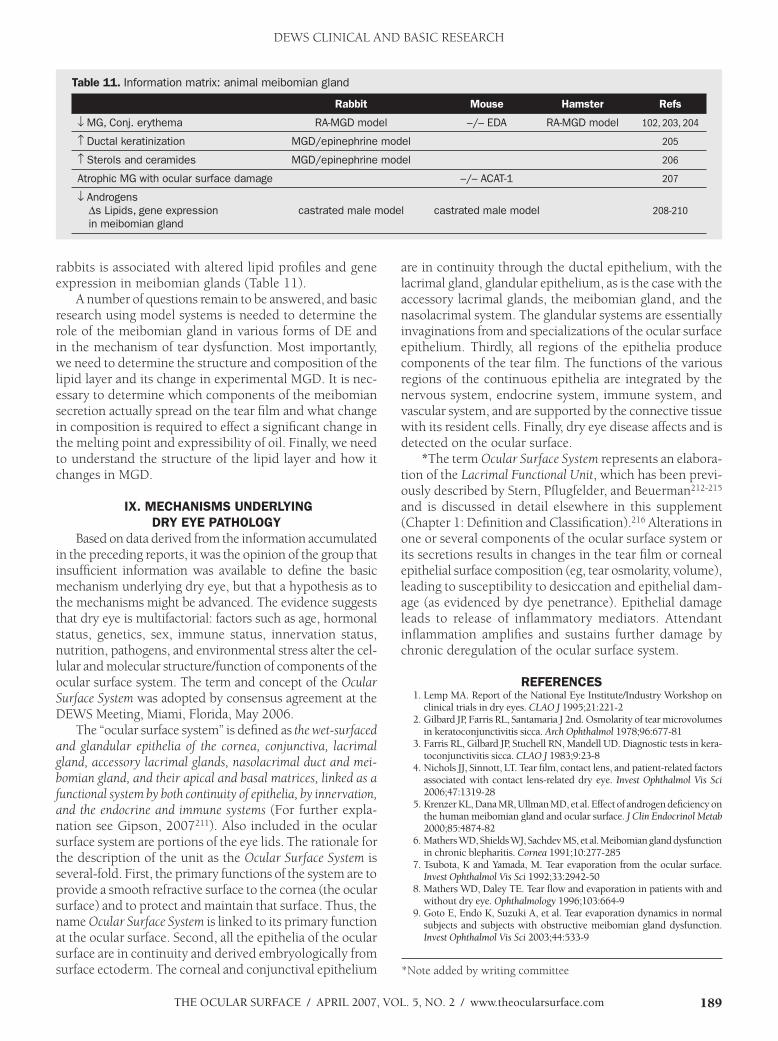
The Ocular Surface / april 2007, VOl. 5, NO. 2 / www.theocularsurface.com 189
rabbits is associated with altered lipid profiles and gene expression in meibomian glands (Table 11).
A number of questions remain to be answered, and basic research using model systems is needed to determine the role of the meibomian gland in various forms of DE and in the mechanism of tear dysfunction. Most importantly, we need to determine the structure and composition of the lipid layer and its change in experimental MGD. It is nec-essary to determine which components of the meibomian secretion actually spread on the tear film and what change in composition is required to effect a significant change in the melting point and expressibility of oil. Finally, we need to understand the structure of the lipid layer and how it changes in MGD.
Ix.MechanISMSunderlyInGdryeyepatholoGy
Based on data derived from the information accumulated in the preceding reports, it was the opinion of the group that insufficient information was available to define the basic mechanism underlying dry eye, but that a hypothesis as to the mechanisms might be advanced. The evidence suggests that dry eye is multifactorial: factors such as age, hormonal status, genetics, sex, immune status, innervation status, nutrition, pathogens, and environmental stress alter the cel-lular and molecular structure/function of components of the ocular surface system. The term and concept of the Ocular Surface System was adopted by consensus agreement at the DEWS Meeting, Miami, Florida, May 2006.
The “ocular surface system” is defined as the wet-surfaced and glandular epithelia of the cornea, conjunctiva, lacrimal gland, accessory lacrimal glands, nasolacrimal duct and mei-bomian gland, and their apical and basal matrices, linked as a functional system by both continuity of epithelia, by innervation, and the endocrine and immune systems (For further expla-nation see Gipson, 2007211). Also included in the ocular surface system are portions of the eye lids. The rationale for the description of the unit as the Ocular Surface System is several-fold. First, the primary functions of the system are to provide a smooth refractive surface to the cornea (the ocular surface) and to protect and maintain that surface. Thus, the name Ocular Surface System is linked to its primary function at the ocular surface. Second, all the epithelia of the ocular surface are in continuity and derived embryologically from surface ectoderm. The corneal and conjunctival epithelium
are in continuity through the ductal epithelium, with the lacrimal gland, glandular epithelium, as is the case with the accessory lacrimal glands, the meibomian gland, and the nasolacrimal system. The glandular systems are essentially invaginations from and specializations of the ocular surface epithelium. Thirdly, all regions of the epithelia produce components of the tear film. The functions of the various regions of the continuous epithelia are integrated by the nervous system, endocrine system, immune system, and vascular system, and are supported by the connective tissue with its resident cells. Finally, dry eye disease affects and is detected on the ocular surface.
*The term Ocular Surface System represents an elabora-tion of the Lacrimal Functional Unit, which has been previ-ously described by Stern, Pflugfelder, and Beuerman212-215 and is discussed in detail elsewhere in this supplement (Chapter 1: Definition and Classification).216 Alterations in one or several components of the ocular surface system or its secretions results in changes in the tear film or corneal epithelial surface composition (eg, tear osmolarity, volume), leading to susceptibility to desiccation and epithelial dam-age (as evidenced by dye penetrance). Epithelial damage leads to release of inflammatory mediators. Attendant inflammation amplifies and sustains further damage by chronic deregulation of the ocular surface system.
referenceS 1. lemp Ma. report of the National eye institute/industry Workshop on
clinical trials in dry eyes. CLAO J 1995;21:221-2 2. Gilbard Jp, farris rl, Santamaria J 2nd. Osmolarity of tear microvolumes
in keratoconjunctivitis sicca. Arch Ophthalmol 1978;96:677-81 3. farris rl, Gilbard Jp, Stuchell rN, Mandell uD. Diagnostic tests in kera-
toconjunctivitis sicca. CLAO J 1983;9:23-8 4. Nichols JJ, Sinnott, lT. Tear film, contact lens, and patient-related factors
associated with contact lens-related dry eye. Invest Ophthalmol Vis Sci 2006;47:1319-28
5. Krenzer Kl, Dana Mr, ullman MD, et al. effect of androgen deficiency on the human meibomian gland and ocular surface. J Clin Endocrinol Metab 2000;85:4874-82
6. Mathers WD, Shields WJ, Sachdev MS, et al. Meibomian gland dysfunction in chronic blepharitis. Cornea 1991;10:277-285
7. Tsubota, K and Yamada, M. Tear evaporation from the ocular surface. Invest Ophthalmol Vis Sci 1992;33:2942-50
8. Mathers WD, Daley Te. Tear flow and evaporation in patients with and without dry eye. Ophthalmology 1996;103:664-9
9. Goto e, endo K, Suzuki a, et al. Tear evaporation dynamics in normal subjects and subjects with obstructive meibomian gland dysfunction. Invest Ophthalmol Vis Sci 2003;44:533-9
table11. Information matrix: animal meibomian gland
rabbit Mouse hamster refs
↓ MG, Conj. erythema RA-MGD model –/– EDA RA-MGD model 102, 203, 204
↑ Ductal keratinization MGD/epinephrine model 205
↑ Sterols and ceramides MGD/epinephrine model 206
Atrophic MG with ocular surface damage –/– ACAT-1 207
↓ Androgens ∆s Lipids, gene expression in meibomian gland
castrated male model castrated male model 208-210
DEWS CLINICAL AND BASIC RESEARCH
*Note added by writing committee

The Ocular Surface / april 2007, VOl. 5, NO. 2 / www.theocularsurface.com190
10. cermak JM, Krenzer Kl, Sullivan rM, et al. is complete androgen insen-sitivity syndrome associated with alterations in the meibomian gland and ocular surface? Cornea 2003;22:516-21
11. Mainstone Jc, Bruce aS, Golding Tr. Tear meniscus measurement in the diagnosis of dry eye. Curr Eye Res 1996;15:653-61
12. Kallarackal Gu, ansari ea, amos N, et al. a comparative study to assess the clinical use of fluorescein meniscus time (fMT) with tear break up time (TBuT) and Schirmer’s tests (ST) in the diagnosis of dry eyes. Eye 2002;16:594-600
13. Savini G, Barboni p, Zanini M. Tear meniscus evaluation by optical coher-ence tomography. Ophthalmic Surg Lasers Imaging 2006;37:112-8
14. craig Jp, Tomlinson a. importance of the lipid layer in human tear film stability and evaporation. Optom Vis Sci 1997;74:8-13
15. craig Jp, Singh i, Tomlinson a, et al. The role of tear physiology in ocular surface temperature. Eye 2000;14 ( pt 4):635-41
16. Tiffany JM, Winter N, Bliss G. Tear film stability and tear surface tension. Curr Eye Res 1989;8:507-15
17. Nagyova, B, Tiffany JM. components responsible for the surface tension of human tears. Curr Eye Res 1999;19:4-11.
18. Glasson MJ, Stapleton f, Keay l, et al. Differences in clinical parameters and tear film of tolerant and intolerant contact lens wearers. Invest Ophthalmol Vis Sci 2003;44:5116-24
19. Begley cG, chalmers rl, abetz l, et al. The relationship between habitual patient-reported symptoms and clinical signs among patients with dry eye of varying severity. Invest Ophthalmol Vis Sci 2003;44:4753-61
20. Yamada M, Mochizuki h, Kawai M, et al. Decreased tear lipocalin con-centration in patients with meibomian gland dysfunction. Br J Ophthalmol 2005;89:803-5
21. Dohlman ch, friend J, Kalevar V, et al. The glycoprotein (mucus) content of tears from normals and dry eye patients. Exp Eye Res 1976;22:359-65
22. argueso p, Balaram M, Spurr-Michaud S, et al. Decreased levels of the goblet cell mucin Muc5ac in tears of patients with Sjogren’s syndrome. Invest Ophthalmol Vis Sci 2002;43:1004-11
23. Zhao h, Jumblatt Je , Wood TO, Jumblatt MM. Quantification of Muc5ac protein in human tears. Cornea 2001;20:873-7
24. Goto e, Tseng Sc. Differentiation of lipid tear deficiency dry eye by ki-netic analysis of tear interference images. Arch Ophthalmol 2003;121:173-80
25. Danjo Y, hamano T. Observation of precorneal tear film in patients with Sjogren’s syndrome. Acta Ophthalmol Scand 1995;73:501-5
26. Shine We, Mcculley Jp. Keratoconjunctivitis sicca associated with meibo-mian secretion polar lipid abnormality. Arch Ophthalmol 1998;116:849-52
27. Grus fh, Sabuncuo p, herber S, augustin aJ. analysis of tear protein pat-terns for the diagnosis of dry eye. Adv Exp Med Biol 2002;506:1213-6
28. Ohashi Y, ishida r, Kojima T, et al. abnormal protein profiles in tears with dry eye syndrome. Am J Ophthalmol 2003;136:291-9
29. Virtanen T, Konttinen YT, harkonen M, Tervo T. Tear fluid plasmin activ-ity of dry eye patients with Sjogren’s syndrome. Acta Ophthalmol Scand 1997;75:137-41
30. afonso aa, Sobrin l, Monroy Dc, et al. Tear fluid gelatinase B activity correlates with il-1alpha concentration and fluorescein clearance in ocular rosacea. Invest Ophthalmol Vis Sci 1999;40:2506-12
31. Sobrin l, liu a, Monroy Dc, et al. regulation of MMp-9 activity in human tear fluid and corneal epithelial culture supernatant. Invest Ophthalmol Vis Sci 2000;41:1703-9
32. Grus fh, podust VN, Bruns K, et al. SelDi-TOf-MS proteinchip array profiling of tears from patients with dry eye. Invest Ophthalmol Vis Sci 2005;46: 863-76
33. Danjo Y, lee M, horimoto K, hamano T. Ocular surface damage and tear lactoferrin in dry eye syndrome. Acta Ophthalmol (copenh) 1994;72:433-7
34. Koo BS, lee DY, ha hS, et al. comparative analysis of the tear protein expression in blepharitis patients using two-dimensional electrophoresis. J Proteome Res 2005;4:719-24
35. Seal DV. The effect of aging and disease on tear constituents. Trans Oph-thalmol Soc UK 1985;104 ( pt 4):355-62
36. Glasson, M, Stapleton f, Willcox M. lipid, lipase and lipocalin differ-ences between tolerant and intolerant contact lens wearers. Curr Eye Res 2002;25:227-35
37. aho VV, Nevalainen TJ, paavilainen V, Saari KM. Group iia phospholipase a2 content of tears in patients with keratoconjunctivitis sicca. Graefes Arch Clin Exp Ophthalmol 2002 Jul;240(7):521-3
38. Tishler M, Yaron i, Geyer O, et al. elevated tear interleukin-6 levels in patients with Sjogren syndrome. Ophthalmology 1998;105:2327-9
39. pflugfelder Sc, Jones D, Ji Z, et al. altered cytokine balance in the tear fluid and conjunctiva of patients with Sjogren’s syndrome keratoconjunctivitis sicca. Curr Eye Res 999;19:201-11
40. Solomon a, Dursun D, liu Z, et al. pro- and anti-inflammatory forms of interleukin-1 in the tear fluid and conjunctiva of patients with dry-eye disease. Invest Ophthalmol Vis Sci 2001;42:2283-92
41. Molloy Mp, Bolis S, herbert Br, et al. establishment of the human reflex tear 2D-paGe reference map: a first step towards the diagnosis of disease. Electrophoresis 1997;18: 2811-5
42. Tsai pS, evans Je, Green KM, et al. proteomic analysis of human meibo-mian gland secretions. Br J Ophthalmol 2006;90:372-7
43. Mulvenna i, Stapleton f, hains pG, et al. low molecular weight analysis of tears using matrix assisted laser desorption ionization-time of flight mass spectrometry. Clin Experiment Ophthalmol 2000;28:205-7
44. fung K, Morris c, Duncan M. Mass spectrometric techniques applied to the analysis of human tears: a focus on the peptide and protein constitu-ents. Adv Exp Med Biol 2002;506(pta):601-5
45. ham BM, Jacob JT, Keese MM, cole rB. identification, quantification and comparison of major non-polar lipids in normal and dry eye tear lipidomes by electrospray tandem mass spectrometry. J Mass Spectrom 2004;39:1321-36
46. Gipson iK, Spurr-Michaud SJ, argueso p, et al. Mucin gene expression in immortalized human corneal-limbal and conjunctival epithelial cell lines. Invest Ophthalmol Vis Sci 2003;44:2496-506
47. robertson DM, li l, fisher S, et al. characterization of growth and dif-ferentiation in a telomerase-immortalized human corneal epithelial cell line. Invest Ophthalmol Vis Sci 2005;46:470-8
48. Gilbard Jp, rossi Sr, heyda KG. Tear film and ocular surface changes after closure of the meibomian gland orifices in the rabbit. Ophthalmology 1989;96:1180-6
49. Stewart p, chen Z, farley W, et al. effect of experimental dry eye on tear sodium concentration in the mouse. Eye Contact Lens 2005;31:175-8
50. Gilbard Jp, rossi Sr, Gray Kl, et al. Tear film osmolarity and ocular surface disease in two rabbit models for keratoconjunctivitis sicca. Invest Ophthalmol Vis Sci 1988;29:374-8
51. Nagelhout TJ, Gamache Da, roberts l, et al. preservation of tear film integrity and inhibition of corneal injury by dexamethasone in a rabbit model of lacrimal gland inflammation-induced dry eye. J Ocul Pharmacol Ther 2005;21:139-48.
52. Salvatore Mf, pedroza l, Beuerman rW. Denervation of rabbit lacrimal gland increases levels of transferrin and unidentified tear proteins of 44 and 36 kDa. Curr Eye Res 1999;18:455-66.
53. Song XJ, li DQ, farley W, et al. Neurturin-deficient mice develop dry eye and keratoconjunctivitis sicca. Invest Ophthalmol Vis Sci 2003;44:4223-9
54. poon ac, Geerling G, Dart JK, et al. autologous serum eyedrops for dry eyes and epithelial defects: clinical and in vitro toxicity studies. Br J Ophthalmol 2001;85:1188-97
55. ralph ra. conjunctival goblet cell density in normal subjects and in dry eye syndromes. Invest Ophthalmol Vis Sci 1975;14:299-302
56. Nelson JD, havener Vr, cameron JD. cellulose acetate impressions of the ocular surface. Dry eye states. Arch Ophthalmol 1983;101:1869-72
57. pflugfelder Sc, Tseng Sc, Yoshino K, et al. correlation of goblet cell density and mucosal epithelial membrane mucin expression with rose bengal staining in patients with ocular irritation. Ophthalmology 1997;104:223-35
58. Blodi Ba, Byrne Ka, Tabbara Kf. Goblet cell population among patients with inactive trachoma. Int Ophthalmol 1988;12:41-5
59. Sommer a. Treatment of corneal xerophthalmia with topical retinoic acid. Am J Ophthalmol 1983;95:349-52
60. lievens cW, connor cG, Murphy h. comparing goblet cell densities in patients wearing disposable hydrogel contact lenses versus silicone hydrogel contact lenses in an extended-wear modality. Eye Contact Lens 2003;29:241-4.
61. albietz JM, Mclennan SG, lenton lM. Ocular surface management of photorefractive keratectomy and laser in situ keratomileusis. J Refract Surg 2003;19:636-44
62. Versura p, Maltarello Mc, cellini M, et al. Detection of mucus glycocon-jugates in human conjunctiva by using the lectin-colloidal gold technique in TeM. ii. a quantitative study in dry-eye patients. Acta Ophthalmol (copenh) 1986;64:451-5
63. Garcher c, Bron aJ, Baudouin c, et al. ca 19-9 eliSa test: a new method for studying mucus changes in tears. Br J Ophthalmol 1998;82:88-90
64. Versura p, Maltarello Mc, cellini M, et al. Detection of mucus glycocon-jugates in human conjunctiva by using the lectin-colloidal gold technique in TeM. iii. a quantitative study in asymptomatic contact lens wearers. Acta Ophthalmol (copenh) 1987;65:661-7
65. hayashi Y, Kao WW, Kohno N, et al. expression patterns of sialylated epitope recognized by Kl-6 monoclonal antibody in ocular surface epithelium of normals and dry eye patients. Invest Ophthalmol Vis Sci
DEWS CLINICAL AND BASIC RESEARCH

The Ocular Surface / april 2007, VOl. 5, NO. 2 / www.theocularsurface.com 191
2004;45:2212-7 66. argueso p, Tisdale a, Mandel u, et al. The cell-layer- and cell-type-specific
distribution of GalNac-transferases in the ocular surface epithelia is altered during keratinization. Invest Ophthalmol Vis Sci 2003;44:86-92
67. Danjo Y, Watanabe h, Tisdale aS, et al. alteration of mucin in human conjunctival epithelia in dry eye. Invest Ophthalmol Vis Sci 1998;39:2602-9
68. Koufakis Di, Karabatsas ch, Sakkas li, et al. conjunctival surface changes in patients with Sjogren’s syndrome: a transmission electron microscopy study. Invest Ophthalmol Vis Sci 2006;47:541-4
69. albietz J, Sanfilippo p, Troutbeck r, lenton lM. Management of filamen-tary keratitis associated with aqueous-deficient dry eye. Optom Vis Sci 2003;80:420-30
70. Murube J, rivas l. Biopsy of the conjunctiva in dry eye patients estalishes a correlation between squamous metaplasia and dry eye clinical severity. Eur J Ophthalmol 2003;13:246-56
71. Kunert KS, Tisdale aS, Gipson iK. Goblet cell numbers and epithelial proliferation in the conjunctiva of patients with dry eye syndrome treated with cyclosporine. Arch Ophthalmol 2002;120:330-7
72. Meller D. The fine structure of chromatin alterations in conjunctival epithelial cells in keratoconjunctivitis sicca. Cornea 1999;18:225-32
73. albietz JM, Bruce aS. The conjunctival epithelium in dry eye subtypes: effect of preserved and non-preserved topical treatments. Curr Eye Res 2001;22:8-18
74. Bjerrum KB. Snake-like chromatin in conjunctival cells of normal elderly persons and of patients with primary Sjogren’s syndrome and other con-nective tissue diseases. Acta Ophthalmol Scand 1995;73:33-6
75. Brignole f, pisella pJ, De Saint Jean M, et al. flow cytometric analysis of inflammatory markers in conjunctival epithelial cells of patients with dry eyes. Invest Ophthalmol Vis Sci 2000;41:1356-63
76. Benitez del castillo JM, Wasfy Ma, fernandez c, Garcia-Sanchez J. an in vivo confocal masked study on corneal epithelium and subbasal nerves in patients with dry eye. Invest Ophthalmol Vis Sci 2004;45:3030-5
77. Xu Kp, Yagi Y, Tsubota K. Decrease in corneal sensitivity and change in tear function in dry eye. Cornea 1996;15:235-9
78. horwath-Winter J, Vidic B, Schantzer G, Schmut O. early changes in corneal sensation, ocular surface integrity, and tear-film function after laser-assisted subepithelial keratectomy. J Cataract Refract Surg 2004;30:2316-21
79. hovanesian Ja, Shah SS, Maloney rK. Symptoms of dry eye and recur-rent erosion syndrome after refractive surgery. J Cataract Refract Surg 2001;27:577-84
80. Wilson Se. laser in situ keratomileusis-induced (presumed) neurotrophic epitheliopathy. Ophthalmology 2001;108:1082-7
81. Scott iu, flynn hW Jr, feuer W, et al. endophthalmitis associated with microbial keratitis. Ophthalmology 1996;103:1864-70
82. Kawasaki S, Kawamoto S, Yokoi N, et al. up-regulated gene expression in the conjunctival epithelium of patients with Sjogren’s syndrome. Exp Eye Res 2003;77:17-26
83. Nakamura T, Nishida K, Dota a, et al. elevated expression of transglu-taminase 1 and keratinization-related proteins in conjunctiva in severe ocular surface disease. Invest Ophthalmol Vis Sci 2001;42:549-56
84. Nakamura T, Nishida K, Dota a, Kinoshita S. changes in conjunctival clusterin expression in severe ocular surface disease. Invest Ophthalmol Vis Sci 2002;43:1702-7
85. Narayanan S, Miller Wl, McDermott aM. conjunctival cytokine expres-sion in symptomatic moderate dry eye subjects. Invest Ophthalmol Vis Sci 2006;47:2445-50
86. argueso p, Tisdale a, Spurr-Michaud S, et al. Mucin characteristics of human corneal-limbal epithelial cells that exclude the rose bengal anionic dye. Invest Ophthalmol Vis Sci 2006;47:113-9
87. hatchell Dl, Sommer a. Detection of ocular surface abnormalities in experimental vitamin a deficiency. Arch Ophthalmol 1984;102:1389-93
88. Gilbard Jp, rossi Sr. Tear film and ocular surface changes in a rabbit model of neurotrophic keratitis. Ophthalmology 1990;97:308-12
89. Tei M, Spurr-Michaud SJ, Tisdale aS, Gipson iK. Vitamin a deficiency alters the expression of mucin genes by the rat ocular surface epithelium. Invest Ophthalmol Vis Sci 2000;41:82-8
90. Dursun D, Wang M, Monroy D, et al. a mouse model of keratoconjunc-tivitis sicca. Invest Ophthalmol Vis Sci 2002;43:632-8
91. ueta M, hamuro a, Yamamoto M, et al. Spontaneous ocular surface in-flammation and goblet cell disappearance in i kappa B zeta gene-disrupted mice. Invest Ophthalmol Vis Sci 2005;46:579-88
92. corfield ap, Donapaty Sr, carrington SD, et al. identification of 9-O-acetyl-N-acetylneuraminic acid in normal canine pre-ocular tear film secreted mucins and its depletion in keratoconjunctivitis sicca. Glycoconj J 2005;22:409-16
93. Tsubota, K, Goto e, fujita h, et al. Treatment of dry eye by autologous serum application in Sjogren’s syndrome. Br J Ophthalmol 1999;83:390-5
94. hori Y, Spurr-Michaud S, russo cl, et al. Differential regulation of mem-brane-associated mucins in the human ocular surface epithelium. Invest Ophthalmol Vis Sci 2004;45:114-22
95. Tseng Sc, hatchell D, Tierney N, et al. expression of specific keratin markers by rabbit corneal, conjunctival, and esophageal epithelia during vitamin a deficiency. J Cell Biol 1984;99:2279-86
96. Gao J, Schwalb Ta, addeo JV, et al. The role of apoptosis in the pathogenesis of canine keratoconjunctivitis sicca: the effect of topical cyclosporin a therapy. Cornea 1998;17:654-63
97. Toshino a, Shiraishi a, Zhang W, et al. expression of keratinocyte transglu-taminase in cornea of vitamin a-deficient rats. Curr Eye Res 2005;30:731-9
98. Yeh S, Song XJ, farley W, et al. apoptosis of ocular surface cells in experi-mentally induced dry eye. invest Ophthalmol Vis Sci 2003;44:124-9
99. luo l, li DQ, Doshi a, et al. experimental dry eye stimulates production of inflammatory cytokines and MMp-9 and activates MapK signaling path-ways on the ocular surface. Invest Ophthalmol Vis Sci 2004;45:4293-301
100. li DQ, chen Z, Song XJ, et al. Stimulation of matrix metalloproteinases by hyperosmolarity via a JNK pathway in human corneal epithelial cells. Invest Ophthalmol Vis Sci 2004;45:4302-11
101. li DQ, luo l, chen Z, et al. JNK and erK Map kinases mediate induction of il-1beta, TNf-alpha and il-8 following hyperosmolar stress in human limbal epithelial cells. Exp Eye Res 2006;82:588-96
102. cui cY, Smith Ja, Schlessinger D, chan cc. X-linked anhidrotic ectoder-mal dysplasia disruption yields a mouse model for ocular surface disease and resultant blindness. Am J Pathol 2005;167:89-95
103. rojas B, cunha r, Zafirakis p, et al. cell populations and adhesion mol-ecules expression in conjunctiva before and after bone marrow transplanta-tion. Exp Eye Res 2005;81:313-25
104. raphael M, Bellefgih S, piette Jc, et al. conjunctival biopsy in Sjogren’s syndrome: correlations between histological and immunohistochemical features. Histopathology 1988;13:191-202
105. Stern Me, Gao J, Schwalb Ta, et al. conjunctival T-cell subpopulations in Sjogren’s and non-Sjogren’s patients with dry eye. Invest Ophthalmol Vis Sci 2002;43:2609-14
106. pflugfelder Sc, huang aJ, feuer W, et al. conjunctival cytologic features of primary Sjögren’s syndrome. Ophthalmology 1990;97:985-91
107. Ogawa Y, Kuwana M, Yamazaki K, et al. periductal area as the primary site for T-cell activation in lacrimal gland chronic graft-versus-host disease. Invest Ophthalmol Vis Sci 2003;44:1888-96
108. Ogawa Y, Yamazaki K, Kuwana M, et al. a significant role of stromal fibroblasts in rapidly progressive dry eye in patients with chronic GVhD. Invest Ophthalmol Vis Sci 2001;42:111-9
109. Gulati a, Sacchetti M, Bonini a, Dana Mr. chemokine receptor ccr5 expression in conjunctival epithelium of patients with dry eye syndrome. Arch Ophthalmol 2006;124:710-6
110. Baudouin c, liang h, Bremond-Gignac D, et al. ccr 4 and ccr 5 expres-sion in conjunctival specimens as differential markers of T(h)1/T(h)2 in ocular surface disorders. J Allergy Clin Immunol 2005;116:614-9
111. aronni S, cortes M, Sacchetti M, et al. upregulation of icaM-1 expression in the conjunctiva of patients with chronic graft-versus-host disease. Eur J Ophthalmol 2006;16:17-23
112. Tsubota K, fujihara T, Saito K, Takeuchi T. conjunctival epithelium ex-pression of hla-Dr in dry eye patients. Ophthalmologica 1999;213:16-9
113. Jones DT, Monroy D, Ji Z, et al. Sjogren’s syndrome: cytokine and epstein-Barr viral gene expression within the conjunctival epithelium. Invest Ophthalmol Vis Sci 1994;35:3493-3504
114. Ogawa Y, Kodama h, Kameyama K, et al. Donor fibroblast chimerism in the pathogenic fibrotic lesion of human chronic graft-versus-host disease. Invest Ophthalmol Vis Sci 2005;46:4519-27
115. Matsumoto i, Tsubota K, Satake Y, et al. common T cell receptor clonotype in lacrimal glands and labial salivary glands from patients with Sjogren’s syndrome. J Clin Invest 1996;97:1969-77
116. pepose JS, akata rf, pflugfelder Sc, Voight W. Mononuclear cell pheno-types and immunoglobulin gene rearrangements in lacrimal gland biopsies from patients with Sjogren’s syndrome. Ophthalmology 1990;97:1599-605
117. Williamson J, Gibson aa, Wilson T, et al. histology of the lacrimal gland in keratoconjunctivitis sicca. Br J Ophthalmol 1973;57:852-8
118. Gao J, Morgan G, Tieu D, et al. icaM-1 expression predisposes ocular tissues to immune-based inflammation in dry eye patients and Sjogrens syndrome-like Mrl/lpr mice. Exp Eye Res 2004;78:823-35
119. Sullivan D, et al. aqueous tear deficiency in Sjogren’s syndrome: possible causes and potential treatment, in pleyer u, et al (eds). Oculodermal dis-eases--immunology of bullous oculo-muco-cutaneous disorders. Buren,
DEWS CLINICAL AND BASIC RESEARCH

The Ocular Surface / april 2007, VOl. 5, NO. 2 / www.theocularsurface.com192
The Netherlands, aeolus press, 1997, pp 95-152 120. Zoukhri D. effect of inflammation on lacrimal gland function. Exp Eye
Res 2006;82:885-98 121. Tsubota K, fujita h, Tsuzaka K, Takeuchi T. Quantitative analysis of
lacrimal gland function, apoptotic figures, fas and fas ligand expression of lacrimal glands in dry eye patients. Exp Eye Res 2003;76:233-40
122. Saito i, Terauchi K, Shimuta M, et al. expression of cell adhesion molecules in the salivary and lacrimal glands of Sjogren’s syndrome. J Clin Lab Anal 1993;7:180-7
123. Ono M, Yoshino K, Tsubota K, Saito i. Subclass expression of iga in lacrimal glands of patients with Sjogren’s syndrome. Adv Exp Med Biol 1994;350:185-8
124. Zhan h, Towler hM, calder Vl. The immunomodulatory role of human conjunctival epithelial cells. Invest Ophthalmol Vis Sci 2003;44:3906-10
125. Barabino S, Shen l, chen l, et al. The controlled-environment chamber: a new mouse model of dry eye. Invest Ophthalmol Vis Sci 2005;46:2766-71
126. Niederkorn J, Stern Me, pflugfelder Sc, et al. Desiccating stress induces T cell-mediated Sjogren’s syndrome-like lacrimal keratoconjunctivitis. J Immunol 2006;176:3950-57
127. van Blokland Sc, Versnel Ma. pathogenesis of Sjogren’s syndrome: char-acteristics of different mouse models for autoimmune exocrinopathy. Clin Immunol 2002;103:111-24
128. hassan aS, clouthier SG, ferrara Jl, et al. lacrimal gland involvement in graft-versus-host disease: a murine model. Invest Ophthalmol Vis Sci 2005;46:2692-7
129. Yang T, Zeng h, Zhang J, et al. Mhc class ii molecules, cathepsins, and la/SSB proeins in lacrimal acinar cell endomembranes. Am J Physiol 1999;277:c994-c1007
130. McDermott a, perez V, huang aJ, et al. pathways of corneal and ocular surface inflammation: a perspecive from the cullen Symposium. Ocul Surf 2005;3:S131-S138
131. lee-Wing MW, hodge WG, Diaz-Mitoma f. investigating a viral etiology for keratoconjunctivitis sicca among patients who are positive for human immunodeficiency virus. Cornea 1999;18:671-4
132. Matoba aY. Ocular disease associated with epstein-Barr virus infection. Surv Ophthalmol 1990;35:145-50
133. Tsubota K, fujishima h, Toda i, et al. increased levels of epstein-Barr virus DNa in lacrimal glands of Sjogren’s syndrome patients. Acta Ophthalmol Scand 1995;73:425-30
134. Zegans Me, anninger W, chapman c, Gordon Sr. Ocular manifestations of hepatitis c virus infection. Curr Opin Ophthalmol 2002;13:423-7
135. Merayo-lloves J, Baltatzis S, foster cS. epstein-Barr virus dacryoad-enitis resulting in keratoconjunctivitis sicca in a child. Am J Ophthalmol 2001;132:922-3
136. Bacman S, perez-leiros c, Sterin-Borda l, et al. autoantibodies against lacrimal gland M3 muscarinic acetylcholine receptors in patients with primary Sjogren’s syndrome. Invest Ophthalmol Vis Sci 1998;39:151-6
137. Tsubota K, fujihara T, Takeuchi T. Soluble interleukin-2 receptors and serum autoantibodies in dry eye patients: correlation with lacrimal gland function. Cornea 1997;16:339-44
138. paulsen f, langer G, hoffman W, Berry M. human lacrimal gland mucins. Cell Tissue Res 2004;316:167-77
139. rivas l, Murube J, Toledano a. [innervation of the lachrymal gland in patients with primary Sjogren’s syndrome. an immunohistopathological study]. Arch Soc Esp Oftalmol 2002;77:623-29
140. Obata h, Yamamoto S, horiuchi h, Machinami r. histopathologic study of human lacrimal gland. Statistical analysis with special reference to ag-ing. Ophthalmology 1995;102:678-86
141. Mccarty ca, Bansal aK, livingston pM, et al. The epidemiology of dry eye in Melbourne, australia. Ophthalmology 1998;105:1114-9
142. Gillette T, allansmith Mr, Greiner JV, Janusz M. histologic and im-munohistologic comparison of main and accessory lacrimal tissue. Am J Ophthalmol 1980;89:724-30
143. Seifert p, Spitznas M, Koch f, cusumano a. The architecture of human accessory lacrimal glands. Ger J Ophthalmol 1993;2:444-54
144. allansmith Mr, Kajiyama G, abelson MB, Simon Ma. plasma cell content of main and accessory lacrimal glands and conjunctiva. Am J Ophthalmol 1976;82:819-26
145. hunt S, Spitznas M, Seifert p, rauwolf M. Organ culture of human main and accessory lacrimal glands and their secretory behaviour. Exp Eye Res 1996;62:541-54
146. Seifert p, Spitznas M. Vasoactive intestinal polypeptide (Vip) innervation of the human eyelid glands. Exp Eye Res 1999;68:685-92
147. esmaeli-Gutstein B, hewlett Br, harvey JT. characterization of adrenergic receptors in the accessory lacrimal glands of the upper eyelid. Ophthal
Plast Reconstr Surg 1999;15:245-51 148. Gillette Te, Greiner JV, allansmith Mr. immunohistochemical localization
of human tear lysozyme. Arch Ophthalmol 1981;99:298-300 149. Obata h, horiuchi h, Dobashi Y, et al. immunohistochemical localization
of epidermal growth factor in human main and accessory lacrimal glands. Jpn J Ophthalmol 1993;37:113-21
150. Yen MT, pflugfelder Sc, feuer WJ. The effect of punctal occlusion on tear production, tear clearance, and ocular surface sensation in normal subjects. Am J Ophthalmol 2001;131:314-23.
151. ayub M, Thale aB, hedderich J, et al. The cavernous body of the human efferent tear ducts contributes to regulation of tear outflow. Invest Oph-thalmol Vis Sci 2003;44:4900-7
152. paulsen fp, Thale aB, hallman uJ, et al. The cavernous body of the human efferent tear ducts: function in tear outflow mechanism. Invest Ophthalmol Vis Sci 2000;41:965-70
153. Guo Z, azzarolo aM, Schecter Je, et al. lacrimal gland epithelial cells stimulate proliferation in autologous lymphocyte preparations. Exp Eye Res 2000;71:11-22
154. Guo Z, Song D, azzarolo aM, et al. autologous lacrimal-lymphoid mixed-cell reactions induce dacryoadenitis in rabbits. Exp Eye Res 2000;71:23-31
155. Zhu Z. lacrimal histopathology and ocular surface disease in a rabbit model of autoimmune dacryoadenitis. Cornea 2003;22:25-32
156. franklin r, McGee DW, Shepard Kf. lacrimal gland-directed B cell re-sponses. J Immunol 1985;135:95-99
157. liu S, Zhou Dh, franklin rM. lacrimal gland-derived lymphocyte pro-liferation potentiating factor. Invest Ophthalmol Vis Sci 1993;34:650-7
158. Jabs Da, enger c, prendergast ra. Murine models of Sjogren’s syndrome. evolution of the lacrimal gland inflammatory lesions. Invest Ophthalmol Vis Sci 1991;32:371-80
159. Jabs Da, prendergast ra, rorer eM, et al. cytokines in autoimmune lacrimal gland disease in Mrl/MpJ mice. Invest Ophthalmol Vis Sci 2001;42:2567-71
160. akpek eK, Jabs Da, Gerard hc, et al. chemokines in autoimmune lacrimal gland disease in Mrl/MpJ mice. Invest Ophthalmol Vis Sci 2004;45:185-90
161. Sato eh, ariga h, Sullivan Da. impact of androgen therapy in Sjogren’s syndrome: hormonal influence on lymphocyte populations and ia expres-sion in lacrimal glands of Mrl/Mp-lpr/lpr mice. Invest Ophthalmol Vis Sci 1992;33:2537-45
162. rocha e, Wickham la, huang Z, et al. presence and testosteroe influence on the levels of anti- and pro-inflammatory cytokines in lacrimal tissues of a mouse model of Sjogren’s syndrome. Adv Exp Med Biol 1998;438:485-91
163. Toda i, Wickham la, Sullivan Da. Gender and androgen treatment influence the expression of proto-oncogenes and apoptotic factors in lacrimal and salivary tissues of Mrl/lpr mice. Clin Immunol Immunopathol 1998;86:59-71
164. Toda i, Sullivan BD, Wickham la, Sullivan Da. Gender- and androgen-related influence on the expression of proto-oncogene and apoptotic factor mrNas in lacrimal glands of autoimmune and non-autoimmune mice. J Steroid Biochem Mol Biol 1999;71:49-61
165. Tornwall J, lane Te, fox ri, fox hS. T cell attractant chemokine expres-sion initiates lacrimal gland destruction in nonobese diabetic mice. Lab Invest 1999;79:1719-26
166. robinson cp, cornelius J, Bounous De, et al. characterization of the changing lymphocyte populations and cytokine expression in the exocrine tissues of autoimmune NOD mice. Autoimmunity 1998;27:29-44
167. Vendramini ac, Soo c, Sullivan Da. Testosterone-induced suppression of autoimmune disease in lacrimal tissue of a mouse model (NZB/NZW f1) of Sjogren’s syndrome. Invest Ophthalmol Vis Sci 1991;32:3002-6
168. Sullivan Da, edwards Ja. androgen stimulation of lacrimal gland func-tion in mouse models of Sjogren’s syndrome. J Steroid Biochem Mol Biol 1997;60:237-45
169. ariga h, edwards J, Sullivan Da. androgen control of autoimmune expression in lacrimal glands of Mrl/Mp-lpr/lpr mice. Clin Immunol Immunopathol 1989;53:499-508
170. Sato e, Sullivan D. comparative influence of steroid hormones and im-munosuppressive agents on autoimmune expression in lacrimal glands of a female mouse model of Sjogren’s syndrome. Invest Ophthalmol Vis Sci 1994;35:2632-42
171. rocha f, et al. effect of androgen analogue treatment and androgen withdrawal on lacrimal gland inflammation in a mouse model (Mrl/Mp-lpr/lpr) of Sjogren’s syndrome. Reg Immunol 1994;6:270-7
172. liu Sh, prendergast ra, Silverstein aM. experimental autoimmune dacryoadenitis. i. lacrimal gland disease in the rat. Invest Ophthalmol Vis Sci 1987;28:270-5
173. Mizejewski G. Studies of autoimmune induction in the rat lacrimal gland.
DEWS CLINICAL AND BASIC RESEARCH

The Ocular Surface / april 2007, VOl. 5, NO. 2 / www.theocularsurface.com 193
Experientia 1978;34:1093-5 174. huang Z, lambert rW, Wickham la, Sullivan Da. analysis of cytomega-
lovirus infection and replication in acinar epithelial cells of the rat lacrimal gland. Invest Ophthalmol Vis Sci 1996;37:1174-86
175. Stern M. Ocular surface inflammation: a causative factor in dry eye. J Rheumatol 1997;50(Suppl):42
176. Gao J, Stern M. Modulators of apoptosis in the lacrimal gland of dry eye dogs. J Rheumatol 1997;50(Suppl):43
177. liu Sh, Zhou Dh, hess aD. adoptive transfer of experimental autoim-mune dacryoadenitis in susceptible and resistant mice. Cell Immunol 1993;150:311-20
178. Ono M, rocha fJ, Sullivan Da. immunocytochemical location and hormonal control of androgen receptors in lacrimal tissues of the fe-male Mrl/Mp-lpr/lpr mouse model of Sjogren’s syndrome. Exp Eye Res 1995;61:659-66
179. Yamamoto h, Sims Ne, Maccauley Sp, et al. alterations in the secretory response of non-obese diabetic (NOD) mice to muscarinic receptor stimu-lation. Clin Immunol Immunopathol 1996;78:245-55
180. Winer S, astsaturov i, cheung r, et al. primary Sjogren’s syndrome and deficiency of ica69. Lancet 2002;360:1063-9
181. Zoukhri D, Kublin cl. impaired neurotransmitter release from lacrimal and salivary gland nerves of a murine model of Sjogren’s syndrome. invest Ophthalmol Vis Sci 2001;42:925-32
182. haneji N, Nakamura T, Takio K, et al. identification of alpha-fodrin as a candidate autoantigen in primary Sjogren’s syndrome. Science 1997;276:604-7
183. fang Y, choi D, Searles rp, Mathers WD.a time course microarray study of gene expression in the mouse lacrimal gland after acute corneal trauma. Invest Ophthalmol Vis Sci 2005;46:461-9
184. paulsen fp, foge M, Thale aB, et al. animal model for the absorption of lipophilic substances from tear fluid by the epithelium of the nasolacrimal ducts. Invest Ophthalmol Vis Sci 2002;43:3137-43
185. paulsen f, Thale aB, Mentlein r.What happens to tears inside the efferent lacrimal passage? an animal experimental study. Graefes Arch Clin Exp Ophthalmol 2000;238:496-9
186. hirt ra. comparative anatomy of the canine efferent tear duct system with regard to mucin production. Ann Anat Supp 2003;185:259-260
187. rios JD, horikawa Y, chen ll, et al. age-dependent alterations in mouse exorbital lacrimal gland structure, innervation and secretory response. Exp eye res 2005;80:477-91
188. Sullivan BD, cermak JM, Sullivan rM, et al. correlations between nutrient intake and the polar lipid profiles of meibomian gland secretions in women with Sjogren’s syndrome. Adv Exp Med Biol 2002;506(pt a):441-7
189. robin JB, Jester JV, Nobe J, et al. in vivo transillumination biomicroscopy and photography of meibomian gland dysfunction. a clinical study. Oph-thalmology 1985;92:1423-6
190. Gutgesell VJ, Stern Ga, hood ci. histopathology of meibomian gland dysfunction. Am J Ophthalmol 1982;94:383-7
191. Matsuoka T. Video-meibographic observations of the meibomian gland. Jpn J Clin Ophthalmol 1996;50:351-4
192. Yokoi N, Mossa f, Tiffany JM, Bron aJ. assessment of meibomian gland function in dry eye by meibometry. Arch Ophthalmol 1999;117:723-9
193. hykin pG, Bron aJ. age-related morphological changes in lid margin and meibomian gland anatomy. Cornea 1992;11:334-42
194. Shimazaki J, Goto e, Ono M, et al. Meibomian gland dysfunction in patients with Sjögren syndrome. Ophthalmology 1998;105:1485-8
195. Shimazaki J, Sakata M, Tsuboto K. Ocular surface changes and dis-
comfort in patients with meibomian gland dysfunction. Arch Ophthalmol 1995;113:1266-70
196. Sullivan BD, evans Je, Dana Mr, Sullivan Da. impact of androgen defi-ciency on the lipid profiles in human meibomian gland secretions. Adv Exp Med Biol 2002;506(pt a):449-58
197. Sullivan BD, evans Je, Krenzer Kl, et al. impact of antiandrogen treat-ment on the fatty acid profile of neutral lipids in human meibomian gland secretions. J Clin Endocrinol Metab 2000;85:4866-73
198. Sullivan BD, evans Je, cermak JM, et al. complete androgen insensitivity syndrome: effect on human meibomian gland secretions. Arch Ophthalmol 2002;120:1689-99
199. Ong Bl, larke Jr. Meibomian gland dysfunction: some clinical, biochemi-cal and physical observations. Ophthalmic Physiol Opt 1990;10:144-8
200. Dougherty JM, Mcculley Jp. Bacterial lipases and chronic blepharitis. Invest Ophthalmol Vis Sci 1986;27:486-91
201. isreb Ma, Greiner VJ, Korb Dr, et al. correlation of lipid layer thickness measurements with fluorescein tear film break-up time and Schirmer’s test. Eye 2003;17:79-83
202. Sullivan BD, evans Je, Dana Mr, Sullivan Da. influence of aging on the polar and neutral lipid profiles in human mebomian gland secretions. Arch Ophthalmol 2006;124:1286-92
203. lambert rW, Smith re. pathogenesis of blepharoconjunctivitis complicat-ing 13-cis-retinoic acid (isotretinoin) therapy in a laboratory model. Invest Ophthalmol Vis Sci 1988;29:1559-64
204. lambert rW, Smith re. effects of 13-cis-retinoic acid on the hamster meibomian gland. J Invest Dermatol 1989;92:321-5
205. Jester JV, Nicolaides N, Kiss-palvolgyi i, Smith re. Meibomian gland dysfunction. ii. The role of keratinization in a rabbit model of MGD. Invest Ophthalmol Vis Sci 1989;30:936-45
206. Nicolaides N, Santos ec, Smith re, Jester JV. Meibomian gland dysfunction. iii. Meibomian gland lipids. Invest Ophthalmol Vis Sci 1989;30:946-51
207. Yagyu h, Kitamine T, Osuga J, et al. absence of acaT-1 attenuates ath-erosclerosis but causes dry eye and cutaneous xanthomatosis in mice with congenital hyperlipidemia. J Biol Chem 2000;275:21324-30
208. Steagall r, Yamagami h, Wickham la, Sullivan Da. androgen control of gene expression in the rabbit mebomian gland. Adv Exp Med Biol 2002;506(pt a):465-476.
209. Sullivan Da, Sullivan BD, ullman MD, et al. androgen influence on the meibomian gland. Invest Ophthalmol Vis Sci 2000;41:3732-42
210. Schirra f, Suzuki T, richards SM, et al. androgen control of gene expression in the mouse meibomian gland. Invest Ophthalmol Vis Sci 2005;46:3666-75
211. Gipson iK. friedenwald lecture: The ocular surface: the challenge to enable and protect vision. Invest Ophthalmol Vis Sci (2007, in press)
212. Stern Me, Beuerman rW, fox ri, et al. The pathology of dry eye: the interaction between the ocular surface and lacrimal glands. Cornea 1998;17:584-9
213. pflugfelder Sc, Solomon a, Stern Me. The diagnosis and management of dry eye: a twenty-five year review. Cornea 2000;19:644-9
214. Beuerman rW, Mircheff aK, pflugfelder Sc, Stern Me. The lacrimal func-tional unit, in pflugfelder Sc, Stern Me, Beuerman rW. Dry eye and the ocular surface—a unified approach. New York, Marcel Dekker, 2004
215. Stern Me, Gao J, Siemasko Kf, et al. The role of the lacrimal functional unit in the pathophysiology of dry eye. Exp Eye Res 2004;78:409-16
216. (No authors listed) Definition and classification of dry eye: report of the Definition and classification Subcommittee of the 2007 Dry eye Work-Shop. Ocul Surf 2007;5:75-92
DEWS CLINICAL AND BASIC RESEARCH

The Ocular Surface / april 2007, VOl. 5, NO. 2 / www.theocularsurface.com194

The Ocular Surface / april 2007, VOl. 5, NO. 2 / www.theocularsurface.com 195
Aabbreviations, 74, 180accessory lacrimal glands, 185-87ace (angiotensin converting enzyme) inhibitors, 82acne rosacea, tetracycline for, 171-72acr50, acr70, 73acute ocular surface inflammation, 185aDDe. See aqueous deficient dry eye (aDDe)affected population, 115-16age and aging, 78, 96age-related dry eye (arDe), 73, 80aiDS, 80aKc. See atopic keratoconjunctivitis (aKc)allergic conjunctivitis, 86allergic eye disease, 87allgrove syndrome, 80allogeneic bone marrow transplantation, 100alpha-fodrin, 186american academy of Ophthalmology preferred
practice patterns, 163american congress of rheumatology (acr)
indices, 159american-european consensus Group, 103tandrogen deficiency, 78, 83-84, 187-89 levels, 100 topically applied, 171androgen insensitivity syndrome, 78angiogenesis, tetracyclines and, 171animal models for dry eye disease, 181-82 meibomian gland, 188-89 ocular surface, 182-83 tear film, 181tantiandrogen therapy, 78, 187-88anti-inflammatory therapy, 170-73 corticosteroids, 171 cyclosporine-a (csa), 170 tetracyclines, 171-73aqueous deficient dry eye (aDDe), 76, 110 classification, 77f, 79 defined, 73, 78 lacrimal secretory response and, 87aqueous tear deficiency (aTD), 73, 85farDe, 73, 80artificial tears, 73, 171 characteristics and effects of, 164-65 hypo-osmotic, 166 ocular surface disorders and, 84 osmolarities of, 165 preservatives, 165-66asymptomatic dry eye disease, 110, 112atopic keratoconjunctivitis (aKc), 73at-risk population, screening, 112aTS. See artificial tear substituteautoantigens, 183autoimmune acinar damage, 79autoimmune disease, research, 183-85autologous serum, 169-70
BBac. See benzalkonium chloride (Bac)basic science studies, 163tBaudoin, christophe, 65, 71Baudouin, c., 86Beaver Dam eye Study, 81, 86, 93, 95tBegley, c. B., 118, 120tBehrens, a., 173benzalkonium chloride (Bac), 165 ocular surface disorders and, 84 tear film instability and, 87-88
Beuerman, r. W., 189bicarbonate-containing solutions, 166bilateral sensory loss, 81biological tear substitutes, 169-70 saliva, 169, 170 serum, 169-70Bjerrum questionnaire, 103tblepharitis chronic posterior, 172 posterior, 82-83blinking time, 168blink rate, 78, 83-84blood, as biological tear substitute, 169-70Blousse, V., 83Blue Mountains eye Study, 95t, 100Bombardieri, S., 119tbone marrow transplantation, 100Brewitt, h., 88Bron, anthony J., 66, 69, 71Brush cytology Technique, 144tBuT. See fluorescein break-up time (test)
Ccae. See controlled adverse environmentcanadian Dry eye epidemiology Study
(caNDeeS), 102t, 104, 118tcancer, 100canine models, immune system, 184carboxymethyl-cellulose (cMc) solution, 166castor oil, 167cataract surgery, in patients with dry eye, 98-99cclr. See centre for contract lens research
(cclr)cevilemine, 169challenge clinical trials, 73chemical burns, 81cholinergic agonists, 169chronic ocular surface inflammation, 185chronic posterior blepharitis, 172cic. See conjunctival impression cytology
(cic)cicatricial pemphigoid, 81classification, 76-88 aqueous tear-deficient dry eye, 78-82 causative mechanisms of dry eye, 86-88 etiopathogenic, 77-86 evaporative dry eye, 82-86 severity, 77, 89 symptoms, 88 systems, 76-77cleK Schema, 73, 118, 128-29tclinical studies, 163tclinical trials, 153-60 administration, 156 challenges in, 153 collaborative, 159-60 controlled adverse environment (cae), 158 data analysis, 155-56 design, 153-54 evaluation and outcome parameters, 158 exclusion criteria, 154-55, 156, 158 goals for, 153 guidelines for, 153-58 inclusion criteria, 154-55, 156, 158 observations from, 158-59 organization of, 157t outcome analysis, 155, 156, 158-59 peculiarities of, 158 placebo effects, 158, 159 primary outcome measures, 159 randomized, 74, 154, 155
sample size, 155, 158 surrogate outcome measures, 155, 159collaborative clinical trials, 159-60colloidal osmolarity, 166complete androgen insensitivity Syndrome, 187-88computer use, 100computer vision syndrome (cVS), 73, 100congenital alacrima, 80congress of the european Society of
Ophthalmology, 77conjunctiva, 118, 184tcontact lens Dry eye Questionnaire
(clDerQ), 84, 103tcontact lenses corneal sensitivity and, 81 dry eye and, 84-86, 98, 100-101 hydrogel, 84 intolerance, 84 protection of corneal surface by, 168 soft, 85 tear film and, 84, 88 visual performance and, 85-86controlled adverse environment (cae), 73, 121, 158corneal surface contact lenses and, 168 fluorescein staining, 118, 171 irregularity, 98 sensitivity, 87correlative surrogate markers, 159corticosteroids, 171cpT, 73, 96craig, J. p., 80cranial nerve Vii, 82crossover design trials, 154crystalloid osmolarity, 166cuckle, h., 115cullen Symposium on corneal & Ocular
Surface inflammation, 185current procedure terminology (cpT), 73, 96cut-off values, 115, 117, 119cVS. See computer vision syndrome (cVS)cyclosporine-a (csa), 170, 184
DDamato, B. e., 80data analysis, for clinical trials, 155-56“Definition and classification of Dry eye
Disease, The” (Definition and classification Subcommittee), 75-89
Definition and classification Subcommittee, 75-89, 110
goals, 75Delphi group, 111Delphi panel, 76, 77demographics, clinical trials and, 154De paiva, c. S., 101depression, in Sjogren syndrome (SS), 98DeQ. See Dry eye Questionnaire (DeQ)DeS. See dry eye syndrome (DeS)Detection rate (Dr), 114, 115DeWS report authorship, 70 glossary, 73-74 introduction, 69-70DeWS research committee report form, 179diabetes mellitus, 81-82
DEWS Report Index*
*index compiled by Marilyn rowland.
f = figure; t = table

The Ocular Surface / april 2007, VOl. 5, NO. 2 / www.theocularsurface.com196
diagnosis, 108-22. See also diagnostic tests criteria for, 108 differential, 111 Japanese criteria for, 127t overdiagnosis, 119-20 recommendations for, 121-22Diagnostic Methodology Subcommittee, 108 goals of, 109diagnostic tests, 111-22 affected population, 115-16 appraisal of, 112, 115-16 characteristics, 114-15t combined, 119 cut-off values, 115, 117, 119 efficacy of, 112, 114 emerging technologies, 120 false positives, 115-16 likelihood ratio (lr), 115 limitations of, 112 listing, 108 Odds of being affected in those with a positive test result (Oapr), 115-16 protocol for evaluating, 116-17 recommendations for, 117-22 screening tests, 112-15 selection bias, 112 sequence of, 117t spectrum bias, 112 templates, 109-10, 126, 128-52 true positives, 115-16 unaffected population, 115-16 uses of, 111 web videos, 110diglycerides, in tear film, 83diquafosol, 168-69direct surrogate markers, 159disodium (eDTa), 165Dogru, M., 120tDougherty, J. M., 171dry eye disease animal models, 181-82 asymptomatic, 110 basis for symptoms, 88 bone marrow transplantation and, 100 burden of, 97-98 causative mechanisms of, 86-88 causes of, 78 challenges of, 69 classification of, 75, 76-89 computer use and, 100 contact lenses and, 98, 100-101 defined, 75-76, 78, 93-94, 110 Delphi panel, 76, 77 diagnosis, 108-22 diagnostic tests, 111-22 environmental influences, 78 essential fatty acids and, 100 etiological causes of, 77f financial costs of, 97 incidence of, 96 low humidity environments and, 100 magnitude of prevalence, 96-97 management and therapy, 163-74 mechanisms of, 85f, 180 menopausal hormonal therapy and, 100 misclassification of, 76 monitoring, 122 morbidity of, 97 natural history of, 96 Nei/industry Workshop classification, 76 ocular morbidity and, 98-99 ocular symptoms, 110
prevalence, 93, 95-96 quality of life and, 97 recommended research in, 99 refractive surgery and, 101 regional prevalence, 96 research, 179-89 risk factors, 96, 99-100, 99t severity, 111, 112, 173t severity grading, 88t, 89 sex hormones and, 100 symptomatic ocular surface disease (SOSD) and, 111 symptoms, 75, 94, 110 treatment, 173-74, 174t Triple classification, 76-77 underreporting, 96-97 vicious circle of, 78, 85f visual function impacts, 98Dry eye epidemiology project (Deep), 102t questionnaire, 104dry eye overdiagnosis, 119-20Dry eye preferred practice patterns of the
american academy of Ophthalmology, 173-74Dry eye Questionnaire (DeQ), 73, 102t, 118, 118tDry eye Questionnaire (DeQ) and contact lens
DeQ, 105dry eye questionnaires. See questionnairesdry eye syndrome (DeS), 73dry eye therapies anti-inflammatory therapy, 170-73 assessment of, 164-73 biological tear substitutes, 169-70 environmental strategies, 173 essential fatty acids, 173 lubricants, 164-67 recommendations, 173-74 salivary gland autotransplantation, 170 tear retention, 167-68 tear stimulation, 168-69 tear supplementation, 164-67Dry eye WorkShop (DeWS), 65Dupin-Spriet, T., 157fdysfunctional tear syndrome, 73, 77, 111
Eecabet sodium, 169ecp. See eosinophil cationic protein (ecp)eDe. See evaporative dry eye (eDe)eDTa, 165efron, N., 84electrolyte composition, of tear
supplementation, 166ellwein, l. B., 96environmental clinical trials, 73, 156environmental influences, 78environmental strategies, 173eosinophil cationic protein (ecp), 73epidemiology challenges in, 94-95 defined, 93 population-based studies, 95t“epidemiology of Dry eye Disease”
(epidemiology Subcommittee), 93-106epidemiology Subcommittee, 93epithelial damage, 85f, 166, 185, 189epstein-Barr virus infection, 185erdelyi, B., 120terickson, Susan, 66erythema multiforme, 81esquivel, e., 166essential fatty acids, 100, 173estrogen, 78
estrogen therapy, 78, 171ethis communications, 66evaporative dry eye (eDe), 76, 82-86, 110, 180 classification, 77f defined, 73, 78 extrinsic causes of, 82 intrinsic causes of, 82-83 lacrimal gland insufficiency and, 87 lacrimal secretory response and, 87evaporative water loss, 78exclusion criteria, for clinical trials, 154-55, 156, 158“expectation of randomization,” 158eye care Technology forum impacting eye
care, The, 103teye drops, 84eye masks, 168
Ffalse-negative results, 112false-positive rate (fpr), 114, 115false positives, 115-16familial dysautonomia (riley Day syndrome), 80fatigue, in Sjogren syndrome (SS), 98fatty acids, 83ferning Test (TfT), 147-48tflow cytometry in impression cytology, 145-46tfluorescein staining, 118, 171fluorometholone, 171fluorophotometry (fluorimetry)—Tear flow
test, 150-51tfoulks, Gary N., 65-66, 70, 71freeman, J. M., 167freeman style punctal plugs, 167, 168fujishima, h., 86
Ggalyfilcon contact lenses, 88gefarnate, 169gene expression, in accessory glands, 186Giles, i., 98Gipson, i. K., 71, 189Glasson, M. J., 84glaucoma, ocular surface disorders and, 84glossary, 73-74glycan array methodologies, 180goblet cell density, 88goblet cell loss, 78 tear hyperosmolarity and, 86 vitamin a deficiency and, 84goblet cells, 73Godaert, G. l., 98Goebbels, M., 82Goto, T., 87Grading Staining: cleK Schema, 128-29tGrading Staining: Oxford Schema, 130-32tgraft vs. host disease (GVhD), 73, 80, 183Grus, f. h., 120t
Hhamill, J. r., 118herrick punctal plugs, 16715(S)-heTe, 169hla-Dr, 88holly, f., 166hospital anxiety and Depression Scale (haDS), 98hp-guar, 167humidity, 100hyaluronic acid, 167hydrogel lenses, 84hydroxymethylcellulose (hMc), 166,167hydroxypropyl-guar (hp-guar), 167
DEWS REpoRt INDEx continued

The Ocular Surface / april 2007, VOl. 5, NO. 2 / www.theocularsurface.com 197
hyperosmolarity, 86-87, 122hypoosmolarity, of salivary glands, 170hypo-osmotic artificial tears, 166
IicaM-1, 73, 88icDM-9-cM codes, 73, 96immune system information matrix, 184t research, 183-85impact of Dry eye on everyday life (iDeel),
73, 97, 102t, 103, 118t features, 104incidence, 73, 96inclusion criteria, for clinical trials, 154-55, 156,
158inflammation anti-inflammatory therapy for, 170-73 information matrix, 184t mechanism of, 185f research, 185inflammatory cytokines, 180inflammatory lacrimal damage, 85finflammatory markers, 88inflammatory mediators, 78“intention-to-treat” principle, 155interblink intervals, 168international classification of Disease, Ninth revision, clinical Modification (icD-9 cM), 96international Dry eye WorkShop (DeWS) abbreviations, 74 defined, 73 glossary, 73-74 membership, 70 report introduction, 69-70 subcommittee members, 70 subcommittees, 69international Sjogren’s classification, 118tinternational Task force (iTf), 173-74in vitro models, 182-83isenberg, D., 98ishida, r., 120tisotretinoin, 83
JJapanese criteria for diagnosis, 127tJapanese dry eye awareness study, 103tJohnson, r., 119t
Kkeratoconjunctivitis sicca (KcS), 73, 78, 180Knop, e., 88Kojima, T., 120tKorb, D. r., 120t, 168Kurihashi, K., 168
Llacrimal acinar damage, 84lacrimal functional unit (lfu), 65, 189damage to, 76defined, 73disturbance of, 76lacrimal gland, 76 ablation, 81 denervation, 81 duct obstruction, 81 excessive reflex stimulation of, 87 hyposecretion, 79 infiltration, 78, 80 information matrix, 184t, 186t, 187t insufficiency, 86-87
primary deficiencies, 80 reflex stimulation of, 86 research, 185-87 secondary deficiencies, 80-81lacrimal secretory compensation, 87lacrimal secretory response, 87lacrimal tear deficiency. See aqueous deficient
dry eye (aDDe)lacrimal tear secretion drug-related reduction of, 78 failure of, 78lanolin, 165-66laser assisted in situ keratomileusis (laSiK)
surgery. See laSiK surgerylaSiK-induced Neuroepitheliopathy (liNe),
73, 101laSiK surgery defined, 73 dry eye following, 81, 101 post-laSiK symptomatic keratitis, 111 tear film instability and, 87last observation carried forward (lOcf), 73, 156lemp, M. a., 69, 71, 118lid oil, 78-79, 85flids, 76 aperture disorders, 83 commensal organisms, 83 lid/globe congruity, 83likelihood ratio (lr) for diagnostic tests, 115lipids, 181t, 188tlissamine green staining, 118liu, h., 120tlow-humidity environments, 100low-income populations, 96-97lubricants characteristics and effects of, 164-67 electrolyte composition, 166 osmolarity, 166 preservatives, 165-66 viscosity agents, 166-67lymphoma, 80
Mmacromolecular complexes, 166Magalhaes, M., 84Management and Therapy Subcommittee, 163 goals, 163Maruyama, K., 168mass spectrometry, 180Mathers, W. D., 80, 120tMccarty, c., 118Mcculley, J. p., 83McMonnies Dry eye Questionnaire, 84, 102t, 118tfeatures, 104Medical Outcome Study Short form-36, 97meibography, 83, 143tmeibometry, 83, 142tmeibomian excreta (meibum), 121meibomian foam, 83meibomian gland, 121 animal models, 188-89 atrophy, 83 information matrix, 188t, 189t lipids, 180 obstruction, 82-83 research, 187-89meibomian gland dysfunction (MGD), 82-83,
85f, 111, 121, 172, 188 allergic conjunctivitis and, 86 amount of oil in lid margin reservoir, 83 causing evaporative dry eye, 82t
cicatricial, 83 degree of, 83 degree of gland dropout, 83 simple, 83 tear hyperosmolarity and, 87meibomianitis, 172meibomian lipids, 121, 180meiboscopy, 143tMelbourne Visual impairment project
Questionnaire, 93, 95t, 103t, 105membrane-spanning mucins, 166menopausal hormone therapy (MhT), 73, 100Mertzanis, p., 97methylprednisolone, 171MGD. See meibomian gland dysfunction (MGD)milieu exterieur, 77f, 78milieu interieur, 77f, 78mineral oil, 167minority populations, 96-97MMp-9 protein, 171moisture chamber spectacles, 168monoglycerides, 83morbidity, 97motor nerves, 76mouse models dry eye disease, 181 immune system, 184 lacrimal gland, 186 ocular surface, 183Muc-4, 73, 166Muc-16, 166Muc5ac, 88, 180mucin markers, 88mucins, 73, 166, 180, 181t, 182tmucous membrane pemphigoid, 81multi-dimensional fatigue inventory (Mfi), 73, 98multidose artificial tears, 165multifactorial diseases, 159multinational clinical trials, 159-60muscarinic receptors, 186
Nnasolacrimal duct information matrix, 186t, 187t research, 185-87National eye institute (Nei), 69, 75, 93National eye institute (Nei) 42-item refractive
error Questionnaire, 103tNational eye institute (Nei)/industry Workshop
classification, 76National eye institute (Nei)-Visual function Questionnaire (Nei-VfQ), 74, 97, 98, 102t, 104-5Nemeth, J., 120tnervus intermedius, 82neurogenic inflammatory cytokine response, 87neurotrophic keratitis, 82neurturin-deficient mice, 181Nichols, J. J., 86, 168Noda-Tsuruya, T., 170“non-autoimmune” dry eye, 78non-invasive TfBuT, 121, 122non-Sjogren syndrome dry eye (NSSDe), 180 age-related, 80 classification, 77f defined, 74, 80 forms of, 80, 80t iDeel questionnaire and, 97 lacrimal gland duct obstruction, 81 lid oil and, 78-79 primary lacrimal gland deficiencies, 80 reflex hyposecretion, 81-82 secondary lacrimal gland deficiencies, 80-81
DEWS REpoRt INDEx continued
The Ocular Surface / april 2007, VOl. 5, NO. 2 / www.theocularsurface.com198
“normal-cholesterol absent” group (N[ca]), 83“normal-cholesterol present” group (N[cp]), 83nutritional deficiencies, 79
oocular allergy, 86ocular comfort moisture chamber spectacles and, 168 tear supplementation and, 164ocular irritation, 76ocular lubricants. See lubricantsocular morbidity, 98-99ocular ointments and gels, 165-66Ocular protection index (Opi), 87, 149tocular sensory loss, 81tocular surface, 76 animal models, 182-83 chronic inflammation, 185 disorders, 84 dryness, 79t, 119 epithelial cell hyperosmolarity, 78 hyperosmolarity, 85f inflammation, 76, 168, 185 information matrix, 182t, 183t protection of, 166 research, 182-83, 182t staining, 118ocular surface disease, 86 classification, 110-11, 111f research, 183Ocular Surface Disease index (OSDi), 97, 102t, 118t defined, 74 features, 104ocular surface system (OSS), 74, 189ocular symptoms, 110Odds of being affected in those with a positive
test result (Oapr) calculating, 116 for diagnostic tests, 114, 115-16 prevalence and, 116foncotic pressure, 166optical aberrations, 98osmolarity colloidal, 166 crystalloid, 166 of tear supplementation, 166osmoprotection, 166Ousler, G. W., 120toutcome analysis, for clinical trials, 155, 156, 158-59overdiagnosis, 119-20Oxford Schema, 118, 130-32t
Ppalpebral aperture natural height of, 78 width of, 78parallel group studies, 154parkinson disease (pD), 83pflugfelder, S. c., 71, 119, 189pharMetrics’ integrated Outcomes, 96photorefractive keratoplasty (prK), 74, 101physicians’ health Study (phS), 74, 93, 95, 95tpilocarpine, 169pisella, p. J., 88placebo effects, 74, 158, 159polychlorinated biphenyls, 83polymacon contact lenses, 88posterior blepharitis, 82-83post-laSiK symptomatic keratitis, 111potassium, 166predictive value of a positive test (ppV), 114
pre-lens tear film (plTf), 84preservatives elimination of, 165-66 ocular surface disorders and, 84 tear film instability and, 87-88 in tear supplements, 165-66 “vanishing,” 165prevalence, 93, 95-97 defined, 74 magnitude of, 96-97 Odds of being affected in those with a positive test result (Oapr) and, 116f regional, 96 underreporting, 96-97 in women, 84, 95prichard, N., 100-101primary lacrimal disease, 80primary lacrimal gland deficiencies, 80primary outcome measures, 155, 159primary Sjogren syndrome, 79proteins, 181tpunctal occlusion, 167-68, 171, 186punctal plugs absorbable, 167 clinical studies, 167-68 complications, 168 contraindications for, 168 freeman style, 167, 168 herrick, 167 indications for, 168 nonabsorbable, 167 Smartplug, 167 tear production and, 168punctate keratoconjunctivitis, 86
Qquality of life (Qol) defined, 74 in dry eye disease, 97 in Sjogren syndrome, 98 tear supplementation and, 164questionnaires characteristics, 113 in current use, 118 evaluation, 105 features of, 104-5 recommendations for, 117-18 research needed, 105 review of, 101-5 symptoms and quality of life instruments, 102-3t
rrabbit models androgen deficiency, 188-89 lacrimal gland, 181, 187trajagopalan, K., 120trandomized clinical trials, 74, 154, 155rebamipide, 169receiver-operator characteristic (rOc) curve, 117reflex hyposecretion, 81-82reflex sensory block, 81reflex tearing, 76, 118-19, 120-21reflex trigeminal activity, 86refractive surgery, 101. See also laSiK surgeryresearch, 179-89 accessory lacrimal glands, 185-87 dry eye mechanisms, 189 immune system, 183-85 inflammation, 185 lacrimal gland, 185-87 meibomian gland, 187-89 nasolacrimal duct, 185-87
ocular surface, 182-83 tear film, 179-82research Subcommittee, 65, 179 goals of, 179riley Day syndrome, 80rose bengal staining, 167
SS. aureus, 83Salisbury eye evaluation Questionnaire, 93, 102t features, 104Salisbury eye Study, 95tsaliva, as tear substitute, 169, 170salivary gland autotransplantation, 170sample size, for clinical trials, 155, 158sarcoidosis, 80Schein, O. D., 82, 118, 120tSchein questionnaire, 118tSchiffman, r. M., 120tSchirmer test, 118-19, 126-27t defined, 74 without anesthesia, 74, 135tscreening tests appraisal of, 112-15 defined, 112 recommendations for, 117-21, 119-20seasonal allergic conjunctivitis, 87secondary lacrimal gland deficiencies, 80-81secondary Sjogren syndrome, 79secretagogues, 74, 168-69selection bias, in diagnostic tests, 112sensitivity, of diagnostic tests, 74, 114sensory nerves, 76serum, as tear substitute, 169-70severity grading, 77, 89sex hormones, 78, 100sex steroid deficiency, 100Shack-hartmann aberrometer, 98Shihpai study, 95t, 100Shimazaki, J., 87Shine, W. e., 83, 171Sicca/Sle Questionnaire, 103tSicca/SS Questionnaire, 103tsilicone hydrogel contact lenses, 88single unit-dose tear substitutes, 165Sjogren syndrome (SS), 111 classification, 76, 77f, 79f criteria for ocular manifestations of, 119t quality of life in, 98Sjogren syndrome dry eye (SSDe), 79-80, 180 autoantibodies, 79t defined, 79 forms of, 79 histopathology, 79t nutritional deficiencies and, 79 ocular dryness, 79 ocular signs, 79t ocular symptoms, 79t oral syndromes, 79t primary, 79 salivary gland involvement, 79t secondary, 79Sjogren syndrome-related KcS iDeel questionnaire and, 97 tear stimulation and, 169Smartplug, 167Smith, J., 71, 118sodium chlorite, 165sodium perborate, 165soft contact lenses, 85specificity, in diagnostic tests, 74, 114-15spectrum bias, in diagnostic tests, 112
DEWS REpoRt INDEx continued

The Ocular Surface / april 2007, VOl. 5, NO. 2 / www.theocularsurface.com 199
Spriet, a., 157fStandards of Good clinical practice, 156staring tear breakup dynamics (S-TBuD), 121Stern, J. J., 173Stern, M. e., 189Stevenson, h. a., 98Strombeck, B., 98Sullivan, amy G., 65, 70Sullivan, B., 120tSullivan, David a., 65, 69, 70, 71Sullivan, rose M., 65, 70Sumatra study, 95tsurrogate markers, 74, 155, 159Survey Manual and Interpretation Guide, 97Sutcliffe, N., 98symptomatic conjunctivitis, 110symptomatic dry eye, 110symptomatic keratitis, 110symptomatic lid disease, 110symptomatic ocular surface disease (SOSD) classification of, 110-11 defined, 111 dry eye disease and, 111symptomatology, 110symptom questionnaires, 94systemic drug use, 82
tTamer, c., 83tear clearance rate (Tcr), 74tear-deficient dry eye. See aqueous deficient dry
eye (aDDe)tear film animal, 181t contact lenses and, 84 destabilization of, 76 hyperosmolarity, 78, 85f, 86-87 information matrix, 181t lacrimal functional unit and, 76 model, 166 noninvasive study techniques, 121, 122 osmolarity, 119, 122, 166, 179-80, 181t research, 179-82, 182 thinning rates, 86tear film analysis system (TMS), 87tear film breakup time (TfBuT), 74, 116, 118,
133-34t blink rate and, 83 optical aberrations and, 98 tear film instability and, 87tear film instability, 85f, 86, 87-88 allergic conjunctivitis and, 86 laSiK surgery and, 87
tear film lipid layer interferometry, 139-40tTear film & Ocular Surface Society, The
(TfOS), 65, 69 officers and staff, 70 website, 70tear film stability test performance, 118 threats to, 76 viscosity agents and, 166Tear function index (liverpool modification), 152ttear function index (Tfi), 122Tear meniscus radius, height, and cross sectional
area, 137-38tTear Osmolarity test, 136ttear retention, 167-68 moisture chamber spectacles, 168 punctual occlusion, 167-68tears composition, 120-21, 180 deficiency, 94 evaporation rate, 87 flow obstruction, 85f flow reduction, 87 sampling, 120 stimulation, 168-69 volume, 181ttear stability analysis system (TSaS), 111, 141ttear supplementation characteristics and effects of, 164-67 electrolyte composition, 166 osmolarity, 166 preservatives in, 165-66 viscosity agents, 166-67tear turnover rate (TTr), 120templates, 179 development of, 109-10 headings, 110 “web video” section, 110testosterone, topically applied, 171tetracyclines, 171-73 for acne rosacea, 171-72 anti-angiogenic properties, 171 antibacterial properties, 171 anti-inflammatory properties, 171 for chronic posterior blepharitis, 172 clinical applications, 171-73 dosage and safety, 172-73 for meibomian gland dysfunction (MGB), 172Thai, l. c., 84thermal burns, 81Thomas, e., 98Tomlinson, a., 71, 80topic anesthesia, 84
trachoma, 81treatment assignments, for clinical trials, 155triple a syndrome (allgrove syndrome), 80Triple classification, 76-77true positives, 115-16Tsubota, K., 69, 71, 168, 169
Uunaffected population, 115-16upgaze, 78u.S. food and Drug administration (fDa), 164, 165utility assessment questionnaire, 103t
VValtysdottir, S. T., 98van Bijsterveld study, 118, 119“vanishing” preservatives, 165vernal keratoconjunctivitis, 74, 87video display terminals, 78, 100viscosity agents, 166-67Vision-Targeted health-related Quality of life
(VT-hrQ), 74, 98visual function contact lenses and, 85-86 dry eye and, 98 vision, 167Vitale, S., 98Vitali, c., 76, 119tvitamin a deficiency, 84, 87
Wwaking tear flow, 76Wald, N. J., 115Wang, J., 120tweb videos, 110women dry eye prevalence, 84, 95, 100 dry eye risk factors, 96 menopausal hormone therapy (MhT), 100Women’s health Study, 74, 93, 95, 95t questionnaire, 102t, 104, 118t
Xxerophthalmia, 74, 84, 87
YYazdani, c., 96yellow barrier filers, 118Yen, M. T., 186Yokoi, N., 120tYoo, S. e., 172
DEWS REpoRt INDEx continued

The Ocular Surface / april 2007, VOl. 5, NO. 2 / www.theocularsurface.com208
SteerInGcoMMItteeMeMberS
Christophe Baudouin has received financial support from alcon, allergan, pfizer, Santen, and Thea. he has been a consultant to alcon, allergan, Bausch & lomb, Novagali, pfizer, Santen, and laboratoires Théa and is currently consulting for allergan, Novartis, pfizer and Novagali.
Anthony J. Bron has a personal financial inter-est in OcuSense. he has been a consultant to acucela, alcon, Novartis, OcuSense, proctor and Gamble, Senju, and Takeda, and he has received gifts from alcon, allergan, and Takeda. cur-rently, professor Bron is a consultant for actelion, acucela, canfite Novagali, OcuSense, proctor and Gamble and Takeda. he receives meeting support from alcon, and allergan.
Murat Dogru has no proprietary interest in any company with products or interests related to dry eye.
Gary N. Foulks has received financial support from alacrity, allergan, inspire, lantibio, Novartis, and Otsuka and he has a personal financial interest in inspire. he is, or has been, a consultant to alacrity, alcon, alimera Sciences, allergan, Bausch & lomb, fovea, inspire, iSTa, lantibio, Nascent, Novagali, Novartis, OcuSense, Otsuka, and pfizer, Santen, Sarcode and XTl Biopharmaceuticals. he has received gifts from alacrity, alcon, Bausch & lomb, inspire, Otsuka, and pfizer.
Ilene K. Gipson has received financial support from alcon.
Michael A. Lemp has received financial support from addition Technologies, alcon, allergan, Bausch & lomb, inspire, Novagali, Novartis, pfizer, OcuSense, and Santen. he is a consultant for Otsuka research corp. and fovea pharma-ceuticals. he has a personal financial interest in inspire and OcuSense, and he is chief Medical Officer of OcuSense.
J. Daniel Nelson has no proprietary interest in any company with products or interests related to dry eye. he is listed on a patent for dry eye that is held by the university of illinois.
Kelly K. Nichols has a personal financial interest in inspire, and she is a consultant to alcon, al-lergan, and inspire.
Stephen C. Pflugfelder has received support from allergan, and he is a consultant to allergan. he is listed on two patents held by the university of Miami that relate to dry eye disease.
Debra A. Schaumberg has received financial support from DSM pharmaceuticals, Daiichi, and pfizer consumer healthcare, inc. She has a personal financial interest in OcuSense, and she is a consultant to OcuSense.
Janine A. Smith is named on a patent held by the uS government.
David A. Sullivan has received financial support from allergan, and has serverd as a consultant to allergan and Novartis. he is named as the inventor on patents for Sjogren Syndrome and Keratoconjunctivitis Sicca treatments held by the Schepens eye research institute.
Alan tomlinson has received financial support from allergan, renaissance pharmaceuticals, and pfizer. he has a personal financial interest in Bausch & lomb.
Kazuo tsubota has received financial support from etech and Santen, and he is a consultant to Nidek and rainbow. he is named on multiple patents both in the u.S. and Japan that relate to dry eye disease.
SubcoMMItteeMeMberS
Mark Abelson is president of Ophthalmic re-search associates, a company that provides consulting and research services to many oph-thalmic industries.
Julie Albietz has no proprietary interest in any company with products or interests related to dry eye.
Pablo Argüeso has received financial support from alcon.
Penny Asbell serves as a Speaker Bureau consul-tant to alcon, allergan, Bausch & lomb, inspire, Novagali, Novartis, Otsuka, pfizer, Santen, and Vistakon pharma.
Jules Baum has no proprietary interest in any company with products or interests related to dry eye.
Carolyn Begley has received financial support from alcon research, allergan, and Vistakon. She is a consultant to alcon and Novartis.
roger Beuerman has received financial support from advanced Ocular Systems and allergan, and he is a consultant to both companies.
Stefano Bonini has received financial support from alcon, allergan, Bausch & lomb, Novartis, Sifi and SOOfT. he is a member of the anabasis Society, Srl, which holds rights to a patent for the use of NGf as it relates to the treatment dry eye disease.
igor Butovich has no proprietary interest in any company with products or interests related to dry eye.
Barbara Caffery has no proprietary interest in any company with products or interests related to dry eye.
Margarita Calonge is a consultant to allergan.
reza Dana has received financial support from allergan, Biogen-idec, and Johnson & Johnson. he is a consultant to Johnson & Johnson.
Darlene Dartt has received financial support from Johnson & Johnson and Otsuka. She is a consultant to Johnson & Johnson, and holds a patent with that company. She has received gifts from Johnson & Johnson and alcon.
Desmond Fonn has received financial support from advanced Medical Optics, alcon, allergan, Bausch & lomb, ciba Vision, cooperVision, and Johnson & Johnson Vision care. he is a consultant to ciba Vision, and he has received gifts from ciba Vision and cooperVision.
Daniel Gamache is employed by alcon and is named on many of its patents related to dry eye disease.
Gerd Geerling is a consultant to pfizer health
care and Novartis pharma. he holds a patent application for a dry eye product. The patent has not been assigned.
Eiki Goto has no proprietary interest in any company with products or interests related to dry eye.
Franz Grus has no proprietary interest in any company with products or interests related to dry eye.
Bryan Ham has no proprietary interest in any company with products or interests related to dry eye.
Marcia Jumblatt has no proprietary interest in any company with products or interests related to dry eye.
Shigeru Kinoshita has received financial support from alcon Japan, alblast ltd, Otsuka, Senju, and Santen. he is a consultant to advanced Medical Optics, Otsuka and Senju.
Donald Korb has a personal financial interest in Ocular research of Boston and Kolis Scientific. he is an employee of and holds patents with both companies.
Friedrich Kruse has no proprietary interest in any company with products or interests related to dry eye.
Peter Laibson has no proprietary interest in any company with products or interests related to dry eye. he is on the Speakers Bureau for Bausch & lomb.
James McCulley is a consultant for alcon labo-ratories.
Juan Murube has no proprietary interest in any company with products or interests related to dry eye disease.
Gary Novack owns and operates pharmalogic, which provides editorial and consulting services to industry.
George ousler is employed by Ophthalmic research associates, a company that provides consulting and research services to many oph-thalmic industries.
Jerry Paugh’s employing institution has received financial support from alcon, allergan, and Bausch & lomb.
Friedrich Paulsen has no proprietary interest in any company with products or interests related to dry eye.
Ian E. Pearce has received support from allergan, pfizer, and Sequani, ltd. he is a consultant to pfizer, and he has received gifts from pfizer.
Maurizio rolando has received gifts from alcon and pfizer.
oliver Schein has received financial support from Bausch & lomb, Novartis, and he has received financial support from ciBa (inactive). he is a consultant to Bausch & lomb.
Jun Shimazaki has no proprietary interest in any company with products or interests related to dry eye.
Michael Stern is employed by allergan.
DEWS DisclosuresDisclosures of financial/proprietary relationships of DEWS participants with companies with
products or interests related to dry eye
continued on page 204
202

The Ocular Surface / april 2007, VOl. 5, NO. 2 / www.theocularsurface.com 209

The Ocular Surface / april 2007, VOl. 5, NO. 2 / www.theocularsurface.com210
Deborah Sweeney has no proprietary interest in any company with products or interests related to dry eye.
John tiffany is a consultant to pfizer consumer healthcare. he holds a patent for lipid-containing eyedrops (no longer active).
Ikuko toda has no proprietary interest in any company with products or interests related to dry eye.
John Ubels has no proprietary interest in any company with products or interests related to dry eye. he is a consultant for alcon research, ltd.
Hitoshi Watanabe has no proprietary interest in any company with products or interests related to dry eye.
Mark Willcox has received financial support from ciBa, alcon, allergan, clearlab international pte, and Ocular Sciences (cooperVision). he cur-rently receives funding from alcon, allergan, Bausch & lomb, and ciBa Vision.
Clive G. Wilson has received financial support from industries in the fields of gastroenterology and ocular drug delivery unrelated to dry eye. he is a consultant to allergan (retinal drug delivery) and to GlaxoSmithKline (biopharamaceutics).
Norihiko Yokoi has no proprietary interest in any company with products or interests related to dry eye.
InduStrylIaISoncoMMIttee
Fouad Amer is employed by Novartis.
Michael J. Brubaker is employed by alcon.
timothy Comstock is employed by Bausch & lomb.
David Eveleth is employed by pfizer.
William Florida formerly employed by Novartis.
Fulvio Foschini is employed by SOOfT.
Sherryl Frisch formerly employed by pfizer, now Johnson & Johnson, McNeil consumer health care Group
Jeff Gilbard is employed by advanced Vision research and is a consultant to that company.
Kate Kline is employed by allergan.
Ami Shah formerly of Bausch & lomb
Masatsugu Nakamura is employed by Santen.
Ian Vessey is employed by Novartis.
non-MeMberattendeeS
Piero Biondi is employed by SOOfT.
Elizabeth Fini is employed by Bascom-palmer eye institute.
Sebastiano Giuffrida is employed by Bausch & lomb.
Marco Marchetti is employed by SOOfT.
Becky Palchek is employed by Santen.
Christopher Paterson is employed by the university of louisville.
Kevin Stanley formerly of alcon laboratories.
DEWS DISCLOSuRES continued
204