2008. principles each human cell has 46 chromosomes = 23 pairs each pair consists of 1 paternal...
TRANSCRIPT

2008

Principles
Each Human cell has 46 chromosomes = 23 pairs Each pair consists of 1 paternal and 1
maternal chromosome 2 genes at equivalent loci each coding
for an individual polypeptide

Principles
Gametes (ova/sperm) has only 50% of parents genetic constitution
The particle randomly selected is one of the 2 genes at each loci
Heterozygote = 2 different allele (genes) at the same locus
Homozygote -= 2 identical alleles at the same locus

Classification of diseases Diseases can be classified
from defects in Whole chromosomes – either number or form
Individual genes Lots of genes and/or the environment

Autosomal disorders
44 autosomes = 22 homologous pairs 1 pair sex chromosomes Genes have strict order on each
autosome Each gene occupies a distinct locus in
unison with its counterpart of maternal/paternal origin
Alleles are alternative genes that have arisen by mutation

Autosomal disorders
If both members of a gene pair are identical then the individual is homozygous
If both members are different then the individual is heterozygous
Gene specified characteristics are called traits

Autosomal disorders
3 types of autosomal disorder Autosomal dominant – trait is seen in
heterozygote Aa and homozygote AA Autosomal recessive – trait is only seen
in homozygote aa Autosomal co-dominant – effect of both
alleles seen in heterozygote AB

Types of autosomal inheritance Autosomal dominant inheritance
Disorder manifest in both homo and heterozygote
Both sexes can be affected but their can be different degrees of severity = variable expression between individuals
Rarely an individual with a mutant gene may have a normal phenotype = non penetrance the gene and trait may still be transmitted to the offspring

Autosomal dominant disorders• 2,200 dominant disorders known– Dominant otosclerosis 3/1000– Familial hypercholesterolemia 2/1000– Adult polycystic kidney disease 1/1000– Multiple exostoses 0.5/1000– Huntingdon’s disease 0.5/1000– Neurofibromatosis 0.4/1000– Myotonic dystrophy 0.2/1000– Polyposis coli 0.1/1000

Autosomal recessive disorders• Only appears in homozygote • Both parents usually heterozygote
carriers• They are not affected by the disease• Incidence should be 1 in 4 of offspring• Affects each sex equally• Very little variability of expression• Parental consanguinity • A few are inborn errors of metabolism
with defective enzymes
Autosomal recessive disorders Some are associated with
ethnic groups beta thalassaemia Cypriots,
Greeks, Italians Sickle cell disease Africans,
Blacks, West Indians Cystic fibrosis
Caucasians

Autosomal recessive disorders 14,000 autosomal recessive traits
known Cystic fibrosis 0.5/1000 Recessive mental retardation 0.5/1000 Congenital deafness 0.2/1000 Phenylketonuria 0.1/1000 Spinal muscular atrophy 0.1/1000
Autosomal co-dominant inheritance Can detect either or both of two
alleles in an individual The fragments can be followed
through the family tree Human blood groups ABO, duffy, kell,
rhesus exhibit this form of inheritance

Autosomal co-dominant inheritance ABO blood groups If parents both AB then
Get offspring who are A, AB, B But the ratio is 1(A) : 2(AB): 1(B)
phenotype If one allele is dominant and the other
recessive would get 3:1 ratio
Chromosomal disorders
If mutations large enough to be seen under light microscope they are called chromosomal disorders
Divided into structural and numeric disorders
The smallest alteration to a chromosome that is visible is 4x10⁶ 6 base pairs
Chromosomal disorders
Affect 7.5% of all conceptions but due to miscarriage only affect 0.6% of live births
60% of spontaneous miscarriages have chromosomal abnormalities
Commonest type of abnormalities are trisomies (Down’s, Edward’s), 45 (Turner’s), x or triploidy
Chromosomal disorders
Disorders result from germ cell mutations in parents that have been passed onto the sex chromosomes or autosomes in the affected individual
Arise out of somatic mutations in the generation affected

Chromosomal disorders
Autosomal chromosome disruptions are more serious than sex chromosomes disruptions
Deletions are more serious than duplications

Chromosomal disorders
Numeric disorders 92 xxyy tetraploidy 69 xyy triploidy 47 xx (21) trisomy 21 47 xy (18) trisomy 18 47 xx (16) trisomy 16 47 xx (13) trisomy 13 47 xxy or xxxxy Klinefelters 47 xxx trisomy x 45 x Turner’s syndrome

Chromosomal disorders
Aneuploidy Exists when the chromosome number is not
46 but not a direct multiple of the haploid number 23
Caused by delayed movement of chromatid in the anaphase or non disjunction of chromosomes in metaphase
Occurs with increasing frequency with Maternal age Maternal hypothyroidism During recent radiation or viral illness

Chromosomal disorders
Polyploidy Occurs with a complete extra set or sets
of chromosomes Triploidy arises from
Fertilisation with 2 sperm or failure of one of the maturation divisions of the egg or sperm so producing a diploid gamete 69 xxy is the commonest
Tetraploidy is due to failure of first zygotic division

Chromosomal disorders
Trilpoidy 69 xxy or more rarely xxx 2% of all conceptions usually leads to
miscarriage If carries on to term
Low birth weight Disproportionally small head to trunk Syndactyly Multiple congenital abnormalities Large placenta with hydatidiform like
changes

Chromosomal disorders
Tetraploidy Describes a situation where the genotype is 96 xxyy or some other combination of sex chromosomes
Is rapidly fatal rarely survives to term

Chromosomal disorders
Trisomy Is having 3 copies of a chromosome Caused by failure of disjunction during
meiosis with unequal separation of the chromosome between the gametes
Most are rapidly fatal only trisomy 21 survives beyond 1yr
Trisomy 13 – Patau’s syndrome severe mental retardation
Trisomy 18 – Edward’s syndrome

Chromosomal disorders
Sex chromosome abnormalities Turner’s xo short stature webbed neck Triple x xxx developmental delay
tall Double y xyy tall fertile psychiatric illness Klinefelter’s xxy tall infertile early germ
cell atrophy poor secondary sexual characteristics
Fragile x dominant x linked gene with 50% penetrance in females developmental delay
Chromosomal disorders
Structural disorders Arise from chromosomal breakage, once
broken attempted repair may rejoin 2 unrelated parts of the chromosome
Breakage facilitated by Ionising radiation Mutagenic chemicals Some rare inherited conditions

Chromosomal disorders
Recognised structural abnormalities Translocation the transference of
material between chromosomes. Carriers with balanced translocations are not affected but offspring are
Deletion this occurs at both ends of a chromosome can lead to ring chromosomes
Duplication of a section of a small section of chromosome often with little harmful consequence

Chromosomal disorders
Recognised structural abnormalities Inversion – breakage at 2 ends of a
chromosome with rotation and rejoining of the part in between so that it lies the wrong way round
Isochrome – deletion of one arm of a chromosome with duplication of the other arm
Centric fragments – small remaining material after translocation

Chromosomal disorders
Chromosomal deletion disorders Angleman syndrome Prader-willi Cri du chat

Chromosomal disorders
Other disorders
Multifactorial disorders
Phenotype is determined by the actions of multiple genetic loci and the environment
Risk in these families is higher than normal population it decreases with distance from affected individual
Twin concordance and family correlational studies are required if multifactorial inheritance is suspected

Multifactorial disorders
Examples Spina bifida
Geographical differences indicate celtic descent
Seasonal variation and greater incidence in lower social class indicate an environmental influence also happening
Cleft palate and lip CDH Diabetes epilepsy

Multifactorial disorders
Examples Hyperthyroidism Multiple sclerosis Psoriasis Pyloric stenosis Schizophrenia Alzheimer’s

Sex linked disorders
Women have two x chromosomes one from each parent one of which is inactivated at random
Males have only one x chromosome X linked disorders can be dominant
or recessive. In dominant disorders they are present in women as well as men
Sex linked disorders
Recessive x linked disorders Only males affected No variation of expression disease
always follows predictable course Heterozygous females are not affected
but carry the gene Rarely occurs in female only if faulty
inactivation of the x chromosome

Sex linked disorders
290 recessive x linked diseases are known Red green colour blind Fragile x Duchenne muscular dystrophy Becker muscular dystrophy Haemophilia A factor 8 Haemophilia B factor 9 X linked agammglobulinaemia

Sex linked disorders
X linked dominant disorders Expressed in both sexes but more
common in females due to greater number of x chromosomes
Females may be homozygous or heterozygous
Males can only be heterozygous Positive father will give trait to all his
daughters but none of his sons Positive mother will give trait to half her
sons and half her daughters
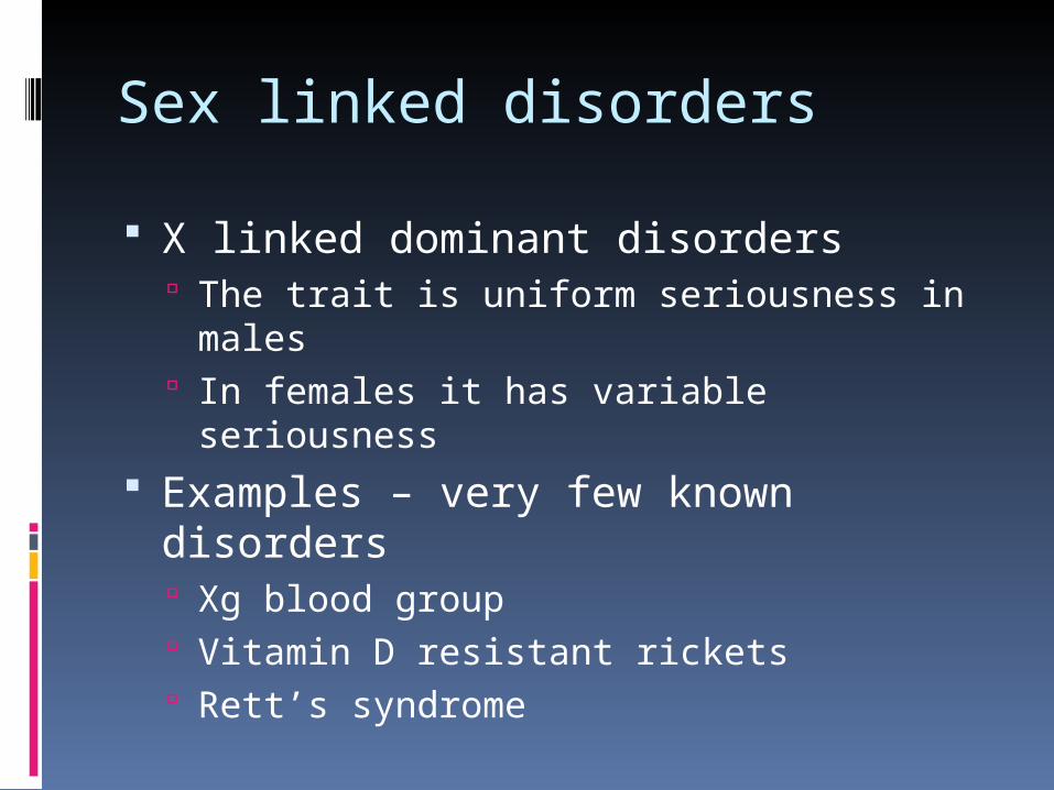
Sex linked disorders
X linked dominant disorders The trait is uniform seriousness in males In females it has variable seriousness
Examples – very few known disorders Xg blood group Vitamin D resistant rickets Rett’s syndrome

Digenic disorders
In these disorders two genes interact to produce the phenotype
Mode of inheritance is often simple mendelian but with another gene interfering to modulate the severity of the disease
Examples Cystic fibrosis Limb girdle dystrophy
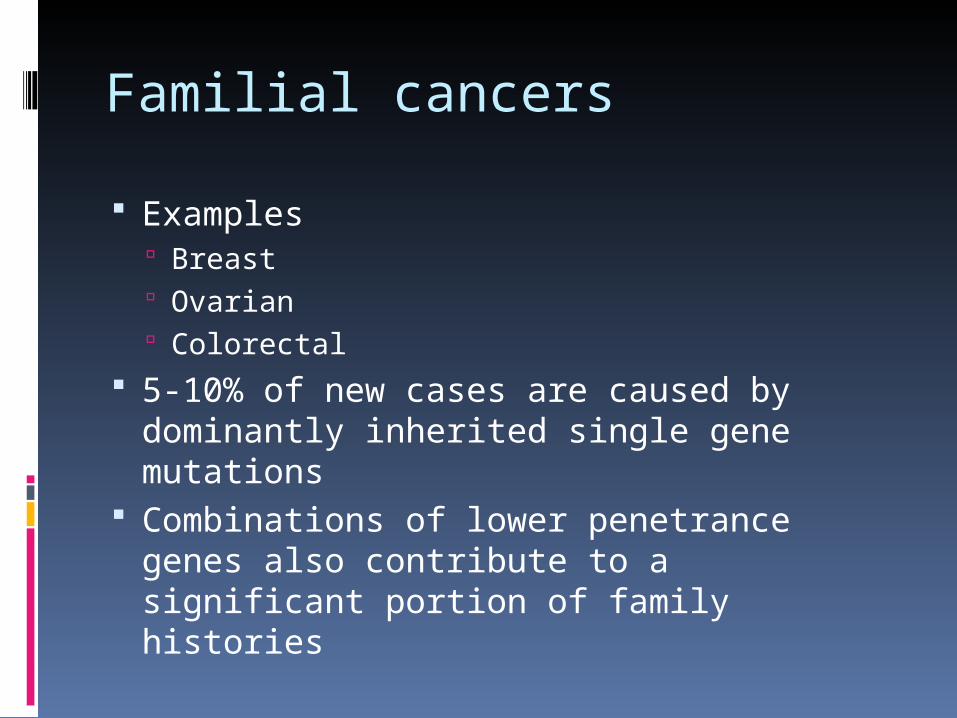
Familial cancers
Examples Breast Ovarian Colorectal
5-10% of new cases are caused by dominantly inherited single gene mutations
Combinations of lower penetrance genes also contribute to a significant portion of family histories

Familial cancers
Features suggestive of inherited cancer High incidence in family in closely related
individuals Early age of onset Multiple primaries in an individual (rockenbach) Certain cancer combinations
Breast and ovary Breast and sarcoma Colorectal, uterine, ovarian and stomach
Ethnicity – Ashkenazi Jews high incidence of 3 common breast and ovarian cancer founder mutations

Familial cancers
Who to refer with FH breast and ovarian cancer Mother or sister breast ca < 40yrs Mother or sister bilateral breast ca any age Father or brother with breast ca any age Mother or sister with breast and ovarian ca
any age One close relative with breast ca < 50 and
relative with ovarian caany age same side of family

Familial cancers
FH breast and ovarian ca who to refer Two close relative breast ca any age Two close relative ovarian ca any
age Three or more close relative with
breast ca, ovarian ca or both on the same side of the family at any age

Familial cancers
Who to refer colorectal cancer 1 first degree relative CRC < 45yrs 1 first degree relative who has 2
separate or multiple CRC or two associated ca – CRC, endometrial, ovarian, small bowel, ureter or renal pelvis.
1 first degree relative with more than 1 bowel polyp < 40 which is tubulovillous, dysplastic, or an adenoma > 10cm

Familial cancers
Who to refer CRC cancers 1 first degree relative with FAP of FH of FAP 1 parent with multiple colorectal polyps
>100 2 close relatives who are first degree
relatives to each other can include both parents with average age < 70 of CRC
2 close relatives who are first degree relatives to each other on same side of family with associated cancers age < 50

Familial cancers
Who to refer CRC 3 close relatives on same side of family
with an associated tumour

Familial cancers
High risk pedigrees 4 close relatives with breast,
ovarian, or both any age 3 close relatives with breast ca
average age < 60 2 close relatives with breast ca
average < 50 2 close relatives ovarian ca any age Known families of carriers of BRCA1,
BRCA2

Familial cancers
High risk pedigrees 3 close relatives CRC or 2 with CRC and
one associated cancer in at least 2 generations. 1 must be under 50 at diagnosis and one should be first degree relative of the other 2
Known gene carriers of hereditary non polyposis colon ca FAP or relatives of known affected family
All others are moderate risk

Familial cancers
Moderate risk pedigrees are normally managed in secondary care Breast ca
Annual mammograms from age 40-50 then will enter national 3 yrly scheme
CRC Offered colonoscopy frequency varies

Familial cancers
High risk pedigrees Normally seen and counselled by
regional genetic centre Breast
Annual mammograms If BRAC1 or 2 then combination of annual
MRI and mammogram between ages 30-49yrs. Age 50-69 mammograms every 18 months then 3x/year after 69

Familial cancers
High risk pedigrees Ovarian ca
Only offered if FH includes either ovarian ca or the person is a known BRAC carrier as part of UKFOCSS trial which offers transvaginal ultrasound and regular ca 125 monitoring every 4 months
CRC cancer People at high risk of hereditary non polyposis crc
and crc are offered 2 colonoscopies a year from ages 25-27 if they have been assessed as a positive pedigree

Familial cancers
Summary Family histories of cancer in primary
care allows GP’s to assess risk and make appropriate referrals.
This allows families to benefit from relevant targeted screening and gene testing as per national guidelines.

Prenatal diagnosis
Investigations include Chorionic villous sampling Amniocentesis Foetoscopy ultrasound
Prenatal diagnosis
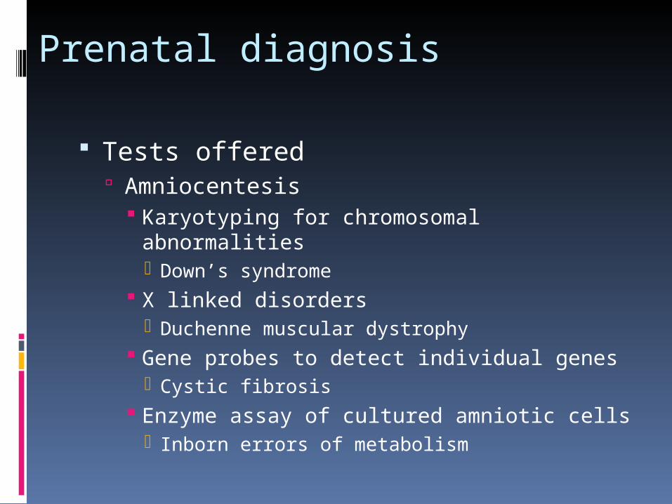
Tests offered Amniocentesis
Karyotyping for chromosomal abnormalities Down’s syndrome
X linked disorders Duchenne muscular dystrophy
Gene probes to detect individual genes Cystic fibrosis
Enzyme assay of cultured amniotic cells Inborn errors of metabolism

Prenatal diagnosis
Risks of amniocentesis Singleton preg 0.5-1% foetal loss Multiple preg 3% risk foetal loss Foetal damage very rare
Loss of one eye damage to brachial plexus Pneumothorax
Lung hypoplasia

Prenatal diagnosis
Tests offered Chorionic villus sampling
Same tests as performed on amniocentesis
Advantages Performed earlier in preg it top needed
done at much earlier stage before preg shows
Results available quicker

Prenatal diagnosis
Chorionic villus sampling Disadvantages
Greater risk of foetal loss 3% Risk of foetal damage – limb agenesis due
to disruption of foetal blood vessels Chromosome analysis less accurate Result sometimes can’t be interpreted
requiring further tests Genetic mosiaicism between chorionic
cells and the foetus resulting in false positives and false negatives i.e. Down’ syndrome

Prenatal diagnosis
Foetoscopy Enables visualisation of foetus
Foetal inspection – facial and limb abnormalities
Foetal blood sampling – haemophilia, thalassaemia, sickle cell, fragile X, alpha 1 antitrypsin deficiency
Foetal skin biopsy – lethal epidermolysis bullosa
Foetal liver biopsy – ornithine transcarbamylase deficiency – loss = 5%

Diagnosis in genetic counselling
If a major chromosomal abnormality exists then a recognised syndrome of 2 or more dysmorphic features will usually be present chromosomal analysis should be carried out if Unexplained mental retardation Known history of structural chromosomal
problem Unexplained stillbirth Female with unexplained short stature Recurrent miscarriages Ambiguous sexual development

Ethical and legal considerations Under congenital disabilities act 1976 an
action can be taken against anyone whose negligent action resulted in a child being born disabled, abnormal or unhealthy.
It is the legal duty of all doctors to provide the most recent valid information about genetic disorders. If omitting to do so and on future pregnancy a foetal abnormality occurred the doctor would be liable to litigation

Genetics in Practice
Neurofibrmatosis Sickle cell disease Beta thalssaemia trait Friedreichs ataxia Facial scapulo humeral dystrophy Beckers muscular dystrophy
Genetics in Practice
Familial breast cancer – 3 families Fragile x