2012 rush orthopedics · 2012 / rush orthopedics journal chairman’s letter began my letter in...
TRANSCRIPT
PLEASE NOTE: All physicians featured in this publication are on the medical faculty of Rush University Medical Center. Many of the physicians featured are in the private practice Midwest Orthopaedics at Rush and, as independent practitioners, are not agents or employees of Rush University Medical Center.
Rush is a not-for-profit health care, education and research enterprise comprising Rush University Medical Center, Rush University, Rush Oak Park Hospital and Rush Health.
ORTHOPEDICS2012 RUSH
JOURNAL
20
12
Ru
sh O
rth
op
ed
ics
Jou
rnal
71795_Cover_u3.indd 1 8/3/12 3:33 PM

THE TOWER AT RUSH UNIVERSITY MEDICAL CENTER, which opened its doors in January 2012, is one of the nation’s most advanced—and visually striking—hospitals.
A TRADITION OF EXCELLENCE
“OUR THRIVING CLINICAL PRACTICE is enhanced by collaborations with specialists at Rush from rheumatology, physical medicine and rehabilitation, physical therapy, and other areas.”
Midwest Orthopaedics at Rush (MOR) is a private practice medical group whose 38 fellowship-trained physicians are on the faculty of Rush University
Medical Center in Chicago. With MOR based primarily at Rush, our renowned surgeons and physicians have access to all the resources of a world-class academic medical center, including the state-of-the-art operating rooms in Rush’s new hospital.
Our thriving clinical practice is enhanced by collaborations with specialists at Rush from rheumatology, physical medicine and rehabilitation, physical therapy, and other areas. As part of our commitment to continually advance orthopedic care, our physicians participate in cutting-edge multidisciplinary research, which is translating into new treatments that benefit patients at Rush—and around the globe.
Physicians from Midwest Orthopaedics at Rush also hold key leadership positions in national societies and organizations, and serve as the team physicians for a variety of professional, collegiate, and high school teams and clubs, including the Chicago Bulls, Chicago White Sox, and DePaul University.
These impressive clinical, research, and administrative activities distinguish orthopedics at Rush as among America’s best. U.S.News & World Report has ranked the orthopedics program at Rush in the top 12 each of the past 10 years—including 7 times in the top 10.
Midwest Orthopaedics at Rush
Chicago, IL • Oak Park, IL • Westchester, IL • Winfield, IL(877) MD-BONES (632-6637) / www.rushortho.com
71795_Cover_u4.indd 2 8/6/12 3:33 PM
FACULTY EDITORS
DAVID FARDON, MD Editor in Chief
BRETT LEVINE, MD, MSSHANE J. NHO, MD, MSROBERT W. WYSOCKI, MD
STEVEN GITELIS, MDEditor Emeritus
2 CHAIRMAN’S LETTER
4 ORTHOPEDIC FACULTY AND FELLOWS (2011)
9 RESEARCH FACULTY
12 DEPARTMENT OF ORTHOPEDIC SURGERY RESIDENTS
13 THREE-DIMENSIONAL ANATOMY OF THE HIP, UTILIZING COMPUTER NAVIGATION AND PLAIN RADIOGRAPHS Rachel M. Frank, MD; Jeremy Alland, BS; Robert C. Grumet, MD; Mark A. Slabaugh, MD; Vincent M. Wang, PhD; Alejandro A. Espinoza Orías, PhD; Bernard R. Bach Jr, MD; Shane J. Nho, MD, MS
18 METAL ION LEVELS AFTER A SECOND JOINT ARTHROPLASTY Andrew R. Hsu, MD; Brett Levine, MD, MS; Anastasia Skipor, MS; Nadim J. Hallab, PhD; Wayne G. Paprosky, MD; Joshua J. Jacobs, MD
23 OUTPATIENT MINIMALLY INVASIVE TOTAL HIP ARTHROPLASTY Darwin Chen, MD; Richard A. Berger, MD
28 ANTERIOR CRUCIATE LIGAMENT RECONSTRUCTION FAILURE: ANALYSIS OF A SINGLE, HIGH-VOLUME ORTHOPEDIC INSTITUTION Rachel M. Frank, MD; Mark A. Slabaugh, MD; Kevin C. McGill, MD, MPH; Charles A. Bush-Joseph, MD; Brian J. Cole, MD, MBA; Bernard R. Bach Jr, MD; Nikhil N. Verma, MD
36 TREATMENT OF CARTILAGE DEFECTS IN YOUNG SHOULDERS: FROM THE LAB TO THE CLINIC Geoffrey S. Van Thiel, MD, MBA; Andrew Riff, MD; Wendell Heard, MD; Vasili Karas, BS; Vincent M. Wang, PhD; Jas Chahal, MD; Anthony A. Romeo, MD; Nikhil N. Verma, MD; Brian J. Cole, MD, MBA
41 OUTCOMES IN THE TREATMENT OF BENIGN BONE LESIONS WITH BIOCERAMIC Yale A. Fillingham, MD; Brett A. Lenart, MD; Steven Gitelis, MD
47 RADIAL HEAD REPLACEMENT WITH A BIPOLAR SYSTEM Mark R. Zunkiewicz, MD; Jill S. Clemente, MS; Mark C. Miller, PhD; Mark E. Baratz, MD; Robert W. Wysocki, MD; Mark S. Cohen, MD
51 OSTEOTOMY-SPARING METHODS FOR CORRECTION OF METATARSUS PRIMUS VARUS IN HALLUX VALGUS George Holmes Jr, MD
54 DYSPHAGIA LUSORIA: AN UNCOMMON CAUSE OF NECK PAIN AND HAZARD TO ANTERIOR CERVICOTHORACIC SURGERY Dominic Scola, BS; Sudeep Bhabad, MD; David Fardon, MD
59 WHAT VOLUME OF BONE GRAFT IS NEEDED FOR PREDICTABLE FUSION IN SINGLE-LEVEL LUMBAR ARTHRODESIS? A PRELIMINARY INVESTIGATION Safdar N. Khan, MD; Kevin U. Park, MD; Shah Dodwad, MD; Thomas Cha, MD; Howard S. An, MD
62 PUBLICATIONS, RESEARCH GRANTS, AND KAPPA DELTA ORTHOPEDIC RESEARCH AWARDS
69 VOLUME AND QUALITY DATA
71 LIFE AND LIMBS An Interview with Pioneering Orthopedic Oncologist and Rush Mainstay Steven Gitelis, MD, by Gunnar B. J. Andersson, MD, PhD
To view the 2012 Rush Orthopedics Journal online, please visit the Rush website at www.rush.edu/orthopedicsjournal.
71795_Body.indd 1 7/25/12 9:38 PM
2012 / RUSH ORTHOPEDICS JOURNAL
CHAIRMAN’S LETTER
began my letter in last year’s Rush Orthopedics Journal with a preview of Rush’s new hospital, known as the Tower, which was scheduled to
open within the next several months. At that time, although I knew the Tower would be an outstanding clinical resource, I could not have forseen how much of an impact it would have on our orthopedic practice. Like the Orthopedic Building before it, the new hospital is transforming the way we care for our patients.
After 7 years of planning and construction, the doors to the Tower opened on January 9, 2012—it’s worth noting that the first patient to cross the threshold into the new facility was a woman who had undergone knee replacement surgery a few days earlier—and already our surgeons are realizing the benefits. In particular, they appreciate the design of the new operating rooms, which was based on input from surgeons across numerous specialties, including orthopedics. Every feature in these new ORs—including state-of-the-art imaging equipment, real-time video conferencing capabilities, and a ventilation system that produces higher-than-OR-quality air to minimize the risk of infections—is helping to improve surgical efficiency and patient outcomes.
The opening of the Tower isn’t the only noteworthy highlight from the past year. Two esteemed faculty members from the Department of Orthopedic Surgery—one former, one present—were recognized for their remarkable career achievements. The Scoliosis Research Society presented its Lifetime Achievement Award to Ronald L. DeWald, MD, professor emeritus. And Gunnar B. J. Andersson, MD, PhD, was given two prestigious awards: the Orthopaedic Research Society’s Alfred R. Shands, Jr. Award in recognition of his contributions to orthopedic research and to furthering knowledge in the field of musculoskeletal disease; and the Henry Farfan Award for outstanding contributions in spine-related basic science research from the North American Spine Society at its 2011 annual meeting.
At that same meeting, two papers co-authored by Frank M. Phillips, MD, were awarded “Best Overall Papers”: “Preclinical Study of Human Allogeneic Amniotic Membrane as a Barrier to Epidural Fibrosis in the Early Wound of a Post Laminectomy Rat Model,” and “Why Don’t Annular Defects Heal? Does Interposed Nucleus Pulposus Prevent Healing? An In Vivo Study.” Among the many other awards and honors bestowed on physicians and researchers from our department, Craig J. Della Valle, MD, was
co-recipient of the Hip Society’s 2012 John Charnley Award for the paper “Clinical Multi-Center Studies of the Wear Performance of Highly Cross-Linked Remelted Polyethylene in THR.” Charles A. Bush-Joseph, MD, who is head team physician for the Chicago White Sox, was appointed president of the Major League Baseball Team Physician Association. And I officially stepped into the role of first vice president of the American Academy of Orthopaedic Surgeons (see opposite page).
Our nationally renowned residency and fellowship programs also enjoyed a stellar year, with a dedicated, talented, and diverse group completing their training at Rush. An important component of our department’s mission is to train specialists who will make significant contributions to their field in the years to come, and our faculty members are committed to providing a strong foundation on which our residents and fellows can build their careers.
Finally, when we talk about advancing orthopedic care, we tend to focus on the front lines—the clinicians and researchers who are directly involved. But so much of the work we do, at the bench and at the bedside, would not be possible without substantial backing from our many donors. The philanthropic support our department received this past year included gifts from the Grainger Foundation, Goldman Sachs Gives, Mrs. William A. Hark, Paul and Joan Rubschlager, the Segal Family, and too many others to list in this space. From creating endowed chairs to providing funding for basic science research to purchasing needed equipment for our labs and clinics, these generous individuals, families, corporations, and organizations enable us to better the lives of our patients while we continue striving to improve the tools of our trade.
I’m pleased to present the 2012 Rush Orthopedics Journal, which represents the breadth and quality of research undertaken by our faculty—and the discoveries that are helping to advance the understanding and treatment of orthopedic conditions.
Joshua J. Jacobs, MD
The William A. Hark, MD/Susanne G. Swift Professor of Orthopedic Surgery
Chairman and professor, Department of Orthopedic Surgery Rush University Medical Center
2
71795_Body_u1.indd 2 8/3/12 10:30 AM
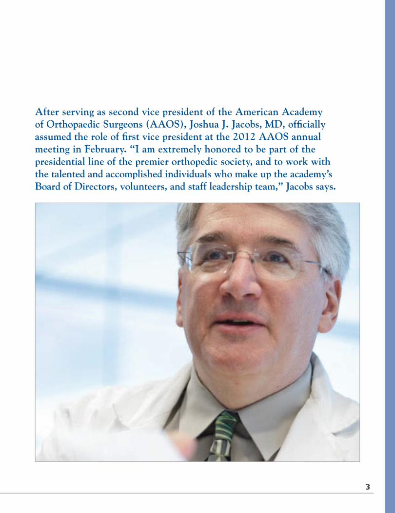
After serving as second vice president of the American Academy of Orthopaedic Surgeons (AAOS), Joshua J. Jacobs, MD, officially assumed the role of first vice president at the 2012 AAOS annual meeting in February. “I am extremely honored to be part of the presidential line of the premier orthopedic society, and to work with the talented and accomplished individuals who make up the academy’s Board of Directors, volunteers, and staff leadership team,” Jacobs says.
3
71795_Body.indd 3 7/25/12 9:38 PM
2012 / RUSH ORTHOPEDICS JOURNAL
CHAIRMAN’S LETTER
began my letter in last year’s Rush Orthopedics Journal with a preview of Rush’s new hospital, known as the Tower, which was scheduled to
open within the next several months. At that time, although I knew the Tower would be an outstanding clinical resource, I could not have forseen how much of an impact it would have on our orthopedic practice. Like the Orthopedic Building before it, the new hospital is transforming the way we care for our patients.
After 7 years of planning and construction, the doors to the Tower opened on January 9, 2012—it’s worth noting that the first patient to cross the threshold into the new facility was a woman who had undergone knee replacement surgery a few days earlier—and already our surgeons are realizing the benefits. In particular, they appreciate the design of the new operating rooms, which was based on input from surgeons across numerous specialties, including orthopedics. Every feature in these new ORs—including state-of-the-art imaging equipment, real-time video conferencing capabilities, and a ventilation system that produces higher-than-OR-quality air to minimize the risk of infections—is helping to improve surgical efficiency and patient outcomes.
The opening of the Tower isn’t the only noteworthy highlight from the past year. Two esteemed faculty members from the Department of Orthopedic Surgery—one former, one present—were recognized for their remarkable career achievements. The Scoliosis Research Society presented its Lifetime Achievement Award to Ronald L. DeWald, MD, professor emeritus. And Gunnar B. J. Andersson, MD, PhD, was given two prestigious awards: the Orthopaedic Research Society’s Alfred R. Shands, Jr. Award in recognition of his contributions to orthopedic research and to furthering knowledge in the field of musculoskeletal disease; and the Henry Farfan Award for outstanding contributions in spine-related basic science research from the North American Spine Society at its 2011 annual meeting.
At that same meeting, two papers co-authored by Frank M. Phillips, MD, were awarded “Best Overall Papers”: “Preclinical Study of Human Allogeneic Amniotic Membrane as a Barrier to Epidural Fibrosis in the Early Wound of a Post Laminectomy Rat Model,” and “Why Don’t Annular Defects Heal? Does Interposed Nucleus Pulposus Prevent Healing? An In Vivo Study.” Among the many other awards and honors bestowed on physicians and researchers from our department, Craig J. Della Valle, MD, was
co-recipient of the Hip Society’s 2012 John Charnley Award for the paper “Clinical Multi-Center Studies of the Wear Performance of Highly Cross-Linked Remelted Polyethylene in THR.” Charles A. Bush-Joseph, MD, who is head team physician for the Chicago White Sox, was appointed president of the Major League Baseball Team Physician Association. And I officially stepped into the role of first vice president of the American Academy of Orthopaedic Surgeons (see opposite page).
Our nationally renowned residency and fellowship programs also enjoyed a stellar year, with a dedicated, talented, and diverse group completing their training at Rush. An important component of our department’s mission is to train specialists who will make significant contributions to their field in the years to come, and our faculty members are committed to providing a strong foundation on which our residents and fellows can build their careers.
Finally, when we talk about advancing orthopedic care, we tend to focus on the front lines—the clinicians and researchers who are directly involved. But so much of the work we do, at the bench and at the bedside, would not be possible without substantial backing from our many donors. The philanthropic support our department received this past year included gifts from the Grainger Foundation, Goldman Sachs Gives, Mrs. William A. Hark, Paul and Joan Rubschlager, the Segal Family, and too many others to list in this space. From creating endowed chairs to providing funding for basic science research to purchasing needed equipment for our labs and clinics, these generous individuals, families, corporations, and organizations enable us to better the lives of our patients while we continue striving to improve the tools of our trade.
I’m pleased to present the 2012 Rush Orthopedics Journal, which represents the breadth and quality of research undertaken by our faculty—and the discoveries that are helping to advance the understanding and treatment of orthopedic conditions.
Joshua J. Jacobs, MD
The William A. Hark, MD/Susanne G. Swift Professor of Orthopedic Surgery
Chairman and professor, Department of Orthopedic Surgery Rush University Medical Center
2
71795_Body_u1.indd 2 8/3/12 10:30 AM
After serving as second vice president of the American Academy of Orthopaedic Surgeons (AAOS), Joshua J. Jacobs, MD, officially assumed the role of first vice president at the 2012 AAOS annual meeting in February. “I am extremely honored to be part of the presidential line of the premier orthopedic society, and to work with the talented and accomplished individuals who make up the academy’s Board of Directors, volunteers, and staff leadership team,” Jacobs says.
3
71795_Body.indd 3 7/25/12 9:38 PM
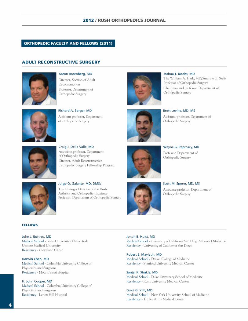
ADULT RECONSTRUCTIVE SURGERY
FELLOWS
Aaron Rosenberg, MD
Director, Section of Adult ReconstructionProfessor, Department of Orthopedic Surgery
Richard A. Berger, MD
Assistant professor, Department of Orthopedic Surgery
Brett Levine, MD, MS
Assistant professor, Department of Orthopedic Surgery
Wayne G. Paprosky, MD
Professor, Department of Orthopedic Surgery
Scott M. Sporer, MD, MS
Associate professor, Department of Orthopedic Surgery
Jorge O. Galante, MD, DMSc
The Grainger Director of the RushArthritis and Orthopedics InstituteProfessor, Department of Orthopedic Surgery
Craig J. Della Valle, MD
Associate professor, Department of Orthopedic SurgeryDirector, Adult Reconstructive Orthopedic Surgery Fellowship Program
Joshua J. Jacobs, MD The William A. Hark, MD/Susanne G. Swift Professor of Orthopedic SurgeryChairman and professor, Department of Orthopedic Surgery
2012 / RUSH ORTHOPEDICS JOURNAL
ORTHOPEDIC FACULTY AND FELLOWS (2011)
4
John J. Bottros, MDMedical School - State University of New York Upstate Medical UniversityResidency - Cleveland Clinic
Darwin Chen, MDMedical School - Columbia University College of Physicians and SurgeonsResidency - Mount Sinai Hospital
H. John Cooper, MDMedical School - Columbia University College of Physicians and SurgeonsResidency - Lenox Hill Hospital
Jonah B. Hulst, MDMedical School - University of California San Diego School of MedicineResidency - University of California San Diego
Robert E. Mayle Jr., MDMedical School - Drexel College of MedicineResidency - Stanford University Medical Center
Sanjai K. Shukla, MDMedical School - Duke University School of MedicineResidency - Rush University Medical Center
Duke G. Yim, MDMedical School - New York University School of MedicineResidency - Tripler Army Medical Center
71795_Body.indd 4 7/25/12 9:38 PM
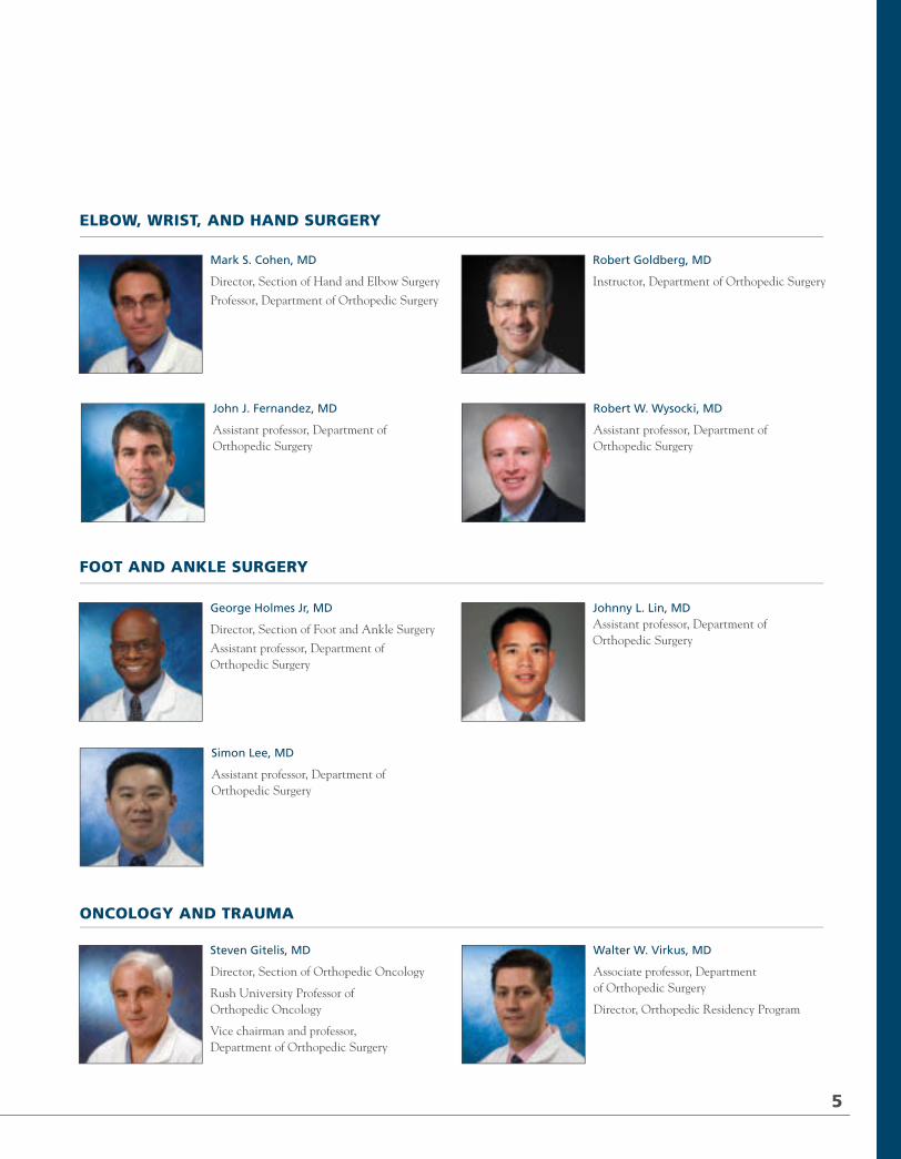
Walter W. Virkus, MD
Associate professor, Department of Orthopedic Surgery
Director, Orthopedic Residency Program
Steven Gitelis, MD
Director, Section of Orthopedic Oncology
Rush University Professor of Orthopedic Oncology
Vice chairman and professor, Department of Orthopedic Surgery
ELBOW, WRIST, AND HAND SURGERY
Mark S. Cohen, MD
Director, Section of Hand and Elbow SurgeryProfessor, Department of Orthopedic Surgery
John J. Fernandez, MD
Assistant professor, Department of Orthopedic Surgery
Robert Goldberg, MD
Instructor, Department of Orthopedic Surgery
Robert W. Wysocki, MD
Assistant professor, Department of Orthopedic Surgery
FOOT AND ANKLE SURGERY
Simon Lee, MD
Assistant professor, Department of Orthopedic Surgery
Johnny L. Lin, MD Assistant professor, Department of Orthopedic Surgery
George Holmes Jr, MD
Director, Section of Foot and Ankle SurgeryAssistant professor, Department of Orthopedic Surgery
ONCOLOGY AND TRAUMA
5
71795_Body.indd 5 7/25/12 9:38 PM
ADULT RECONSTRUCTIVE SURGERY
FELLOWS
Aaron Rosenberg, MD
Director, Section of Adult ReconstructionProfessor, Department of Orthopedic Surgery
Richard A. Berger, MD
Assistant professor, Department of Orthopedic Surgery
Brett Levine, MD, MS
Assistant professor, Department of Orthopedic Surgery
Wayne G. Paprosky, MD
Professor, Department of Orthopedic Surgery
Scott M. Sporer, MD, MS
Associate professor, Department of Orthopedic Surgery
Jorge O. Galante, MD, DMSc
The Grainger Director of the RushArthritis and Orthopedics InstituteProfessor, Department of Orthopedic Surgery
Craig J. Della Valle, MD
Associate professor, Department of Orthopedic SurgeryDirector, Adult Reconstructive Orthopedic Surgery Fellowship Program
Joshua J. Jacobs, MD The William A. Hark, MD/Susanne G. Swift Professor of Orthopedic SurgeryChairman and professor, Department of Orthopedic Surgery
2012 / RUSH ORTHOPEDICS JOURNAL
ORTHOPEDIC FACULTY AND FELLOWS (2011)
4
John J. Bottros, MDMedical School - State University of New York Upstate Medical UniversityResidency - Cleveland Clinic
Darwin Chen, MDMedical School - Columbia University College of Physicians and SurgeonsResidency - Mount Sinai Hospital
H. John Cooper, MDMedical School - Columbia University College of Physicians and SurgeonsResidency - Lenox Hill Hospital
Jonah B. Hulst, MDMedical School - University of California San Diego School of MedicineResidency - University of California San Diego
Robert E. Mayle Jr., MDMedical School - Drexel College of MedicineResidency - Stanford University Medical Center
Sanjai K. Shukla, MDMedical School - Duke University School of MedicineResidency - Rush University Medical Center
Duke G. Yim, MDMedical School - New York University School of MedicineResidency - Tripler Army Medical Center
71795_Body.indd 4 7/25/12 9:38 PM
Walter W. Virkus, MD
Associate professor, Department of Orthopedic Surgery
Director, Orthopedic Residency Program
Steven Gitelis, MD
Director, Section of Orthopedic Oncology
Rush University Professor of Orthopedic Oncology
Vice chairman and professor, Department of Orthopedic Surgery
ELBOW, WRIST, AND HAND SURGERY
Mark S. Cohen, MD
Director, Section of Hand and Elbow SurgeryProfessor, Department of Orthopedic Surgery
John J. Fernandez, MD
Assistant professor, Department of Orthopedic Surgery
Robert Goldberg, MD
Instructor, Department of Orthopedic Surgery
Robert W. Wysocki, MD
Assistant professor, Department of Orthopedic Surgery
FOOT AND ANKLE SURGERY
Simon Lee, MD
Assistant professor, Department of Orthopedic Surgery
Johnny L. Lin, MD Assistant professor, Department of Orthopedic Surgery
George Holmes Jr, MD
Director, Section of Foot and Ankle SurgeryAssistant professor, Department of Orthopedic Surgery
ONCOLOGY AND TRAUMA
5
71795_Body.indd 5 7/25/12 9:38 PM
2012 / RUSH ORTHOPEDICS JOURNAL
6
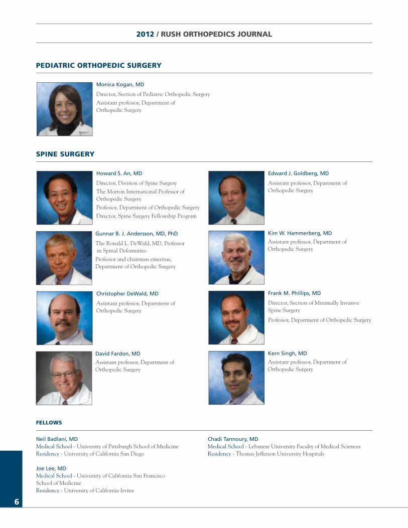
PEDIATRIC ORTHOPEDIC SURGERY
Monica Kogan, MD
Director, Section of Pediatric Orthopedic SurgeryAssistant professor, Department of Orthopedic Surgery
SPINE SURGERY
Howard S. An, MD
Director, Division of Spine SurgeryThe Morton International Professor of Orthopedic SurgeryProfessor, Department of Orthopedic SurgeryDirector, Spine Surgery Fellowship Program
Gunnar B. J. Andersson, MD, PhD
The Ronald L. DeWald, MD, Professor in Spinal DeformitiesProfessor and chairman emeritus, Department of Orthopedic Surgery
Kim W. Hammerberg, MD
Assistant professor, Department of Orthopedic Surgery
Frank M. Phillips, MD
Director, Section of Minimally Invasive Spine Surgery
Professor, Department of Orthopedic Surgery
Kern Singh, MD
Assistant professor, Department of Orthopedic Surgery
David Fardon, MD
Assistant professor, Department of Orthopedic Surgery
Christopher DeWald, MD
Assistant professor, Department of Orthopedic Surgery
Edward J. Goldberg, MD
Assistant professor, Department of Orthopedic Surgery
FELLOWS
Neil Badlani, MDMedical School - University of Pittsburgh School of MedicineResidency - University of California San Diego
Joe Lee, MDMedical School - University of California San Francisco School of MedicineResidency - University of California Irvine
Chadi Tannoury, MDMedical School - Lebanese University Faculty of Medical SciencesResidency - Thomas Jefferson University Hospitals
71795_Body_u1.indd 6 8/3/12 10:30 AM
7
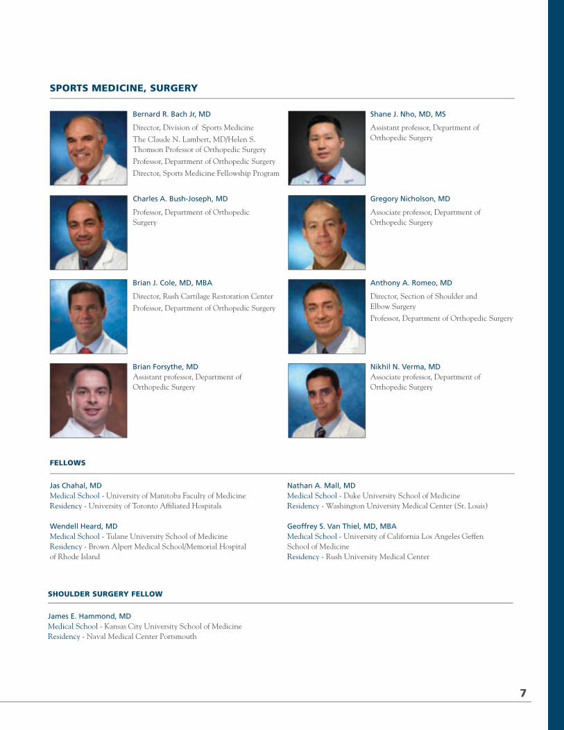
SPORTS MEDICINE, SURGERY
Bernard R. Bach Jr, MD
Director, Division of Sports Medicine The Claude N. Lambert, MD/Helen S. Thomson Professor of Orthopedic SurgeryProfessor, Department of Orthopedic SurgeryDirector, Sports Medicine Fellowship Program
Charles A. Bush-Joseph, MD
Professor, Department of Orthopedic Surgery
Anthony A. Romeo, MD
Director, Section of Shoulder and Elbow SurgeryProfessor, Department of Orthopedic Surgery
Brian Forsythe, MD Assistant professor, Department of Orthopedic Surgery
Shane J. Nho, MD, MS
Assistant professor, Department of Orthopedic Surgery
Brian J. Cole, MD, MBA
Director, Rush Cartilage Restoration CenterProfessor, Department of Orthopedic Surgery
Gregory Nicholson, MD
Associate professor, Department of Orthopedic Surgery
FELLOWS
Jas Chahal, MDMedical School - University of Manitoba Faculty of MedicineResidency - University of Toronto Affiliated Hospitals
Wendell Heard, MDMedical School - Tulane University School of MedicineResidency - Brown Alpert Medical School/Memorial Hospital of Rhode Island
Nathan A. Mall, MDMedical School - Duke University School of MedicineResidency - Washington University Medical Center (St. Louis)
Geoffrey S. Van Thiel, MD, MBAMedical School - University of California Los Angeles Geffen School of MedicineResidency - Rush University Medical Center
SHOULDER SURGERY FELLOW
James E. Hammond, MDMedical School - Kansas City University School of MedicineResidency - Naval Medical Center Portsmouth
Nikhil N. Verma, MD Associate professor, Department of Orthopedic Surgery
71795_Body.indd 7 7/25/12 9:38 PM
2012 / RUSH ORTHOPEDICS JOURNAL
6
PEDIATRIC ORTHOPEDIC SURGERY
Monica Kogan, MD
Director, Section of Pediatric Orthopedic SurgeryAssistant professor, Department of Orthopedic Surgery
SPINE SURGERY
Howard S. An, MD
Director, Division of Spine SurgeryThe Morton International Professor of Orthopedic SurgeryProfessor, Department of Orthopedic SurgeryDirector, Spine Surgery Fellowship Program
Gunnar B. J. Andersson, MD, PhD
The Ronald L. DeWald, MD, Professor in Spinal DeformitiesProfessor and chairman emeritus, Department of Orthopedic Surgery
Kim W. Hammerberg, MD
Assistant professor, Department of Orthopedic Surgery
Frank M. Phillips, MD
Director, Section of Minimally Invasive Spine Surgery
Professor, Department of Orthopedic Surgery
Kern Singh, MD
Assistant professor, Department of Orthopedic Surgery
David Fardon, MD
Assistant professor, Department of Orthopedic Surgery
Christopher DeWald, MD
Assistant professor, Department of Orthopedic Surgery
Edward J. Goldberg, MD
Assistant professor, Department of Orthopedic Surgery
FELLOWS
Neil Badlani, MDMedical School - University of Pittsburgh School of MedicineResidency - University of California San Diego
Joe Lee, MDMedical School - University of California San Francisco School of MedicineResidency - University of California Irvine
Chadi Tannoury, MDMedical School - Lebanese University Faculty of Medical SciencesResidency - Thomas Jefferson University Hospitals
71795_Body_u1.indd 6 8/3/12 10:30 AM
7
SPORTS MEDICINE, SURGERY
Bernard R. Bach Jr, MD
Director, Division of Sports Medicine The Claude N. Lambert, MD/Helen S. Thomson Professor of Orthopedic SurgeryProfessor, Department of Orthopedic SurgeryDirector, Sports Medicine Fellowship Program
Charles A. Bush-Joseph, MD
Professor, Department of Orthopedic Surgery
Anthony A. Romeo, MD
Director, Section of Shoulder and Elbow SurgeryProfessor, Department of Orthopedic Surgery
Brian Forsythe, MD Assistant professor, Department of Orthopedic Surgery
Shane J. Nho, MD, MS
Assistant professor, Department of Orthopedic Surgery
Brian J. Cole, MD, MBA
Director, Rush Cartilage Restoration CenterProfessor, Department of Orthopedic Surgery
Gregory Nicholson, MD
Associate professor, Department of Orthopedic Surgery
FELLOWS
Jas Chahal, MDMedical School - University of Manitoba Faculty of MedicineResidency - University of Toronto Affiliated Hospitals
Wendell Heard, MDMedical School - Tulane University School of MedicineResidency - Brown Alpert Medical School/Memorial Hospital of Rhode Island
Nathan A. Mall, MDMedical School - Duke University School of MedicineResidency - Washington University Medical Center (St. Louis)
Geoffrey S. Van Thiel, MD, MBAMedical School - University of California Los Angeles Geffen School of MedicineResidency - Rush University Medical Center
SHOULDER SURGERY FELLOW
James E. Hammond, MDMedical School - Kansas City University School of MedicineResidency - Naval Medical Center Portsmouth
Nikhil N. Verma, MD Associate professor, Department of Orthopedic Surgery
71795_Body.indd 7 7/25/12 9:38 PM

2012 / RUSH ORTHOPEDICS JOURNAL
8
SPORTS MEDICINE, PRIMARY CARE
Jeffrey M. Mjaanes, MD
Assistant professor, Department of Orthopedic Surgery and Department of Pediatrics
Krystian Bigosinski, MD
Assistant professor, Department of Family Medicine and Department of Orthopedic Surgery
Joshua Blomgren, DO
Assistant professor, Department of Family Medicine and Department of Orthopedic Surgery
Kathleen M. Weber, MD
Director, Primary Care/Sports Medicine Program and Women’s Sports Medicine Program
Assistant professor, Department of Orthopedic Surgery
FELLOW
Michael S. Erickson, MDMedical School - University of Washington School of MedicineResidency - Swedish Medical Center, Cherry Hill
ORTHOPEDIC PHYSICAL MEDICINE AND REHABILITATION
Yejia Zhang, MD, PhD
Assistant professor, Department of Orthopedic Surgery
April M. Fetzer, DO
Assistant professor, Department of Orthopedic Surgery and Department of Physical Medicine and Rehabilitation
71795_Body.indd 8 7/25/12 9:38 PM
9
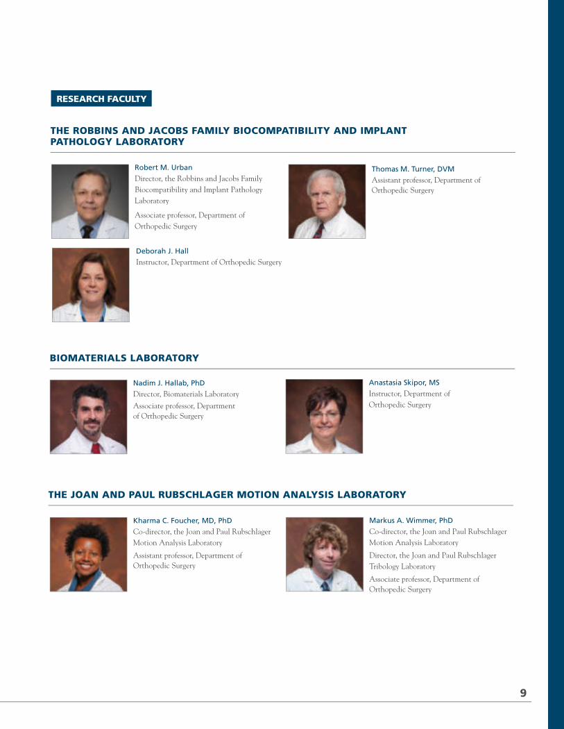
THE ROBBINS AND JACOBS FAMILY BIOCOMPATIBILITY AND IMPLANT PATHOLOGY LABORATORY
Robert M. Urban
Director, the Robbins and Jacobs Family Biocompatibility and Implant Pathology Laboratory
Associate professor, Department of Orthopedic Surgery
Deborah J. Hall
Instructor, Department of Orthopedic Surgery
Thomas M. Turner, DVM Assistant professor, Department of Orthopedic Surgery
BIOMATERIALS LABORATORY
Nadim J. Hallab, PhD
Director, Biomaterials Laboratory Associate professor, Department of Orthopedic Surgery
THE JOAN AND PAUL RUBSCHLAGER MOTION ANALYSIS LABORATORY
Markus A. Wimmer, PhD
Co-director, the Joan and Paul Rubschlager Motion Analysis Laboratory
Director, the Joan and Paul Rubschlager Tribology Laboratory
Associate professor, Department of Orthopedic Surgery
Kharma C. Foucher, MD, PhD
Co-director, the Joan and Paul Rubschlager Motion Analysis Laboratory
Assistant professor, Department of Orthopedic Surgery
RESEARCH FACULTY
Anastasia Skipor, MS
Instructor, Department of Orthopedic Surgery
71795_Body.indd 9 7/25/12 9:39 PM
2012 / RUSH ORTHOPEDICS JOURNAL
8
SPORTS MEDICINE, PRIMARY CARE
Jeffrey M. Mjaanes, MD
Assistant professor, Department of Orthopedic Surgery and Department of Pediatrics
Krystian Bigosinski, MD
Assistant professor, Department of Family Medicine and Department of Orthopedic Surgery
Joshua Blomgren, DO
Assistant professor, Department of Family Medicine and Department of Orthopedic Surgery
Kathleen M. Weber, MD
Director, Primary Care/Sports Medicine Program and Women’s Sports Medicine Program
Assistant professor, Department of Orthopedic Surgery
FELLOW
Michael S. Erickson, MDMedical School - University of Washington School of MedicineResidency - Swedish Medical Center, Cherry Hill
ORTHOPEDIC PHYSICAL MEDICINE AND REHABILITATION
Yejia Zhang, MD, PhD
Assistant professor, Department of Orthopedic Surgery
April M. Fetzer, DO
Assistant professor, Department of Orthopedic Surgery and Department of Physical Medicine and Rehabilitation
71795_Body.indd 8 7/25/12 9:38 PM
9
THE ROBBINS AND JACOBS FAMILY BIOCOMPATIBILITY AND IMPLANT PATHOLOGY LABORATORY
Robert M. Urban
Director, the Robbins and Jacobs Family Biocompatibility and Implant Pathology Laboratory
Associate professor, Department of Orthopedic Surgery
Deborah J. Hall
Instructor, Department of Orthopedic Surgery
Thomas M. Turner, DVM Assistant professor, Department of Orthopedic Surgery
BIOMATERIALS LABORATORY
Nadim J. Hallab, PhD
Director, Biomaterials Laboratory Associate professor, Department of Orthopedic Surgery
THE JOAN AND PAUL RUBSCHLAGER MOTION ANALYSIS LABORATORY
Markus A. Wimmer, PhD
Co-director, the Joan and Paul Rubschlager Motion Analysis Laboratory
Director, the Joan and Paul Rubschlager Tribology Laboratory
Associate professor, Department of Orthopedic Surgery
Kharma C. Foucher, MD, PhD
Co-director, the Joan and Paul Rubschlager Motion Analysis Laboratory
Assistant professor, Department of Orthopedic Surgery
RESEARCH FACULTY
Anastasia Skipor, MS
Instructor, Department of Orthopedic Surgery
71795_Body.indd 9 7/25/12 9:39 PM
2012 / RUSH ORTHOPEDICS JOURNAL
8
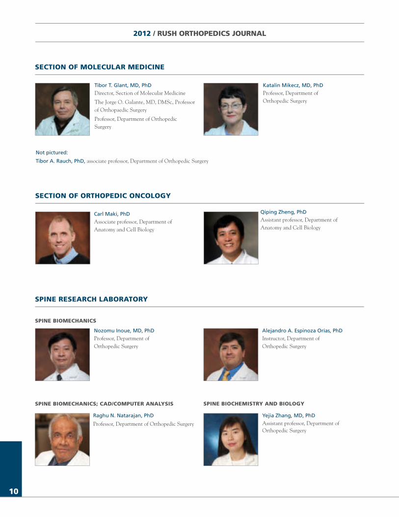
SECTION OF ORTHOPEDIC ONCOLOGY
Carl Maki, PhD
Associate professor, Department of Anatomy and Cell Biology
Qiping Zheng, PhD
Assistant professor, Department of Anatomy and Cell Biology
SECTION OF MOLECULAR MEDICINE
Tibor T. Glant, MD, PhD
Director, Section of Molecular Medicine
The Jorge O. Galante, MD, DMSc, Professor of Orthopaedic Surgery
Professor, Department of Orthopedic Surgery
Katalin Mikecz, MD, PhD
Professor, Department of Orthopedic Surgery
Not pictured:
Tibor A. Rauch, PhD, associate professor, Department of Orthopedic Surgery
SPINE RESEARCH LABORATORY
Nozomu Inoue, MD, PhD
Professor, Department of Orthopedic Surgery
Yejia Zhang, MD, PhD
Assistant professor, Department of Orthopedic Surgery
SPINE BIOMECHANICS
Raghu N. Natarajan, PhD
Professor, Department of Orthopedic Surgery
Alejandro A. Espinoza Orías, PhD
Instructor, Department of Orthopedic Surgery
10
SPINE BIOMECHANICS; CAD/COMPUTER ANALYSIS SPINE BIOCHEMISTRY AND BIOLOGY
71795_Body_u1.indd 10 8/3/12 10:30 AM
11
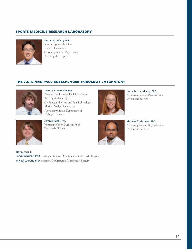
SPORTS MEDICINE RESEARCH LABORATORY
Vincent M. Wang, PhD
Director, Sports Medicine Research Laboratory
Assistant professor, Department of Orthopedic Surgery
THE JOAN AND PAUL RUBSCHLAGER TRIBOLOGY LABORATORY
Alfons Fischer, PhD
Visiting professor, Department of Orthopedic Surgery
Mathew T. Mathew, PhD Assistant professor, Department of Orthopedic Surgery
Markus A. Wimmer, PhD
Director, the Joan and Paul Rubschlager Tribology Laboratory
Co-director, the Joan and Paul Rubschlager Motion Analysis Laboratory
Associate professor, Department of Orthopedic Surgery
Not pictured:
Joachim Kunze, PhD, visiting instructor, Department of Orthopedic Surgery
Michel Laurent, PhD, scientist, Department of Orthopedic Surgery
Hannah J. Lundberg, PhD Assistant professor, Department of Orthopedic Surgery
71795_Body.indd 11 7/25/12 9:39 PM
2012 / RUSH ORTHOPEDICS JOURNAL
8
SECTION OF ORTHOPEDIC ONCOLOGY
Carl Maki, PhD
Associate professor, Department of Anatomy and Cell Biology
Qiping Zheng, PhD
Assistant professor, Department of Anatomy and Cell Biology
SECTION OF MOLECULAR MEDICINE
Tibor T. Glant, MD, PhD
Director, Section of Molecular Medicine
The Jorge O. Galante, MD, DMSc, Professor of Orthopaedic Surgery
Professor, Department of Orthopedic Surgery
Katalin Mikecz, MD, PhD
Professor, Department of Orthopedic Surgery
Not pictured:
Tibor A. Rauch, PhD, associate professor, Department of Orthopedic Surgery
SPINE RESEARCH LABORATORY
Nozomu Inoue, MD, PhD
Professor, Department of Orthopedic Surgery
Yejia Zhang, MD, PhD
Assistant professor, Department of Orthopedic Surgery
SPINE BIOMECHANICS
Raghu N. Natarajan, PhD
Professor, Department of Orthopedic Surgery
Alejandro A. Espinoza Orías, PhD
Instructor, Department of Orthopedic Surgery
10
SPINE BIOMECHANICS; CAD/COMPUTER ANALYSIS SPINE BIOCHEMISTRY AND BIOLOGY
71795_Body_u1.indd 10 8/3/12 10:30 AM
11
SPORTS MEDICINE RESEARCH LABORATORY
Vincent M. Wang, PhD
Director, Sports Medicine Research Laboratory
Assistant professor, Department of Orthopedic Surgery
THE JOAN AND PAUL RUBSCHLAGER TRIBOLOGY LABORATORY
Alfons Fischer, PhD
Visiting professor, Department of Orthopedic Surgery
Mathew T. Mathew, PhD Assistant professor, Department of Orthopedic Surgery
Markus A. Wimmer, PhD
Director, the Joan and Paul Rubschlager Tribology Laboratory
Co-director, the Joan and Paul Rubschlager Motion Analysis Laboratory
Associate professor, Department of Orthopedic Surgery
Not pictured:
Joachim Kunze, PhD, visiting instructor, Department of Orthopedic Surgery
Michel Laurent, PhD, scientist, Department of Orthopedic Surgery
Hannah J. Lundberg, PhD Assistant professor, Department of Orthopedic Surgery
71795_Body.indd 11 7/25/12 9:39 PM
2012 / RUSH ORTHOPEDICS JOURNAL
8
DEPARTMENT OF ORTHOPEDIC SURGERY RESIDENTS
CLASS OF 2012
Cara A. Cipriano, MD Medical school - University of Pennsylvania School of Medicine
Nickolas G. Garbis, MD Medical school - University of Illinois College of Medicine at Chicago
Richard W. Kang, MD Medical school - Rush Medical College
Sameer J. Lodha, MD Medical school - Washington University School of Medicine
CLASS OF 2013
Christopher O. Bayne, MD Medical school - Harvard Medical School
Michael B. Ellman, MD Medical school - University of Michigan Medical School
James M. Gregory, MD Medical school - University of Pennsylvania School of Medicine
Brett A. Lenart, MD Medical school - Weill Cornell Medical College
Adam B. Yanke, MD Medical school - Rush Medical College
CLASS OF 2014
Sanjeev Bhatia, MD Medical school - Northwestern University Feinberg School of Medicine
Debdut Biswas, MD Medical school - Yale University School of Medicine
Christopher E. Gross, MD Medical school - Harvard Medical School
Andrew R. Hsu, MD Medical school - Stanford University School of Medicine
Kevin U. Park, MD Medical school - Tulane University School of Medicine
CLASS OF 2015
Laith M. Al-Shihabi, MD Medical school - Medical College of Wisconsin
Peter N. Chalmers, MD Medical school - Columbia University College of Physicians and Surgeons
Jonathan M. Frank, MD Medical school - University of California Los Angeles Geffen School of Medicine
William Slikker III, MD Medical school - Stanford University School of Medicine
David M. Walton, MD Medical school - Case Western Reserve University School of Medicine
CLASS OF 2016
Nicholas M. Brown, MD Medical school - Columbia University College of Physicians and Surgeons
Rachel M. Frank, MD Medical school - Northwestern University Feinberg School of Medicine
Bryan D. Haughom, MD Medical school - University of California San Francisco School of Medicine
Michael D. Hellman, MD Medical school - Jefferson Medical College
Andrew Riff, MD Medical school - Georgetown University School of Medicine
CLASS OF 2017
Gregory Cvetanovich, MD Medical school - Harvard Medical School
Brandon Erickson, MD Medical school - Tufts University School of Medicine
Yale A. Fillingham, MD Medical school - Rush Medical College
David Levy, MD Medical school - Columbia University College of Physicians and Surgeons
Nathan Wetters, MD Medical school - University of Illinois College of Medicine at Rockford
12
71795_Body.indd 12 7/25/12 9:39 PM
13
Three-Dimensional Anatomy of the Hip, Utilizing Computer Navigation and Plain Radiographs
RACHEL M. FRANK, MD / JEREMY ALLAND, BS / ROBERT C. GRUMET, MD / MARK A. SLABAUGH, MD / VINCENT M.
WANG, PHD / ALEJANDRO A. ESPINOZA ORÍAS, PHD / BERNARD R. BACH JR, MD / SHANE J. NHO, MD, MS
“These descriptions of normal hip morphology may improve preoperative planning and postoperative outcomes of hip joint preservation surgery.”
ARTICLES
AUTHOR AFFILIATIONS
Department of Orthopedic Surgery, Rush University Medical Center, Chicago, Illinois (Drs Frank, Grumet, Slabaugh, Wang, Espinoza Orías, Nho, and Bach; and Mr Alland); and Midwest Orthopaedics at Rush, Chicago, Illinois (Drs Bach, Nho).
CORRESPONDING AUTHOR
Shane J. Nho, MD, MS; Rush University Medical Center and Midwest Orthopaedics at Rush, 1611 W Harrison St, Suite 300, Chicago, IL 60612 ([email protected]).
With femoro-acetabular impingement (FAI) as a known etiology of hip osteoarthritis,1-4 medical researchers have recently expressed interest in hip joint preservation
surgery.5-9 As FAI becomes increasingly recognized as a source of intra-articular hip pathology, particularly in the young, active patient population, doctors are beginning to favor nonarthroplasty treatment options. As with any orthopedic intervention, appropriate patient selection is critical for successful surgical outcomes; however, there remains a lack of consensus among orthopedic surgeons on the diagnosis, assessment, and surgical treatment of hips with FAI.
The surgical treatment options for young patients with FAI remain controversial. As techniques in diagnostic modalities as well as instrumentation and implant design continue to improve, surgical treatment options will also continue to evolve. Current options include nonoperative approaches,10-12 hip arthroscopy (Figures 1 and 2),6,13-18 open surgical dislocation (Figure 3),19-23
and periacetabular osteotomy.24 Open surgical hip dislocation has traditionally been considered the gold standard for FAI, with good to excellent technically reproducible results. In 2004, Murphy et al7 described a cohort of patients representing 23 hips at an average of 5 years following open hip dislocation and found that 7 (30%) had converted to total hip arthroplasty. While this open hip dislocation provides excellent visualization and typically allows for muscle preservation, the open nature of the surgery provides a source of morbidity not seen with less-invasive techniques, including arthroscopically assisted and all-arthroscopic procedures.
While the underlying etiology causing FAI varies, the mechanical process that leads to early osteoarthritis occurs when the proximal femur has repeated contact with the native acetabular rim upon normal range of motion. Pincer FAI is caused by excessive acetabular coverage with linear contact between the acetabular rim and head-neck junction, which across time and with continued impact can cause degeneration and ossification of the anterior labrum. Etiologies underlying abnormal acetabular anatomy that may lead to pincer FAI include acetabular retroversion, coxa profunda, coxa protrusio, dysplasia, and trauma. Cam FAI is caused by an abnormal femoral head-neck junction with an increased radius at the waist, causing shearing forces on the acetabular rim, which can lead to cartilage and labral damage. Etiologies underlying abnormal femoral anatomy that may lead to cam FAI include coxa vara, slipped capital femoral epiphysis (SCFE), femoral head avascular necrosis, retrotorsion, Legg-Calve-Perthes disease, and trauma. Many cases of abnormal acetabular and femoral anatomy are idiopathic. FAI may result from cam or pincer impingement; however, components of both mechanisms often contribute. In general, the most common location for FAI is along the anterosuperior acetabular rim, and thus, a precise
71795_Body.indd 13 7/25/12 9:39 PM
2012 / RUSH ORTHOPEDICS JOURNAL
14
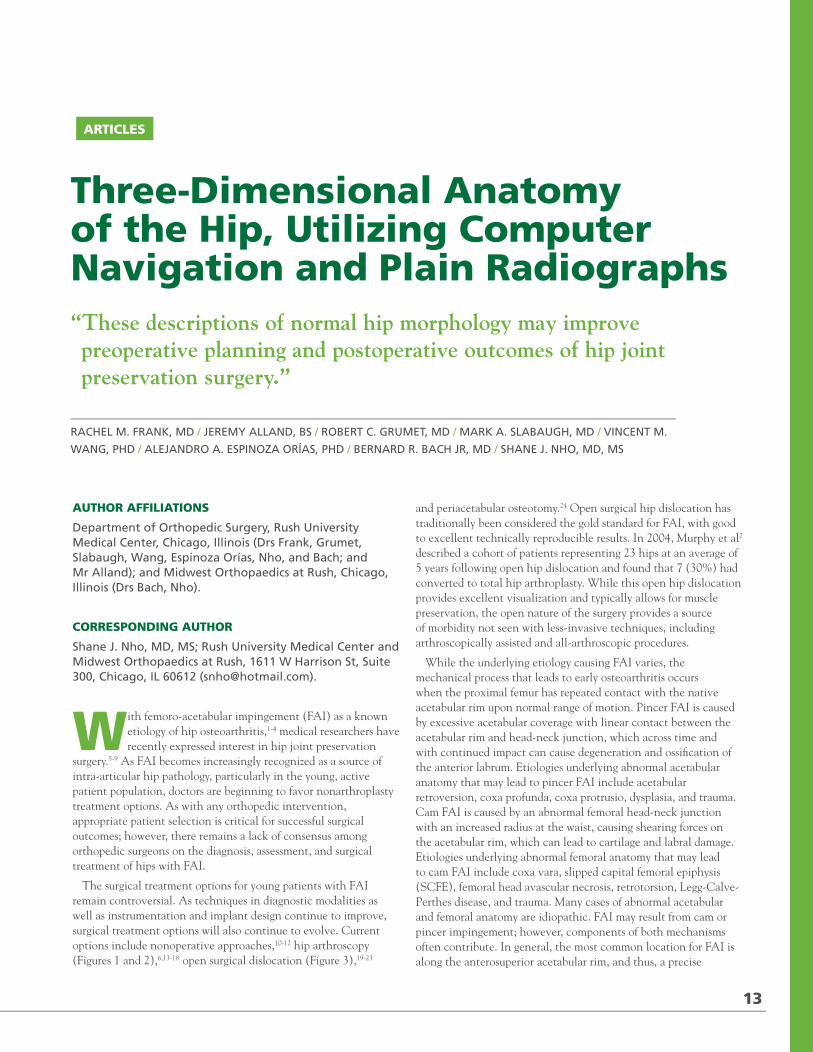
Figure 1. Intraoperative figures of hip arthroscopy for FAI. A, Pincer. B, Labral repair. C, Rim Trim. D, Cam. E, Cam osteochondroplasty. F, Cam osteochondroplasty.
understanding of normal acetabular anatomy is of critical importance.
The normal morphologic characteristics of the acetabular joint are poorly understood, and there remains a paucity of basic studies available in the literature that adequately describe normal hip osseous morphology. For example, while the anatomy of the anterior acetabular ridge has been consistently described as irregular, including curved, angular, irregular, and straight configurations,25-27 the posterior acetabular rim has been described as hypoplastic in some studies28 but not in others.29,30 Additionally, the majority of published morphologic studies were designed to
help with the design and sizing of acetabular components for total hip arthroplasty surgery. While the anatomical data presented in these studies25-29,31 are clearly relevant, there still remains a need for improved understanding of normal hip osseous morphology from an arthroscopic perspective. Specifically, the goals of arthroscopic treatment for FAI include creating “normal” acetabular and/or femoral neck anatomy in order to promote normal articulation of the femoro-acetabular joint during physiologic range of motion. In order to create more “normal” anatomy for a patient with pincer and/or cam FAI, the surgeon must intimately understand the normal osseous morphology of the hip joint.
A B C
D E F
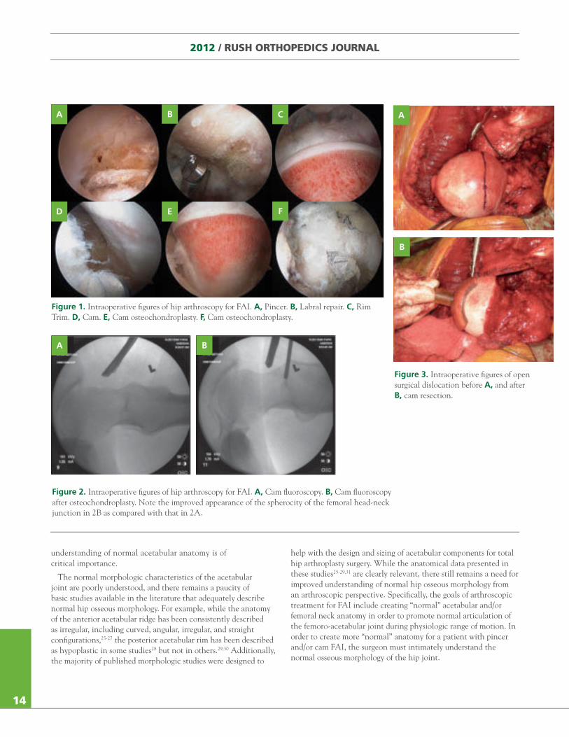
Figure 2. Intraoperative figures of hip arthroscopy for FAI. A, Cam fluoroscopy. B, Cam fluoroscopy after osteochondroplasty. Note the improved appearance of the spherocity of the femoral head-neck junction in 2B as compared with that in 2A.
Figure 3. Intraoperative figures of open surgical dislocation before A, and after B, cam resection.
A B
A
B
71795_Body.indd 14 7/25/12 9:39 PM
15
Thus, when considering surgical treatment of FAI, one of the major challenges for the orthopedic surgeon is the difficulty in determining the precise anatomic location and severity of the impingement. A reproducible, noninvasive method utilizing computer navigation for both diagnostic assessment and proper treatment planning may present a possible future solution.32 Nevertheless, equally as important as a noninvasive model for assessing FAI is a functional and comprehensive understanding of normal hip osseous morphology, which is currently not available in the literature. An improved understanding of 3-dimensional (3D) hip anatomy will allow the orthopedic surgeon to plan appropriate hip joint preservation surgery. Our study creates a novel method that would allow for comprehensive and reproducible mapping of the osseous morphology of the acetabulum using 3D analysis.
BRIEF DESCRIPTION OF LABORATORY WORK
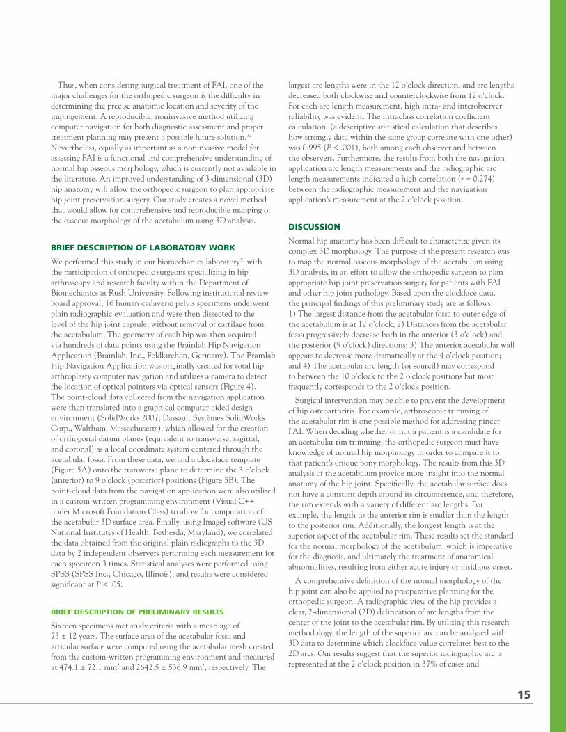
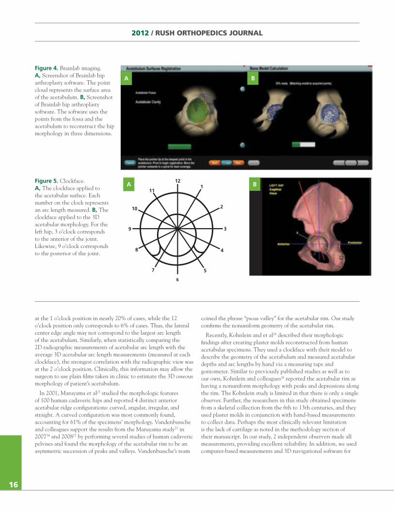
We performed this study in our biomechanics laboratory33 with the participation of orthopedic surgeons specializing in hip arthroscopy and research faculty within the Department of Biomechanics at Rush University. Following institutional review board approval, 16 human cadaveric pelvis specimens underwent plain radiographic evaluation and were then dissected to the level of the hip joint capsule, without removal of cartilage from the acetabulum. The geometry of each hip was then acquired via hundreds of data points using the Brainlab Hip Navigation Application (Brainlab, Inc., Feldkirchen, Germany). The Brainlab Hip Navigation Application was originally created for total hip arthroplasty computer navigation and utilizes a camera to detect the location of optical pointers via optical sensors (Figure 4). The point-cloud data collected from the navigation application were then translated into a graphical computer-aided design environment (SolidWorks 2007; Dassault Systèmes SolidWorks Corp., Waltham, Massachusetts), which allowed for the creation of orthogonal datum planes (equivalent to transverse, sagittal, and coronal) as a local coordinate system centered through the acetabular fossa. From these data, we laid a clockface template (Figure 5A) onto the transverse plane to determine the 3 o’clock (anterior) to 9 o’clock (posterior) positions (Figure 5B). The point-cloud data from the navigation application were also utilized in a custom-written programming environment (Visual C++ under Microsoft Foundation Class) to allow for computation of the acetabular 3D surface area. Finally, using ImageJ software (US National Institutes of Health, Bethesda, Maryland), we correlated the data obtained from the original plain radiographs to the 3D data by 2 independent observers performing each measurement for each specimen 3 times. Statistical analyses were performed using SPSS (SPSS Inc., Chicago, Illinois), and results were considered significant at P < .05.
BRIEF DESCRIPTION OF PRELIMINARY RESULTS
Sixteen specimens met study criteria with a mean age of 73 ± 12 years. The surface area of the acetabular fossa and articular surface were computed using the acetabular mesh created from the custom-written programming environment and measured at 474.1 ± 72.1 mm2 and 2642.5 ± 536.9 mm2, respectively. The
largest arc lengths were in the 12 o’clock direction, and arc lengths decreased both clockwise and counterclockwise from 12 o’clock. For each arc length measurement, high intra- and interobserver reliability was evident. The intraclass correlation coefficient calculation, (a descriptive statistical calculation that describes how strongly data within the same group correlate with one other) was 0.995 (P < .001), both among each observer and between the observers. Furthermore, the results from both the navigation application arc length measurements and the radiographic arc length measurements indicated a high correlation (r = 0.274) between the radiographic measurement and the navigation application’s measurement at the 2 o’clock position.
DISCUSSION
Normal hip anatomy has been difficult to characterize given its complex 3D morphology. The purpose of the present research was to map the normal osseous morphology of the acetabulum using 3D analysis, in an effort to allow the orthopedic surgeon to plan appropriate hip joint preservation surgery for patients with FAI and other hip joint pathology. Based upon the clockface data, the principal findings of this preliminary study are as follows: 1) The largest distance from the acetabular fossa to outer edge of the acetabulum is at 12 o’clock; 2) Distances from the acetabular fossa progressively decrease both in the anterior (3 o’clock) and the posterior (9 o’clock) directions; 3) The anterior acetabular wall appears to decrease more dramatically at the 4 o’clock position; and 4) The acetabular arc length (or sourcil) may correspond to between the 10 o’clock to the 2 o’clock positions but most frequently corresponds to the 2 o’clock position.
Surgical intervention may be able to prevent the development of hip osteoarthritis. For example, arthroscopic trimming of the acetabular rim is one possible method for addressing pincer FAI. When deciding whether or not a patient is a candidate for an acetabular rim trimming, the orthopedic surgeon must have knowledge of normal hip morphology in order to compare it to that patient’s unique bony morphology. The results from this 3D analysis of the acetabulum provide more insight into the normal anatomy of the hip joint. Specifically, the acetabular surface does not have a constant depth around its circumference, and therefore, the rim extends with a variety of different arc lengths. For example, the length to the anterior rim is smaller than the length to the posterior rim. Additionally, the longest length is at the superior aspect of the acetabular rim. These results set the standard for the normal morphology of the acetabulum, which is imperative for the diagnosis, and ultimately the treatment of anatomical abnormalities, resulting from either acute injury or insidious onset.
A comprehensive definition of the normal morphology of the hip joint can also be applied to preoperative planning for the orthopedic surgeon. A radiographic view of the hip provides a clear, 2-dimensional (2D) delineation of arc lengths from the center of the joint to the acetabular rim. By utilizing this research methodology, the length of the superior arc can be analyzed with 3D data to determine which clockface value correlates best to the 2D arcs. Our results suggest that the superior radiographic arc is represented at the 2 o’clock position in 37% of cases and
71795_Body.indd 15 7/25/12 9:39 PM
2012 / RUSH ORTHOPEDICS JOURNAL
14
Figure 1. Intraoperative figures of hip arthroscopy for FAI. A, Pincer. B, Labral repair. C, Rim Trim. D, Cam. E, Cam osteochondroplasty. F, Cam osteochondroplasty.
understanding of normal acetabular anatomy is of critical importance.
The normal morphologic characteristics of the acetabular joint are poorly understood, and there remains a paucity of basic studies available in the literature that adequately describe normal hip osseous morphology. For example, while the anatomy of the anterior acetabular ridge has been consistently described as irregular, including curved, angular, irregular, and straight configurations,25-27 the posterior acetabular rim has been described as hypoplastic in some studies28 but not in others.29,30 Additionally, the majority of published morphologic studies were designed to
help with the design and sizing of acetabular components for total hip arthroplasty surgery. While the anatomical data presented in these studies25-29,31 are clearly relevant, there still remains a need for improved understanding of normal hip osseous morphology from an arthroscopic perspective. Specifically, the goals of arthroscopic treatment for FAI include creating “normal” acetabular and/or femoral neck anatomy in order to promote normal articulation of the femoro-acetabular joint during physiologic range of motion. In order to create more “normal” anatomy for a patient with pincer and/or cam FAI, the surgeon must intimately understand the normal osseous morphology of the hip joint.
A B C
D E F
Figure 2. Intraoperative figures of hip arthroscopy for FAI. A, Cam fluoroscopy. B, Cam fluoroscopy after osteochondroplasty. Note the improved appearance of the spherocity of the femoral head-neck junction in 2B as compared with that in 2A.
Figure 3. Intraoperative figures of open surgical dislocation before A, and after B, cam resection.
A B
A
B
71795_Body.indd 14 7/25/12 9:39 PM
15
Thus, when considering surgical treatment of FAI, one of the major challenges for the orthopedic surgeon is the difficulty in determining the precise anatomic location and severity of the impingement. A reproducible, noninvasive method utilizing computer navigation for both diagnostic assessment and proper treatment planning may present a possible future solution.32 Nevertheless, equally as important as a noninvasive model for assessing FAI is a functional and comprehensive understanding of normal hip osseous morphology, which is currently not available in the literature. An improved understanding of 3-dimensional (3D) hip anatomy will allow the orthopedic surgeon to plan appropriate hip joint preservation surgery. Our study creates a novel method that would allow for comprehensive and reproducible mapping of the osseous morphology of the acetabulum using 3D analysis.
BRIEF DESCRIPTION OF LABORATORY WORK
We performed this study in our biomechanics laboratory33 with the participation of orthopedic surgeons specializing in hip arthroscopy and research faculty within the Department of Biomechanics at Rush University. Following institutional review board approval, 16 human cadaveric pelvis specimens underwent plain radiographic evaluation and were then dissected to the level of the hip joint capsule, without removal of cartilage from the acetabulum. The geometry of each hip was then acquired via hundreds of data points using the Brainlab Hip Navigation Application (Brainlab, Inc., Feldkirchen, Germany). The Brainlab Hip Navigation Application was originally created for total hip arthroplasty computer navigation and utilizes a camera to detect the location of optical pointers via optical sensors (Figure 4). The point-cloud data collected from the navigation application were then translated into a graphical computer-aided design environment (SolidWorks 2007; Dassault Systèmes SolidWorks Corp., Waltham, Massachusetts), which allowed for the creation of orthogonal datum planes (equivalent to transverse, sagittal, and coronal) as a local coordinate system centered through the acetabular fossa. From these data, we laid a clockface template (Figure 5A) onto the transverse plane to determine the 3 o’clock (anterior) to 9 o’clock (posterior) positions (Figure 5B). The point-cloud data from the navigation application were also utilized in a custom-written programming environment (Visual C++ under Microsoft Foundation Class) to allow for computation of the acetabular 3D surface area. Finally, using ImageJ software (US National Institutes of Health, Bethesda, Maryland), we correlated the data obtained from the original plain radiographs to the 3D data by 2 independent observers performing each measurement for each specimen 3 times. Statistical analyses were performed using SPSS (SPSS Inc., Chicago, Illinois), and results were considered significant at P < .05.
BRIEF DESCRIPTION OF PRELIMINARY RESULTS
Sixteen specimens met study criteria with a mean age of 73 ± 12 years. The surface area of the acetabular fossa and articular surface were computed using the acetabular mesh created from the custom-written programming environment and measured at 474.1 ± 72.1 mm2 and 2642.5 ± 536.9 mm2, respectively. The
largest arc lengths were in the 12 o’clock direction, and arc lengths decreased both clockwise and counterclockwise from 12 o’clock. For each arc length measurement, high intra- and interobserver reliability was evident. The intraclass correlation coefficient calculation, (a descriptive statistical calculation that describes how strongly data within the same group correlate with one other) was 0.995 (P < .001), both among each observer and between the observers. Furthermore, the results from both the navigation application arc length measurements and the radiographic arc length measurements indicated a high correlation (r = 0.274) between the radiographic measurement and the navigation application’s measurement at the 2 o’clock position.
DISCUSSION
Normal hip anatomy has been difficult to characterize given its complex 3D morphology. The purpose of the present research was to map the normal osseous morphology of the acetabulum using 3D analysis, in an effort to allow the orthopedic surgeon to plan appropriate hip joint preservation surgery for patients with FAI and other hip joint pathology. Based upon the clockface data, the principal findings of this preliminary study are as follows: 1) The largest distance from the acetabular fossa to outer edge of the acetabulum is at 12 o’clock; 2) Distances from the acetabular fossa progressively decrease both in the anterior (3 o’clock) and the posterior (9 o’clock) directions; 3) The anterior acetabular wall appears to decrease more dramatically at the 4 o’clock position; and 4) The acetabular arc length (or sourcil) may correspond to between the 10 o’clock to the 2 o’clock positions but most frequently corresponds to the 2 o’clock position.
Surgical intervention may be able to prevent the development of hip osteoarthritis. For example, arthroscopic trimming of the acetabular rim is one possible method for addressing pincer FAI. When deciding whether or not a patient is a candidate for an acetabular rim trimming, the orthopedic surgeon must have knowledge of normal hip morphology in order to compare it to that patient’s unique bony morphology. The results from this 3D analysis of the acetabulum provide more insight into the normal anatomy of the hip joint. Specifically, the acetabular surface does not have a constant depth around its circumference, and therefore, the rim extends with a variety of different arc lengths. For example, the length to the anterior rim is smaller than the length to the posterior rim. Additionally, the longest length is at the superior aspect of the acetabular rim. These results set the standard for the normal morphology of the acetabulum, which is imperative for the diagnosis, and ultimately the treatment of anatomical abnormalities, resulting from either acute injury or insidious onset.
A comprehensive definition of the normal morphology of the hip joint can also be applied to preoperative planning for the orthopedic surgeon. A radiographic view of the hip provides a clear, 2-dimensional (2D) delineation of arc lengths from the center of the joint to the acetabular rim. By utilizing this research methodology, the length of the superior arc can be analyzed with 3D data to determine which clockface value correlates best to the 2D arcs. Our results suggest that the superior radiographic arc is represented at the 2 o’clock position in 37% of cases and
71795_Body.indd 15 7/25/12 9:39 PM
2012 / RUSH ORTHOPEDICS JOURNAL
16
at the 1 o’clock position in nearly 20% of cases, while the 12 o’clock position only corresponds to 6% of cases. Thus, the lateral center edge angle may not correspond to the largest arc length of the acetabulum. Similarly, when statistically comparing the 2D radiographic measurements of acetabular arc length with the average 3D acetabular arc length measurements (measured at each clockface), the strongest correlation with the radiographic view was at the 2 o’clock position. Clinically, this information may allow the surgeon to use plain films taken in clinic to estimate the 3D osseous morphology of patient’s acetabulum.
In 2001, Maruyama et al25 studied the morphologic features of 100 human cadaveric hips and reported 4 distinct anterior acetabular ridge configurations: curved, angular, irregular, and straight. A curved configuration was most commonly found, accounting for 61% of the specimens’ morphology. Vandenbussche and colleagues support the results from the Maruyama study25 in 200734 and 200827 by performing several studies of human cadaveric pelvises and found the morphology of the acetabular rim to be an asymmetric succession of peaks and valleys. Vandenbussche’s team
coined the phrase “psoas valley” for the acetabular rim. Our study confirms the nonuniform geometry of the acetabular rim.
Recently, Kohnlein and et al26 described their morphologic findings after creating plaster molds reconstructed from human acetabular specimens. They used a clockface with their model to describe the geometry of the acetabulum and measured acetabular depths and arc lengths by hand via a measuring tape and goniometer. Similar to previously published studies as well as to our own, Kohnlein and colleagues26 reported the acetabular rim as having a nonuniform morphology with peaks and depressions along the rim. The Kohnlein study is limited in that there is only a single observer. Further, the researchers in this study obtained specimens from a skeletal collection from the 6th to 13th centuries, and they used plaster molds in conjunction with hand-based measurements to collect data. Perhaps the most clinically relevant limitation is the lack of cartilage as noted in the methodology section of their manuscript. In our study, 2 independent observers made all measurements, providing excellent reliability. In addition, we used computer-based measurements and 3D navigational software for
Figure 4. Brainlab imaging. A, Screenshot of Brainlab hip arthroplasty software. The point cloud represents the surface area of the acetabulum. B, Screenshot of Brainlab hip arthroplasty software. The software uses the points from the fossa and the acetabulum to reconstruct the hip morphology in three dimensions.
Figure 5. Clockface. A, The clockface applied to the acetabular surface. Each number on the clock represents an arc length measured. B, The clockface applied to the 3D acetabular morphology. For the left hip, 3 o’clock corresponds to the anterior of the joint. Likewise, 9 o’clock corresponds to the posterior of the joint.
A B
6
121
2
3
4
57
8
9
10
11A B
71795_Body.indd 16 7/25/12 9:39 PM
17
all data measurements, providing excellent precision and accuracy. Overall, while the Kohnlein study focused more on acetabular version, tilt, and inclination, our study focused more on surface area, radius, depth, and arc length of the acetabulum. Thus, the aim of our research better allows for the characterization of the overall acetabular osseous morphology. Both our study and the Kohnlein study provide a better understanding of the functional anatomy of the acetabulum within the hip joint itself.
The preliminary research conducted in our lab had some limitations. First, the data were collected from an elderly cadaveric population, which is not representative of the young, active patient population typically under consideration for surgical treatment of FAI. Second, the Brainlab system was originally designed for total hip arthroplasty procedures and not for FAI procedures, which may make data collection less reliable. Nevertheless, the use of a computer-based 3D measurement system is a strength of our preliminary methodology. The Brainlab system, despite its intent as a total hip arthroplasty system, is able to recreate a 3D model of a hip using data points, which provides more accuracy and precision than using human-based measurements.
CONCLUSIONS
The normal morphologic characteristics of the acetabular joint are poorly understood, and very few studies adequately describe normal acetabular osseous anatomy. As FAI becomes increasingly recognized as a precursor to early hip osteoarthritis, less-invasive treatment options may become more favorable. Arthroscopy has proven to be a successful short- and medium-term surgical solution for appropriately indicated patients; however, a comprehensive understanding of “normal” hip osseous morphology is absolutely critical prior to any attempt at surgical correction. Our preliminary research provided a comprehensive description of “normal” acetabular osseous morphology by using 3D computer navigation and correlating the resulting data with plain radiographs. These descriptions of normal hip morphology may improve preoperative planning and postoperative outcomes of hip joint preservation surgery. Future areas of research will be conducted to further analyze the correlation between 3D measurements and radiographic measurements, as well as to map the osseous morphology of the femoral head-neck junction.
REFERENCES1. Ganz R, Leunig M, Leunig-Ganz K, Harris WH. The etiology of osteoarthritis of the hip: an integrated mechanical concept. Clin Orthop Relat Res. 2008;466(2):264-272.
2. Allen D, Beaule PE, Ramadan O, Doucette S. Prevalence of associated deformities and hip pain in patients with cam-type femoro-acetabular impingement. J Bone Joint Surg Br. 2009;91(5):589-594.
3. Beck M, Kalhor M, Leunig M, Ganz R. Hip morphology influences the pattern of damage to the acetabular cartilage: femoro-acetabular impingement as a cause of early osteoarthritis of the hip. J Bone Joint Surg Br. 2005;87(7):1012-1018.
4. Parvizi J, Leunig M, Ganz R. Femoro-acetabular impingement. J Am Acad Orthop Surg. 2007;15(9):561-570.
5. Leunig M, Huff TW, Ganz R. Femoro-acetabular impingement: treatment of the acetabular side. Instr Course Lect. 2009;58:223-229.
6. Byrd JW, Jones KS. Arthroscopic management of femoro-acetabular impingement. Instr Course Lect. 2009;58:231-239.
7. Murphy S, Tannast M, Kim YJ, Buly R, Millis MB. Debridement of the adult hip for femoro-acetabular impingement: indications and preliminary clinical results. Clin Orthop Relat Res. 2004(429):178-181.
8. Philippon MJ, Stubbs AJ, Schenker ML, Maxwell RB, Ganz R, Leunig M. Arthroscopic management of femoro-acetabular impingement: osteoplasty technique and literature review. Am J Sports Med. 2007;35(9):1571-1580.
9. Sierra RJ, Trousdale RT, Ganz R, Leunig M. Hip disease in the young, active patient: evaluation and nonarthroplasty surgical options. J Am Acad Orthop Surg. 2008;16(12):689-703.
10. Grumet R, Frank RM, Slabaugh M, Virkus W, Bush-Joseph C, Nho S. Lateral hip pain in an athletic population: differential diagnosis and treatment options. Sports Health: A Multidisciplinary Approach. 2010;2(3):191-196.
11. Frank RM, Grumet R, Slabaugh M, Virkus W, Bush-Joseph C, Nho S. Posterior hip pain in an athletic population: differential diagnosis and treatment options. Sports Health: A Multidisciplinary Approach. 2010;2(3):237-246.
12. Keogh MJ, Batt ME. A review of femoro-acetabular impingement in athletes. Sports Med. 2008;38(10):863-878.
13. Brunner A, Horisberger M, Herzog RF. Sports and recreation activity of patients with femoro-acetabular impingement before and after arthroscopic osteoplasty. Am J Sports Med. 2009;37(5): 917-922.
14. Horisberger M, Brunner A, Herzog RF. Arthroscopic treatment of femoral acetabular impingement in patients with preoperative generalized degenerative changes. Arthroscopy. 2010;26(5):623-629.
15. Ilizaliturri VM, Jr., Orozco-Rodriguez L, Acosta-Rodriguez E, Camacho-Galindo J. Arthroscopic treatment of cam-type femoro-acetabular impingement: preliminary report at 2 years minimum follow-up. J Arthroplasty. 2008;23(2):226-234.
16. Larson CM, Giveans MR. Arthroscopic management of femoro-acetabular impingement: early outcomes measures. Arthroscopy. 2008;24(5):540-546.
17. Philippon MJ, Schenker ML. Arthroscopy for the treatment of femoro-acetabular impingement in the athlete. Clin Sports Med. 2006;25(2):299-308, ix.
18. Philippon MJ, Weiss DR, Kuppersmith DA, Briggs KK, Hay CJ. Arthroscopic labral repair and treatment of femoro-acetabular impingement in professional hockey players. Am J Sports Med. 2010;38(1):99-104.
19. Anderson LA, Crofoot CD, Erickson J, Morton DA, Peters CL. Acetabular osteochondroplasty and simultaneous reorientation: background and validation of concept. Orthopedics. 2010;33(5):299.
20. Beaule PE, Le Duff MJ, Zaragoza E. Quality of life following femoral head-neck osteochondroplasty for femoro-acetabular impingement. J Bone Joint Surg Am. 2007;89(4):773-779.
21. Bizzini M, Notzli HP, Maffiuletti NA. Femoro-acetabular impingement in professional ice hockey players: a case series of 5 athletes after open surgical decompression of the hip. Am J Sports Med. 2007;35(11):1955-1959.
22. Graves ML, Mast JW. Femoro-acetabular impingement: do outcomes reliably improve with surgical dislocations? Clin Orthop Relat Res. 2009;467(3):717-723.
23. Peters CL, Schabel K, Anderson L, Erickson J. Open treatment of femoro-acetabular impingement is associated with clinical improvement and low complication rate at short-term followup. Clin Orthop Relat Res. 2010;468(2):504-510.
24. Siebenrock KA, Schoeniger R, Ganz R. Anterior femoro-acetabular impingement due to acetabular retroversion. Treatment with periacetabular osteotomy. J Bone Joint Surg Am. 2003;85(2):278-286.
25. Maruyama M, Feinberg JR, Capello WN, DʼAntonio JA. The Frank Stinchfield Award: morphologic features of the acetabulum and femur: anteversion angle and implant positioning. Clin Orthop Relat Res. 2001;(393):52-65.
26. Kohnlein W, Ganz R, Impellizzeri FM, Leunig M. Acetabular morphology: implications for joint-preserving surgery. Clin Orthop Relat Res.2009;467(3):682-691.
27. Vandenbussche E, Saffarini M, Taillieu F, Mutschler C. The asymmetric profile of the acetabulum. Clin Orthop Relat Res. 2008;466(2):417-423.
28. Giori NJ, Trousdale RT. Acetabular retroversion is associated with osteoarthritis of the hip. Clin Orthop Relat Res. 2003(417):263-269.
29. Jamali AA, Mladenov K, Meyer DC, et al. Anteroposterior pelvic radiographs to assess acetabular retroversion: high validity of the “cross-over-sign”. J Orthop Res. 2007;25(6):758-765.
30. Kalberer F, Sierra RJ, Madan SS, Ganz R, Leunig M. Ischial spine projection into the pelvis: a new sign for acetabular retroversion. Clin Orthop Relat Res. 2008;466(3):677-683.
31. Thompson MS, Dawson T, Kuiper JH, Northmore-Ball MD, Tanner KE. Acetabular morphology and resurfacing design. J Biomech. 2000;33(12):1645-1653.
32. Tannast M, Kubiak-Langer M, Langlotz F, Puls M, Murphy SB, Siebenrock KA. Noninvasive three-dimensional assessment of femoro-acetabular impingement. J Orthop Res. 2007;25(1):122-131.
33. Frank RM, Alland J, Grumet RC, et al. Hip osseous morphologic characteristics using computer navigation and plain radiographs. Amer J Orthop. In press.
34. Vandenbussche E, Saffarini M, Deloge N, Moctezuma JL, Nogler M. Hemispheric cups do not reproduce acetabular rim morphology. Acta Orthop. 2007;78(3):327-332.
Disclosures for the preceding article are listed on p. 22.
71795_Body_u1.indd 17 8/3/12 10:30 AM
2012 / RUSH ORTHOPEDICS JOURNAL
Metal Ion Levels After a Second Joint Arthroplasty
ANDREW R. HSU, MD / BRETT LEVINE, MD, MS / ANASTASIA SKIPOR, MS / NADIM J. HALLAB, PHD /
WAYNE G. PAPROSKY, MD / JOSHUA J. JACOBS, MD
“Determining serum metal ion concentrations after total joint arthroplasty may be useful in the work-up of patients with local pain or unexplained systemic symptoms...”
ARTICLES
AUTHOR AFFILIATIONS
Department of Orthopedic Surgery, Rush University Medical Center, Chicago, Illinois (Drs Hsu, Levine, Hallab, Paprosky, and Jacobs; and Ms Skipor); and Midwest Orthopaedics at Rush, Chicago, Illinois (Drs Levine, Paprosky, and Jacobs).
CORRESPONDING AUTHOR
Joshua J. Jacobs, MD; Rush University Medical Center and Midwest Orthopaedics at Rush, 1611 W Harrison St, Suite 300, Chicago, IL 60612 ([email protected]).
Elevated serum metal ion levels have been found after primary total hip arthroplasty (THA) regardless of the type of articulation couple used.1,2,3,4 With the recent
focus on reported adverse responses to metal-on-metal THA, there is mounting concern regarding local soft tissue reactions, systemic immune modulation, and end-organ deposition and damage associated with elevated serum metal ion concentrations. The metal components of joint replacements can undergo electrochemical corrosion resulting in the formation of chemically active metal ion degradation products.3 Tower reported 2 patients with metal-on-metal THA who developed systemic neurological and cardiac complications associated with an elevation in serum cobalt levels.5 Tower suggested a cause-effect relationship due to the fact that systemic symptoms improved following revision surgery, presumably lowering serum cobalt concentrations although no serum measurements were reported following revision and there was limited information regarding case details. Adverse systemic responses to metal ion debris from joint replacement are extremely
rare. It is also important to note that increased serum metal ion levels are not unique to metal-on-metal bearings, because other metal devices with accelerated damage due to wear or corrosion can lead to elevated levels of metal in local and remote tissues.
Currently, there is no generally accepted threshold above which serum concentrations of metal ions such as cobalt, chromium, and titanium are known to be toxic in patients with a joint replacement. However, cobalt and chromium are both toxic in high concentrations in vivo, and titanium has been shown to be potentially carcinogenic at high levels in animal models.6 Previous work has also shown that wear particles from total hip and knee replacements can disseminate to the liver, spleen, and abdominal lymph nodes, causing as-yet-unknown long-term effects.7 Degenerative joint disease of the hip will often impact the contralateral hip and knee, thus requiring a future second total joint replacement.8 However, we know little about the effect of this second arthroplasty on metal ion levels in the body. The purpose of the present study was to determine the effect of a second joint replacement on serum concentrations of cobalt, chromium, and titanium after a primary metal-on-polyethylene THA as well as the time course of concentration changes.
METHODS
We prospectively enrolled in this study after obtaining research approval from the Rush University Institutional Review Board. We obtained informed consent from all patients prior to study inclusion. The average age of patients was 63.4 years (range, 55-76 years). We included 4 males and 8 females who were all undergoing an initial primary metal-on-polyethylene THA for the treatment of osteoarthritis. All patients had a well-functioning prosthesis determined by Harris hip score, and none had
18
71795_Body.indd 18 7/25/12 9:39 PM
radiographic evidence of loosening or osteolysis at interval follow-up or just prior to their second operation. We included 8 patients who had received cementless proximally porous-coated titanium-alloy femoral stems and titanium acetabular components. The femoral stem was made of Ti-6A1-4V with a titanium fiber-metal porous-coated surface diffusion bonded onto the proximal aspect of the stem. We used 6 Anatomic and 2 Harris-Galante Multilock femoral components (Zimmer, Warsaw, Indiana). The acetabular component, which we inserted without cement, consisted of a titanium shell with a diffusion-bonded, titanium fiber-metal porous-coated surface (Zimmer). We used 2 titanium alloy self-tapping screws for fixation. The bearing surface consisted of a modular cobalt-alloy femoral head and ultrahigh molecular weight polyethylene liner.
Two patients received cementless, extensively porous-coated, cobalt-alloy femoral stems with titanium-alloy acetabular components. We inserted anatomical medullary locking (AML) femoral components (DePuy, Warsaw, Indiana) based on preoperative templating and intraoperative bone quality. We used Duraloc acetabular sockets, consisting of titanium-alloy shells with a beaded titanium porous coating (DePuy). Using 3 porous-coated titanium-alloy spikes, we secured the acetabular shells to the pelvis. We used modular cobalt-alloy femoral head and ultra-high molecular weight polyethylene liner for the articulation surface.
The final 2 patients received a hybrid THA that consisted of a modular uncoated cobalt-alloy femoral stem inserted with cement along with a cobalt-alloy head. We used 1 Iowa and 1 Precoat femoral component (Zimmer). We inserted the Harris-Galante II titanium acetabular component as described above and secured it to the pelvis with 2 titanium alloy screws. A snap-fit ultra-high molecular weight polyethylene liner served as the bearing surface.
Second joint replacements consisted of 8 subsequent THAs and 4 total knee arthroplasties (TKA) at an average of 102.7 months (range, 36-144 months) after the index operation. We performed all primary and secondary arthroplasties between 1989 and 1998. Secondary THAs consisted of 3 cementless titanium-alloy femoral stem and titanium acetabular component reconstructions and 5 hybrid constructs following the preceding descriptions. We cemented all TKAs in place with a cobalt-chromium femur and a titanium tibial tray (Zimmer). We excluded patients if additional metal implants other than the second total joint replacements were inserted during the follow-up intervals. Exclusion criteria also included component failure requiring component revision, medication use that would alter serum metal ion levels, and advanced systemic disease.
We collected blood samples for serum metal ion level analysis before second joint replacement and at 3-, 6-, and 12-month intervals and annually thereafter. We verified all vessels and utensils used for serum collection to be free of metal contamination using techniques described previously.3 Blood samples were obtained with use of siliconized butterfly needles in triplicate polypropylene syringes. We used the first syringe to rinse the system and then performed metal-ion analysis on the contents of the second and third syringes. The blood was immediately separated and frozen at –80°C after collection for long-term preservation
as serum and clot fractions using a Class 100 biological safety hood. To prevent contamination, we used ultrapure reagents and processed vessels in an acid wash.
We analyzed the serum samples for cobalt, chromium, and titanium levels, using a high-resolution sector field inductively coupled plasma mass spectrophotometer (HR-SF-ICPMS) (Element 2, Thermo Finnigan, Bremen, Germany), according to the method of additions described in detail in Iavicoli et al.9 The technique for measuring serum metal ion levels was consistent and reproducible throughout the duration of the study. We used detection limits of 0.04 nanograms per milliliter (ng/ml) for cobalt, 0.015 ng/ml for chromium, and 0.2 ng/ml for titanium. Data reported in figures are the means for each group at each time interval with standard deviations. We performed Friedman tests using SPSS statistics software (SPSS Science, Chicago, Illinois) to determine statistical significance with a P value < .05 as the threshold for significance.
RESULTS
Minimum 5-year data were available for all patients after their second joint arthroplasty, with an average follow-up of 96.4 months (range, 61-168 months). None of the concentrations of serum cobalt, chromium, or titanium was below the standard detection limits described above. Normal serum levels of the metal ions analyzed for the current study have been previously reported as 0.15 ng/ml (parts per billion) for chromium and 0.1 to 0.2 ng/ml for cobalt.10 The normal level of titanium has been reported as being less than 4.1 ng/ml.3
Patients undergoing a secondary TKA after a primary THA had no significant trends or differences in cobalt, chromium, or titanium ion levels from baseline up to 72 months after their operation. In addition, there were no significant differences in cobalt, chromium, or titanium levels at baseline between unilateral THA and secondary THA groups. Patients with a secondary THA had significantly elevated cobalt ion levels at 36 months follow-up, reaching a peak at 48 months (Figure 1A). Chromium levels in patients with a secondary THA were significantly elevated at 12 months follow-up with sustained increased concentration at 24 months (Figure 1B). Patients with a secondary THA also had significantly elevated titanium levels at 48 and 72 months follow-up (Figure 1C).
DISCUSSION
Elevation of serum metal ion levels after primary THA is a well-described phenomenon3,11-15; however, the impact of a second joint arthroplasty on these levels has not been well investigated. It is commonly found that patients who develop degenerative joint disease requiring hip arthroplasty often go on to develop joint disease in an adjacent or contralateral joint necessitating future joint arthroplasty.8 In the present study, cobalt ion concentrations after secondary THA were significantly increased at 36 months follow-up, reaching a peak at 48 months, while chromium levels were increased at 12 and 48 months. Titanium levels were
19
71795_Body.indd 19 7/25/12 9:39 PM
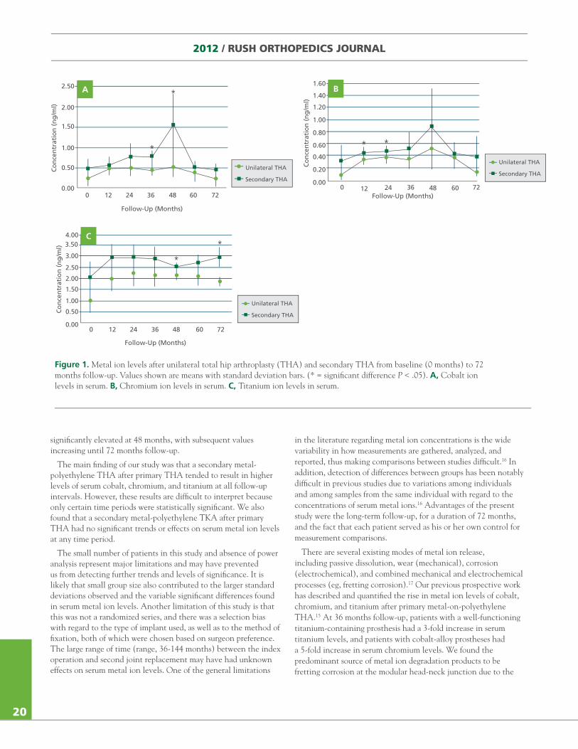
significantly elevated at 48 months, with subsequent values increasing until 72 months follow-up.
The main finding of our study was that a secondary metal-polyethylene THA after primary THA tended to result in higher levels of serum cobalt, chromium, and titanium at all follow-up intervals. However, these results are difficult to interpret because only certain time periods were statistically significant. We also found that a secondary metal-polyethylene TKA after primary THA had no significant trends or effects on serum metal ion levels at any time period.
The small number of patients in this study and absence of power analysis represent major limitations and may have prevented us from detecting further trends and levels of significance. It is likely that small group size also contributed to the larger standard deviations observed and the variable significant differences found in serum metal ion levels. Another limitation of this study is that this was not a randomized series, and there was a selection bias with regard to the type of implant used, as well as to the method of fixation, both of which were chosen based on surgeon preference. The large range of time (range, 36-144 months) between the index operation and second joint replacement may have had unknown effects on serum metal ion levels. One of the general limitations
in the literature regarding metal ion concentrations is the wide variability in how measurements are gathered, analyzed, and reported, thus making comparisons between studies difficult.16 In addition, detection of differences between groups has been notably difficult in previous studies due to variations among individuals and among samples from the same individual with regard to the concentrations of serum metal ions.16 Advantages of the present study were the long-term follow-up, for a duration of 72 months, and the fact that each patient served as his or her own control for measurement comparisons.
There are several existing modes of metal ion release, including passive dissolution, wear (mechanical), corrosion (electrochemical), and combined mechanical and electrochemical processes (eg, fretting corrosion).17 Our previous prospective work has described and quantified the rise in metal ion levels of cobalt, chromium, and titanium after primary metal-on-polyethylene THA.15 At 36 months follow-up, patients with a well-functioning titanium-containing prosthesis had a 3-fold increase in serum titanium levels, and patients with cobalt-alloy prostheses had a 5-fold increase in serum chromium levels. We found the predominant source of metal ion degradation products to be fretting corrosion at the modular head-neck junction due to the
2012 / RUSH ORTHOPEDICS JOURNAL
20
Figure 1. Metal ion levels after unilateral total hip arthroplasty (THA) and secondary THA from baseline (0 months) to 72 months follow-up. Values shown are means with standard deviation bars. (* = significant difference P < .05). A, Cobalt ion levels in serum. B, Chromium ion levels in serum. C, Titanium ion levels in serum.
2.50
2.00
1.50
1.00
0.50
0.000 12 24 36 48 60 72
Follow-Up (Months)
Co
nce
ntr
atio
n (
ng
/ml)
Unilateral THA
Secondary THA
1.60
1.40
1.20
1.00
0.80
0 12 24 36 48 60 72Follow-Up (Months)
Co
nce
ntr
atio
n (
ng
/ml)
Unilateral THA
Secondary THA
0.60
0.40
0.20
0.00
4.003.50
3.00
2.50
2.00
0 12 24 36 48 60 72
Follow-Up (Months)
Co
nce
ntr
atio
n (
ng
/ml)
Unilateral THA
Secondary THA
1.50
1.00
0.50
0.00
A B
C
71795_Body.indd 20 7/25/12 9:39 PM
geometry of the coupling (Figure 2). We did not find that passive dissolution of extensively porous-coated cobalt-alloy stems was a significant mode of metal ion release.
The absence of a modular taper in knee replacements and the lack of any significant increases in any of the measured metal ions suggest that corrosion at the head-neck junction and possible fretting at the screw-cup interface may be responsible for the elevated metal ion levels observed after a secondary THA, but not after TKA. Metal ion release from corrosion at the modular head-neck junction documented in the present study supports previously reported findings.15,18 However, Sunderman et al19 reported increased levels of cobalt in the serum 6 to 120 weeks after total knee replacement. Michel et al16 found that, in a prospective study of 10 patients undergoing TKA with cobalt-based-alloy components, there was a 2-fold increase in the level of cobalt in the serum at the 90-day follow-up. We have also previously investigated concentrations of titanium, aluminum, and vanadium in serum and urine of patients with cementless titanium-alloy primary TKA components.14 We found that serum concentrations of titanium were 50 times greater in patients with failed patellar components and 10 times greater in patients with carbon-fiber-reinforced polyethylene bearing surfaces, when compared with control subjects.
Increased serum metal ion concentrations derived from total joint arthroplasty may have yet-unknown long-term deleterious biological effects, requiring future investigation. Accurate and longitudinal monitoring of serum metal concentrations is especially critical now, given the dramatic increase over the past decade in the use of metal-on-metal bearings and in the modularity of femoral prostheses. It is important to note that even in the
absence of increases in the concentrations of metal in the serum during work-up, previous research has shown that deposition and accumulation of metal ions can occur locally and in remote organ stores in association with a well-functioning device.4 In addition, the kinetics of metal ion production, transport, and excretion are complex, making it difficult to interpret the significance of elevated metal ions. The present study contributes to the growing body of literature investigating the bioreactivity and bioavailability of metal ion degradation products generated by the presence of joint replacements.
Routine periodic surveillance is necessary for any patient who has undergone a total joint replacement in order to expedite care in the event of complications. Determining serum metal ion concentrations may be useful in the work-up of patients with local pain or otherwise unexplained systemic symptoms that may indicate an adverse reaction to metal ions. Surgeons need to be aware of the rare systemic complications of metal devices undergoing accelerated wear and corrosion so that a timely revision operation can be considered. Studies involving larger numbers of patients and longer-term follow-up are necessary to determine the biomechanical and biochemical extent and impact of elevated metal ion levels.
REFERENCES1. Agins HJ, Alcock NW, Bansal M, Salvati EA, Wilson PD Jr, Pellicci PM, Bullough PG. Metallic wear in failed titanium-alloy total hip replacements. A histological and quantitative analysis. J Bone Joint Surg Am. 1988;70(3):347-356.
2. Black J, Sherk H, Bonini J, Rostoker WR, Schajowicz F, Galante JO. Metallosis associated with a stable titanium-alloy femoral component in total hip replacement. A case report. J Bone Joint Surg Am. 1990;72(1):126-130.
Figure 2. Intraoperative photo from a case outside of the present study showing fretting corrosion at the modular femoral head-neck junction resulting in metallosis and metal ion release.
21
71795_Body.indd 21 7/25/12 9:39 PM
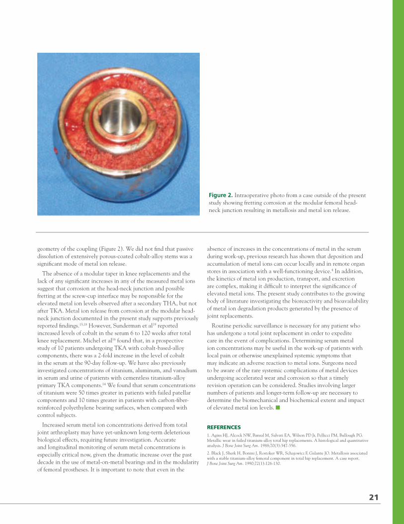

3. Jacobs JJ, Skipor AK, Black J, Urban R, Galante JO. Release and excretion of metal in patients who have a total hip-replacement component made of titanium-base alloy. J Bone Joint Surg Am. 1991;73(10):1475-1486.
4. Jacobs JJ, Skipor AK, Urban RM, Black J, Manion LM, Galante JO. Transport of metal degradation products of titanium alloy total hip replacements to reticuloendothelial organs. An autopsy study. Trans Soc Biomater. 1994;17: 318-325.
5. Tower SS. Arthroprosthetic cobaltism: neurological and cardiac manifestations in two patients with metal-on-metal arthroplasty: a case report. J Bone Joint Surg Am. 2010;92(17):2847-2851.
6. Angle CR. Organ-specific therapeutic intervention. In: Goyer, RA, Klaassen, CD, Waalkes, MP, eds. Metal Toxicology. San Diego, CA: Academic Press; 1995:71-110.
7. Urban RM, Jacobs JJ, Tomlinson MJ, Gavrilovic J, Black J, Peoc’h M. Dissemination of wear particles to the liver, spleen, and abdominal lymph nodes of patients with hip or knee replacement. J Bone Joint Surg Am. 2000;82(4): 457-476.
8. Ritter MA, Carr K, Herbst SA, Eizember LE, Keating EM, Faris PM, Meding JB. Outcome of the contralateral hip
following total hip arthroplasty for osteoarthritis. J Arthroplasty. 1996;11(3):242-246.
9. Iavicoli I, Falcone G, Alessandrelli M, Cresti R, De Santis V, Salvatori S, Alimonti A, Carelli G. The release of metals from metal-on-metal surface arthroplasty of the hip. J Trace Elem Med Biol. 2006;20(1):25-31.
10. Versieck J, Cornelis R. Trace Elements in Human Plasma or Serum. Boca Raton, FL: CRC Press; 1989.
11. Bartolozzi A, Black J. Chromium concentrations in serum, blood clot and urine from patients following total hip arthroplasty. Biomaterials. 1985;6(1):2-8.
12. Black J, Maitin EC, Gelman H, Morris DM. Serum concentrations of chromium, cobalt and nickel after total hip replacement: a six month study. Biomaterials. 1983;4(3):160-164.
13. Brien WW, Salvati EA, Betts F, Bullough P, Wright T, Rimnac C, Buly R, Garvin K. Metal levels in cemented total hip arthroplasty. A comparison of well-fixed and loose implants. Clin Orthop Relat Res. 1992;(276):66-74.
14. Jacobs JJ, Silverton C, Hallab NJ, Skipor AK, Patterson L, Black J, Galante JO. Metal release and excretion from cementless titanium alloy total knee replacements. Clin Orthop Relat Res. 1999;(358):173-180.
15. Jacobs JJ, Skipor AK, Patterson LM, Hallab NJ, Paprosky WG, Black J, Galante JO. Metal release in patients who have had a primary total hip arthroplasty. A prospective, controlled, longitudinal study. J Bone Joint Surg Am. 1998;80(10):1447-1458.
16. Michel R, Nolte M, Reich M, Loer F. Systemic effects of implanted prostheses made of cobalt-chromium alloys. Arch Orthop Trauma Surg. 1991;110(2):61-74.
17. Jacobs JJ, Shanbhag A, Glant TT, Black J, Galante JO. Wear debris in total joint replacements. J Am Acad Orthop Surg. 1994;2(4):212-220.
18. Gilbert JL, Buckley CA, Jacobs JJ. In vivo corrosion of modular hip prosthesis components in mixed and similar metal combinations. The effect of crevice, stress, motion, and alloy coupling. J Biomed Mater Res. 1993;27(12): 1533-1544.
19. Sunderman FW Jr, Hopfer SM, Swift T, Rezuke WN, Ziebka L, Highman P, Edwards B, Folcik M, Gossling HR. Cobalt, chromium, and nickel concentrations in body fluids of patients with porous-coated knee or hip prostheses. J Orthop Res. 1989;7(3):307-315.
2012 / RUSH ORTHOPEDICS JOURNAL
22
Medical devices and drugs discussed in the preceding article are approved by the FDA for use in the manner described. This work was supported in part by NIH/NIAMS Grant AR39310. Authors Hsu, Skippor, and Hallab receive no funds from commercial sources. Dr Levine is a paid consultant to Zimmer and to Janssen Pharmaceutical; and receives funds to support research from Zimmer and Biomet. Dr Paprosky is a paid consultant for Zimmer and Biomet; receives medical device royalties from Zimmer and Wright Medical and research funding from Zimmer; and is paid for service on Zimmer’s speakers’ bureau. Dr Jacobs is a paid consultant to Zimmer, Medtronic, and Spinal Motion; and has stock options in Implant Protection.
“Three-Dimensional Anatomy of the Hip, Utilizing Computer Navigation and Plain Radiographs,” beginning on p. 13, does not discuss the use of medical devices or drugs that are regulated by the FDA. No funds were received in support of this work. Authors Frank, Alland, Grumet, Slabaugh, Wang, and Orías have no commercial interests to disclose. Dr Bach’s financial disclosures are listed above. Dr Nho is a paid consultant to Stryker and for Pivot Medical, and receives funding for research from Stryker, Arthrex, and Allosource.
71795_Body_u1.indd 22 8/3/12 10:30 AM
23
Outpatient Minimally Invasive Total Hip Arthroplasty
DARWIN CHEN, MD / RICHARD A. BERGER, MD
“Outpatient total hip arthroplasty can successfully and safely be performed in select patient populations.”
ARTICLES
AUTHOR AFFILIATIONS
Department of Orthopedic Surgery, Rush University Medical Center, Chicago, Illinois (Drs Chen and Berger); and Midwest Orthopaedics at Rush, Chicago, Illinois (Dr Berger).
CORRESPONDING AUTHOR
Darwin Chen, MD; Rush University Medical Center, 1611 W Harrison St, Suite 300, Chicago, IL 60612 ([email protected]).
Modern total hip arthroplasty (THA) faces a challenging new era in health care. Orthopedic surgeons are now confronted by a medical landscape colored by
high patient expectations, direct-to-consumer marketing, and pressure from insurers and hospital administrators to minimize length of stay.1,2,3 Patients also demand a highly functioning hip, rapid recovery, and a pain-free postoperative course. Today’s arthroplasty surgeon, while executing a safe and well-performed hip replacement, must respond to these additional challenges. Minimally invasive hip surgery, in combination with multimodal anesthesia techniques, rapid rehabilitation, and proper patient education, can allow surgeons to achieve these goals and enable THA to become an outpatient procedure.4
Surgical approach is one of the main components of outpatient minimally invasive THA. The original Watson-Jones anterolateral approach was described through the interval between the tensor fascia lata and the gluteus medius, detaching the anterior aspect of the abductor complex.5 Bertin and Rottinger described a modified, minimally invasive anterolateral approach through the same muscular interval as the Watson-Jones approach while preserving the abductors and preventing a postoperative limp.6 We have further modified and incorporated this approach as a part of an outpatient total hip replacement pathway.
Specialized anesthesia and rapid rehabilitation protocols play equally important roles in enabling a minimally invasive outpatient approach to total hip replacement.4,7,8 The use of regional anesthesia allows for less narcotic administration, which leads to decreased postoperative nausea and hypotension.9 Initiation of pre-emptive oral analgesia and antiemetics early in the recovery phase also aids in pain management and nausea control.10,11,12 Physical therapy, preoperative education, and discharge planning are also critical in managing patient expectations and ensuring adequate care at home.13
Currently the only reported experience with outpatient THA has been with the 2-incision7,14 and miniposterior15 techniques. In both of these studies, the authors demonstrated that outpatient hip replacement is both possible and safe in appropriately selected patients. In the current study, we describe outpatient THA utilizing a minimally invasive, abductor-sparing, anterolateral approach, coupled with a rapid rehabilitation-and-recovery protocol. The purpose of our study is to evaluate the effectiveness of such a pathway for outpatient hip replacement and to assess its feasibility and safety.
MATERIALS AND METHODS
We retrospectively reviewed 199 consecutive patients who underwent primary THA between January 2011 and August 2011. Of these patients, 110 were scheduled for outpatient surgery, and 87 of their surgeries were completed in the operating room by 12 pm. All surgeries were performed by the senior author (R.A.B.). For this study, we included all 87 patients who were scheduled for outpatient THA and who had surgery completed by 12 pm. We performed a retrospective chart review following approval of our institutional review board.
Of the 87 patients enrolled in this study, 53 (60.9%) were males and 34 (39.1%) were females. The average overall age was 56 years (range, 38-73 years). Of the 87, 15 patients (17.2%) were younger than 50 years, 65 patients (74.7%) were between the ages of 50 and 65, and 7 patients (8.1%) were older than 65 years.
71795_Body.indd 23 7/25/12 9:39 PM

2012 / RUSH ORTHOPEDICS JOURNAL
24
The average overall weight of the patients was 85.7 kg (range, 47.2-145.2 kg). The average weight of the male patients was 94.2 kg (range, 63.5-145.2 kg) and 73.0 kg (range, 47.2-113.4 kg) for the female patients. The average body mass index (BMI) overall was 27.9 (range, 18.5-43.3). The average BMI for the male patients was 29 (range, 22.5-43.3), and the average BMI for the female patients was 26.8 (range, 18.5-40.2).
PERIOPERATIVE CARE
We enrolled all patients in a comprehensive clinical pathway including preoperative, intraoperative, and postoperative care. The pathway specifically addresses pain management, postoperative nausea and hypotension, and home aftercare.
Preoperatively, all patients attended a class taught by a clinical nurse and a physical therapist. On the morning of surgery, prior to coming to the hospital, patients took 10 mg of oxycodone hydrochloride controlled release (CR). All patients received an epidural catheter, Foley catheter, and intravenous (IV) antibiotics prior to incision. We gave patients propofol for sedation at the discretion of the anesthesiologist. We achieved nausea prophylaxis with IV ondansetron and metoclopramide. All patients received 1 unit of autologous blood, and IV and epidural narcotics were avoided during surgery.
Postoperatively, patients were given additional ondansetron for pre-emptive nausea control. Within 4 hours after surgery, we discontinued the Foley catheter and epidural. We achieved pain control with scheduled oral oxycodone and as-needed oral hydrocodone or IV morphine. The patients started physical therapy 6 hours later and were allowed to bear weight as tolerated, using crutches, a cane, or independently.
A clinical nurse was available to address any postoperative issues. We discharged patients once they met standard physical therapy discharge criteria and had stable vital signs and adequate pain control. Upon discharge, we instructed patients to take 325 mg of aspirin as prophylaxis against venous thromboembolism twice a day for 3 weeks. In addition to oxycodone CR and hydrocodone for pain relief, patients also took diclofenac as an anti-inflammatory medication.
OPERATIVE TECHNIQUE
POSITIONING
We positioned the patient in the lateral decubitus position, utilizing an operating table that allowed us to move the leg sections independently. This allowed us to place the operative leg in a position of extension, adduction, and external rotation, facilitating femoral exposure. We used a leg holder to support the operative leg in a position of 5°-15° of abduction during the approach and acetabular exposure.
APPROACH
We use a modified, abductor-sparing anterolateral approach through the Watson-Jones interval. The proximal end of the
incision begins slightly posterior to the interval between the tensor fascia lata and the gluteus medius. The incision then curves gently towards the anterosuperior corner of the greater trochanter. The distal end of the incision parallels and is slightly posterior to the anterior border of the femur. Total incision length is approximately 3.5 to 4 inches (Figure 1).
After incision and subcutaneous dissection, we identified the fascial border between the tensor fascia lata and the gluteus medius. We made the fascial incision over the anterior border of the gluteus medius, anterior to the greater trochanter. Next we gently split the interval between the tensor fascia lata and the gluteus medius. We placed 2 curved Hohmann retractors extracapsularly over the superior and inferior aspects of the femoral neck (Figure 2). We identified the anterior capsule and performed an anterior capsulectomy. We then identified the saddle point between the inner aspect of the greater trochanter and the base of the superior femoral neck and cleared off the capsular remnants, marking the superolateral border of the femoral neck osteotomy (Figure 3). We then repositioned the retractors intracapsularly over the acetabular rim.
FEMORAL HEAD AND NECK OSTEOTOMY
Without dislocating the hip, we sequentially cut the femoral head and neck in situ into thin wafer slices using an oscillating saw (Figure 4). We removed the wafer fragments, decompressing the hip. We then placed the leg in the figure-4 position into a sterile hip pouch. Using the oscillating saw, we finalized the neck cut, which extends from the saddle point to the templated level above the lesser trochanter.
ACETABULAR PREPARATION
We moved the leg from the pouch and placed it onto the leg holder. Next we placed acetabular retractors, retracting the femur posteriorly. Once we achieved 360° visualization of the acetabulum, we excised the pulvinar and labrum (Figure 5). Next, we performed acetabular reaming until we achieved a good press fit. We then impacted the acetabular component in the appropriate abduction and anteversion, with or without adjunctive screw fixation, and then placed the acetabular liner (Figure 6).
FEMORAL PREPARATION
We placed the leg into a sterile kangaroo pouch attached to an assistant’s gown, allowing the hip to be extended, adducted, and externally rotated. Retractors were placed around the proximal femur, presenting it anteriorward. It is often necessary to release the posterior superior capsule from the saddle point and the inner aspect of the greater trochanter (Figure 7) in order to further expose the femur for instrumentation (Figure 8). We used a curved awl to enter the femoral canal and then reamed out the inner aspect of the greater trochanter with a large motorized burr, preventing varus positioning of the femoral stem. We performed broaching with offset handles until adequate fit, depth, and version were achieved (Figure 9). After inserting the final stem
71795_Body.indd 24 7/25/12 9:39 PM
25
(Figure 10), we performed trial reduction with varying neck lengths until adequate hip stability, soft tissue tension, and leg lengths were achieved. Finally we removed the trial head and impacted the final head into place. We then reduced the hip for a final time.
RESULTS
Of the 87 patients who were scheduled for outpatient THA and whose surgery was completed by 12 pm, 86 (98.9%) were discharged home the same day. Of the 87 patients, 1 (1.1%) did not pass physical therapy and was discharged the following day. Mean surgical time was 59 minutes (range, 38-91 minutes). Mean estimated blood loss was 251.1 cc (range, 50-1500 cc). All 87 patients (100%) received epidural anesthesia, and all 87 (100%) patients were discharged home with home physical therapy services.
All patients received uncemented acetabular and femoral THA components. Of the 87 patients, 34 patients (39.1%) had a DePuy
Pinnacle Porocoat acetabular shell (DePuy, Warsaw, Indiana), and 53 patients (60.9%) had a Zimmer Trilogy fiber metal mesh acetabular shell (Zimmer, Warsaw, Indiana). Of the 87 patients, 27 patients (31.0%) had a Zimmer VerSys beaded fullcoat stem, 26 patients (29.9%) had a Zimmer M/L Taper Kinectiv modular tapered stem, 33 patients (37.9%) had a DePuy Tri-Lock tapered stem, and 1 patient (1.1%) had a Zimmer VerSys fiber metal tapered stem.
Of the 87 patients, 0 patients had any significant medical complications in the acute (<2 weeks) postoperative period. There were no DVTs/PEs, cardiopulmonary events, deaths, or other serious medical complications. There were no readmissions for postoperative complications during this period. One patient had a deep hip infection at 3 weeks postoperatively that was treated with a 1-stage exchange revision hip arthroplasty and IV antibiotics. To date, this patient has not had a recurrence of infection.
Figure 1. Curvilinear incision bordering the Waston-Jones interval.
Figure 2. Retractors around the femoral neck, opening the interval between the tensor fascia lata and the gluteus medius.
Figure 3. Exposure of the femoral head and neck after anterior capsulectomy.
Figure 4. Femoral head removed after sequential cuts during head/neck osteotomy.
1 2
3 4
71795_Body.indd 25 7/25/12 9:39 PM
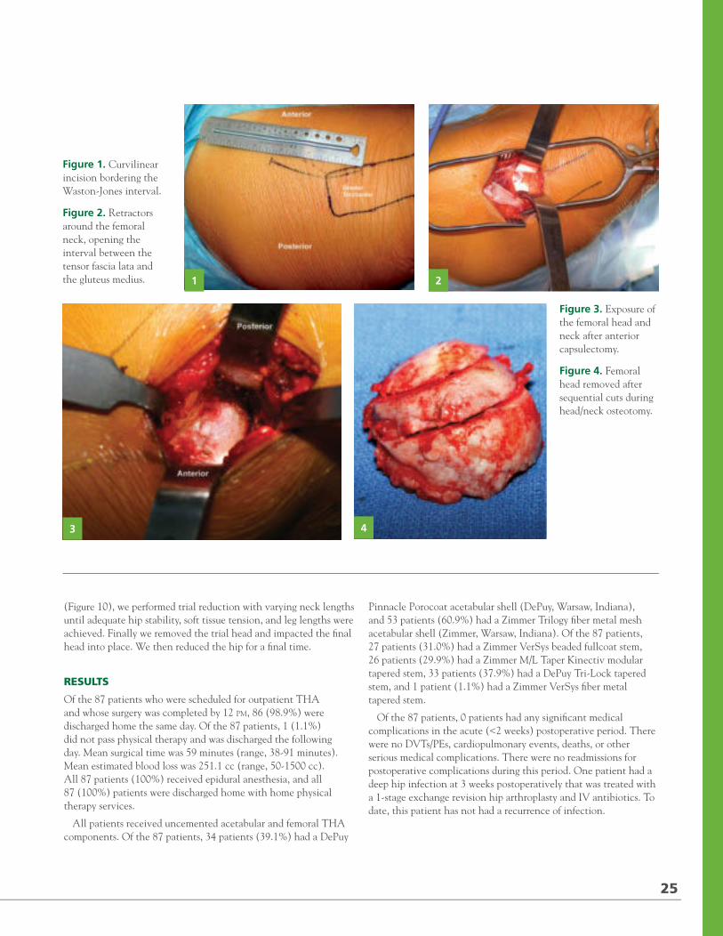
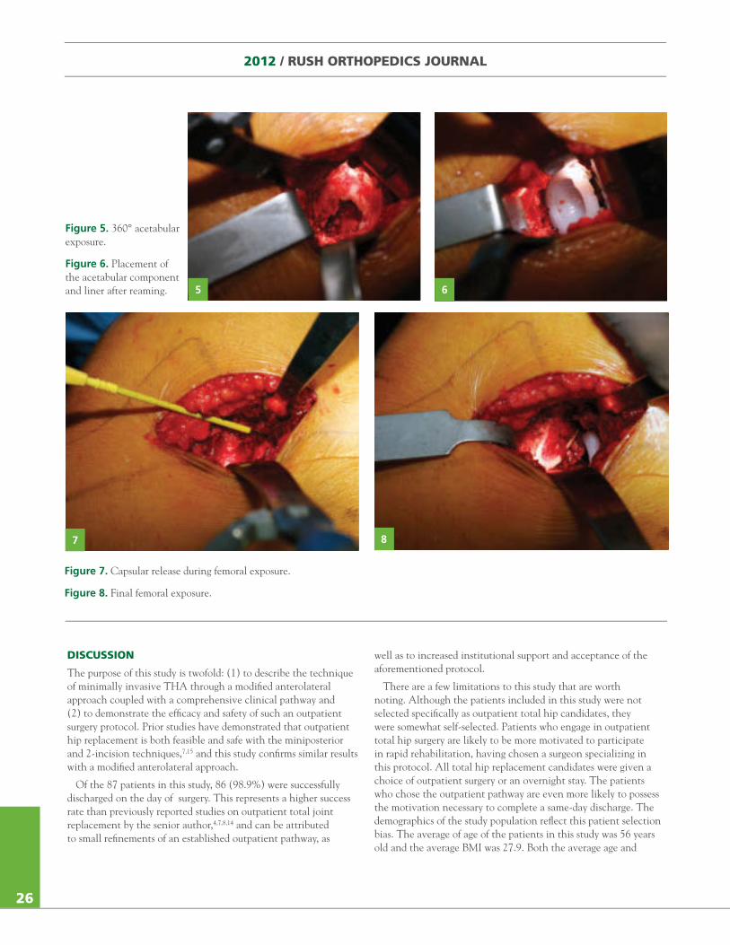
2012 / RUSH ORTHOPEDICS JOURNAL
826
DISCUSSION
The purpose of this study is twofold: (1) to describe the technique of minimally invasive THA through a modified anterolateral approach coupled with a comprehensive clinical pathway and (2) to demonstrate the efficacy and safety of such an outpatient surgery protocol. Prior studies have demonstrated that outpatient hip replacement is both feasible and safe with the miniposterior and 2-incision techniques,7,15 and this study confirms similar results with a modified anterolateral approach.
Of the 87 patients in this study, 86 (98.9%) were successfully discharged on the day of surgery. This represents a higher success rate than previously reported studies on outpatient total joint replacement by the senior author,4,7,8,14 and can be attributed to small refinements of an established outpatient pathway, as
well as to increased institutional support and acceptance of the aforementioned protocol.
There are a few limitations to this study that are worth noting. Although the patients included in this study were not selected specifically as outpatient total hip candidates, they were somewhat self-selected. Patients who engage in outpatient total hip surgery are likely to be more motivated to participate in rapid rehabilitation, having chosen a surgeon specializing in this protocol. All total hip replacement candidates were given a choice of outpatient surgery or an overnight stay. The patients who chose the outpatient pathway are even more likely to possess the motivation necessary to complete a same-day discharge. The demographics of the study population reflect this patient selection bias. The average of age of the patients in this study was 56 years old and the average BMI was 27.9. Both the average age and
Figure 5. 360° acetabular exposure.
Figure 6. Placement of the acetabular component and liner after reaming. 5 6
7 8
Figure 7. Capsular release during femoral exposure.
Figure 8. Final femoral exposure.
71795_Body.indd 26 7/25/12 9:39 PM
27
4
BMI of patients in this study are lower than the averages of most arthroplasty practices.
Length of stay after THA has dramatically decreased in the past 20 years. The average stay has dropped from over 1 week to 3 to 4 days, and is now approaching an overnight stay or same-day surgery. In summary, outpatient THA can successfully and safely be performed in select patient populations. While minimally invasive surgery plays a critical role in outpatient THA, equally important are the contributions from multimodal anesthesia techniques as well as rapid rehabilitation and preoperative patient education. When combined into the comprehensive clinical pathway outlined in this study, outpatient THA has become a reality that is safe, effective, and reproducible.
REFERENCES1. Mason JB. The new demands by patients in the modern era of total joint arthroplasty: a point of view. Clin Orthop Relat Res. 2008;466(1):146-152.
2. Booth RE Jr. Truth in advertising: the ethical limits of direct-to-consumer marketing. Orthopedics. 2006;29:780-781.
3. Bozic KJ, Smith AR, Hariri S, Adeoye O, Gourville J, Maloney WJ, Parsley B, Rubash HE. The 2007 ABJS Marshall Urist Award: the impact of direct-to-consumer advertising in orthopaedics. Clin Orthop Relat Res. 2007;458:202-219.
4. Berger RA, Sanders SA, Thill ES, Sporer SM, Della Valle C. Newer anesthesia and rehabilitation protocols enable outpatient hip replacement in selected patients. Clin Orthop Relat Res. 2009;467(6):1424-1430.
5. Watson-Jones R: Fractures of the neck of the femur. Br J Surg. 1936;23(92):787-808.
6. Bertin KC, Rottinger H. Anterolateral mini-incision hip replacement surgery: a modified Watson-Jones approach. Clin Orthop Relat Res. 2004;(429):248-255.
7. Berger RA, Jacobs JJ, Meneghini RM, Della Valle C, Paprosky W, Rosenberg AG. Rapid rehabilitation and recovery with minimally invasive total hip arthroplasty. Clin Orthop Relat Res. 2004;(429):239-247.
8. Berger RA. A comprehensive approach to outpatient total hip arthroplasty. Am J Orthop. 2007;36(suppl 9):4-5.
9. Macfarlane AJ, Prasad GA, Chan VW, Brull R. Does regional anaesthesia improve outcome after total hip arthroplasty? A systematic review. Br J Anaesth. 2009;103(3):335-345.
10. Dorr LD, Raya J, Long WT, Boutary M, Sirianni LE. Multimodal analgesia without parenteral narcotics for total knee arthroplasty. J Arthroplasty. 2008;23(4):502-508.
11. DiIorio TM, Sharkey PF, Hewitt AM, Parvizi J. Antiemesis after total joint arthroplasty: does a single preoperative dose of aprepitant reduce nausea and vomiting? Clin Orthop Relat Res. 2010;468(9):2405-2409.
12. Duellman TJ, Gaffigan C, Milbrandt JC, Allan DG. Multi-modal, pre-emptive analgesia decreases the length of hospital stay following total joint arthroplasty. Orthopedics. 2009;32(3):167.
13. Kearney M, Jennrich MK, Lyons S, Robinson R, Berger B. Effects of preoperative education on patient outcomes after joint replacement surgery. Orthop Nurs. 2011;30(6):391-396.
14. Berger RA. Total hip arthroplasty using the minimally invasive two-incision approach. Clin Orthop Relat Res. 2003;417:232-241.
15. Dorr LD, Thomas DJ, Zhu J, Dastane M, Chao L, Long WT. Outpatient total hip arthroplasty. J Arthroplasty. 2010;25(4):501-506.
The preceding article does not discuss use of medical devices or drugs that are not approved by the FDA. No funds were received in support of the work described. Dr Chen receives no funding from any commercial sources. Dr Berger receives medical device royalties from Zimmer.
Figure 9. Femoral broaching using offset handles.
Figure 10. Final femoral component insertion. 9 10
71795_Body.indd 27 7/25/12 9:39 PM
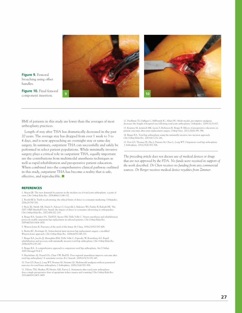
2012 / RUSH ORTHOPEDICS JOURNAL
Anterior Cruciate Ligament Reconstruction Failure: Analysis of a Single, High-Volume Orthopedic Institution
RACHEL M. FRANK, MD / MARK A. SLABAUGH, MD / KEVIN C. MCGILL, MD, MPH / CHARLES A. BUSH-JOSEPH, MD
/ BRIAN J. COLE, MD, MBA / BERNARD R. BACH JR, MD / NIKHIL N. VERMA, MD
“With strict attention to good principles of anterior cruciate ligament reconstruction, a lower failure rate can be expected with the majority of the failures resulting from traumatic reinjuries.”
ARTICLES
AUTHOR AFFILIATIONS
Division of Sports Medicine, Department of Orthopedic Surgery, (Drs Frank, Slabaugh, Bush-Joseph, Cole, Bach, Verma), Rush University Medical Center, Chicago, Illinois; Department of Radiology, Henry Ford Hospital, Detroit, Michigan (Dr McGill); and Midwest Orthopaedics at Rush, Chicago, Illinois (Drs Bush-Joseph, Cole, Bach, and Verma).
CORRESPONDING AUTHOR
Bernard R. Bach Jr, MD; Rush University Medical Center and Midwest Orthopaedics at Rush, 1611 W Harrison St, Suite 300, Chicago, IL 60612 ([email protected]).
Anterior cruciate ligament (ACL) tears remain one of the most common knee injuries in the United States, with an estimated 175,000 ACL reconstructions performed each
year.1,2 The surgical technique of ACL reconstruction (ACLR) has evolved over the last 40 years from open, extra-articular procedures to arthroscopically assisted intra-articular procedures. Recently, several reports in the literature have stated that the success rates of primary intra-articular ACLR surgery range from 75% to 90%.3-8 Despite these excellent reported outcomes considering the numbers of ACL reconstructions performed annually, many patients will require a revision procedure.
Failed ACL reconstruction can be defined as a knee withpersistent pathological laxity, persistent pain, and/or without
laxity but with a limited range of motion.9 Potential etiologies of ligament failure include new trauma, technical errors during the index operation, biological failure of graft incorporation, failure to address concomitant instability pathologies, and poor postoperative patient compliance with rehabilitation protocol. Typically, if a primary ACLR fails within 6 months and trauma has been ruled out, the likely explanation is a technical issue.3 Specific technical errors include incorrect bone tunnel placement, inappropriate graft tensioning, and inadequate graft fixation. Among the potential factors causing ACLR failure, surgical technique and/or surgeon error remain the most commonly reported causes in the literature.3,10-13
While several studies report the various causes of failed ACLR leading to a revision procedure, there are no reports describing both the primary and revision procedures when both operations are performed by the same surgeon.3,14-16 The purpose of this study was to determine the causes of failed primary ACL reconstructions performed by experienced, fellowship-trained, sports medicine surgeons at a single, high-volume institution. Our hypothesis was that the most common cause of failed ACLR would be traumatic reinjury rather than technical error. In addition, we aimed to analyze postrevision ACLR outcomes as determined by assessment with the KT1000 Arthrometer (MEDMetric, Inc., San Diego, California) and physical examination, as well as knee-specific outcomes surveys. For this aspect of the study, we hypothesized that patients undergoing revision ACLR would demonstrate good to excellent clinical outcomes.
28
71795_Body_u1.indd 28 8/3/12 10:30 AM
METHODS
An independent research fellow conducted a retrospective blinded review of the surgical logs of 4 senior fellowship-trained sports medicine surgeons to find all patients who had undergone ACLR from 2002 through 2009.17 This list of 1944 patients was further narrowed to include only those on whom the same surgeon had performed a subsequent revision ACLR on the ipsilateral knee
within the 9-year time period. Patients who had undergone their index procedure with another surgeon were excluded from the study. Patients who had undergone more than one revision (7 patients within the 8-year time period) were included in the statistical analysis of overall failure; however, these patients were not included in the clinical follow-up portion of the study. Out of 1944 ACL reconstructions, 28 patients (56 reconstructions) were included in the study.
Figure 1. Radiographs of properly positioned ACL femoral bone tunnel, anteroposterior (AP) (left) and lateral (right) views. Tibial and femoral tunnel diameters on both anterior and posterior radiographs were measured in addition to the location of the center of the tibial tunnel on the lateral radiograph. Femoral tunnel position was measured on the lateral radiograph in relationship to Blumensaat’s line. Angle of the femoral tunnel was measured in relationship to the anatomic axis of the femur, whereas the angle of the tibial tunnel was measured in relationship to the line parallel to the medial and lateral tibial plateau.
1
2
Figure 2. Radiographs of vertically placed femoral bone tunnel, anteroposterior (AP) (left) and lateral (right) views. Tibial and femoral tunnel diameters on both anterior and posterior radiographs were measured in addition to the location of the center of the tibial tunnel on the lateral radiograph. Femoral tunnel position was measured on the lateral radiograph in relationship to Blumensaat’s line. Angle of the femoral tunnel was measured in relationship to the anatomic axis of the femur, whereas the angle of the tibial tunnel was measured in relationship to the line parallel to the medial and lateral tibial plateau.
29
71795_Body.indd 29 7/25/12 9:39 PM
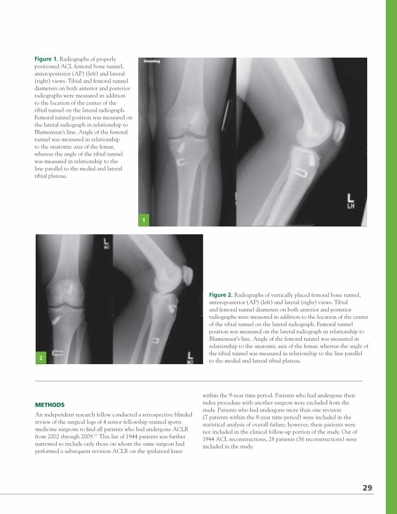
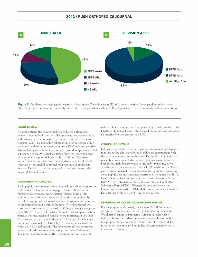
INDEX ACLR
14%
11%
29%
46%
BPTB Auto
BPTB Allo
HS Auto
HS Allo
2012 / RUSH ORTHOPEDICS JOURNAL
CHART REVIEW
For each patient, the research fellow conducted a thorough review of the medical chart to collect preoperative, postoperative, and intraoperative information pertinent to both the index and revision ACLR. Demographic information, graft selection, clinic notes, physical exam measures including KT1000 scores, operative data including concomitant pathologies, procedures performed, and appearance of the ACL graft (at time of revision) were analyzed to determine any potential mechanism of failure. Operative notes (index and revision) were analyzed for evidence of possible technical errors, including tunnel malposition and inadequate fixation. Particular attention was paid to the time between the index ACLR and failure.
RADIOGRAPHIC ANALYSIS
Radiographic measurements were obtained on both anteroposterior (AP) and lateral views on radiographs obtained between the primary and secondary reconstruction (Figures 1 and 2). In addition, the position of the center of the tibial tunnel on the lateral radiograph was measured as a percentage in relation to the entire anteroposterior depth of the tibia. The tibial tunnel was considered too posterior (too vertical) if the percentage was greater than 50%.18 The angle of the tibial tunnel relationship to the tibial plateaus was measured and considered malpositioned if less than 55 degrees or greater than 75 degrees.19 The angle of the femoral tunnel was measured in relationship to the anatomic axis of the femur on the AP radiograph. The femoral tunnel was considered too vertical if this measurement was greater than 10 degrees.20 The position of the center of the femoral tunnel on the lateral
radiograph was also measured as a percentage in relationship to the length of Blumensaat’s line. The femoral tunnel was considered too far anterior if it was greater than 15%.
CLINICAL FOLLOW-UP
Following the chart review, each patient was invited by telephone to return to the clinic for a blinded follow-up examination with the lone independent research fellow. During the clinic visit, the research fellow conducted a thorough physical examination of both knees, including provocative and stability testing, as well as arthrometric evaluation with the KT1000 Arthrometer.2 Each patient was also asked to complete a follow-up survey containing demographic data and outcomes assessments, including the SF-12 Health Survey, Knee Injury and Osteoarthritis Outcome Score (KOOS), the International Knee Documentation Committee Subjective Form (IKDC), Western Ontario and McMaster Universities Osteoarthritis (WOMAC) index, modified Cincinnati Knee-Rating Scale (10-point), and Lysholm score.
DEFINITION OF ACL RECONSTRUCTION FAILURE
For the purpose of this study, the cause of ACLR failure was categorized into 3 groups: iatrogenic, biologic, and traumatic. We classified failure as iatrogenic (surgeon or technical) if radiographs indicated that the femoral and/or tibial tunnels were inappropriately positioned, or if at the time of revision ACLR, intra- or postoperative findings indicated tunnel malposition or inadequate fixation.
A REVISION ACLR
BPTB Auto
BPTB Allo
Achilles Allo
14%4%
B
Figure 3. Pie charts portraying graft selection in both index (A) and revision (B) ACL reconstruction. Bone–patellar tendon–bone (BPTB) autografts were most commonly used in the index procedures, while BTPB allografts were more commonly used in the revision.
30
82%
71795_Body.indd 30 7/25/12 9:39 PM
We classified failures as biologic based upon definitive findings of incorporation failure, allograft rejection, or notch impingement from regrowth at the time of revision ACLR, or if failure occurred within the first 6 months following index ACLR in patients with a properly placed and well-fixed graft (if there was no report of a recurrent traumatic event).
We considered a failure to be traumatic only if the patient had a distinct traumatic reinjury after returning to full activity in his or her sport and if other causes of failure could be ruled out. Further, in order to be considered a traumatic failure, intraoperative findings at the time of revision ACLR had to have shown a midsubstance tear or tear off the femur/tibia with good tissue remaining opposite the tear. Only those patients who met these 3 criteria (clinical history, exclusion of other causes, and intraoperative findings) were included as traumatic failures.
STATISTICAL ANALYSIS
Statistical analyses, including descriptive data analysis, correlation analysis, and inferential analysis, were performed using SPSS (SPSS Inc., Chicago, Illinois) statistical software. We considered results to be statistically significant for all analyses when P < .05.
RESULTS
CHART REVIEW AND RADIOGRAPHIC ANALYSIS RESULTS
From January 2002 through December 2009, all 4 senior knee surgeons at our institution were performing arthroscopic-assisted ACLR with a transtibial technique. After incorporation of the study’s inclusion and exclusion criteria, 28 patients (56 reconstructions) were included in the study. During this same time period, a total of 1944 primary ACL reconstructions were performed, with 35 revision cases (1.8%) (includes only patients, who, to the best of our knowledge, needed a revision
CHARACTERISTICS NO. (%) OF PATIENTS
Total patients 28 (100)
Sex
Male 15 (54)
Female 13 (46)
Operative knee
Left 17 (61)
Right 11 (39)
TIME BETWEEN INDEX AND REVISION ACLR, MONTHS 21.7 ± 16.6
SURGERY AGE OF PATIENT
Index ACLR
Age, y 22 ± 11 (range, 12-50)
Revision ACLR
Age, y 24 ± 11 (range, 14-57)
CONCOMITANT PROCEDURES NO. (%) OF PATIENTS
At index ACLR
Partial medial meniscectomy 5 (18)
Medial meniscal repair 6 (21)
Open MCL repair 1 (4)
Partial lateral meniscectomy and medial meniscus repair 1 (4)
At revision ACLR
Partial medial meniscectomy 6 (21)
Medial meniscal repair 4 (14)
Partial lateral meniscectomy and medial meniscus repair 4 (14)
Table 1. Summary of Demographic Data
Abbreviations: ACLR, anterior cruciate ligament reconstruction; MCL, medial collateral ligament
31
71795_Body.indd 31 7/25/12 9:39 PM
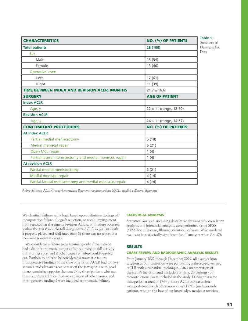
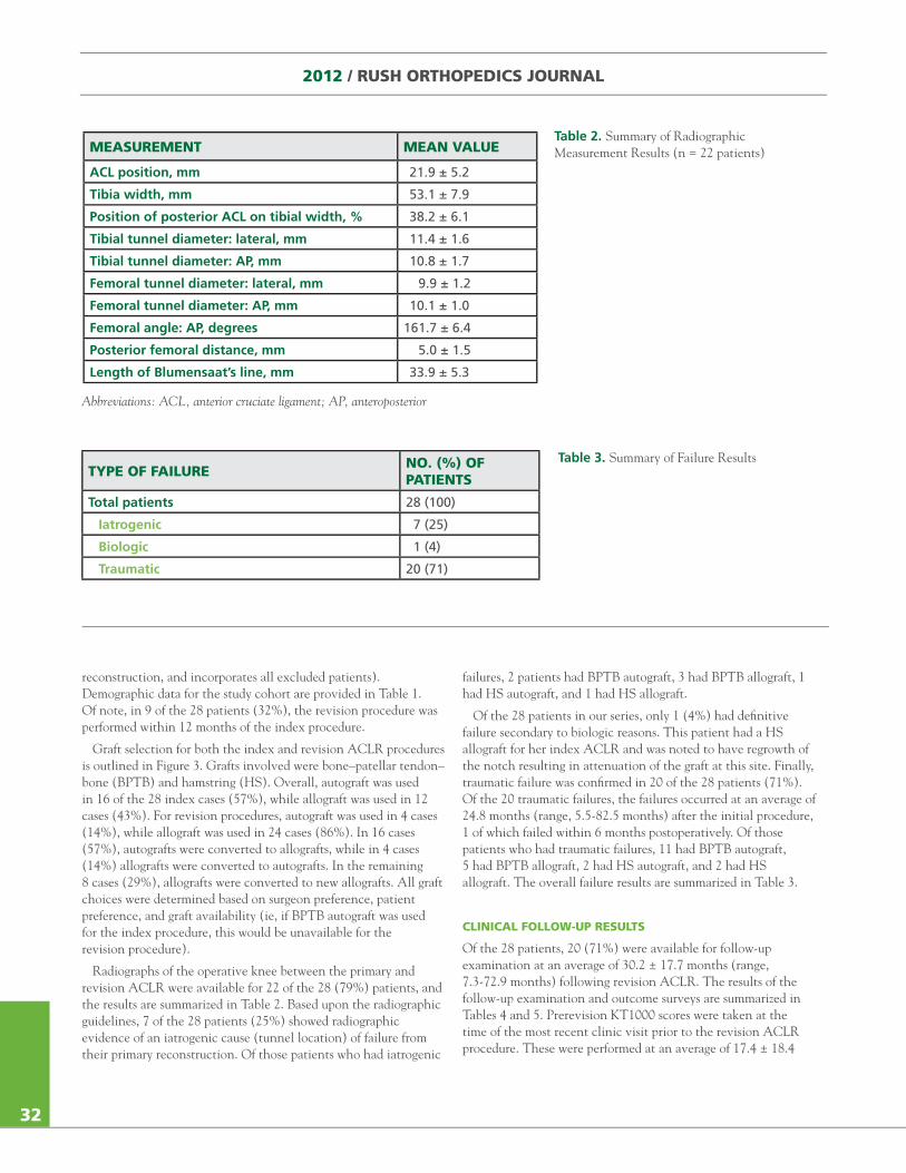
2012 / RUSH ORTHOPEDICS JOURNAL
reconstruction, and incorporates all excluded patients). Demographic data for the study cohort are provided in Table 1. Of note, in 9 of the 28 patients (32%), the revision procedure was performed within 12 months of the index procedure.
Graft selection for both the index and revision ACLR procedures is outlined in Figure 3. Grafts involved were bone–patellar tendon–bone (BPTB) and hamstring (HS). Overall, autograft was used in 16 of the 28 index cases (57%), while allograft was used in 12 cases (43%). For revision procedures, autograft was used in 4 cases (14%), while allograft was used in 24 cases (86%). In 16 cases (57%), autografts were converted to allografts, while in 4 cases (14%) allografts were converted to autografts. In the remaining 8 cases (29%), allografts were converted to new allografts. All graft choices were determined based on surgeon preference, patient preference, and graft availability (ie, if BPTB autograft was used for the index procedure, this would be unavailable for the revision procedure).
Radiographs of the operative knee between the primary and revision ACLR were available for 22 of the 28 (79%) patients, and the results are summarized in Table 2. Based upon the radiographic guidelines, 7 of the 28 patients (25%) showed radiographic evidence of an iatrogenic cause (tunnel location) of failure from their primary reconstruction. Of those patients who had iatrogenic
failures, 2 patients had BPTB autograft, 3 had BPTB allograft, 1 had HS autograft, and 1 had HS allograft.
Of the 28 patients in our series, only 1 (4%) had definitive failure secondary to biologic reasons. This patient had a HS allograft for her index ACLR and was noted to have regrowth of the notch resulting in attenuation of the graft at this site. Finally, traumatic failure was confirmed in 20 of the 28 patients (71%). Of the 20 traumatic failures, the failures occurred at an average of 24.8 months (range, 5.5-82.5 months) after the initial procedure, 1 of which failed within 6 months postoperatively. Of those patients who had traumatic failures, 11 had BPTB autograft, 5 had BPTB allograft, 2 had HS autograft, and 2 had HS allograft. The overall failure results are summarized in Table 3.
CLINICAL FOLLOW-UP RESULTS
Of the 28 patients, 20 (71%) were available for follow-up examination at an average of 30.2 ± 17.7 months (range, 7.3-72.9 months) following revision ACLR. The results of the follow-up examination and outcome surveys are summarized in Tables 4 and 5. Prerevision KT1000 scores were taken at the time of the most recent clinic visit prior to the revision ACLR procedure. These were performed at an average of 17.4 ± 18.4
MEASUREMENT MEAN VALUE
ACL position, mm 21.9 ± 5.2
Tibia width, mm 53.1 ± 7.9
Position of posterior ACL on tibial width, % 38.2 ± 6.1
Tibial tunnel diameter: lateral, mm 11.4 ± 1.6
Tibial tunnel diameter: AP, mm 10.8 ± 1.7
Femoral tunnel diameter: lateral, mm 9.9 ± 1.2
Femoral tunnel diameter: AP, mm 10.1 ± 1.0
Femoral angle: AP, degrees 161.7 ± 6.4
Posterior femoral distance, mm 5.0 ± 1.5
Length of Blumensaat’s line, mm 33.9 ± 5.3
Table 2. Summary of Radiographic Measurement Results (n = 22 patients)
Abbreviations: ACL, anterior cruciate ligament; AP, anteroposterior
TYPE OF FAILURE NO. (%) OF PATIENTS
Total patients 28 (100)
Iatrogenic 7 (25)
Biologic 1 (4)
Traumatic 20 (71)
Table 3. Summary of Failure Results
32
71795_Body.indd 32 7/25/12 9:39 PM
months (range, 1.6-80.7 months) after the index procedure, which was an average of 4.9 ± 5.9 months (range, 0.5-20.3 months) prior to the revision procedure. Postrevision KT1000 data were taken at the time of follow-up examination, again an average of 30.2 ± 17.7 months following revision ACLR.
With regard to the clinical outcomes assessments, 18 of the 28 patients (65%) had good to excellent Lysholm scores. Higher IKDC scores were correlated with younger patients at both primary and revision surgery (P < .0001). Greater differences in postrevision KT1000 scores between the operative and nonoperative knees were associated with elevated KOOS pain and symptomatic values (P < .0001). Furthermore, a shorter time period between index and revision ACLR was associated with better KOOS pain scores. Those patients who had surgery within 18 months of their primary reconstruction had KOOS pain scores of 96.0 vs 84.2 in patients who had their reconstruction later than 18 months (P = .0001).
DISCUSSION
The principal findings of this study indicate that traumatic rerupture is the most common cause of graft failure following index ACLR. Historically, the most common reason cited for revision reconstruction has been technical and/or surgeon errors, and published reviews of revision ACLR cite several studies done in the mid 1990s to support this claim.22-25 However, the majority of these studies were published during a time period when ACLR may not have been performed anatomically, with either the femoral tunnel too vertical or the tibial tunnel too posterior. Four of these studies were published in a single issue of Clinical Orthopedics and Related Research and are similar to our study in that they are single-institution analyses of ACLR failure.22-25 It is difficult, however, to draw conclusions as to definitive etiologies of failure based upon these 4 studies because specific numbers on causes of failure other than iatrogenic are not always provided. The study by Johnson et al22 is the only study of this group in which concrete data were given regarding their cohort of patients undergoing ACLR. In this group of 25 patients, 13 (52%) failed ACLR due to technical errors, while biologic failure was cited as the cause in 5 cases (20%) and trauma in 7 cases (28%).
A study from a paper presented at a national arthroscopy meeting in 1995 also has been cited frequently to support the assumption that technical error is the most common cause of failure after ACLR.26 In this study, 77% of patients undergoing revision ACLR had procedures that were considered failures due to technical errors such as tunnel malposition, inadequate fixation or inadequate/insufficient graft. Despite this high percentage of technical errors, there was no definition of what constituted tunnel malposition, inadequate fixation, or inadequate/insufficient gait. Their data are in direct contrast to our study where we found that 63% of primary ACLR failures were due to traumatic recurrent ACL tears after a successful reconstruction.
When comparing our follow-up results of revision ACLR to other studies previously published, our results compare similarly. Denti et al27 reported on their experience with revision ACLR in 66 patients. In their series of 66 patients, 46 (70%) had good/excellent Lysholm scores, which compares favorably to 18 of our 28 patients (65%) who had good/excellent Lysholm scores. Similarly, Salmon et al28 reported on their experience with revision ACLR in 50 consecutive patients at an average of 9 years following revision ACLR. The average Lysholm score for their patient population was 85 points, similar to the average of 84 points reported in our study. The authors additionally reported on the method of failure of the primary ACLR and while not giving completely specific definitions to determine their causes of failure, they reported etiologies of failure (65% recurrent trauma, 35% biologic or technical) similar to those in our cohort (71% trauma, 29% biologic or technical errors).
There were several limitations to the current study. We acknowledge that all ACLR failures might not have presented to our institution after a failure, falsely lowering our rate of revisions. Further, some patients who may be classified as a clinical failure may not have elected to undergo revision surgery and therefore would not have been identified during our case log review. However, the 28 patients do provide insight into several key aspects of revision ACLR, such as probable cause of failure. Another limitation was all patients did not have radiographs to determine the accuracy of tunnel placement. This could lead to a falsely lowered number of patients who failed the initial
PREREVISION(n = 22)
POSTREVISION(n = 20) SIGNIFICANCE
Operative knee KT1000 (MMT) 12.1 ± 1.4 6.8 ± 2.8 P < .001
Nonoperative knee KT1000 (MMT) 6.7 ± 1.3 5.9 ± 2.8 P > .05
Difference between operative and nonoperative knee 1.4 ± 1.2
Table 4. Summary of KT1000 Results (measured in millimeters, mm)
Abbreviation: MMT, manual maximum test
33
71795_Body.indd 33 7/25/12 9:39 PM
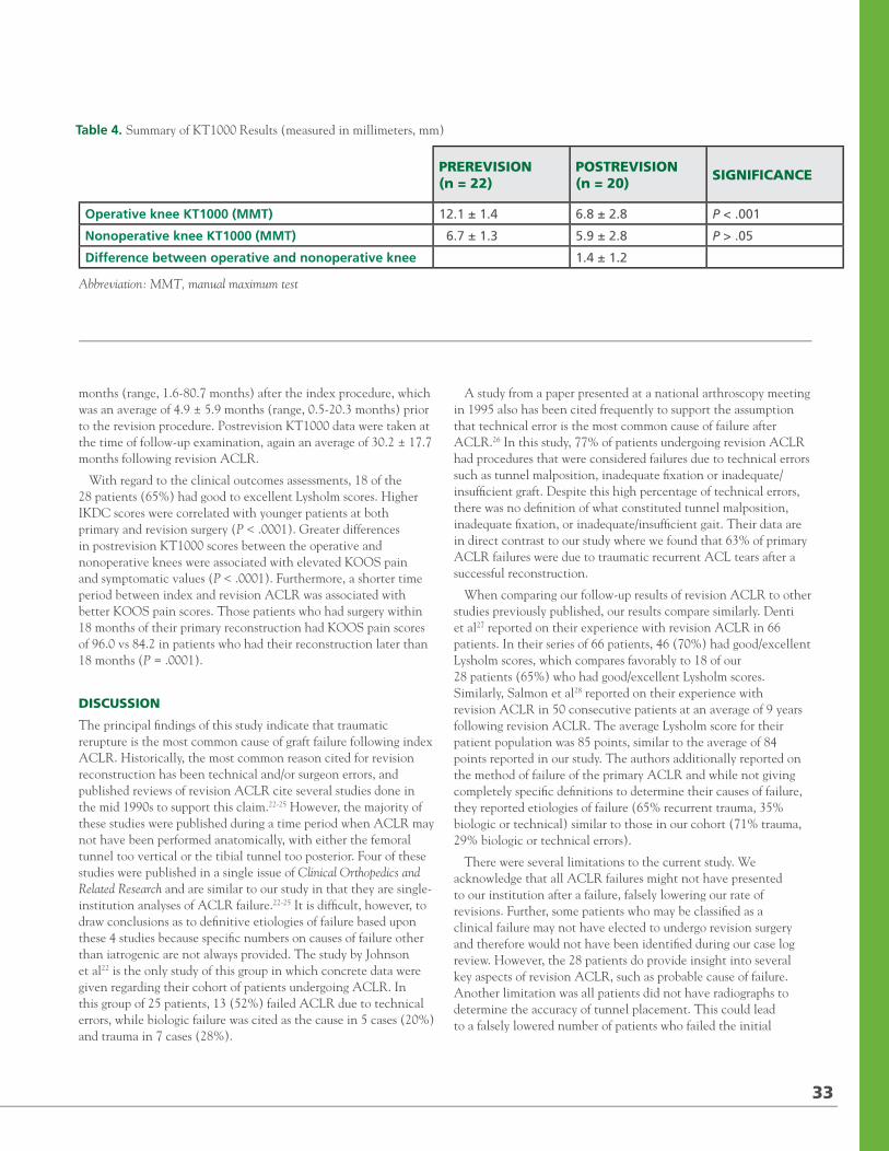
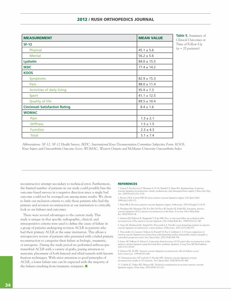
2012 / RUSH ORTHOPEDICS JOURNAL
reconstructive attempt secondary to technical error. Furthermore, the limited number of patients in our study could possibly bias the outcome-based surveys in a negative direction since a single bad outcome could not be averaged out among many results. We chose to limit our inclusion criteria to only those patients who had the primary and revision reconstruction at our institution to critically look at our failures and outcomes.
There were several advantages to the current study. This study is unique in that specific radiographic, clinical, and intraoperative criteria were used to define the cause of failure in a group of patients undergoing revision ACLR in patients who had their primary ACLR at the same institution. This allows a retrospective review of patients who presented with a failed primary reconstruction to categorize their failure as biologic, traumatic, or iatrogenic. During the study period we performed arthroscopic transtibial ACLR with a variety of grafts, paying attention to anatomic placement of both femoral and tibial tunnels with known fixation techniques. With strict attention to good principles of ACLR, a lower failure rate can be expected with the majority of the failures resulting from traumatic reinjuries.
REFERENCES1. Lyman S, Koulouvaris P, Sherman S, Do H, Mandl LA, Marx RG. Epidemiology of anterior cruciate ligament reconstruction: trends, readmissions, and subsequent knee surgery. J Bone Joint Surg Am. 2009;91(10):2321-2328.
2. Brown CH Jr, Carson EW. Revision anterior cruciate ligament surgery. Clin Sports Med. 1999;18(1):109-171.
3. Bach BR Jr. Revision anterior cruciate ligament surgery. Arthroscopy. 2003;19(suppl 1):14-29.
4. Friedman MJ, Sherman OH, Fox JM, Del Pizzo W, Snyder SJ, Ferkel RJ. Autogenic anterior cruciate ligament (ACL) anterior reconstruction of the knee. A review. Clin Orthop Relat Res.1985(196):9-14.
5. Johnson RJ, Eriksson E, Haggmark T, Pope MH. Five- to ten-year follow-up evaluation after reconstruction of the anterior cruciate ligament. Clin Orthop Relat Res. 1984(183):122-140.
6. Yunes M, Richmond JC, Engels EA, Pinczewski LA. Patellar versus hamstring tendons in anterior cruciate ligament reconstruction: a meta-analysis. Arthroscopy. 2001;17(3):248-257.
7. Pinczewski LA, Lyman J, Salmon LJ, Russell VJ, Roe J, Linklater J. A 10-year comparison of anterior cruciate ligament reconstructions with hamstring tendon and patellar tendon autograft: a controlled, prospective trial. Am J Sports Med. 2007;35(4):564-574.
8. Jones AP, Sidhom S, Sefton G. Long-term clinical review (10-20 years) after reconstruction of the anterior cruciate ligament using the Leeds-Keio synthetic ligament. J Long Term Eff Med Implants. 2007;17(1):59-69.
9. Johnson DL, Fu FH. Anterior cruciate ligament reconstruction: why do failures occur? Instr Course Lect. 1995;44:391-406.
10. Diamantopoulos AP, Lorbach O, Paessler HH. Anterior cruciate ligament revision reconstruction: results in 107 patients. Am J Sports Med. 2008;36(5):851-860.
11. Carlisle JC, Parker RD, Matava MJ. Technical considerations in revision anterior cruciate ligament surgery. J Knee Surg. 2007;20(4):312-322.
MEASUREMENT MEAN VALUE
SF-12
Physical 45.1 ± 5.6
Mental 56.2 ± 5.6
Lysholm 84.0 ± 15.5
IKDC 77.4 ± 14.2
KOOS
Symptoms 82.9 ± 15.3
Pain 88.0 ± 11.4
Activities of daily living 95.9 ± 7.3
Sport 41.1 ± 12.5
Quality of life 89.5 ± 10.4
Cincinnati Satisfaction Rating 8.4 ± 1.6
WOMAC
Pain 1.3 ± 2.1
Stiffness 1.5 ± 1.5
Function 2.3 ± 4.3
Total 5.1 ± 7.4
Table 5. Summary of Clinical Outcomes at Time of Follow-Up (n = 20 patients)
Abbreviations: SF-12, SF-12 Health Survey; IKDC, International Knee Documentation Committee Subjective Form; KOOS, Knee Injury and Osteoarthritis Outcome Score; WOMAC, Western Ontario and McMaster University Osteoarthritis Index
34
71795_Body_u1.indd 34 8/3/12 10:30 AM
12. George MS, Dunn WR, Spindler KP. Current concepts review: revision anterior cruciate ligament reconstruction. Am J Sports Med. 2006;34(12):2026-2037.
13. Kamath GV, Redfern JC, Greis PE, Burks RT. Revision anterior cruciate ligament reconstruction. Am J Sports Med. 2011;39(1):199-217.
14. Grossman MG, ElAttrache NS, Shields CL, Glousman RE. Revision anterior cruciate ligament reconstruction: three- to nine-year follow-up. Arthroscopy. 2005;21(4):418-423.
15. Weiler A, Schmeling A, Stohr I, Kaab MJ, Wagner M. Primary versus single-stage revision anterior cruciate ligament reconstruction using autologous hamstring tendon grafts: a prospective matched-group analysis. Am J Sports Med. 2007;35(10):1643-1652.
16. Johnson DL, Coen MJ. Revision ACL surgery. Etiology, indications, techniques, and results. Am J Knee Surg. 1995;8(4):155-167.
17. Frank RM, McGill KC, Cole BJ, et al. An institution-specific analysis of ACL reconstruction failure [published online ahead of print Oct. 5, 2011]. Journal of Knee Surgery. doi: 10.1007/s00167-011-1793-5.
18. Behrend H, Stutz G, Kessler MA, Rukavina A, Giesinger K, Kuster MS. Tunnel placement in anterior cruciate ligament (ACL) reconstruction: quality control in a teaching hospital. Knee Surg Sports Traumatol Arthrosc. 2006;14(11):1159-1165.
19. Morgan CD, Kalman VR, Grawl DM. Definitive landmarks for reproducible tibial tunnel placement in anterior cruciate ligament reconstruction. Arthroscopy. 995;11(3):275-288.
20. Gougoulias N, Khanna A, Griffiths D, Maffulli N. ACL reconstruction: can the transtibial technique achieve optimal tunnel positioning? A radiographic study. Knee. 2008;15(6):486-490.
21. Bach BR Jr, Jones GT, Hager CA, Sweet FA, Luergans S. Arthrometric results of arthroscopically assisted anterior cruciate ligament reconstruction using autograft patellar tendon substitution. Am J Sports Med.1995;23(2):179-185.
22. Johnson DL, Swenson TM, Irrgang JJ, Fu FH, Harner CD. Revision anterior cruciate ligament surgery: experience from Pittsburgh. Clin Orthop Relat Res.1996;(325):100-109.
23. Noyes FR, Barber-Westin SD. Revision anterior cruciate ligament surgery: experience from Cincinnati. Clin Orthop Relat Res.1996;(325):116-129.
24. Uribe JW, Hechtman KS, Zvijac JE, Tjin ATEW. Revision anterior cruciate ligament surgery: experience from Miami. Clin Orthop Relat Res.1996;(325):91-99.
25. Wirth CJ, Kohn D. Revision anterior cruciate ligament surgery: experience from Germany. Clin Orthop Relat Res.1996;(325):110-115.
26. Getelman MH, Schepsis AA, Zimmer J. Revision ACL reconstruction: autograft versus allograft. Arthroscopy. 1995;11:378.
27. Denti M, Lo Vetere D, Bait C, Schonhuber H, Melegati G, Volpi P. Revision anterior cruciate ligament reconstruction: causes of failure, surgical technique, and clinical results. Am J Sports Med. 2008;36(10):1896-1902.
28. Salmon LJ, Pinczewski LA, Russell VJ, Refshauge K. Revision anterior cruciate ligament reconstruction with hamstring tendon autograft: 5- to 9-year follow-up. Am J Sports Med. 2006;34(10):1604-1614.
35
The authors wish to acknowledge that the data presented in this article were published in the Journal of Knee Surgery.
The preceding article does not discuss the use of medical devices or drugs that are regulated by the FDA. No funds were received in support of the work described in this article. All authors are associated with the Division of Sports Medicine at Rush University and Midwest Orthopaedics at Rush, which receives financial support for research and education from: Arthrex, Conmed, Linvatec, Smith & Nephew Endoscopy, Mitek, Ossur, Athletico Physical Therapy, and Sheck and Siress Brace and Orthotics. Authors Frank, Slabaugh, McGill, and Bush-Joseph have no financial interests to disclose. Dr Cole receives medical device royalties from Arthrex and DSOrtho; is a paid consultant to Zimmer, Arthrex, and Depuy; and receives research support from Johnson and Johnson, Regenerative Therapeutics, and Arthrex Surface. Dr Bach receives book royalties from Slack Publishers. Dr Verma is a paid consultant to Arthrex and Smith & Nephew; holds stock in Omerex; and receives medical device royalties from Smith & Nephew.
71795_Body.indd 35 7/25/12 9:39 PM
2012 / RUSH ORTHOPEDICS JOURNAL
Treatment of Cartilage Defects in Young Shoulders: From the Lab to the Clinic
GEOFFREY S. VAN THIEL, MD, MBA / ANDREW RIFF, MD / WENDELL HEARD, MD / VASILI KARAS, BS / VINCENT M. WANG, MD
/ JAS CHAHAL, MD / ANTHONY A. ROMEO, MD / NIKHIL N. VERMA, MD / BRIAN J. COLE, MD, MBA
“Overall, this study provides a solid foundation for continued basic science research.”
ARTICLES
AUTHOR AFFILIATIONS
Department of Orthopedic Surgery (Drs Van Thiel, Riff, Heard, Wang, Romeo, Verma, and Cole), Division of Sports Medicine, Department of Orthopedic Surgery (Drs Heard and Chahal), Rush University Medical Center; Midwest Orthopaedics at Rush (Drs Romeo, Verma, and Cole); and Rush University (Mr Karas), Chicago, Illinois.
CORRESPONDING AUTHOR
Brian J. Cole, MD, MBA; Rush University Medical Center and Midwest Orthopaedics at Rush, 1611 W Harrison St, Suite 300, Chicago, IL 60612 ([email protected]).
Glenohumeral cartilage defects in the young patient are challenging clinical problems given the longer life expectancy after surgery of these patients and the greater
demands their more vigorous lifestyles place on their shoulders. Numerous etiologies can lead to glenohumeral cartilage disease: trauma, instability, inflammatory arthridites, postinfectious degeneration, foreign body reaction, and glenohumeral chondrolysis.1,2
The initial treatment of glenohumeral cartilage disease is always nonsurgical, but when measures are needed beyond conservative management, there are a variety of treatment options available, including palliative, reparative, restorative, and reconstructive techniques for cartilage defects in the shoulder.
This study is one of a series from this institution that analyzes new bioconstructs and collagen matrices to augment cartilage in shoulder surgery. In this study, we evaluate whether autologous matrix-induced chondrogenesis (AMIC), which involves using a
collagen I/III matrix with microfracture, can promote the formation of tissue with similar architecture to native cartilage by organizing adhesion, migration, and differentiation of mesenchymal stem cells to chondrocytes.
In order to understand the potential applications of this basic science research, we have employed a framework of clinical needs, which includes palliative, reparative, restorative, and reconstructive treatments, to guide a clinical management algorithm. Thus, we report on a novel treatment method and discuss the background framework into which it and other pieces are being fitted to improve care of shoulder disorders.
METHODS
We hypothesized that a collagen I/III matrix superimposed on a chondral defect that has been concomitantly treated with microfracture will provide a superior medium on which functional cartilage will form and heal.
To test this hypothesis, we divided 12 rabbits into 3 groups. Each group underwent the same surgical approach to the rabbit glenohumeral joint, including incision and repair of the superior rotator cuff. Group 1, the surgical control, consisted of rabbits that underwent removal of the cartilage layer on the glenohumeral joint only. Group 2 rabbits underwent microfracture to the glenohumeral defect (Figure 1). Group 3 underwent the autologous matrix-induced chondrogenesis (AMIC) procedure: microfracture of the glenohumeral defect followed by the application of a collagen I/III matrix (Figure 2). Each rabbit had 1 operative shoulder and 1 control nonoperative shoulder. All operations were completed with the same exposure and closure.
The rabbits were then allowed to ambulate as tolerated. All rabbits recovered well from the procedure, indicating that the
36
71795_Body_u1.indd 36 8/3/12 10:30 AM
operations were tolerable from a physiologic standpoint and reaffirming the fact that a rabbit shoulder is a good model for glenohumeral surgical analysis. At 8 weeks post-op, we dissected and analyzed the glenohumeral joints of the rabbits. On inspection of the rabbits’ glenohumeral joints, we found that they anatomically resembled the human shoulder joint with similar osseous and soft-tissue anatomy. Using a new micro-computed tomography (micro-CT) protocol, we also evaluated fill of the glenohumeral defect for each rabbit and every shoulder.
Based on the assumption that the glenoid cartilage would be approximately 100-500 μm in thickness, we set the micro-CT scanner to 20 μm resolution in all three spatial planes. These scans were carried out at 45 kV, 177 μA, and 300 ms integration time. The average scan consisted of approximately 412 slices. We used analysis of variance (ANOVA) results and Tukey post-hoc testing to determine significant differences between the normalized values.
RESULTS
The results for total cartilage volume and average cartilage thickness in both native and operative shoulders are displayed in Figures 3 and 4. There were no significant differences in the statistical results between all groups; however, there was a trend toward increased defect fill and thickness in the microfracture and AMIC groups (Groups 2 and 3, respectively). The topographical surface maps for the surgical control and AMIC procedures are shown in Figure 5 as an illustrative example of the subjective improvement in the AMIC fill patterns. There were also no significant trends in the attenuation values of the defect fill. Post-hoc power analysis showed each group would need to have 10 specimens in order to find statistical differences.
Figure 1. Microfracture to a rabbit glenoid.
Figure 2. Collagen I/III patch placed on the glenohumeral rabbit joint after microfracture.
Figure 3. Total cartilage volume of the glenohumeral defect in the rabbit joint.
1 2
Total Cartilage Volume of Defect
mm
3
Pooled Average Total Volume
Native
Surg Cont
MicroFx
AMIC
7.62
2.28
6.19
5.43
3
Average Cartilage Thickness in Defect
mm
3
Pooled Average Mean Thickness (mm)
Native
Surg Cont
MicroFx
AMIC
0.36
0.12
0.22
0.18
4
37
Figure 4. Average cartilage thickness in the glenohumeral defect of the rabbit joint.
1
2
3
4
5
6
7
8
9
0.0
0.1
0.2
0.3
0.4
Abbreviations: Native, no surgery; Surg Cont, surgical control; MicroFx, microfracture; AMIC, autologous matrix-induced chondrogenesis
71795_Body.indd 37 7/25/12 9:39 PM
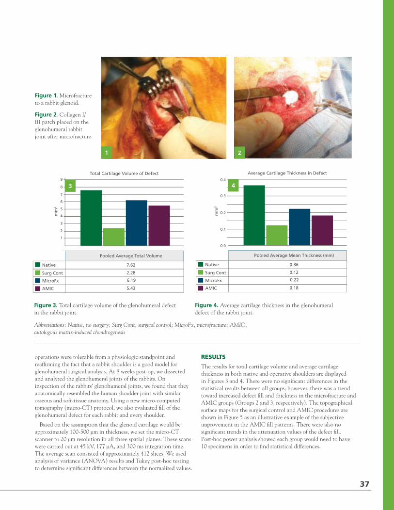
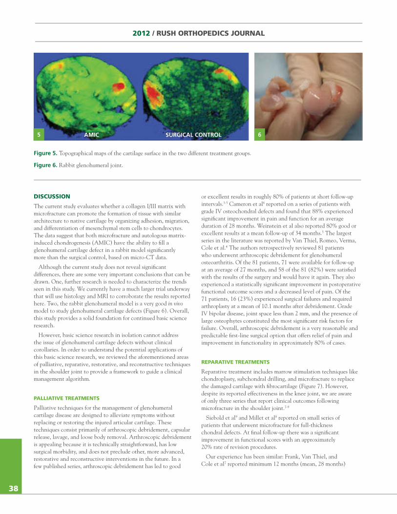
2012 / RUSH ORTHOPEDICS JOURNAL
DISCUSSION
The current study evaluates whether a collagen I/III matrix with microfracture can promote the formation of tissue with similar architecture to native cartilage by organizing adhesion, migration, and differentiation of mesenchymal stem cells to chondrocytes. The data suggest that both microfracture and autologous matrix-induced chondrogenesis (AMIC) have the ability to fill a glenohumeral cartilage defect in a rabbit model significantly more than the surgical control, based on micro-CT data.
Although the current study does not reveal significant differences, there are some very important conclusions that can be drawn. One, further research is needed to characterize the trends seen in this study. We currently have a much larger trial underway that will use histology and MRI to corroborate the results reported here. Two, the rabbit glenohumeral model is a very good in vivo model to study glenohumeral cartilage defects (Figure 6). Overall, this study provides a solid foundation for continued basic science research.
However, basic science research in isolation cannot address the issue of glenohumeral cartilage defects without clinical corollaries. In order to understand the potential applications of this basic science research, we reviewed the aforementioned areas of palliative, reparative, restorative, and reconstructive techniques in the shoulder joint to provide a framework to guide a clinical management algorithm.
PALLIATIVE TREATMENTS
Palliative techniques for the management of glenohumeral cartilage disease are designed to alleviate symptoms without replacing or restoring the injured articular cartilage. These techniques consist primarily of arthroscopic debridement, capsular release, lavage, and loose body removal. Arthroscopic debridement is appealing because it is technically straightforward, has low surgical morbidity, and does not preclude other, more advanced, restorative and reconstructive interventions in the future. In a few published series, arthroscopic debridement has led to good
or excellent results in roughly 80% of patients at short follow-up intervals.3-5 Cameron et al6 reported on a series of patients with grade IV osteochondral defects and found that 88% experienced significant improvement in pain and function for an average duration of 28 months. Weinstein et al also reported 80% good or excellent results at a mean follow-up of 34 months.5 The largest series in the literature was reported by Van Thiel, Romeo, Verma, Cole et al.4 The authors retrospectively reviewed 81 patients who underwent arthroscopic debridement for glenohumeral osteoarthritis. Of the 81 patients, 71 were available for follow-up at an average of 27 months, and 58 of the 81 (82%) were satisfied with the results of the surgery and would have it again. They also experienced a statistically significant improvement in postoperative functional outcome scores and a decreased level of pain. Of the 71 patients, 16 (23%) experienced surgical failures and required arthroplasty at a mean of 10.1 months after debridement. Grade IV bipolar disease, joint space less than 2 mm, and the presence of large osteophytes constituted the most significant risk factors for failure. Overall, arthroscopic debridement is a very reasonable and predictable first-line surgical option that offers relief of pain and improvement in functionality in approximately 80% of cases.
REPARATIVE TREATMENTS
Reparative treatment includes marrow stimulation techniques like chondroplasty, subchondral drilling, and microfracture to replace the damaged cartilage with fibrocartilage (Figure 7). However, despite its reported effectiveness in the knee joint, we are aware of only three series that report clinical outcomes following microfracture in the shoulder joint.7-9
Siebold et al9 and Millet et al8 reported on small series of patients that underwent microfracture for full-thickness chondral defects. At final follow-up there was a significant improvement in functional scores with an approximately 20% rate of revision procedures.
Our experience has been similar: Frank, Van Thiel, and Cole et al7 reported minimum 12 months (mean, 28 months)
Figure 5. Topographical maps of the cartilage surface in the two different treatment groups.
Figure 6. Rabbit glenohumeral joint.
5 6AMIC SURGICAL CONTROL
38
71795_Body.indd 38 7/25/12 9:39 PM
follow-up on 16 patients (17 shoulders) who underwent arthroscopic microfracture of the humeral head or glenoid surface. The 14 patients that were available for follow-up had statistically significant improvements in pain and function. Of the 16 patients, 3 (20%) had subsequent shoulder surgery and therefore, their initial surgeries were considered to be failures. Additional research is needed before definitive statements can be made, but microfracture does appear to be a viable treatment option for select patient populations.
RESTORATIVE TREATMENTS
Restorative treatments aim to reestablish hyaline or hyaline-like cartilage by transferring hyaline cartilage via osteochondral grafting (autograft or allograft) or by growing hyaline-like cartilage using autologous chondrocyte implantation (ACI). At present, osteochondral autograft and ACI require a shoulder arthrotomy
and a second surgical procedure at the knee for graft harvest. Consequently, both procedures are more invasive and more technically demanding, and they expose the patient to significantly greater surgical morbidity than arthroscopic palliative or reparative techniques. Therefore, restorative modalities are best reserved for the young, active individual with a distinct chondral lesion of the humerus or glenoid who has already failed conservative, palliative, and reparative treatment.
Habermeyer et al10 have published good results for 7 patients who received osteochondral autograft transfer from the knee to the shoulder with almost 9-year follow-up. The authors based their results on both functional as well as MRI criteria. Osteochondral allograft transfer employs a similar technique, matching a donor plug to a recipient site, but without the concern for donor-site morbidity. Therefore, allograft transfer can be used to treat more sizable lesions than can be treated effectively by autograft transfer.
Figure 7. Microfracture of the glenoid in a young patient.
Figure 8. Humeral head allograft in a patient with severe degeneration of the humeral head. A, Allograft implanted into the patient’s humerus. B, Inset showing humeral head allograft prior to implantation. 7 8
9 10
Figure 9. Autologous chondrocyte implantation to the humeral head.
Figure 10. Lateral meniscus allograft (LMA) A, LMA that will be used to resurface the glenoid. B, LMA sutured to the glenoid.
A
B
A B
39
71795_Body_u2.indd 39 8/3/12 3:51 PM
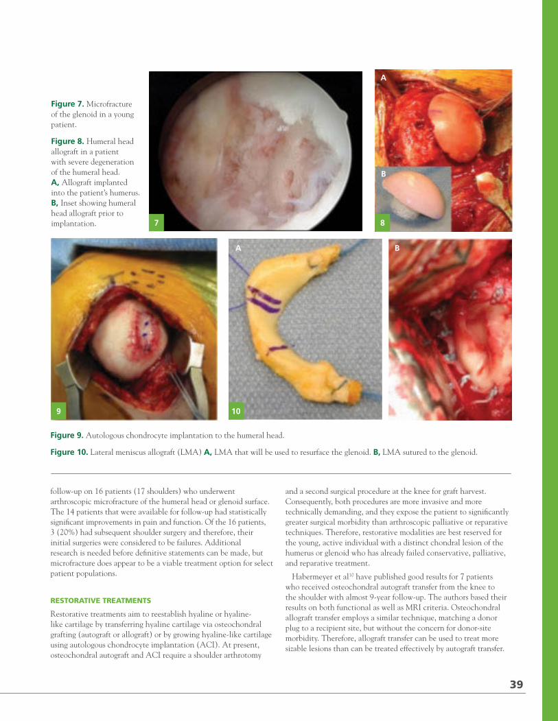
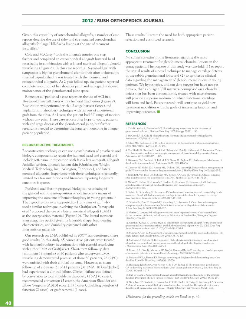
Given this versatility of osteochondral allografts, a number of case reports describe the use of side- and size-matched osteochondral allografts for large Hill-Sachs lesions at the site of recurrent instability.11-13
Cole and McCarty14 took the allograft transfer one step further and completed an osteochondral allograft humeral head resurfacing in combination with a lateral meniscal allograft glenoid resurfacing (Figure 8). In this case report, a 16-year-old girl with symptomatic bipolar glenohumeral chondrolysis after arthroscopic thermal capsulorrhaphy was treated with the meniscal and osteochondral allografts. At 2-year follow-up, the patient reported complete resolution of her shoulder pain, and radiographs showed maintenance of the glenohumeral joint space.
Romeo et al15 published a case report on the use of ACI in a 16-year-old baseball player with a humeral head lesion (Figure 9). Restoration was performed with a 2-stage harvest (knee) and implantation (shoulder) technique with harvest of a periosteal graft from the tibia. At 1 year, the patient had full range of motion without any pain. These case reports offer hope to young patients with end stage disease of the glenohumeral joint, but further research is needed to determine the long term outcome in a larger patient population.
RECONSTRUCTIVE TREATMENTS
Reconstructive techniques can use a combination of prosthetic and biologic components to repair the humeral head and glenoid and include soft-tissue interposition with fascia lata autograft, allograft Achilles tendon, allograft human skin (GraftJacket; Wright Medical Technology, Inc., Arlington, Tennessee), and lateral meniscal allografts. Experience with these techniques is generally limited to a few institutions and literature reporting long-term outcomes is sparse.
Burkhead and Hutton proposed biological resurfacing of the glenoid with the interposition of soft tissue as a means of improving the outcome of hemiarthroplasty in young patients.16 Their good results were supported by Huijsmans et al,17 who used a similar technique involving the GraftJacket. Yamaguchi et al18 proposed the use of a lateral meniscal allograft (LMA) as the interposition material (Figure 10). The lateral meniscus is an attractive option given its favorable shape, load-bearing characteristics, and durability compared with other interposition materials.
Our research on LMA published in 200719 has questioned these good results. In this study, 45 consecutive patients were treated with hemiarthroplasty in conjunction with glenoid resurfacing with either LMA or GraftJacket. Short-term follow-up data (minimum 18 months) of 30 patients who underwent LMA resurfacing demonstrated promise; of those 30 patients, 28 (94%) were satisfied with their clinical outcome. However, at mean follow-up of 2.8 years, 21 of 41 patients (31 LMA, 10 GraftJacket) had experienced a clinical failure. Clinical failure was defined by conversion to total shoulder arthroplasty (TSA) (8 cases), recommended conversion (5 cases), the American Shoulder and Elbow Surgeons (ASES) score ≤ 5 (5 cases), disabling pain/loss of function (2 cases), or graft removal (1 case).
These results illustrate the need for both appropriate patient selection and continued research.
CONCLUSION
No consensus exists in the literature regarding the most appropriate treatment for glenohumeral chondral lesions in the young patient. The purpose of this study was two-fold: (1) to report the initial results of a novel technique to manage cartilage defects in the rabbit glenohumeral joint and (2) to synthesize clinical data regarding the management of glenohumeral lesions in young patients. We hypothesize, and our data suggest but have not yet proven, that a collagen I/III matrix superimposed on a chondral defect that has been concomitantly treated with microfracture will provide a superior medium on which functional cartilage will form and heal. Future research will continue to yield new treatment modalities with the goals of increasing function and improving outcomes.
REFERENCES1. Cole BJ, Yanke A, Provencher MT. Nonarthroplasty alternatives for the treatment of glenohumeral arthritis. J Shoulder Elbow Surg. 2007;16(suppl 5):S231-240.
2. McCarty LP III, Cole BJ. Nonarthroplasty treatment of glenohumeral cartilage lesions. Arthroscopy.2005;21(9):1131-1142.
3. Safran MR, Baillargeon D. The role of arthroscopy in the treatment of glenohumeral arthritis. Sports Med Arthrosc. 2004;12(2):139-145.
4. Van Thiel GS, Sheehan S, Frank RM, Slabaugh M, Cole BJ, Nicholson GP, Romeo AA, Verma NN. Retrospective analysis of arthroscopic management of glenohumeral degenerative disease. Arthroscopy. 2010;26(11):1451-1455.
5. Weinstein DM, Bucchieri JS, Pollock RG, Flatow EL, Bigliani LU. Arthroscopic debridement of the shoulder for osteoarthritis. Arthroscopy. 2000;16(5):471-476.
6. Cameron BD, Galatz LM, Ramsey ML, Williams GR, Iannotti JP. Non-prosthetic management of grade IV osteochondral lesions of the glenohumeral joint. J Shoulder Elbow Surg. 2002;11(1):25-32.
7. Frank RM, Van Thiel GS, Slabaugh MA, Romeo AA, Cole BJ, Verma NN. Clinical outcomes after microfracture of the glenohumeral joint. Am J Sports Med. 2010;38(4):772-781.
8. Millett PJ, Huffard BH, Horan MP, Hawkins RJ, Steadman JR. Outcomes of full-thickness articular cartilage injuries of the shoulder treated with microfracture. Arthroscopy. 2009;25(8):856-863.
9. Siebold R, Lichtenberg S, Habermeyer P. Combination of microfracture and periostal-flap for the treatment of focal full thickness articular cartilage lesions of the shoulder: a prospective study. Knee Surg Sports Traumatol Arthrosc. 2003;11(3):183-189.
10. Scheibel M, Bartl C, Magosch P, Lichtenberg S, Habermeyer P. Osteochondral autologous transplantation for the treatment of full-thickness articular cartilage defects of the shoulder. J Bone Joint Surg Br. 2004;86(7):991-997.
11. Gerber C, Lambert SM. Allograft reconstruction of segmental defects of the humeral head for the treatment of chronic locked posterior dislocation of the shoulder. J Bone Joint Surg Am. 1996;78(3):376-382.
12. Giannini S, Buda R, Cavallo M, et al. Bipolar fresh osteochondral allograft for the treatment of glenohumeral post-traumatic arthritis [published online ahead of print Nov. 23, 2011]. Knee Surg Sports Traumatol Arthrosc. doi: 10.1007/s00167-011-1793-5.
13. Miniaci A, Gish W. Management of anterior glenohumeral instability associated with large Hill-Sachs defects. Tech Shoulder Elbow Surg. 2004;5(3):170-175.
14. McCarty LP III, Cole BJ. Reconstruction of the glenohumeral joint using a lateral meniscal allograft to the glenoid and osteoarticular humeral head allograft after bipolar chondrolysis. J Shoulder Elbow Surg. 2007;16(6):e20-24.
15. Romeo AA, Cole BJ, Mazzocca AD, Fox JA, Freeman KB, Joy E. Autologous chondrocyte repair of an articular defect in the humeral head. Arthroscopy. 2002;18(8):925-929.
16. Burkhead WZ Jr, Hutton KS. Biologic resurfacing of the glenoid with hemiarthroplasty of the shoulder. J Shoulder Elbow Surg.1995;4(4):263-270.
17. Huijsmans P, Roberts C, van Rooyen K, du T DF, de Beer JF. The treatment of glenohumeral OA in the young and active patient with the Graft Jacket: preliminary results. J Bone Joint Surg Br. 2004;87-B(suppl 3):275.
18. Ball C, Galatz L, Yamaguchi K. Meniscal allograft interposition arthroplasty for the arthritic shoulder. Description of a new surgical technique. Tech Shoulder Elbow Surg. 2001;2(4):247-254.
19. Nicholson GP, Goldstein JL, Romeo AA, Cole BJ, Hayden JK, Twigg SL, McCarthy, LP, Detterline AJ. Lateral meniscus allograft biologic glenoid arthroplasty in total shoulder arthroplasty for young shoulders with degenerative joint disease. J Shoulder Elbow Surg. 2007;16(suppl 5):S261-266.
2012 / RUSH ORTHOPEDICS JOURNAL
40Disclosures for the preceding article are listed on p. 46.
71795_Body_u1.indd 40 8/3/12 10:30 AM
Outcomes in the Treatment of Benign Bone Lesions with Bioceramic
YALE A. FILLINGHAM, MD / BRETT A. LENART, MD / STEVEN GITELIS, MD
“Functional results were excellent, complications were infrequent, and composite bioceramic seems to be a reasonable alternative to autogenous bone graft [for treatment of benign bone lesions].”
ARTICLES
AUTHOR AFFILIATIONS
Department of Orthopedic Surgery, Rush University Medical Center, Chicago, Illinois (Drs Lenart and Gitelis); Midwest Orthopaedics at Rush, Chicago, Illinois (Dr Gitelis); and Rush University Medical Center, Chicago, Illinois (Dr Fillingham).
CORRESPONDING AUTHOR
Steven Gitelis, MD, Rush University Medical Center and Midwest Orthopaedics at Rush, 1611 W Harrison St, Suite 300, Chicago, IL 60612 ([email protected]).
B enign bone tumors and cysts are relatively common entities encountered in a general orthopedic and orthopedic oncology practice. This broad category encompasses lesions
with widely varying clinical behaviors and natural histories. Treatment, therefore, must be individualized based on factors such as the specific tissue diagnosis, size of the lesion, location, associated symptoms, risk of pathologic fracture, and individual patient characteristics.
MATERIALS
Traditionally, autogenous bone graft has been the “gold standard” for all grafting procedures.1,2 Limited supply and significant donor site morbidity, however, make this option much less desirable.1-11 Bone-graft substitutes composed of calcium sulfate (CaSO4) or calcium phosphate [Ca3(PO4)2] are attractive alternatives because they are both biodegradable and osteoconductive. Furthermore, they do not contain potent cytokines, which may be contraindicated in the oncology setting. Unfortunately,
few data exist in the literature regarding the use of bone-graft substitutes in orthopedic oncology.12 Most of the reported series using surgical grade CaSO4
1,13-15 or Ca3(PO4)216 graft materials to
treat patients with benign bone tumors feature relatively small numbers of patients and short-duration follow-up, ranging from 6 to 72 months. Results have been generally acceptable in terms of function and recurrence rates. However, relatively common complications still exist: the most common problem reported is serous drainage.1,12-14,16 Radiological appearance and demonstration of resorption with bony replacement are inconsistent at best.
In 2006 Wright Medical Technology (Arlington, Tennessee) released Pro-Dense, an injectable CaSO4-Ca3(PO4)2 composite graft, or bioceramic material with high compressive strength and an intermediate degradation profile. A preclinical canine study showed this material to be superior to CaSO4 with regard to the quantity and quality of bone formed in a contained humeral defect.2,17,18
The bioceramic is a composite graft that incorporates a matrix of CaSO4 and dicalcium phosphate dihydrate (DCPD) into which β-tricalcium phosphate (β-TCP, or β-Ca3(PO4)2) granules are distributed.2 The graft is prepared intraoperatively by mixing the powdered graft materials with an aqueous diluent (Figure 1A). The resulting composite is injectable for approximately 5 minutes (Figure 1B) and sets up over a period of 20-30 minutes. The resorption profile is triphasic. The CaSO4 resorbs first through simple dissolution, leaving behind an open-pore structure that allows for vascular infiltration and new bone deposition on the remaining Ca3(PO4)2 scaffold. DCPD has an intermediate profile,2 resorbing by both osteoclastic resorption and simple dissolution. Finally, β-TCP only undergoes osteoclastic resorption and thus exhibits the longest profile. While a preclinical study2 was extremely promising in the ability to promote bony replacement
41
71795_Body_u1.indd 41 8/3/12 10:30 AM
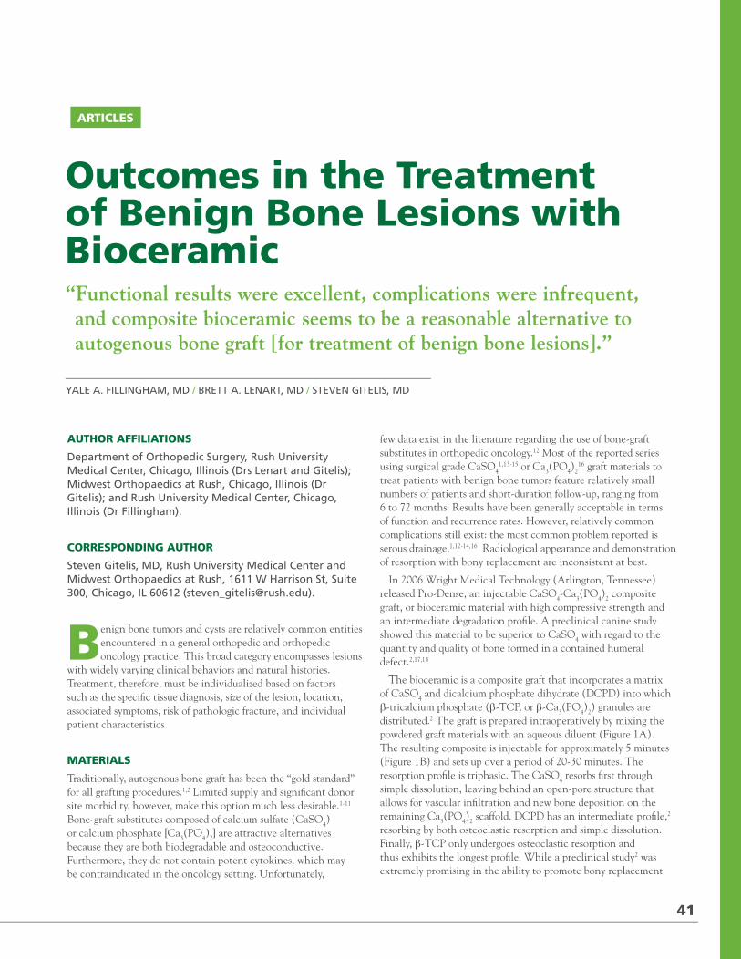
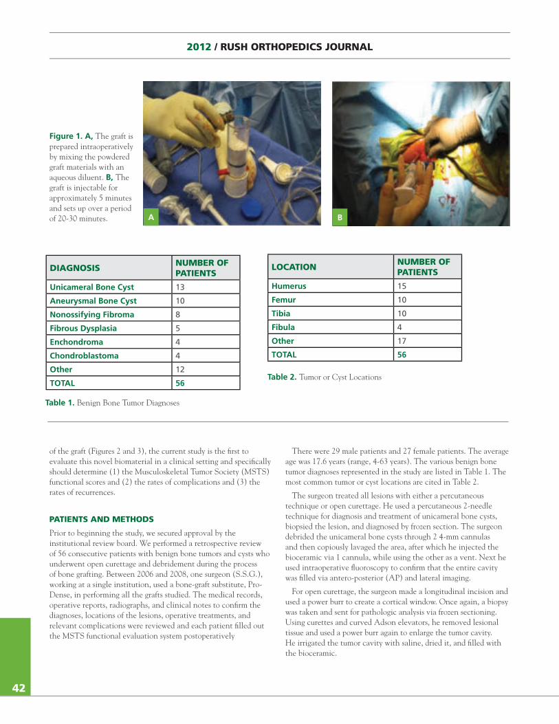
2012 / RUSH ORTHOPEDICS JOURNAL
of the graft (Figures 2 and 3), the current study is the first to evaluate this novel biomaterial in a clinical setting and specifically should determine (1) the Musculoskeletal Tumor Society (MSTS) functional scores and (2) the rates of complications and (3) the rates of recurrences.
PATIENTS AND METHODS
Prior to beginning the study, we secured approval by the institutional review board. We performed a retrospective review of 56 consecutive patients with benign bone tumors and cysts who underwent open curettage and debridement during the process of bone grafting. Between 2006 and 2008, one surgeon (S.S.G.), working at a single institution, used a bone-graft substitute, Pro-Dense, in performing all the grafts studied. The medical records, operative reports, radiographs, and clinical notes to confirm the diagnoses, locations of the lesions, operative treatments, and relevant complications were reviewed and each patient filled out the MSTS functional evaluation system postoperatively
There were 29 male patients and 27 female patients. The average age was 17.6 years (range, 4-63 years). The various benign bone tumor diagnoses represented in the study are listed in Table 1. The most common tumor or cyst locations are cited in Table 2.
The surgeon treated all lesions with either a percutaneous technique or open curettage. He used a percutaneous 2-needle technique for diagnosis and treatment of unicameral bone cysts, biopsied the lesion, and diagnosed by frozen section. The surgeon debrided the unicameral bone cysts through 2 4-mm cannulas and then copiously lavaged the area, after which he injected the bioceramic via 1 cannula, while using the other as a vent. Next he used intraoperative fluoroscopy to confirm that the entire cavity was filled via antero-posterior (AP) and lateral imaging.
For open curettage, the surgeon made a longitudinal incision and used a power burr to create a cortical window. Once again, a biopsy was taken and sent for pathologic analysis via frozen sectioning. Using curettes and curved Adson elevators, he removed lesional tissue and used a power burr again to enlarge the tumor cavity. He irrigated the tumor cavity with saline, dried it, and filled with the bioceramic.
Figure 1. A, The graft is prepared intraoperatively by mixing the powdered graft materials with an aqueous diluent. B, The graft is injectable for approximately 5 minutes and sets up over a period of 20-30 minutes. A B
42
DIAGNOSIS NUMBER OF PATIENTS
Unicameral Bone Cyst 13
Aneurysmal Bone Cyst 10
Nonossifying Fibroma 8
Fibrous Dysplasia 5
Enchondroma 4
Chondroblastoma 4
Other 12
TOTAL 56
LOCATION NUMBER OF PATIENTS
Humerus 15
Femur 10
Tibia 10
Fibula 4
Other 17
TOTAL 56
Table 1. Benign Bone Tumor Diagnoses
Table 2. Tumor or Cyst Locations
71795_Body.indd 42 7/25/12 9:39 PM
After discharge from the hospital, the patient was followed up in the clinic at intervals of 1 week and 1, 6, 12, and 18 months. The surgeon altered the follow-up intervals as needed due to variable healing rates and encountered complications. Each routine follow-up visit in the clinic included assessment of strength, range of motion, functional status, and radiographic investigation to evaluate for potential fractures and the rate of absorption of the bone-graft substitute (Figures 4 and 5).
The MSTS functional evaluation system was employed to measure the clinical outcomes. This survey consists of a list of qualitative responses for multiple categories associated with a graded, numerical score for both upper and lower limbs. For lower limbs, the survey questions evaluate emotional acceptance, supports (eg, brace, prosthesis, cane or crutches), walking ability, and gait. For upper limbs, the survey questions evaluate function, emotional acceptance, hand positioning, dexterity, and lifting ability. Patients rated each category from 0 to 5. A score of 5 for each section represented the best possible outcome, with a total maximum score of 30.
RESULTS
Of the 56 patients in the initial review, we could not reach 10 of them, leaving 46 patients. The average follow-up was 41.9 months (range, 26-57 months).
The average MSTS functional evaluation score was 28.6 (range, 20-30). The average score for patients with lower-limb lesions was 29.1. The average for patients with upper-limb lesions was slightly lower, at 27.8. The lowest score was 20, in a 32-year-old male with fibrous dysplasia of the humerus at 31 months follow-up. Of the 46 patients, 23 patients reported a perfect score of 30; 8 of these patients with perfect scores had upper-limb lesions, while 15 had lower-limb lesions.
Of the 46 patients, 3 (6.5%) suffered local recurrences. A 14-year-old boy with a chondroblastoma of the proximal femur
Figure 3. Computed tomogram 19 months after percutaneous injection of the bioceramic graft material in the proximal humerus unicameral bone cyst of a 12-year-old male (see Figure 1). Full resorption of the ceramic material with complete bone repair is demonstrated.
43
Figure 2. A, Histology of the bone repair using the composite (bioceramic) graft at 13 weeks in the animal model. B, Histology of the bone repair using the autogenous bone graft at 13 weeks in the animal model. C, Normal cancellous bone histology in this animal model.
A B C
71795_Body.indd 43 7/25/12 9:39 PM
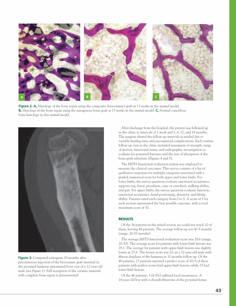
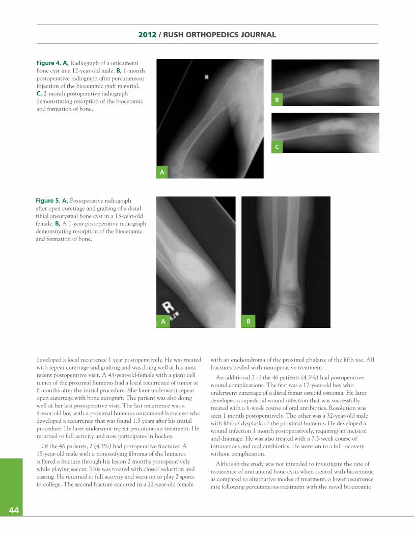
2012 / RUSH ORTHOPEDICS JOURNAL
developed a local recurrence 1 year postoperatively. He was treated with repeat curettage and grafting and was doing well at his most recent postoperative visit. A 43-year-old-female with a giant cell tumor of the proximal humerus had a local recurrence of tumor at 6 months after the initial procedure. She later underwent repeat open curettage with bone autograft. The patient was also doing well at her last postoperative visit. The last recurrence was a 9-year-old boy with a proximal humerus unicameral bone cyst who developed a recurrence that was found 1.5 years after his initial procedure. He later underwent repeat percutaneous treatment. He returned to full activity and now participates in hockey.
Of the 46 patients, 2 (4.3%) had postoperative fractures. A 15-year-old male with a nonossifying fibroma of the humerus suffered a fracture through his lesion 2 months postoperatively while playing soccer. This was treated with closed reduction and casting. He returned to full activity and went on to play 2 sports in college. The second fracture occurred in a 22-year-old female
with an enchondroma of the proximal phalanx of the fifth toe. All fractures healed with nonoperative treatment.
An additional 2 of the 46 patients (4.3%) had postoperative wound complications. The first was a 17-year-old boy who underwent curettage of a distal femur osteoid osteoma. He later developed a superficial wound infection that was successfully treated with a 1-week course of oral antibiotics. Resolution was seen 1 month postoperatively. The other was a 32-year-old male with fibrous dysplasia of the proximal humerus. He developed a wound infection 1 month postoperatively, requiring an incision and drainage. He was also treated with a 7.5-week course of intravenous and oral antibiotics. He went on to a full recovery without complication.
Although the study was not intended to investigate the rate of recurrence of unicameral bone cysts when treated with bioceramic as compared to alternative modes of treatment, a lower recurrence rate following percutaneous treatment with the novel bioceramic
Figure 5. A, Postoperative radiograph after open curettage and grafting of a distal tibial aneurysmal bone cyst in a 13-year-old female. B, A 1-year postoperative radiograph demonstrating resorption of the bioceramic and formation of bone.
A B
44
Figure 4. A, Radiograph of a unicameral bone cyst in a 12-year-old male. B, 1-month postoperative radiograph after percutaneous injection of the bioceramic graft material. C, 2-month postoperative radiograph demonstrating resorption of the bioceramic and formation of bone.
A
B
C
71795_Body.indd 44 7/25/12 9:39 PM
was observed as compared to that in the literature. In our case report, only 1 of the 13 (7.7%) unicameral bone cysts treated percutaneously required a second injection of the bioceramic.19-26,28
DISCUSSION
We performed this retrospective case series to evaluate the initial clinical results using a novel bioceramic injectable graft in 56 consecutive patients with diagnoses of benign bone tumors and cysts who treated at a single orthopedic oncology center. Functional results were excellent, as evidenced by an average MSTS functional evaluation score of 28.6. Complications were infrequent and in most cases were likely not attributable to the graft material itself. Three patients experienced local recurrence. Two patients sustained postoperative fractures, all of which healed with nonoperative treatment. Two patients experienced wound complications, none of which required removal of the graft material. There were no other complications.
Functional results in this series were excellent. This study is comparable to other reported series of CaSO4 and Ca3(PO4)2
bone-graft substitutes reported in the literature.1,2,12,13,14,15,16,27 Complications, specifically tissue reaction and serous wound drainage, appear to be decreased in this series as compared to other series of patients treated with CaSO4.
1,13,14 We hypothesize that the rapid dissolution profile of pure CaSO4 contributes to these complications. Thus, as expected, a composite graft with an intermediate profile appears to lessen the severities of tissue reaction and wound drainage. Furthermore, although it was not specifically quantified in this study, the composite graft material appears to exhibit the expected intermediate resorption profile and is gradually replaced by host bone (Figures 2 and 3). This is in contrast to patients treated with pure Ca3(PO4)2 bone-graft substitutes in whom residual graft material can be seen for years postoperatively and possibly permanently.12,16
The success of the novel bioceramic for the percutaneous treatment of unicameral bone cysts was a surprising finding. Whereas recurrence rates for other benign bone lesions are most likely related solely to the thoroughness of the initial curettage, recurrence rates after percutaneous treatment of unicameral bone cysts is possibly attributable to the injected material itself. With
SLOW RESORPTION OF CEMENT
Post Op 2 wk 6 wk 13 wk 26 wk
Figure 6. A, Animal model of a critically sized cancellous (latticed) defect grafted with this bioceramic material. Note the progressive dissolution and bone repair across time. B, Transverse radiographs of the animal model at 13 weeks comparing the bioceramic graft (left) to autogenous bone graft (right).
A
B
45
71795_Body.indd 45 7/25/12 9:39 PM
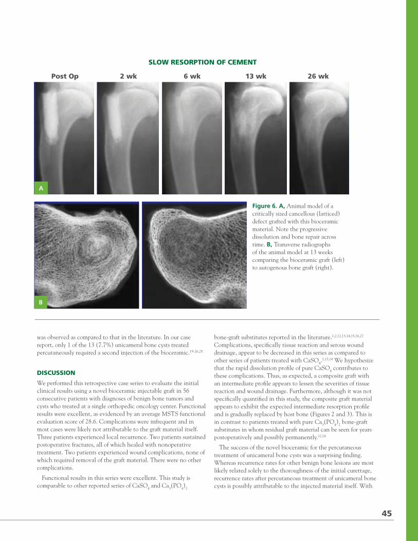
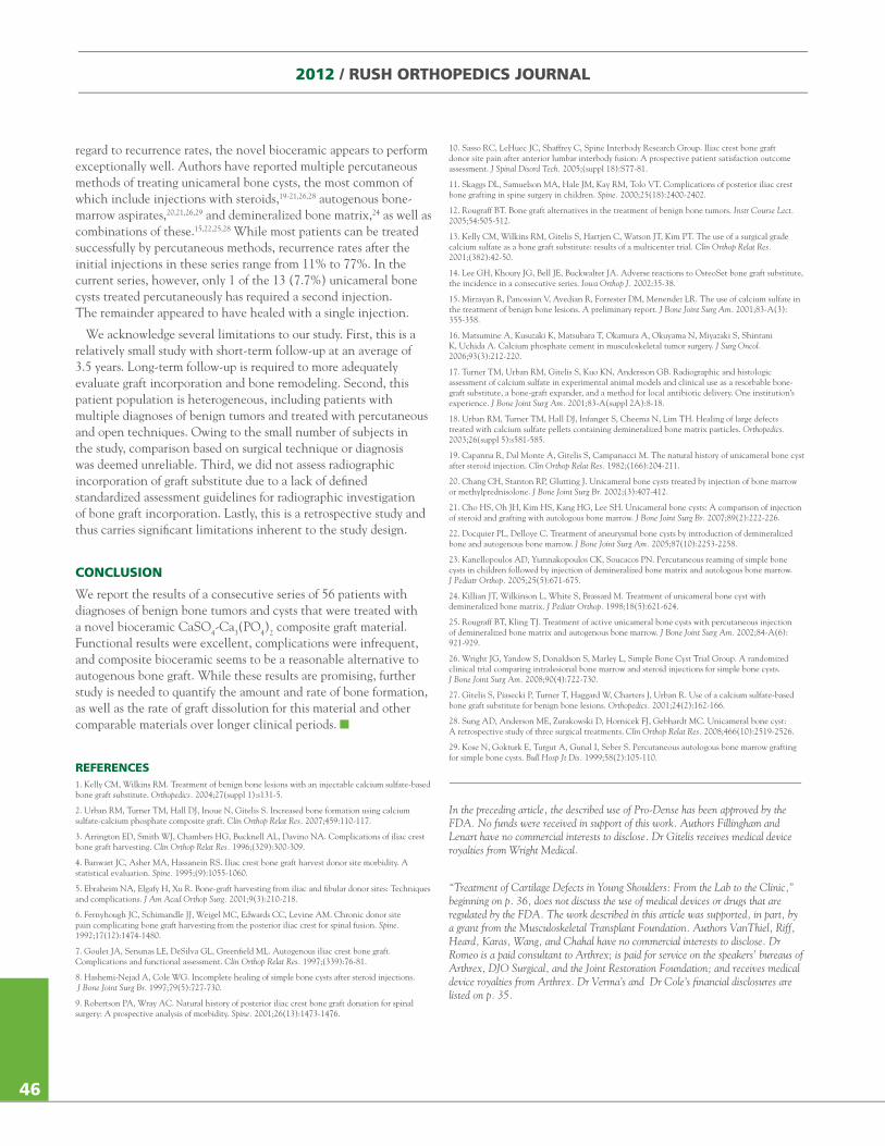
2012 / RUSH ORTHOPEDICS JOURNAL
regard to recurrence rates, the novel bioceramic appears to perform exceptionally well. Authors have reported multiple percutaneous methods of treating unicameral bone cysts, the most common of which include injections with steroids,19-21,26,28 autogenous bone-marrow aspirates,20,21,26,29 and demineralized bone matrix,24 as well as combinations of these.15,22,25,28 While most patients can be treated successfully by percutaneous methods, recurrence rates after the initial injections in these series range from 11% to 77%. In the current series, however, only 1 of the 13 (7.7%) unicameral bone cysts treated percutaneously has required a second injection. The remainder appeared to have healed with a single injection.
We acknowledge several limitations to our study. First, this is a relatively small study with short-term follow-up at an average of 3.5 years. Long-term follow-up is required to more adequately evaluate graft incorporation and bone remodeling. Second, this patient population is heterogeneous, including patients with multiple diagnoses of benign tumors and treated with percutaneous and open techniques. Owing to the small number of subjects in the study, comparison based on surgical technique or diagnosis was deemed unreliable. Third, we did not assess radiographic incorporation of graft substitute due to a lack of defined standardized assessment guidelines for radiographic investigation of bone graft incorporation. Lastly, this is a retrospective study and thus carries significant limitations inherent to the study design.
CONCLUSION
We report the results of a consecutive series of 56 patients with diagnoses of benign bone tumors and cysts that were treated with a novel bioceramic CaSO4-Ca3(PO4)2 composite graft material. Functional results were excellent, complications were infrequent, and composite bioceramic seems to be a reasonable alternative to autogenous bone graft. While these results are promising, further study is needed to quantify the amount and rate of bone formation, as well as the rate of graft dissolution for this material and other comparable materials over longer clinical periods.
REFERENCES1. Kelly CM, Wilkins RM. Treatment of benign bone lesions with an injectable calcium sulfate-based bone graft substitute. Orthopedics. 2004;27(suppl 1):s131-5.
2. Urban RM, Turner TM, Hall DJ, Inoue N, Gitelis S. Increased bone formation using calcium sulfate-calcium phosphate composite graft. Clin Orthop Relat Res. 2007;459:110-117.
3. Arrington ED, Smith WJ, Chambers HG, Bucknell AL, Davino NA. Complications of iliac crest bone graft harvesting. Clin Orthop Relat Res. 1996;(329):300-309.
4. Banwart JC, Asher MA, Hassanein RS. Iliac crest bone graft harvest donor site morbidity. A statistical evaluation. Spine. 1995;(9):1055-1060.
5. Ebraheim NA, Elgafy H, Xu R. Bone-graft harvesting from iliac and fibular donor sites: Techniques and complications. J Am Acad Orthop Surg. 2001;9(3):210-218.
6. Fernyhough JC, Schimandle JJ, Weigel MC, Edwards CC, Levine AM. Chronic donor site pain complicating bone graft harvesting from the posterior iliac crest for spinal fusion. Spine. 1992;17(12):1474-1480.
7. Goulet JA, Senunas LE, DeSilva GL, Greenfield ML. Autogenous iliac crest bone graft. Complications and functional assessment. Clin Orthop Relat Res. 1997;(339):76-81.
8. Hashemi-Nejad A, Cole WG. Incomplete healing of simple bone cysts after steroid injections. J Bone Joint Surg Br. 1997;79(5):727-730.
9. Robertson PA, Wray AC. Natural history of posterior iliac crest bone graft donation for spinal surgery: A prospective analysis of morbidity. Spine. 2001;26(13):1473-1476.
10. Sasso RC, LeHuec JC, Shaffrey C, Spine Interbody Research Group. Iliac crest bone graft donor site pain after anterior lumbar interbody fusion: A prospective patient satisfaction outcome assessment. J Spinal Disord Tech. 2005;(suppl 18):S77-81.
11. Skaggs DL, Samuelson MA, Hale JM, Kay RM, Tolo VT. Complications of posterior iliac crest bone grafting in spine surgery in children. Spine. 2000;25(18):2400-2402.
12. Rougraff BT. Bone graft alternatives in the treatment of benign bone tumors. Instr Course Lect. 2005;54:505-512.
13. Kelly CM, Wilkins RM, Gitelis S, Hartjen C, Watson JT, Kim PT. The use of a surgical grade calcium sulfate as a bone graft substitute: results of a multicenter trial. Clin Orthop Relat Res. 2001;(382):42-50.
14. Lee GH, Khoury JG, Bell JE, Buckwalter JA. Adverse reactions to OsteoSet bone graft substitute, the incidence in a consecutive series. Iowa Orthop J. 2002:35-38.
15. Mirzayan R, Panossian V, Avedian R, Forrester DM, Menendez LR. The use of calcium sulfate in the treatment of benign bone lesions. A preliminary report. J Bone Joint Surg Am. 2001;83-A(3): 355-358.
16. Matsumine A, Kusuzaki K, Matsubara T, Okamura A, Okuyama N, Miyazaki S, Shintani K, Uchida A. Calcium phosphate cement in musculoskeletal tumor surgery. J Surg Oncol. 2006;93(3):212-220.
17. Turner TM, Urban RM, Gitelis S, Kuo KN, Andersson GB. Radiographic and histologic assessment of calcium sulfate in experimental animal models and clinical use as a resorbable bone-graft substitute, a bone-graft expander, and a method for local antibiotic delivery. One institution’s experience. J Bone Joint Surg Am. 2001;83-A(suppl 2A):8-18.
18. Urban RM, Turner TM, Hall DJ, Infanger S, Cheema N, Lim TH. Healing of large defects treated with calcium sulfate pellets containing demineralized bone matrix particles. Orthopedics. 2003;26(suppl 5):s581-585.
19. Capanna R, Dal Monte A, Gitelis S, Campanacci M. The natural history of unicameral bone cyst after steroid injection. Clin Orthop Relat Res. 1982;(166):204-211.
20. Chang CH, Stanton RP, Glutting J. Unicameral bone cysts treated by injection of bone marrow or methylprednisolone. J Bone Joint Surg Br. 2002;(3):407-412.
21. Cho HS, Oh JH, Kim HS, Kang HG, Lee SH. Unicameral bone cysts: A comparison of injection of steroid and grafting with autologous bone marrow. J Bone Joint Surg Br. 2007;89(2):222-226.
22. Docquier PL, Delloye C. Treatment of aneurysmal bone cysts by introduction of demineralized bone and autogenous bone marrow. J Bone Joint Surg Am. 2005;87(10):2253-2258.
23. Kanellopoulos AD, Yiannakopoulos CK, Soucacos PN. Percutaneous reaming of simple bone cysts in children followed by injection of demineralized bone matrix and autologous bone marrow. J Pediatr Orthop. 2005;25(5):671-675.
24. Killian JT, Wilkinson L, White S, Brassard M. Treatment of unicameral bone cyst with demineralized bone matrix. J Pediatr Orthop. 1998;18(5):621-624.
25. Rougraff BT, Kling TJ. Treatment of active unicameral bone cysts with percutaneous injection of demineralized bone matrix and autogenous bone marrow. J Bone Joint Surg Am. 2002;84-A(6): 921-929.
26. Wright JG, Yandow S, Donaldson S, Marley L, Simple Bone Cyst Trial Group. A randomized clinical trial comparing intralesional bone marrow and steroid injections for simple bone cysts. J Bone Joint Surg Am. 2008;90(4):722-730.
27. Gitelis S, Piasecki P, Turner T, Haggard W, Charters J, Urban R. Use of a calcium sulfate-based bone graft substitute for benign bone lesions. Orthopedics. 2001;24(2):162-166.
28. Sung AD, Anderson ME, Zurakowski D, Hornicek FJ, Gebhardt MC. Unicameral bone cyst: A retrospective study of three surgical treatments. Clin Orthop Relat Res. 2008;466(10):2519-2526.
29. Kose N, Gokturk E, Turgut A, Gunal I, Seber S. Percutaneous autologous bone marrow grafting for simple bone cysts. Bull Hosp Jt Dis. 1999;58(2):105-110.
In the preceding article, the described use of Pro-Dense has been approved by the FDA. No funds were received in support of this work. Authors Fillingham and Lenart have no commercial interests to disclose. Dr Gitelis receives medical device royalties from Wright Medical.
“Treatment of Cartilage Defects in Young Shoulders: From the Lab to the Clinic,” beginning on p. 36, does not discuss the use of medical devices or drugs that are regulated by the FDA. The work described in this article was supported, in part, by a grant from the Musculoskeletal Transplant Foundation. Authors VanThiel, Riff, Heard, Karas, Wang, and Chahal have no commercial interests to disclose. Dr Romeo is a paid consultant to Arthrex; is paid for service on the speakers’ bureaus of Arthrex, DJO Surgical, and the Joint Restoration Foundation; and receives medical device royalties from Arthrex. Dr Verma’s and Dr Cole’s financial disclosures are listed on p. 35.
46
71795_Body_u2.indd 46 8/3/12 3:51 PM
Radial Head Replacement with a Bipolar System
MARK R. ZUNKIEWICZ, MD / JILL S. CLEMENTE, MS / MARK C. MILLER, PHD / MARK E. BARATZ, MD /
ROBERT W. WYSOCKI, MD / MARK S. COHEN, MD.
“Patients [given this implant] typically recovered functional range of motion, albeit decreased compared to the unaffected elbow.”
ARTICLES
AUTHOR AFFILIATIONS
Department of Orthopedic Surgery, Rush University Medical Center, Chicago, Illinois, and Midwest Orthopaedics at Rush, Chicago, Illinois (Drs Wysocki and Cohen); Allegheny General Hospital (Drs Baratz and Zunkiewicz and Ms Clemente).
CORRESPONDING AUTHOR
Robert W. Wysocki, MD, Rush University Medical Center and Midwest Orthopaedics at Rush, 1611 W Harrison St, Suite 300, Chicago, IL 60612 ([email protected]).
R adial head and neck fractures are common orthopedic injuries. They are estimated to account for 1.7% to 5.4% of all fractures1 and represent one-third of all elbow
fractures.2,3 In addition, 85% of all radial head and neck fractures occur in patients between 20 and 60 years of age.3
The goals of treatment for radial head fractures are to restore elbow stability, preserve elbow motion, restore forearm rotation, and maintain the length of the radius. Depending on the fracture pattern, these goals may be achieved with open reduction and fracture fixation or radial head replacement. Radial head excision without replacement should be performed with caution in the setting of an acute elbow injury. Associated injury to the collateral ligaments or interosseous membrane may lead to valgus elbow instability, longitudinal forearm instability, and loss of strength.1
A variety of radial head replacements is currently available. Fragmentation of silicone implants led to the development of metallic devices, and the majority of these are 1-piece monoblock or unipolar implants. Due to the complex nature of the proximal radioulnar and radiocapitellar joints, bipolar radial head implants were developed to enhance joint congruency throughout elbow and forearm motion. There is basic scientific evidence that these
bipolar designs better reproduce kinematics of the elbow following radial head replacement.4
The Katalyst Bipolar Radial Head System (Integra, Plainsboro, New Jersey) is a bipolar implant with a smooth telescoping stem. The stem design allows for the precise restoration of radial column “length” in situ without the need to release the lateral collateral ligament and common extensor origin when they are intact. This study reports the early clinical outcome of a cohort of patients undergoing radial head replacement with this implant.
MATERIALS AND METHODS
Prior to beginning the study, we received approval from the institutional review board. We placed 36 prostheses in 34 patients (21 female, 13 male) between March 2004 and October 2006. All individuals underwent resection of the radial head followed by replacement arthroplasty with the Katalyst implant. We treated 27 patients after acute injury (fracture or fracture-dislocation), where the radial head was deemed irreparable at the time of surgery (Figure 1), and 7 patients for posttraumatic arthritis or in the setting of elbow reconstruction indicated by failed previous surgery or fixation at outside institutions. We performed radial head arthroplasty following acute trauma where the radial head could not be repaired and following late radial head resection done for posttraumatic arthritis where there was residual valgus laxity of the elbow.
The surgical technique consisted of a lateral ligament-sparing approach in which the extensor tendon origin and lateral ligament were incised at the midline of the radiocapitellar axis. We sectioned the radial head with a saw and chose the appropriate diameter implant. We measured for the appropriate length of the implant, using the cutting guide system under fluoroscopy with the ulnohumeral joint reduced. The head size varied from 18 to 24 mm in diameter, and the head offset from the stem was between 1 and 3 mm, depending on the radiocapitellar gap to be restored.
47
71795_Body.indd 47 7/25/12 9:39 PM
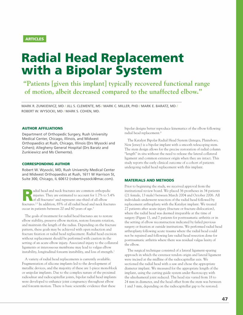
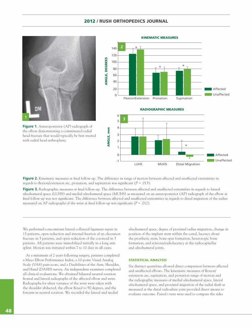
2012 / RUSH ORTHOPEDICS JOURNAL
We performed concomitant lateral collateral ligament repair in 15 patients, open reduction and internal fixation of an olecranon fracture in 5 patients, and open reduction of the coronoid in 3 patients. All patients were immobilized initially in a long arm splint. Motion was initiated within 7 to 10 days in all cases.
At a minimum of 2 years following surgery, patients completed a Mayo Elbow Performance Index, a 10-point Visual Analog Scale (VAS) pain score, and a Disabilities of the Arm, Shoulder, and Hand (DASH) survey. An independent examiner completed all clinical evaluations. We obtained bilateral neutral rotation frontal and lateral radiographs of the affected elbow and wrist. Radiographs for ulnar variance of the wrist were taken with the shoulder abducted, the elbow flexed to 90 degrees, and the forearm in neutral rotation. We recorded the lateral and medial
ulnohumeral space, degree of proximal radius migration, change in position of the implant stem within the canal, lucency about the prosthetic stem, bone-spur formation, heterotopic bone formation, and sclerosis/radiolucency at the radiocapitellar and ulnohumeral joints.
STATISTICAL ANALYSIS
Six distinct quantities allowed direct comparison between affected and unaffected elbows. The kinematic measures of flexion/extension arc, supination, and pronation range of motion and the radiographic measures of medial ulnohumeral space, lateral ulnohumeral space, and proximal migration of the radial shaft as measured at the distal radioulnar joint provided direct means to evaluate outcome. Paired t tests were used to compare the sides
Figure 1. Anteroposterior (AP) radiograph of the elbow demonstrating a comminuted radial head fracture that would typically be best treated with radial head arthroplasty.
Figure 2. Kinematic measures at final follow-up. The difference in range of motion between affected and unaffected extremities in regards to flexion/extension arc, pronation, and supination was significant (P = .013).
Figure 3. Radiographic measures at final follow-up. The difference between affected and unaffected extremities in regards to lateral ulnohumeral space (LUHS) and medial ulnohumeral space (MUHS) as measured on an anteroposterior (AP) radiograph of the elbow at final follow-up was not significant. The difference between affected and unaffected extremities in regards to distal migration of the radius measured on AP radiographs of the wrist at final follow-up was significant (P = .012).
1
RADIOGRAPHIC MEASURESA
NG
LE,
mm
3
LUHS MUHS Distal Migration
5
4
3
2
1
0
-1
Affected
Unaffected
KINEMATIC MEASURES
AN
GLE
, D
EG
REES
2
Flexion/Extension Pronation Supination
140
120
100
80
60
40
20
0
Affected
Unaffected
48
6
71795_Body.indd 48 7/25/12 9:39 PM
in each case, and unpaired t tests were used for comparison of functional scores between those with acute radial head arthroplasty for trauma and those with previous surgery or chronic conditions (ie, arthritis). A significance level of 0.05 was utilized.
RESULTS
Of the 36 implants, 30 (83.3%) were available for review. Follow-up averaged 34 months (range, 24-48 months). The Mayo Elbow Performance Index score averaged 92.1 (range, 65-100), the VAS (from 0 = no pain to 10 = severe pain) averaged 1.4 (range, 0-5), and the DASH score averaged 13.8 (range, 0-52.5) for the entire cohort. When broken down into cases performed for acute injury (n = 23) versus those for chronic conditions (n = 7), the Mayo score in the chronic group demonstrated poorer function (mean, 85.0) compared to the acute group (mean, 94.6) (P = .034). The VAS (means, 1.2 vs. 2.2) and DASH (means, 12.01 vs. 21.8) scores in the acute and chronic groups respectively did not reach statistical significance (P = .166 and P = .127).
Clinical evaluation of the affected elbow revealed an average flexion/extension arc of 126 degrees (range, 95-150 degrees). Forearm pronation averaged 69 degrees (range, 45-90 degrees), and supination averaged 74 degrees (range, 60-85 degrees). In comparison, the unaffected elbow measured an average flexion/extension arc of 138 degrees (range, 120-150 degrees), an average 72 degrees of pronation (range, 45-90 degrees), and an average 80 degrees of supination (range, 60-85 degrees). The difference in range of motion between the affected and unaffected arms for flexion/extension arc, pronation, and supination was statistically significant (P = .013) The kinematic measures are shown in Figure 2.
Analysis of orthogonal radiographs of both the affected and unaffected elbows revealed an average lateral ulnohumeral space (LUHS) of 2.71 mm on the operative side (range, 0.5-5.1 mm),
as compared to 2.72 mm on the unaffected side (range, 1.0-6.3 mm). This difference was not significant (P = .317). The medial ulnohumeral space (MUHS) on the affected side averaged 2.14 mm (range, 1.0-4.1 mm) and on the unaffected side averaged 2.34 mm (range, 1.0-5.6 mm). This difference was also not significant (P = .120). Wrist radiographs revealed an average proximal migration of the radius of 0.34 mm on the affected side (range, –3.2 to 4.4 mm) as compared to an average ulnar positive variance of 0.26 mm (range, –4.0 to 1.8 mm) on the unaffected side. This difference was statistically significant (P = .012) The radiographic measures are shown in Figure 3. No patient reported wrist pain or exhibited any evidence of distal radioulnar joint instability on clinical stress testing. Further analysis of the radiographs revealed the presence of minimal ulnohumeral bone spurs in 22 patients, small degrees of non-motion-limiting calcification in 13 patients, and stem lucency in 24 patients (Figure 4). Despite this radiolucency, there was very little (if any) change in the position of the stem within the canal across time.
Two patients suffered complications significant enough to require repeat surgery. The first was overstuffing of the radiocapitellar joint. This was a technical error in a patient treated early in the series, for which we returned to the operating room, removed the set screw on the adjustable neck, shortened the neck by 2 mm and reinserted the set screw. The operating room implant tray now includes wands upon which are the 6 combinations of head diameter and neck length that can be used to trial the head diameter and neck length to avoid this complication.
The second patient had a grossly unstable elbow despite repair of the lateral ligament complex, due to incompetent anterior and posterior capsule and medial collateral ligament. This was recognized at his first postoperative visit and was treated with reduction and stabilization of the joint with a hinged external fixator. Neither of these complications was deemed specifically related to the implant.
A B
Figure 4. A, Lateral and B, anteroposterior (AP) radiograph of a right elbow at final follow-up reveals the presence of minimal ulnohumeral bone spurs, no soft tissue calcification (heterotopic ossification), and minimal stem lucency. When we compared immediate postoperative radiographs to radiographs obtained at final follow-up, there was very little (if any) change in the position of the stem within the canal over time.
49
71795_Body.indd 49 7/25/12 9:39 PM
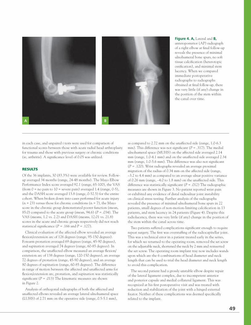
2012 / RUSH ORTHOPEDICS JOURNAL
We performed concomitant lateral collateral ligament repair in 15 patients, open reduction and internal fixation of an olecranon fracture in 5 patients, and open reduction of the coronoid in 3 patients. All patients were immobilized initially in a long arm splint. Motion was initiated within 7 to 10 days in all cases.
At a minimum of 2 years following surgery, patients completed a Mayo Elbow Performance Index, a 10-point Visual Analog Scale (VAS) pain score, and a Disabilities of the Arm, Shoulder, and Hand (DASH) survey. An independent examiner completed all clinical evaluations. We obtained bilateral neutral rotation frontal and lateral radiographs of the affected elbow and wrist. Radiographs for ulnar variance of the wrist were taken with the shoulder abducted, the elbow flexed to 90 degrees, and the forearm in neutral rotation. We recorded the lateral and medial
ulnohumeral space, degree of proximal radius migration, change in position of the implant stem within the canal, lucency about the prosthetic stem, bone-spur formation, heterotopic bone formation, and sclerosis/radiolucency at the radiocapitellar and ulnohumeral joints.
STATISTICAL ANALYSIS
Six distinct quantities allowed direct comparison between affected and unaffected elbows. The kinematic measures of flexion/extension arc, supination, and pronation range of motion and the radiographic measures of medial ulnohumeral space, lateral ulnohumeral space, and proximal migration of the radial shaft as measured at the distal radioulnar joint provided direct means to evaluate outcome. Paired t tests were used to compare the sides
Figure 1. Anteroposterior (AP) radiograph of the elbow demonstrating a comminuted radial head fracture that would typically be best treated with radial head arthroplasty.
Figure 2. Kinematic measures at final follow-up. The difference in range of motion between affected and unaffected extremities in regards to flexion/extension arc, pronation, and supination was significant (P = .013).
Figure 3. Radiographic measures at final follow-up. The difference between affected and unaffected extremities in regards to lateral ulnohumeral space (LUHS) and medial ulnohumeral space (MUHS) as measured on an anteroposterior (AP) radiograph of the elbow at final follow-up was not significant. The difference between affected and unaffected extremities in regards to distal migration of the radius measured on AP radiographs of the wrist at final follow-up was significant (P = .012).
1
RADIOGRAPHIC MEASURES
AN
GLE
, m
m
3
LUHS MUHS Distal Migration
5
4
3
2
1
0
-1
Affected
Unaffected
KINEMATIC MEASURES
AN
GLE
, D
EG
REES
2
Flexion/Extension Pronation Supination
140
120
100
80
60
40
20
0
Affected
Unaffected
48
6
71795_Body.indd 48 7/25/12 9:39 PM
in each case, and unpaired t tests were used for comparison of functional scores between those with acute radial head arthroplasty for trauma and those with previous surgery or chronic conditions (ie, arthritis). A significance level of 0.05 was utilized.
RESULTS
Of the 36 implants, 30 (83.3%) were available for review. Follow-up averaged 34 months (range, 24-48 months). The Mayo Elbow Performance Index score averaged 92.1 (range, 65-100), the VAS (from 0 = no pain to 10 = severe pain) averaged 1.4 (range, 0-5), and the DASH score averaged 13.8 (range, 0-52.5) for the entire cohort. When broken down into cases performed for acute injury (n = 23) versus those for chronic conditions (n = 7), the Mayo score in the chronic group demonstrated poorer function (mean, 85.0) compared to the acute group (mean, 94.6) (P = .034). The VAS (means, 1.2 vs. 2.2) and DASH (means, 12.01 vs. 21.8) scores in the acute and chronic groups respectively did not reach statistical significance (P = .166 and P = .127).
Clinical evaluation of the affected elbow revealed an average flexion/extension arc of 126 degrees (range, 95-150 degrees). Forearm pronation averaged 69 degrees (range, 45-90 degrees), and supination averaged 74 degrees (range, 60-85 degrees). In comparison, the unaffected elbow measured an average flexion/extension arc of 138 degrees (range, 120-150 degrees), an average 72 degrees of pronation (range, 45-90 degrees), and an average 80 degrees of supination (range, 60-85 degrees). The difference in range of motion between the affected and unaffected arms for flexion/extension arc, pronation, and supination was statistically significant (P = .013) The kinematic measures are shown in Figure 2.
Analysis of orthogonal radiographs of both the affected and unaffected elbows revealed an average lateral ulnohumeral space (LUHS) of 2.71 mm on the operative side (range, 0.5-5.1 mm),
as compared to 2.72 mm on the unaffected side (range, 1.0-6.3 mm). This difference was not significant (P = .317). The medial ulnohumeral space (MUHS) on the affected side averaged 2.14 mm (range, 1.0-4.1 mm) and on the unaffected side averaged 2.34 mm (range, 1.0-5.6 mm). This difference was also not significant (P = .120). Wrist radiographs revealed an average proximal migration of the radius of 0.34 mm on the affected side (range, –3.2 to 4.4 mm) as compared to an average ulnar positive variance of 0.26 mm (range, –4.0 to 1.8 mm) on the unaffected side. This difference was statistically significant (P = .012) The radiographic measures are shown in Figure 3. No patient reported wrist pain or exhibited any evidence of distal radioulnar joint instability on clinical stress testing. Further analysis of the radiographs revealed the presence of minimal ulnohumeral bone spurs in 22 patients, small degrees of non-motion-limiting calcification in 13 patients, and stem lucency in 24 patients (Figure 4). Despite this radiolucency, there was very little (if any) change in the position of the stem within the canal across time.
Two patients suffered complications significant enough to require repeat surgery. The first was overstuffing of the radiocapitellar joint. This was a technical error in a patient treated early in the series, for which we returned to the operating room, removed the set screw on the adjustable neck, shortened the neck by 2 mm and reinserted the set screw. The operating room implant tray now includes wands upon which are the 6 combinations of head diameter and neck length that can be used to trial the head diameter and neck length to avoid this complication.
The second patient had a grossly unstable elbow despite repair of the lateral ligament complex, due to incompetent anterior and posterior capsule and medial collateral ligament. This was recognized at his first postoperative visit and was treated with reduction and stabilization of the joint with a hinged external fixator. Neither of these complications was deemed specifically related to the implant.
A B
Figure 4. A, Lateral and B, anteroposterior (AP) radiograph of a right elbow at final follow-up reveals the presence of minimal ulnohumeral bone spurs, no soft tissue calcification (heterotopic ossification), and minimal stem lucency. When we compared immediate postoperative radiographs to radiographs obtained at final follow-up, there was very little (if any) change in the position of the stem within the canal over time.
49
71795_Body.indd 49 7/25/12 9:39 PM
2012 / RUSH ORTHOPEDICS JOURNAL
DISCUSSION
A variety of implants has been used to replace the radial head. These include those made of ferrule caps, metal, acrylic, and silicone.5 While silicone was initially popular, fragmentation and an appreciation of the limited load-bearing capacity of silicone led to the development of metallic implants. Currently, there are a variety of implant designs available. However, few data exist on the superiority of one design over another. Most current implants function as monoblock or unipolar devices. Malalignment of these implants can lead to decreased radiocapitellar contact area and increased cartilage wear from stress concentration.6 Bipolar radial head implants were developed to maximize radiocapitellar congruency and contact forces.
The Katalyst implant is a bipolar implant with a smooth, stainless-steel stem that is inserted without cement. Within the stem is a telescoping shaft with fluted walls and a ball on the proximal end. The flutes provide a surface to engage a set screw to fix the shaft in a distracted position. The ball on the telescoping shaft couples with the polyethylene liner of the head to allow assembly of the head to shaft in situ as a “snap fit.”
The stem was designed to be a smooth, “loose” fit within the canal. This was chosen due to excellent results with a similar stem in a monoblock head (Evolve; Wright Medical, Arlington, Tennessee). Since this implant’s conception over 10 years ago, the “loose,” smooth stem design has held up well in long-term follow-up.7 In this series we had similarly good experience with this stem design. The stem has a surrounding lucency by design but did not migrate or tip within the canal during nearly 3-year average follow-up.
The polyethylene-stem coupling mechanism allows in situ assembly. Polyethylene wear is a concern in any implant. To date we have not had the chance to retrieve an implant that has been in place for more than 7 weeks. While wear could become an issue, it has not been apparent clinically or on radiographs. With rotational loads shared between stem and proximal radius, stem and polyethylene liner, and head and capitellum, it is our expectation that the stresses seen at the polyethylene-stem interface are relatively low.
The polyethylene-stem connection is also the heart of the bipolar nature of the implant. There are conflicting data regarding the function of bipolar prostheses compared with unipolar designs. Using a cadaver model, Moon et al concluded that a bipolar implant would impart less posterolateral stability to the elbow joint in the face of an incompetent lateral ligament complex.6 In another laboratory study using cadaver elbows, Yian et al determined that only a bipolar implant would restore physiological radiocapitellar alignment and tracking.4 Clinical short- to midterm outcome studies have shown favorable results with both implant designs.7-18 It is unknown how long the Katalyst implant retains its bipolar nature, and this is currently a subject of investigation in our institutions.
Implant dissociation at the polyethylene-stem interface is another potential concern, but we did not experience it in this series. In our experience, the coupling is very secure when a loud
“snap” is heard when the head is coupled to the stem, the elbow is not overstuffed, and when the lateral complex is intact or repaired.
CONCLUSION
At a minimum 2-year follow-up, we did not identify any major complications specifically related to the implant. Radiographic and clinical evaluations revealed re-establishment of a congruous elbow joint in both posttraumatic and reconstructive applications, although function as assessed by the Mayo Elbow Performance Index was better in the acutely posttraumatic setting than in reconstructive cases. There was no evidence of capitellar osteopenia, significant proximal radial translation, or migration of the implant itself. Patients typically recovered functional range of motion, albeit decreased compared to the unaffected elbow. Further study will be required to see if these short-term results are maintained across time.
REFERENCES1. Morrey BF. Radial head fracture. In: Morrey BF, ed. The Elbow and Its Disorders. 3rd ed. Philadelphia: WB Saunders; 2000:341-364.
2. Harrington IJ, Tountas AA. Replacement of the radial head in the treatment of unstable elbow fractures. Injury. 1981;12(5):405-412.
3. Mason ML. Some observations on fracture of the head of the radius with a review of one hundred cases. Br J Surg. 1954;42(172):123-132.
4. Yian E, Steens W, Lingenfelter E, Schneeberger AG. Malpositioning of radial head prosthesis: an in vitro study. J Shoulder Elbow Surg. 2008;17(4):663-670.
5. Swanson AB, Jaeger SH, La Rochelle D. Comminuted fractures of the radial head. The role of silicone-implant replacement arthroplasty. J Bone Joint Surg Am. 1981;63(7):1039-1049.
6. Moon JG, Berglund LJ, Zachary D, An KN, O’Driscoll SW. Radiocapitellar joint stability with bipolar versus monopolar radial head prostheses. J Shoulder Elbow Surg. 2009;18(5):779-784.
7. Grewal R, MacDermid JC, Faber KJ, Drosdowech DS, King GJ. Comminuted radial head fracture treated with a modular metallic radial head arthroplasty. Study of outcomes. J Bone Joint Surg Am. 2006;88(10):2192-2200.
8. Ashwood N, Bian GI, Unni R. Management of Mason type-III radial head fractures with a titanium prosthesis, ligament repair, and early mobilization. J Bone Joint Surg Am. 2004;86-A(2): 274-280.
9. Burkhart KJ, Mattyasovszky SG, Runkel M, Schwarz C, Kuchle R, Hessmann MH, et al. Mid- to long-term results after bipolar radial head arthroplasty. J Shoulder Elbow Surg. 2010;19(7):965-972.
10. Chapman CB, Su BW, Sinicropi SM, Bruno R, Stauch RJ, Rosenwasser MP. Vitallium radial head prosthesis for acute and chronic elbow fractures and fracture-dislocations involving the radial head. J Shoulder Elbow Surg. 2006;15(4):463-473.
11. Frosch KH, Kropp W, Dresing K, Langer C, Sturmer KM. A bipolar radial head prosthesis after comminuted radial head fractures: Indications, treatment, and outcome after 5 years. Unfallchirurg. 2003;106(5):367-373.
12. Holmenschlager F, Halm JP, Piatek S, Schubert S, Winckler S. Comminuted radial head fractures. Initial experiences with a Judet radial head prosthesis. Unfallchirug. 2002;105(4):344-352.
13. Judet T, Garreau de Loubresse C, Piriou P, Charnley G. A floating prosthesis for radial head fractures. J Bone Joint Surg Br. 1996;78(2):244-249.
14. King GJ, Zarzour ZD, Rath DA, Dunning CE, Patterson SD, Johnson JA. Metallic radial head arthroplasty improves valgus stability of the elbow. Clin Orthop Relat Res. 1999;(368):114-125.
15. Knight DJ, Rymaszewski LA, Amis AA, Miller JH. Primary replacement of the fractured radial head with a metal prosthesis. J Bone Joint Surg Br. 1993;75(4):572-576.
16. Moro JK, Werier J, MacDermid JC, Patterson S, King GJ. Arthroplasty with a metal radial head for unreconstructable fractures of the radial head. J Bone Joint Surg Am. 2001;83-A(8):1201-1211.
17. Muller MC, Burger C, Striepens N, Wirtz CD, Weber O. Clinical results after replacement of comminuted radial head fractures (Mason III and IV) by the bipolar radial head prosthesis of Judet. Z Orthop Unfall. 2008;146(2):218-226.
18. Syczewska M, Skalski K, Pomianowski S, Szczerbik E. Functional outcome following the implantation of the modal/bipolar radial head endoprosthesis. Preliminary results. Acta Bioeng Biomech. 2008;10(2):43-49.
Disclosures for the preceding article are listed on p. 58.
50
71795_Body_u1.indd 50 8/3/12 10:30 AM
Osteotomy-Sparing Methods for Correction of Metatarsus Primus Varus in Hallux Valgus
GEORGE HOLMES JR, MD
“Osteotomy-sparing techniques provide viable alternatives to the use of osteotomies or fusions for the correction of the metatarsus primus varus.”
ARTICLES
AUTHOR AFFILIATIONS
Department of Orthopedic Surgery, Section of Foot and Ankle Surgery, Rush University Medical Center, and Midwest Orthopaedics at Rush, Chicago, Illinois.
CORRESPONDING AUTHOR
George Holmes Jr, MD, Rush University Medical Center and Midwest Orthopaedics at Rush, 1611 West Harrison St, Suite 300, Chicago, IL 60612 ([email protected]).
Many surgical procedures have been developed for the correction of the various components of hallux valgus deformity. The predominant approach to the correction
of the primus varus deformity or intermetatarsal angle (IMA) has relied upon various osteotomies or fusions of the first metatarsal.1-4 Whether performed distally, midshaft, or proximally, the goal of these osteotomies and fusions is to decrease the IMA by an angular realignment or translation of the first metatarsal closer to the second metatarsal.
In general, a reliance solely on soft-tissue procedures has been reserved for deformities with a relatively small IMA.5,6 This article will explore various osteotomy-sparing options that have been developed for narrowing of the IMA and correction of the metatarsus primus varus associated with mild and moderate hallux valgus deformity.
TECHNIQUE DESCRIPTIONS
The modified McBride procedure, popularized by Dr. Roger Mann and others, has been used as a stand-alone procedure and also as an adjunct to other procedures for the correction of hallux valgus
deformity. The core of the McBride procedure requires the release of the dynamic deforming forces of the adductor tendon as well as the static deforming forces of the contracted lateral capsule of the first metatarsophalangeal joint (MTPJ). In the presence of a flexible foot and a small IMA, correction of the IMA may be accomplished solely by suturing together the lateral capsule of the first metatarsal, the medial capsule of the second metatarsal, and the distal stump of the released adductor tendon.
However, this method will not be effective in patients with more moderate IMAs, particularly those greater than 12 degrees. Additional osteotomy-sparing techniques have been developed to address patients with this more widened IMA deformity (Figure 1).
In 2005, Wu demonstrated the efficacy of narrowing the IMA by creating a synostosis between the first and second metatarsals.7 Wu reduced the IMA encircling the first and second metatarsals with suture tape. The reduction was maintained by the development of a synostosis between the first and second metatarsal, created by scarification of the periosteum of the medial second metatarsal and the lateral aspect of the first metatarsal (Figure 2). Using a similar methodology in 1950, Joplin described the use of a sling procedure for correction of the metatarsus primus deformity.8
More recently surgeons have been using ultra-high-molecular-weight polyethylene (UHMWPE) braided suture (Fiberwire; Arthrex, Naples, Florida) to attach endobuttons as a means of reducing the IMA in patients with metatarsus primus varus deformity.3,9 UHMWPE suture has been critical to the advancement of soft-tissue repair in orthopedic surgery. It has been critical for enhancing the biomechanical fixation of tendon-to-bone healing in shoulder rotator-cuff repair, and numerous biomechanical studies have demonstrated the increased strength of UHMWPE sutures over traditional high-strength braided sutures. The ability of UHMWPE has been able to supplant the use of
51
71795_Body.indd 51 7/25/12 9:39 PM
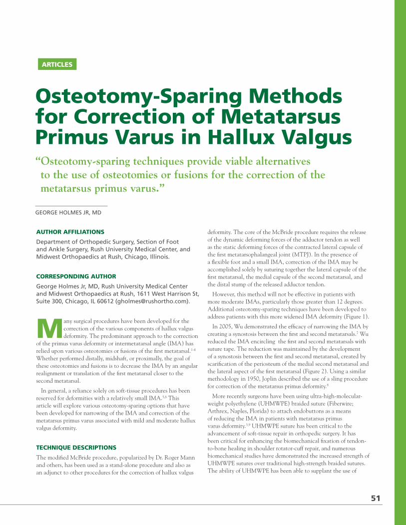
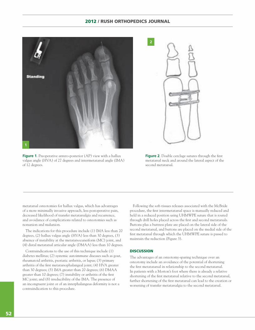
2012 / RUSH ORTHOPEDICS JOURNAL
metatarsal osteotomies for hallux valgus, which has advantages of a more minimally invasive approach, less postoperative pain, decreased likelihood of transfer metatarsalgia and recurrence, and avoidance of complications related to osteotomies such as nonunion and malunion.
The indications for this procedure include (1) IMA less than 20 degrees, (2) hallux valgus angle (HVA) less than 30 degrees, (3) absence of instability at the metatarsocuneiform (MC) joint, and (4) distal metatarsal articular angle (DMAA) less than 10 degrees.
Contraindications to the use of this technique include (1) diabetes mellitus; (2) systemic autoimmune diseases such as gout, rheumatoid arthritis, psoriatic arthritis, or lupus; (3) primary arthritis of the first metatarsophalangeal joint; (4) HVA greater than 30 degrees; (5) IMA greater than 20 degrees; (6) DMAA greater than 10 degrees; (7) instability or arthritis of the first MC joint; and (8) irreducibility of the IMA. The presence of an incongruent joint or of an interphalangeus deformity is not a contraindication to this procedure.
Following the soft-tissues releases associated with the McBride procedure, the first intermetatarsal space is manually reduced and held in a reduced position using UHMWPE suture that is routed through drill holes placed across the first and second metatarsals. Buttons plus a buttress plate are placed on the lateral side of the second metatarsal, and buttons are placed on the medial side of the first metatarsal through which the UHMWPE suture is passed to maintain the reduction (Figure 3).
DISCUSSION
The advantages of an osteotomy-sparing technique over an osteotomy include an avoidance of the potential of shortening the first metatatarsal in relationship to the second metatarsal. In patients with a Morton’s foot where there is already a relative shortening of the first metatarsal relative to the second metatarsal, further shortening of the first metatarsal can lead to the creation or worsening of transfer metatarsalgia to the second metatarsal.
Figure 1. Preoperative antero-posterior (AP) view with a hallux valgus angle (HVA) of 27 degrees and intermetatarsal angle (IMA) of 12 degrees.
1
2
52
Figure 2. Double cerclage sutures through the first metatarsal neck and around the lateral aspect of the second metatarsal.
71795_Body.indd 52 7/25/12 9:39 PM
Osteotomy-sparing techniques avoid the potential complications of malunion. The distal portion of a first metatarsal osteotomy may heal with a dorsal angular deformity, creating a transfer metatarsalgia to the second metatarsal.The complications of avascular necrosis, delayed union and nonunion can also be avoided using an osteotomy-sparing technique.
The available selection of safe, midrange narcotic-analgesic medications has been reduced due to the recent withdrawal of Darvocet- (propoxyphene/acetaminophen) related products from the market. Other alternatives include oxycodone, morphine and Nucynta (tapentadol). In some cases, these medications may be either stronger than is clinically needed for pain control or may not be well tolerated by patients. However, these concerns can be successfully addressed in patients using the UHMWPE-attached endobutton technique because there is potentially less pain without an osteotomy. In addition, these patients are able to use nonsteroidal anti-inflammatory (NSAID) medications without the risk of delaying bone healing.
CONCLUSIONS
Osteotomy-sparing techniques provide viable alternatives to the use of osteotomies or fusions for the correction of the metatarsus primus varus. Common complications associated with hallux valgus corrective surgery include recurrence, nonunion, malunion, and transfer metatarsalgia. The use of osteotomy-sparing techniques
can eliminate most of these potential complications and more importantly does not create the obstacles to revision surgery associated with osteotomies and fusions.
REFERENCES1. Easley ME, et al. Prospective, randomized comparison of proximal cresentic and proximal chevron osteotomies for correction of hallus valgus deformity. Foot Ankle Int. 1996;17(6):307-316.
2. Easley ME, Trnka HJ. Current concepts review: hallux valgus part II: operative treatment. Foot Ankle Int. 2007;28(6):748-758.
3. Holmes GB. Correction of hallux valgus deformity using the Mini TightRope device. Techniques Foot Ankle Surg. 2008;7(1):9-16.
4. Myerson MS, Badekas A. Hypermobility of the first ray. Foot Ankle Clin. 2000;5(3):469-484.
5. Mann RA, Coughlin MJ. Adult hallux valgus. In Coughlin M, Mann RA, eds.: Surgery of the foot and ankle. Philadelphia: Mosby; 1999:150-269.
6. McBride E. A conservative operation for bunions. J Bone Joint Surg. 1928;10:735-739.
7. Wu DY. Syndesmosis procedure: a non-osteotomy approach to metatarsus primus varus correction. Foot Ankle Int. 2007;28(9):1000-1006.
8. Joplin RJ. Sling procedure for correction of splay-foot, metatarsus primus varus, and hallux valgus. J Bone Joint Surg Am. 1950;32(A:4):779-785.
9. Holmes GB. Correction of hallux valgus without osteotomy allows for faster recovery. Orthopedics Today. 2011;31(2):5-9.
In the preceding article, the use of the Mini TightRope device has been approved by the FDA. No funds were received in support of the work described in this article. Dr Holmes is a paid consultant to Arthrex.
3
Figure 3. Antero-posterior (AP) view after insertion of Fiberwire-attached endobuttons for correction of metatarsus primus varus deformity.
53
71795_Body.indd 53 7/25/12 9:39 PM
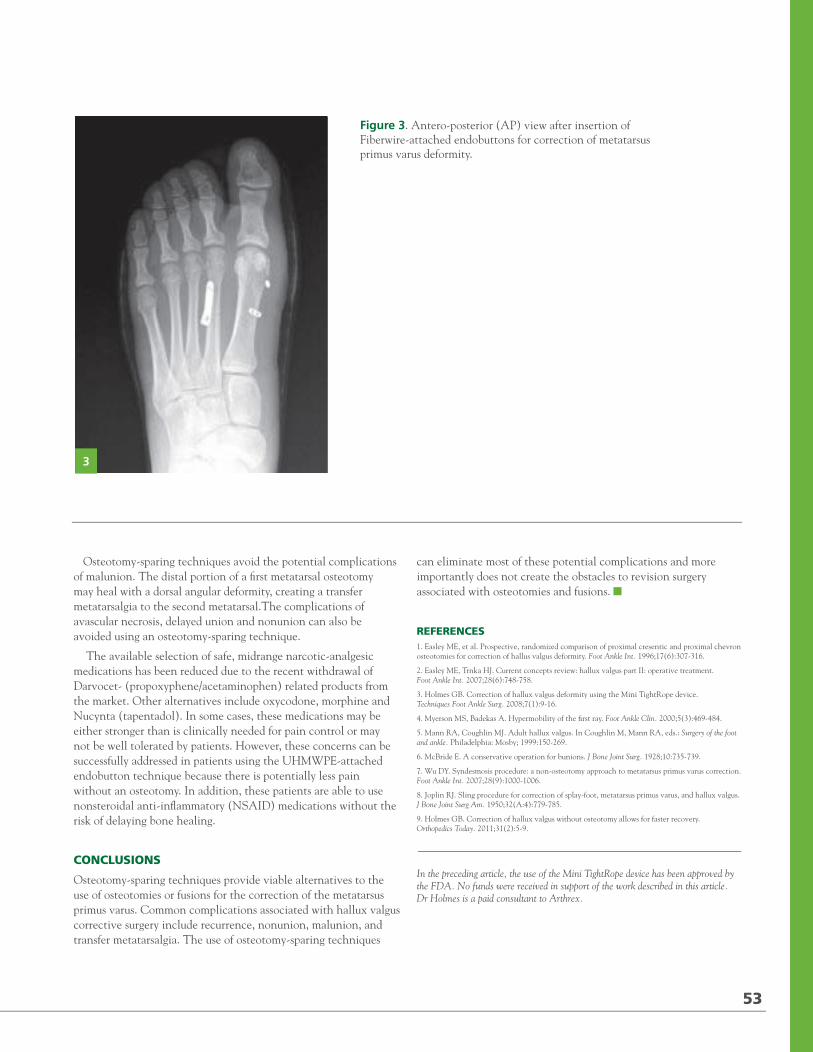
2012 / RUSH ORTHOPEDICS JOURNAL
Dysphagia Lusoria: An Uncommon Cause of Neck Pain and Hazard to Anterior Cervicothoracic Surgery
DOMINIC SCOLA, BS / SUDEEP BHABAD, MD / DAVID FARDON, MD
“Dysphagia lusoria, a source of incompletely diagnosed neck pain, is caused by an aberrant right subclavian artery, an anomaly that may require special planning for cervicothoracic spine surgery done through an anterior approach.”
ARTICLES
AUTHOR AFFILIATIONS
Department of Orthopedic Surgery (Dr Fardon), Department of Neuroradiology (Dr Bhabad), Rush University Medical Center; Rush University (Mr Scola), Chicago, Illinois.
CORRESPONDING AUTHOR
David Fardon, MD, Rush University Medical Center and Midwest Orthopaedics at Rush, 1611 W Harrison St, Suite 300, Chicago, IL 60612 ([email protected]).
When evaluating neck pain, the presence of degenerative or arthritic changes does not always identify the sole source of symptoms. Lymphadenopathy, thyroid
pathology, esophageal and paraesophageal disease, embryologic malformations, and referred pain from lung or heart disease should be considered as primary or concomitant secondary sources of pain.
Dysphagia may accompany neck pain because osteophytes efface the esophagus or because another disorder causes both neck pain and dysphagia. Dysphagia lusoria, a condition that may accompany neck pain, is due to an aberrant right subclavian artery (ARSA).Dysphagia lusoria should be in the differential diagnosis of a patient with anterior neck pain and dysphagia. Presence of ARSA and associated anomalies should be considered during preoperative
evaluation or in the event of unusual findings during anterior cervical and upper thoracic surgical procedures.
CASE REPORT
A 72-year-old woman had experienced neck pain for about 1 year. Her primary care doctor evaluated her for cardiorespiratory disease and performed computed tomography (CT) of the soft tissues of her neck, resulting in observations of mural calcification of the aorta and endplate anterior osteophytes at C5-6 and C6-7. The doctor advised the patient that she might have fibromyalgia, treated her with a serotonin uptake inhibitor, and referred her to the Rush orthopedic spine clinic for further evaluation and treatment of the cervical spondylosis that was thought to be the source of her symptoms.
The patient stated that her throat discomfort and upper anterior chest pain were mostly on the left side. Some pain was present while upright and at rest, but it was exacerbated by movement and by lying down. She recalled that, as a child, she had suffered a severe laceration of the right side of her neck, which resulted in copious bleeding and required surgical repair. She knew nothing else about the nature of the injury or the treatment and had no symptoms after the wound healed. Until she reached her 70s, she had enjoyed good health.
Physical exam showed the patient to be 5 ft, 7 in tall; 168 lb; and healthy in appearance. Her blood pressure was 110/70 mm/Hg in the
54
71795_Body.indd 54 7/25/12 9:39 PM
left arm and 130/72 mm/Hg in the right arm. Her radial pulses were normal. There was a long, oblique scar along the right anterolateral surface of her neck with no mass or abnormal pulsation either beneath it or in the supraclavicular fossa. Her neck motions were mildly limited. There were no signs of radiculopathy or myelopathy. On a pain drawing, she marked the left side of her neck, both anteriorly and posteriorly, and the manubrial area as sites of aching pain.
Cervical spine radiographs showed hypertrophic spondylosis along the anterior aspect of the mid-lower cervical spine. Magnetic resonance imaging (MRI) without contrast showed prominent osteophytes at the anterior disc margins and along
the ventral canal and uncovertebral joints of C5-C7, creating mild foraminal and central stenosis. The MRI also revealed that the esophagus was displaced anteriorly from the body of the first thoracic vertebra by a mass seen in axial projection to extend across the anterior surface of the vertebra and in sagittal projection to appear round, compatible with an anomalous vessel (Figure 1). Volumetric computer helical angiography of the chest before and after administration of 75.5 g iopamidol (10 0 mL of Isovue 370) showed an anomalous branching of the aortic arch, with the right and left carotid arteries arising from a common trunk (truncus bicaroticus), and the right and left subclavian arteries arising from a second common trunk. The right subclavian artery
A B
Figure 1. Magnetic resonance images showing the aberrant right subclavian artery (ARSA). A, Sagittal T2 weighted image of the cervical spine shows ARSA (arrow) immediately in front of the C7-T1 disc. There is spondylosis at C5-6 and C6-7 levels with anterior osteophytes. B, Axial T2-weighted image at the level of thoracic inlet shows ARSA (arrow) traversing left to right immediately in front of thoracic spine and posterior to the trachea (wedge) and esophagus, which is compressed between the trachea and the ARSA.
A B
Figure 2. Thoracic angiogram CT images showing an aberrant right subclavian artery (ARSA). A, Coronal reformatted image of CT thoracic angiogram indicating RSA (arrow). B, Axial image of CT thoracic angiogram showing ARSA (straight arrow) traversing left to right immediately in front of thoracic spine and posterior to the trachea and esophagus (curved arrow), which is compressed between the trachea and the ARSA.
55
71795_Body.indd 55 7/25/12 9:39 PM
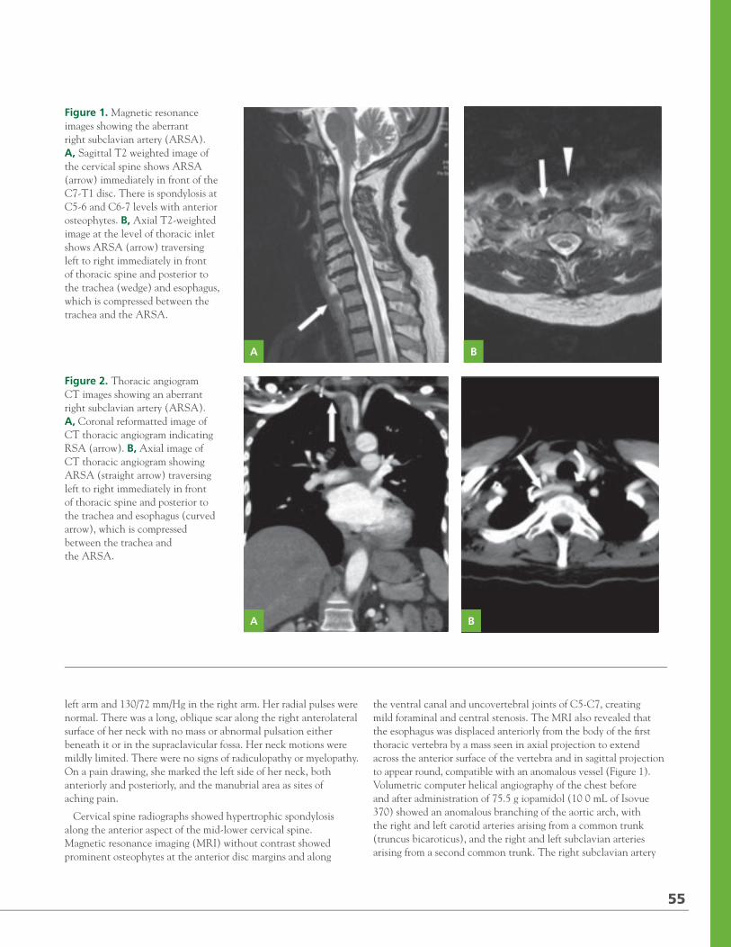
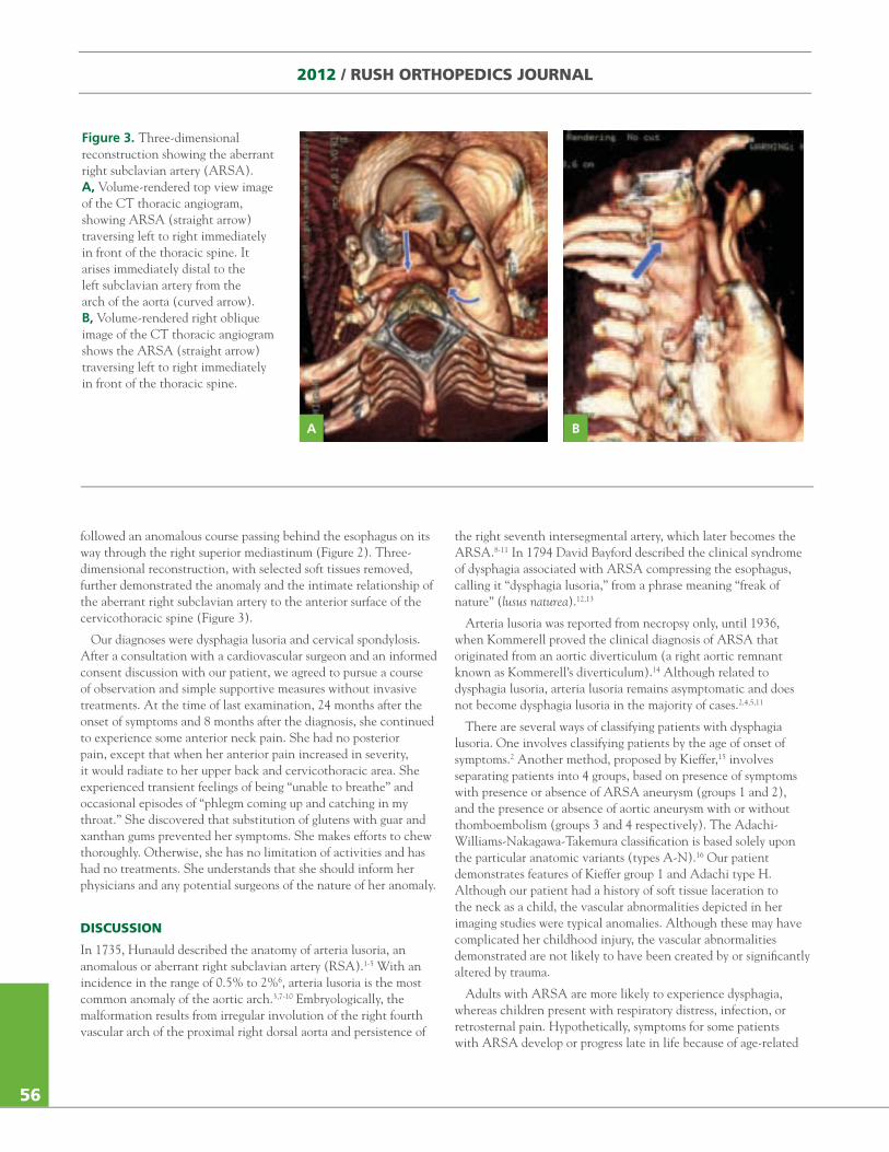
2012 / RUSH ORTHOPEDICS JOURNAL
followed an anomalous course passing behind the esophagus on its way through the right superior mediastinum (Figure 2). Three-dimensional reconstruction, with selected soft tissues removed, further demonstrated the anomaly and the intimate relationship of the aberrant right subclavian artery to the anterior surface of the cervicothoracic spine (Figure 3).
Our diagnoses were dysphagia lusoria and cervical spondylosis. After a consultation with a cardiovascular surgeon and an informed consent discussion with our patient, we agreed to pursue a course of observation and simple supportive measures without invasive treatments. At the time of last examination, 24 months after the onset of symptoms and 8 months after the diagnosis, she continued to experience some anterior neck pain. She had no posterior pain, except that when her anterior pain increased in severity, it would radiate to her upper back and cervicothoracic area. She experienced transient feelings of being “unable to breathe” and occasional episodes of “phlegm coming up and catching in my throat.” She discovered that substitution of glutens with guar and xanthan gums prevented her symptoms. She makes efforts to chew thoroughly. Otherwise, she has no limitation of activities and has had no treatments. She understands that she should inform her physicians and any potential surgeons of the nature of her anomaly.
DISCUSSION
In 1735, Hunauld described the anatomy of arteria lusoria, an anomalous or aberrant right subclavian artery (RSA).1-5 With an incidence in the range of 0.5% to 2%6, arteria lusoria is the most common anomaly of the aortic arch.3,7-10 Embryologically, the malformation results from irregular involution of the right fourth vascular arch of the proximal right dorsal aorta and persistence of
the right seventh intersegmental artery, which later becomes the ARSA.8-11 In 1794 David Bayford described the clinical syndrome of dysphagia associated with ARSA compressing the esophagus, calling it “dysphagia lusoria,” from a phrase meaning “freak of nature” (lusus naturea).12,13
Arteria lusoria was reported from necropsy only, until 1936, when Kommerell proved the clinical diagnosis of ARSA that originated from an aortic diverticulum (a right aortic remnant known as Kommerell’s diverticulum).14 Although related to dysphagia lusoria, arteria lusoria remains asymptomatic and does not become dysphagia lusoria in the majority of cases.2,4,5,11
There are several ways of classifying patients with dysphagia lusoria. One involves classifying patients by the age of onset of symptoms.2 Another method, proposed by Kieffer,15 involves separating patients into 4 groups, based on presence of symptoms with presence or absence of ARSA aneurysm (groups 1 and 2), and the presence or absence of aortic aneurysm with or without thomboembolism (groups 3 and 4 respectively). The Adachi-Williams-Nakagawa-Takemura classification is based solely upon the particular anatomic variants (types A-N).16 Our patient demonstrates features of Kieffer group 1 and Adachi type H. Although our patient had a history of soft tissue laceration to the neck as a child, the vascular abnormalities depicted in her imaging studies were typical anomalies. Although these may have complicated her childhood injury, the vascular abnormalities demonstrated are not likely to have been created by or significantly altered by trauma.
Adults with ARSA are more likely to experience dysphagia, whereas children present with respiratory distress, infection, or retrosternal pain. Hypothetically, symptoms for some patients with ARSA develop or progress late in life because of age-related
A B
Figure 3. Three-dimensional reconstruction showing the aberrant right subclavian artery (ARSA). A, Volume-rendered top view image of the CT thoracic angiogram, showing ARSA (straight arrow) traversing left to right immediately in front of the thoracic spine. It arises immediately distal to the left subclavian artery from the arch of the aorta (curved arrow). B, Volume-rendered right oblique image of the CT thoracic angiogram shows the ARSA (straight arrow) traversing left to right immediately in front of the thoracic spine.
56
71795_Body.indd 56 7/25/12 9:39 PM
increasing rigidity of the esophagus and/or the artery, and loss of elasticity of surrounding tissues.2 Changes in associated arterial malformations, such as in our patient with Adachi type H, may predispose to symptom progression.17
Saeed reviewed a “bowstring” phenomenon resulting from tautness of the artery across the esophagus with changes in posture.5 We postulated that bowstringing, along with loss of tissue elasticity, contributed to development of symptoms in our patient, though ordinary reflux phenomena could also explain why her symptoms are exacerbated by recumbence.
Osteophytes along the anterior surface of the spine, with or without concomitant soft tissue inflammation, can further displace and compromise the mobility of the esophagus, resulting in neck pain and dysphagia.18,19 Large anterior osteophytes at C5-C7 probably contributed to the onset of our patient’s symptoms. Because only 10% of adults with arteria lusoria have symptoms, including neck pain,20,21 careful search for other sources of cervical symptoms is always warranted in patients with the anomaly.
For patients presenting with neck pain, clinical symptoms that suggest the possibility of the presence of ARSA include dysphagia, stridor, cough, substernal pain, and pain with recumbence or other deviation from typical mechanically induced pain patterns. Radiographs and noncontrast CT are not likely to reveal the anomaly, but, as in our patient’s case, careful perusal of a cervical spine noncontrast MRI may reveal it. In patients who have undergone gastroenterologic work-up, the diagnosis may result from barium swallow if the test shows a typical diagonal impression on the esophagus or by endoscopic observation of a pulsatile mass. Confirming the diagnosis by digital subtraction CT angiography with contrast or by MRI angiography may, as in our case, reveal associated anomalies and facilitate subtyping.13,16,22 Three-dimensional color reconstructions, with deletion of obscuring tissues, clarifies the relationship between the anomalous vessels and contiguous tissues. Multidisciplinary evaluation may require a spine-care specialist, cardiovascular surgeon, gastroenterologist, laryngologist, radiologist, and primary care physician.
Surgical treatment of the anomaly, often complex and accompanied by significant risk, is usually reserved for younger patients with severe and/or progressive symptoms that are clearly related to the anomaly. For older patients with less severe symptoms, the best strategies to treat the dysphagia and the inflammatory and mechanical aspects of the pain include observation, food selection, and chewing precautions, and pharmacologic and physical therapeutic measures.
When planning an anterior approach to the cervical and upper thoracic spine, spinal surgeons should be aware that ARSAs and related anomalies present special hazards. Thorpe cited a case of death resulting from injury to an ARSA during anterior exposure of T2.23 Awkward exposures that challenge the surgeon working anteriorly at C7-T1 in a patient with a short and immobile neck could result in retraction or dissection of such precariously close arteries. The presence of very large anterior osteophytes, as in patients with diffuse idiopathic skeletal hyperostosis, or other extensive anterior pathology raises the risks imposed by the presence of ARSA. Reported associated anomalies relevant to the
surgeon include absence of a recurrent laryngeal nerve; anomalous branches of the vagus nerve, including direct branch to the larynx; right-sided thoracic duct; and anomalies of carotid and vertebral arteries.7,20
CONCLUSION
Dysphagia lusoria, a source of incompletely diagnosed neck pain, is caused by an aberrant right subclavian artery, an anomaly that may require special planning for cervicothoracic spine surgery done through an anterior approach. When considering surgery on patients with an ARSA, the surgeon should consider the challenges imposed by the aberrant artery, the possibility of associated anomalies, and the likelihood of difficult mobilization of the esophagus. These challenges include deciding whether or not to operate, choosing the approach (posterior, left anterior, or right anterior), and electing special monitoring and technique modifications. Surgeons should be aware of the possibility of the unrecognized presence of an ARSA and associated anomalies while exposing the anterior cervical and upper thoracic spine.
REFERENCES1. Kamiya H, Knobloch K, Lotz J, Bog A, Lichtenberg A, Hagl C, et al. Surgical treatment of aberrant right subclavian artery (arteria lusoria) aneurysm using three different methods. Ann Thorac Surg. 2006;82(1):187-190.
2. Atay Y, Engin C, Posacioglu H, Ozyurek R, Ozcan C, Yagdi T, et al. Surgical approaches to the aberrant right subclavian artery. Tex Heart Inst J. 2006;33(4):477-481.
3. Frank MW, Blakeman BP. Two-stage elephant trunk reconstruction for aneurysm of an aberrant right subclavian artery in association with aneurysmal distal aortic arch and descending thoracic aorta. Tex Heart Inst J. 2000;27(4):412-413.
4. Roland CF, Cherry KJ Jr. Symptomatic atherosclerotic stenotic disease of an aberrant right subclavian artery. Ann Vasc Surg.1991;5(2):196-198.
5. Saeed, G, Ganster G, Friedel N. Arteria lusoria aneurysm with truncus bicaroticus: surgical resection without restoring blood supply to the right arm. Tex Heart Inst J. 2010;37(5):602-607.
6. Epstein, DA, DeBord JR. Abnormalities associated with aberrant right subclavian arteries: a case report. Vasc Endovascular Surg. 2002;36(4):297-303.
7. Rodgers BM, Tabert JL, Hollenbeck JI. Aneurysm of anomalous subclavian artery: an unusual cause of dysphagia lusoria in childhood. Ann Surg.1978;187(2):158-160.
8. Bisognano JD, Young B, Brown JM, Gill EA, Fang FC, Zisman LS. Diverse presentation of aberrant origin of the right subclavian artery: two case reports. Chest.1997;112(6):1693-1697.
9. Duff SB, Hicks GL. Aortic arch dissection with an aberrant right subclavian artery: surgical treatment. Tex Heart Inst J. 1986;13(2):233-235.
10. Alvarez JR, Quiroga SJ, Nazar AB, Comendador MJ, Carro GJ. Aberrant right subclavian artery and calcified aneurysm of Kommerell’s diverticulum: an alternative approach. J Cardiothorac Surg. 2008;3:43.
11. Carrizo GJ, Marjani MA. Dysphagia lusoria caused by an aberrant right subclavian artery. Tex Heart Inst J. 2004;31(2):168-171.
12. Asherson N. David Bayford. His syndrome and sign of dysphagia lusoria. Ann R Coll Surg Engl. 1979;61:63-67.
13. Schouten van der Velden A, Berger P, Krasznai A, van Duijvendijk P, Adam van der Vliet J: Aberrant insertion of the right subclavian artery: an unusual cause of dysphagia in an adult. J Gastrointest Surg. 2009;13(11):2064–2067.
14. van Son JA, Konstantinov IE. Burckhard F. Kommerell and Kommerell’s diverticulum. Tex Heart Inst J. 2002;29(2):109-112.
15. Kieffer E, Bahnini A, Koskas F. Aberrant subclavian artery: surgical treatment in thirty-three adult patients. J Vasc Surg. 1994;19(1):100-111.
16. Rogers, AD, Nel M, Eloff EP, Naidoo NG. Dysphagia lusoria: a case of an aberrant right subclavian artery and a bicarotid trunk. ISRN Surg. 2011;(2011):819295. doi:10.5402/2011/819295.
17. Srinivas P, George J. Cervical osteoarthropathy: an unusual cause of dysphagia. Age Aging. 1999;28(3):321-325.
18. Lambert JR, Tepperman PS, Jimenez J, Newman A. Cervical spine disease and dysphagia. Four new cases and a review of the literature. Am J Gastroenterol. 1981; 76(1):35-40.
19. Uzunca K, Birtane M, Tezel A. Dysphagia induced by a cervical osteophyte: a case report of cervical spondylosis. Chin Med J (Engl). 2004;117(3):478-480.
20. Epstein, DA, DeBord JR. Abnormalities associated with aberrant right subclavian arteries: a case report. Vasc Endovascular Surg. 2002;36(4):297-303.
57
71795_Body.indd 57 7/25/12 9:39 PM
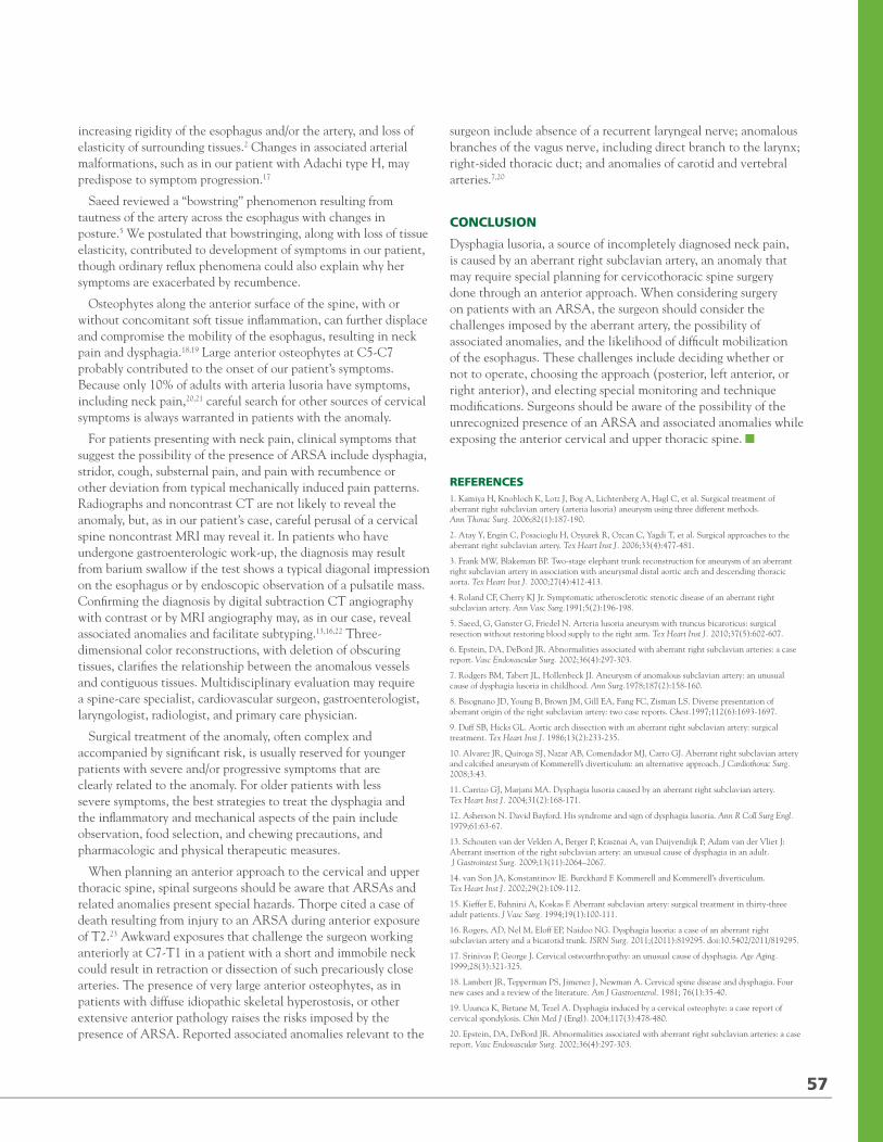
2012 / RUSH ORTHOPEDICS JOURNAL
21. Delap TG, Jones SE, Johnson DR. Aneurysm of an aberrant right subclavian artery presenting as dysphagia lusoria. Ann Otol Rhinol Laryngol. 2000;109(2):231-234.
22. Unlu Z, Orguc S, Eskiizmir G, Aslan A, Tasci S. The role of phonophoresis in dysphagia due to cervical osteophytes. Int J Gen Med. 2009;1:11-13.
23. Thorpe SW, Hohl JB, Gilbert S, Tannoury CA, Lee JY. Aberrant right subclavian artery encountered during debridement of T2 osteomyelitis and associated phlegmon. Spine J. 2011;11(2):e6-e10. doi:10.1016/j.spinee.2010.11.016.
ADDITIONAL REFERENCESAustin EH, Wolfe WG. Aneurysm of aberrant subclavian artery with a review of the literature. J Vasc Surg. 1985;2(4):571-577.
Brean HP, Neuhauser EBD. Syndrome of aberrant right subclavian artery with patent ductus arteriosus. Am J Roentgenol Rad Ther. 1947;58(6):708-716.
Bone RC, Nahum AM, Harris AS. Evaluation and correlation of dysphagia-producing cervical ostephytosis. Laryngoscope. 1974;84(11):2045-2050.
Cave EM, Virdi IS, Ruttley MS. Case report: An unusual case of the superior vena caval syndrome aneurysm of an aberrant right subclavian artery. Clin Radiol. 1994;49(11):834-837.
Chambers JL, Neale ML, Appleberg M. Fibromuscular hyperplasia in an aberrant subclavian artery and neurogenic thoracic outlet syndrome: an unusual combination. J Vasc Surg. 1994;20(5):834-838.
Engelman RM, Madayag M. Aberrant right subclavian artery aneurysm: a rare cause of a superior mediastinal tumor. Chest. 1972;62(1):45-47.
Esposito RA, Khalil I, Galloway AC, et al. Surgical treatment for aneurysm of aberrant subclavian artery based on a case report and a review of the literature. J Thorac Cardiovasc Surg.1988;95(5):888-891.
Gross RE. Surgical treatment for dysphagia lusoria. Ann Surg. 1946;124(3):532-534.
Hartyanszky IL, Lozsadi K, Marcsek P, Huttl T, Sapi E, Kovacs AB. Congenital vascular rings: surgical management of 111 cases. Eur J Cardiothorac Surg. 1989;3(3):250-254.
Jelkunen H, Heindnen O, Pyörälä K. Hyperostosis of the spine in an adult population. Its relation to hyperglycaemia and obesity. Ann Rheum Dis. 1971;30(6):605-612.
Karadag B, Cat H, Aksoy S, Ozulu B, Ozturk AO, Oguz S, Altuntas Y. A geriatric patient with diffuse idiopathic skeletal hyperostosis. World J Gastroenterol. 2010;16(13):1673-1675.
Klinkhamer AC. Aberrant right subclavian artery. Clinical and roentgenologic aspects. Am J Roentgenol Radium Ther Nucl Med. 1966;97(2):438-446.
Klinkhamer AC. Het vaststellen van aberrante arterien in het mediastinum superius door middel van oesophagogram [thesis]. Ned Tijdschr Geneeskd. 1963;107:510.
Mok CK, Cheung KL, Kong SM, et al. Translocating the aberrant right subclavian artery in dysphagia lusoria. Br J Surg. 1979;66(2):113-116.
Murzi M, Mariani M, Tiwari KK, Farneti P, Berti S, Karimov JH, Glauber M. Aberrant right subclavian artery aneurysm in coexistence with a common carotid trunk. Ann Thorac Surg. 2009;88(1):e8.
Nathan H, Seidel MR. The association of a retroesophageal right subclavian artery, a right-sided terminating thoracic duct, and a left vertebral artery of aortic origin: Anatomical and clinical considerations. Acta Anat. 1983;117(4):362-373.
Resnick D, Niwayama G. Diagnosis of Bone and Joint Disorders. Philadelphia, PA: WB Saunders; 1982.
Rotes-Querol, J, Balmes C. Clinical manifestations of diffuse idiopathic skeletal hyperostosis: editorial. Br J Rheumatol. 1996;35:1193-1196.
Sanders G, Uyeda RY, Karian MS. Nonrecurrent inferior laryngeal nerves and their association with a recurrent branch. Am J Surg. 1983;146(4):501-503.
Stone WM, Brewster DC, Moncure AC, et al. Aberrant right subclavian artery: varied presentations and management options. J Vasc Surg. 2002;11(6):812-817.
58
The preceding article does not discuss the use of medical devices or drugs that are regulated by the FDA. No funds were received in support of this work. None of the authors receive financial benefits from commercial sources.
In “Radial Head Replacement With a Bipolar System,” beginning on p. 47, the Katalyst device has been approved by the FDA for the described use. No funds were received in support of the work. Authors Zunkiewicz, Clemente, and Miller have no financial disclosures. Dr Wysocki is a paid consultant for Acumed. Drs Cohen and Baratz are paid consultants and receive royalties from Integra Life Sciences.
71795_Body_u1.indd 58 8/3/12 10:30 AM
What Volume of Bone Graft Is Needed for Predictable Fusion in Single-Level Lumbar Arthrodesis? A Preliminary Investigation
SAFDAR N. KHAN, MD / KEVIN U. PARK, MD / SHAH DODWAD, MD / THOMAS CHA, MD / HOWARD S. AN, MD
“…We believe that predictable fusion in instrumented posterolateral arthrodesis for single-level degenerative spondylolisthesis can be achieved by meticulous fusion-bed preparation with local bone graft placed prior to pedicle screw/rod placement at a minimum volume of 15 cc per side.”
ARTICLES
AUTHOR AFFILIATIONS
Department of Orthopaedic Surgery, The Ohio State University Medical Center, Columbus, Ohio (Drs Dodwad and Khan); Department of Orthopedic Surgery, Rush University Medical Center (Drs Park and An) and Midwest Orthopaedics at Rush, (Dr An) Chicago, Illinois; and the Department of Orthopaedic Surgery, Massachusetts General Hospital, Boston, Massachusetts (Dr Cha).
CORRESPONDING AUTHOR
Howard S. An, MD, Rush University Medical Center andMidwest Orthopaedics at Rush, 1611 W Harrison St, Suite300, Chicago, IL 60612 ([email protected]).
There has been increasing interest in alternatives to autogenous bone graft for patients undergoing instrumented spinal fusion surgery. Reducing the need to harvest the
patient’s own bone from iliac crest diminishes problems with pain and potential complications associated with the additional procedure. By introducing proper “ingredients” (osteoinduction/osteoconduction/osteogenic cells), a bone-graft substitute may more predictably lead to fusion.1,2
Single-level instrumented posterolateral spinal fusion for degenerative spondylolisthesis is a common procedure performed by spinal surgeons. What constitutes the optimal mechano-biological
environment has been the subject of intense debate. The best bone-graft substitute to supplement rigid instrumentation is still unclear.
Autogenous iliac crest bone graft has been used with great success but carries the risks mentioned previously. Various studies have divided the complications of autogenous iliac crest harvest into major and minor complications. Minor complications (approximately 10%) include superficial infections, superficial seromas, and minor hematomas. Major complications (approximately 6%) include herniation of abdominal contents through massive bone-graft donor sites, vascular injuries, deep infections at the donor site, neurologic injuries (cluneal nerve damage), deep hematoma formation requiring surgical intervention, and iliac wing fractures.3,4
Several alternatives to autogenous iliac graft have been introduced with varying degrees of success. Bone-graft extenders, such as corticocancellous allograft, or ceramics, such as hydroxyapatite-tricalcium phosphate, have been used as bulking agents to add to the volume of local bone graft. Bone-graft enhancers, such as demineralized bone matrix (DBM) and DBM-stem cell composites, are alternatives that have met with recent criticism.5 More data are needed to draw any firm clinical conclusions about their performance. Finally, bone-graft substitutes such as recombinant bone morphogenetic proteins (rhBMP-2/INFUSE Bone Graft/Medtronic Sofamor Danek, Memphis, Tennessee) were introduced a decade ago as effective
59
71795_Body.indd 59 7/25/12 9:39 PM
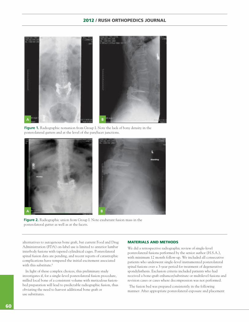
2012 / RUSH ORTHOPEDICS JOURNAL
alternatives to autogenous bone graft, but current Food and Drug Administration (FDA) on-label use is limited to anterior lumbar interbody fusions with tapered cylindrical cages. Posterolateral spinal fusion data are pending, and recent reports of catastrophic complications have tempered the initial excitement associated with this substitute.6
In light of these complex choices, this preliminary study investigates if, for a single-level posterolateral fusion procedure, milled local bone of a consistent volume with meticulous fusion-bed preparation will lead to predictable radiographic fusion, thus obviating the need to harvest additional bone graft or use substitutes.
MATERIALS AND METHODS
We did a retrospective radiographic review of single-level posterolateral fusions performed by the senior author (H.S.A.), with minimum 12 month follow-up. We included all consecutive patients who underwent single-level instrumented posterolateral spinal fusions over a 3-year period for treatment of degenerative spondylisthesis. Exclusion criteria included patients who had received a bone-graft enhancer/substitute or multilevel fusions and revision cases or cases where decompression was not performed.
The fusion bed was prepared consistently in the following manner: After appropriate posterolateral exposure and placement
A B
Figure 1. Radiographic nonunion from Group I. Note the lack of bony density in the posterolateral gutters and at the level of the pars/facet junctions.
A B
Figure 2. Radiographic union from Group I. Note exuberant fusion mass in the posterolateral gutter as well as at the facets.
60
71795_Body.indd 60 7/25/12 9:39 PM
of retractors, a thorough decompression was performed. All decompression-related bone (laminectomy bone from Kerrisons/bone dust collected from suction trap device) was saved, meticulously cleaned of soft tissue with a #15 blade scalpel, and milled with a commercially available bone mill. The final volume of local bone was measured, and we prepared the pedicles for pedicle screw placement. Using a gear shift, the pedicles were probed and tapped, and screw trajectory was confirmed with markers using biplanar fluoroscopy. A matchstick burr was then used to completely decorticate the segmental facet joints and create a bilateral trough from pars to pars that was wide enough for manual insertion of bone graft. The transverse processes were also decorticated and, depending on the volume of local bone graft available, the decorticated trough/pars/transverse process beds were packed with the graft. We determined the screw entry point, and placed the pedicle screws. We inserted appropriately sized rods, tightened sets screws, and took final radiographs. Wound closure was performed in the standard fashion.
We divided our series into the following groups:
Group I: Patients with at least 30 cc of local bone graft placed at the fusion bed.
Group II: Patients with less than 30 cc of local bone to which an additional 15-30 cc of corticocancellous allograft chips were added as a bulking agent. Total volume of graft and additive was no less than 15 cc on each side.
Two sets of blinded reviewers assessed the available radiographs at final follow-up. The reviewers included fellowship-trained spinal surgeons as well as midlevel orthopedic surgery residents. A preliminary set of 10 radiographs was used between blinded reviewers to set an equivalent standard for fusion criteria. The criteria used to determine fusion included intertransverse bridging bone, fusion radiodensity over the facet joints, continued segmental motion at the fusion site and any evidence of instrumentation loosening or failure. Attempted fusions were deemed either fused or unfused. All results were tabulated and analyzed.
RESULTS
A total of 40 consecutive patients met our inclusion criteria and had radiographs that were available for clinical review in this preliminary study (Group I: n = 20, Group II: n = 20). Radiographic review revealed 18 of the 20 Group I patients and 18 of the 20 Group II patients was radiographically fused. See Figures 1 and 2 for examples of radiographic union and nonunion. Final follow-up clinical notes did not indicate any complications including hardware loosening or revisions secondary to nonunions.
DISCUSSION
The bone graft that most predictably leads to fusion for single-level instrumented posterolateral spinal arthrodesis is still debatable. We maintain that both the ingredients of the bone-graft substitute and the preparation of the fusion bed contribute to the predictability of the fusion. Each patient in our series underwent the same preparation of the fusion bed. A key point in our series is that we prepared the fusion bed and packed bone graft prior to placing the rigid pedicle screw-rod construct. We believe that the predictability
of our fusions (as determined by final radiographic evidence of arthrodesis with no clinical evidence of nonunion) is directly related to this method. Once the pedicle screws/rods are placed, the biologic “real estate” for packing bone graft (whether autograft or composite grafts) severely diminishes. Packing 15 cc of bone graft per side within the fresh, bleeding cancellous surface of the trough made within the pars-facet-pars segment leads to a better biologic fusion environment than attempting to place bone graft in the posterolateral gutter after the pedicle screws/rods have been placed.
From a cost perspective, this preliminary study has some important considerations. In Group I, we achieved 80% radiographic fusion without the additional cost of using a bone-graft enhancer such as a DBM or even a ceramic bulking agent. Additionally in Group II, we achieved comparable radiographic fusion with the use of corticocancellous allograft chips, which are vastly cheaper than commercially available DBM products or osteoinductive proteins.
Our method of posterolateral fusion also differs in that it is not a true intertransverse process fusion. Our primary focus was to prepare the facet and pars at the motion segment in question and pack as much bone graft as possible within the surgically created trough. The remaining bone graft was then manually packed along the decorticated transverse processes.
This study is not without methodological drawbacks. It was a retrospective review, and we did not have computed tomography to assess the fusions. In addition we did not include iliac crest bone-graft group or a rhBMP-2 group as a positive control. As a preliminary study, the numbers were too small to reveal statistical significance of differences between groups in this study and between our data and other published results. However, our intent was to demonstrate equivalency in radiographic fusion rates by using a set amount of local autogenous bone graft alone in single-level lumbar fusion, rather than to show a statistical difference between two groups.
In conclusion, we believe that predictable fusion in instrumented posterolateral arthrodesis for single-level degenerative spondylolisthesis can be achieved by meticulous fusion-bed preparation with local bone graft placed prior to pedicle screw/rod placement at a minimum volume of 15 cc per side.
REFERENCES1. Agarwal R et al. Osteoinductive bone graft substitutes for lumbar fusion: a systematic review. J Neurosurg Spine. 2009;11(6):729-740.
2. Rihn J et al. Graft options in posterolateral and posterior interbody lumbar fusion. Spine. 2010;35(17):1629-1639.
3. Mazock JB et al. Posterior iliac crest bone harvest: review of technique, complications, and use of an epidural catheter for postoperative pain control. J Oral Maxillofac Surg. 2003;61(12):1497-1503.
4. Meyeroff C et al. Autogenous bone graft: donor sites and techniques. J Bone Joint Surg Am. 2011;93(23):2227-2236.
5. Epstein NE. Efficacy of different bone volume expanders for augmenting lumbar fusions. Surg Neurol. 2008;69(1):16-19; discussion 19.
6. Carragee EJ et al. Retrograde ejaculation after anterior lumbar interbody fusion using rhBMP-2: a cohort controlled study. Spine J. 2011;11(6):511-516.
In the preceding article, FDA approval status of described biomaterials is discussed in the text. No funds were received in support of this work. None of the authors receive financial benefits from commercial sources.
61
71795_Body.indd 61 7/25/12 9:39 PM
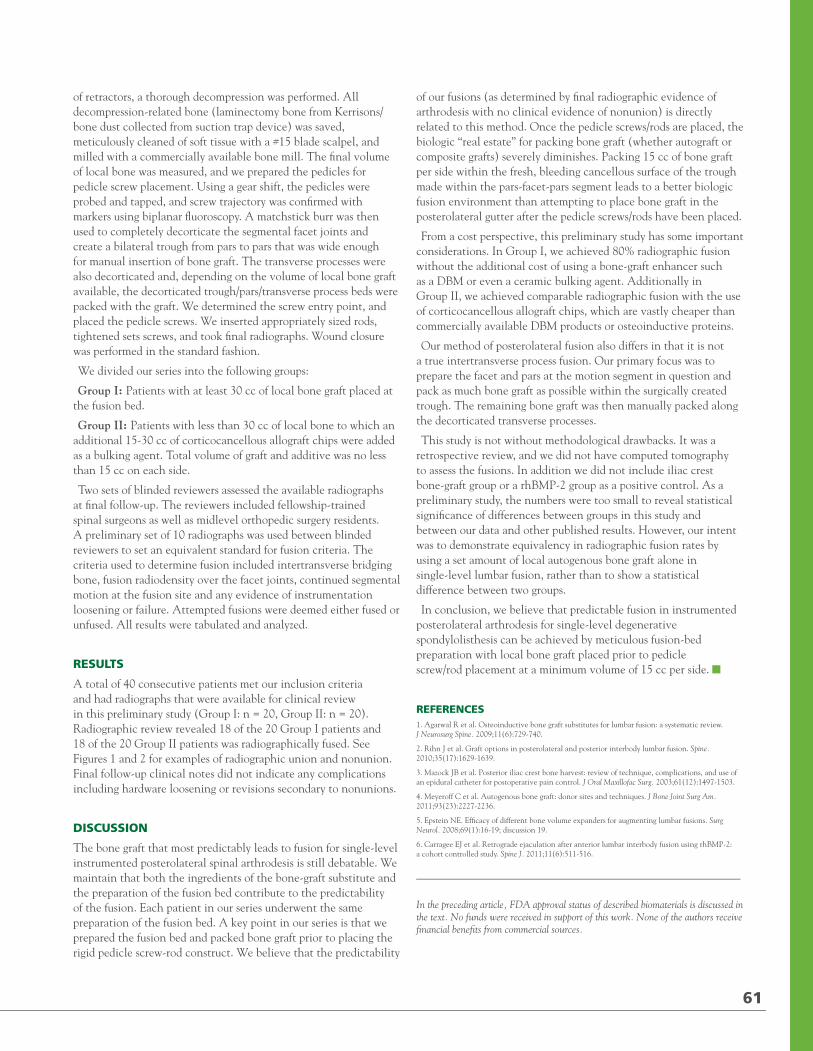
2012 / RUSH ORTHOPEDICS JOURNAL
PUBLICATIONS (2011)*
Publications, Research Grants, and Kappa Delta Orthopedic Research Awards
AN, HOWARD S.
An HS, Masuda K, Cs-Szabo G, Zhang Y, Chee A, Andersson GB, Im HJ, Thonar EJ, Kwon YM. Biologic repair and regeneration of the intervertebral disc. J Am Acad Orthop Surg. 2011;19(7): 450-452.
Kim JS, Kroin JS, Li X, An HS, Buvanendran A, Yan D, Tuman KJ, van Wijnen AJ, Chen D, Im HJ. The rat intervertebral disc degeneration pain model: relationships between biological and structural alterations and pain. Arthritis Res Ther. 2011;13(5):R165.
Zhang Y, Drapeau S, An HS, Markova D, Lenart BA, Anderson DG. Histological features of the degenerating intervertebral disc in a goat disc-injury model. Spine. 2011;36(19):1519-1527.
ANDERSSON, GUNNAR B. J.
Karppinen J, Shen FH, Luk KD, Andersson GB, Cheung KM, Samartzis D. Management of degenerative disc disease and chronic low back pain. Orthop Clin North Am. 2011;42(4):513-528, viii.
BACH, JR, BERNARD R.
Hsu AR, Ghodadra NS, Provencher MT, Lewis PB, Bach BR Jr. Biceps tenotomy versus tenodesis: a review of clinical outcomes and biomechanical results. J Shoulder Elbow Surg. 2011;20(2): 326-332.
Kang RW, Strauss EJ, Barker JU, Bach BR Jr. Effect of donor age on bone mineral density in irradiated bone-patellar tendon-bone allografts of the anterior cruciate ligament. Am J Sports Med. 2011;39(2):380-383.
Piasecki DP, Bach BR Jr, Espinoza Orías AA, Verma NN. Anterior cruciate ligament reconstruction: can anatomic femoral placement be achieved with a transtibial technique? Am J Sports Med. 2011;39(6):1306-1315.
Strauss EJ, Salata MJ, Kercher J, Barker JU, McGill K, Bach BR Jr, Romeo AA,Verma NN. Multimedia article. The arthroscopic management of partial-thickness rotator cuff tears: a systematic review of the literature. Arthroscopy. 2011;27(4):568-580.
BUSH-JOSEPH, CHARLES
Strauss EJ, Kang R, Bush-Joseph C, Bach BR Jr. The diagnosis and management of spontaneous and post-arthroscopy osteonecrosis of the knee. Bull NYU Hosp Jt Dis. 2011;69(4):320-330.
COHEN, MARK S.
Shukla SK, Cohen MS. Symptomatic medial epicondyle nonunion: treatment by open reduction and fixation with a tension band construct. J Shoulder Elbow Surg. 2011;20(3):455-460.
COLE, BRIAN J.
Anderson AF, Irrgang JJ, Dunn W, Beaufils P, Cohen M, Cole BJ, Coolican M, Ferretti M, Glenn RE Jr, Johnson R, Neyret P, Ochi M, Panarella L, Siebold R, Spindler KP, Ait Si Selmi T, Verdonk P, Verdonk R, Yasuda K, Kowalchuk DA. Interobserver reliability of the International Society of Arthroscopy, Knee Surgery and Orthopaedic Sports Medicine (ISAKOS) classification of meniscal tears. Am J Sports Med. 2011;39(5):926-932.
Batty L, Dance S, Bajaj S, Cole BJ. Autologous chondrocyte implantation: an overview of technique and outcomes. ANZ J Surg. 2011;81(1-2):18-25.
Cole BJ, Farr J, Winalski CS, Hosea T, Richmond J, Mandelbaum B, De Deyne PG. Outcomes after a single-stage procedure for cell-based cartilage repair: a prospective clinical safety trial with 2-year follow-up. Am J Sports Med. 2011;39(6):1170-1179.
62
*This is a partial list of published works for the faculty members of the Department of Orthopedic Surgery at Rush in 2011. Works featuring more than 1 physician from Rush are listed under the first physician from Rush to appear in the citation. Works with electronic publication dates in 2011 and print publication dates in 2012 are not included in this list. Although only faculty members are cited, the department gratefully acknowledges the co-authorship of students, nurses, practitioners, therapists, residents, fellows, and colleagues at Rush. Source: PubMed.
71795_Body.indd 62 7/25/12 9:39 PM
Daley EL, Bajaj S, Bisson LJ, Cole BJ. Improving injection accuracy of the elbow, knee, and shoulder: does injection site and imaging make a difference? A systematic review. Am J Sports Med. 2011;39(3):656-662.
Encalada-Diaz I, Cole BJ, Macgillivray JD, Ruiz-Suarez M, Kercher JS, Friel NA, Valero-Gonzalez F. Rotator cuff repair augmentation using a novel polycarbonate polyurethane patch: preliminary results at 12 months’ follow-up. J Shoulder Elbow Surg. 2011;20(5):788-794.
Fortier LA, Barker JU, Strauss EJ, McCarrel TM, Cole BJ. The role of growth factors in cartilage repair. Clin Orthop Relat Res. 2011;469(10):2706-2715.
Provencher MT, Kercher JS, Galatz LM, Elattrache NS, Frank RM, Cole BJ. Evolution of rotator cuff repair techniques: are our patients really benefiting? Instr Course Lect. 2011;60:123-136.
Strauss EJ, Barker JU, McGill K, Cole BJ, Bach BR Jr, Verma NN. Can anatomic femoral tunnel placement be achieved using a transtibial technique for hamstring anterior cruciate ligament reconstruction? Am J Sports Med. 2011;39(6):1263-1269.
Sundman EA, Cole BJ, Fortier LA. Growth factor and catabolic cytokine concentrations are influenced by the cellular composition of platelet-rich plasma. Am J Sports Med. 2011;39(10):2135-2140.
DELLA VALLE, CRAIG J.
Bedair H, Ting N, Bozic KJ, Della Valle CJ, Sporer SM. Treatment of early postoperative infections after THA: a decision analysis. Clin Orthop Relat Res. 2011;469(12):3477-3485.
Bedair H, Ting N, Jacovides C, Saxena A, Moric M, Parvizi J, Della Valle CJ. The Mark Coventry Award: diagnosis of early postoperative TKA infection using synovial fluid analysis. Clin Orthop Relat Res. 2011;469(1):34-40.
Del Gaizo DJ, Della Valle CJ. Instability in primary total knee arthroplasty. Orthopedics. 2011;34(9):e519-e21.
Della Valle C, Parvizi J, Bauer T et. al. AAOS Clinical practice guideline: The diagnosis of periprosthetic joint infection of the hip and knee. J Bone Joint Surg. 2011;93:1355-1357.
Jacofsky DJ, Della Valle CJ, Meneghini RM, Sporer SM, Cercek RM. Revision total knee arthroplasty: what the practicing orthopaedic surgeon needs to know. Instr Course Lect. 2011;60:269-281.
Miller M, Brown NB, Della Valle C, Rosenberg AG, Galante JO. Posterior cruciate retaining total knee arthroplasty in patients with rheumatoid arthritits: a concise follow-up of a previous report at a mnimum of 20 years. J Bone Joint Surg. 2011;93:e130.
Koyonos L, Zmistowski B, Della Valle CJ, Parvizi J. Infection control rate of irrigation and débridement for periprosthetic joint infection. Clin Orthop Relat Res. 2011;469(11):3043-3048.
Nunley RM, Ruh EL, Zhang Q, Della Valle CJ, Engh CA Jr, Berend ME, Parvizi J, Clohisy JC, Barrack RL. Do patients return to work after hip arthroplasty surgery. J Arthroplasty. 2011;26(6)(suppl):92-98.e3.
Parvizi J, Zmistowski B, Berbari EF, Bauer TW, Springer BD, Della Valle CJ, Garvin KL, Mont MA, Wongworawat MD, Zalavras CG. New definition for periprosthetic joint infection: from the Workgroup of the Musculoskeletal Infection Society. Clin Orthop Relat Res. 2011;469(11):2992-2994.
Sah AP, Shukla S, Della Valle CJ, Rosenberg AG, Paprosky WG. Modified hybrid stem fixation in revision TKA is durable at 2 to 10 years. Clin Orthop Relat Res. 2011;469(3):839-836.
Squire MW, Della Valle CJ, Parvizi J. Preoperative diagnosis of periprosthetic joint infection: role of aspiration. AJR Am J Roentgenol. 2011;196(4):875-879.
Van Thiel GS, Berend KR, Klein GR, Gordon AC, Lombardi AV, Della Valle CJ. Intraoperative molds to create an articulating spacer for the infected knee arthroplasty. Clin Orthop Relat Res. 2011;469(4):994-1001.
ESPINOZA ORÍAS, ALEJANDRO, A.
Bhatia A, Albazzaz M, Espinoza Orías AA, Inoue N, Miller LM, Acerbo A, George A, Sumner DR. Overexpression of DMP1 accelerates mineralization and alters cortical bone biomechanical properties in vivo. J Mech Behav Biomed Mater. 2012;5(1):1-8.
Hong JT, Takigawa T, Udayakunmar R, Shin HK, Simon P, Espinoza Orías AA, Inoue N, An HS. Biomechanical effect of the C2 laminar decortication on the stability of C2 intralaminar screw construct and biomechanical comparison of C2 intralaminar screw and C2 pars screw. Neurosurgery. 2011;69(1) (suppl operative):ons1-7.
Hong JT, Tomoyuki T, Udayakumar R, Espinoza Orías AA, Inoue N, An HS. Biomechanical comparison of three different types of C7 fixation techniques. Spine. 2011;36(5):393-398.
Rudy DJ, Deuerling JM, Espinoza Orías AA, Roeder RK. Anatomic variation in the elastic inhomogeneity and anisotropy of human femoral cortical bone tissue is consistent across multiple donors. J Biomech. 2011;44(9):1817-1820.
FOUCHER, KHARMA C.
Foucher KC, Wimmer MA, Moisio KC, Hildebrand M, Berli MC, Walker MR, Berger RA, Galante JO. Time course and extent of functional recovery during the first postoperative year after minimally invasive total hip arthroplasty with two different surgical approaches—a randomized controlled trial. J Biomech. 2011;44(3):372-378.
63
71795_Body.indd 63 7/25/12 9:39 PM
2012 / RUSH ORTHOPEDICS JOURNAL
GITELIS, STEVEN
Kang RW, McGill KC, Lin J, Gitelis S. Chronic ankle pain and swelling in a 25-year-old woman: an unusual case. Clin Orthop Relat Res. 2011;469(5):1517-1521.
GLANT, TIBOR T.
Glant TT, Radacs M, Nagyeri G, Olasz K, Laszlo A, Boldizsar F, Hegyi A, Finnegan A, Mikecz K. Proteoglycan-induced arthritis and recombinant human proteoglycan aggrecan G1 domain-induced arthritis in BALB/c mice resembling two subtypes of rheumatoid arthritis. Arthritis Rheum. 2011;63(5):1312-1321.
Hamel KM, Cao Y, Ashaye S, Wang Y, Dunn R, Kehry MR, Glant TT, Finnegan A. B cell depletion enhances T regulatory cell activity essential in the suppression of arthritis. J Immunol. 2011;187(9):4900-4906.
Nagyeri G, Radacs M, Ghassemi-Nejad S, Tryniszewska B, Olasz K, Hutas G, Gyorfy Z, Hascall VC, Glant TT, Mikecz K. TSG-6 protein, a negative regulator of inflammatory arthritis, forms a ternary complex with murine mast cell tryptases and heparin. J Biol Chem. 2011;286(26):23559-23569.
Nesterovitch AB, Hoffman MD, Simon M, Petukhov PAT, Glant TT. Mutations in PSTPIP1 gene and aberrant splicing variant in patients with pyoderma gangrenosum. Clin Exp Dermatol 2011(8);36:889-895.
HALLAB, NADIM J.
Mishra PK, Wu W, Rozo C, Hallab NJ, Benevenia J, Gause WC. Micrometer-sized titanium particles can induce potent Th2-type responses through TLR4-independent pathways. J Immunol. 2011;187(12):6491-6498.
Orhue V, Kanaji A, Caicedo MS, Virdi AS, Sumner DR, Hallab NJ, Jahr H, Sena K. Calcineurin/nuclear factor of activated T cells (NFAT) signaling in cobalt-chromium-molybdenum (CoCrMo) particles-induced tumor necrosis factor-ʼ (TNFʼ) secretion in MLO-Y4 osteocytes. J Orthop Res. 2011;29(12):1867-1873.
HAMMERBERG, KIM W.
O’Leary PT, Sturm PF, Hammerberg KW, Lubicky JP, Mardjetko SM. Convex hemiepiphysiodesis: the limits of vertebral stapling. Spine. 2011;36(19):1579-1583.
INOUE, NOZOMU
Hioki A, Miyamoto K, Shimizu K, Inoue N. Test-retest repeatability of lumbar sagittal alignment and disc height measurements with or without axial loading: a computed tomography study. J Spinal Disord Tech. 2011;24(2):93-8.
Hong JT, Takigawa T, Sugisaki K, Orías AA, Inoue N, An HS. Biomechanical and morphometric evaluation of occipital
condyle for occipitocervical segmental fixation. Neurol Med Chir. 2011;51(10):701-706.
Imai K, Ikoma K, Maki M, Kido M, Tsuji Y, Takatori R, Tokunaga D, Inoue N, Kubo T. Features of hindfoot 3D kinetics in flat foot in ankle-joint maximal dorsiflexion and plantarflexion. J Orthop Sci. 2011;16(5):638-643.
Inoue N, Espinoza Orías AA. Biomechanics of intervertebral disc degeneration. Orthop Clin North Am. 2011;42(4):487-499, vii.
JACOBS, JOSHUA J.
Bedair H, Berli M, Gezer S, Jacobs JJ, Della Valle CJ. Hematologic genetic testing in high-risk patients before knee arthroplasty: a pilot study. Clin Orthop Relat Res. 2011;469(1):131-137.
Gallardo LA, Knowlton CB, Kunze J, Jacobs JJ, Wimmer MA, Laurent MP. Effect of europium(II) stearate on the mechanical properties and the oxidation resistance of UHMWPE. J Mech Behav Biomed Mater. 2011;4(5):821-826.
Guyer RD, Shellock J, MacLennan B, Hanscom D, Knight RQ, McCombe P, Jacobs JJ, Urban RM, Bradford D, Ohnmeiss DD. Early failure of metal-on-metal artificial disc prostheses associated with lymphocytic reaction: diagnosis and treatment experience in four cases. Spine. 2011;36(7):e492-e497.
Mont MA, Jacobs JJ. AAOS clinical practice guideline: preventing venous thromboembolic disease in patients undergoing elective hip and knee arthroplasty. J Am Acad Orthop Surg. 2011;19(12): 777-778.
Mont MA, Jacobs JJ, Boggio LN, Bozic KJ, Della Valle CJ, Goodman SB, Lewis CG, Yates AJ Jr, Watters WC III, Turkelson CM, Wies JL, Donnelly P, Patel N, Sluka P. Preventing venous thromboembolic disease in patients undergoing elective hip and knee arthroplasty. J Am Acad Orthop Surg. 2011;19(12):768-776.
LAURENT, MICHEL
Gallardo LA, Carpentieri I, Laurent MP, Costa L, Wimmer MA. Europium stearate additives delay oxidation of UHMWPE for orthopaedic applications: a pilot study. Clin Orthop Relat Res. 2011;469(8):2294-2301.
Laurent MP, Pourzal R, Fischer A, Bertin KC, Jacobs JJ, Wimmer MA. In vivo wear of a squeaky alumina-on-alumina hip prosthesis: a case report. J Bone Joint Surg Am. 2011;93(7):e27.
LEVINE, BRETT
Levine BR, Ting N, Della Valle CJ. Use of a barbed suture in the closure of hip and knee arthroplasty wounds. Orthopedics. 2011;34(9):e473-e475.
MAKI, CARL
Shen H, Maki CG. Pharmacologic activation of p53 by small-molecule MDM2 antagonists. Curr Pharm Des. 2011;17(6):560-568.
64
71795_Body.indd 64 7/25/12 9:39 PM
MIKECZ, KATALIN
Li J, Anemaet W, Diaz MA, Buchanan S, Tortorella M, Malfait AM, Mikecz K, Sandy JD, Plaas A. Knockout of ADAMTS5 does not eliminate cartilage aggrecanase activity but abrogates joint fibrosis and promotes cartilage aggrecan deposition in murine osteoarthritis models. J Orthop Res. 2011;29(4):516-522.
Nesterovitch AB, Szanto S, Gonda A, Bardos T, Kis-Toth K, Adarichev VA, Olasz K, Ghassemi-Najad S, Hoffman MD, Tharp MD, Mikecz K, Glant TT. Spontaneous insertion of a B2 element in the PTPN6 gene drives a systemic autoinflammatory disease in mice resembling neutrophilic dermatosis in humans. Am J Pathol. 2011;178(4):1701-1714.
Yan D, Chen D, Cool SM, van Wijnen AJ, Mikecz K, Murphy G, Im HJ. Fibroblast growth factor receptor 1 is principally responsible for fibroblast growth factor 2-induced catabolic activities in human articular chondrocytes. Arthritis Res Ther. 2011;13(4):R130.
NATARAJAN, RAGHU N.
Hussain M, Nassr A, Natarajan RN, An HS, Andersson GB. Biomechanical effects of anterior, posterior, and combined anterior-posterior instrumentation techniques on the stability of a multilevel cervical corpectomy construct: a finite element model analysis. Spine J. 2011;11(4):324-330.
Hussain M, Natarajan RN, Chaudhary G, An HS, Andersson GB. Relative contributions of strain-dependent permeability and fixed charged density of proteoglycans in predicting cervical disc biomechanics: a poroelastic C5-C6 finite element model study. Med Eng Phys. 2011;33(4):438-445.
Hussain M, Natarajan RN, Chaudhary G, An HS, Andersson GB. Simulation of inhomogeneous rather than homogeneous poroelastic tissue material properties within disc annulus and nucleus better predicts cervical spine response: a C3-T1 finite element model analysis under compression and moment loadings. Spine. 2011;36(4):e245-e255.
Rajasekaran S, Natarajan RN, Babu JN, Kanna PR, Shetty AP, Andersson GB. Lumbar vertebral growth is governed by “chondral growth force response curve” rather than “Hueter-Volkmann law”: a clinico-biomechanical study of growth modulation changes in childhood spinal tuberculosis. Spine. 2011;36(22):e1435-e1445.
NHO, SHANE J.
Gulotta LV, Nho SJ, Dodson CC, Adler RS, Altchek DW, MacGillivray JD; HSS Arthroscopic Rotator Cuff Registry. Prospective evaluation of arthroscopic rotator cuff repairs at 5 years: part I—functional outcomes and radiographic healing rates. J Shoulder Elbow Surg. 2011;20(6):934-940.
Gulotta LV, Nho SJ, Dodson CC, Adler RS, Altchek DW, MacGillivray JD; HSS Arthroscopic Rotator Cuff Registry. Prospective evaluation of arthroscopic rotator cuff repairs at
5 years: part II—prognostic factors for clinical and radiographic outcomes. J Shoulder Elbow Surg. 2011;20(6):941-946.
Nho SJ, Magennis EM, Singh CK, Kelly BT. Outcomes after the arthroscopic treatment of femoroacetabular impingement in a mixed group of high-level athletes. Am J Sports Med. 2011;39(suppl):14S-19S.
NICHOLSON, GREGORY
Jia X, Yokota A, McCarty EC, Nicholson GP, Weber SC, McMahon PJ, Dunn WR, McFarland EG. Reproducibility and reliability of the Snyder classification of superior labral anterior posterior lesions among shoulder surgeons. Am J Sports Med. 2011;39(5):986-991.
Nicholson GP, Strauss EJ, Sherman SL. Scapular notching: recognition and strategies to minimize clinical impact. Clin Orthop Relat Res. 2011;469(9):2521-2530.
PHILLIPS, FRANK M.
Kim CW, Siemionow K, Anderson DG, Phillips FM. The current state of minimally invasive spine surgery. J Bone Joint Surg Am. 2011;93(6):582-596.
Kim CW, Siemionow K, Anderson DG, Phillips FM. The current state of minimally invasive spine surgery. Instr Course Lect. 2011;60:353-370.
Lee MJ, Dumonski M, Phillips FM, Voronov LI, Renner SM, Carandang G, Havey RM, Patwardhan AG. Disc replacement adjacent to cervical fusion: a biomechanical comparison of hybrid construct versus two-level fusion. Spine. 2011;36(23):1932-1939.
Park DK, Lin EL, Phillips FM. Index and adjacent level kinematics after cervical disc replacement and anterior fusion: in vivo quantitative radiographic analysis. Spine. 2011;36(9):721-730.
Phillips FM, Garfin SR. Minimally invasive sacroiliac joint fusion. Orthopedics. 2011;34(8):600-601.
RAUCH, TIBOR A.
Ghassemi-Nejad S, Kobezda T, Rauch TA, Matesz C, Glant TT, Mikecz K. Osteoarthritis-like damage of cartilage in the temporomandibular joints in mice with autoimmune inflammatory arthritis. Osteoarthritis Cartilage. 2011;19(4):458-465.
Jin SG, Jiang Y, Qiu R, Rauch TA, Wang Y, Schackert G, Krex D, Lu Q, Pfeifer GP. 5-Hydroxymethylcytosine is strongly depleted in human cancers but its levels do not correlate with IDH1 mutations. Cancer Res. 2011;71(24):7360-7365.
Nesterovitch AB, Gyorfy Z, Hoffman MD, Moore EC, Elbuluk N, Tryniszewska B, Rauch TA, Simon M, Kang S, Fisher GJ, Mikecz K, Tharp MD, Glant TT. Alteration in the gene encoding protein tyrosine phosphatase nonreceptor type 6 (PTPN6/SHP1) may contribute to neutrophilic dermatoses. Am J Pathol. 2011;178(4):1434-1441.
65
71795_Body.indd 65 7/25/12 9:39 PM
2012 / RUSH ORTHOPEDICS JOURNAL
Singh P, Wu X, Lee DH, Li AX, Rauch TA, Pfeifer GP, Mann JR, Szabó PE. Chromosome-wide analysis of parental allele-specific chromatin and DNA methylation. Mol Cell Biol. 2011;31(8): 1757-1770.
ROMEO, ANTHONY A.
Provencher MT, Barker JU, Strauss EJ, Frank RM, Romeo AA, Matsen FA III, Cole BJ. Glenohumeral arthritis in the young adult. Instr Course Lect. 2011;60:137-153.
Provencher MT, LeClere LE, King S, McDonald LS, Frank RM, Mologne TS, Ghodadra NS, Romeo AA. Posterior instability of the shoulder: diagnosis and management. Am J Sports Med. 2011;39(4):874-886.
Provencher MT, Navaie M, Solomon DJ, Smith JC, Romeo AA, Cole BJ. Joint chondrolysis. J Bone Joint Surg Am. 2011;93(21):2033-2044.
Van Thiel GS, Halloran JP, Twigg S, Romeo AA, Nicholson GP. The vertical humeral osteotomy for stem removal in revision shoulder arthroplasty: results and technique. J Shoulder Elbow Surg. 2011;20(8):1248-1254.
ROSENBERG, AARON
Scuderi GR, Bertin K, Booth R, Rosenberg A. Comparison of a standard and a gender-specific posterior cruciate-substituting high-flexion knee prosthesis. A prospective, randomized, short-term outcome study. J Bone Joint Surg Am. 2011;93(4):e12; author reply e12.
SINGH, KERN
Singh K, Dumonski M, Stanley T, Ponnappan R, Phillips FM. Repeat use of human recombinant bone morphogenetic protein-2 for second level lumbar arthrodesis. Spine. 2011;36(3):192-196.
SPORER, SCOTT M.
Berend KR, Sporer SM, Sierra RJ, Glassman AH, Morris MJ. Achieving stability and lower limb length in total hip arthroplasty. Instr Course Lect. 2011;60:229-246.
Brown NM, Cipriano CA, Moric M, Sporer SM, Della Valle CJ. Dilute betadine lavage before closure for the prevention of acute postoperative deep periprosthetic joint infection. J Arthroplasty. 2012;27(1):27-30.
Foran JR, Whited BW, Sporer SM. Early aseptic loosening with a precoated low-profile tibial component: a case series. J Arthroplasty. 2011;26(8):1445-1450.
Kusuma SK, Ward J, Jacofsky M, Sporer SM, Della Valle CJ. What is the role of serological testing between stages of two-stage reconstruction of the infected prosthetic knee? Clin Orthop Relat Res. 2011;469(4):1002-1008.
Sporer SM. How to do a revision total hip arthroplasty: revision of the acetabulum. J Bone Joint Surg Am. 2011;93(14):1359-1366.
WANG, VINCENT M.
Karas V, Wang VM, Dhawan A, Cole BJ. Biomechanical factors in rotator cuff pathology. Sports Med Arthrosc. 2011;19(3):202-206.
Plaas A, Sandy JD, Liu H, Diaz MA, Schenkman D, Magnus RP, Bolam-Bretl C, Kopesky PW, Wang VM, Galante JO. Biochemical identification and immunolocalizaton of aggrecan, ADAMTS5 and inter-alpha-trypsin-inhibitor in equine degenerative suspensory ligament desmitis. J Orthop Res. 2011;29(6):900-906.
Slabaugh MA, Frank RM, Van Thiel GS, Bell RM, Wang VM, Trenhaile S, Provencher MT, Romeo AA, Verma NN. Biceps tenodesis with interference screw fixation: a biomechanical comparison of screw length and diameter. Arthroscopy. 2011;27(2):161-166.
Van Thiel GS, Frank RM, Gupta A, Ghodadra N, Shewman EF, Wang VM, Bach BR, Verma NN, Cole BJ, Provencher MT. Biomechanical evaluation of a high tibial osteotomy with a meniscal transplant. J Knee Surg. 2011;24(1):45-53.
WIMMER, MARKUS A.
Catelas I, Wimmer MA. New insights into wear and biological effects of metal-on-metal bearings. J Bone Joint Surg Am. 2011;93(2)(suppl):76-83.
Catelas I, Wimmer MA, Utzschneider S. Polyethylene and metal wear particles: characteristics and biological effects. Semin Immunopathol. 2011;33(3):257-271.
Liao Y, Pourzal R, Wimmer MA, Jacobs JJ, Fischer A, Marks LD. Graphitic tribological layers in metal-on-metal hip replacements. Science, 2011;334:1687-1690.
Shakoor N, Dua A, Thorp LE, Mikolaitis RA, Wimmer MA, Foucher KC, Fogg LF, Block JA. Asymmetric loading and bone mineral density at the asymptomatic knees of patients with unilateral hip osteoarthritis. Arthritis Rheum. 2011;63(12): 3853-3858.
WYSOCKI, ROBERT W.
Mather RC III, Wysocki RW, Mack Aldridge J III, Pietrobon R, Nunley JA. Effect of facility on the operative costs of distal radius fractures. J Hand Surg Am. 2011;36(7):1142-1148.
Payne DE, Kaufman AM, Wysocki RW, Richard MJ, Ruch DS, Leversedge FJ. Vascular perfusion of a flexor carpi ulnaris muscle turnover pedicle flap for posterior elbow soft tissue reconstruction: a cadaveric study. J Hand Surg Am. 2011;36(2):246-251.
Pérez CA, Stanley SA, Wysocki RW, Havranova J, Ahrens-Nicklas R, Onyimba F, Friedman JM. Molecular annotation of integrative feeding neural circuits. Cell Metab. 2011;13(2):222-232.
66
71795_Body.indd 66 7/25/12 9:39 PM
Strauss JB, Wysocki RW, Shah A, Chen SS, Shah AP, Abrams RA, Cohen MS. Radiation therapy for heterotopic ossification prophylaxis afer high-risk elbow surgery. Am J Orthop. 2011;40(8):400-405.
Wysocki RW, Cohen MS. Primary osteoarthritis and posttraumatic arthritis of the elbow. Hand Clin. 2011;27(2):131-137, v.
ZHANG, YEJIA
Kondo N, Yuasa T, Shimono K, Tung W, Okabe T, Yasuhara R, Pacifici M, Zhang Y, Iwamoto M, Enomoto-Iwamoto M. Intervertebral disc development is regulated by Wnt/ʼ-catenin signaling. Spine. 2011;36(8):e513-e518.
Zhang Y, Chee A, Thonar EJ, An HS. Intervertebral disc repair by protein, gene, or cell injection: a framework for rehabilitation-focused biologics in the spine. PM R. 2011;3(6)(suppl 1):S88-S94.
Zhang Y, Drapeau S, An HS, Markova D, Lenart BA, Anderson DG. Histological features of the degenerating intervertebral disc in a goat disc-injury model. Spine. 2011;36(19):1519-1527.
Zhang Y, Drapeau S, Howard SA, Thonar EJ, Anderson DG. Transplantation of goat bone marrow stromal cells to the degenerating intervertebral disc in a goat disc injury model. Spine. 2011;36(5):372-377.
ZHENG, QIPING
Li F, Jiang Y, Zheng Q, Yang X, Wang S. TEC protein tyrosine kinase is involved in the Erk signaling pathway induced by HGF. Biochem Biophys Res Commun. 2011;404(1):79-85.
Li F, Lu Y, Ding M, Napierala D, Abbassi S, Chen Y, Duan X, Wang S, Lee B, Zheng Q. Runx2 contributes to murine Col10a1 gene regulation through direct interaction with its cis-enhancer. J Bone Miner Res. 2011;26(12):2899-2910.
Yang H, Gu J, Zheng Q, Li M, Lian X, Miao J, Jiang J, Wei W. RPB5-mediating protein is required for the proliferation of hepatocellular carcinoma cells. J Biol Chem. 2011;286(13): 11865-11874.
SELECT RESEARCH GRANTS (2010-2011)
HOWARD S. AN, MD
Biomechanical Effects on In Situ Distracting Corpectomy Cage in the Cervical Spine
Development of Rat Discogenic Pain Model and Molecular Therapy
Epidermal Stem Cell Transplantation for Intervertebral Disc Degeneration: In Vivo Repair
Intervertebral Disc Degeneration and Pain
Use of Actifuse and Actifuse Plus for Posterolateral Fusion in the Rabbit Model
BRIAN J. COLE, MD, MBA
Arthroscopic Partial Meniscectomy in Osteoarthritis
Effects of Serial Sectioning and Repair on Lateral Meniscus
CRAIG J. DELLA VALLE, MD
Comprehensive Comparison of Hip Arthroplasty Options in Young Active Patients
Study of Acetablular System in Patients with Degenerative Hip Disease
TIBOR T. GLANT, MD, PHD
Mapping of Arthritis Susceptibility Genes
EDWARD J. GOLDBERG, MD
Disc Treatment of Radicular Symptoms for the Cervical Spine
DEBORAH J. HALL
Early Histology of CaS04/CaS04-DBM Composite Bone Graft
NADIM J. HALLAB, PHD
Battlefield-Acquired Immunogenicity to Metals Affects Orthopedic Implant Outcomes
JOSHUA J. JACOBS, MD
Biotribological Layers in Metal-on-Metal Hip Replacement
Prospective Randomized Trial of Head-Neck Modularity in Total Hip Arthroplasty
HANNAH J. LUNDBERG, PHD
Calculation of Total Joint Replacement Contact Forces During Walking
KATALIN MIKECZ, MD, PHD
Myeloid-Derived Suppressor Cells in Autoimmune Arthritis
67
71795_Body.indd 67 7/25/12 9:39 PM
2012 / RUSH ORTHOPEDICS JOURNAL
KAPPA DELTA ORTHOPEDIC RESEARCH AWARDS
Since 1950, the American Academy of Orthopaedic Surgeons has presented Kappa Delta Orthopedic Research Awards annually to researchers whose key discoveries have led to major advancements in orthopedics. Often referred to as the Nobel Prizes of orthopedic research, Kappa Delta awards are bestowed for outstanding manuscripts that represent either a large body of cohesive scientific work reflecting years of investigation, or a single project of high significance and impact. Currently, 3 annual awards are given: the Elizabeth Winston Lanier Award, the Ann Doner Vaughn Award, and the Young Investigator Award.
The following research teams from Rush University Medical Center have received Kappa Delta awards:
2011Elizabeth Winston Lanier Award
Howard S. An, MD; Koichi Masuda, MD; Gabriella Cs-Szabo, PhD; Yejia Zhang, MD, PhD; Ana Chee, PhD; Gunnar B. J. Andersson, MD, PhD; Hse-Jeong IM, PhD; and Eugene J-M. A. Thonar, PhD
Project – Intervertebral Disc Repair or Regeneration by Growth Factor and/or Cytokine Inhibitor Protein Injection
2002Ann Doner Vaughn Award
Joshua J. Jacobs, MD; Anastasia Skipor, MD; Robert M. Urban; Nadim J. Hallab, PhD; Leslie Pattern, RN; and Jonathan Black, PhD
Project – Systemic Implications of Total Joint Replacement
1993Young Investigator Award
D. Rick Sumner, PhD; Thomas M. Turner, DVM; Robert M. Urban; and Jorge O. Galante, MD, DMSc
1983*Thomas Andriacchi, PhDProject – Interaction Between Knee Joint Mechanics and Patient Function Following Total Knee Replacement
1978*Klaus E. Kuettner, PhD
Project – The Resistance of Cartilage to Normal and Neoplastic Invasion
1970*Jorge O. Galante, MD, DMSc; William Rostoker, PhD; Roger Lueck, MD; and Robert D. Ray, PhD
Project – Sintered Fiber Metal Composites as a Basis for Attachment of Implants to Bone
*Prior to 1989, the awards were not individually named.
SHANE J. NHO, MD, MS
Biomechanical and Functional Analyses of Hip Labral Repairs
ANTHONY A. ROMEO, MD
Prospective Study of the Encore Reverse Shoulder Prosthesis
ROBERT M. URBAN
Evaluation of Bone Regeneration Using Composites in a Metaphyseal Defect
Implant and Tissue Retrieval Studies
NIKHIL N. VERMA, MD
Biomechanical Evaluation of Remplissae Procedure for Engaging Hill-Sach Lesions
VINCENT M. WANG, PHD
Biologic Repair of Focal Chondral Defects with the Use of Collagen I/III Matrix in Rabbits
MR Monitoring of Engineered Tissues
QIPING ZHENG, PHD
Chondrocyte Maturation and Osteosarcoma
68
71795_Body_u1.indd 68 8/3/12 10:30 AM
TOTAL ORTHOPEDIC SURGICAL CASES+
0
2 000
4 000
6 000
8 000
10 000
12 000
Year FY07 FY08 FY09 FY10 FY11
8 7479 310 9 679
10 43410 794
* Source: University HealthSystem Consortium (UHC) orthopedics service line criteria applied to index cases with standard UHC exclusion criteria (FY2011).
** Source: UHC orthopedics service line with standard UHC exclusion criteria (FY2011).
+ Volumes include surgeries performed at Rush University Medical Center, Rush Oak Park Hospital, and the outpatient Rush SurgiCenter for each fiscal year, covering July 1 to June 30.
Volume and Quality Data
ABOUT RUSH UNIVERSITY MEDICAL CENTER
Rush is a not-for-profit health care, education, and research enterprise located on the West Side of Chicago. Rush encompasses the academic medical center Rush University Medical Center; Rush Oak Park Hospital; Rush University; and Rush Health, a clinically integrated network of providers covering the full spectrum of patient care. In January 2012, Rush opened its new, state-of-the-art hospital, known as the Tower. One of the nation’s most advanced hospitals, the 14-story Tower houses acute and critical care patients, as well as technologically sophisticated surgical, diagnostic, and therapeutic services.
QUALITY RECOGNITION
• The orthopedics program at Rush has been ranked among the top 12 by U.S.News & World Report each of the last 10 years—including 7 times in the top 10.
• Rush’s nurses have been awarded Magnet status—the highest honor a hospital can receive for outstanding achievement in nursing services—3 times. Rush was the first medical center in Illinois caring for adults and children to receive this prestigious designation, and the first in Illinois to earn a third consecutive 4-year designation.
• Rush has been named among the top hospitals in the country for quality, safety, and efficiency by the Leapfrog Group, a national organization that promotes health care safety, and quality improvement. Rush is one of only 65 hospitals that made the list of top hospitals for 2011 from among nearly 1200 hospitals surveyed.
• University HealthSystem Consortium (UHC) has awarded Rush the highest possible score for “equity of care” in each of the 6 years of its annual quality and accountablity study. This ranking measures whether patients receive the same quality of treatment and have the same outcomes regardless of their gender, race, or socioeconomic status. UHC is an alliance of 116 academic medical centers and 261 of their affiliated hospitals, representing approximately 90% of the nation’s nonprofit academic medical centers.
• In 2011, the orthopedics program at Rush had the lowest 30-day readmission rate (2.12%) among UHC hospitals with a case volume of 2000 or higher.*
• The mortality index for Rush orthopedic patients in 2011 was 41% less than expected by UHC risk adjustment algorithms.** Additionally, US News & World Report identified orthopedics at Rush as having a “much better than expected” survival rate in 2011.
69
Nu
mb
er o
f C
ases
71795_Body.indd 69 7/25/12 9:39 PM
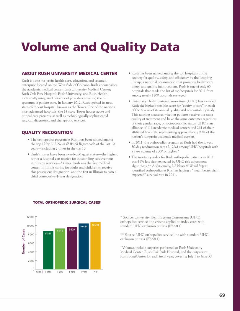
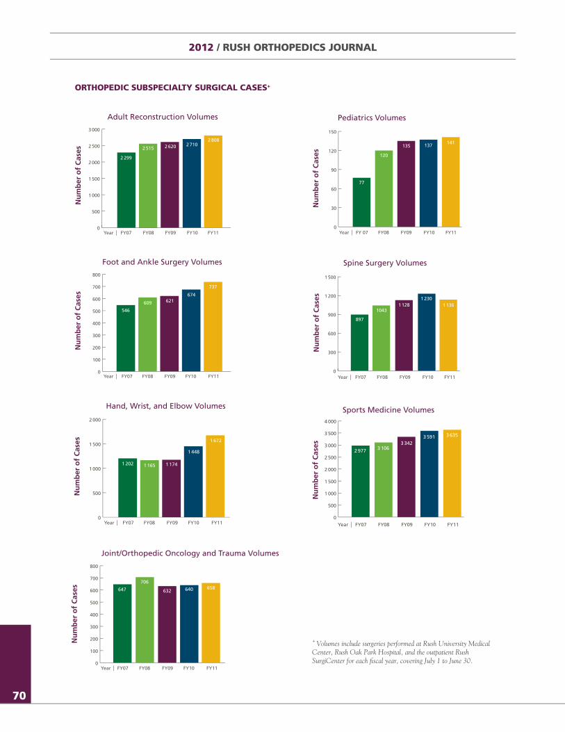
Year FY 07 FY08 FY09 FY10 FY110
30
60
90
120
150
77
120
135 137 141
Year FY07 FY08 FY09 FY10 FY110
300
600
900
1 200
1 500
897
10431 128
1 2301 136
Year FY07 FY08 FY09 FY10 FY110
500
1 000
1 500
2 000
2 500
3 000
3 500
4 000
2 977 3 1063 342
3 591 3 635
0
500
1 000
1 500
2 000
2 500
3 000
Year FY07 FY08 FY09 FY10 FY11
2 299
2 515 2 620 2 7102 808
0
500
1 000
1 500
2 000
Year FY07 FY08 FY09 FY10 FY11
1 202 1 165 1 174
1 448
1 672
0
100
200
300
400
500
600
700
800
Year FY07 FY08 FY09 FY10 FY11
546
609 621674
737
Year FY07 FY08 FY09 FY10 FY110
100
200
300
400
500
600
700
800
647
706
632 640 658
2012 / RUSH ORTHOPEDICS JOURNAL
ORTHOPEDIC SUBSPECIALTY SURGICAL CASES+
Adult Reconstruction Volumes
Nu
mb
er o
f C
ases
Hand, Wrist, and Elbow Volumes
Nu
mb
er o
f C
ases
Pediatrics Volumes
Nu
mb
er o
f C
ases
Spine Surgery Volumes
Nu
mb
er o
f C
ases
Foot and Ankle Surgery Volumes
Nu
mb
er o
f C
ases
Joint/Orthopedic Oncology and Trauma Volumes
Nu
mb
er o
f C
ases
Sports Medicine Volumes
Nu
mb
er o
f C
ases
70
+ Volumes include surgeries performed at Rush University Medical Center, Rush Oak Park Hospital, and the outpatient Rush SurgiCenter for each fiscal year, covering July 1 to June 30.
71795_Body.indd 70 7/25/12 9:39 PM
Steven Gitelis, MD (right), with Gunnar B. J. Andersson, MD, PhD, on the orthopedic floor of Rush’s new hospital. Gitelis has made lasting contributions to Rush and to the field of orthopedic oncology.
BY GUNNAR B. J. ANDERSSON, MD, PHD
LIFE AND LIMBSAn Interview with Pioneering Orthopedic Oncologist and Rush Mainstay Steven Gitelis, MD
When Steven Gitelis, MD, first arrived on the Rush campus in 1972 as a first year medical student, the medical college occupied just one floor of one building,
the Department of Orthopedic Surgery had only a handful of members, and amputation was the predominant treatment for osteosarcoma.
Four decades later, Gitelis is still at Rush—and, as his nurse practitioner and residents will attest, he still has the energy of a 25-year-old. But thanks in part to his dedication and vision, the Medical Center, the department, and the field of orthopedic
oncology are all dramatically different. As director of the first limb preservation center in Chicago, Gitelis helped to pioneer surgical techniques, implants, and bone substitutes that have made limb salvage possible for the majority of sarcoma patients. And he has become a respected leader at Rush whose current titles include president-elect of the medical staff; director of the Section of Orthopedic Oncology; and vice chairman of the Department of Orthopedic Surgery, which today has nearly 60 clinical and research faculty. He also holds the prestigious Rush University Chair of Orthopedic Oncology.
71
Steven Gitelis, MD (right), with Gunnar B. J. Andersson, MD, PhD, on the orthopedic floor of Rush’s new hospital. Gitelis has made lasting contributions to Rush and to the field of orthopedic oncology.
BY GUNNAR B. J. ANDERSSON, MD, PHD
71795_Body_u1.indd 71 8/3/12 10:30 AM
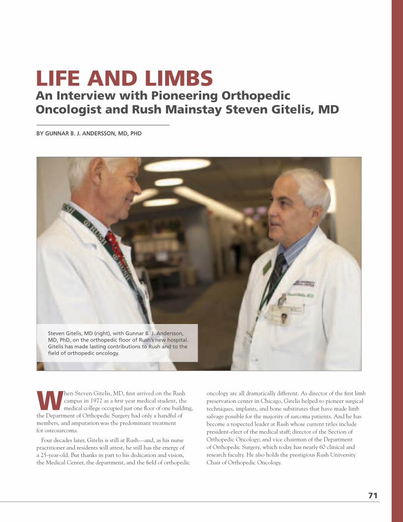
2012 / RUSH ORTHOPEDICS JOURNAL
One of Gitelis’ mentors at Rush has been Gunnar B. J. Andersson, MD, PhD, chairman emeritus of the department and past medical staff president. The two recently sat down to discuss Gitelis’ prolific career—and why he won’t be retiring anytime soon.
ANDERSSON: You’ve been at Rush most of your life, as a student, a resident, and an attending. What keeps you here?
GITELIS: It’s been wonderful to grow up in this institution. I do feel as if I started out here as a child. I still wear my original ID badge from the 1970s because I want to show people what I looked like when I came to Rush 40 years ago. I’ve been mentored by so many great people, first by former Rush President and CEO Leo Henikoff, MD, who chose me to become a student at Rush Medical College, then by former department chairman Jorge O. Galante, MD, DMSc, who recruited me for my residency. I’ve also had the opportunity to work in your lab and co-author numerous articles with you, which really launched my academic career. When I first joined Rush as an attending, we were a small group of six orthopedic surgeons, and we became very close both academically and socially. That’s a major reason I’ve stayed: the interpersonal relationships I’ve developed.
However, you may remember that I’ve also left Rush a few times. I spent time at Denver Children’s Hospital in pediatric orthopedics. I did my orthopedic oncology fellowships at the Rizzoli Institute in Bologna, Italy, and at Mayo Clinic, so I experienced both a foreign perspective and the Mayo perspective. I served in the United States Navy during Desert Shield and Desert Storm at the Bethesda Naval Hospital, ultimately rising to the rank of commander. So I’ve had opportunities to see medicine practiced in different ways, which helped me develop as a clinician and as a person.
ANDERSSON: Your twin brother, Mike, also did his residency here, but he went into private practice, which at the time was the more common choice for orthopedic residents. Why did you decide to remain in the academic world?
GITELIS: Why does one choose academic medicine? You have to have a love for education, and you have to love to mentor students, which I do. I enjoy research, and I’ve done a lot of both clinical and basic research—not as much as you, but what I have done has been interesting. We are blessed to have the best of both worlds at this institution: We can be academicians, but we’re also private orthopedic surgeons with Midwest Orthopaedics at Rush. That’s a unique combination not offered at many other places. So I haven’t had to sacrifice anything. I’ve been at this now 30 years, and I’ll be around at least another 10 years because my work is still exciting to me.
ANDERSSON: You also chose a fairly narrow and somewhat unique subspecialty. Why orthopedic oncology?
GITELIS: For the interpersonal relationships, the bonds that develop between me and my patients, as well as their families. They put a lot of trust in me because they have serious illnesses and their
survival is linked to the care my colleagues and I provide. The good news is that more of my patients are surviving. When I started my practice in 1981, survival outcomes were around 30 percent for the cancers I treat; now we’re achieving 75 percent survival.
ANDERSSON: To what do you attribute that increase?
GITELIS: One big factor is that treatment of orthopedic cancers is now multidisciplinary. We have a sarcoma group at Rush that meets regularly to discuss management for these patients. In addition, the medical treatments—chemotherapy and radiation therapy—have improved tremendously, along with diagnostic capabilities. Diagnostics were a bit dicey in the olden days; now there are very precise ways of making a diagnosis pathologically, so we know the optimal treatments for the diseases we face.
Of course, I also believe that the operations I’m doing are better, from a cancer perspective and a reconstructive perspective. I’m pleased to say that in addition to surviving their cancers, today most patients are also able to keep their limbs. I now do about 95 percent limb salvage and only 5 percent amputation. Back when I was a resident, limb salvage was in its infancy, so I’ve had the opportunity to help develop and refine surgical techniques and technology—including a self-lengthening prosthetic for pediatric patients, bone substitutes, and the use of allografts and autografts—that are now widely used to reconstruct limbs. It’s been exciting to play a role in this amazing progress.
ANDERSSON: You don’t have as many now, but how do you cope with the bad outcomes?
GITELIS: I run a lot on the treadmill, and I’m being very serious about that. What sticks with you most is not the 75 percent you cure, but the 25 percent you lose. Another aspect of my practice is joint replacement, and while there may be complications, they don’t threaten a patient’s life. The stress of orthopedic oncology can be overwhelming, like when I recently had to tell a patient whom I operated on 8 years ago that his cancer has relapsed. You must have release valves, because if you don’t, it can be very destructive personally.
ANDERSSON: Do you think there will be continued improvement in outcomes?
GITELIS: Frankly, we’ve reached the limit of what we can do with traditional treatments for sarcomas. We’ve had these great improvements, but we’re stymied now, so we have to get into a more fundamental approach. As is the case with many other cancers, the key is going to be targeted therapies, therapies that attack the cancer cells at a molecular level. There’s going to be targeted immunotherapy; we’ve just started with the use of protons, a highly targeted form of radiation therapy, for sarcomas; and we’re going to see nanotechnology in the future. So right now we’re at a roadblock, but I think that roadblock will fall apart once these new technologies are refined for clinical application.
72
71795_Body_u1.indd 72 8/3/12 10:30 AM
ANDERSSON: In terms of your personal contributions, is there something you feel has had a particular impact on the field?
GITELIS: One of the things I’m most proud of is that I’ve been medical director of the Bone and Tissue Bank of the Regional Organ Bank of Illinois (ROBI) since its inception. It’s an interesting story, actually. When I began practicing in 1981, there was no tissue bank in Illinois. I had a young patient who needed a bone transplant, so I made arrangements to procure a bone from a tissue bank in Miami. I met the plane at O’Hare to pick up the bone—the pilot almost fainted when I told him what he was carrying in that cooler—but then I had to figure out what to do with the bone, because I wasn’t transplanting it until the next day. So I brought it home, my wife and I threw out all the food in our freezer, and we stashed the bone in the freezer. I still credit my wife with being the first tissue banker in the state.
Shortly thereafter, Jorge O. Galante, MD, DMSc, who was department chairman at that time, asked me to start a tissue bank at Rush. He received a $5,000 grant from the Rush Woman’s Board to buy a Revco freezer, and I handled everything: the procurements, overseeing the operations of the tissue bank, etc. From there, it just grew and grew. However, when the Organ Procurement Organization (OPO) was developed in the U.S., I realized that a hospital-based tissue bank couldn’t exist for very long because there’s just too much regulation. That’s when I joined forces with one of the OPOs, ROBI. ROBI then joined forces with AlloSource, which is now the fourth largest tissue bank in the U.S. ROBI eventually morphed into the Gift of Hope Organ and Tissue Donor Network (a nonprofit that coordinates organ and tissue donation in Illinois and northwest Indiana). So I’m extremely proud of the role I’ve played in the development of tissue banking and transplantation; many patients benefit from these tissues, which include bone, cartilage, and skin.
ANDERSSON: In addition to your many clinical and research activities, you are president-elect of the medical staff, as I once was. It’s an honor, but it’s also a huge responsibility and requires a lot of your time. What compelled you to accept the position?
GITELIS: Sometimes I ask myself that question. It’s particularly challenging for me because I remain a very active surgeon, whereas many presidents are in the waning years of their career, or they’re in a specialty where they have a lot of time. Fortunately, I’m pretty efficient, so I can still maintain a large cancer practice and attend the many meetings that are required as president. Why did I take it? Quite simply, I’m honored. I’ve been at Rush 40 years, and this represents the medical staff ’s recognition of the contributions I’ve made.
ANDERSSON: What do you think of Rush’s new facilities, particularly the Orthopedic Building and the new hospital?
GITELIS: Our orthopedic services were much more spread out before the Orthopedic Building opened. I think this type of consolidated facility greatly enhances patient care because patients can see their doctor, have their imaging, and get their tests done all in the same place. And by housing the clinical programs, plus our residency program and laboratories, the building enables greater collegiality and collaboration. For instance, I frequently walk down to the laboratories on a clinic day, talk to my research colleagues and get updates on what’s happening. I couldn’t do that before because the labs were relatively far from our clinical offices.
As for the new hospital, both my patients and I love it. It’s an outstanding facility. I’ve been impressed with the attention to detail throughout the hospital, from the ORs to the patient floors. In my opinion, it will serve as a model for other institutions around the country to follow.
ANDERSSON: As much time as you spend here, I know family is also very important to you. You have five children, some of whom are following in your footsteps. Did you influence them to become physicians, or did it just happen?
GITELIS: It really did just happen. I try to set an example but not interfere in their lives too much, so my children who opted for careers in medicine did so of their own accord. One of my
73
“Back when I was a resident, limb salvage was in its infancy, so I’ve had the opportunity to help develop and refine surgical techniques and technology—including a self-lengthening prosthetic for pediatric patients, bone substitutes, and the use of allografts and autografts—that are now widely used to reconstruct limbs. It’s been exciting to play a role in this amazing progress.”
71795_Body_u1.indd 73 8/3/12 10:30 AM
Principal photography provided by Andrew Campbell, Eric Herzog, Kevin Horan, and the Rush Photo Group.
daughters is a pediatric resident, another is a medical student, and I just found out recently that one of my sons is now considering becoming a physician. So that’s more tuition I’ll have to pay, but I’m proud of the fact that he chose it on his own.
ANDERSSON: You’ve said that retirement is far in the future, but you must at least think about it occasionally.
GITELIS: Not right now. In fact, after my son’s announcement and the new medical school tuition that I face, my retirement plans have been put on indefinite hold. I have more or less committed to be active for another 10 years, assuming I can maintain my surgical skills. Right now I believe I’m the best surgeon I’ve ever been in my career, and as long as I stay sharp, stay healthy, and continue to enjoy the work I’m doing I’ll keep going.
ANDERSSON: What would you like to be remembered for when you do retire?
GITELIS: For my long-term durability and my involvement in many different aspects of the Medical Center: research, education, administration, and surgery. As an academic orthopedic surgeon, you can choose to expand your horizons within your own institution, or externally through involvement in the American Academy of Orthopaedic Surgeons and other societies. I don’t think I could do both very well, so I’ve chosen to focus internally. I did serve as president of both the Musculoskeletal Tumor Society and the Illinois Orthopaedic Society in the late 1990s, but the vast majority of my leadership roles and committee work have been within the department, Rush Medical College, and the Medical Center. I’m a Rush person, and I’d like to be remembered for all that I’ve contributed to Rush’s success.
74
71795_Body_u1.indd 74 8/3/12 10:30 AM
THE TOWER AT RUSH UNIVERSITY MEDICAL CENTER, which opened its doors in January 2012, is one of the nation’s most advanced—and visually striking—hospitals.
A TRADITION OF EXCELLENCE
“OUR THRIVING CLINICAL PRACTICE is enhanced by collaborations with specialists at Rush from rheumatology, physical medicine and rehabilitation, physical therapy, and other areas.”
Midwest Orthopaedics at Rush (MOR) is a private practice medical group whose 38 fellowship-trained physicians are on the faculty of Rush University
Medical Center in Chicago. With MOR based primarily at Rush, our renowned surgeons and physicians have access to all the resources of a world-class academic medical center, including the state-of-the-art operating rooms in Rush’s new hospital.
Our thriving clinical practice is enhanced by collaborations with specialists at Rush from rheumatology, physical medicine and rehabilitation, physical therapy, and other areas. As part of our commitment to continually advance orthopedic care, our physicians participate in cutting-edge multidisciplinary research, which is translating into new treatments that benefit patients at Rush—and around the globe.
Physicians from Midwest Orthopaedics at Rush also hold key leadership positions in national societies and organizations, and serve as the team physicians for a variety of professional, collegiate, and high school teams and clubs, including the Chicago Bulls, Chicago White Sox, and DePaul University.
These impressive clinical, research, and administrative activities distinguish orthopedics at Rush as among America’s best. U.S.News & World Report has ranked the orthopedics program at Rush in the top 12 each of the past 10 years—including 7 times in the top 10.
Midwest Orthopaedics at Rush
Chicago, IL • Oak Park, IL • Westchester, IL • Winfield, IL(877) MD-BONES (632-6637) / www.rushortho.com
71795_Cover_u4.indd 2 8/6/12 3:33 PM
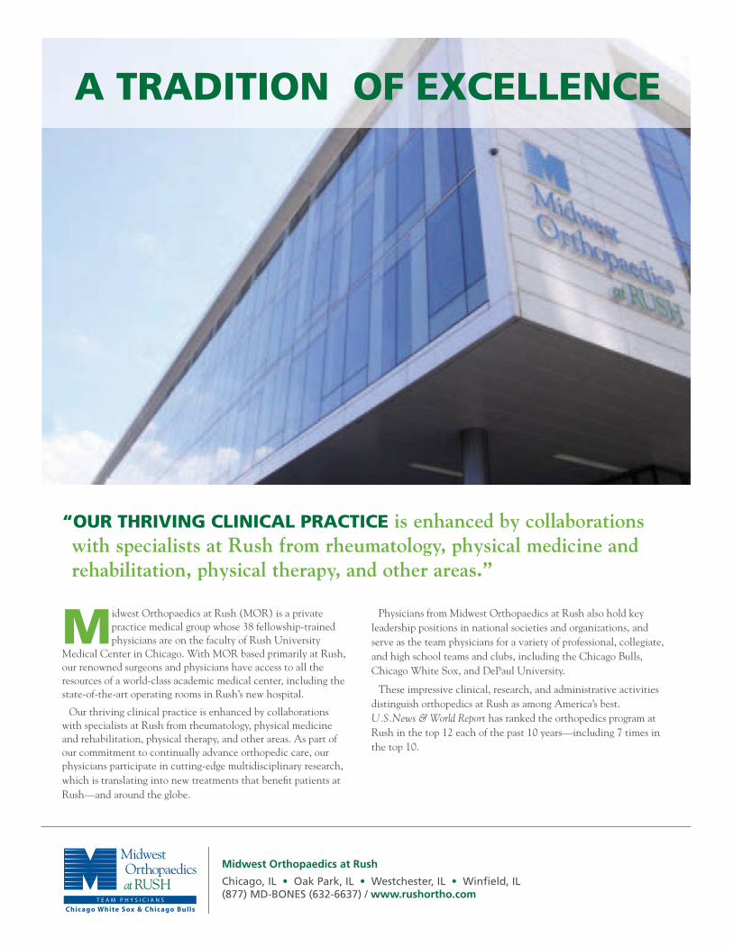
PLEASE NOTE: All physicians featured in this publication are on the medical faculty of Rush University Medical Center. Many of the physicians featured are in the private practice Midwest Orthopaedics at Rush and, as independent practitioners, are not agents or employees of Rush University Medical Center.
Rush is a not-for-profit health care, education and research enterprise comprising Rush University Medical Center, Rush University, Rush Oak Park Hospital and Rush Health.
ORTHOPEDICS2012 RUSH
JOURNAL
20
12
Ru
sh O
rth
op
ed
ics
Jou
rnal
71795_Cover_u3.indd 1 8/3/12 3:33 PM