2014 aha/acc guideline for the management of patients...
TRANSCRIPT

2014 AHA/ACC Guideline for the
Management of Patients With
Valvular Heart Disease
Developed in Collaboration with the American Association for Thoracic Surgery,
American Society of Echocardiography, Society for Cardiovascular Angiography and
Interventions, Society of Cardiovascular Anesthesiologists, and Society of Thoracic
Surgeons
© American College of Cardiology Foundation and American Heart Association

Citation
This slide set is adapted from the 2014 AHA/ACC Guideline for the
Management of Patients With Valvular Heart Disease (Journal of the
American College of Cardiology). Published on March 3, 2014,
available at:
http://content.onlinejacc.org/cgi/content/full/j.jacc.2014.02.536 and
http://circ.ahajournals.org/content/early/2014/02/27/CIR.0000000000
000029.citation
The full-text guidelines are also available on the following Web sites:
ACC (www.cardiosource.org) and AHA (my.americanheart.org)
Robert O. Bonow, MD, MACC, FAHA† Carlos E. Ruiz, MD, PhD, FACC†
Blase A. Carabello, MD, FACC*† Nikolaos J. Skubas, MD, FASE¶
John P. Erwin III, MD, FACC, FAHA‡ Paul Sorajja, MD, FACC, FAHA#
Robert A. Guyton, MD, FACC*§ Thoralf M. Sundt III, MD* **††
Patrick T. O’Gara, MD, FACC, FAHA† James D. Thomas, MD, FASE, FACC, FAHA‡‡
Slide Set EditorsRick A. Nishimura and Catherine M. Otto
Valvular Heart Disease Guideline
Writing Committee MembersRick A. Nishimura, MD, MACC, FAHA, Co-Chair†
Catherine M. Otto, MD, FACC, FAHA, Co-Chair†
*Writing committee members are required to recuse themselves from voting on sections to which their specific
relationships with industry and other entities may apply.
†ACC/AHA representative. ‡ACC/AHA Task Force on Performance Measures liaison. §ACC/AHA Task Force on
Practice Guidelines liaison. ¶SCA Representative. #SCAI Representative. **AATS Representative. ††STS
Representative. ‡‡ASE Representative.

Classification of Recommendations and Levels of Evidence
A recommendation with Level of
Evidence B or C does not imply
that the recommendation is weak.
Many important clinical questions
addressed in the guidelines do
not lend themselves to clinical
trials. Although randomized trials
are unavailable, there may be a
very clear clinical consensus that
a particular test or therapy is
useful or effective.
*Data available from clinical trials
or registries about the usefulness/
efficacy in different
subpopulations, such as sex, age,
history of diabetes mellitus,
history of prior myocardial
infarction, history of heart failure,
and prior aspirin use.
†For comparative-effectiveness
recommendations (Class I and
IIa; Level of Evidence A and B
only), studies that support the use
of comparator verbs should
involve direct comparisons of the
treatments or strategies being
evaluated.
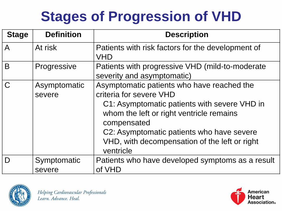
Stages of Progression of VHDStage Definition Description
A At risk Patients with risk factors for the development of
VHD
B Progressive Patients with progressive VHD (mild-to-moderate
severity and asymptomatic)
C Asymptomatic
severe
Asymptomatic patients who have reached the
criteria for severe VHD
C1: Asymptomatic patients with severe VHD in
whom the left or right ventricle remains
compensated
C2: Asymptomatic patients who have severe
VHD, with decompensation of the left or right
ventricle
D Symptomatic
severe
Patients who have developed symptoms as a result
of VHD

Diagnostic Testing – Diagnosis and Follow-Up
Recommendations COR LOE
TTE is recommended in the initial evaluation of
patients with known or suspected VHD to confirm
the diagnosis, establish etiology, determine
severity, assess hemodynamic consequences,
determine prognosis, and evaluate for timing of
intervention
I B
TTE is recommended in patients with known VHD
with any change in symptoms or physical
examination findings
I C
Periodic monitoring with TTE is recommended in
asymptomatic patients with known VHD at intervals
depending on valve lesion, severity, ventricular
size, and ventricular function
I C

Diagnostic Testing – Diagnosis and Follow-Up
Recommendations COR LOE
Cardiac catheterization for hemodynamic
assessment is recommended in symptomatic
patients when noninvasive tests are inconclusive
or when there is a discrepancy between the
findings on noninvasive testing and physical
examination regarding severity of the valve lesion
I C
Exercise testing is reasonable in selected
patients with asymptomatic severe VHD to 1)
confirm the absence of symptoms, or 2) assess
the hemodynamic response to exercise, or 3)
determine prognosis
IIa B

Frequency of Echocardiograms in Asymptomatic
Patients With VHD and Normal Left Ventricular Function
Stage Valve Lesion
Stage Aortic StenosisAortic
RegurgitationMitral Stenosis
Mitral
Regurgitation
Progressive
(stage B)
Every 3–5 y
(mild severity
Vmax 2.0–2.9 m/s)
Every 1–2 y
(moderate
severity
Vmax 3.0–3.9 m/s)
Every 3-5 y
(mild severity)
Every 1-2 y
(moderate
severity)
Every 3–5 y
(MVA >1.5 cm2)
Every 3–5 y
(mild severity)
Every 1–2 y
(moderate
severity)
Severe
(stage C)
Every 1 y
(Vmax ≥4 m/s)
Every 1 y
Dilating LV–
more frequent
Every 1–2 y
(MVA 1.0–1.5 cm2)
Every 1 y
(MVA <1 cm2)
Every 6 months
to 1 y
Dilating LV–
more frequent

Basic Principles of Medical TherapyRecommendations COR LOE
Secondary prevention of rheumatic fever is indicated in
patients with rheumatic heart disease, specifically
mitral stenosis
I C
Prophylaxis against infective endocarditis (IE) is
reasonable for the following patients at highest risk for
adverse outcomes from IE prior to dental procedures
that involve manipulation of gingival tissue,
manipulation of the periapical region of teeth, or
perforation of the oral mucosa:
Patients with prosthetic cardiac valves;
Patients with previous IE;
Cardiac transplant recipients with valve regurgitation
due to a structurally abnormal valve; or (continued
on next page)
IIa B

Basic Principles of Medical TherapyRecommendations COR LOE
(continued)
Patients with CHD with:
o Unrepaired cyanotic CHD, including palliative shunts
and conduits;
o Completely repaired congenital heart defect repaired
with prosthetic material or device, whether placed by
surgery or by catheter intervention, during the first 6
months after the procedure; or
o Repaired CHD with residual defects at the site or
adjacent to the site of a prosthetic patch or
prosthetic device
IIa B
Prophylaxis against IE is not recommended in patients
with VHD at risk of IE for nondental procedures (e.g., TEE,
esophagogastroduodenoscopy, colonoscopy, or
cystoscopy) in the absence of active infection
III: No
BenefitB

Low Risk (must
meet ALL criteria
in this column )
Intermediate Risk
(any 1 criteria in
this column)
High Risk
(any 1 criteria in
this column)
Prohibitive Risk
(any 1 criteria in this
column)
STS PROM <4%
AND
4% to 8%
OR
>8%
OR
Predicted risk with surgery
of death or major morbidity
(all-cause) >50% at 1 y
OR
Frailty None
AND
1 index (mild)
OR
2 or more indices
(moderate-to-
severe)
OR
Major organ
system
compromise not
to be improved
postoperatively
None
AND
1 organ system
OR
No more than 2
organ systems
OR
3 or more organ systems
OR
Procedure-
specific
impediment
None Possible procedure-
specific impediment
Possible procedure-
specific impediment
Severe procedure-specific
impediment
Risk Assessment Combining STS Risk Estimate, Frailty, Major
Organ System Dysfunction, and Procedure-Specific Impediments

The Heart Valve Team and Heart
Valve Centers of ExcellenceRecommendations COR LOE
Patients with severe VHD should be evaluated by
a multidisciplinary Heart Valve Team when
intervention is consideredI C
Consultation with or referral to a Heart Valve
Center of Excellence is reasonable when
discussing treatment options for 1) asymptomatic
patients with severe VHD, 2) patients who may
benefit from valve repair versus valve
replacement, or 3) patients with multiple
comorbidities for whom valve intervention is
considered
IIa C

Stages of Valvular Aortic Stenosis
Stage Definition Valve Anatomy Valve
Hemodynamics
Hemodynamic
Consequences
Symptoms
A At risk of AS ● Bicuspid aortic
valve (or other
congenital valve
anomaly)
● Aortic valve
sclerosis
● Aortic
Vmax <2 m/s
● None ● None
B Progressive
AS
● Mild-to-moderate
leaflet calcification
of a bicuspid or
trileaflet valve with
some reduction in
systolic motion or
● Rheumatic valve
changes with
commissural fusion
● Mild AS: Aortic
Vmax 2.0–2.9
m/s or mean
P <20 mm Hg
● Moderate AS:
Aortic Vmax
3.0–3.9 m/s or
mean P 20–
39 mm Hg
● Early LV
diastolic
dysfunction
may be
present
● Normal LVEF
● None

Stages of Valvular Aortic StenosisStage Definition Valve Anatomy Valve
Hemodynamics
Hemodynamic
Consequences
Symptoms
C - Asymptomatic severe AS
C1 Asymptomatic
severe AS
● Severe leaflet
calcification or
congenital
stenosis with
severely
reduced leaflet
opening
● Aortic Vmax 4 m/s
or mean P ≥40
mm Hg
● AVA typically is
≤1 cm2 (or AVAi
0.6 cm2/m2)
● Very severe AS is
an aortic Vmax
≥5 m/s, or mean
P ≥60 mm Hg
● LV diastolic
dysfunction
● Mild LV
hypertrophy
● Normal LVEF
● None–
exercise
testing is
reasonable
to confirm
symptom
status
C2 Asymptomatic
severe AS with
LV
dysfunction
● Severe leaflet
calcification or
congenital
stenosis with
severely
reduced leaflet
opening
● Aortic Vmax ≥4 m/s
or mean P ≥40
mm Hg
● AVA typically is
≤1 cm2 (or AVAi
0.6 cm2/m2)
● LVEF <50% ● None
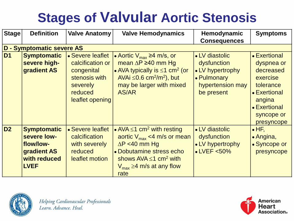
Stage Definition Valve Anatomy Valve Hemodynamics Hemodynamic
Consequences
Symptoms
D - Symptomatic severe AS
D1 Symptomatic
severe high-
gradient AS
● Severe leaflet
calcification or
congenital
stenosis with
severely
reduced
leaflet opening
● Aortic Vmax ≥4 m/s, or
mean P ≥40 mm Hg
● AVA typically is 1 cm2 (or
AVAi 0.6 cm2/m2), but
may be larger with mixed
AS/AR
● LV diastolic
dysfunction
● LV hypertrophy
● Pulmonary
hypertension may
be present
● Exertional
dyspnea or
decreased
exercise
tolerance
● Exertional
angina
● Exertional
syncope or
presyncope
D2 Symptomatic
severe low-
flow/low-
gradient AS
with reduced
LVEF
● Severe leaflet
calcification
with severely
reduced
leaflet motion
● AVA 1 cm2 with resting
aortic Vmax <4 m/s or mean
P <40 mm Hg
● Dobutamine stress echo
shows AVA 1 cm2 with
Vmax 4 m/s at any flow
rate
● LV diastolic
dysfunction
● LV hypertrophy
● LVEF <50%
● HF,
● Angina,
● Syncope or
presyncope
Stages of Valvular Aortic Stenosis

Stage Definition Valve Anatomy Valve
Hemodynamics
Hemodynamic
Consequences
Symptoms
D - Symptomatic severe AS
D3 Symptomatic
severe low-
gradient AS
with normal
LVEF or
paradoxical
low-flow
severe AS
● Severe leaflet
calcification
with severely
reduced leaflet
motion
● AVA 1 cm2 with
aortic Vmax <4 m/s,
or mean P <40
mm Hg
● Indexed AVA 0.6
cm2/m2 and
● Stroke volume
index <35 mL/m2
● Measured when
the patient is
normotensive
(systolic BP <140
mm Hg)
● Increased LV
relative wall
thickness
● Small LV chamber
with low-stroke
volume.
● Restrictive diastolic
filling
● LVEF ≥50%
● HF,
● Angina,
● Syncope or
presyncope
Stages of Valvular Aortic Stenosis

Aortic Stenosis: Diagnosis and Follow-UpRecommendations COR LOE
TTE is indicated in patients with signs or symptoms of
AS or a bicuspid aortic valve for accurate diagnosis of
the cause of AS, hemodynamic severity, LV size and
systolic function, and for determining prognosis and
timing of valve intervention
I B
Low-dose dobutamine stress testing using
echocardiographic or invasive hemodynamic
measurements is reasonable in patients with stage D2
AS with all of the following:
a. Calcified aortic valve with reduced systolic opening;
b. LVEF less than 50%;
c. Calculated valve area 1.0 cm2 or less; and
d. Aortic velocity less than 4.0 m per second or mean
pressure gradient less than 40 mm Hg
IIa B

Aortic Stenosis: Diagnosis and Follow-Up
Recommendations COR LOE
Exercise testing is reasonable to assess
physiological changes with exercise and to
confirm the absence of symptoms in
asymptomatic patients with a calcified aortic valve
and an aortic velocity 4.0 m per second or greater
or mean pressure gradient 40 mm Hg or higher
(stage C)
IIa B
Exercise testing should not be performed in
symptomatic patients with AS when the aortic
velocity is 4.0 m per second or greater or mean
pressure gradient is 40 mm Hg or higher (stage
D)
III:
HarmB

Aortic Stenosis: Medical Therapy
Recommendations COR LOE
Hypertension in patients at risk for developing
AS (stage A) and in patients with asymptomatic
AS (stages B and C) should be treated
according to standard GDMT, started at a low
dose, and gradually titrated upward as needed
with frequent clinical monitoring
I B
Vasodilator therapy may be reasonable if used
with invasive hemodynamic monitoring in the
acute management of patients with severe
decompensated AS (stage D) with New York
Heart Association (NYHA) class IV heart failure
(HF) symptoms
IIb C

Aortic Stenosis: Medical Therapy
Recommendations COR LOE
Statin therapy is not indicated for prevention of
hemodynamic progression of AS in patients
with mild-to-moderate calcific valve disease
(stages B to D)
III: No
BenefitA

Aortic Stenosis: Timing of Intervention
Recommendations COR LOE
AVR is recommended with severe high-gradient
AS who have symptoms by history or on exercise
testing (stage D1)
I B
AVR is recommended for asymptomatic patients
with severe AS (stage C2) and LVEF <50%I B
AVR is indicated for patients with severe AS (stage
C or D) when undergoing other cardiac surgeryI B

Aortic Stenosis: Timing of Intervention (cont.)
Recommendations COR LOE
AVR is reasonable for asymptomatic patients with
very severe AS (stage C1, aortic velocity ≥5 m/s)
and low surgical risk
IIa B
AVR is reasonable in asymptomatic patients (stage
C1) with severe AS and decreased exercise
tolerance or an exercise fall in BP
IIa B
AVR is reasonable in symptomatic patients with
low-flow/low-gradient severe AS with reduced
LVEF (stage D2) with a low-dose dobutamine
stress study that shows an aortic velocity 4 m/s
(or mean pressure gradient 40 mm Hg) with a
valve area 1.0 cm2 at any dobutamine dose
IIa B

Aortic Stenosis: Timing of Intervention (cont.)
Recommendations COR LOE
AVR is reasonable in symptomatic patients who
have low-flow/low-gradient severe AS (stage D3)
who are normotensive and have an LVEF ≥50% if
clinical, hemodynamic, and anatomic data support
valve obstruction as the most likely cause of
symptoms
IIa C
AVR is reasonable for patients with moderate AS
(stage B) (aortic velocity 3.0–3.9 m/s) who are
undergoing other cardiac surgery
IIa C
AVR may be considered for asymptomatic patients
with severe AS (stage C1) and rapid disease
progression and low surgical risk
IIb C
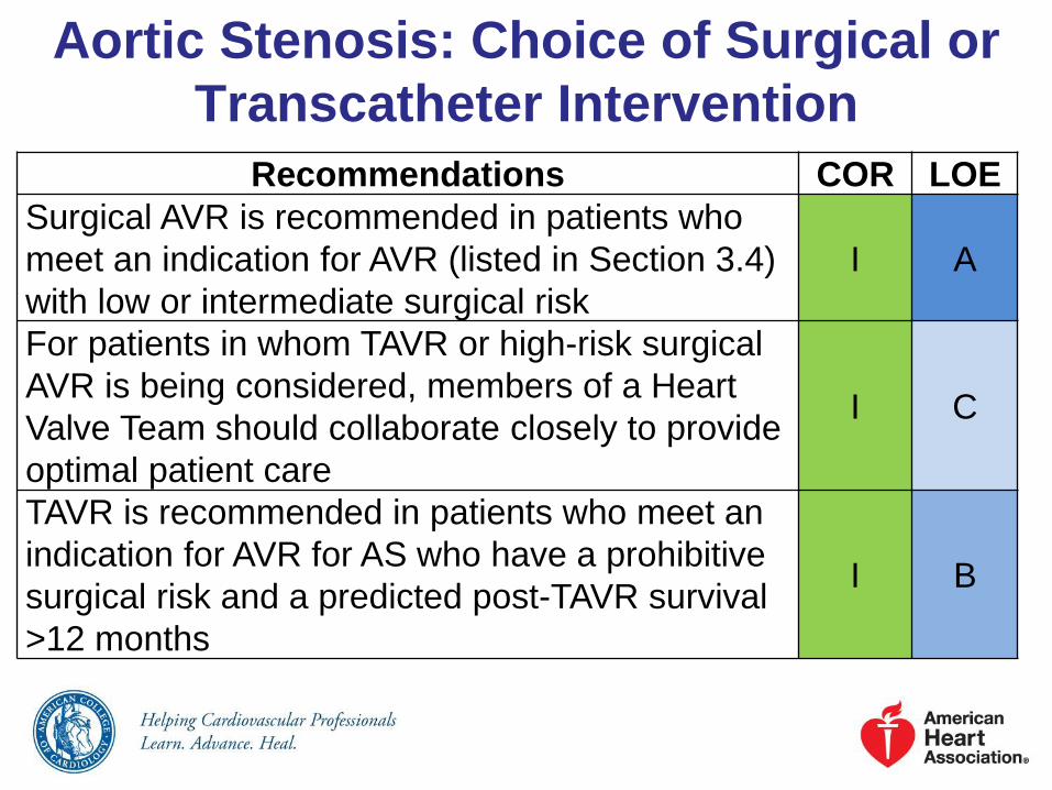
Aortic Stenosis: Choice of Surgical or
Transcatheter Intervention
Recommendations COR LOE
Surgical AVR is recommended in patients who
meet an indication for AVR (listed in Section 3.4)
with low or intermediate surgical risk
I A
For patients in whom TAVR or high-risk surgical
AVR is being considered, members of a Heart
Valve Team should collaborate closely to provide
optimal patient care
I C
TAVR is recommended in patients who meet an
indication for AVR for AS who have a prohibitive
surgical risk and a predicted post-TAVR survival
>12 months
I B
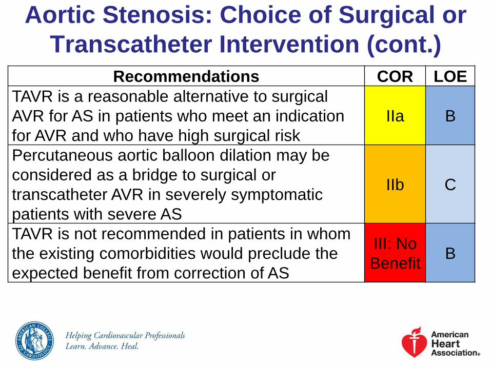
Aortic Stenosis: Choice of Surgical or
Transcatheter Intervention (cont.)
Recommendations COR LOE
TAVR is a reasonable alternative to surgical
AVR for AS in patients who meet an indication
for AVR and who have high surgical risk
IIa B
Percutaneous aortic balloon dilation may be
considered as a bridge to surgical or
transcatheter AVR in severely symptomatic
patients with severe AS
IIb C
TAVR is not recommended in patients in whom
the existing comorbidities would preclude the
expected benefit from correction of AS
III: No
BenefitB

Indications for Aortic Valve Replacement in Patients With Aortic Stenosis
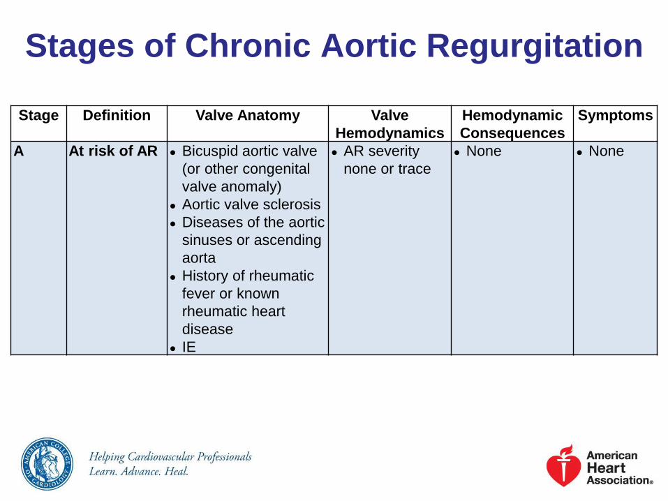
Stages of Chronic Aortic Regurgitation
Stage Definition Valve Anatomy Valve
Hemodynamics
Hemodynamic
Consequences
Symptoms
A At risk of AR ● Bicuspid aortic valve
(or other congenital
valve anomaly)
● Aortic valve sclerosis
● Diseases of the aortic
sinuses or ascending
aorta
● History of rheumatic
fever or known
rheumatic heart
disease
● IE
● AR severity
none or trace
● None ● None

Stages of Chronic Aortic Regurgitation (cont.)
Stage Definition Valve Anatomy Valve Hemodynamics Hemodynamic
Consequences
Symptoms
B Progressive
AR
● Mild-to-
moderate
calcification of
a trileaflet
valve bicuspid
aortic valve (or
other
congenital
valve
anomaly)
● Dilated aortic
sinuses
● Rheumatic
valve changes
● Previous IE
● Mild AR:
o Jet width <25% of LVOT
o Vena contracta <0.3 cm
o RVol <30 mL/beat
o RF <30%
o ERO <0.10 cm2
o Angiography grade 1+
● Moderate AR:
o Jet width 25%–64% of
LVOT
o Vena contracta 0.3–0.6
cm
o RVol 30–59 mL/beat
o RF 30%–49%
o ERO 0.10–0.29 cm2
o Angiography grade 2+
● Normal LV
systolic function
● Normal LV
volume or mild
LV dilation
● None
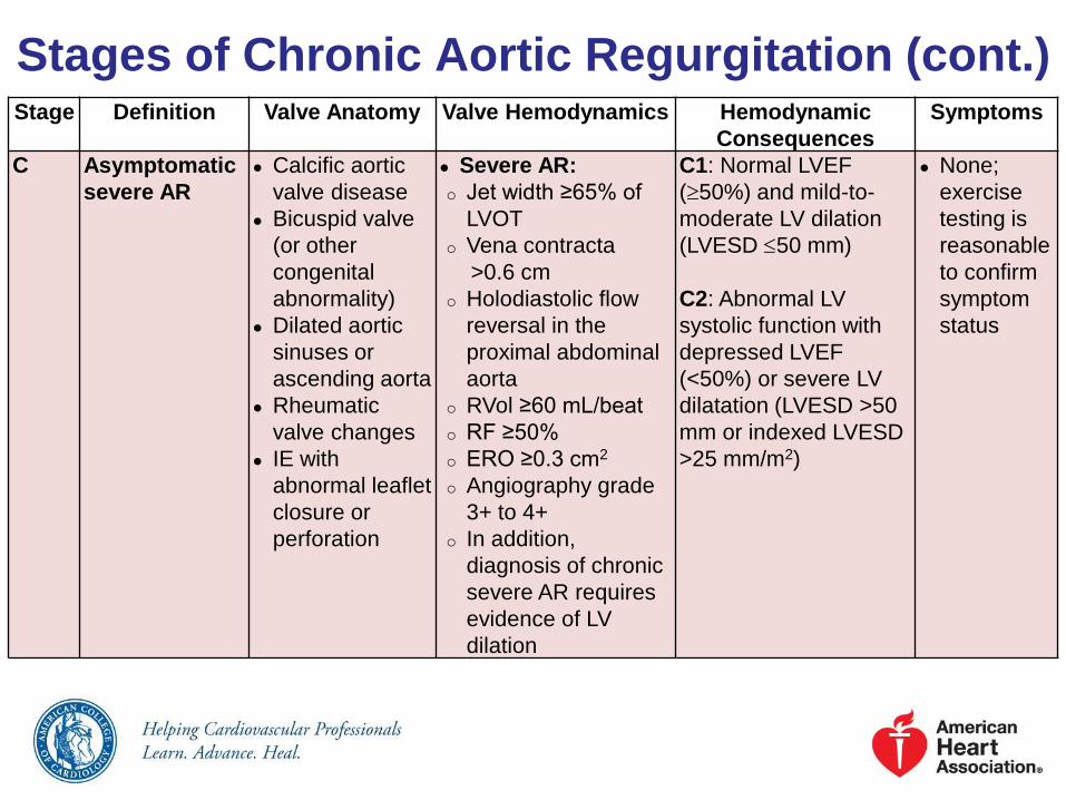
Stages of Chronic Aortic Regurgitation (cont.)Stage Definition Valve Anatomy Valve Hemodynamics Hemodynamic
Consequences
Symptoms
C Asymptomatic
severe AR
● Calcific aortic
valve disease
● Bicuspid valve
(or other
congenital
abnormality)
● Dilated aortic
sinuses or
ascending aorta
● Rheumatic
valve changes
● IE with
abnormal leaflet
closure or
perforation
● Severe AR:
o Jet width ≥65% of
LVOT
o Vena contracta
>0.6 cm
o Holodiastolic flow
reversal in the
proximal abdominal
aorta
o RVol ≥60 mL/beat
o RF ≥50%
o ERO ≥0.3 cm2
o Angiography grade
3+ to 4+
o In addition,
diagnosis of chronic
severe AR requires
evidence of LV
dilation
C1: Normal LVEF
(50%) and mild-to-
moderate LV dilation
(LVESD 50 mm)
C2: Abnormal LV
systolic function with
depressed LVEF
(<50%) or severe LV
dilatation (LVESD >50
mm or indexed LVESD
>25 mm/m2)
● None;
exercise
testing is
reasonable
to confirm
symptom
status

Stages of Chronic Aortic Regurgitation (cont.)
Stage Definition Valve Anatomy Valve Hemodynamics Hemodynamic
Consequences
Symptoms
D Symptomatic
severe AR
● Calcific valve
disease
● Bicuspid valve
(or other
congenital
abnormality)
● Dilated aortic
sinuses or
ascending aorta
● Rheumatic valve
changes
● Previous IE with
abnormal leaflet
closure or
perforation
● Severe AR:
o Doppler jet width
≥65% of LVOT;
o Vena contracta >0.6
cm,
o Holodiastolic flow
reversal in the
proximal abdominal
aorta,
o RVol ≥60 mL/beat;
o RF ≥50%;
o ERO ≥0.3 cm2;
o Angiography grade
3+ to 4+
o In addition, diagnosis
of chronic severe AR
requires evidence of
LV dilation
● Symptomatic severe
AR may occur with
normal systolic
function (LVEF
50%), mild-to-
moderate LV
dysfunction (LVEF
40% to 50%) or
severe LV
dysfunction (LVEF
<40%);
● Moderate-to-severe
LV dilation is
present.
● Exertional
dyspnea or
angina, or
more
severe HF
symptoms

Aortic Regurgitation: Diagnosis and Follow-Up
Recommendations COR LOE
TTE is indicated in patients with signs or symptoms
of AR (stages A to D) for accurate diagnosis of the
cause of regurgitation, regurgitant severity, and LV
size and systolic function, and for determining
clinical outcome and timing of valve intervention
I B
TTE is indicated in patients with dilated aortic
sinuses or ascending aorta or with a bicuspid aortic
valve (stages A and B) to evaluate the presence and
severity of AR
I B
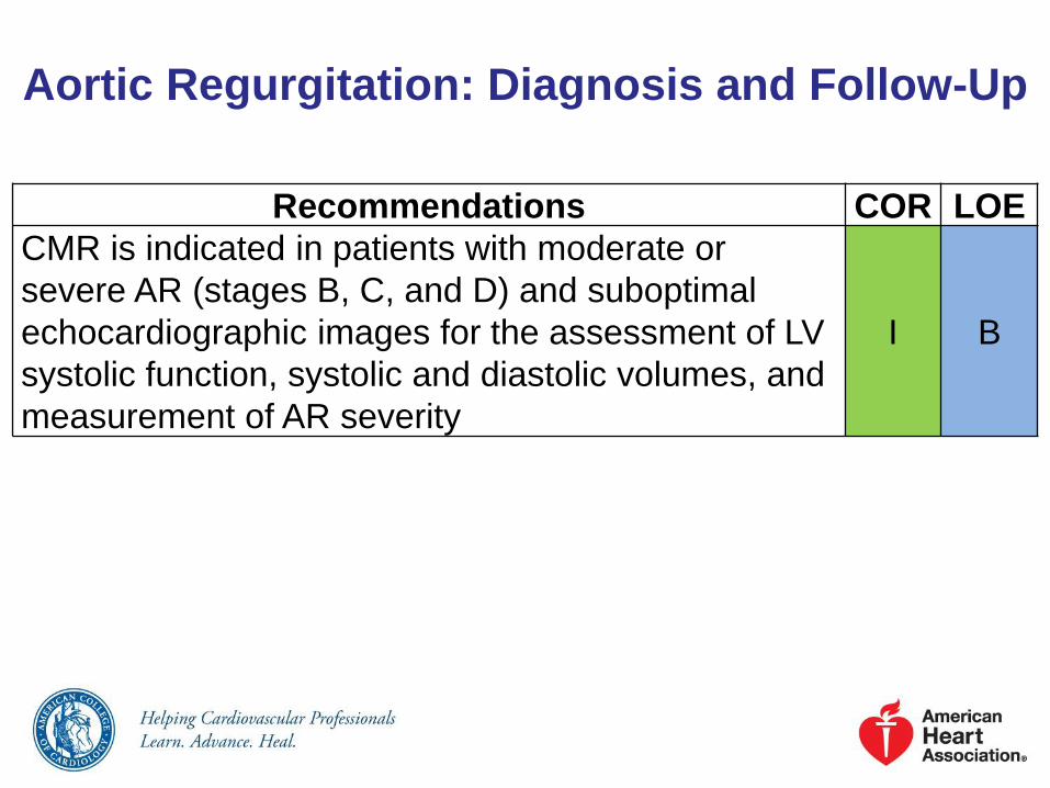
Aortic Regurgitation: Diagnosis and Follow-Up
Recommendations COR LOE
CMR is indicated in patients with moderate or
severe AR (stages B, C, and D) and suboptimal
echocardiographic images for the assessment of LV
systolic function, systolic and diastolic volumes, and
measurement of AR severity
I B

Aortic Regurgitation: Medical Therapy
Recommendations COR LOE
Treatment of hypertension (systolic BP >140 mm
Hg) is recommended in patients with chronic AR
(stages B and C), preferably with dihydropyridine
calcium channel blockers or angiotensin-
converting enzyme (ACE) inhibitors/angiotensin-
receptor blockers (ARBs)
I B
Medical therapy with ACE inhibitors/ARBs and
beta blockers is reasonable in patients with
severe AR who have symptoms and/or LV
dysfunction (stages C2 and D) when surgery is
not performed because of comorbidities
IIa B
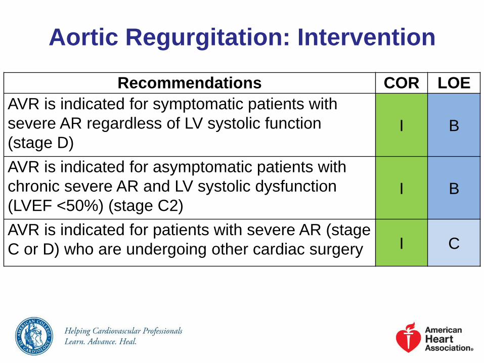
Recommendations COR LOE
AVR is indicated for symptomatic patients with
severe AR regardless of LV systolic function
(stage D)I B
AVR is indicated for asymptomatic patients with
chronic severe AR and LV systolic dysfunction
(LVEF <50%) (stage C2)I B
AVR is indicated for patients with severe AR (stage
C or D) who are undergoing other cardiac surgery I C
Aortic Regurgitation: Intervention

Recommendations COR LOE
AVR is reasonable for asymptomatic patients with
severe AR with normal LV systolic function (LVEF
50%), but severe LV dilation (stage C2, LVESD
>50 mm)
IIa B
AVR is reasonable in patients with moderate AR
(stage B) who are undergoing other cardiac
surgery
IIa C
AVR may be considered for asymptomatic patients
with severe AR and normal LV systolic function
(stage C1, LVEF ≥50%) but severe LV dilation
(LVEDD >65 mm) if surgical risk is low*
IIb C
Aortic Regurgitation: Intervention (cont.)
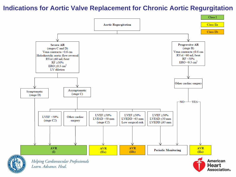
Indications for Aortic Valve Replacement for Chronic Aortic Regurgitation

Bicuspid Aortic Valve and Aortopathy:
Diagnosis and Follow-UpRecommendations COR LOE
An initial TTE is indicated in patients with a known
bicuspid aortic valve to evaluate valve morphology,
to measure the severity of AS and AR, and to
assess the shape and diameter of the aortic sinuses
and ascending aorta for prediction of clinical
outcome and to determine timing of intervention
I B
Aortic magnetic resonance angiography or CT
angiography is indicated in patients with a bicuspid
aortic valve when morphology of the aortic sinuses,
sinotubular junction, or ascending aorta cannot be
assessed accurately or fully by echocardiography
I C

Bicuspid Aortic Valve and Aortopathy:
Diagnosis and Follow-Up
Recommendations COR LOE
Serial evaluation of the size and morphology of
the aortic sinuses and ascending aorta by
echocardiography, CMR, or CT angiography is
recommended in patients with a bicuspid aortic
valve and an aortic diameter greater than 4.0 cm,
with the examination interval determined by the
degree and rate of progression of aortic dilation
and by family history. In patients with an aortic
diameter greater than 4.5 cm, this evaluation
should be performed annually
I C
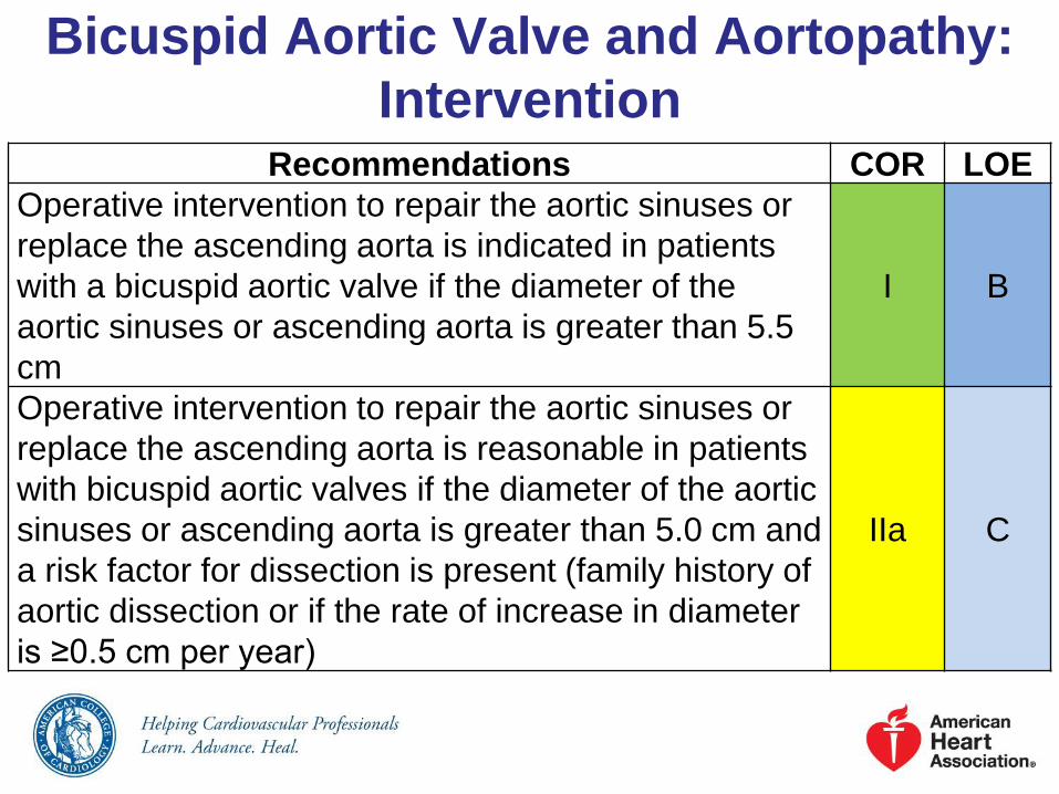
Bicuspid Aortic Valve and Aortopathy:
InterventionRecommendations COR LOE
Operative intervention to repair the aortic sinuses or
replace the ascending aorta is indicated in patients
with a bicuspid aortic valve if the diameter of the
aortic sinuses or ascending aorta is greater than 5.5
cm
I B
Operative intervention to repair the aortic sinuses or
replace the ascending aorta is reasonable in patients
with bicuspid aortic valves if the diameter of the aortic
sinuses or ascending aorta is greater than 5.0 cm and
a risk factor for dissection is present (family history of
aortic dissection or if the rate of increase in diameter
is ≥0.5 cm per year)
IIa C

Bicuspid Aortic Valve and Aortopathy:
Intervention
Recommendations COR LOE
Replacement of the ascending aorta is reasonable
in patients with a bicuspid aortic valve who are
undergoing aortic valve surgery because of severe
AS or AR (Sections 3.4 and 4.4) if the diameter of
the ascending aorta is greater than 4.5 cm
IIa C

Stages of Mitral Stenosis
Stage Definition Valve Anatomy Valve Hemodynamics Hemodynamic
Consequences
Symptoms
A At risk of MS Mild valve doming
during diastole
Normal transmitral
flow velocity
None None
B Progressive
MS
Rheumatic valve
changes with
commissural fusion
and diastolic
doming of the mitral
valve leaflets
Planimetered MVA
>1.5 cm2
Increased transmitral
flow velocities
MVA >1.5 cm2
Diastolic pressure
half-time <150 msec
Mild-to-moderate
LA enlargement
Normal
pulmonary
pressure at rest
None

Stages of Mitral Stenosis
Stage Definition Valve Anatomy Valve Hemodynamics Hemodynamic
Consequences
Symptoms
C Asymptomatic
severe MS
Rheumatic valve
changes with
commissural
fusion and
diastolic doming
of the mitral valve
leaflets
Planimetered
MVA ≤1.5 cm2
(MVA ≤1 cm2 with
very severe MS)
MVA ≤1.5 cm2
(MVA ≤1 cm2 with very
severe MS)
Diastolic pressure
half-time ≥150 msec
(Diastolic pressure
half-time ≥220 msec
with very severe MS)
Severe LA
enlargement
Elevated PASP
>30 mm Hg
None

Stages of Mitral Stenosis
Stage Definition Valve Anatomy Valve
Hemodynamics
Hemodynamic
Consequences
Symptoms
D Symptomatic
severe MS
Rheumatic
valve changes
with
commissural
fusion and
diastolic doming
of the mitral
valve leaflets
Planimetered
MVA ≤1.5 cm2
MVA≤1.5 cm2
(MVA ≤1 cm2 with
very severe MS)
Diastolic pressure
half-time ≥150
msec
(Diastolic pressure
half-time ≥220
msec with very
severe MS)
Severe LA
enlargement
Elevated PASP
>30 mm Hg
Decreased
exercise
tolerance
Exertional
dyspnea

Mitral Stenosis: Diagnosis and Follow-Up
Recommendations COR LOE
TTE is indicated in patients with signs or
symptoms of MS to establish the diagnosis,
quantify hemodynamic severity (mean pressure
gradient, mitral valve area, and pulmonary artery
pressure), assess concomitant valvular lesions,
and demonstrate valve morphology (to
determine suitability for mitral commissurotomy)
I B
TEE should be performed in patients considered
for percutaneous mitral balloon commissurotomy
to assess the presence or absence of left atrial
thrombus and to further evaluate the severity of
mitral regurgitation
I B

Mitral Stenosis: Diagnosis and
Follow-Up
Recommendations COR LOE
Exercise testing with Doppler or invasive
hemodynamic assessment is recommended to
evaluate the response of the mean mitral
gradient and pulmonary artery pressure in
patients with MS when there is a discrepancy
between resting Doppler echocardiographic
findings and clinical symptoms or signs
I C
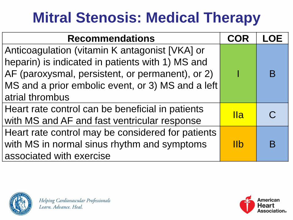
Mitral Stenosis: Medical Therapy
Recommendations COR LOE
Anticoagulation (vitamin K antagonist [VKA] or
heparin) is indicated in patients with 1) MS and
AF (paroxysmal, persistent, or permanent), or 2)
MS and a prior embolic event, or 3) MS and a left
atrial thrombus
I B
Heart rate control can be beneficial in patients
with MS and AF and fast ventricular responseIIa C
Heart rate control may be considered for patients
with MS in normal sinus rhythm and symptoms
associated with exercise
IIb B

Mitral Stenosis: Intervention
Recommendations COR LOE
PMBC is recommended for symptomatic patients
with severe MS (MVA <1.5 cm2, stage D) and
favorable valve morphology in the absence of
contraindications
I A
Mitral valve surgery is indicated in severely
symptomatic patients (NYHA class III/IV) with severe
MS (MVA <1.5 cm2, stage D) who are not high risk
for surgery and who are not candidates for or failed
previous PMBC
I B
Concomitant mitral valve surgery is indicated for
patients with severe MS (MVA ≤1.5 cm2, stages C or
D) undergoing other cardiac surgery
I C
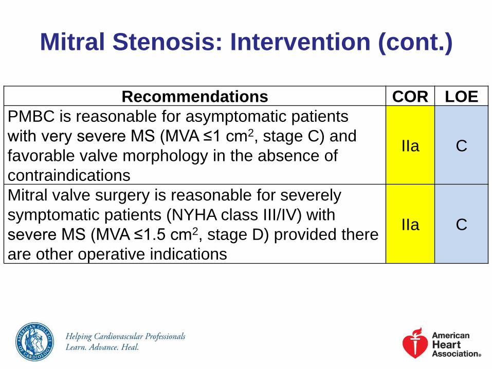
Mitral Stenosis: Intervention (cont.)
Recommendations COR LOE
PMBC is reasonable for asymptomatic patients
with very severe MS (MVA ≤1 cm2, stage C) and
favorable valve morphology in the absence of
contraindications
IIa C
Mitral valve surgery is reasonable for severely
symptomatic patients (NYHA class III/IV) with
severe MS (MVA ≤1.5 cm2, stage D) provided there
are other operative indications
IIa C

Mitral Stenosis: Intervention (cont.)
Recommendations COR LOE
PMBC may be considered for asymptomatic
patients with severe MS (MVA ≤1.5 cm2, stage C)
and favorable valve morphology who have new
onset of AF in the absence of contraindications
IIb C
PMBC may be considered for symptomatic patients
with MVA >1.5 cm2 if there is evidence of
hemodynamically significant MS during exercise
IIb C
PMBC may be considered for severely
symptomatic patients (NYHA class III-IV) with
severe MS (MVA ≤1.5 cm2, stage D) who have
suboptimal valve anatomy and are not candidates
for surgery or at high risk for surgery
IIb C

Mitral Stenosis: Intervention (cont.)
Recommendations COR LOE
Concomitant mitral valve surgery might be
considered for patients with moderate MS (MVA
1.6–2.0 cm2) undergoing other cardiac surgery
IIb C
Mitral valve surgery and excision of the left atrial
appendage may be considered for patients with
severe MS (MVA ≤1.5 cm2, stages C and D) who
have had recurrent embolic events while receiving
adequate anticoagulation
IIb C

Indications for Intervention for Rheumatic Mitral Stenosis

Stages of Primary Mitral Regurgitation
Stage Definition Valve Anatomy Valve Hemodynamics Hemodynamic
Consequences
Symptoms
A At risk of
MR
Mild mitral valve
prolapse with
normal coaptation
Mild valve
thickening and
leaflet restriction
No MR jet or small central
jet area <20% LA on
Doppler
Small vena contracta
<0.3 cm
● None ● None
B Progressive
MR
Severe mitral valve
prolapse with
normal coaptation
Rheumatic valve
changes with
leaflet restriction
and loss of central
coaptation
Prior IE
Central jet MR 20%–40%
LA or late systolic
eccentric jet MR
Vena contracta <0.7 cm
Regurgitant volume
<60 cc
Regurgitant fraction <50%
ERO <0.40 cm2
Angiographic grade 1–2+
Mild LA
enlargement
No LV
enlargement
Normal
pulmonary
pressure
● None

Stages of Primary Mitral Regurgitation (cont.)
Stage Definition Valve Anatomy Valve
Hemodynamics
Hemodynamic
Consequences
Symptoms
C Asymptomatic
severe MR
Severe mitral valve
prolapse with loss
of coaptation or
flail leaflet
Rheumatic valve
changes with
leaflet restriction
and loss of central
coaptation
Prior IE
Thickening of
leaflets with
radiation heart
disease
Central jet MR
>40% LA or
holosystolic
eccentric jet MR
Vena contracta
≥0.7 cm
Regurgitant volume
≥60 cc
Regurgitant fraction
≥50%
ERO ≥0.40 cm2
Angiographic grade
3–4+
Moderate or
severe LA
enlargement
LV enlargement
Pulmonary
hypertension may
be present at rest
or with exercise
C1: LVEF >60%
and LVESD
<40 mm
C2: LVEF ≤60%
and LVESD
≥40 mm
● None

Stages of Primary Mitral Regurgitation (cont.)
Stage Definition Valve Anatomy Valve
Hemodynamics
Hemodynamic
Consequences
Symptoms
D Symptomatic
severe MR
Severe mitral valve
prolapse with loss
of coaptation or flail
leaflet
Rheumatic valve
changes with leaflet
restriction and loss
of central
coaptation
Prior IE
Thickening of
leaflets with
radiation heart
disease
Central jet MR
>40% LA or
holosystolic
eccentric jet MR
Vena contracta
≥0.7 cm
Regurgitant volume
≥60 cc
Regurgitant fraction
≥50%
ERO ≥0.40 cm2
Angiographic grade
3–4+
Moderate or
severe LA
enlargement
LV enlargement
Pulmonary
hypertension
present
Decreased
exercise
tolerance
Exertional
dyspnea
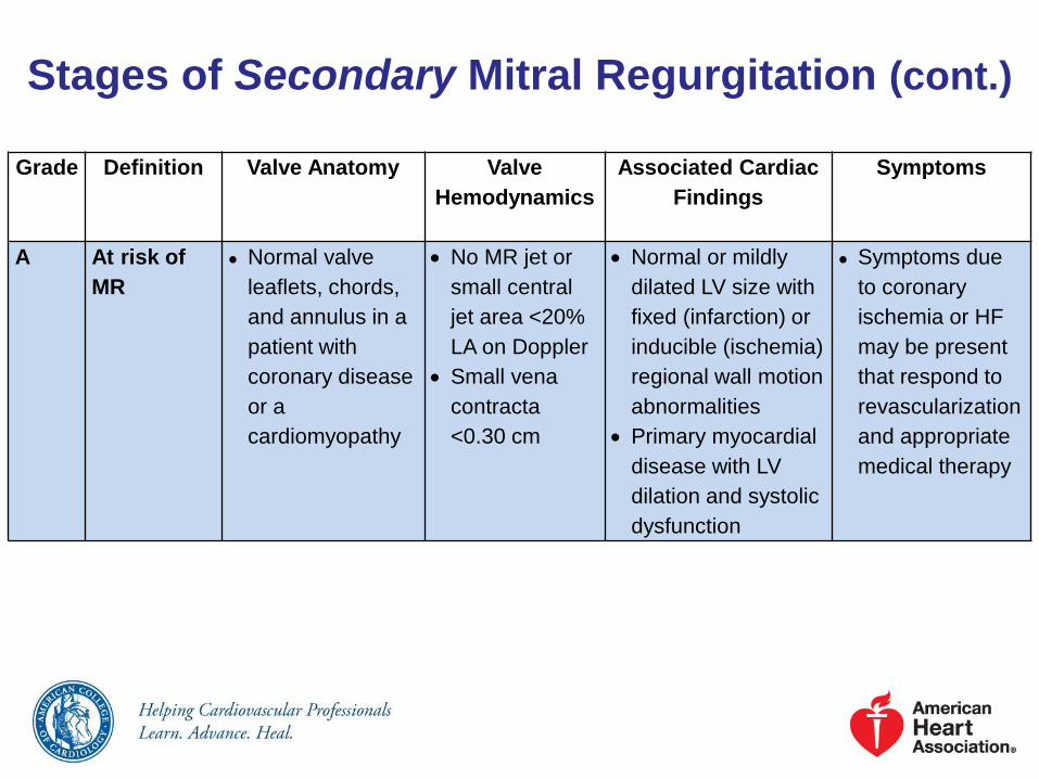
Stages of Secondary Mitral Regurgitation (cont.)
Grade Definition Valve Anatomy Valve
Hemodynamics
Associated Cardiac
Findings
Symptoms
A At risk of
MR
● Normal valve
leaflets, chords,
and annulus in a
patient with
coronary disease
or a
cardiomyopathy
No MR jet or
small central
jet area <20%
LA on Doppler
Small vena
contracta
<0.30 cm
Normal or mildly
dilated LV size with
fixed (infarction) or
inducible (ischemia)
regional wall motion
abnormalities
Primary myocardial
disease with LV
dilation and systolic
dysfunction
● Symptoms due
to coronary
ischemia or HF
may be present
that respond to
revascularization
and appropriate
medical therapy

Stages of Secondary Mitral Regurgitation (cont.)
Grade Definition Valve Anatomy Valve
Hemodynamics
Associated Cardiac
Findings
Symptoms
B Progressive
MR
Regional wall
motion
abnormalities
with mild
tethering of
mitral leaflet
Annular dilation
with mild loss of
central
coaptation of the
mitral leaflets
ERO <0.20
cm2
Regurgitant
volume <30 cc
Regional wall
motion
abnormalities with
reduced LV systolic
function
LV dilation and
systolic dysfunction
due to primary
myocardial disease
● Symptoms due
to coronary
ischemia or HF
may be present
that respond to
revascularization
and appropriate
medical therapy
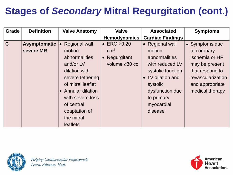
Stages of Secondary Mitral Regurgitation (cont.)
Grade Definition Valve Anatomy Valve
Hemodynamics
Associated
Cardiac Findings
Symptoms
C Asymptomatic
severe MR
Regional wall
motion
abnormalities
and/or LV
dilation with
severe tethering
of mitral leaflet
Annular dilation
with severe loss
of central
coaptation of
the mitral
leaflets
ERO ≥0.20
cm2
Regurgitant
volume ≥30 cc
Regional wall
motion
abnormalities
with reduced LV
systolic function
LV dilation and
systolic
dysfunction due
to primary
myocardial
disease
● Symptoms due
to coronary
ischemia or HF
may be present
that respond to
revascularization
and appropriate
medical therapy

Stages of Secondary Mitral Regurgitation (cont.)
Grade Definition Valve Anatomy Valve
Hemodynamics
Associated
Cardiac Findings
Symptoms
D Symptomatic
severe MR
Regional wall
motion
abnormalities
and/or LV
dilation with
severe
tethering of
mitral leaflet
Annular
dilation with
severe loss of
central
coaptation of
the mitral
leaflets
ERO ≥0.20 cm2
Regurgitant
volume ≥30 cc
Regional wall
motion
abnormalities
with reduced LV
systolic function
LV dilation and
systolic
dysfunction due
to primary
myocardial
disease.
HF symptoms
due to MR
persist even after
revascularization
and optimization
of medical
therapy
Decreased
exercise
tolerance
Exertional
dyspnea

Chronic Primary Mitral Regurgitation:
Diagnosis and Follow-Up
Recommendations COR LOE
TTE is indicated for baseline evaluation of LV
size and function, right ventricular (RV) function
and left atrial size, pulmonary artery pressure,
and mechanism and severity of primary MR
(stages A to D) in any patient suspected of having
chronic primary MR
I B
CMR is indicated in patients with chronic primary
MR to assess LV and RV volumes, function, or
MR severity and when these issues are not
satisfactorily addressed by TTE
I B

Chronic Primary Mitral Regurgitation:
Diagnosis and Follow-Up (cont.)
Recommendations COR LOE
Intraoperative TEE is indicated to establish the
anatomic basis for chronic primary MR (stages C
and D) and to guide repair
I B
TEE is indicated for evaluation of patients with
chronic primary MR (stages B to D) in whom
noninvasive imaging provides nondiagnostic
information about severity of MR, mechanism of
MR, and/or status of LV function
I C

Chronic Primary Mitral Regurgitation:
Diagnosis and Follow-Up (cont.)
Recommendations COR LOE
Exercise hemodynamics with either Doppler
echocardiography or cardiac catheterization is
reasonable in symptomatic patients with chronic
primary MR where there is a discrepancy
between symptoms and the severity of MR at rest
(stages B and C)
IIa B
Exercise treadmill testing can be useful in
patients with chronic primary MR to establish
symptom status and exercise tolerance (stages B
and C)
IIa C

Chronic Primary Mitral Regurgitation:
Medical Therapy
Recommendations COR LOE
Medical therapy for systolic dysfunction is
reasonable in symptomatic patients with chronic
primary MR (stage D) and LVEF less than 60%
in whom surgery is not contemplated
IIa B
Vasodilator therapy is not indicated for
normotensive asymptomatic patients with
chronic primary MR (stages B and C1) and
normal systolic LV function
III: No
BenefitB

Recommendations COR LOE
MV surgery is recommended for symptomatic
patients with chronic severe primary MR (stage D)
and LVEF >30%
I B
MV surgery is recommended for asymptomatic
patients with chronic severe primary MR and LV
dysfunction (LVEF 30%–60% and/or LVESD ≥40
mm, stage C2)
I B
MV repair is recommended in preference to MVR
when surgical treatment is indicated for patients
with chronic severe primary MR limited to the
posterior leaflet
I B
Chronic Primary Mitral Regurgitation:
Intervention

Recommendations COR LOE
MV repair is recommended in preference to MVR
when surgical treatment is indicated for patients
with chronic severe primary MR involving the
anterior leaflet or both leaflets when a successful
and durable repair can be accomplished
I B
Concomitant MV repair or replacement is
indicated in patients with chronic severe primary
MR undergoing other cardiac surgery
I B
Chronic Primary Mitral Regurgitation:
Intervention (cont.)
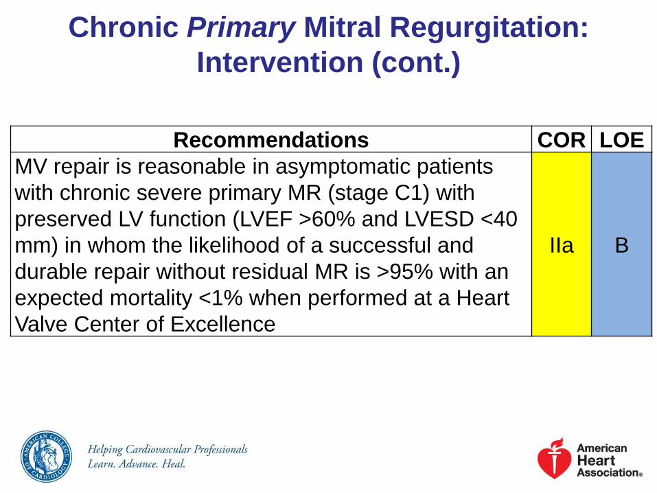
Recommendations COR LOE
MV repair is reasonable in asymptomatic patients
with chronic severe primary MR (stage C1) with
preserved LV function (LVEF >60% and LVESD <40
mm) in whom the likelihood of a successful and
durable repair without residual MR is >95% with an
expected mortality <1% when performed at a Heart
Valve Center of Excellence
IIa B
Chronic Primary Mitral Regurgitation:
Intervention (cont.)

Recommendations COR LOE
MV repair is reasonable for asymptomatic
patients with chronic severe nonrheumatic
primary MR (stage C1) and preserved LV
function in whom there is a high likelihood of a
successful and durable repair with 1) new onset
of AF or 2) resting pulmonary hypertension (PA
systolic arterial pressure >50 mm Hg)
IIa B
Concomitant MV repair is reasonable in patients
with chronic moderate primary MR (stage B)
undergoing other cardiac surgery
IIa C
Chronic Primary Mitral Regurgitation:
Intervention (cont.)
Recommendations COR LOE
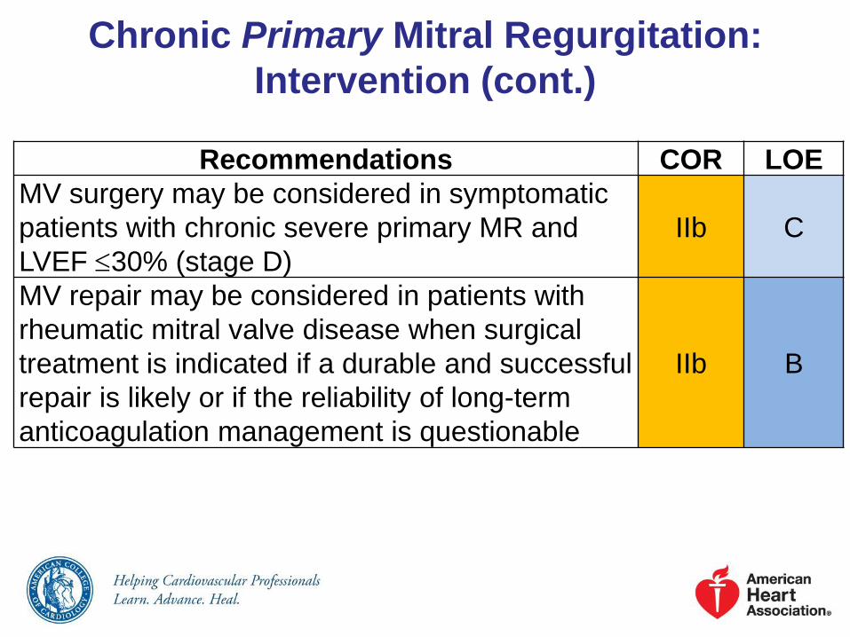
MV surgery may be considered in symptomatic
patients with chronic severe primary MR and
LVEF 30% (stage D)
IIb C
MV repair may be considered in patients with
rheumatic mitral valve disease when surgical
treatment is indicated if a durable and successful
repair is likely or if the reliability of long-term
anticoagulation management is questionable
IIb B
Chronic Primary Mitral Regurgitation:
Intervention (cont.)

Recommendations COR LOE
Percutaneous MV repair may be considered for
severely symptomatic patients (NYHA class III-
IV) with chronic severe primary MR (stage D)
who have a reasonable life expectancy, but a
prohibitive surgical risk because of severe
comorbidities
IIb B
MVR should not be performed for the treatment
of isolated severe primary MR limited to less
than one half of the posterior leaflet unless MV
repair has been attempted and was
unsuccessful
III:
HarmB
Chronic Primary Mitral Regurgitation:
Intervention (cont.)
Chronic Secondary Mitral Regurgitation:
Diagnosis and Follow-UpRecommendations COR LOE
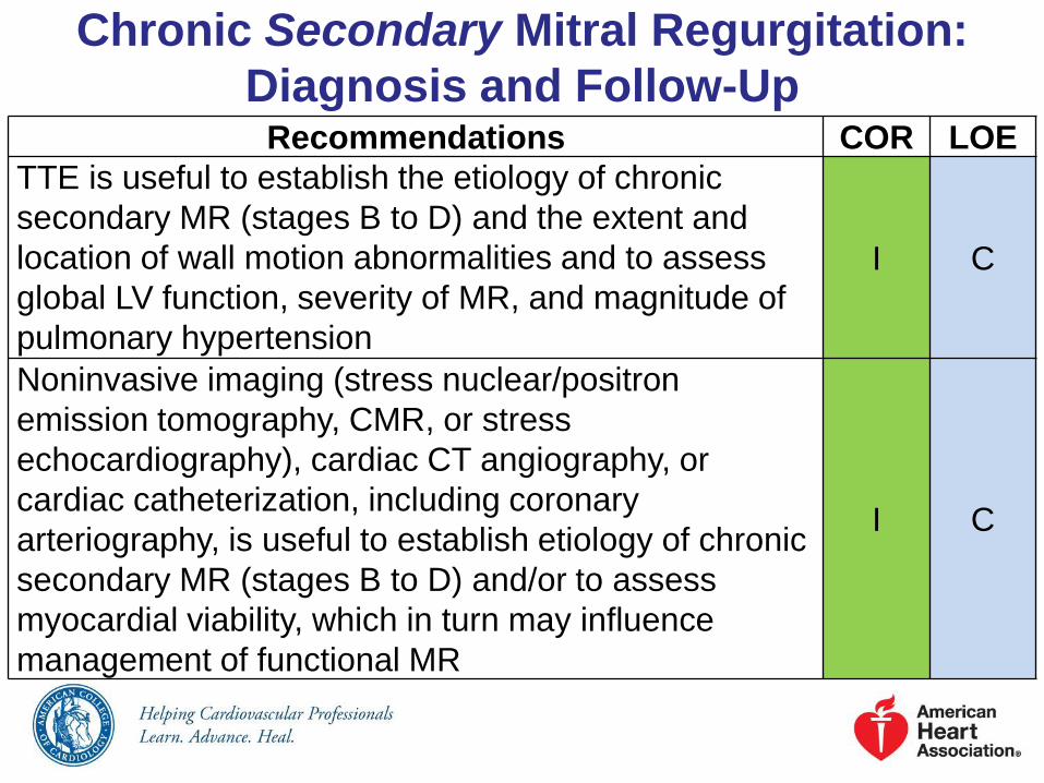
TTE is useful to establish the etiology of chronic
secondary MR (stages B to D) and the extent and
location of wall motion abnormalities and to assess
global LV function, severity of MR, and magnitude of
pulmonary hypertension
I C
Noninvasive imaging (stress nuclear/positron
emission tomography, CMR, or stress
echocardiography), cardiac CT angiography, or
cardiac catheterization, including coronary
arteriography, is useful to establish etiology of chronic
secondary MR (stages B to D) and/or to assess
myocardial viability, which in turn may influence
management of functional MR
I C

Chronic Secondary Mitral Regurgitation:
Medical TherapyRecommendations COR LOE
Patients with chronic secondary MR (stages B to D)
and HF with reduced LVEF should receive standard
GDMT therapy for HF, including ACE inhibitors,
ARBs, beta blockers, and/or aldosterone antagonists
as indicated
I A
Noninvasive imaging (stress nuclear/positron
emission tomography, CMR, or stress
echocardiography), cardiac CT angiography, or
cardiac catheterization, including coronary
arteriography, is useful to establish etiology of chronic
secondary MR (stages B to D) and/or to assess
myocardial viability, which in turn may influence
management of functional MR
I A
Chronic Severe Secondary Mitral
Regurgitation: Intervention
Recommendations COR LOE
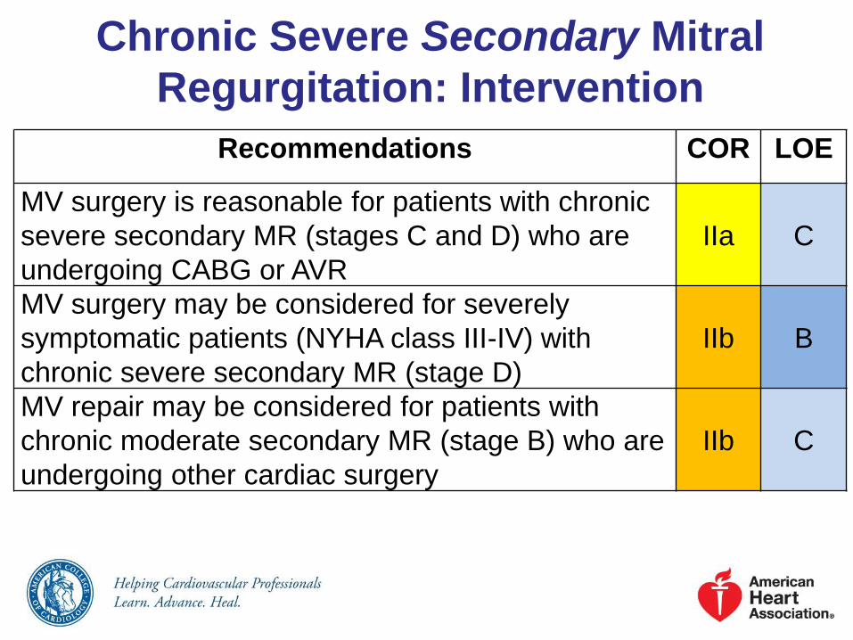
MV surgery is reasonable for patients with chronic
severe secondary MR (stages C and D) who are
undergoing CABG or AVR
IIa C
MV surgery may be considered for severely
symptomatic patients (NYHA class III-IV) with
chronic severe secondary MR (stage D)
IIb B
MV repair may be considered for patients with
chronic moderate secondary MR (stage B) who are
undergoing other cardiac surgery
IIb C
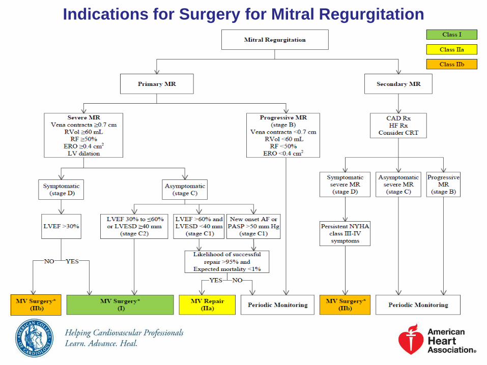
Indications for Surgery for Mitral Regurgitation

Stages of Tricuspid Regurgitation
Stage Definition Valve Anatomy Valve
Hemodynamics
Hemodynamic
Consequences
Symptoms
A At risk of
TR
Primary
Mild rheumatic change
Mild prolapse
Other (e.g., IE with
vegetation, early
carcinoid deposition,
radiation)
Intra-annular RV
pacemaker or ICD lead
Postcardiac transplant
(biopsy-related)
Functional
Normal
Early annular dilation
● No or trace TR ● None ● None or in
relation to other
left heart or
pulmonary/
pulmonary
vascular
disease

Stages of Tricuspid Regurgitation (cont.)Stage Definition Valve
Anatomy
Valve Hemodynamics Hemodynamic
Consequences
Symptoms
B Progressive
TR
Primary
Progressive
leaflet
deterioration/
destruction
Moderate-to-
severe
prolapse,
limited
chordal
rupture
Functional
Early
annular
dilation
Moderate
leaflet
tethering
Mild TR
Central jet area <5 cm2
Vena contracta width not
defined
CW jet density and contour:
soft and parabolic
Hepatic vein flow: systolic
dominance
Moderate TR
Central jet area 5–10 cm2
Vena contracta width not
defined, but <0.70 cm
CW jet density and contour:
dense, variable contour
Hepatic vein flow: systolic
blunting
Mild TR
RV/RA/IVC size
normal
Moderate TR
No RV
enlargement
No or mild RA
enlargement
No or mild IVC
enlargement with
normal
respirophasic
variation
Normal RA
pressure
● None or in
relation to
other left
heart or
pulmonary/
pulmonary
vascular
disease

Stages of Tricuspid Regurgitation (cont.)
Stage Definition Valve Anatomy Valve
Hemodynamics
Hemodynamic
Consequences
Symptoms
C Asymptomatic,
severe TR
Primary
Flail or grossly
distorted leaflets
Functional
Severe annular
dilation (>40 mm
or 21 mm/m2)
Marked leaflet
tethering
Central jet area
>10 cm2
Vena contracta
width >0.7 cm
CW jet density
and contour:
dense, triangular
with early peak
Hepatic vein
flow: systolic
reversal
RV/RA/IVC dilated
with decreased IVC
respirophasic
variation
Elevation RA
pressure with “c-V”
wave
Diastolic
interventricular septal
flattening may be
present
● None, or in
relation to
other left
heart or
pulmonary/
pulmonary
vascular
disease

Stages of Tricuspid Regurgitation (cont.)
Stage Definition Valve Anatomy Valve
Hemodynamics
Hemodynamic
Consequences
Symptoms
D Symptomatic
severe TR
Primary
Flail or grossly
distorted leaflets
Functional
Severe annular
dilation (>40 mm
or >21 mm/m2)
Marked leaflet
tethering
Central jet area
>10 cm2
Vena contracta
width >0.70 cm
CW jet density
and contour:
dense, triangular
with early peak
Hepatic vein
flow: systolic
reversal
RV/RA/IVC dilated
with decreased IVC
respirophasic
variation
Elevation RA
pressure with “c-V”
wave
Diastolic
interventricular septal
flattening
Reduced RV systolic
function in late phase
● Fatigue,
palpitations,
dyspnea,
abdominal
bloating,
anorexia,
edema

Tricuspid Regurgitation: Diagnosis and
Follow-Up
Recommendations COR LOE
TTE is indicated to evaluate severity of TR,
determine etiology, measure sizes of right-sided
chambers and inferior vena cava, assess RV
systolic function, estimate pulmonary artery
systolic pressure, and characterize any
associated left-sided heart disease
I C
Invasive measurement of pulmonary artery
pressures and pulmonary vascular resistance
can be useful in patients with TR when clinical
and noninvasive data regarding their values are
discordant
IIa C

Tricuspid Regurgitation: Diagnosis and
Follow-Up (cont.)
Recommendations COR LOE
CMR or real-time 3-dimensional
echocardiography may be considered for
assessment of RV systolic function and systolic
and diastolic volumes in patients with severe
TR (stages C and D) and suboptimal 2-
dimensional echocardiograms
IIb C
Exercise testing may be considered for the
assessment of exercise capacity in patients
with severe TR with no or minimal symptoms
(stage C)
IIb C

Tricuspid Regurgitation: Medical Therapy
Recommendations COR LOE
Diuretics can be useful for patients with severe
TR and signs of right-sided HF (stage D)IIa C
Medical therapies to reduce elevated
pulmonary artery pressures and/or pulmonary
vascular resistance might be considered in
patients with severe functional TR (stages C
and D)
IIb C

Tricuspid Regurgitation: Intervention
Recommendations COR LOE
Tricuspid valve surgery is recommended for
patients with severe TR (stages C and D)
undergoing left-sided valve surgery
I C
Tricuspid valve repair can be beneficial for
patients with mild, moderate, or greater
functional TR (stage B) at the time of left-sided
valve surgery with either 1) tricuspid annular
dilation or 2) prior evidence of right HF
IIa B
Tricuspid valve surgery can be beneficial for
patients with symptoms due to severe primary
TR that are unresponsive to medical therapy
(stage D)
IIa C

Tricuspid Regurgitation: Intervention (cont.)
Recommendations COR LOE
Tricuspid valve repair may be considered for
patients with moderate functional TR (stage B)
and pulmonary artery hypertension at the time
of left-sided valve surgery
IIb C
Tricuspid valve surgery may be considered for
asymptomatic or minimally symptomatic
patients with severe primary TR (stage C) and
progressive degrees of moderate or greater RV
dilation and/or systolic dysfunction
IIb C
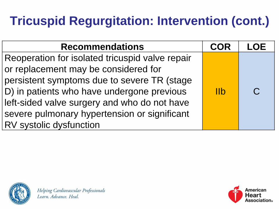
Tricuspid Regurgitation: Intervention (cont.)
Recommendations COR LOE
Reoperation for isolated tricuspid valve repair
or replacement may be considered for
persistent symptoms due to severe TR (stage
D) in patients who have undergone previous
left-sided valve surgery and who do not have
severe pulmonary hypertension or significant
RV systolic dysfunction
IIb C

Indications for Surgery for Tricuspid Regurgitation
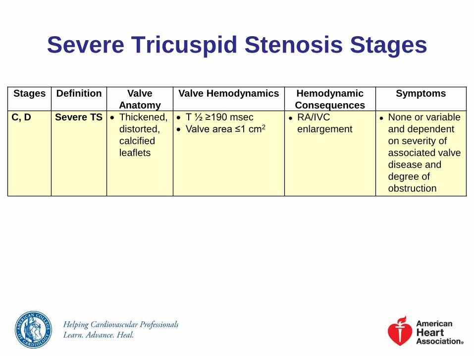
Severe Tricuspid Stenosis Stages
Stages Definition Valve
Anatomy
Valve Hemodynamics Hemodynamic
Consequences
Symptoms
C, D Severe TS Thickened,
distorted,
calcified
leaflets
T ½ ≥190 msec
Valve area ≤1 cm2
● RA/IVC
enlargement
● None or variable
and dependent
on severity of
associated valve
disease and
degree of
obstruction

Tricuspid Stenosis: Diagnosis and Follow-Up
Recommendations COR LOE
TTE is indicated in patients with TS to assess
the anatomy of the valve complex, evaluate
severity of stenosis, and characterize any
associated regurgitation and/or left-sided valve
disease
I C
Invasive hemodynamic assessment of severity
of TS may be considered in symptomatic
patients when clinical and noninvasive data are
discordant
IIb C

Tricuspid Stenosis: Intervention
Recommendations COR LOE
Tricuspid valve surgery is recommended for
patients with severe TS at the time of operation
for left-sided valve diseaseI C
Tricuspid valve surgery is recommended for
patients with isolated, symptomatic severe TS I C
Percutaneous balloon tricuspid
commissurotomy might be considered in
patients with isolated, symptomatic severe TS
without accompanying TR
IIb C
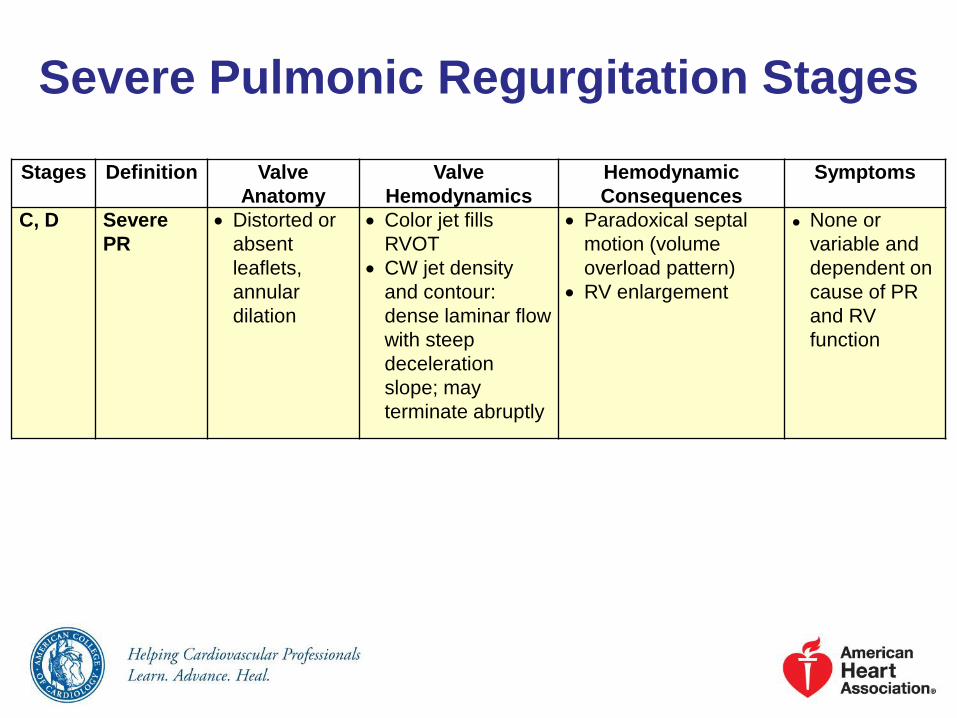
Severe Pulmonic Regurgitation Stages
Stages Definition Valve
Anatomy
Valve
Hemodynamics
Hemodynamic
Consequences
Symptoms
C, D Severe
PR
Distorted or
absent
leaflets,
annular
dilation
Color jet fills
RVOT
CW jet density
and contour:
dense laminar flow
with steep
deceleration
slope; may
terminate abruptly
Paradoxical septal
motion (volume
overload pattern)
RV enlargement
● None or
variable and
dependent on
cause of PR
and RV
function

Severe Pulmonic Stenosis Stages
Stages Definition Valve Anatomy Valve
Hemodynamics
Hemodynamic
Consequences
Symptoms
C, D Severe
PS
Thickened,
distorted, possibly
calcified leaflets
with systolic
doming and/or
reduced excursion
Other anatomic
abnormalities may
be present, such as
narrowed RVOT
● Vmax >4 m/s; peak
instantaneous
gradient >64 mm
Hg
RVH
Possible RV,
RA
enlargement
Poststenotic
enlargement of
main PA
● None or
variable and
dependent
on severity of
obstruction

Prosthetic Valve: Diagnosis and Follow-Up
Recommendations COR LOE
An initial TTE study is recommended in patients
after prosthetic valve implantation for evaluation
of valve hemodynamics
I B
Repeat TTE is recommended in patients with
prosthetic heart valves if there is a change in
clinical symptoms or signs suggesting valve
dysfunction
I C
TEE is recommended when clinical symptoms or
signs suggest prosthetic valve dysfunctionI C
Annual TTE is reasonable in patients with a
bioprosthetic valve after the first 10 years, even in
the absence of a change in clinical status
IIa C
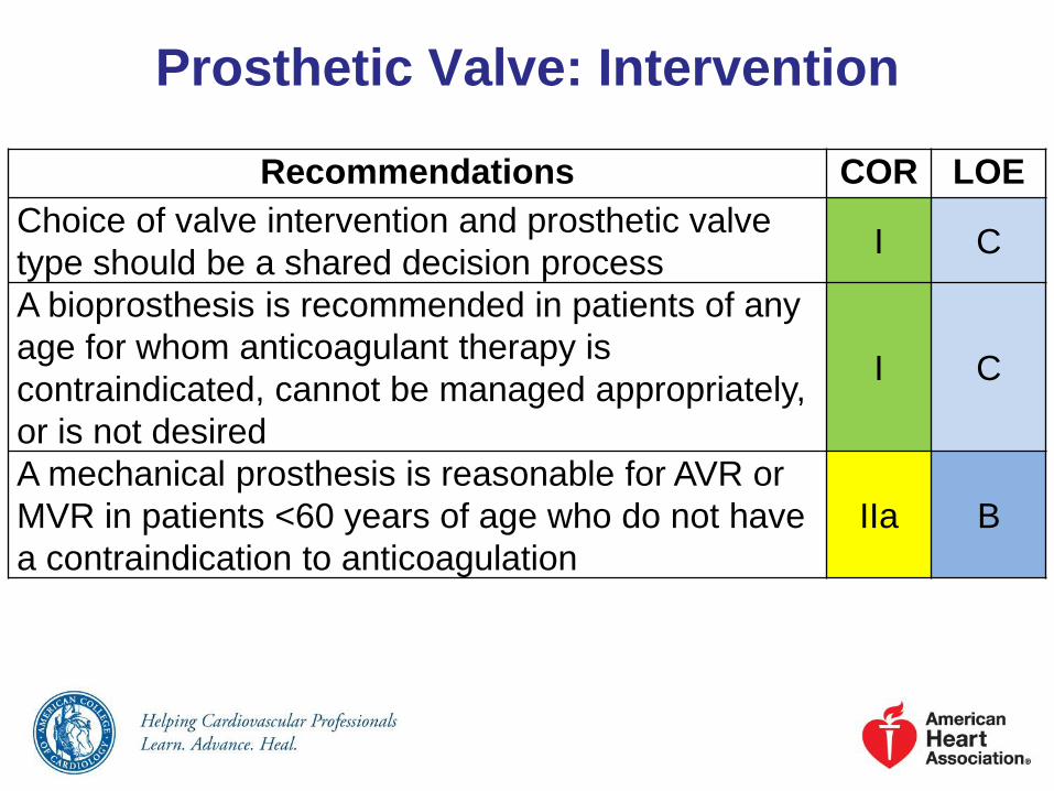
Prosthetic Valve: Intervention
Recommendations COR LOE
Choice of valve intervention and prosthetic valve
type should be a shared decision processI C
A bioprosthesis is recommended in patients of any
age for whom anticoagulant therapy is
contraindicated, cannot be managed appropriately,
or is not desired
I C
A mechanical prosthesis is reasonable for AVR or
MVR in patients <60 years of age who do not have
a contraindication to anticoagulation
IIa B

Prosthetic Valve: Intervention (cont.)
Recommendations COR LOE
A bioprosthesis is reasonable in patients >70 years
of age IIa B
Either a bioprosthetic or mechanical valve is
reasonable in patients between 60 years of age
and 70 years of age
IIa B
Replacement of the aortic valve by a pulmonary
autograft (the Ross procedure), when performed by
an experienced surgeon, may be considered in
young patients when VKA anticoagulation is
contraindicated or undesirable
IIb C
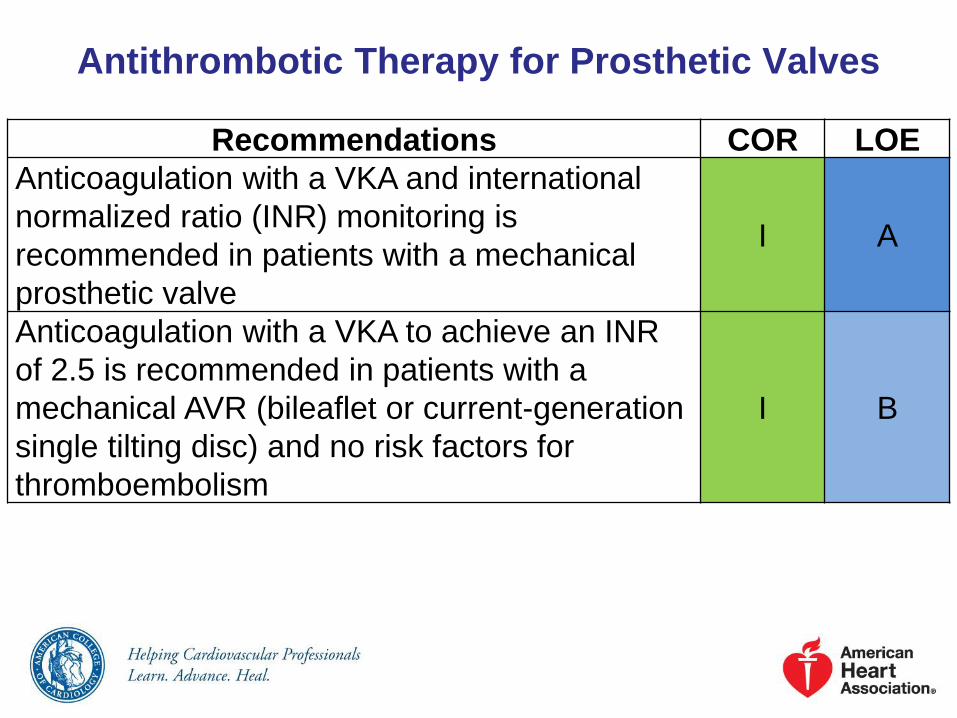
Antithrombotic Therapy for Prosthetic Valves
Recommendations COR LOE
Anticoagulation with a VKA and international
normalized ratio (INR) monitoring is
recommended in patients with a mechanical
prosthetic valve
I A
Anticoagulation with a VKA to achieve an INR
of 2.5 is recommended in patients with a
mechanical AVR (bileaflet or current-generation
single tilting disc) and no risk factors for
thromboembolism
I B
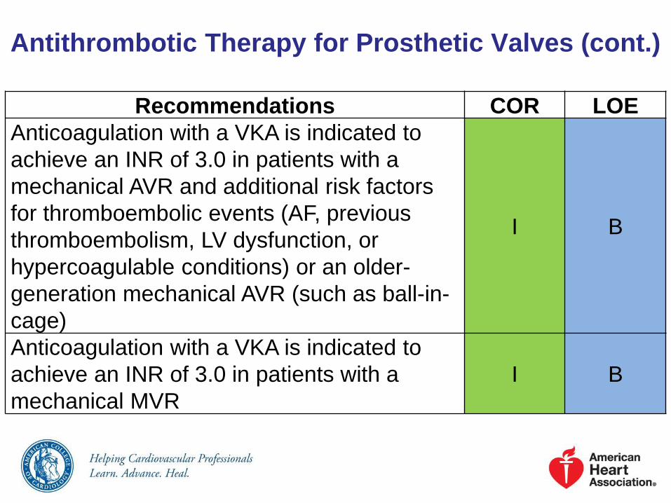
Antithrombotic Therapy for Prosthetic Valves (cont.)
Recommendations COR LOE
Anticoagulation with a VKA is indicated to
achieve an INR of 3.0 in patients with a
mechanical AVR and additional risk factors
for thromboembolic events (AF, previous
thromboembolism, LV dysfunction, or
hypercoagulable conditions) or an older-
generation mechanical AVR (such as ball-in-
cage)
I B
Anticoagulation with a VKA is indicated to
achieve an INR of 3.0 in patients with a
mechanical MVR
I B

Antithrombotic Therapy for Prosthetic Valves (cont.)
Recommendations COR LOE
Aspirin 75 mg to 100 mg daily is recommended
in addition to anticoagulation with a VKA in
patients with a mechanical valve prosthesis
I A
Aspirin 75 mg to 100 mg per day is reasonable
in all patients with a bioprosthetic aortic or
mitral valve
IIa B
Anticoagulation with a VKA is reasonable for
the first 3 months after bioprosthetic MVR or
repair to achieve an INR of 2.5
IIa C

Antithrombotic Therapy for Prosthetic Valves (cont.)
Recommendations COR LOE
Anticoagulation, with a VKA, to achieve an INR
of 2.5 may be reasonable for the first 3 months
after bioprosthetic AVR
IIb B
Clopidogrel 75 mg daily may be reasonable for
the first 6 months after TAVR in addition to life-
long aspirin 75 mg to 100 mg daily
IIb C
Anticoagulant therapy with oral direct thrombin
inhibitors or anti-Xa agents should not be used
in patients with mechanical valve prostheses
III:
HarmB

Bridging Therapy for Prosthetic Valves
Recommendations COR LOE
Continuation of VKA anticoagulation with a
therapeutic INR is recommended in patients
with mechanical heart valves undergoing minor
procedures (such as dental extractions or
cataract removal) where bleeding is easily
controlled
I C
Temporary interruption of VKA anticoagulation,
without bridging agents while the INR is
subtherapeutic, is recommended in patients
with a bileaflet mechanical AVR and no other
risk factors for thrombosis who are undergoing
invasive or surgical procedures
I C

Bridging Therapy for Prosthetic Valves (cont.)
Recommendations COR LOE
Bridging anticoagulation with either
intravenous unfractionated heparin (UFH) or
subcutaneous low-molecular-weight heparin
(LMWH) is recommended during the time
interval when the INR is subtherapeutic
preoperatively in patients who are undergoing
invasive or surgical procedures with a 1)
mechanical AVR and any thromboembolic risk
factor, 2) older-generation mechanical AVR, or
3) mechanical MVR
I C

Bridging Therapy for Prosthetic Valves (cont.)
Recommendations COR LOE
Administration of fresh frozen plasma or
prothrombin complex concentrate is
reasonable in patients with mechanical valves
receiving VKA therapy who require emergency
noncardiac surgery or invasive procedures
IIa C
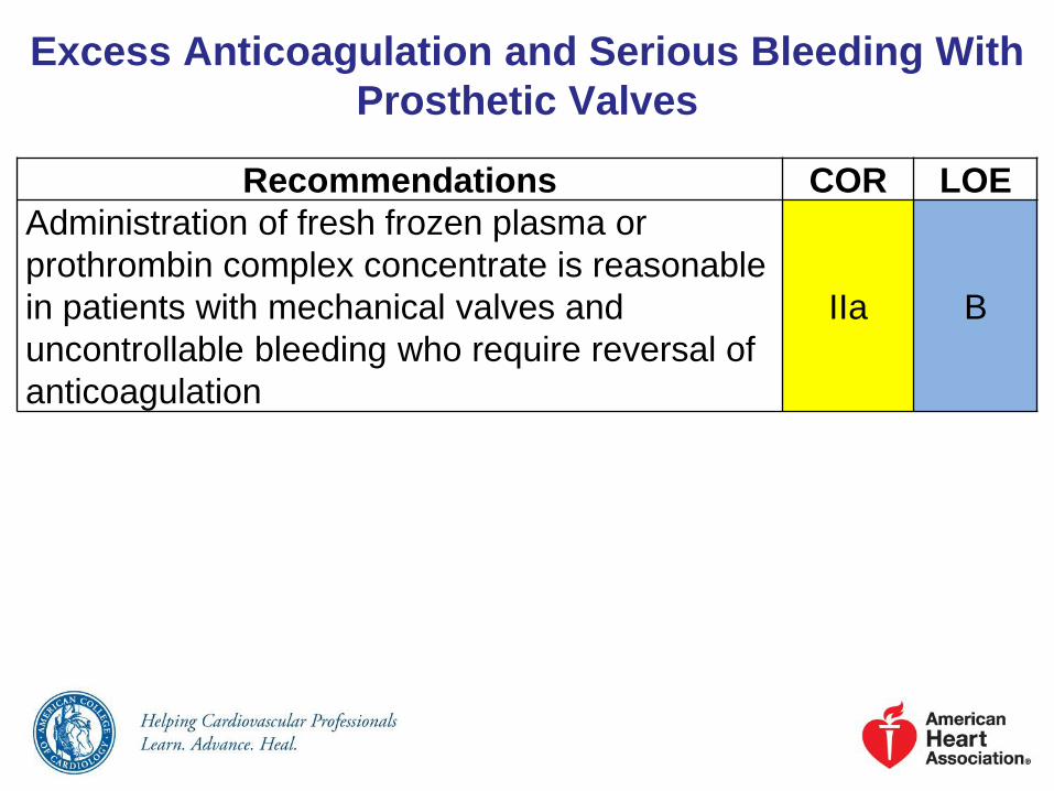
Excess Anticoagulation and Serious Bleeding With
Prosthetic Valves
Recommendations COR LOE
Administration of fresh frozen plasma or
prothrombin complex concentrate is reasonable
in patients with mechanical valves and
uncontrollable bleeding who require reversal of
anticoagulation
IIa B

Anticoagulation for Prosthetic Valves

Prosthetic Valve Thrombosis: Diagnosis and
Follow-Up
Recommendations COR LOE
TTE is indicated in patients with suspected
prosthetic valve thrombosis to assess hemodynamic
severity and follow resolution of valve dysfunction
I B
TEE is indicated in patients with suspected
prosthetic valve thrombosis to assess thrombus size
and valve motion
I B
Fluoroscopy or CT is reasonable in patients with
suspected valve thrombosis to assess valve motionIIa C

Prosthetic Valve Thrombosis: Medical Therapy
Recommendations COR LOE
Fibrinolytic therapy is reasonable for patients
with a thrombosed left-sided prosthetic heart
valve, recent onset (<14 days) of NYHA class I
to II symptoms, and a small thrombus
IIa B
Fibrinolytic therapy is reasonable for
thrombosed right-sided prosthetic heartIIa B

Prosthetic Valve Thrombosis: Intervention
Recommendations COR LOE
Emergency surgery is recommended for
patients with a thrombosed left-sided
prosthetic heart valve with NYHA class III to IV
symptoms
I B
Emergency surgery is reasonable for patients
with a thrombosed left-sided prosthetic heart
valve with a mobile or large thrombus (>0.8
cm2)
IIa C

Evaluation and Management of Suspected Prosthetic Valve Thrombosis
Suspected Prosthetic Valve
Thrombosis
Fibrinolytic Rx if persistent valve thrombosis after
IV heparin therapy*
(IIa)
Emergency Surgery
(IIa)
Right-sided prosthetic valve
thrombosis
CT or fluoroscopy to evaluate
valve motion
(IIa)
Left-sided prosthetic valve
thrombosis
Mobile or large
(≥0.8 cm2) thrombus
NYHA class III-IV
symptoms
Recent onset (<14 d)
NYHA class I-II symptoms
Small thrombus (<0.8 cm2)
TTE to evaluate
hemodynamic severity
(I)
TEE for thrombus size
(I)
Class I
Class IIa
Emergency Surgery
(I)

Prosthetic Valve Stenosis
Recommendations COR LOE
Repeat valve replacement is indicated for
severe symptomatic prosthetic valve stenosis I C

Prosthetic Valve RegurgitationRecommendations COR LOE
Surgery is recommended for operable patients with
mechanical heart valves with intractable hemolysis
or HF due to severe prosthetic or paraprosthetic
regurgitation
I B
Surgery is reasonable for operable patients with
severe symptomatic or asymptomatic bioprosthetic
regurgitation
IIa C
Percutaneous repair of paravalvular regurgitation is
reasonable in patients with prosthetic heart valves
and intractable hemolysis or NYHA class III/IV HF
who are at high risk for surgery and have anatomic
features suitable for catheter-based therapy when
performed in centers with expertise in the procedure
IIa B

Imaging Studies in Native Valve Endocarditis and
Prosthetic Valve Endocarditis

Infective Endocarditis: Diagnosis and Follow-Up
Recommendations COR LOE
At least 2 sets of blood cultures should be obtained
in patients at risk for IE (e.g., those with congenital
or acquired VHD, previous IE, prosthetic heart
valves, certain congenital or heritable heart
malformations, immunodeficiency states, or injection
drug users) who have unexplained fever for more
than 48 hours
I B
At least 2 sets of blood cultures should be obtained
in patients with newly diagnosed left-sided valve
regurgitation
I C
The Modified Duke Criteria should be used in
evaluating a patient with suspected IE (Tables 24
and 25 in the full-text guideline)
I B

Infective Endocarditis: Diagnosis and
Follow-Up (cont.)
Recommendations COR LOE
Patients with IE should be evaluated and managed
with consultation of a multispecialty Heart Valve Team
including an infectious disease specialist, cardiologist,
and cardiac surgeon. In surgically managed patients,
this team should also include a cardiac
anesthesiologist
I B
TTE is recommended in patients with suspected IE to
identify vegetations, characterize the hemodynamic
severity of valvular lesions, assess ventricular
function and pulmonary pressures, and detect
complications
I B

Infective Endocarditis: Diagnosis and
Follow-Up (cont.)
Recommendations COR LOE
TEE is recommended in all patients with known or
suspected IE when TTE is nondiagnostic, when
complications have developed or are clinically
suspected, or when intracardiac device leads are
present
I B
TTE and/or TEE are recommended for reevaluation of
patients with IE who have a change in clinical signs or
symptoms (e.g., new murmur, embolism, persistent
fever, HF, abscess, or atrioventricular heart block) and in
patients at high risk of complications (e.g., extensive
infected tissue/large vegetation on initial echocardiogram
or staphylococcal, enterococcal, or fungal infections)
I B

Infective Endocarditis: Diagnosis and
Follow-Up (cont.)
Recommendations COR LOE
Intraoperative TEE is recommended for patients
undergoing valve surgery for IEI B
TEE is reasonable to diagnose possible IE in
patients with Staphylococcal aureus bacteremia
without a known source
IIa B
TEE is reasonable to diagnose IE of a prosthetic
valve in the presence of persistent fever without
bacteremia or a new murmur
IIa B
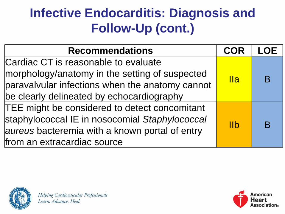
Infective Endocarditis: Diagnosis and
Follow-Up (cont.)
Recommendations COR LOE
Cardiac CT is reasonable to evaluate
morphology/anatomy in the setting of suspected
paravalvular infections when the anatomy cannot
be clearly delineated by echocardiography
IIa B
TEE might be considered to detect concomitant
staphylococcal IE in nosocomial Staphylococcal
aureus bacteremia with a known portal of entry
from an extracardiac source
IIb B
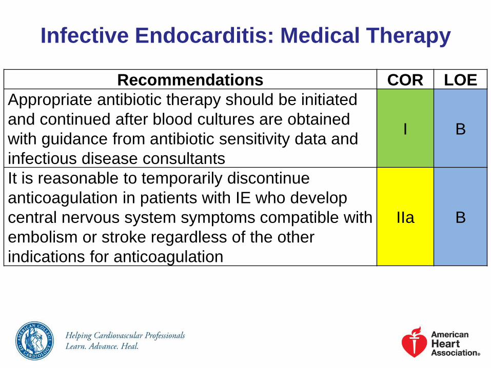
Infective Endocarditis: Medical Therapy
Recommendations COR LOE
Appropriate antibiotic therapy should be initiated
and continued after blood cultures are obtained
with guidance from antibiotic sensitivity data and
infectious disease consultants
I B
It is reasonable to temporarily discontinue
anticoagulation in patients with IE who develop
central nervous system symptoms compatible with
embolism or stroke regardless of the other
indications for anticoagulation
IIa B
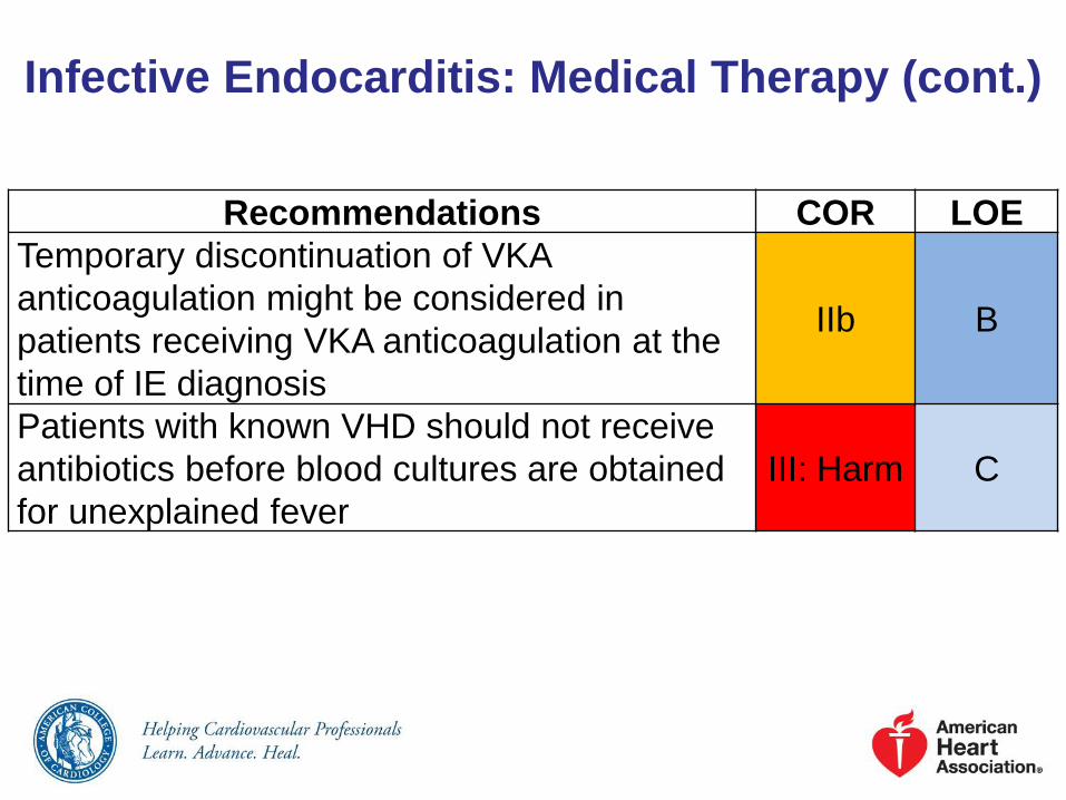
Infective Endocarditis: Medical Therapy (cont.)
Recommendations COR LOE
Temporary discontinuation of VKA
anticoagulation might be considered in
patients receiving VKA anticoagulation at the
time of IE diagnosis
IIb B
Patients with known VHD should not receive
antibiotics before blood cultures are obtained
for unexplained fever
III: Harm C

Infective Endocarditis: Intervention
Recommendations COR LOE
Decisions about timing of surgical intervention
should be made by a multispecialty Heart Valve
Team of cardiology, cardiothoracic surgery, and
infectious disease
I B
Early surgery (during initial hospitalization
before completion of a full therapeutic course of
antibiotics) is indicated in patients with IE who
present with valve dysfunction resulting in
symptoms of HF
I B

Infective Endocarditis: Intervention (cont.)
Recommendations COR LOE
Early surgery (during initial hospitalization
before completion of a full therapeutic course of
antibiotics) is indicated in patients with left-sided
IE caused by Staphylococcal aureus, fungal, or
other highly resistant organisms
I B
Early surgery (during initial hospitalization
before completion of a full therapeutic course of
antibiotics) is indicated in patients with IE
complicated by heart block, annular or aortic
abscess, or destructive penetrating lesions
I B

Infective Endocarditis: Intervention (cont.)Recommendations COR LOE
Early surgery (during initial hospitalization before
completion of a full therapeutic course of
antibiotics) for IE is indicated in patients with
evidence of persistent infection as manifested by
persistent bacteremia or fevers lasting longer than
5 to 7 days after onset of appropriate antimicrobial
therapy
I B
Surgery is recommended for patients with
prosthetic valve endocarditis and relapsing infection
(defined as recurrence of bacteremia after a
complete course of appropriate antibiotics and
subsequently negative blood cultures) without other
identifiable source for portal of infection
I C

Infective Endocarditis: Intervention (cont.)
Recommendations COR LOE
Complete removal of pacemaker or defibrillator
systems, including all leads and the generator, is
indicated as part of the early management plan
in patients with IE with documented infection of
the device or leads
I B
Complete removal of pacemaker or defibrillator
systems, including all leads and the generator, is
reasonable in patients with valvular IE caused by
Staphylococcal aureus or fungi, even without
evidence of device or lead infection
IIa B

Infective Endocarditis: Intervention (cont.)
Recommendations COR LOE
Complete removal of pacemaker or
defibrillator systems, including all leads and
the generator, is reasonable in patients
undergoing valve surgery for valvular IE
IIa C
Early surgery (during initial hospitalization
before completion of a full therapeutic course
of antibiotics) is reasonable in patients with IE
who present with recurrent emboli and
persistent vegetations despite appropriate
antibiotic therapy
IIa B

Infective Endocarditis: Intervention (cont.)
Recommendations COR LOE
Early surgery (during initial hospitalization before
completion of a full therapeutic course of
antibiotics) may be considered in patients with
native valve endocarditis who exhibit mobile
vegetations greater than 10 mm in length (with
or without clinical evidence of embolic
phenomenon)
IIb B

Diagnosis and Treatment of Infective Endocarditis

Native Valve Stenosis
Recommendations COR LOE
All patients with suspected valve stenosis should
undergo a clinical evaluation and TTE before
pregnancy
I C
All patients with severe valve stenosis (stages C
and D) should undergo prepregnancy counseling
by a cardiologist with expertise in managing
patients with VHD during pregnancy
I C
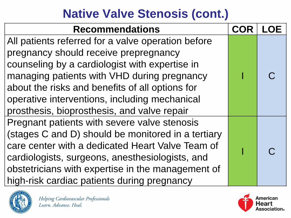
Native Valve Stenosis (cont.)
Recommendations COR LOE
All patients referred for a valve operation before
pregnancy should receive prepregnancy
counseling by a cardiologist with expertise in
managing patients with VHD during pregnancy
about the risks and benefits of all options for
operative interventions, including mechanical
prosthesis, bioprosthesis, and valve repair
I C
Pregnant patients with severe valve stenosis
(stages C and D) should be monitored in a tertiary
care center with a dedicated Heart Valve Team of
cardiologists, surgeons, anesthesiologists, and
obstetricians with expertise in the management of
high-risk cardiac patients during pregnancy
I C

Pregnancy and VHD: Diagnosis and
Follow-Up
Recommendations COR LOE
Exercise testing is reasonable in asymptomatic
patients with severe AS (aortic velocity ≥4 m per
second or mean pressure gradient ≥40 mm Hg,
stage C) before pregnancy
IIa C
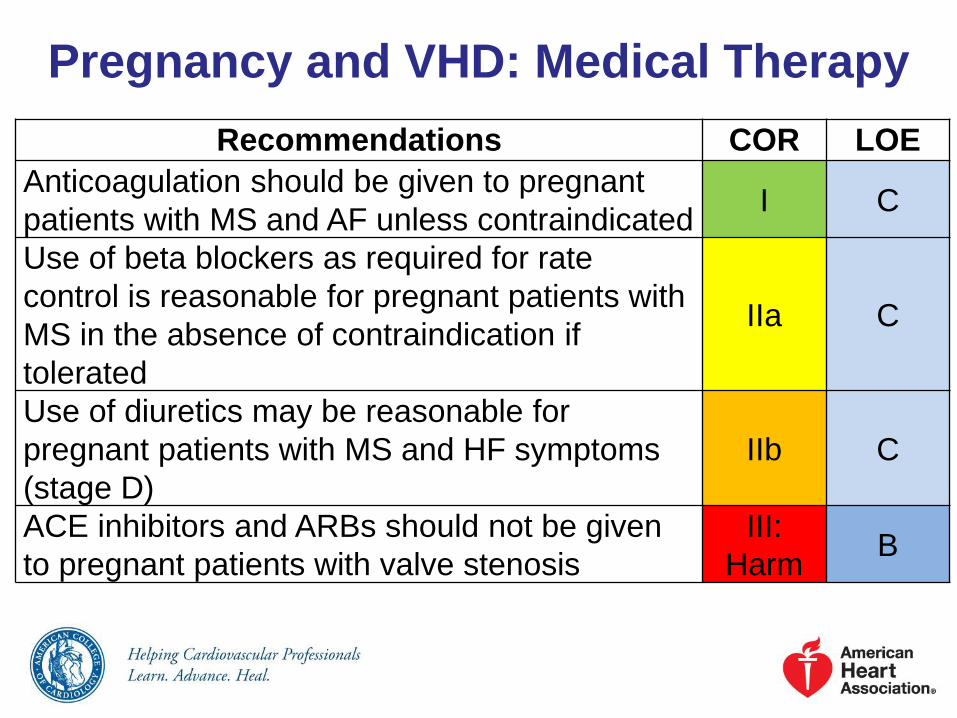
Pregnancy and VHD: Medical Therapy
Recommendations COR LOE
Anticoagulation should be given to pregnant
patients with MS and AF unless contraindicatedI C
Use of beta blockers as required for rate
control is reasonable for pregnant patients with
MS in the absence of contraindication if
tolerated
IIa C
Use of diuretics may be reasonable for
pregnant patients with MS and HF symptoms
(stage D)
IIb C
ACE inhibitors and ARBs should not be given
to pregnant patients with valve stenosis
III:
HarmB

Pregnancy and VHD: InterventionRecommendations COR LOE
Valve intervention is recommended before
pregnancy for symptomatic patients with severe
AS (aortic velocity ≥4.0 m per second or mean
pressure gradient ≥40 mm Hg, stage D)
I C
Valve intervention is recommended before
pregnancy for symptomatic patients with severe
MS (mitral valve area ≤1.5 cm2, stage D)
I C
Percutaneous mitral balloon commissurotomy is
recommended before pregnancy for
asymptomatic patients with severe MS (mitral
valve area ≤1.5 cm2, stage C) who have valve
morphology favorable for percutaneous mitral
balloon commissurotomy
I C

Pregnancy and VHD: Intervention (cont.)
Recommendations COR LOE
Valve intervention is reasonable before pregnancy
for asymptomatic patients with severe AS (aortic
velocity ≥4.0 m per second or mean pressure
gradient ≥40 mm Hg, stage C)
IIa C
Percutaneous mitral balloon commissurotomy is
reasonable for pregnant patients with severe MS
(mitral valve area ≤1.5 cm2, stage D) with valve
morphology favorable for percutaneous mitral
balloon commissurotomy who remain
symptomatic with NYHA class III to IV HF
symptoms despite medical therapy
IIa B

Pregnancy and VHD: Intervention (cont.)
Recommendations COR LOE
Valve intervention is reasonable for pregnant
patients with severe MS (mitral valve area ≤1.5
cm2, stage D) and valve morphology not favorable
for percutaneous mitral balloon commissurotomy
only if there are refractory NYHA class IV HF
symptoms
IIa C
Valve intervention is reasonable for pregnant
patients with severe AS (mean pressure gradient
≥40 mm Hg, stage D) only if there is
hemodynamic deterioration or NYHA class III to IV
HF symptoms
IIa B

Pregnancy and VHD: Intervention (cont.)
Recommendations COR LOE
Valve operation should not be performed in
pregnant patients with valve stenosis in the
absence of severe HF symptoms
III: Harm C

Native Valve Regurgitation: Diagnosis
and Follow-Up
Recommendations COR LOE
All patients with suspected valve regurgitation
should undergo a clinical evaluation and TTE
before pregnancy
I C
All patients with severe valve regurgitation
(stages C and D) should undergo
prepregnancy counseling by a cardiologist
with expertise in managing patients with VHD
during pregnancy
I C

Native Valve Regurgitation: Diagnosis
and Follow-Up (cont.)
Recommendations COR LOE
All patients referred for a valve operation
before pregnancy should receive prepregnancy
counseling by a cardiologist with expertise in
managing patients with VHD during pregnancy
regarding the risks and benefits of all options
for operative interventions, including
mechanical prosthesis, bioprosthesis, and
valve repair
I C

Native Valve Regurgitation: Diagnosis
and Follow-Up (cont.)
Recommendations COR LOE
Pregnant patients with severe regurgitation
(stages C and D) should be monitored in a
tertiary care center with a dedicated Heart
Valve Team of cardiologists, surgeons,
anesthesiologists, and obstetricians with
expertise in managing high-risk cardiac
patients
I C
Exercise testing is reasonable in
asymptomatic patients with severe valve
regurgitation (stage C) before pregnancy
IIa C

Native Valve Regurgitation: Medical
Therapy
Recommendations COR LOE
ACE inhibitors and ARBs should not be
given to pregnant patients with valve III: Harm B

Native Valve Regurgitation: Intervention
Recommendations COR LOE
Valve repair or replacement is recommended
before pregnancy for symptomatic women with
severe valve regurgitation (stage D)
I C
Valve operation for pregnant patients with
severe valve regurgitation is reasonable only if
there are refractory NYHA class IV HF
symptoms (stage D)
IIa C

Native Valve Regurgitation:
Intervention (cont.)
Recommendations COR LOE
Valve repair before pregnancy may be
considered in the asymptomatic patient with
severe MR (stage C) and a valve suitable for
valve repair, but only after detailed discussion
with the patient about the risks and benefits of
the operation and its outcome on future
pregnancies
IIb C
Valve operations should not be performed in
pregnant patients with valve regurgitation in
the absence of severe intractable HF
symptoms
III: Harm C

Prosthetic Valves in Pregnancy:
Diagnosis and Follow-UpRecommendations COR LOE
All patients with a prosthetic valve should
undergo a clinical evaluation and baseline TTE
before pregnancy
I C
All patients with a prosthetic valve should
undergo prepregnancy counseling by a
cardiologist with expertise in managing
patients with VHD during pregnancy.
I C
TTE should be performed in all pregnant
patients with a prosthetic valve if not done
before pregnancy
I C

Prosthetic Valves in Pregnancy:
Diagnosis and Follow-Up (cont.)
Recommendations COR LOE
Repeat TTE should be performed in all
pregnant patients with a prosthetic valve who
develop symptoms
I C
TEE should be performed in all pregnant
patients with a mechanical prosthetic valve
who have prosthetic valve obstruction or
experience an embolic event
I C

Prosthetic Valves in Pregnancy:
Diagnosis and Follow-Up (cont.)
Recommendations COR LOE
Pregnant patients with a mechanical
prosthesis should be monitored in a tertiary
care center with a dedicated Heart Valve Team
of cardiologists, surgeons, anesthesiologists,
and obstetricians with expertise in the
management of high-risk cardiac patients
I C
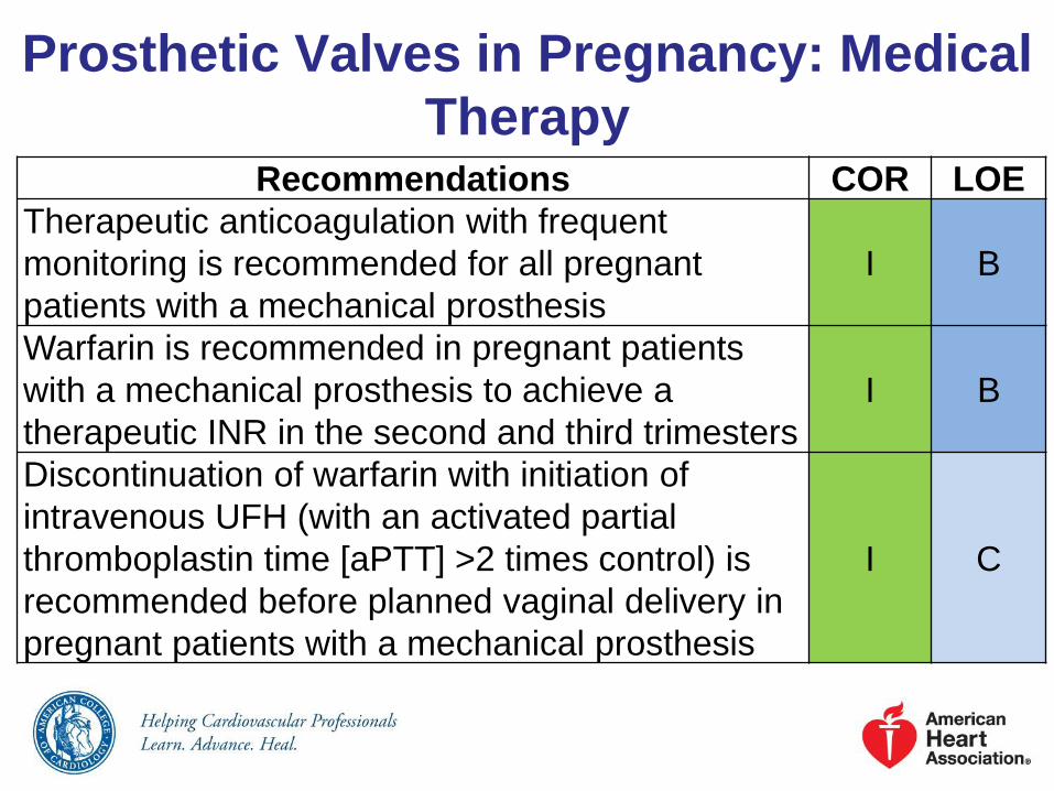
Prosthetic Valves in Pregnancy: Medical
TherapyRecommendations COR LOE
Therapeutic anticoagulation with frequent
monitoring is recommended for all pregnant
patients with a mechanical prosthesis
I B
Warfarin is recommended in pregnant patients
with a mechanical prosthesis to achieve a
therapeutic INR in the second and third trimesters
I B
Discontinuation of warfarin with initiation of
intravenous UFH (with an activated partial
thromboplastin time [aPTT] >2 times control) is
recommended before planned vaginal delivery in
pregnant patients with a mechanical prosthesis
I C

Prosthetic Valves in Pregnancy: Medical
Therapy (cont.)
Recommendations COR LOE
Low-dose aspirin (75 mg to 100 mg) once per day
is recommended for pregnant patients in the
second and third trimesters with either a
mechanical prosthesis or bioprosthesis
I C
Continuation of warfarin during the first trimester is
reasonable for pregnant patients with a mechanical
prosthesis if the dose of warfarin to achieve a
therapeutic INR is 5 mg per day or less after full
discussion with the patient about risks and benefits
IIa B

Prosthetic Valves in Pregnancy: Medical
Therapy (cont.)
Recommendations COR LOE
Dose-adjusted LMWH at least 2 times per day (with a
target anti-Xa level of 0.8 U/mL to 1.2 U/mL, 4 to 6
hours postdose) during the first trimester is reasonable
for pregnant patients with a mechanical prosthesis if the
dose of warfarin is greater than 5 mg per day to achieve
a therapeutic INR
IIa B
Dose-adjusted continuous intravenous UFH (with an
aPTT at least 2 times control) during the first trimester
is reasonable for pregnant patients with a mechanical
prosthesis if the dose of warfarin is greater than 5 mg
per day to achieve a therapeutic INR
IIa B

Prosthetic Valves in Pregnancy: Medical
Therapy (cont.)Recommendations COR LOE
Dose-adjusted LMWH at least 2 times per day (with
a target anti-Xa level of 0.8 U/mL to 1.2 U/mL, 4 to 6
hours postdose) during the first trimester may be
reasonable for pregnant patients with a mechanical
prosthesis if the dose of warfarin is 5 mg per day or
less to achieve a therapeutic INR
IIb B
Dose-adjusted continuous infusion of UFH (with
aPTT at least 2 times control) during the first
trimester may be reasonable for pregnant patients
with a mechanical prosthesis if the dose of warfarin
is 5 mg per day or less to achieve a therapeutic INR
IIb B

Prosthetic Valves in Pregnancy: Medical
Therapy (cont.)
Recommendations COR LOE
LMWH should not be administered to pregnant
patients with mechanical prostheses unless
anti-Xa levels are monitored 4 to 6 hours after
administration
III:
HarmB

Anticoagulation of Pregnant Patients With Mechanical Valves

Evaluation of Coronary Anatomy
Recommendations COR LOE
Coronary angiography is indicated before valve
intervention in patients with symptoms of
angina, objective evidence of ischemia,
decreased LV systolic function, history of CAD,
or coronary risk factors (including men age >40
years and postmenopausal women)
I C
Coronary angiography should be performed as
part of the evaluation of patients with chronic
severe secondary MR
I C

Evaluation of Coronary Anatomy (cont.)
Recommendations COR LOE
Surgery without coronary angiography is
reasonable for patients having emergency
valve surgery for acute valve regurgitation,
disease of the aortic sinuses or ascending
aorta, or IE
IIa C
CT coronary angiography is reasonable to
exclude the presence of significant obstructive
CAD in selected patients with a
low/intermediate pretest probability of CAD. A
positive coronary CT angiogram (the presence
of any epicardial CAD) can be confirmed with
invasive coronary angiography
IIa B
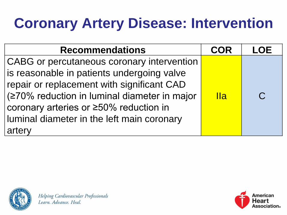
Coronary Artery Disease: Intervention
Recommendations COR LOE
CABG or percutaneous coronary intervention
is reasonable in patients undergoing valve
repair or replacement with significant CAD
(≥70% reduction in luminal diameter in major
coronary arteries or ≥50% reduction in
luminal diameter in the left main coronary
artery
IIa C

Evaluation and Management of Coronary Artery Disease in
Patients Undergoing Valve Surgery

Atrial Fibrillation: Intervention
Recommendations COR LOE
A concomitant maze procedure is reasonable at
the time of mitral valve repair or replacement for
treatment of chronic, persistent AF
IIa C
A full bi-atrial maze procedure, when technically
feasible, is reasonable at the time of mitral valve
surgery, compared with a lesser ablation
procedure, in patients with chronic, persistent AF
IIa B
A concomitant maze procedure or pulmonary vein
isolation may be considered at the time of mitral
valve repair or replacement in patients with
paroxysmal AF that is symptomatic or associated
with a history of embolism on anticoagulation
IIb C

Atrial Fibrillation: Intervention (cont.)
Recommendations COR LOE
Concomitant maze procedure or pulmonary vein
isolation may be considered at the time of
cardiac surgical procedures other than mitral
valve surgery in patients with paroxysmal or
persistent AF that is symptomatic or associated
with a history of emboli on anticoagulation
IIb C
Catheter ablation for AF should not be
performed in patients with severe MR when
mitral repair or replacement is anticipated, with
preference for the combined maze procedure
plus mitral valve repair
III: No
BenefitB

Noncardiac Surgery in Patients With VHD
Recommendations COR LOE
Moderate-risk elective noncardiac surgery with
appropriate intraoperative and postoperative
hemodynamic monitoring is reasonable to
perform in patients with asymptomatic severe
AS
IIa B
Moderate-risk elective noncardiac surgery with
appropriate intraoperative and postoperative
hemodynamic monitoring is reasonable to
perform in patients with asymptomatic severe
MR
IIa C
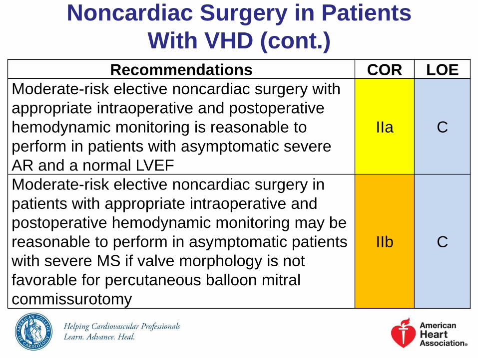
Noncardiac Surgery in Patients
With VHD (cont.)Recommendations COR LOE
Moderate-risk elective noncardiac surgery with
appropriate intraoperative and postoperative
hemodynamic monitoring is reasonable to
perform in patients with asymptomatic severe
AR and a normal LVEF
IIa C
Moderate-risk elective noncardiac surgery in
patients with appropriate intraoperative and
postoperative hemodynamic monitoring may be
reasonable to perform in asymptomatic patients
with severe MS if valve morphology is not
favorable for percutaneous balloon mitral
commissurotomy
IIb C