2014 taylor & francis group, llc issn ... - vanderlick lab
TRANSCRIPT

Water-Soluble Anisotropic Iron Oxide Nanoparticles:Dextran-Coated Crystalline Nanoplates and Nanoflowers
SOUBANTIKA PALCHOUDHURY,1 FAHMEED HYDER,2 T. KYLE VANDERLICK,1 and NIENKE GEERTS1
1Department of Chemical and Environmental Engineering, Yale University, New Haven, Connecticut, USA2Department of Biomedical Engineering and Diagnostic Radiology, Yale University, New Haven, Connecticut, USA
We report a simple phase transfer based synthesis route for two novel anisotropic water soluble iron oxide nanoparticle shapes,namely, nanoplates and nanoflowers. The nanoplates and nanoflowers are initially prepared in an organic solvent via a modified‘‘heat-up’’ method. Then, the crystalline nanoparticles are rendered hydrophilic via sonication in the presence of dextran and water.These nanoparticles are highly monodisperse and superparamagnetic at room temperature. High resolution transmission electronmicroscopy indicates that the iron oxides cores are not affected by the phase transfer. Dextran coating is confirmed by dynamiclight scattering, Fourier transform infrared spectroscopy, and thermogravimetric analysis. The obtained dextran coverage was26wt% for the nanoplates and 37wt% for the nanoflowers. The nanoplates and nanoflowers were not only water soluble, but alsoremained stable at different pH (4–7) and in common aqueous buffer solutions. Thorough characterizations of the nonsphericaliron oxide nanoparticles indicate that these particles could be useful for potential biomedical applications and magnetic resonanceimaging.
Keywords: Materials, nanoparticles, nonspherical particle, particle characteristics, particle shape
1. Introduction
Iron oxide nanoparticles (NPs) have been extensively studiedfor biomedical applications, such as targeted drug delivery,localized cancer therapy, and as contrast agents for magneticresonance imaging (MRI) (Sonvico et al. 2005; Sun et al.2008; Laurent et al. 2008; Fang and Zhang 2009; Yiu et al.2013). These extensive studies led to the realization ofclinically approved magnetic iron oxide particles, such asFerridex and Ferucarbotran. Although these particles arecurrently extensively used, their shape and preparationmethod present two limitations. First, the mentioned studiesfocus on spherical NPs, primarily because the synthesis ofanisotropic iron oxide NPs is more challenging. Second,due to their preparation method, the materials are often oflow crystallinity.
Compared to nanospheres, nonspherical iron oxide NPshave shown promising advantages in biomedical applica-tions. Nonspherical NPs could be preferred because of theirshape-dependent applications in sensing and catalysis (Junet al. 2006). Anisotropic worm-like shaped particles exhibitedprolonged blood circulation times (Park et al. 2008), andnonspherical NPs showed increased intercellular adhesionand retention in tumors (Tao et al. 2008). Therefore, an
emerging strategy is to control the shape-dependentproperties of iron oxide NPs for biological applications.Although the above mentioned anisotropic NPs show greatpromise, their preparation method (co-precipitation) posesthe second limitation.
Particles produced by a co-precipitation method resultsinto iron oxide NPs with a wide size distribution and lowcrystallinity. As these parameters directly influence themagnetic properties of these NPs, it limits their perfor-mances as magnetic resonance imaging (MRI) contrastagents. High quality iron oxide NPs regarding monodisper-sity, size distribution, and crystallinity can currently only beproduced in organic solvents at high temperatures. This socalled ‘‘heat-up’’ method generates iron oxide NPs with agreat amount of control over particle size, composition,and shape (Park et al. 2004). A well-controlled synthesis ofiron oxide nanoplates and nanoflowers in organic solventwas recently reported. These novel NPs could be potentiallyuseful in exploring the shape-dependent bio-applications(Palchoudhury et al. 2012). For biological applications, sur-face modification to achieve water soluble NPs is essential.Typically, the NPs can be transferred from organic solventsto aqueous solutions through attachment of amphiphilicligands (Gonzales and Krishnan 2007; Prakash et al. 2009;Yu et al. 2006) or replacement of the hydrophobic coatingsby hydrophilic molecules (Xu et al. 2011; Binder et al. 2008).
Here, we report the transfer of crystalline iron oxidenanoplates and nanoflowers into the aqueous phase withthe use of dextran. Although the reproducible synthesis of
Address correspondence to: Nienke Geerts, Department ofChemical and Environmental Engineering, Yale University,New Haven, CT 06511, USA. E-mail: [email protected] versions of one or more of the figures in the article can befound online at www.tandfonline.com/upst.
Particulate Science and Technology, 32: 224–233
Copyright # 2014 Taylor & Francis Group, LLC
ISSN: 0272-6351 print=1548-0046 online
DOI: 10.1080/02726351.2013.850460

these anisotropic iron oxide particles has been reportedbefore (Palchoudhury et al. 2012), this is to our knowledgethe first attempt at rendering the NPs hydrophilic using adextran based bilayer phase transfer (Figure 1). The phasetransfer through attachment of amphiphilic ligands is nota trivial execution as this often results in multiple NPs beingcollectively coated within an envelope of the capping mol-ecule (Euliss et al. 2003). Full ligand replacement is in thiscase not desirable, while the organic ligands are partiallyresponsible for the anisotropic shapes. Dextran was selectedas it is readily available, inexpensive, and approved as acoating for NPs in clinical studies by the Food & DrugAdministration (FDA) (Reimer and Balzer 2003).
To confirm the surface attachment of dextran, the ironoxide nanoplates and nanoflowers are extensively character-ized. Transmission electron microscopy (TEM) is used toconfirm the shape and size of the NPs. With two dimensionalpower x-ray diffraction (XRD2) the crystallinity of the NPsis further confirmed. The hydrodynamic dimensions of theaqueous nanoplate and nanoflower dispersions were mea-sured with dynamic light scattering (DLS). Fourier trans-form infrared spectroscopy (FTIR) is used to show thechange in outer surface ligands after the phase transfer fromorganic solvents to aqueous solutions, while thermogravi-metric analysis (TGA) was used to examine the completecoating. Moreover, magnetic measurements confirmed thesuperparamagnetic nature of the water soluble particles.Additionally, the stability of the dextran-coated nanoplatesand nanoflowers was characterized over a 28 day period atdifferent pH and for three commonly used buffer solutions(phosphate buffered saline (PBS), saline sodium citrate(SSC), and 4-(2-hydroxyethyl)-1-piperazineethanesulfonicacid (HEPES)).
In summary, this study provides a phase transfer basedfacile route for the formation of crystalline dextran-coatediron oxide NPs of two different anisotropic shapes. Theseresults will be useful for the growing application of nonsphe-rical NPs in biomedical fields (Geng et al. 2007; Zhao et al.2013) and potentially even for blood volume imaging withMRI where dextran-coated iron oxide NPs have high speci-ficity for the plasma compartment (Herman et al. 2009).
2. Materials and Methods
2.1 Materials
Reagents were used as purchased, including: iron (III)chloride (anhydrous, 98%, Alfa Aesar, Ward Hill, MA,USA), sodium oleate (Spectrum), 1-octadecene (90%, Acros,Bridgewater, MA, USA), tri-n-octylphosphine oxide (TOPO,90%, Sigma-Aldrich), oleic acid (OA, 90%, Alfa Aesar), dex-tran (high fraction, MW¼ 250 000, Acros), chloroform(�99.8%, Sigma Aldrich, St. Louis, MO, USA), acetone(�99.5%, Sigma Aldrich), n-hexane (Sigma Aldrich), ethylalcohol (Pharmco Aaper, Brookfield, CT, USA), 4-(2-hydroxyethyl)-1-piperazineethanesulfonic acid (HEPES,�99.5%, Sigma), saline sodium citrate (SSC, Life Technolo-gies, Grand Island, NY, USA), and phosphate buffered saline(PBS, Sigma).
2.2 Synthesis of the Iron Oxide Nanoplates and Nanoflowers
Iron oxide nanoplates and nanoflowers were prepared fromthe thermal decomposition of an iron oleate complex, follow-ing a similar procedure reported earlier (Palchoudhury et al.2012) The iron oleate precursor was prepared as follows:sodium oleate (36.5 g) and ferric chloride (6.5 g) were mixedin a solvent mixture (hexane, 140mL; ethanol, 80mL;de-ionized water, DI, 60mL) at 60�C. After phase separ-ation, the organic phase containing iron oleate complexwas washed three times with deionized (DI) water to removeby-products such as KCl. The resulting product, an iron ole-ate complex with 6wt% hexane, served as the precursor forsubsequent nanoplate and nanoflower synthesis.
To obtain nanoplates and nanoflowers the iron oleatehexane precursor solution (1.82 g) was heated at 290�C for1 h in 1-octadecene (13mL) in the presence of two additionalligands, OA (0.1mL) and TOPO (0.2 g nanoplates and 1 gnanoflowers). The crystalline products were precipitated outof the solution with an acetone=chloroformmixture, magneti-cally separated to wash off 1-octadecene and unreactedorganics, and vacuum dried overnight. The well driedpowders of nanoplates and nanoflowers were redispersed inhexane via sonication to obtain stock solutions of 50mg=mL.
Fig. 1. Schematic illustration of the addition of dextran to make the iron-oxide particles water soluble. A full ligand exchange wasnot attempted as this will affect the shape of the particles.
Water-Soluble Anisotropic Iron Oxide Nanoparticles 225

2.3 Calculation for the Quantity of Dextran Required forEffective Phase Transfer
The molar ratio of dextran to Fe atoms on nanoparticle (NP)surface was targeted at 5:1. The fraction of surface iron atomswas computed from the total number of atoms per particle asfollows. Nanoplates and nanoflowers in 1mL of the respectivestock solutions (50mg=mL) were well dried for thephase transfer procedure. First, the number of nanoplatesand nanoflowers were calculated via dividing the weight used(50mg) by the mass of a single NP (4=3pr3q). Here, q is thedensity of bulk iron oxide (c-Fe2O3, 5.24 g=cm
3 for nanoplatesand Fe3O4, 5.18 g=cm
3 for nanoflowers) and r is the longestradius of the NPs estimated from transmission electronmicroscopy (TEM) images. To estimate the moles of nano-plates and nanoflowers used, the resultant number wasdivided by the Avogadro’s constant (NA¼ 6.023� 1023).Second, the cubic reactant on the nanoplate and nanoflowersurface was computed using the formula, 4=3pr3 (i.e., NPvolume)=volume of cubic unit cell (cell parameter 8.5 A).Third, the fraction of unit cells on the nanoplate and nano-flower surface was estimated to 18%. Each of these unit cellswas assumed to contribute a surface Fe atom. Therefore, thenumber of iron atoms on the nanoplate and nanoflower sur-face was obtained by multiplying the moles of NP with thesurface cubic reactant and the surface unit cells. The molesof dextran used for phase transfer was set at 5 times the calcu-lated number of Fe atoms on the NP surfaces.
2.4 Phase Transfer of the Nanoplates and Nanoflowers
The nanoplate or nanoflower stock solution (1mL) was mixedwith dextran (MW 250 000) such that the molar ratio ofbiocompatible ligand-to-NP surface Fe atoms was approxi-mately set to 5:1 (Park et al. 2004). After sonication (5min),slight mixing was observed in this solution. Ultra-pure water(5mL, Millipore) was added to the nanoplate-dextransolution forming two phase separated layers. To transfer theplates or flowers to the aqueous phase, the solution wasvortexed (Vortex genie 2; Scientific Industries, room tempera-ture (RT)) and sonicated (Branson 5510 sonicator; FisherScientific, Waltham, MA, USA, RT) for 30min. Thehomogeneous solution was left to phase separate overnight.Next, the aqueous dispersion of dextran-iron oxide nanoplateswas collectedwith a syringe from the aqueous phase. The nano-plates weremagnetically separated, weighed, and redispersed inwater, prior to filtration through a syringe filter (pore size0.2mm, Whatman, Pittsburgh, PA, USA). The concentrationof the nanoplates in the final aqueous dispersion was estimatedto be 5mg=mL. The water soluble particles were stable at RTfor months without notable precipitation.
2.5 Characterization
The size and morphology of the iron oxide nanoplates andnanoflowers were investigated on a FEI Tecnai Osiris trans-mission electron microscope (TEM) using a double tiltholder. The nanoplates and nanoflowers in organic phasewere centrifuged (1500 rpm, 15min RT, EppendorfMiniSpin) out with 1:1 ethanol=hexane (by volume),
redispersed in hexane, and dropped on TEM grids forviewing. The aqueous phase samples were imaged withoutfurther treatment. Size histograms were obtained by measur-ing and plotting the size of 100 NPs. The hydrodynamicdiameter and zeta potential of the nanoplates and nano-flowers were measured on a Malvern, Zetasizer nano DLS.The crystal structure of the dextran coated nanoplates andnanoflowers were examined with 2D powder diffraction(XRD2) using a Cu source (Rigaku; MicroMax-007HFgenerator and Saturn 944þ CCD detector; using 2h scans(20–80�) and Ka1, k¼ 1.5416 A). Magnetic properties ofthe hydrophobic and aqueous phase nanoplates and nano-flowers were determined on a superconducting quantuminterference device (SQUID) magnetometer at RT.
The surface attachment of dextran on the nanoplates andnanoflowers was evaluated with a Nicolet 6700 (ThermoScientific) Fourier transform infrared spectrophotometer(FTIR), and thermogravimetric analyzer (TGA Q1000, TAInstruments). Powdered samples for XRD2, SQUID,TGA, and FTIR were prepared via centrifugation and over-night vacuum drying.
2.6 Stability Tests
The pH-dependent tests were conducted by carefully adjust-ing the nanoplate and nanoflower suspensions in water(pH 7, 1mL) to the target values (pH 4, 5, 6, and 8) withHCl or NaOH, keeping all other parameters the same. Theadjusted NP dispersions were sonicated (Branson 5510;Fisher Scientific) and left undisturbed till no change in pHwas observed. The hydrodynamic diameter and zeta potentialof these nanoplates and nanoflowers at different pH weremeasured over time (0, 7, 14, 21, and 28 days) using DLS.
The stability study was followed by examining the hydro-dynamic diameter of nanoplates and nanoflowers in threecommon buffers (PBS, SSC, and HEPES). HEPES powderwas dissolved in ultra-pure water via hand-shaking toachieve target aqueous buffer concentrations (0.1, 0.2, 0.5,0.8, and 1M). PBS (10 X; 80 g NaCl, 2 g KCl, 14.4 gNa2HPO4, and 2.4 g KH2PO4 in 950ml H2O) and SSC(10 X; 1.5M NaCl and 0.15M sodium citrate) were usedas purchased. These buffers were diluted with ultra-purewater to obtain the targeted concentrations (1, 2, 5, 8, and10 X). Final volumes of the HEPES, PBS, and SSC buffersof different concentrations were kept the same (5mL). TheNPs were added at target buffer concentrations (0.1, 0.2,0.5, 0.8, and 1M). Aqueous dispersions of the NPs (5mL)were added to each buffer solution, prior to mixing bysonication (Branson 5510; Fisher Scientific). Well mixednanoplate and nanoflower suspensions in the three bufferswere measured for their hydrodynamic diameter and zetapotential with DLS.
3. Results
This work presents the phase transfer of two hydrophobicanisotropic iron oxide NPs, into the aqueous phase byattachment of dextran to the outer ligand surface. Theexchange process is illustrated in Figure 1, where dextran
226 Palchoudhury et al.

is added in addition to the two original coatings of thehydrophobic iron oxide NPs (oleic acid: OA) [Huang et al.2013; Park et al. 2004] and trioctylphosphine oxide: TOPO[Palchoudhury et al. 2010]). The iron oxide nanoplates andnanoflowers are synthesized in an organic solvent in thepresence of these two ligands as OA provides a compactbinding to the NP surfaces to prevent inter-particleaggregation (Park et al. 2004), while TOPO, the weaker
binding ligand, facilitates shape control (Palchoudhuryet al. 2012).
Transmission electron microscopy (TEM) confirmed theformation of the two anisotropic shapes in the organicphase. Figure 2a shows a TEM image of monodispersednanoplates. The nanoplates are crystalline (c-Fe2O3) asshown by the lattice fringes in the high resolution trans-mission electron microscope (HRTEM) insert and�12� 14 nm in size (Figure 2b). The iron oxide nanoflowerswere formed from the coalescence of small nanocrystals(Figure 2c). The longest dimension of the nanoflowers is�14 nm (Figure 2d). The visible lattice fringes in theHRTEM insert proves the crystallinity (Fe3O4) for theseparticles as well. The hydrodynamic size of NPs was deter-mined by DLS: OA- and TOPO-coated plates were 30 nm,the flowers 60 nm in diameter. The increase in size comparedto the TEM is due to the fact that the TEM size histogramsonly reflect the iron oxide core, not the ligand coating.
To bring the as-synthesized hydrophobic anisotropic NPsinto aqueous solution, a subsequent ligand attachment pro-cess was preceded by mixing the NP solution (50mg=ml; themass concentration includes both the iron oxide core and theorganic coatings) with dextran in hexane. The relative ratioof the NP-to-ligand was calculated to ensure the ratio of thesurface iron atoms to ligands were roughly 1 to 5. (A pre-vious determined optimum (Xu et al. 2011), see Materialsand Methods for detailed explanation of the calculation.)After addition of water (pH 7) and 30min of sonicationthe homogeneous solution was left to phase separate over-night. The nanoplates were magnetically separated, weighed,and redispersed in water. The efficacy of the ligand exchangeprocess was confirmed by FTIR, TGA, the hydrodynamicsizes, and the zeta potentials of the resultant NPs.
Figure 3 shows the TEM images of the nanoplates (a) andnanoflowers (d) in the aqueous phase. Compared with theas-synthesized NPs (Figure 2) the uniformity, morphology,
Fig. 2. TEM images of iron oxide nanoparticles in the organicphase: a) nanoplates, the insert is provided to show particlescrystallinity; b) size distribution histogram of the nanoplatesin the organic phase; c) nanoflowers, the insert is provided toshow particles crystallinity; d) size distribution histogram ofthe nanoflowers in the organic phase. Scale bars 50 nm, insertscale bars 5 nm.
Fig. 3. Iron oxide nanoparticles in the aqueous phase. a) TEM image of nanoplates, the insert is provided to show particlescrystallinity; b) size distribution histogram of the nanoplates in the aqueous phase; c) average size of the nanoplates as determinedby DLS (the insert picture is to show plates stability in water); d) TEM image of nanoflowers, the insert is provided to showparticles crystallinity (scale bars 50 nm, insert scale bars 5 nm); e) size distribution histogram of the nanoflowers in the aqueousphase; f) average size of the nanoflowers as determined by DLS. (the insert picture is to show flowers stability in water).
Water-Soluble Anisotropic Iron Oxide Nanoparticles 227

and core size (Figures 3b and 3e) did not show evidentchanges. Both the nanoplates and the nanoflowers remainin solution over prolonged periods of time, without anynoticeable precipitation (Figures 3c and 3f inserts). Thehydrodynamic sizes of the dextran coated NPs are about80 nm for the plates and 76 nm for the flowers. The sizeincrease for both particle types is indicative for theadditional dextran coating.
The XRD2 scans of dextran-coated nanoplates and nano-flowers (Figure 4) show typical iron oxide peaks, suggestinggood crystallinity. The size broadening of the diffractionpeaks is owed to the nanometer sizes of the crystallites.The 2h peaks of the hydrophilic nanoplates at 30�, 35.4�,42.7�, 53�, 57�, 62.6�, 71�, and 75.5� (Cu source,Ka1 k¼ 1.5416 A) correspond well to (220), (311), (400),(422), (511), (440), (620), and (533) expected crystal planesof c-Fe2O3 (Figure 4a). Particularly, the presence of the(620) and (533) peaks of comparable intensity distinguishthe maghemite phase of the nanoplates from the more com-mon magnetite phase (Liu et al. 2003). The iron oxide nano-plates were already characterized to be of maghemite crystalphase in an earlier report (Palchoudhury et al. 2012), here weshow that addition of a dextran coating does not affect this.Figure 4b shows the XRD2 scan of dextran-coated nano-flowers. A completely dry powdered sample of thedextran-nanoflowers was difficult to prepare, likely due to
the heavy ligand coverage. Therefore, the nanoflowers showless prominent peaks compared to the nanoplates. For thenanoflowers, the 2h peaks found at 35.05�, 38.08�, 41�,52.5�, 56.5�, and 62� match well with the (311), (222),(400), (422), (511), and (440) crystal planes of magnetite.Especially, the presence of a (222) peak and comparableintensities of the (311) and (400) peak are indicative of amagnetite (Fe3O4) crystal phase (Liu et al. 2003; Palchoudh-ury et al. 2012). Hence, the iron oxide core of thenanoflowers is also preserved after the addition of a dextrancoating.
The FTIR spectra (Figures 5a and 5b) of the hydrophilicNPs further confirmed the attachment of dextran. Com-pared to the spectra of the as-synthesized NPs, the hydrophi-lic nanoplates and flowers have distinctive OH peaks at�3300 cm�1, and water molecule bending at �1640 cm�1,characteristic of dextran binding. The slight shift in thisOH peak compared to pure dextran suggests adsorption tothe surface. Additionally, the bands around 1010 cm�1 canbe assigned to the alcoholic C-O stretch from the carbonattached to OH in dextran. Furthermore, typical peaks foras-synthesized particles, like the saturated C-H stretch(Sherman Hsu 1997) around 2900 and 2840 cm�1, the P=Opresence (Young et al. 2008) of TOPO around 980 and sur-face conjugated carboxyl groups (Fang et al. 2009; Bronsteinet al. 2007) of OA, indicated by the peaks at 1564 and
Fig. 4. Two-dimensional powder diffraction scans: a) dextran-coated nanoplates; maghetite (c-Fe2O3); b) dextran-coatednanoflowers; magnemite (Fe3O4).
Fig. 5. a) FTIR spectra of iron oxide nanoplates in the organic and aqueous phase b) FTIR spectra of iron oxide nanoflowers in theorganic and aqueous phase. For comparison a FTIR spectra of dextran is added to both graphs.
228 Palchoudhury et al.
1448 cm�1, were absent in the spectra of the dextran-coatedNPs. Another important observation is the absence of typi-cal peaks for covalent dextran iron oxide bonds around900–800 cm�1 (Jung 1995), indicating that the dextranindeed formed an outer bilayer instead of binding covalentlyto the nanoplate and nanoflower surfaces. For clarity thespectrum of dextran is added to both graphs. Compared topure dextran, the dextran-nanoplates and nanoflowers showa stronger band at 1640 cm�1, presumably due to adsorbedwater during processing.
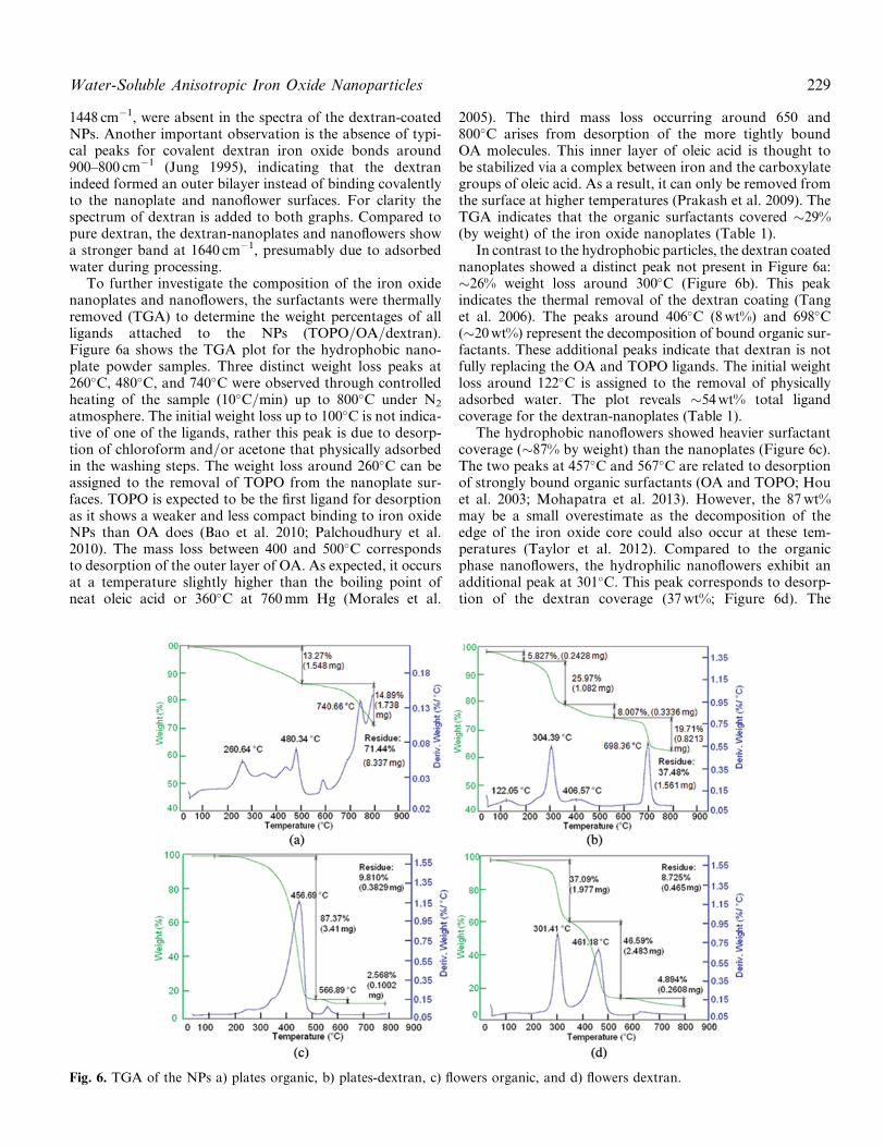
To further investigate the composition of the iron oxidenanoplates and nanoflowers, the surfactants were thermallyremoved (TGA) to determine the weight percentages of allligands attached to the NPs (TOPO=OA=dextran).Figure 6a shows the TGA plot for the hydrophobic nano-plate powder samples. Three distinct weight loss peaks at260�C, 480�C, and 740�C were observed through controlledheating of the sample (10�C=min) up to 800�C under N2
atmosphere. The initial weight loss up to 100�C is not indica-tive of one of the ligands, rather this peak is due to desorp-tion of chloroform and=or acetone that physically adsorbedin the washing steps. The weight loss around 260�C can beassigned to the removal of TOPO from the nanoplate sur-faces. TOPO is expected to be the first ligand for desorptionas it shows a weaker and less compact binding to iron oxideNPs than OA does (Bao et al. 2010; Palchoudhury et al.2010). The mass loss between 400 and 500�C correspondsto desorption of the outer layer of OA. As expected, it occursat a temperature slightly higher than the boiling point ofneat oleic acid or 360�C at 760mm Hg (Morales et al.
2005). The third mass loss occurring around 650 and800�C arises from desorption of the more tightly boundOA molecules. This inner layer of oleic acid is thought tobe stabilized via a complex between iron and the carboxylategroups of oleic acid. As a result, it can only be removed fromthe surface at higher temperatures (Prakash et al. 2009). TheTGA indicates that the organic surfactants covered �29%(by weight) of the iron oxide nanoplates (Table 1).
In contrast to the hydrophobic particles, the dextran coatednanoplates showed a distinct peak not present in Figure 6a:�26% weight loss around 300�C (Figure 6b). This peakindicates the thermal removal of the dextran coating (Tanget al. 2006). The peaks around 406�C (8wt%) and 698�C(�20wt%) represent the decomposition of bound organic sur-factants. These additional peaks indicate that dextran is notfully replacing the OA and TOPO ligands. The initial weightloss around 122�C is assigned to the removal of physicallyadsorbed water. The plot reveals �54wt% total ligandcoverage for the dextran-nanoplates (Table 1).
The hydrophobic nanoflowers showed heavier surfactantcoverage (�87% by weight) than the nanoplates (Figure 6c).The two peaks at 457�C and 567�C are related to desorptionof strongly bound organic surfactants (OA and TOPO; Houet al. 2003; Mohapatra et al. 2013). However, the 87wt%may be a small overestimate as the decomposition of theedge of the iron oxide core could also occur at these tem-peratures (Taylor et al. 2012). Compared to the organicphase nanoflowers, the hydrophilic nanoflowers exhibit anadditional peak at 301�C. This peak corresponds to desorp-tion of the dextran coverage (37wt%; Figure 6d). The
Fig. 6. TGA of the NPs a) plates organic, b) plates-dextran, c) flowers organic, and d) flowers dextran.
Water-Soluble Anisotropic Iron Oxide Nanoparticles 229

additional peak at 461�C accounts for the loss of remainingorganic ligands (OA and TOPO). Once again the TGA con-firms that the dextran coating is added on top of the organicligands. The small mass loss (�5%) above 570�C is from thetransformation of Fe3O4 to FeO, the thermally stable phaseat that elevated temperature (Zhao et al. 2006). The totalsurfactant coverage for the dextran-nanoflowers wasapproximately 84% (Table 1). The heavy dextran coveragefor the nanoplates and nanoflowers is a promising aspectof our synthesis method, as a thick dextran layer preventsinteraction with plasma proteins and opsonization (Jung1995). This prevention is highly desirable for potentialimaging and therapeutic applications.
The zeta potential value of a NP solution is an indicatorof its stability, where a colloidal system is generally stable ifthis value is >�30mV or <30mV, but stable solutions of
dextran coated iron oxide NPs have been reported with anout of range negative zeta potential (��10mV—Sonvicoet al. 2005; Griffiths et al. 2011; ��2mV—Karmali et al.2012). The nanoplates have a zeta potential of �13mV inwater, the nanoflowers �9mV. The low absolute values ofthe zeta potentials indicate a lack of strong electrostaticcharges on the NP surfaces. Therefore, the aqueous nano-plates and nanoflowers are most likely stabilized via sterichindrance of the dextran outer coating (Amstad et al. 2011).
To assess their preliminary potential in bio-applicationssuch as MRI contrast enhancement, the magnetic propertiesof the particles are characterized using superconductingquantum interference magnetometer (SQUID). The magne-tizations (M) versus applied magnetic field (H) curves of ironoxide plates and flowers before and after dextran attachmentare shown in Figure 7. For the nanoplates, the saturationmagnetization decreased from 60 emu=g (as-synthesizedplates) to 53 emu=g for the hydrophilic plates, but the parti-cles remained purely superparamagnetic (Figure 7b). Theiron oxide nanoflower samples displayed a combination ofsuperparamagnetism and paramagnetism, characteristic ofNPs with high surface ligand coverage (Figure 7a). Boththe hydrophobic and dextran-coated nanoflowers showedsimilar M-H curves without saturation. The magnetizationof the nanoflowers at the highest observed values decreasedfrom 58 emu=g to 47 emu=g after aqueous phase transfer.The absence of any ferromagnetic loops in the M-H curveof the iron oxide nanoplates and nanoflowers samplesconfirms stability (negligible aggregation).
The physicochemical properties of NPs in solution aredynamic and can be altered by the environment. The definedproperties in water (e.g., hydrodynamic size, zeta potential),do not necessarily predict their performance during applica-tions. Therefore, the stability of aqueous NP dispersions atvarious pH and in different buffer solutions is critical forin vivo bio-applications. For example, endocytosis is a com-mon pathway of cellular internalization of iron oxide NPsfor targeted imaging and drug delivery. During this uptake,theNPs are subjected to pH changes from 6.2 in the early endo-somes to 4.6–5 for the lysosomes (Scott and Gruenberg 2011).
We monitored the aqueous dispersions of nanoplates andnanoflowers under related pH conditions (pH 4, 5, 6, and 7)over a 28 day period to examine the aggregation behavior(Figure 8). The size of the nanoplates changed instantly
Table 1. Ligand coverage of the iron oxide nanoplates andnanoflowers
Sample
Ligand coverage (�wt%)
Organic ligands Dextran Total
Hydrophobic nanoplates 29 X 29Hydrophilic nanoplates 28 26 54Hydrophobic nanoflowers 90 X 90Hydrophobic nanoflowers 47 37 84
Fig. 7. Magnetic measurement of iron oxide nanoparticles;a) nanoflowers; b) nanoplates.
Fig. 8. Particle stability in water at different pH values; a) nanoplates; b) nanoflowers.
230 Palchoudhury et al.

upon all pH changes, but stayed approximately the samewith time for each pH tested (Figure 8a). This suggests goodtime-dependent stability, but also indicates that the pHchanges do affect the dextran coating. For instance, at thelowest pH tested (pH 4), protonation of the hydroxyl groupsof dextran takes place, strengthening the change of hydrogenbond formation between adjacent nanoplates, explaining theobserved increase in size (Xu et al. 2011). Figure 8b showsthe stability of the nanoflowers at different pH. The beha-vior is similar to the nanoplates, with smaller overallchanges. Again, the slight increase in dimension at the moreacidic values (pH 4 and 5) could be due to hydrogen bondformation. We would like to stress that no visible aggre-gation was observed for either shape and the hydrodynamicsizes remained well below 200 nm in all instances. This ispromising for bio-applications because NPs of dimensions<200 nm can evade rapid clearance via macrophages.
The stability of NPs could also be affected by the ionicstrength of a solution. While NPs eventually will be usedunder physiological conditions and not in pure water, theirstability in common biology buffer is tested. Thereforethe nanoplates and nanoflowers are monitored for their sizeand their zeta potential in HEPES (0.1–1M), PBS (1–10 X),and SSC (1–10 X). The hydrodynamic size of thedextran-nanoplates is most stable in PBS (Figure 9a) withnegligible variation from 63 nm to 68 nm. In SSC, the dex-tran coated nanoplates are stable as well. However, thenanoplates showed an appreciable size increase in HEPES(76–119 nm). The obtained zeta potentials are consistentwith these findings. Values for dextran-nanoplates remainedfairly constant for PBS (highly stable) and for SSC (stable),but displayed a significant increase (from �21 to �9mV) for
HEPES, suggesting instability (Figure 9c). Figure 9b showsthe variation in hydrodynamic size of the dextran-nanoflowers with increasing ionic strength. Excellentstability was once again observed in PBS. The increase inhydrodynamic dimension (133–174 nm) suggests slightaggregation in HEPES. Different from the nanoplates, thedextran coated nanoflowers showed low stability in SSC,according to the significant change in size (135–248 nm). Apossible explanation for this could be the different crystal-line phase of the nanoflowers (Fe3O4 versus c-Fe2O3 forthe plates).
Aggregation was not visible in the TEM images of thedextran-nanoflowers at low buffer concentrations. There-fore, the aggregation behavior could be the result of saltbridge formation caused by the salt of the concentrated buf-fers. The zeta potentials of the nanoflowers confirmed wellwith the size measurements (Figure 9d) for the nanoflowers.For example, the zeta potentials of the nanoflowers werefairly constant in PBS and HEPES, but notably increasedfor SSC (�14mV to �6mV). The slight negative zeta poten-tial of the nanoplates and nanoflowers is consistent with ourhypothesis that the –OH groups of dextran protruded out inwater to render the NPs hydrophilic. The �OH functional-ities on the iron oxide nanoplates and nanoflowers couldpotentially provide active sites for bioconjugation (Pathaket al. 2001; Sperling and Parak 2010).
4. Conclusion
In summary, crystalline iron oxide nanoplates and nano-flowers with hydrophilic dextran coating were prepared.The anisotropic NPs were synthesized via the modified
Fig. 9. Particle stability tested in three common buffers: a) nanoplates size (DLS); b) nanoflowers size (DLS); c) nanoplates zetapotential; d) nanoflowers zeta potential.
Water-Soluble Anisotropic Iron Oxide Nanoparticles 231
‘‘heat-up’’ method to obtain excellent shape and size control.A facile phase transfer route was explored to coat theorganic NPs with an additional dextran layer. FTIR analy-ses indicated successful adsorption of a dextran bilayer onthe nanoplate and nanoflower surfaces. The dextran-coatedhydrophilic nanoplates and nanoflowers were most stablein aqueous dispersions of pH 6–7 for water and in PBS. Boththe nanoplates and nanoflowers are sterically stabilized viadextran with the polyhydroxylated groups of dextranextending out to the solution phase, as suggested by thelow absolute values of zeta potential. Detailed theoreticalinvestigations and computer simulations on the dextranbilayer-NP binding interactions are being pursued. Webelieve the present work can be extended to other aniso-tropic NPs. The dextran based phase transfer for anisotropicNPs will offer promising applications in targeted imagingand therapy (Nidhin et al. 2013).
Acknowledgments
We acknowledge Yale Institute of Nanoscience and Quan-tum Engineering (YINQE) for TEM facilities. We thankElias Quijano and Dr. Saltzman for use of the DLS. Thanksare due to Teng-hooi Goh and Dr. Andre Taylor for use ofthe TGA. We also acknowledge the Chemical and Biophysi-cal Instrumentation and specifically Dr. Wuyi Meng at Yalefor FTIR and XRD2 facilities.
ReferencesAmstad, E., M. Textor, and E. Reimhult. 2011. Stabilization and
functionalization of iron oxide nanoparticles for biomedicalapplications. Nanoscale 3:2819–2843.
Bao, Y., W. An, C. H. Turner, and K. M. Krishnan. 2010. The criticalrole of surfactants in the growth of cobalt nanoparticles. Langmuir26:478–83.
Binder, W. H., H. Weinstabl, and R. Sachsenhofer. 2008. Superpara-magnetic ironoxide nanoparticles via ligand exchange reactions:organic 1, 2-diols as versatile building blocks for surface engineer-ing. Journal of Nanomaterials 383020(1–10).
Bronstein, L. M., X. Huang, J. Retrum, A. Schmucker, M. Pink,B. Stein, and B. Dragnea. 2007. Influence of iron oleate complexstructure on iron oxide nanoparticle formation. Chemistry ofMaterials 19:3624–3632.
Euliss, L. E., S. G. Grancharov, S. O’Brien, T. J. Deming, G. D.Stucky, C. B. Murray, and G. A. Held. 2003. Cooperative assemblyof magnetic nanoparticles and block copolypeptides in aqueousmedia. Nano Letters 3:1489–1493.
Fang, C., N. Bhattarai, C. Sun, and M. Zhang. 2009. Functionalizednanoparticles with long-term stability in biological media. Small5:1637–1641.
Fang, C., and M. Zhang. 2009. Multifunctional magnetic nanoparticlesfor medical imaging applications. Journal of Materials Chemistry19:6258–6266.
Geng, Y., P. Dalhaimer, S. Cai, R. Tsai, M. Tewari, T. Minko, andD. E. Discher. 2007. Shape effects of filaments versus spherical par-ticles in flow and drug delivery. Nature Nanotechnology 2:249–255.
Gonzales, M., and K. M. Krishnan. 2007. Synthesis of magnetolipo-somes with mondisperse iron oxide nanocrystals cores for hyperther-mia. Journal of Magnetism and Magnetic Materials 311:59–62.
Griffiths, S. M., N. Singh, G. J. S. Jenkins, P. M. Williams, A. W.Orbaek, A. R. Barron, C. J. Wright, and S. H. Doak. 2001. Dextrancoated ultrafine superparamagnetic iron oxide nanoparticles:
compatibility with common fluorometric and colorimetric dyes.Analytical Chemistry 83:3778–3785.
Herman, P., B. G. Sanganahalli, and F. Hyder. 2009. Multi-modalmeasurements of blood plasma and red blood cell volumes duringfunctional brain activation. Journal of Cerebral Blood Flow &Metabolism 29:19–24.
Huang, J., L. Wang, R. Lin, A. Y. Wang, L. Yang, M. Kuang, W. Qian,and H. Mao. 2013. Casein-coated iron oxide nanoparticles for highMRI contrast enhancement and efficient cell targeting. ACS AppliedMaterials & Interfaces 5:4632–4639.
Hou, Y., J. Yu, and S. Gao. 2003. Solvothermal reduction synthesisand characterization of superparamagnetic magnetite nanoparticles.Journal of Materials Chemistry 13:1983–1987.
Jun, Y.W., J. S. Choi, and J. Cheon. 2006. Shape control of semiconduc-tor and metal oxide nanocrystals through nonhydrolytic colloidalroutes. Angewandte Chemie International Edition 45:3414–3439.
Jung, C. W. 1995. Surface properties of superparamagnetic ironoxide MR contrast agents: ferumoxides, ferumoxtran, ferumoxsil.Magnetic Resonance Imaging 13:675–691.
Karmali, P. P., Y. Chao, J-H. Park, M. J. Sailor, E. Ruoslahti, S. C.Esener, and D. Simberg. 2012. Different effect of hydrogelationon antifouling and circulation properties of dextran-iron oxidenanoparticles. Molecular Pharmacology 9:539–545.
Laurent, S., D. Forge, M. Port, A. Roch, C. Robic, L. Vander Elst, andR. N. Muller. 2008. Magnetic iron oxide nanoparticles: synthesis,stabilization, vectorization, physicochemical characterizations, andbiological applications. Chemical Reviews 108:2064–2110.
Liu, K., L. Zhao, P. Klavins, F. E. Osterloh, and H. Hiramatsu. 2003.Extrinsic magnetoresistance in magnetite nanoparticles. Journal ofApplied Physics 93:7951–7954.
Mohapatra, J., A. Mitra, D. Bahadur, and M. Aslam. 2013. Surfacecontrolled synthesis of MFe2O4 (M=Mn, Fe, Co, Ni and Zn) nano-particles and their magnetic characteristics. Cryst. Eng. Comm.15:524–532.
Morales, M. A., T. K. Jain, V. Labhasetwar, and D. L. Leslie-Pelecky.2005. Magnetic studies of iron oxide nanoparticles coated with oleicacid and pluronic (R) block copolymer. Journal of Applied Physics97:10Q905.
Nidhin, M., S. S. Nazeer, R. S. Jayasree, M. S. Kiran, B. U. Nair, andK. J. Sreeram. 2013. Flower shaped assembly of cobalt ferrite nano-particles: application as T2 contrast agent in MRI. RSC Advances3:6909–6912.
Palchoudhury, S., Y. Xu, W. An, C. H. Turner, and Y. Bao. 2010.Platinum attachments on iron oxide nanoparticle surfaces. Journalof Applied Physics 107:09B311.
Palchoudhury, S., Y. Xu, A. Rushdi, R. A. Holler, and Y. Bao. 2012.Controlled synthesis of iron oxide nanoplates and nanoflowers.Chemical Communications. 48:10499–10501.
Park, J., K. An, Y. Hwang, J. G. Park, H. J. Noh, J. Y. Kim, J. H.Park, N. M. Hwang, and T. Hyeon. 2004. Ultra-large-scale syn-thesis of monodispersed nanocrystals. Nature Materials 3:891–895.
Park, J. H., G. Von Maltzahn, L. Zhang, M. P. Schwartz, E. Ruoslahti,S. N. Bhatia, and M. J. Sailor. 2008. Magnetic iron oxide nano-worms for tumor targeting and imaging. Advanced Materials20:1630–1635.
Pathak, S., S. K. Choi, N. Arnheim, and M. E. Thompson. 2001.Hydroxylated quantum dots as luminescent probes for in situ hybri-dization. Journal of the American Chemical Society 123:4103–4104.
Prakash, A., H. Zhu, C. J. Jones, D. N. Benoit, E. J. Ellsworth, B. N.Bryant, and V. L. Colvin. 2009. Bilayers as phase transfer agentsfor nanocrystals prepared in nonpolar solvents. ACS Nano3:2139–2146.
Reimer, P., and T. Balzer. 2003. Ferucarbotran (resovist): A new clini-cally approved RES-specific contrast agent for contrast-enhanchedMRI of the liver: properties clinical development, and applications.European Radiology 13:1266–1276.
232 Palchoudhury et al.

Scott, C., and J. Gruenberg. 2011. Ion flux and the function of endo-somes and lysosomes: pH is just the start. Bioessays 33:103–110.
Sherman Hsu, C. P. 1997. Infrared spectroscopy. In Handbook of instru-mental techniques for analytical chemistry, (F. A. Settle, ed.),247–283. Upper Saddle River, NJ: Prentice Hall.
Sonvico, F., S. Mornet, S. Vasseur, C. Dubernet, D. Jaillard,J. Degrouard, et al. 2005. Folate-conjugated iron oxide nanoparticlesfor solid tumor targeting as potential specific magnetic hyperthermiamediators: Synthesis, Physicochemical characterization, and invitro experiments. Bioconjugate Chemistry 16:1181–1188.
Sperling, R. A., and W. J. Parak. 2010. Surface modification,functionalization and bioconjugation of colloidal inorganic nano-particles. Philosophical Transactions of the Royal Society A368:1333–1383.
Sun, C., J. S. H. Lee, and M. Zhang. 2008. Magnetic nanoparticles inMR imaging and drug delivery. Advanced Drug Delivery Reviews60:1252–1265.
Tang, M., H. Dou, and K. Sun. 2006. One-step synthesis of dextran-based stable nanoparticles assisted by self-assembly. Polymer47:728–734.
Tao, L., W. Hu, Y. Liu, G. Huang, B. D. Sumer, and J. Gao. 2011.Shape-specific polymeric nanomedicine: Emerging opportunitiesand challenges. Experimental Biology and Medicine 236:20–29.
Taylor, R. M., D. L. Huber, T. C. Monson, V. Esch, and L. O. Sillerud.2012. Structural andmagnetic characterization of superparamagnetic
iron platinum nanoparticle contrast agents for magnetic resonanceimaging. Journal of Vacuum Science & Technology B 30:1–6.
Xu, Y., Y. Qin, S. Palchoudhury, and Y. Bao. 2011. Water-soluble ironoxide nanoparticles with high stability and selective surface func-tionality. Langmuir. 27:8990–8997.
Yiu, H. H. P., L. Bouffier, P. Boldrin, J. Long, J. B. Claridge, and M. J.Rosseinsky. 2013. comprehensive study of DNA binding oniron(II,III) oxide nanoparticles with a positively charged polyaminethree-dimensional coating. Langmuir. 29:11354–11365.
Young, A. G., N. Al-Salim, D. P. Green, and A. J. McQuillan. 2008.Attenuated total reflection infrared studies of oleate and trioctyl-phosphine oxide ligand adsorption and exchange reactions onCdS quantum dot films. Langmuir. 24:3841–3849.
Yu, W. W., E. Chang, C. M. Sayes, R. Drezek, and V. L. Colvin.2006. Aqueous dispersion of monodisperse magnetic iron oxidenanocrystals through phase transfer. Nanotechnology. 17:4483–4487.
Zhao, S-Y., D. K. Lee, C. W. Kim, H. G. Cha, Y. H. Kim, and Y. S.Kang. 2006. Synthesis of Magnetic Nanoparticles of Fe3O4 andCoFe2O4 and Their Surface Modification by Surfactant Adsorption.Bulletin of the Korean Chemical Society 27:237–242.
Zhao, Z., Z. Zhou, J. Bao, Z. Wang, J. Hu, X. Chi, et al. 2013. Octapodiron oxide nanoparticles as high-performance T2 contrast agents formagnetic resonance imaging. Nature Communications Article No.2266.
Water-Soluble Anisotropic Iron Oxide Nanoparticles 233

Copyright of Particulate Science & Technology is the property of Taylor & Francis Ltd andits content may not be copied or emailed to multiple sites or posted to a listserv without thecopyright holder's express written permission. However, users may print, download, or emailarticles for individual use.