20140824 human embryonic stem cells derived by somatic cell nuclear transfer
TRANSCRIPT
Human Embryonic Stem Cells Derivedby Somatic Cell Nuclear TransferMasahito Tachibana,1 Paula Amato,2 Michelle Sparman,1 Nuria Marti Gutierrez,1 Rebecca Tippner-Hedges,1 Hong Ma,1
Eunju Kang,1 Alimujiang Fulati,1 Hyo-Sang Lee,1,6 Hathaitip Sritanaudomchai,3 Keith Masterson,2 Janine Larson,2
Deborah Eaton,2 Karen Sadler-Fredd,2 David Battaglia,2 David Lee,2 Diana Wu,2 Jeffrey Jensen,1,4 Phillip Patton,2
Sumita Gokhale,5 Richard L. Stouffer,1,2 Don Wolf,1 and Shoukhrat Mitalipov1,2,*1Division of Reproductive & Developmental Sciences, Oregon National Primate Research Center, Oregon Health & Science University, 505NW 185th Avenue, Beaverton, OR 97006, USA2Division of Reproductive Endocrinology, Department of Obstetrics and Gynecology, Oregon Health & Science University, 3181 SW Sam
Jackson Park Road, Portland, OR 97239, USA3Department of Oral Biology, Faculty of Dentistry, Mahidol University, Bangkok 10400, Thailand4Women’s Health Research Unit, Oregon Health & Science University, 3303 SW Bond Avenue, Portland, OR 79239, USA5Boston University School of Medicine, 72 East Concord Street, Boston, MA 02118, USA6Present address: Laboratory Animal Center, Osong Medical Innovation Foundation, Chungbuk 363-951, Republic of Korea*Correspondence: [email protected]
http://dx.doi.org/10.1016/j.cell.2013.05.006
SUMMARY
Reprogramming somatic cells into pluripotent em-bryonic stem cells (ESCs) by somatic cell nucleartransfer (SCNT) has been envisioned as an approachfor generating patient-matched nuclear transfer (NT)-ESCs for studies of disease mechanisms and fordeveloping specific therapies. Past attempts to pro-duce human NT-ESCs have failed secondary to earlyembryonic arrest of SCNT embryos. Here, we identi-fied premature exit from meiosis in human oocytesand suboptimal activation as key factors that areresponsible for these outcomes. Optimized SCNTapproaches designed to circumvent these limita-tions allowed derivation of human NT-ESCs. Whenapplied to premium quality human oocytes, NT-ESC lines were derived from as few as two oocytes.NT-ESCs displayed normal diploid karyotypes andinherited their nuclear genome exclusively fromparental somatic cells. Gene expression and differ-entiation profiles in human NT-ESCs were similar toembryo-derived ESCs, suggesting efficient reprog-ramming of somatic cells to a pluripotent state.
INTRODUCTION
Cytoplasmic factors present in mature, metaphase II (MII)-ar-
rested oocytes have a unique ability to reset the identity of trans-
planted somatic cell nuclei to the embryonic state. Since the
initial discovery in amphibians (Gurdon, 1962), somatic cell nu-
clear transfer (SCNT) success in a range of different mammalian
species has demonstrated that such reprogramming activity in
enucleated or spindle-free oocytes (cytoplasts) is universal
(Campbell et al., 1996; Solter, 2000; Wilmut et al., 1997, 2002).
However, despite numerous applications of SCNT for animal
1228 Cell 153, 1228–1238, June 6, 2013 ª2013 Elsevier Inc.
cloning, the nature of reprogramming oocyte factors and their
mechanism of action remain largely unknown.
In humans, SCNT was envisioned as a means of generating
personalized embryonic stem cells from patients’ somatic cells,
which could be used to study disease mechanisms and ulti-
mately for cell-based therapies (Lanza et al., 1999; Yang et al.,
2007). However, the derivation of human nuclear transfer-em-
bryonic stem cells (NT-ESCs) has not been achieved despite
numerous attempts during the past decade. The roadblock
responsible for failure is early embryonic arrest of human
SCNT embryos precluding derivation of stable NT-ESCs. Typi-
cally, SCNT embryos fail to progress beyond the eight-cell stage,
presumably due to an inability to activate critical embryonic
genes from the somatic donor cell nucleus (Egli et al., 2011; Nog-
gle et al., 2011). In a few cases, when SCNT embryos did reach
the blastocyst stage, either stable ESCs were not recovered or
derivation was not attempted (Fan et al., 2011; French et al.,
2008). Though the underlying cause of early developmental
arrest remains unclear, most of these studies involving human
oocytes applied SCNT protocols developed for nonprimate spe-
cies. Previously, we demonstrated that SCNT procedures, when
adapted to primates, succeeded in reprogramming rhesus ma-
caque adult skin fibroblasts into NT-ESCs (Byrne et al., 2007;
Sparman et al., 2009). Therefore, we reasoned that, similar to
other mammals, human MII oocytes must contain reprogram-
ming activity.
Several recent observations are relevant. Removal of human
oocytes’ nuclear genetic material (chromosomes) negatively im-
pacts the cytoplast’s subsequent ability to induce reprogram-
ming (Noggle et al., 2011). However, when a somatic cell nucleus
is transplanted into an intact oocyte containing its own chromo-
somes, the resulting polyploid embryos are able to develop to
blastocysts and support ESC derivation. One possible explana-
tion for these observations is that critical reprogramming factors
in humanMII oocytes are physically associated with the chromo-
somes or spindle apparatus and are depleted or critically dimin-
ished upon enucleation. Alternatively, it is possible that one or
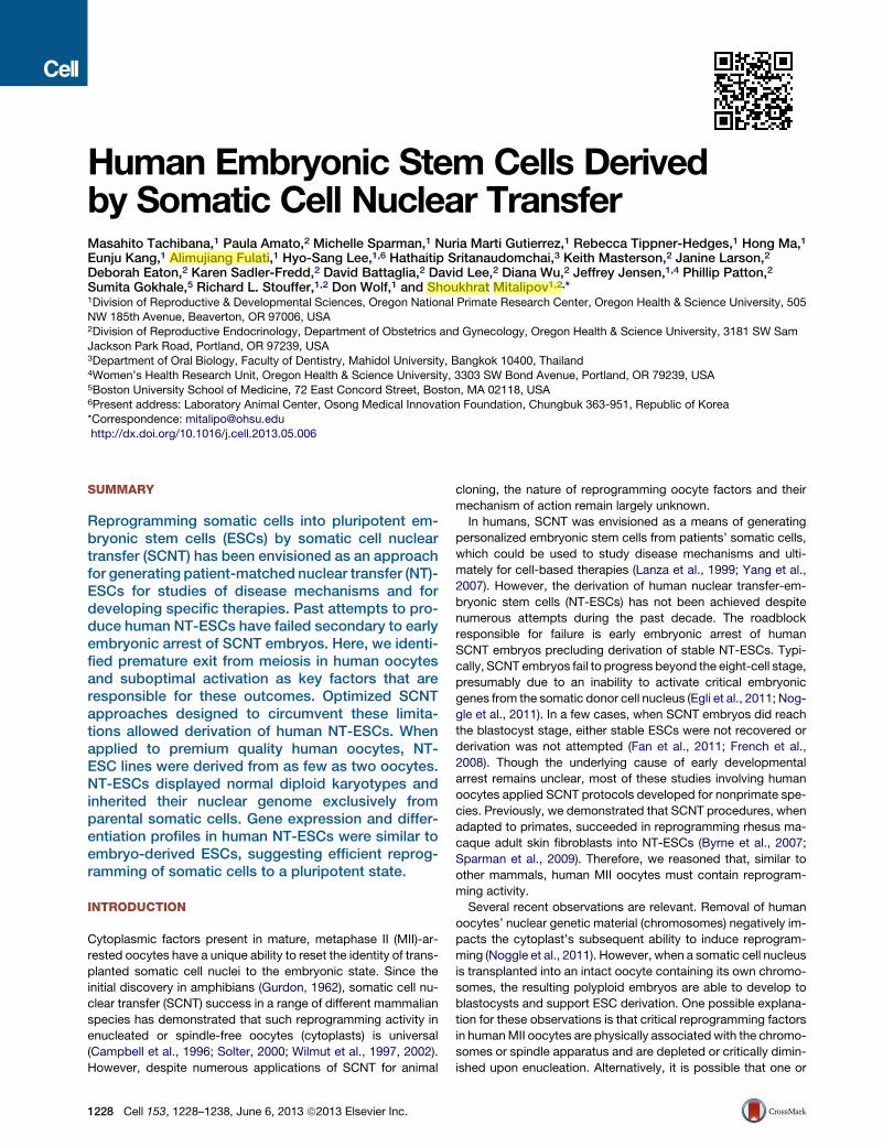
Figure 1. Development of Monkey SCNT
Embryos Reconstructed with Optimized
Protocols
Although HVJ-E fusion was efficient, SCNT con-
structs required activation by electroporation for
blastocyst formation. Elimination of ionomycin
from the activation treatment further improved
blastocyst development. I, ionomycin; DMAP,
6-DMAP; CM, compact morula.
See also Tables S1 and S2.
more of the steps in SCNT—namely, oocyte enucleation, donor
cell nucleus introduction, or cytoplast activation—negatively
impact cytoplast quality, rendering it incapable of inducing suffi-
cient reprogramming.
In considering distinct biological features of human oocytes
that could be involved, we focused on our recent observation
that meiotic arrest in human MII oocytes is unstable and can
be easily perturbed by mechanical manipulations (Tachibana
et al., 2013). Earlier, we suggested that retention of meiosis-spe-
cific activities in the cytoplast is critical for nuclear remodeling af-
ter transplantation of an interphase-stage somatic cell nucleus
(Mitalipov et al., 2007). This remodeling is positively correlated
with onward development of SCNT embryos after activation.
Therefore, we systematically evaluated modifications in oocyte
enucleation and donor cell introduction that might work to retain
meiosis factors in human cytoplasts. We also determined that
routine cytoplast activation treatments were insufficient to sup-
port subsequent human SCNT embryo development. We initially
used rhesus macaque oocytes to evaluate factors affecting suc-
cessful SCNT reprogramming in a primate system. Subse-
quently, we refined SCNT approaches with high-quality human
oocytes donated by healthy volunteers and demonstrated that
methodological alterations significantly improve blastocyst for-
mation from human SCNT embryos. Moreover, we derived
several human NT-ESC lines from these embryos and validated
that their nuclear DNA is an exclusive match to parental somatic
cells, whereas mitochondrial DNA originated almost exclusively
from oocytes. We also conducted extensive pluripotency assays
on human NT-ESCs to verify reprogramming.
RESULTS
SCNT Protocol Optimization in a Nonhuman PrimateModelOur recent studies demonstrated humanMII oocyte sensitivity to
premature activation induced by removal and reintroduction of
meiotic spindles (Tachibana et al., 2013) and to the use of elec-
trofusion in the context of cytoplast activation (Tachibana et al.,
2009). Consequently, our present investigation began with opti-
mizing the use of envelope from inactivated hemagglutinating
Cell 153, 1228–12
virus of Japan (HVJ-E) to fuse nuclear
donor cells with enucleated MII oocytes
while maintaining cytoplasts in meiosis
(Tachibana et al., 2009). Because of
limited oocyte availability, we first tested
both the feasibility and efficacy of HVJ-
E-based cell fusion on the development of rhesus macaque
SCNT embryos.
The fusion rate of adult fibroblasts with cytoplasts was 100%
after HVJ-E treatment; however, and unexpectedly, the SCNT
embryos generated by HVJ-E fusion failed to progress beyond
the compact morula (CM) stage following standard ionomycin/
DMAP (I/DMAP) activation. We previously demonstrated that
monkey SCNT embryos produced by electrofusion developed
into blastocysts (Byrne et al., 2007; Sparman et al., 2009).
Therefore, we postulated that exposure of the cytoplast to an
electropulse (electroporation) could be beneficial for SCNT re-
programming, perhaps as a supplemental activation stimulus.
To test this possibility, we exposed HVJ-E-fused SCNT embryos
to electroporation before the standard I/DMAP activation treat-
ment. Ten percent of SCNT embryos were capable of reaching
the blastocyst stage (Figure 1). Interestingly, this SCNT blasto-
cyst formation rate was unaffected even when exposure to
ionomycin was omitted and SCNT embryos were activated
with electroporation followed by DMAP treatment (Figure 1).
Together, these results indicate that, although an electroporation
stimulus is not required for cell fusion, it is supportive of proper
cytoplast activation following SCNT.
Histone deacetylase inhibitors, such as trichostatin A (TSA),
have been associated with improved SCNT reprogramming in
several mammalian species (Ding et al., 2008; Kishigami et al.,
2006; Li et al., 2008). We previously demonstrated enhanced
development of monkey SCNT embryos treated with 37.5 nM
TSA (from 4% up to 18% blastocyst development rate [Sparman
et al., 2010]). However, blastocyst quality and potential to give
rise to stable ESCs remained unknown. Here, we plated 16mon-
key SCNT blastocysts produced during TSA treatment on mitot-
ically inactivated mouse embryonic fibroblast (mEF) feeders, but
none resulted in NT-ESC line isolation (Table S1). We reasoned
that, although TSA treatment was promoting blastocyst forma-
tion, high TSA concentrations may negatively affect blastocyst
quality and epiblast lineage integrity. Therefore, we tested
several lower TSA concentrations, as well as shorter exposure
times, on monkey SCNT blastocyst development and ESC isola-
tion. Reducing the TSA concentration to 10 nM or shortening the
TSA exposure time from 24 to 12 hr did not affect blastocyst
38, June 6, 2013 ª2013 Elsevier Inc. 1229
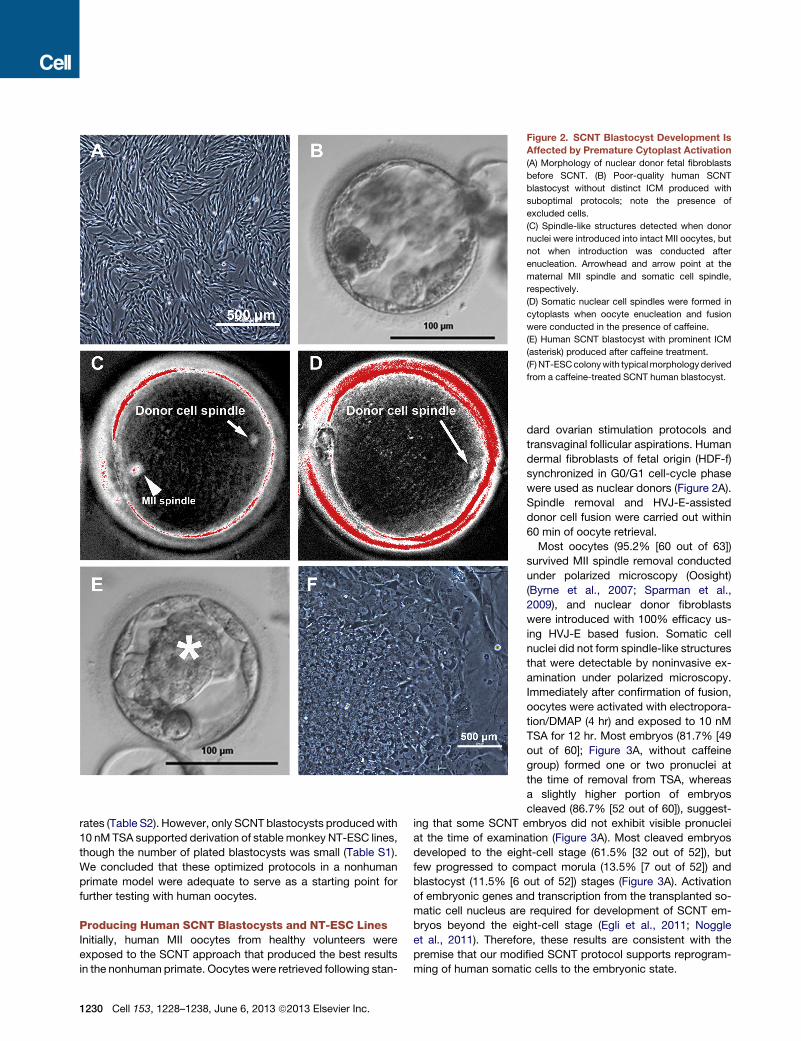
Figure 2. SCNT Blastocyst Development Is
Affected by Premature Cytoplast Activation
(A) Morphology of nuclear donor fetal fibroblasts
before SCNT. (B) Poor-quality human SCNT
blastocyst without distinct ICM produced with
suboptimal protocols; note the presence of
excluded cells.
(C) Spindle-like structures detected when donor
nuclei were introduced into intact MII oocytes, but
not when introduction was conducted after
enucleation. Arrowhead and arrow point at the
maternal MII spindle and somatic cell spindle,
respectively.
(D) Somatic nuclear cell spindles were formed in
cytoplasts when oocyte enucleation and fusion
were conducted in the presence of caffeine.
(E) Human SCNT blastocyst with prominent ICM
(asterisk) produced after caffeine treatment.
(F)NT-ESCcolonywith typicalmorphologyderived
from a caffeine-treated SCNT human blastocyst.
rates (Table S2). However, only SCNT blastocysts producedwith
10 nM TSA supported derivation of stable monkey NT-ESC lines,
though the number of plated blastocysts was small (Table S1).
We concluded that these optimized protocols in a nonhuman
primate model were adequate to serve as a starting point for
further testing with human oocytes.
Producing Human SCNT Blastocysts and NT-ESC LinesInitially, human MII oocytes from healthy volunteers were
exposed to the SCNT approach that produced the best results
in the nonhuman primate. Oocytes were retrieved following stan-
1230 Cell 153, 1228–1238, June 6, 2013 ª2013 Elsevier Inc.
dard ovarian stimulation protocols and
transvaginal follicular aspirations. Human
dermal fibroblasts of fetal origin (HDF-f)
synchronized in G0/G1 cell-cycle phase
were used as nuclear donors (Figure 2A).
Spindle removal and HVJ-E-assisted
donor cell fusion were carried out within
60 min of oocyte retrieval.
Most oocytes (95.2% [60 out of 63])
survived MII spindle removal conducted
under polarized microscopy (Oosight)
(Byrne et al., 2007; Sparman et al.,
2009), and nuclear donor fibroblasts
were introduced with 100% efficacy us-
ing HVJ-E based fusion. Somatic cell
nuclei did not form spindle-like structures
that were detectable by noninvasive ex-
amination under polarized microscopy.
Immediately after confirmation of fusion,
oocytes were activated with electropora-
tion/DMAP (4 hr) and exposed to 10 nM
TSA for 12 hr. Most embryos (81.7% [49
out of 60]; Figure 3A, without caffeine
group) formed one or two pronuclei at
the time of removal from TSA, whereas
a slightly higher portion of embryos
cleaved (86.7% [52 out of 60]), suggest-
ing that some SCNT embryos did not exhibit visible pronuclei
at the time of examination (Figure 3A). Most cleaved embryos
developed to the eight-cell stage (61.5% [32 out of 52]), but
few progressed to compact morula (13.5% [7 out of 52]) and
blastocyst (11.5% [6 out of 52]) stages (Figure 3A). Activation
of embryonic genes and transcription from the transplanted so-
matic cell nucleus are required for development of SCNT em-
bryos beyond the eight-cell stage (Egli et al., 2011; Noggle
et al., 2011). Therefore, these results are consistent with the
premise that our modified SCNT protocol supports reprogram-
ming of human somatic cells to the embryonic state.
Figure 3. Development of Human SCNT Embryos and NT-ESC
Derivation after Caffeine Treatment
(A) Improved blastocyst development of human SCNT embryos treated with
caffeine. A total of 63 (five cycles) and 43 (three cycles) oocytes were utilized
for SCNT without or with caffeine, respectively. Sixty (95.2%) and 42 (97.7%)
oocytes survived after SCNT micromanipulations.
(B) NT-ESCs were derived only from blastocysts produced with caffeine.
See also Figures S1, S2 and S3.
Of note, human SCNT blastocysts exhibited poorly organized
trophectoderm and small or undetectable inner cell masses
(ICMs). In addition, large blastomere-like cells were often
excluded (Figure 2B). Nevertheless, we plated six SCNT blasto-
cysts onto feeder layers to examine their ability to support ESC
derivation. Though four blastocysts attached to mEFs, only
one gave an outgrowth that, after further passaging, failed to
produce stable ESC-like cells (Figure 3B).
Though the observed SCNT blastocyst development rate was
encouraging, further optimization of the human protocol focused
on improvements in embryo quality. To assess whether spindle
removal in human oocytes could cause spontaneous exit from
meiosis, we introduced somatic nuclei into intact MII oocytes
and examined their birefringence properties under polarized mi-
croscopy. All introduced somatic cell nuclei efficiently formed
spindle-like structures that were visible within 30 min of fusion
(17 out of 17) (Figure 2C). Again, spindle formation was not
observed when somatic cell nuclei were fused with manipulated,
spindle-free oocytes (0 out of 3). These observations are consis-
tent with recent conclusions that human MII oocytes undergo
premature activation secondary to spindle removal (Tachibana
et al., 2013). Assuming that meiosis-specific factors are retained
during spindle removal but decline due to spontaneous activa-
tion, we focused on noninvasive treatments to maintain meiotic
arrest during manipulation.
We reported previously that the exposure of monkey oocytes
to caffeine, a protein phosphatase inhibitor, was effective in
protecting the cytoplast from premature activation and improved
development of SCNT embryos (Mitalipov et al., 2007). There-
fore, human oocytes weremaintained in 1.25mMcaffeine during
spindle removal and somatic cell fusion. As expected, somatic
cell nuclei introduced into cytoplasts under these conditions
efficiently formed spindle-like structures that were detectable
under birefringence microscopy (83.3% [10 out of 12]) (Fig-
ure 2D). More importantly, the blastocyst development rate of
caffeine-treated embryos was notably enhanced (23.5%)
compared to the standard SCNT group (Figure 3A, with caffeine
group), and blastocysts were characterized by visible and prom-
inent ICMs, similar to those observed for IVF-produced embryos
(Figure 2E).
Remarkably, when eight SCNT blastocysts produced with
caffeine exposure were utilized for ESCs isolation, all attached
to mEFs and four formed ICM outgrowths (Figure 3B), which
gave rise to ESC-like colonies upon manual splitting onto fresh
mEF plates (Figure 2F). Subsequent passaging resulted in the
propagation of stable ESC colonies with typical morphology
and growth characteristics. This surprisingly high ESC derivation
ratewas similar to that reported in our previous studywith human
IVF-derived blastocysts (50%) and was even higher than in
manipulated spindle transfer embryos (38%) (Figure 3B) (Tachi-
bana et al., 2013).
Collectively, our findings indicate that a protocol developed in
the monkey model supported blastocyst development for hu-
man SCNT embryos. However, poor SCNT blastocyst quality
precluded ESC isolation. Subsequent incorporation of caffeine
during enucleation and fusion allowed improved blastocyst
development and ESC line derivation.
Reproducibility of Human SCNT ResultsInterestingly, all four human NT-ESC lines were derived from oo-
cytes retrieved from one egg donor (egg donor A). Eight mature
MII oocytes were recovered after a single stimulation cycle. Us-
ing fetal dermal fibroblasts as nuclear donor cells and following
our caffeine-incorporated SCNT protocol, five blastocysts
were produced (62.5%) that gave rise to four NT-ESC lines
(80%) (Figure S1 available online). In the context of generating
patient-specific pluripotent stem cells, reproducible results
with various patient-derived somatic cells and with different
egg donors are a necessity.
We therefore acquired a skin fibroblast culture from a patient
with Leigh syndrome. A total of 15 and 5 MII oocytes were
collected from two unrelated egg donor volunteers (B and C)
and were used for SCNT with these fibroblasts. All oocytes sur-
vived spindle removal and successfully fused with nuclear donor
cells. Following activation and culture, four (27%, [4 out of 15])
and three (60% [3 out of 5]) blastocysts were produced from
these egg donors (Figure 4A). After plating on mEFs and manual
passaging, we established two stable NT-ESC lines—one from
each oocyte cohort (Figure 4B). Thus, these outcomes confirm
the reproducibility of our human SCNT protocols.
Cell 153, 1228–1238, June 6, 2013 ª2013 Elsevier Inc. 1231
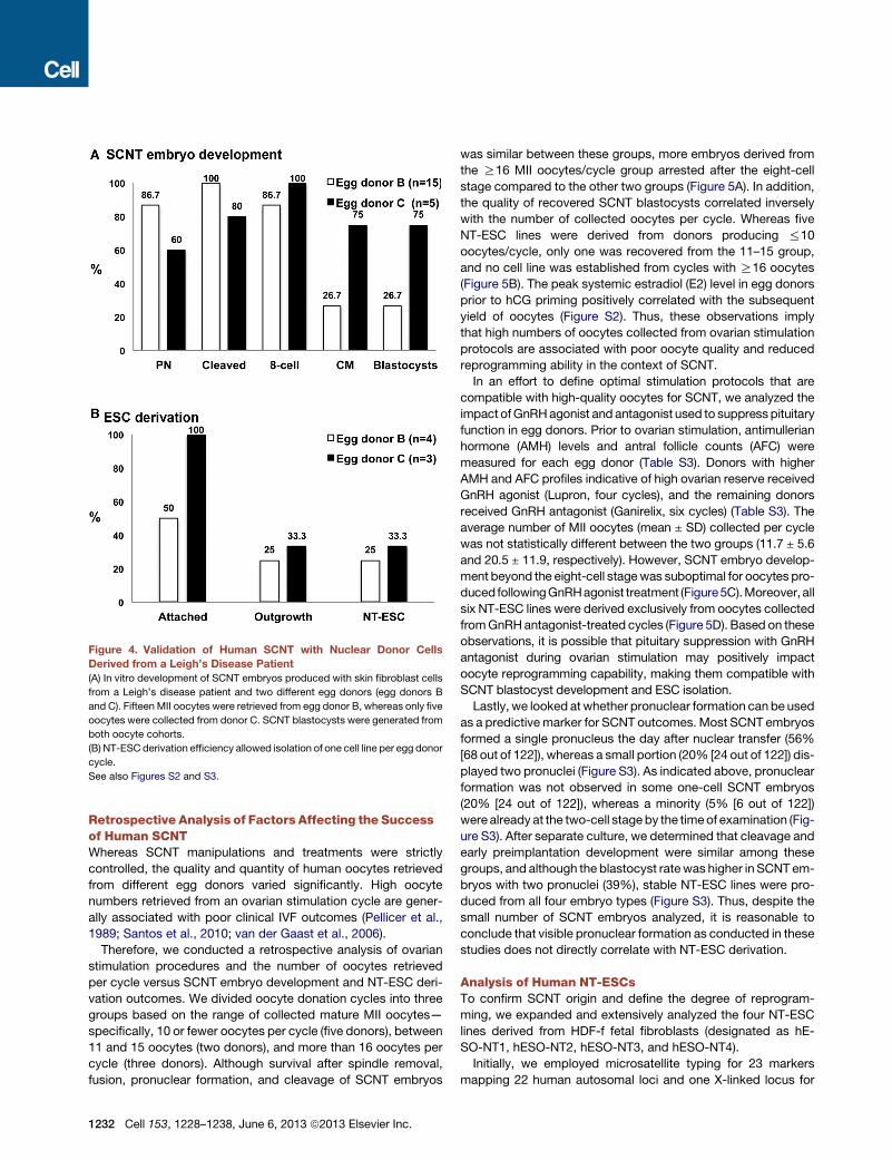
Figure 4. Validation of Human SCNT with Nuclear Donor Cells
Derived from a Leigh’s Disease Patient
(A) In vitro development of SCNT embryos produced with skin fibroblast cells
from a Leigh’s disease patient and two different egg donors (egg donors B
and C). Fifteen MII oocytes were retrieved from egg donor B, whereas only five
oocytes were collected from donor C. SCNT blastocysts were generated from
both oocyte cohorts.
(B) NT-ESC derivation efficiency allowed isolation of one cell line per egg donor
cycle.
See also Figures S2 and S3.
Retrospective Analysis of Factors Affecting the Successof Human SCNTWhereas SCNT manipulations and treatments were strictly
controlled, the quality and quantity of human oocytes retrieved
from different egg donors varied significantly. High oocyte
numbers retrieved from an ovarian stimulation cycle are gener-
ally associated with poor clinical IVF outcomes (Pellicer et al.,
1989; Santos et al., 2010; van der Gaast et al., 2006).
Therefore, we conducted a retrospective analysis of ovarian
stimulation procedures and the number of oocytes retrieved
per cycle versus SCNT embryo development and NT-ESC deri-
vation outcomes. We divided oocyte donation cycles into three
groups based on the range of collected mature MII oocytes—
specifically, 10 or fewer oocytes per cycle (five donors), between
11 and 15 oocytes (two donors), and more than 16 oocytes per
cycle (three donors). Although survival after spindle removal,
fusion, pronuclear formation, and cleavage of SCNT embryos
1232 Cell 153, 1228–1238, June 6, 2013 ª2013 Elsevier Inc.
was similar between these groups, more embryos derived from
the R16 MII oocytes/cycle group arrested after the eight-cell
stage compared to the other two groups (Figure 5A). In addition,
the quality of recovered SCNT blastocysts correlated inversely
with the number of collected oocytes per cycle. Whereas five
NT-ESC lines were derived from donors producing %10
oocytes/cycle, only one was recovered from the 11–15 group,
and no cell line was established from cycles with R16 oocytes
(Figure 5B). The peak systemic estradiol (E2) level in egg donors
prior to hCG priming positively correlated with the subsequent
yield of oocytes (Figure S2). Thus, these observations imply
that high numbers of oocytes collected from ovarian stimulation
protocols are associated with poor oocyte quality and reduced
reprogramming ability in the context of SCNT.
In an effort to define optimal stimulation protocols that are
compatible with high-quality oocytes for SCNT, we analyzed the
impact ofGnRHagonist and antagonist used to suppress pituitary
function in egg donors. Prior to ovarian stimulation, antimullerian
hormone (AMH) levels and antral follicle counts (AFC) were
measured for each egg donor (Table S3). Donors with higher
AMH and AFC profiles indicative of high ovarian reserve received
GnRH agonist (Lupron, four cycles), and the remaining donors
received GnRH antagonist (Ganirelix, six cycles) (Table S3). The
average number of MII oocytes (mean ± SD) collected per cycle
was not statistically different between the two groups (11.7 ± 5.6
and 20.5 ± 11.9, respectively). However, SCNT embryo develop-
ment beyond the eight-cell stagewas suboptimal for oocytes pro-
duced followingGnRHagonist treatment (Figure5C).Moreover, all
six NT-ESC lines were derived exclusively from oocytes collected
fromGnRHantagonist-treated cycles (Figure 5D). Based on these
observations, it is possible that pituitary suppression with GnRH
antagonist during ovarian stimulation may positively impact
oocyte reprogramming capability, making them compatible with
SCNT blastocyst development and ESC isolation.
Lastly, we looked atwhether pronuclear formation can be used
as a predictivemarker for SCNT outcomes. Most SCNT embryos
formed a single pronucleus the day after nuclear transfer (56%
[68 out of 122]), whereas a small portion (20% [24 out of 122]) dis-
played two pronuclei (Figure S3). As indicated above, pronuclear
formation was not observed in some one-cell SCNT embryos
(20% [24 out of 122]), whereas a minority (5% [6 out of 122])
were already at the two-cell stageby the timeof examination (Fig-
ure S3). After separate culture, we determined that cleavage and
early preimplantation development were similar among these
groups, and although the blastocyst ratewas higher in SCNT em-
bryos with two pronuclei (39%), stable NT-ESC lines were pro-
duced from all four embryo types (Figure S3). Thus, despite the
small number of SCNT embryos analyzed, it is reasonable to
conclude that visible pronuclear formation as conducted in these
studies does not directly correlate with NT-ESC derivation.
Analysis of Human NT-ESCsTo confirm SCNT origin and define the degree of reprogram-
ming, we expanded and extensively analyzed the four NT-ESC
lines derived from HDF-f fetal fibroblasts (designated as hE-
SO-NT1, hESO-NT2, hESO-NT3, and hESO-NT4).
Initially, we employed microsatellite typing for 23 markers
mapping 22 human autosomal loci and one X-linked locus for
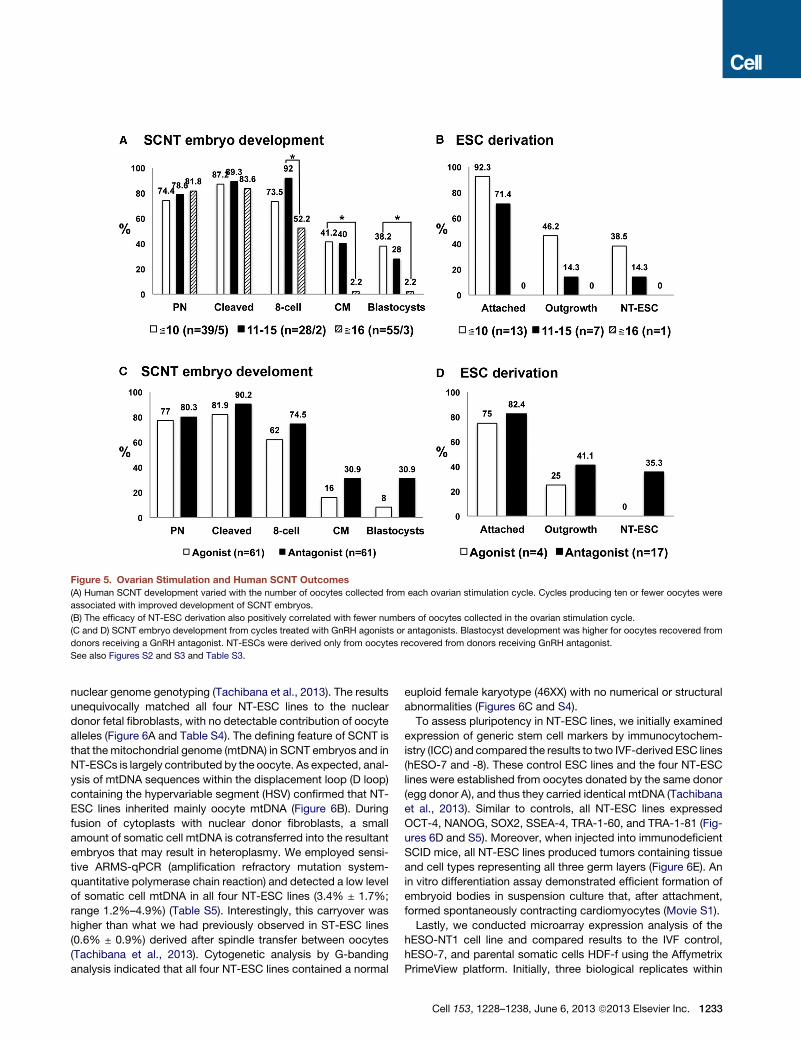
Figure 5. Ovarian Stimulation and Human SCNT Outcomes
(A) Human SCNT development varied with the number of oocytes collected from each ovarian stimulation cycle. Cycles producing ten or fewer oocytes were
associated with improved development of SCNT embryos.
(B) The efficacy of NT-ESC derivation also positively correlated with fewer numbers of oocytes collected in the ovarian stimulation cycle.
(C and D) SCNT embryo development from cycles treated with GnRH agonists or antagonists. Blastocyst development was higher for oocytes recovered from
donors receiving a GnRH antagonist. NT-ESCs were derived only from oocytes recovered from donors receiving GnRH antagonist.
See also Figures S2 and S3 and Table S3.
nuclear genome genotyping (Tachibana et al., 2013). The results
unequivocally matched all four NT-ESC lines to the nuclear
donor fetal fibroblasts, with no detectable contribution of oocyte
alleles (Figure 6A and Table S4). The defining feature of SCNT is
that the mitochondrial genome (mtDNA) in SCNT embryos and in
NT-ESCs is largely contributed by the oocyte. As expected, anal-
ysis of mtDNA sequences within the displacement loop (D loop)
containing the hypervariable segment (HSV) confirmed that NT-
ESC lines inherited mainly oocyte mtDNA (Figure 6B). During
fusion of cytoplasts with nuclear donor fibroblasts, a small
amount of somatic cell mtDNA is cotransferred into the resultant
embryos that may result in heteroplasmy. We employed sensi-
tive ARMS-qPCR (amplification refractory mutation system-
quantitative polymerase chain reaction) and detected a low level
of somatic cell mtDNA in all four NT-ESC lines (3.4% ± 1.7%;
range 1.2%–4.9%) (Table S5). Interestingly, this carryover was
higher than what we had previously observed in ST-ESC lines
(0.6% ± 0.9%) derived after spindle transfer between oocytes
(Tachibana et al., 2013). Cytogenetic analysis by G-banding
analysis indicated that all four NT-ESC lines contained a normal
euploid female karyotype (46XX) with no numerical or structural
abnormalities (Figures 6C and S4).
To assess pluripotency in NT-ESC lines, we initially examined
expression of generic stem cell markers by immunocytochem-
istry (ICC) and compared the results to two IVF-derived ESC lines
(hESO-7 and -8). These control ESC lines and the four NT-ESC
lines were established from oocytes donated by the same donor
(egg donor A), and thus they carried identical mtDNA (Tachibana
et al., 2013). Similar to controls, all NT-ESC lines expressed
OCT-4, NANOG, SOX2, SSEA-4, TRA-1-60, and TRA-1-81 (Fig-
ures 6D and S5). Moreover, when injected into immunodeficient
SCID mice, all NT-ESC lines produced tumors containing tissue
and cell types representing all three germ layers (Figure 6E). An
in vitro differentiation assay demonstrated efficient formation of
embryoid bodies in suspension culture that, after attachment,
formed spontaneously contracting cardiomyocytes (Movie S1).
Lastly, we conducted microarray expression analysis of the
hESO-NT1 cell line and compared results to the IVF control,
hESO-7, and parental somatic cells HDF-f using the Affymetrix
PrimeView platform. Initially, three biological replicates within
Cell 153, 1228–1238, June 6, 2013 ª2013 Elsevier Inc. 1233
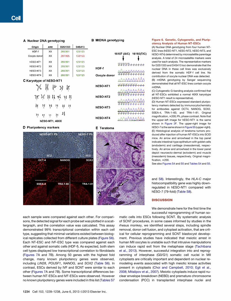
Figure 6. Genetic, Cytogenetic, and Plurip-
otency Analysis of Human NT-ESCs
(A) Nuclear DNA genotyping from four human NT-
ESC lines (hESO-NT1, hESO-NT2, hESO-NT3, and
hESO-NT4) determined bymicrosatellite parentage
analysis. A total of 24 microsatellite markers were
used for each analysis. The representative markers
for D2S1333 and D4S413 loci demonstrate that the
nuclear DNA in these cell lines was exclusively
derived from the somatic HDF-f cell line. No
contribution of oocyte nuclear DNA was detected.
(B) mtDNA genotyping by Sanger sequencing
demonstrated that all NT-ESC lines contain oocyte
mtDNA.
(C) Cytogenetic G-banding analysis confirmed that
all NT-ESCs exhibited a normal 46XX karyotype
(hESO-NT1 result is representative).
(D) Human NT-ESCs expressed standard pluripo-
tency markers detected by immunocytochemistry
for antibodies against OCT4, NANOG, SOX2,
SSEA-4, TRA-1–60, and TRA-1–81. Original
magnification,3200; Ph, phase contrast. Note that
the upper-left image for hESO-NT1 is the same
shown in Figure 2F. The upper-right image for
hESO-7 is thesameshown inFigureS5 (upper-right).
(E) Histological analysis of teratoma tumors pro-
duced after injection of human NT-ESCs into SCID
mice. An arrow and arrowhead in the top panel
indicate intestinal-type epitheliumwith goblet cells
(endoderm) and cartilage (mesodermal), respec-
tively. An arrow and arrowhead in the lower panel
depict neuroecto-dermal (ectoderm) and muscle
(mesoderm) tissues, respectively. Original magni-
fication, 3200.
See also Figures S4 and S5 and Tables S4 and S5.
each sample were compared against each other. For compari-
sons, thedetected signal for eachprobe setwasplotted in a scat-
tergraph, and the correlation value was calculated. This assay
demonstrated 99% transcriptional correlation within each cell
type, suggesting that minimal variations existed between biolog-
ical replicates collected from different culture plates (Figure S6).
Each NT-ESC and IVF-ESC type was compared against each
other and against somatic cells (HDF-f). As expected, both stem
cell types displayed low transcriptional correlation to fibroblasts
(Figures 7A and 7B). Among 50 genes with the highest fold
change, many known pluripotency genes were observed,
including LIN28, POU5F1, NANOG, and SOX2 (Table S6). In
contrast, ESCs derived by IVF and SCNT were similar to each
other (Figures 7A and 7B). Some transcriptional differences be-
tween human NT-ESCs and IVF-ESCs were observed. However
no known pluripotency genes were included in this list (Tables S7
1234 Cell 153, 1228–1238, June 6, 2013 ª2013 Elsevier Inc.
and S8). Interestingly, the HLA-C major
histocompatibility gene was highly down-
regulated in hESO-NT1 compared with
hESO-7 (79-fold) (Table S8).
DISCUSSION
We demonstrate here for the first time the
successful reprogramming of human so-
matic cells into ESCs following SCNT. By systematic analysis
of SCNT procedures, in some cases informed by studies in the
rhesus monkey, we identified several steps, including spindle
removal, donor cell fusion, and cytoplast activation, that are crit-
ical for cellular reprogramming and SCNT blastocyst develop-
ment. Previous studies have indicated that meiotic arrest in
human MII oocytes is unstable such that intrusive manipulations
can induce rapid exit from the metaphase stage (Tachibana
et al., 2013). However, successful integration into and reprog-
ramming of interphase (G0/G1) somatic cell nuclei in MII
cytoplasts are critically important and dependent on nuclear re-
modeling events associated with high meiotic kinase activities
present in cytoplasts (Choi and Campbell, 2010; Egli et al.,
2008; Mitalipov et al., 2007). Meiotic cytoplasts induce rapid nu-
clear envelope breakdown (NEBD) and premature chromosome
condensation (PCC) in transplanted interphase nuclei and
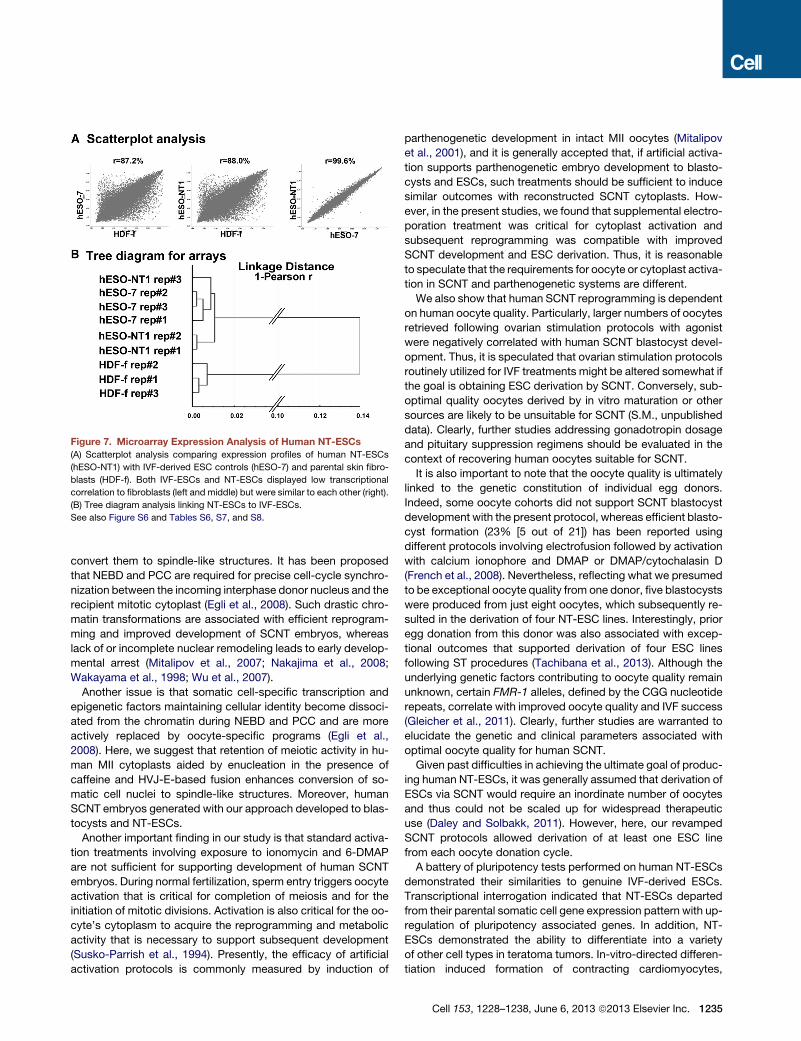
Figure 7. Microarray Expression Analysis of Human NT-ESCs
(A) Scatterplot analysis comparing expression profiles of human NT-ESCs
(hESO-NT1) with IVF-derived ESC controls (hESO-7) and parental skin fibro-
blasts (HDF-f). Both IVF-ESCs and NT-ESCs displayed low transcriptional
correlation to fibroblasts (left and middle) but were similar to each other (right).
(B) Tree diagram analysis linking NT-ESCs to IVF-ESCs.
See also Figure S6 and Tables S6, S7, and S8.
convert them to spindle-like structures. It has been proposed
that NEBD and PCC are required for precise cell-cycle synchro-
nization between the incoming interphase donor nucleus and the
recipient mitotic cytoplast (Egli et al., 2008). Such drastic chro-
matin transformations are associated with efficient reprogram-
ming and improved development of SCNT embryos, whereas
lack of or incomplete nuclear remodeling leads to early develop-
mental arrest (Mitalipov et al., 2007; Nakajima et al., 2008;
Wakayama et al., 1998; Wu et al., 2007).
Another issue is that somatic cell-specific transcription and
epigenetic factors maintaining cellular identity become dissoci-
ated from the chromatin during NEBD and PCC and are more
actively replaced by oocyte-specific programs (Egli et al.,
2008). Here, we suggest that retention of meiotic activity in hu-
man MII cytoplasts aided by enucleation in the presence of
caffeine and HVJ-E-based fusion enhances conversion of so-
matic cell nuclei to spindle-like structures. Moreover, human
SCNT embryos generated with our approach developed to blas-
tocysts and NT-ESCs.
Another important finding in our study is that standard activa-
tion treatments involving exposure to ionomycin and 6-DMAP
are not sufficient for supporting development of human SCNT
embryos. During normal fertilization, sperm entry triggers oocyte
activation that is critical for completion of meiosis and for the
initiation of mitotic divisions. Activation is also critical for the oo-
cyte’s cytoplasm to acquire the reprogramming and metabolic
activity that is necessary to support subsequent development
(Susko-Parrish et al., 1994). Presently, the efficacy of artificial
activation protocols is commonly measured by induction of
parthenogenetic development in intact MII oocytes (Mitalipov
et al., 2001), and it is generally accepted that, if artificial activa-
tion supports parthenogenetic embryo development to blasto-
cysts and ESCs, such treatments should be sufficient to induce
similar outcomes with reconstructed SCNT cytoplasts. How-
ever, in the present studies, we found that supplemental electro-
poration treatment was critical for cytoplast activation and
subsequent reprogramming was compatible with improved
SCNT development and ESC derivation. Thus, it is reasonable
to speculate that the requirements for oocyte or cytoplast activa-
tion in SCNT and parthenogenetic systems are different.
We also show that human SCNT reprogramming is dependent
on human oocyte quality. Particularly, larger numbers of oocytes
retrieved following ovarian stimulation protocols with agonist
were negatively correlated with human SCNT blastocyst devel-
opment. Thus, it is speculated that ovarian stimulation protocols
routinely utilized for IVF treatments might be altered somewhat if
the goal is obtaining ESC derivation by SCNT. Conversely, sub-
optimal quality oocytes derived by in vitro maturation or other
sources are likely to be unsuitable for SCNT (S.M., unpublished
data). Clearly, further studies addressing gonadotropin dosage
and pituitary suppression regimens should be evaluated in the
context of recovering human oocytes suitable for SCNT.
It is also important to note that the oocyte quality is ultimately
linked to the genetic constitution of individual egg donors.
Indeed, some oocyte cohorts did not support SCNT blastocyst
development with the present protocol, whereas efficient blasto-
cyst formation (23% [5 out of 21]) has been reported using
different protocols involving electrofusion followed by activation
with calcium ionophore and DMAP or DMAP/cytochalasin D
(French et al., 2008). Nevertheless, reflecting what we presumed
to be exceptional oocyte quality from one donor, five blastocysts
were produced from just eight oocytes, which subsequently re-
sulted in the derivation of four NT-ESC lines. Interestingly, prior
egg donation from this donor was also associated with excep-
tional outcomes that supported derivation of four ESC lines
following ST procedures (Tachibana et al., 2013). Although the
underlying genetic factors contributing to oocyte quality remain
unknown, certain FMR-1 alleles, defined by the CGG nucleotide
repeats, correlate with improved oocyte quality and IVF success
(Gleicher et al., 2011). Clearly, further studies are warranted to
elucidate the genetic and clinical parameters associated with
optimal oocyte quality for human SCNT.
Given past difficulties in achieving the ultimate goal of produc-
ing human NT-ESCs, it was generally assumed that derivation of
ESCs via SCNT would require an inordinate number of oocytes
and thus could not be scaled up for widespread therapeutic
use (Daley and Solbakk, 2011). However, here, our revamped
SCNT protocols allowed derivation of at least one ESC line
from each oocyte donation cycle.
A battery of pluripotency tests performed on human NT-ESCs
demonstrated their similarities to genuine IVF-derived ESCs.
Transcriptional interrogation indicated that NT-ESCs departed
from their parental somatic cell gene expression pattern with up-
regulation of pluripotency associated genes. In addition, NT-
ESCs demonstrated the ability to differentiate into a variety
of other cell types in teratoma tumors. In-vitro-directed differen-
tiation induced formation of contracting cardiomyocytes,
Cell 153, 1228–1238, June 6, 2013 ª2013 Elsevier Inc. 1235
demonstrating their potential for regenerative medicine. Genetic
analyses showed that all four NT-ESC lines tested to date con-
tained normal diploid karyotypes, with no detectable gross chro-
mosomal abnormalities or contribution from the oocyte genome
apart from mtDNA.
An approach to patient-specific pluripotent stem cell deriva-
tion that precludes the use of embryos is based on somatic
cell reprogramming by induced expression of a few critical tran-
scription factors (referred as induced pluripotent stem cells
[iPSCs]) (Takahashi et al., 2007; Takahashi and Yamanaka,
2006; Yu et al., 2007). Recent studies have concluded that
human iPSCs are characterized by high frequencies of subchro-
mosomal copy number alterations compared to IVF-derived
ESCs (Laurent et al., 2011). Some of these genetic changes
were associated with the reprogramming process itself, whereas
others could have been inherited from the parental somatic cells
(Laurent et al., 2011). In addition, iPSC-specific methylation and
transcriptional abnormalities in imprinted regions and X chromo-
somes were also described (Nazor et al., 2012). Direct compar-
isons between iPSCs and NT-ESCs in the mouse indicated that
such abnormalities are less frequent in the latter case,
concluding that SCNT-based reprogramming is more efficient
in resetting the epigenetic identity of parental somatic cells
(Kim et al., 2010). Further comparisons of the genetic, epige-
netic, and transcriptional characteristics of human NT-ESCs,
IVF-ESCs, and iPSCs are clearly justified.
Lastly, one of the fundamental differences of SCNT-based
reprogramming is that NT-ESCs contain mtDNA almost exclu-
sively originating from the oocyte. This fact is generally underap-
preciated but may represent an advantage over iPSC derivation
because it ensures that NT-ESCs acquire the potential to pro-
duce metabolically functional cells and tissues for cell therapies,
irrespective of the nuclear donor cell mtDNA. Thus, SCNT offers
a strategy for correcting of mtDNA mutations and rescuing the
metabolic function of pluripotent cells from patients with in-
herited or acquired mtDNA diseases.
EXPERIMENTAL PROCEDURES
Rhesus Macaque SCNT
All animal procedures were approved by the Institutional Animal Care and Use
Committee at the Oregon National Primate Research Center. Oocyte collec-
tions, SCNT, embryo culture, and NT-ESC isolation procedures were per-
formed as previously described (Byrne et al., 2007; Sparman et al., 2009,
2010).
Human Oocyte Donations
The study protocols were approved by both the OHSU Embryonic Stem Cell
Research Oversight Committee and the Institutional Review Board. Anony-
mous egg donors of ages 23–33 were recruited through the OHSU Women’s
Health Research Unit via print and web-based advertising. Responding
women were screened with respect to their reproductive, medical, and psy-
chosocial health. Healthy nonobese (BMI < 28 kg/m2) women who passed
the initial medical and psychological evaluations were invited to participate
in a research egg donation program. Egg donors were financially compen-
sated for the time, effort, discomfort, and inconvenience associated with the
donation process.
Ovarian stimulation protocols followed established clinical IVF guidelines as
described previously (Tachibana et al., 2013). In brief, a combination of recom-
binant human-follicle-stimulating hormone (rFSH) and humanmenopausal go-
nadotropins (hMG) and either GnRH agonist (Lupron, Tap Pharmaceutical
1236 Cell 153, 1228–1238, June 6, 2013 ª2013 Elsevier Inc.
Products) or antagonist (Ganirelix, Merck) were given. Human chorionic
gonadotropin (hCG) was prescribed to trigger oocyte maturation. Self-admin-
istration of injectable rFSH (sc, Follistim, Merck) commenced on cycle day 2
or 3 and continued for �8–12 days. The starting gonadotropin dose was
75–125 IU/day and 1–2 A hMG (sc, Menopur, Ferring Pharmaceuticals). The
dose was adjusted per individual response using an established stepdown
regimen until the day of hCG injection. Ovarian response and follicular growth
were monitored by transvaginal ultrasound and measurement of serum estra-
diol levels. When two or more follicles reachedR 18 mm in diameter, subjects
received hCG (104 IU, sc, Ovidrel, EMDSerono) to trigger follicle and oocyte
maturation. Thirty-six hours following hCG injection, subjects underwent
oocyte retrieval via transvaginal follicular aspiration.
Cumulus-oocyte complexes (COCs) were collected from aspirates and
placed in HTF w/HEPES medium (LifeGlobal, IVFonline) supplemented with
10% serum substitute supplement (SSS; Quinns Advantage Serum, Cooper-
Surgical) (HTF w/HEPES 10%) at 37�C. COCs were treated with hyaluronidase
to disaggregate cumulus and granulosa cells. Oocytes were isolated and
classified as germinal vesicle (GV), meiotic metaphase I (MI), andmaturemeta-
phase II (MII) stage and were then placed in Global medium (LifeGlobal, IVFon-
line) supplemented with 10% SSS (Global 10%) at 37�C in 5% CO2 and
covered with tissue culture oil (Sage IVF, Cooper Surgical).
Nuclear Donor Cell Preparations
Commercially available female dermal fibroblasts of fetal origin (HDF-f) were
obtained from ScienCell Research Laboratories, and Leigh syndrome patient
cells were acquired from the Coriell Cell Repositories. Cells were expanded
in 75 cm3 cell culture flasks (Corning) containing DMEM/F12 supplemented
with 100 IU ml�1 penicillin, 100 mg ml�1 streptomycin (Invitrogen), 10% FBS
at 37�C in 5% CO2. Fibroblasts were then disaggregated with trypsin treat-
ment and were frozen down in aliquots of 3 3 105 cells in medium containing
10% dimethyl sulphoxide (DMSO, Sigma). Cells were subsequently thawed
prior to SCNT and cultured in four-well dishes (Nunc) under standard condi-
tions until they reached confluency. Confluent cells were synchronized in the
G0/G1 phase of the cell cycle by culture in DMEM/F12 medium with 0.5%
FBS for 2–4 days before SCNT.
Human SCNT Procedure and Embryo Culture
Enucleations were performed as described previously (Tachibana et al., 2013).
Oocytes were placed into a 50 ml manipulation droplet of HTF w/HEPES 10%
medium containing 5 mg/ml cytochalasin B and 1.25 mM caffeine in a glass-
bottom dish. The droplet was covered with tissue culture oil, and oocytes
were maintained at 37�C for 10–15 min before spindle removal. The dish
was then mounted on the stage of an inverted microscope (Olympus IX71)
equipped with a stage warmer (http://www.tokaihit.com), Narishige microma-
nipulators, Oosight Imaging System (http://www.cri-inc.com), and a laser
objective (http://www.hamiltonthorne.com). An oocyte was positioned using
a holding pipette so that the spindle was situated close to the 2 to 4 o’clock
position. The zona pellucida next to the spindle was drilled with laser pulses,
and an enucleation pipette was inserted through the opening. A small amount
of cytoplasm surrounded by plasma membrane and contacting the spindle
was aspirated into the pipette. Next, a disaggregated fibroblast was aspirated
into a micropipette and was briefly transferred to the drop containing HVJ-E
extract (Ishihara Sangyo Kaisha). The cell was then placed into the perivitelline
space of the cytoplast on the side opposite the first polar body. This construct
was rinsed with HTF w/HEPES 10%, transferred to global 10% medium, and
incubated at 37�C in 5% CO2 for 30 min until fusion occurred as confirmed
visually by the disappearance of the donor cell from the perivitelline space.
Constructs were then subjected to artificial activation consisting of electropo-
ration pulses (two 50 ms DC pulses of 2.7 kV cm�1) (Electro Square Porator
T-820, BTX) in 0.25 M d-sorbitol buffer containing 0.1 mM calcium acetate,
0.5 mM magnesium acetate, 0.5 mM HEPES, and 1 mg ml�1 fatty-acid-free
BSA. Activated SCNT constructs were then incubated in Global medium
(without serum) containing 2 mM DMAP at 37�C in 5% CO2 for 4 hr. After
DMAP, SCNT embryos were rinsed with HTF w/HEPES 10% and transferred
into four-well dishes containing Global medium supplemented with 10%
FBS, 12 mM b-mercaptoethanol (BME), and 10 nM Trichostatin A (TSA, Sigma)
and cultured at 37�C in 5% CO2, 5% O2, and 90% N2 for 12 hr. Embryos were
then rinsed, checked for pronuclear formation, and cultured in Global medium
supplemented with 10% FBS and 12 mM b-mercaptoethanol (BME) at 37�C in
5% CO2, 5% O2, and 90% N2 for a maximum of 7 days. The medium was
changed only once, at day 3 of culture.
Isolation, Culture, and Characterization of Human NT-ESCs
After zona pellucida removal via brief exposure to 0.5% protease (Sigma),
SCNT blastocysts were plated onto confluent feeder layers of mitomycin-C-
inactivated mouse embryonic fibroblasts (mEFs) and were cultured for
5–7 days at 37�C, 3% CO2, 5% O2, and 92% N2 in ESC derivation medium
consisting of DMEM/F12 (Invitrogen) supplemented with 0.1 mM nonessential
amino acids, 1 mM l-glutamine, 0.1 mM b-mercaptoethanol, 5 ng/ml basic
fibroblast growth factor, 10 mM ROCK inhibitor (Sigma), 10% FBS, and 10%
knockout serum replacement (KSR; Invitrogen). Before use, fresh ESC deriva-
tionmediumwasmixed (50%:50%, v/v) with derivationmedium conditioned in
a 24 hr culture with growing human ESCs. Outgrowths of the inner cell mass
(ICMs) were manually dissociated into small clumps with a microscalpel and
were replated on fresh mEF plates. After the first passage of ICM outgrowths,
FBS and ROCK inhibitor were omitted, and KSR was increased to 20%. Col-
onies with ESC-like morphologies were selected for further propagation, char-
acterization, and cytogenetic analyses.
Immunocytochemistry, in vivo and in vitro differentiation, andmicroarray an-
alyses were performed as described (Byrne et al., 2007; Mitalipov et al., 2007;
Tachibana et al., 2013). Detailed protocols are also available in the Extended
Experimental Procedures.
Nuclear DNA Genotyping and Cytogenetic Analyses
Genotyping of NT-ESCs was performed by microsatellite typing using 23
markers representing 22 human autosomal loci and one X-linked locus, as pre-
viously described (Tachibana et al., 2013). Karyotyping was performed by
GWT banding on 20 metaphase cells from each human NT-ESC line at the Hu-
man Genetics Laboratory, University of Nebraska Medical Center, as previ-
ously described (Tachibana et al., 2013).
Mitochondrial DNA Genotyping
Genotyping of mtDNA was performed as previously described (Tachibana
et al., 2013). The human mitochondrial displacement loop region (D loop)
harboring the hypervariable segment 1(HSV-1) was amplified using published
primers (Danan et al., 1999). PCR products were sequenced, and the informa-
tive single nucleotide polymorphic (SNP) sites were identified using
Sequencher v. 4.7 software (GeneCodes). Quantitative mtDNA analysis was
performed by ARMS-qPCR as described (Tachibana et al., 2013). The detailed
protocol is also available in Extended Experimental Procedures.
SUPPLEMENTAL INFORMATION
Supplemental Information includes Extended Experimental Procedures, six
figures, eight tables, and one movie and can be found with this article online
at http://dx.doi.org/10.1016/j.cell.2013.05.006.
ACKNOWLEDGMENTS
The authors would like to acknowledge the OHSU Embryonic Stem Cell
Research Oversight Committee and the Institutional Review Board for
providing oversight and guidance with human embryo and ESC studies. We
thank egg donor volunteers and the staff at the Women’s Health Research
Unit at the Center for Women’s Health, the Division of Reproductive Endocri-
nology & Infertility of the Department of Obstetrics & Gynecology of Oregon
Health & Science University for their support and procurement of human gam-
etes. The Division of Animal Resources, Surgery Team, Assisted Reproductive
Technology & Embryonic Stem Cell Core, Endocrine Technology Core, and
Imaging & Morphology Core at the Oregon National Primate Research Center
provided expertise and services for the nonhuman primate studies. Hamilton
Thorne donated the XYClone laser system for this study. We are grateful to
Dr. Warren Sanger and Dianna Zaleski for karyotyping services, Dr. Cecilia Pe-
nedo for microsatellite analysis, and Dr. Cary Harding for assistance with nu-
clear donor cells. We are also indebted to Vanessa Domush, Elizabeth Smo-
lens, Cathy Ramsey, Ying Li, Riffat Ahmed, Brittany Daughtry, and Erin
Wolff for their technical support. The human oocyte/embryo researchwas sup-
ported by OHSU institutional funds and by the grant from Leducq Foundation.
The nonhuman primate studies were supported by grants from the National
Institutes of Health HD063276, HD057121, HD059946, EY021214, and
8P51OD011092.
Received: April 30, 2013
Revised: May 3, 2013
Accepted: May 3, 2013
Published: May 15, 2013
REFERENCES
Byrne, J.A., Pedersen, D.A., Clepper, L.L., Nelson, M., Sanger, W.G., Gokhale,
S., Wolf, D.P., and Mitalipov, S.M. (2007). Producing primate embryonic stem
cells by somatic cell nuclear transfer. Nature 450, 497–502.
Campbell, K.H., McWhir, J., Ritchie, W.A., andWilmut, I. (1996). Sheep cloned
by nuclear transfer from a cultured cell line. Nature 380, 64–66.
Choi, I., and Campbell, K.H. (2010). Treatment of ovine oocytes with caffeine
increases the accessibility of DNase I to the donor chromatin and reduces
apoptosis in somatic cell nuclear transfer embryos. Reprod. Fertil. Dev. 22,
1000–1014.
Daley, G.Q., and Solbakk, J.H. (2011). Stem cells: Triple genomes go far.
Nature 478, 40–41.
Danan, C., Sternberg, D., Van Steirteghem, A., Cazeneuve, C., Duquesnoy, P.,
Besmond, C., Goossens, M., Lissens, W., and Amselem, S. (1999). Evaluation
of parental mitochondrial inheritance in neonates born after intracytoplasmic
sperm injection. Am. J. Hum. Genet. 65, 463–473.
Ding, X., Wang, Y., Zhang, D., Wang, Y., Guo, Z., and Zhang, Y. (2008).
Increased pre-implantation development of cloned bovine embryos treated
with 5-aza-20-deoxycytidine and trichostatin A. Theriogenology 70, 622–630.
Egli, D., Birkhoff, G., and Eggan, K. (2008). Mediators of reprogramming: tran-
scription factors and transitions through mitosis. Nat. Rev. Mol. Cell Biol. 9,
505–516.
Egli, D., Chen, A.E., Saphier, G., Ichida, J., Fitzgerald, C., Go, K.J., Acevedo,
N., Patel, J., Baetscher, M., Kearns, W.G., et al. (2011). Reprogramming within
hours following nuclear transfer into mouse but not human zygotes. Nat. Com-
mun. 2, 488.
Fan, Y., Jiang, Y., Chen, X., Ou, Z., Yin, Y., Huang, S., Kou, Z., Li, Q., Long, X.,
Liu, J., et al. (2011). Derivation of cloned human blastocysts by histone deace-
tylase inhibitor treatment after somatic cell nuclear transfer with b-thalassemia
fibroblasts. Stem Cells Dev. 20, 1951–1959.
French, A.J., Adams, C.A., Anderson, L.S., Kitchen, J.R., Hughes, M.R., and
Wood, S.H. (2008). Development of human cloned blastocysts following so-
matic cell nuclear transfer with adult fibroblasts. Stem Cells 26, 485–493.
Gleicher, N., Weghofer, A., Lee, I.H., and Barad, D.H. (2011). Association of
FMR1 genotypes with in vitro fertilization (IVF) outcomes based on ethnicity/
race. PLoS ONE 6, e18781.
Gurdon, J.B. (1962). The developmental capacity of nuclei taken from intesti-
nal epithelium cells of feeding tadpoles. J. Embryol. Exp. Morphol. 10,
622–640.
Kim, K., Doi, A., Wen, B., Ng, K., Zhao, R., Cahan, P., Kim, J., Aryee, M.J., Ji,
H., Ehrlich, L.I., et al. (2010). Epigenetic memory in induced pluripotent stem
cells. Nature 467, 285–290.
Kishigami, S., Mizutani, E., Ohta, H., Hikichi, T., Thuan, N.V., Wakayama, S.,
Bui, H.T., andWakayama, T. (2006). Significant improvement of mouse cloning
technique by treatment with trichostatin A after somatic nuclear transfer. Bio-
chem. Biophys. Res. Commun. 340, 183–189.
Lanza, R.P., Cibelli, J.B., and West, M.D. (1999). Prospects for the use of nu-
clear transfer in human transplantation. Nat. Biotechnol. 17, 1171–1174.
Laurent, L.C., Ulitsky, I., Slavin, I., Tran, H., Schork, A., Morey, R., Lynch, C.,
Harness, J.V., Lee, S., Barrero, M.J., et al. (2011). Dynamic changes in the
Cell 153, 1228–1238, June 6, 2013 ª2013 Elsevier Inc. 1237
copy number of pluripotency and cell proliferation genes in human ESCs and
iPSCs during reprogramming and time in culture. Cell Stem Cell 8, 106–118.
Li, J., Svarcova, O., Villemoes, K., Kragh, P.M., Schmidt, M., Bøgh, I.B., Zhang,
Y., Du, Y., Lin, L., Purup, S., et al. (2008). High in vitro development after so-
matic cell nuclear transfer and trichostatin A treatment of reconstructed
porcine embryos. Theriogenology 70, 800–808.
Mitalipov, S.M., Nusser, K.D., and Wolf, D.P. (2001). Parthenogenetic activa-
tion of rhesus monkey oocytes and reconstructed embryos. Biol. Reprod.
65, 253–259.
Mitalipov, S.M., Zhou, Q., Byrne, J.A., Ji, W.Z., Norgren, R.B., and Wolf, D.P.
(2007). Reprogramming following somatic cell nuclear transfer in primates is
dependent upon nuclear remodeling. Hum. Reprod. 22, 2232–2242.
Nakajima, N., Inomata, T., Ito, J., and Kashiwazaki, N. (2008). Treatment with
proteasome inhibitor MG132 during cloning improves survival and pronuclear
number of reconstructed rat embryos. Cloning Stem Cells 10, 461–468.
Nazor, K.L., Altun, G., Lynch, C., Tran, H., Harness, J.V., Slavin, I., Garitaonan-
dia, I., Muller, F.J., Wang, Y.C., Boscolo, F.S., et al. (2012). Recurrent
variations in DNA methylation in human pluripotent stem cells and their differ-
entiated derivatives. Cell Stem Cell 10, 620–634.
Noggle, S., Fung, H.L., Gore, A., Martinez, H., Satriani, K.C., Prosser, R., Oum,
K., Paull, D., Druckenmiller, S., Freeby, M., et al. (2011). Human oocytes repro-
gram somatic cells to a pluripotent state. Nature 478, 70–75.
Pellicer, A., Ruiz, A., Castellvi, R.M., Calatayud, C., Ruiz, M., Tarin, J.J., Miro,
F., and Bonilla-Musoles, F. (1989). Is the retrieval of high numbers of oocytes
desirable in patients treatedwith gonadotrophin-releasing hormone analogues
(GnRHa) and gonadotrophins? Hum. Reprod. 4, 536–540.
Santos, M.A., Kuijk, E.W., and Macklon, N.S. (2010). The impact of ovarian
stimulation for IVF on the developing embryo. Reproduction 139, 23–34.
Solter, D. (2000). Mammalian cloning: advances and limitations. Nat. Rev.
Genet. 1, 199–207.
Sparman, M., Dighe, V., Sritanaudomchai, H., Ma, H., Ramsey, C., Pedersen,
D., Clepper, L., Nighot, P., Wolf, D., Hennebold, J., and Mitalipov, S. (2009).
Epigenetic reprogramming by somatic cell nuclear transfer in primates.
Stem Cells 27, 1255–1264.
Sparman, M.L., Tachibana, M., and Mitalipov, S.M. (2010). Cloning of non-
human primates: the road ‘‘less traveled by’’. Int. J. Dev. Biol. 54, 1671–1678.
Susko-Parrish, J.L., Leibfried-Rutledge, M.L., Northey, D.L., Schutzkus, V.,
and First, N.L. (1994). Inhibition of protein kinases after an induced calcium
transient causes transition of bovine oocytes to embryonic cycles without
meiotic completion. Dev. Biol. 166, 729–739.
1238 Cell 153, 1228–1238, June 6, 2013 ª2013 Elsevier Inc.
Tachibana, M., Sparman, M., Sritanaudomchai, H., Ma, H., Clepper, L., Wood-
ward, J., Li, Y., Ramsey, C., Kolotushkina, O., and Mitalipov, S. (2009). Mito-
chondrial gene replacement in primate offspring and embryonic stem cells.
Nature 461, 367–372.
Tachibana, M., Amato, P., Sparman,M., Woodward, J., Sanchis, D.M., Ma, H.,
Gutierrez, N.M., Tippner-Hedges, R., Kang, E., Lee, H.S., et al. (2013). To-
wards germline gene therapy of inherited mitochondrial diseases. Nature
493, 627–631.
Takahashi, K., and Yamanaka, S. (2006). Induction of pluripotent stem cells
from mouse embryonic and adult fibroblast cultures by defined factors. Cell
126, 663–676.
Takahashi, K., Tanabe, K., Ohnuki, M., Narita, M., Ichisaka, T., Tomoda, K.,
and Yamanaka, S. (2007). Induction of pluripotent stem cells from adult human
fibroblasts by defined factors. Cell 131, 861–872.
van der Gaast, M.H., Eijkemans, M.J., van der Net, J.B., de Boer, E.J., Burger,
C.W., van Leeuwen, F.E., Fauser, B.C., and Macklon, N.S. (2006). Optimum
number of oocytes for a successful first IVF treatment cycle. Reprod. Biomed.
Online 13, 476–480.
Wakayama, T., Perry, A.C., Zuccotti, M., Johnson, K.R., and Yanagimachi, R.
(1998). Full-term development of mice from enucleated oocytes injected with
cumulus cell nuclei. Nature 394, 369–374.
Wilmut, I., Schnieke, A.E., McWhir, J., Kind, A.J., and Campbell, K.H. (1997).
Viable offspring derived from fetal and adult mammalian cells. Nature 385,
810–813.
Wilmut, I., Beaujean, N., de Sousa, P.A., Dinnyes, A., King, T.J., Paterson, L.A.,
Wells, D.N., and Young, L.E. (2002). Somatic cell nuclear transfer. Nature 419,
583–586.
Wu, Y.G., Zhou, P., Lan, G.C., Wang, G., Luo, M.J., and Tan, J.H. (2007). The
effects of delayed activation and MG132 treatment on nuclear remodeling and
preimplantation development of embryos cloned by electrofusion are corre-
lated with the age of recipient cytoplasts. Cloning Stem Cells 9, 417–431.
Yang, X., Smith, S.L., Tian, X.C., Lewin, H.A., Renard, J.P., and Wakayama, T.
(2007). Nuclear reprogramming of cloned embryos and its implications for
therapeutic cloning. Nat. Genet. 39, 295–302.
Yu, J., Vodyanik, M.A., Smuga-Otto, K., Antosiewicz-Bourget, J., Frane, J.L.,
Tian, S., Nie, J., Jonsdottir, G.A., Ruotti, V., Stewart, R., et al. (2007). Induced
pluripotent stem cell lines derived from human somatic cells. Science 318,
1917–1920.