2015 toma convention learning objectives basic wound care · 2015 toma convention glennon r....
TRANSCRIPT

8/9/2015
1
Basic Wound CareThe 9 Essentials of Wound Healing
2015 TOMA Convention
Glennon R. Einspanier, DO, FACOS, CWSPKatherine A. Lincoln, DO, FAAFPC. Tad Stiles. MD, FACEP, ATP, CFII, MEIIH. Sprague Taveau IV, DO, MBA, FACOFPJim R. Wood, MDJoElla Gibbs-Stiles, RN, BSN, FNP-C
Central Texas Wound Healing Associates
Learning Objectives
• To understand the role of each of the 9 essentials in wound healing:– Perfusion
– Non-Viable Tissue
– Inflammation/Infection
8/9/2015 2
– Inflammation/Infection
– Edema
– Wound Microenvironment
– Tissue Growth
– Off-loading
– Pain Control
– Host Factors
Pre-test
1 Infection/Inflammation have little to do with ultimate wound healing. T F
2 In the wounded patient, pain control usually means improved compliance. T F
3 Knowing the patient’s current medications can be key to remedying delayed healing T Fremedying delayed healing. T F
4 Removal of dead tissue from the wound every once in awhile is an important essential of early wound closure. T F
5 The invasive Vascular Surgeon/Cardiologist/Radiologist is a wound care physician’s best friend. T F
8/9/2015 3
Central Texas Wound Healing Associates
Presents
The Nine Essentials of Wound Healing
Central Texas Wound Healing Associates
The Nine Essentials of Wound Healing
1. Adequate Perfusion2. Non-Viable Tissue3. Inflammation or Infection4. Edema5. Wound Microenvironment6. Tissue Growth Optimized7. Off-Loading8. Pain Control9. Host Factors

8/9/2015
2
If you can’t get water
The Nine Essentials of Wound Healing
If you can t get water to the garden……the garden won’t grow!!!!
It has been suggested that decreased It has been suggested that decreased oxygen down regulates or diminishes oxygen down regulates or diminishes
certain critical cellular functions.certain critical cellular functions.
It has been suggested that decreased It has been suggested that decreased oxygen down regulates or diminishes oxygen down regulates or diminishes
certain critical cellular functions.certain critical cellular functions.
Epidemiology and Impact of PAD• PAD is a manifestation of atherosclerosis
characterized by progressive lower extremity arterial occlusive disease
• PAD serves as a marker of atherothrombotic disease in other vascular beds
• Estimated to affect over 10 million people in the US
Epidemiology and Impact of PAD• Framingham Heart Study suggested that
20% of symptomatic patients with PAD also had diabetes
• Probably underestimates prevalence as more people with PAD are assymptomaticmore people with PAD are assymptomatic than symptomatic– Over half are probably assymptomatic– Only a third have claudication– Remainder have more severe forms of
disease
Patterns of Peripheral Arterial Occlusive Disease in Diabetics
• Earlier age of onset
• Characteristic distribution pattern (Strandness, 1964)
Nondiabetics 68% 57%Diabetics 27% 81%
Aortoiliac Tibial/Peroneal
• Stenoses causing vessel lumen reduction 50% or greater detected in 99% of 104 consecutively admitted diabetic patients with foot ulcers, 1993-1995
• Stenoses were detected in patients with palpable foot l kl b hi l i d t th 1
Angiographic Evaluation of PVD as a Prognostic Determinant for Major Amputation in Diabetics with Foot Ulcers
pulses, ankle-brachial indexes greater than 1
• Stenoses were detected in patients with PtcO2 values greater than 50 mmHg
• Risk of amputation significantly increased when total occlusion present in popliteal and infrapopliteal arteries
Faglia, et al. Diabetes Care. 1998; 21(4):625-530

8/9/2015
3
Epidemiology and Impact of PAD
• PAD risk factors include…–– DiabetesDiabetes
–– SmokingSmoking
– Male
Ad d– Advanced age
– Hypertension
– Hyperlipidemia
– Obesity
• Other risk factors may include…– Elevated CRP, fibrinogen, homocysteine,
apolipoprotein B, lipoprotein A
– Plasma viscosity
Epidemiology and Impact of PAD• PAD is a major risk factor for lower
extremity amputation, especially in patients with diabetes
• PAD is a marker for systemic vascular• PAD is a marker for systemic vascular disease– Coronary
– Cerebral
– Renal
• PAD is associated with increased risk of MI, stroke, death
Advanced Atherosclerosis
Peripheral Vascular Disease: Primary Reason for Limb Loss
Diabetes MellitusCoronary Artery DiseaseCerebrovascular DiseaseAdvanced Age
Criqui MH. Langer RD. Fronek A. Feigelson HS. Klauber MR. McCann TJ. Browner D. Mortality over a period of 10 years in patients with peripheral arterial disease. N Engl.J Med 1992; 326: 381-386.
5- Year Risk: • 5% Amputation • 30% Death
•Poor Quality of Life•Increased Health Care Cost•Lost Income •Decrease in Functional Status
g
Major Pathophysiological Mechanisms• Neuropathy
• Ischemia
• Infection
• Wound healing failure
Pecoraro Diabetes Care 1990; 13(5):513-521
Two Common Soft Tissue Complications• Cutaneous ulcer
• Gangrene
Frequent Initiating Events• Minor trauma
• Acute arterial insufficiency
Increased microvascular pressures leads to microvascular sclerosis
Basement membrane thickeningIncreased extracellular matrix as diffusion barrier
Microvascular Dysfunction
Increased extracellular matrix as diffusion barrierIncreased capillary fragility
Micro vascular failure with loss of vasodilatory responseLoss of flow reserveDelayed hyperemic response to pressureImpaired hyperemic response to injury
Endothelial dysfunction leads to vascular functional abnormality in the absence of luminal obstruction
• Endothelial cells line the inner surface of all blood vessels – Arteries – Veins – Heart (endocardial cells)
L h ti l– Lymphatic vessels
• Normal functions includes:– Mediation of coagulation– Platelet adhesion– Immune function– Control of volume and electrolyte content

8/9/2015
4
Endothelial dysfunction is the loss of normal physiological and biochemical properties of the innermost lining of blood vessels
• Endothelial dysfunction can result from disease or environmental processes and can exist without atherosclerotic disease– Septic shock
– Hypertension– Hypertension
– Hypercholesterolemia
– Diabetes
– Inflammatory disease
– Smoking tobacco products• Arteries and arterioles can not vasodilate in
response to stimuli- low levels of nitric oxide (NO)
Remember Key Caveats:• Endothelial dysfunction can be present and
significant even in the absence of atherosclerotic disease
Diagnosing Peripheral Arterial Occlusive Disease (PAOD): Comprehensive History
• Typical predispositions for “arterial disease” may not be positive in patients suffering from endothelial dysfunction
Bottom Line• In the Diabetic Patient:
– Far greater chance of PAD than non-diabetics
– Far greater chance of concurrent venous disease
F t h f l ti l di t
8/9/2015 21
– Far greater chance of ulceration leading to amputation
– Far greater chance of mortality as a result of amputation
– Far greater chance of requiring a 2nd
amputation within a short period of time
Refer early……the life you save
8/9/2015 22
life you save may be your patient’s!!!
Please welcome
Glen R Einspanier DO FACOS CWSP
8/9/2015 23
Glen R. Einspanier, DO, FACOS, CWSP
The Nine Essentials of Wound Healing
1. Adequate Perfusion2. Non-Viable Tissue3. Inflammation or Infection4. Edema5. Wound Microenvironment6. Tissue Growth Optimized7. Off-Loading8. Pain Control9. Host Factors
8/9/2015
5
•Essential 2: Nonviable TissueNonviable TissueDebridement
Goal: Understand the principles of local and excisional wound debridement
Objectives:1. Know the essentials of initial excisional
wound debridement and why this should occur in the operating room environment
2 Review the principles of limited local2. Review the principles of limited local wound debridement in the wound care center setting
3. Review anesthetic techniques for local wound debridement
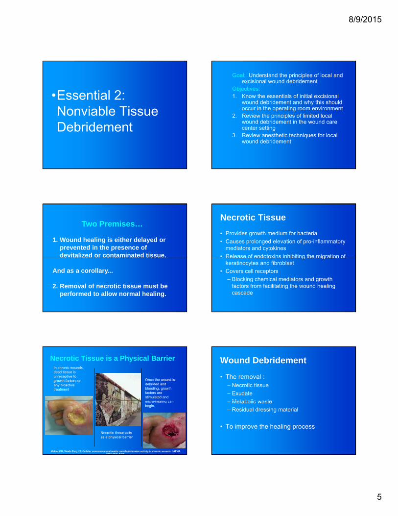
Two Premises…
1. Wound healing is either delayed or prevented in the presence of devitalized or contaminated tissue.
And as a corollary...
2. Removal of necrotic tissue must be performed to allow normal healing.
Necrotic Tissue
• Provides growth medium for bacteria
• Causes prolonged elevation of pro-inflammatory mediators and cytokines
• Release of endotoxins inhibiting the migration ofRelease of endotoxins inhibiting the migration of keratinocytes and fibroblast
• Covers cell receptors
– Blocking chemical mediators and growth factors from facilitating the wound healing cascade
Necrotic Tissue is a Physical BarrierIn chronic wounds, dead tissue is unreceptive to growth factors or any bioactive treatment
Once the wound is debrided and bleeding, growth factors are stimulated and micro healing canmicro-healing can begin.
Necrotic tissue acts as a physical barrier
Mulder GD, Vande Berg JS. Cellular senescence and matrix metalloproteinase activity in chronic wounds. JAPMA 2002;92(1):3407.
Wound Debridement
• The removal :– Necrotic tissue
– Exudate
Metabolic waste– Metabolic waste
– Residual dressing material
• To improve the healing process

8/9/2015
6
Uncontrolled MMP’s in Wound Healing
Excessive MMP activity may result in:
• Degradation ofDegradation of newly deposited tissue components
• Destruction of GF’s, cell surface receptors
• Chronic, non-healing wounds
Debridement of Wounds• Reduces bacterial load
• Decreases production of MMPs
• Increases the production and release of growth factors
E t ll f th f t• Exposes receptors on cells, for growth factor interface
• Removes senescent cells
• Facilitates angiogenesis
• Wound bed prep for advanced therapies
• Allows for determining depth and character of the wound bed
Cleansing vs Debridement
• Cleansing is the use of fluids to remove loosely adherent material
D b id t i ti h• Debridement is an enzymatic or sharp dissection process to remove tightly adherent or necrotic material.
Types of Debridement
• Autolytic
• Enzymatic
• Mechanical
• Sharp
• Surgical
• Biologic (Larva or Maggot therapy)
• Chemical
Debridement Principles
• Clean wound base of all devitalized tissue
• Remove “undermining”

8/9/2015
7
Which Debridement Technique is NEEDED?
• All debridement techniques might be appropriate in the same patient, at different times, depending on:– Type and extent of devitalized tissue – Pain control– Infection or bleeding riskInfection or bleeding risk– Cost– Access to care– Underlying nutritional status– Many other complex factors
Autolytic Debridement
• Moist Wound Healing Goal– Promotes physiological healing
A t l ti d b id t G l• Autolytic debridement Goal– The promotion of endogenous enzymes to
debride necrotic tissue
Autolytic Debridement
indications
• All wounds with necrotic tissue
Contraindications
• Dry gangrene or dry ischemic wounds
• Must determine vascular status
Autolytic Debridement
• ADVANTAGES– Does not damage surrounding skin
– Fast--should see significant improvement within one week
– Painless
– Easy to perform
– Always occurs when using MOIST WOUND HEALING
– Can and should be used in conjunction with other methods of debridement
Autolytic Debridement
• DISADVANTAGES– Not as rapid as surgical, mechanical or
enzymatic debridement
– Occlusive dressings may promote anaerobicOcclusive dressings may promote anaerobic growth
– Maceration of skin edge
– Sensitivity to adhesives
Autolytic Debridement
• CONTRAINDICATIONS– Dry stable gangrene
– Dry ischemic wounds
Contraindicated for infected wounds– Contraindicated for infected wounds
RELATIVE CONTRAINDICATIONS– Diabetic wounds

8/9/2015
8
Dressing Absorb Wound Type
Saline Gauze Yes All, undermined, cavernous
Hydrogel Limited All except heavily
Autolytic Debridement
y g p yexudative
Alginate Yes All except dry eschar
Hydrocolloid Yes All. Use filler for dead space
Foam Yes All. Use filler for dead space
Film No Avoid heavily exudative
Autolytic Debridement
• Necrotic tissue is liquified using the body’s natural enzymes– phagocytic cells
proteolytic enzymes– proteolytic enzymes. • Accomplished by
keeping the wound moist with occlusive or semiocclusive dressings.
• Only works if wound stays moist
Autolytic Debridement• Slow, painless• Can be used when patient:
– Is stable from medical and nutritional standpoint– Is not a candidate for sharp debridement
• Should not be used when the wound is infected
80 y.o. woman with COPD and active lung cancer on prednisone. Multiple skin tears treated with saline wet to dry for 16 weeks with no improvement. Debrided
and healing wound after 14 weeks of moist wound care using a hydrocolloid.
Mechanical
• Application of outside force or energy to dislodge necrotic tissue
• Does not discriminate between viable and nonviable tissuenonviable tissue
• Examples: – Wet to dry
– Pulse Lavage
– Whirlpool
Wet to Dry
• Non-selective• Cost of dressing is not a factor
although labor costs may be significantg
• Various interpretations of what wet to dry means
• Various gauze types used, most common open weave, woven,
• Issues of linting and pain• Traumatic to wound bed
Wound Irrigation Systems:
Maximum safe irrigation pressure is 15 psi
• Bulb syringe 2 psi
• #19 needle on syringe 8-10 psi
• Tume syringe 4 psi• Tume syringe 4 psi
• Water pik– low setting, 6 psi
– med setting, 35 psi
– high setting, 70 psi
• Stryker system, 8-15 psi

8/9/2015
9
Pulsatile Lavage
• Provides cleansing and debridement with pulsed irrigation combined with suction
• Negative pressure removes irrigation and debris to reduce infection and encouragedebris to reduce infection and encourage granulation tissue formation
Pulse Lavage
• Quick removal of necrotic tissue, bacterial load, foreign debris
• Controlled pressure >15 psi• Controlled pressure >15 psi
• Site specific
• Less risk of maceration
• Less risk of cross contamination
• Can be done at bedside
Pulse Lavage
• Used in…– Pressure ulcers
– Diabetic ulcer
– Venous stasis ulcers
– Cavernous wounds
– Tunneled or undermined wounds
– Infected wounds
– Multiple wound sites
Enzymatic Debridement
• Application of topical agents that disrupt or digest extracellular proteins.
• Collagenase: derived from the fermentation of Clostridium histolyticum.
– It possesses the unique ability to digest collagen in t possesses t e u que ab ty to d gest co agenecrotic tissue.
• Papain: proteolytic enzyme from the fruit of carica papaya, a potent digestant of nonviable protein matter but harmless to viable tissue.
– Relatively ineffective when used alone – Usually combined with urea, a substance which denatures
proteins. – Urea makes the proteins more susceptible to enzymatic
digestion.
Enzymatic Debridement
54 y.o. man with painful mixed arterial and venous disease ulcer. The patient applied collagenase daily for 5 weeks.
Chemical Debridement
• Silver Nitrate– Skin, wound cauterization following sharp
debridement
– Debridement of hypergranulated tissueDebridement of hypergranulated tissue
– Educate the patient regarding black or gray staining

8/9/2015
10
Medicinal MaggotsMaggot Debridement Therapy (MDT)
FDA approved Medical Device as of 2002005
Monarchlabs.com
Medicinal Maggot Therapy
• Facultative Myiasis: Infestation that is not harmful to the host
• Action of Maggots:D b id th d b di l i ti– Debride the wound by dissolving necrotic tissue
– Kill bacteria
– Stimulate wound healing
Medicinal Maggots
COST:
1 Vial (250-500) $80
4 4 d i $44x4 dressing $4
8x8 dressing $8
FEDEX $35
Indications for Sharp Debridement
• Wound Bed Preparation: As an adjunct to allow other methods to be more effective
• Presence of deep eschar such thatPresence of deep eschar such that autolytic or enzymatic debridement will not be effective
• Should be first consideration in an infected wound
When Not to Debride
• If the wound is dry ischemia, and vascular status is not yet determined
• If a clear demarcation line is not establishedestablished
• If you are not prepared to follow through on wound care
• If you are unsure of what you will find and how deep it will be
Debridement…Critical Principles1. Necrotic tissue must be debrided to
facilitate healing and management of microbial burden.
2 Debridement of lower extremity2. Debridement of lower extremity wounds/ulcers, especially those associated with arterial insufficiency or diabetes mellitus should undergo sharp, surgical, excisional debridement only after evaluation of perfusion status.

8/9/2015
11
Debridement…Critical Principles
3. Do not debride stable, dry, black, non tender, non fluctuant, non erythematous and non suppurative eschars until perfusion status is determined.
4. Debridement of nonviable and non infected tissue should be performed ONLY AFTER the revascularization procedure. Pre revascularization debridement should be indicated only in a septic foot with and without ischemic signs.
5. Soft, fluctuant eschar should be unroofed when identified.
Debridement…Critical Principles6. For diabetic neuropathic foot ulcers, the
following are always indications for debridement:
• Presence of callus• Presence of skin undermining at the ulcer edges
or margination of epithelium• Wound bed necrotic tissue
7. These indications are probably appropriate for other wounds/ulcers as well.
Debridement…Critical Principles8. There is no evidence for the
effectiveness of hydrotherapy/whirlpool in chronic wound/ulcer patients (see DCS white paper)DCS white paper).
Excisional Debridement CPT Codes
• The removal of tissue by surgical means by cutting outside or beyond the wound margin in whole or in part (CPT 2007 AMA).
• 11040: debridement skin, partial thickness
• 11041: debridement of skin full thickness11041: debridement of skin, full thickness
• 11042: debridement of skin, and subcutaneous tissue
• 11043: debridement of skin, subcutaneous tissue, and muscle (would also include fascia, tendon, joint capsule; 10 day global)
• 11044: debridement of skin, subcutaneous tissue, muscle, and bone (bone is the discriminator; 10 day global)
Selective “Debridement CPT Codes
• The removal of devitalized tissue including slough, fibrin, exudates, crusts, and other non-tissue materials from wounds
• 97597 total wound(s) surface area ≤ 20• 97597 total wound(s) surface area ≤ 20 cm2;
• 97598 total wound(s) surface area > 20 cm2 and is billed once per patient per event
When and how often should chronic wounds be debrided?
• When there is necrotic tissue present
• When there is tunneling or undermining detected
• When the wound edges need sculpting in order to favor epithelial migration

8/9/2015
12
Maintenance Debridement
Repeated removal of necrotic tissue throughout the lifespan of the chronic wound
• Required for chronic wounds– Fibrotic and necrotic tissue continue to accumulate in
the wound
• Continually prepares the wound bed for healing
Falanga V. Wound bed preparation and the role of enzymes: A case of multiple actions of therapeutic agents. WOUNDS 2002;14(2):47-57.
Falanga V. Introdusing the concept of wound bed preparation. International Forum on Wound Care 2001;16(1):1-4.
CONCLUSION
• A key to wound healing is a clean wound
• Autolysis always occurs in a moist wound environment
• Mechanical debridement should be discontinuedMechanical debridement should be discontinued as soon as possible
• Enzymatic and Autolytic debridement may be used in conjunction with each other and with Sharp Debridement
Please welcome
Katherine A Lincoln DO FAAFP
8/9/2015 70
Katherine A. Lincoln, DO, FAAFP
The Nine Essentials of Wound Healing
1. Adequate Perfusion2. Non-Viable Tissue3. Inflammation or Infection4. Edema5. Wound Microenvironment6. Tissue Growth Optimized7. Off-Loading8. Pain Control9. Host Factors
Essential 3: Wounds with bugs
don’t heal!!don t heal!!
Katherine A. Lincoln, DO, FAAFP

8/9/2015
13
Essential 3: Signs and Symptoms of Infection and/or Inflammation
• AKA Wounds with Bugs don’t Heal
• AKA Dear ER, please stop putting everyone on Clindamycin
“To cure, sometimes;
To help, often,To help, often,
To care, always.”-Hippocrates
8/9/2015 74
Essential 3: Signs and Symptoms of Infection and/or Inflammation• Hallmarks of
Infection:
• Calor
• DolorDolor
• Rubor
• Tumor
Essential 3: Signs and Symptoms of Infection and/or Inflammation
• Evaluation is subjective and objective
• Culture +/- biopsy
• Labs
Getting to the Truth
• Culture vs Biopsy
• “gold standard” is biopsy of deep tissue within wound
Oft ti d h bi ith h• Often practiced: punch biopsy with punch biotome, swab of wound, needle-aspiration of fluid
• Don’t start IV or po Abx before wound culture
Biopsy of Lesions
• Biopsy ANY atypical wound
• Biopsy ANY wound that has been unresponsive to 2-4 wk of appropriateunresponsive to 2 4 wk of appropriate therapy
• (we usually use 3-4mm punch biotome, 2 punches, one to pathology and one to histology)
8/9/2015
14
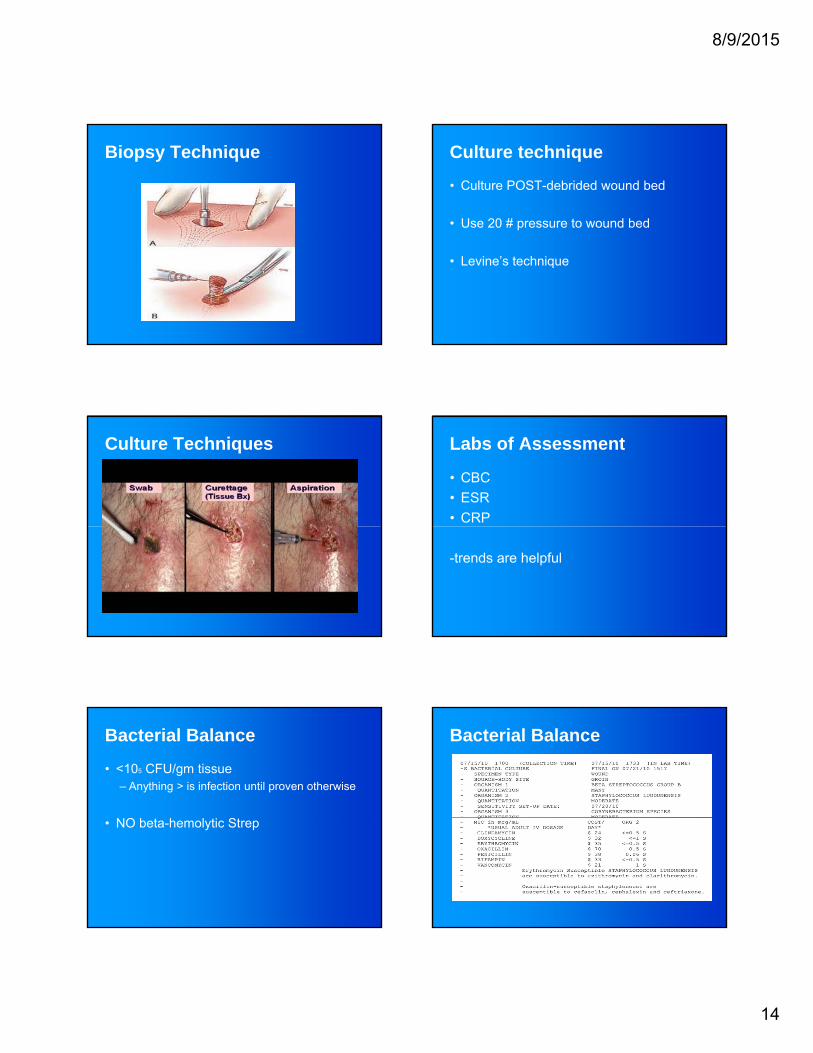
Biopsy Technique Culture technique
• Culture POST-debrided wound bed
• Use 20 # pressure to wound bed
• Levine’s technique
Culture Techniques Labs of Assessment
• CBC
• ESR
• CRP
-trends are helpful
Bacterial Balance
• <105 CFU/gm tissue– Anything > is infection until proven otherwise
• NO beta-hemolytic Strep
Bacterial Balance

8/9/2015
15
Treatment of Inflammation vs. Infection• Topical antimicrobials
• Topical Antibotics
• Systemic Antibotics• Bacteremia• Sepsis• Advancing Cellulitis• Osteomylitis
Use Evidence Based Medicine
Use Evidence Based Medicine
8/9/2015 87
Essential 3: Signs and Symptoms of Infection and/or Inflammation
Pearles:
Assess bacterial bioburden BEFORE:
S i l l• Surgical closure
• Direct wound approximation
• Free flap
• Bioengineered tissue graft
Essential 3: Signs and Symptoms of Infection and/or Inflammation
Pearles:
• If Osteo is suspected in DFUserial x ray– serial x-ray
– MRI
– CT
– Tagged WBC radionuclide study
Essential 3: Signs and Symptoms of Infection and/or Inflammation
• Pearles:
• EARLY closure is the MOST important intervention to prevent wound infection

8/9/2015
16
Essential 3: Signs and Symptoms of Infection and/or Inflammation
• Pearles:
• S/s of disease may be subtle due to decreased blood flow +/- immune compromised stateblood flow / immune compromised state
Comments, Additions
Please welcome back
Dr Sprague Taveau
8/9/2015 93
Dr. Sprague Taveau
The Nine Essentials of Wound Healing
1. Adequate Perfusion2. Non-Viable Tissue3. Inflammation or Infection4. Edema5. Wound Microenvironment6. Tissue Growth Optimized7. Off-Loading8. Pain Control9. Host Factors
Anatomic Layers of the Venous system
Deep
Superficial
Inferior Vena Cava
Iliac Vein
Femoral Vein
Deep Veins
Femoral Vein
Popliteal VeinPosterior TibialisAnterior Tibialis

8/9/2015
17
Superficial Veins
Superficial venous system- tremendously complicated and extremely variable network of i t ti iinterconnecting veins
Most veins are unnamed
A few larger superficialveins are fairly constantin location
Main superficial veins of the leg
Short saphenous vein
Long saphenous vein
Perforators pass through anatomic defects in the deep fascia to connect the superficial
Perforating Veins
the superficial “collecting web” and the larger superficial veins with the deep veins of the calf or thigh
At Rest- Legs Elevated
0 - 10 mmHg
Walking
30 - 40 mmHg
Standing
90 mmHg±

8/9/2015
18
30 mm Hg
40 mm Hg
50 mm Hg
60 H60 mm Hg
70 mm Hg
80 mm Hg
90 mm Hg
100 mm Hg
30 mmHg
40 mmHg
Th i
“The Battle Against Gravity”
The calf muscle pump
50 mmHg
60 mmHg
70 mmHg
There is a continuous column of
blood from foot to heart
Calf Muscle Pump
The deep veins
Th fi i l i
“The Battle Against Gravity”
Four components working together
against the force of gravity
The superficial veins
The venous valves
The calf muscles insidethe inelastic fascia
Venous Valves- normal competent valves allow blood flow in only one direction
open
closedclose
d
open
deep vein
superficial vein
Muscle Systole
Every time the
Calf muscles inside the inelastic fascia effects blood flow during muscle systole
calf muscles contracts veins are compressed
and blood is pushed upwards
Muscle Diastole
Every time the calf l
Calf muscles inside the inelastic fascia effects blood flow during muscle diastole
musclesare relaxed, blood
is suckedin from the
peripheralregions

8/9/2015
19
Injection of radiografic contrast dye in the femoral vein shows a competent valve, which is preventing backward flow down the vein
Leaky Valves allow deep to superficial flow during muscle systole and bidirectional during muscle diastole.
CompetentValve
IncompetentValve
35 mmHg
PC = 25 mmHg
FiltrationResorptionPA
Blood Capillary
Normal Vascular Fluid Balance
15
Lymphatic Capillary
~30 liters/dayPV
protein
~27 liters/day
~3 liters/day(10% of filtered)
Dr. HN Mayrovitz
• Natural secondary response
• Often temporary
• Normal lymphatic system
Edema
• Primarily water
Passive HyperemiaVenous insufficiencyCardiacPulmonaryPregnancy
HypoproteinemiaMalabsorbtionMalnutritionRenal diseaseC li idi thi
Etiology of Edema
g yInactivityDependencyTravel
Active HyperemiaInflammationAllergy
Cyclic idiopathic edema syndromes
DrugsAge
• High protein edema consisting of both• Lymph
• Water
Lymphedema
• Results from• Damage to lymphatic vasculature
• Absence of the normal lymphatic anatomy

8/9/2015
20
Primary• Congenital (Birth)• Praecox (adolescent)• Tarda (age 35+)
Secondary• Surgery• Infection• Tumor
Etiology of Lymphedema
( g )• Radiation• Wounds• Venous • Trauma• Neurological• Filiariasis
• Overload = Edema
• Lymphatic Transport Malfunction +Edema + Protein = Lymphedema= Lymphedema
If Net Filtration Exceeds Lymphatic Transport Capacity
Therapy OptionsTherapy Options•• Reduce FiltrationReduce Filtration
••CompressionCompression•• Increase Transport CapacityIncrease Transport Capacity
••MassageMassageDr. HN Mayrovitz
Edema + Protein = Lymphedema= Lymphedema
Calf Pump Failure
Venous Hypertension
WBC Adhesion
Cell Wall Damage
Increased Permeability
Etiology of an Edema
Related Ulcer
y
EDEMAActivated CellsProteins
InflammationImpairedNutrition
Tissue Death
Ulceration
LYMPHEDEMA
Bottom Line
Wounds won’t heal in a swamp!!
8/9/2015 118
Please welcome back
Dr Glen Einspanier
8/9/2015 119
Dr. Glen Einspanier
The Nine Essentials of Wound Healing
1. Adequate Perfusion2. Non-Viable Tissue3. Inflammation or Infection4. Edema5. Wound Microenvironment6. Tissue Growth Optimized7. Off-Loading8. Pain Control9. Host Factors

8/9/2015
21
Goals: Understand the key factors in wound examination, documentation and classification and a general approach to selecting products for topical wound care.
Objectives:Objectives:1. Outline the Essentials in wound
examination2. Know the technology useful in wound
examination and documentation.3. Know the important wound
classification systems4 Know a topical wound care product
Wound Assessment
• Good complete History and Physical– We treat the whole patient not just the hole in
the patient
Purposes of Assessment
• Initial definition/diagnosis
• Monitoring the effect of treatment
• Failure to progressp g
• Monitoring for occurrence of infection
• Prediction of outcome
• Reimbursement qualification
David H. Keast MSc, MD, FCFP
Parkwood Hospital, St. Joseph’s Health Care London, London, Canada
Partial vs Full Thickness wounds
– Partial• Loss of epidermis into but not through the dermis
– Abrasions, Skin tears, Blisters
– Full• Through the dermis into subcutaneous tissue,
muscle, and may exposes deep structures
Skin Assessment
• Temperature– Normally warm to the touch
• Warmer could indicate inflammation
• Coolness could indicate vascular issuesCoolness could indicate vascular issues
– Color• Intensity
– Pallor
– Rubor
Skin Assessment
• Hyper – or Hypopigmentation
• Moisture– Dry (xerosis) or moist
H k t i (fl ki l )– Hyperkeratosis (flaking, scales)
– Eczema
– Dermatitis, psoriasis, rash
• Turgor– Dehydration vs effects of aging

8/9/2015
22
Location
• Document in reference to Commonly used terms– Proximal, distal
Superior inferior– Superior, inferior
– Medial, lateral
– Anterior, posterior
– Dorsal, plantar
Location
• Pressure Ulcers– Identify the location by the boney prominence
beneath the wound • Left iliac crestLeft iliac crest
• Left trochanter
• Thoracic spine
MEASURE
• M = Measure
• E = Exudate
• A = Appearance
• S = Suffering
• E = Exudate
• E = Edge• A = Appearance
• S = Suffering
• U = Undermining
• R = Reevaluation
• E = Edge
Keast DH, et al. Wound Rep Reg 2004; 12:S1-S17.
• E = Edge
• U = Undermining
• A = Appearance
• M = Measure
• R = Reevaluation
Measure: Length, Width, Depth
David H. Keast MSc, MD, FCFP
Parkwood Hospital, St. Joseph’s Health Care London, London, Canada
• Document in centimeters
• Length (measurement head to toe or longest dimension)
• Width (perpendicular to length)
• Depth (at deepest point)
Measure: Length, Width, Depth
• Calculations of area and volume
• Tools:
– linear measuring ruler
– acetate wound tracings
– computerized planimetry
– digital photography
• Issue of inter-rater reliability
Key PointThe percentage of decrease in
wound area in first 2-4 weeks has been found to be predictive of
Measure: Length, Width, Depth
phealing at 12-24 weeks.
Keast DH, et al. Wound Rep Reg 2004; 12:S1-S17.

8/9/2015
23
Wound Size: Depth
• Insert tip of probe to greatest depth and measure perpendicular di t t d d
Measure: Length, Width, Depth
distance to wound edge
• 15% variability
David H. Keast MSc, MD, FCFP
Parkwood Hospital, St. Joseph’s Health Care London, London, Canada
Wound Depth
• Foam Tip Measuring Device with centimeter calibrations
Measure: Length, Width, Depth
• Too little exudate results in a desiccated wound
• Excessive exudate complicates wound management
mEasure: Exudate
management
Look at the dressing upon removal…
mEasure: Exudate
Strike through.. Leakage….Intact?
Exudate: Character..Amount..Color…Odor
mEasure: ExudateExudate Descriptions
• Character– Serous, Serosanguineous, Sanguineous,
PurulentPurulent
– Opaque, clear, cloudy
– Liquefying necrotic tissue (slough)
– Dressing residue
Exudate Descriptions
SMALL
mEasure: Exudate
NONE

8/9/2015
24
Exudate Descriptions
MODERATE
LARGE
mEasure: Exudate
Moisture Balance
MEasure: Exudate
Condition Of The Wound Bed
• Necrosis
• Granulation tissue
Exposed structures
meAsure: Appearance
• Exposed structures
• Fibrin
• Exudate
• Eschar
• Foreign body
• Inflammation, infection
• Tunneling, sinuses
Tissue type Description
Granulation Red, firm, and pebbled. Friability may indicate infection.
Fibrin Yello and firm Represents collagen
meAsure: Appearance
Keast DH, et al. Wound Rep Reg 2004; 12:S1-S17.
Fibrin Yellow and firm. Represents collagen or other proteinaceous material in the wound bed.
Slough Yellow to gray-green and loose. Soft, loose necrotic tissue.
Eschar Black, soft and wet or hard and dry. Necrotic tissue
• Evaluation…By standard grading scale in every patient at every visit
• Consider…Monitor and reevaluate pain level adjust intervention strategy and
meaSure: Suffering (pain, disability)Essential 8
level, adjust intervention strategy and continue reevaluation and treatment adjustment
• A change in pain (worsening) should prompt reevaluation for infection, ischemia.
Keast DH, et al. Wound Rep Reg 2004; 12:S1-S17.Krasner D. in Chronic Wound Care 2nd ed, 1997;336-
343
Space between the surrounding skin and the wound bed– Usually involves significant
proportion of wound edgesM t d ti l d
measUre: Undermining
– May extend entirely around wound
– Subcutaneous fat necrosis– Result of:
1. Necrotizing infection2. Primary inflammatory etiology3. Friction, shearing

8/9/2015
25
measUre: UnderminingTunneling
• Extends into tissue in any direction• May be more than one present in any
wound
measUre: Undermining
• Document number if more than one present
• Document direction of each using face of clock
• Document depth of each if more than one is present
Tunneling
measUre: UnderminingTunneling
measUre: Undermining
Condition Of The Surrounding Skin
• Color
• Pigmentation
measurE: Edge…surrounding skin
• Inflammation, induration
• Dermatologic abnormalities
• Satellite lesions
• Suppleness
• Edema
• Maceration
NPUAP Pressure Ulcer Classification System
• Stage I: Non-blanchable erythema of intact skin
• Stage II: Partial thickness skin loss involving epidermis or dermis
• Stage III: Full thickness involving subcutaneous tissue that may extend down to, but not through, the underlying fascia
• Stage IV: Full thickness with extensive tissue necrosis involving muscle, bone, or supporting structures
National Pressure Ulcer Advisory Panel. Consensus Development Conference Statement, West Dundee, IL, S-N Publications, Inc., 1989.

8/9/2015
26
Stage I (New from NPUAP)
– An observable pressure related alteration of intactskin whose indicators as compared to an adjacent or opposite area on the body may include changes in one or more of the following:
• Skin temperature (warmth or coolness)• Skin temperature (warmth or coolness)
• Tissue Consistency (Firm or boggy feel)
• Sensation (pain or itching)
– Non-blanchable erythema of intact skin (press 15 seconds)
– In darker skin tones, the ulcer may appear with persistent red, blue or purple hues
Stage I Press for 15 seconds.If remains red, Stage I
Blanching is NOTStage I
Deep Tissue Injury (DTI)
• Often skin is still intact
• Deep purple or deep bruising present
• Obvious deeper muscle damage
• Unfortunately, staged as a Stage I
• Typically will demonstrate that it is actually Stage III or IV in days to follow
• “… Stage I pressure wound with DTI…”
Stage II – Partial thickness skin loss involving epidermis and or dermis. The ulcer is superficial and presents clinically as an abrasion, blister or shallow crater.
Stage III – Full thickness skin loss involving damage/necrosis of subq tissue. Presents as a deep crater, possible undermining.
Stage IV – Full thickness skin loss with extensive destruction,necrosis, damage to muscle, bone or tendon
8/9/2015
27
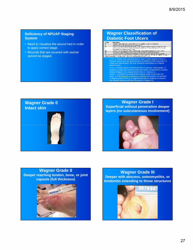
Deficiency of NPUAP Staging System
• Need to visualize the wound bed in order to apply correct stage.
• Wounds that are covered with escharcannot be stagedcannot be staged.
Wagner Classification of Diabetic Foot Ulcers
• Grade II: Deeper ulcer reaching tendon, bone, or joint capsule (if bone is exposed, based upon the preponderance of evidence of the association of osteitis and osteomyelitis, the ulcer should be reclassified as a Wagner grade III ulcer)
• Grade III: Deeper tissues involved and there is abscess, osteomyelitis, or tendonitis usually with extension along the midfoot compartments of tendon sheaths (Wagner in his original article refers to “abscess or osteitis”…remember that anatomically Wagner grade II and III are the same…it is the presence or absence of infection that defines the transition to Wagner grade III from II, so aggressively look for evidence of infection; any ischemic necrosis will move the grade to Wagner IV)
Wagner Grade 0 Intact skin
Wagner Grade I Superficial without penetration deeper layers (no subcutaneous involvement)
Wagner Grade II Deeper reaching tendon, bone, or joint
capsule (full thickness)
Wagner Grade IIIDeeper with abscess, osteomyelitis, or
tendonitis extending to those structures

8/9/2015
28
Wagner Grade IV Gangrene of some portion of the
toe, toes, and/or forefoot
Wagner Grade V Gangrene involving the whole foot or
enough of the foot that no local procedures are possible
UTHSCSA Diabetic Wound Classification MEASURE
• M = Measure
• E = Exudate
• A = Appearance
• S = Suffering
• E = Exudate
• E = Edge• A = Appearance
• S = Suffering
• U = Undermining
• R = Reevaluation
• E = Edge
Keast DH, et al. Wound Rep Reg 2004; 12:S1-S17.
• E = Edge
• U = Undermining
• A = Appearance
• M = Measure
• R = Reevaluation
Bottom Line
Wounds won’t heal unless the environment is conducive to
healing…..
8/9/2015 167
Please welcome back
Dr Katie Lincoln
8/9/2015 168
Dr. Katie Lincoln

8/9/2015
29
The Nine Essentials of Wound Healing
1. Adequate Perfusion2. Non-Viable Tissue3. Inflammation or Infection4. Edema5. Wound Microenvironment6. Tissue Growth Optimized7. Off-Loading8. Pain Control9. Host Factors
ADVANCED THERAPIES IN WOUND
CARE
Focus: Essential 6Enhance Tissue Growth
Options to stimulate tissue growth
AFTER:
• Proper debridement
• Control of microbial burden
• Optimized wound bed moisture balance
• Optimization of Host Factors
Enhance Tissue Growth
Options:• Growth
factors/cytokine replacementD l b t t
• Negative pressure wound therapy
• Correction of local hypoxia (stimulate angiogenesis)• Dermal substrate
replacements• Bioengineered tissue
grafts• Surgical
closure/reconstruction
angiogenesis)• Misc “other”: laser,
US• Soft tissue splinting• Hyperbaric oxygen
therapy (HBO)
Enhance Tissue Growth: 4 different methods• 1-Bioengineered tissue
• 2-Negative Pressure Wound Therapy
• 3-Hyperbaric oxygen therapy
• 4-Suction bubble epidermal grafting

8/9/2015
30
Bioengineered tissues
• 3 main types: Dermagraft, Apligraf, Epifix
• Dermagraft- human derived dermal substitute; only FDA approved for chronic diabetic foot ulcers (DFU)diabetic foot ulcers (DFU)
• Apligraf- 2 layered living cell based product; Only FDA approved for DFU and chronic venous insuffiency ulcers
Bioengineered tissues• Epifix is dehydrated Human
Amnion/Chorion Membrane dHACM
• Available to place on any wound anywhere
• Uses the PURION Process for tissueUses the PURION Process for tissue safety
• Multiple layers including a single layer of epithelial cells, a basement membrane, and an avascular connective tissue matrix
EpifixEnhance Tissue Growth: 4 different methods
• 1-Bioengineered tissue
• 2-Negative Pressure Wound Therapy
• 3-Hyperbaric oxygen therapy
• 4-Suction bubble epidermal grafting
Negative Pressure Wound Therapy (V.A.C. devices)
• The V.A.C. Therapy System is comprised of three essential components that actively work together to help promote wound healing through granulation tissue formation.
VAC Therapy Unit: Provides intermittent and continuous– VAC Therapy Unit: Provides intermittent and continuous therapy with integrated patient safety features
– SensaTRAC Technologytm
: Regulates pressure at the wound site to provide accurate delivery of prescribed therapy settings
– VAC GranuFoampatienttm
Dressings: Help provide the necessary mechanisms to promote granulation tissue formation
Negative Pressure Wound Therapy (V.A.C. devices)

8/9/2015
31
Negative Pressure Wound Therapy (V.A.C. devices)FDA approved indications of VAC• Cavitary wounds (acute trauma, post surgical,
chronic-diabetic wounds, pressure wounds)• Heavily exudative if infection is treated
S i l d d hi• Surgical wound dehiscence• Wounds requiring soft tissue mechanical
stabilization• Split thickness grafts/ support of flaps• Thermal partial thickness burns• Management of certain fistulas
Enhance Tissue Growth: 4 different methods• 1-Bioengineered tissue
• 2-Negative Pressure Wound Therapy
• 3-Hyperbaric oxygen therapy
• 4-Suction bubble epidermal grafting
Hyperbaric oxygen therapy
• “HBO”
• Defined as “breathing 100% oxygen at a pressure greater than 1.5 ATA for the purpose of elevating the PtO2 and therebypurpose of elevating the PtO2 and thereby tissue PO2 values improve oxygen delivery and diffusion to malperfused tissue”
• Used as adjunctive therapy in wound care
Hyperbaric oxygen therapy
HBO CMS indications
• Acute carbon monoxide intoxication
• Cyanide poisoning• Decompression illness• Gas embolism• Gas gangrene
• Necrotizing fasciitis • Acute peripheral arterial
insufficiency• Prep/preservation of
compromised grafts• Chronic refractory Gas gangrene
• Acute traumatic peripheral ischemia
• Crush injuries/suturing of severed limbs
yosteomyelitis
• Osteonecrosis• Soft tissue radionecrosis• Actinomycosis, refr to Abx• Diabetic Wound of the LE,
Wagner Grade 3 or more
Enhance Tissue Growth: 4 different methods• 1-Bioengineered tissue
• 2-Negative Pressure Wound Therapy
• 3-Hyperbaric oxygen therapy
• 4-Suction bubble epidermal grafting

8/9/2015
32
Suction Bubble Epidermal Grafting (SBEG)• Commercially called “CelluTome”, which is
an epidermal harvesting system
• Epidermal harvesting system in out patient settingsetting
• No “down time” or OR complications
• Uses heat and suction for auto-donation of Microdomes, transfer to chronic wound site
Suction Bubble Epidermal Grafting (SBEG)
Questions, additions, subtractions, and edifications
Katherine Lincoln, DO, FAAFP [email protected]
Please welcome back
Dr Sprague Taveau
8/9/2015 190
Dr. Sprague Taveau
The Nine Essentials of Wound Healing
1. Adequate Perfusion2. Non-Viable Tissue3. Inflammation or Infection4. Edema5. Wound Microenvironment6. Tissue Growth Optimized7. Off-Loading8. Pain Control9. Host Factors
Positioning and Off-Loading• Sustained pressure causes most ulcerations• Factors for unrelieved pressure:
– Inactivity, immobility, decreased sensation
• Use pillows, bolsters, pads, support surfaces• Turning schedules:• Turning schedules:
– Every two hours, more often in higher risk patient– Typically supine to sidelying, opposite sidelying, then
back to supine (3/4 position)– Protective boots or pillow bridging for heels

8/9/2015
33
Positioning and Off-Loading
• Pressure relief techniques– Volitional off-loading at periodic intervals
Capillary refill and tissue reperfusion
Wheelchair pushups– Wheelchair pushups
– Passive repositioning by care provider
– Greater risk: support surface modification
Positioning and Off-Loading
• Support surfaces – 2 groups– Pressure reducing – low risk patient
• Tend to lower tissue interface pressure
• Don’t provide full reliefDon t provide full relief
• Pads, cushions, overlays, foam mattresses
– Pressure relieving• Consistent reduction of tissue interface pressure
• Low air, high air, dynamic air devices
Positioning and Off-Loading
• Mattress overlays– Pressure reducing surface
– Inexpensive, easy to clean and transport
Bottoming out phenomenon– Bottoming out phenomenon
– Leaks and moisture build up
– Less forgiving
– Intended for the patient of < minimal risk
Positioning and Off-Loading
• Specialized pads – dense foams & gels
• Foam pads– Low cost, ease of application
I b d t t t i i t– Increase body temperature, retain moisture
– “bottoming out “ phenomenon
• High quality gel pads– None of the above disadvantages
– Higher cost, heavy
Positioning and Off-Loading
• Low air loss beds– Pressure relieving surface
– Patients with high risk for breakdown
Immobile patients or those with ulcerations– Immobile patients or those with ulcerations
– Bed frame, air filled cushions, low friction/shear surface material
– Low noise pump, continuous air flow, multiple areas or zones
– High cost
Positioning and Off-Loading
• High air loss beds– “air-fluidized” glass beads
– Dry environment, heat controlled
Highest risk patient– Highest risk patient
– Patients with recalcitrant wounds
– Total bed rest patient
– Highest cost range
– Large and heavy (2000 lbs)
8/9/2015
34
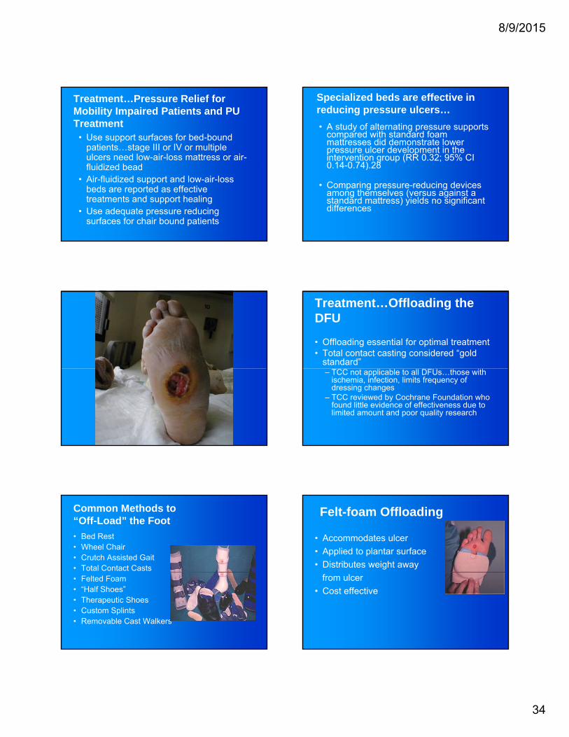
Treatment…Pressure Relief for Mobility Impaired Patients and PU Treatment
• Use support surfaces for bed-bound patients…stage III or IV or multiple ulcers need low-air-loss mattress or air-fl idi d b dfluidized bead
• Air-fluidized support and low-air-loss beds are reported as effective treatments and support healing
• Use adequate pressure reducing surfaces for chair bound patients
Specialized beds are effective in reducing pressure ulcers…
• A study of alternating pressure supports compared with standard foam mattresses did demonstrate lower pressure ulcer development in the intervention group (RR 0.32; 95% CI 0 14 0 74) 280.14-0.74).28
• Comparing pressure-reducing devices among themselves (versus against a standard mattress) yields no significant differences
Treatment…Offloading the DFU
• Offloading essential for optimal treatment• Total contact casting considered “gold
standard”– TCC not applicable to all DFUs…those with
ischemia, infection, limits frequency of dressing changes
– TCC reviewed by Cochrane Foundation who found little evidence of effectiveness due to limited amount and poor quality research
Common Methods to “Off-Load” the Foot
• Bed Rest• Wheel Chair• Crutch Assisted Gait• Total Contact Casts• Felted Foam• “Half Shoes”• Therapeutic Shoes• Custom Splints• Removable Cast Walkers
Felt-foam Offloading
• Accommodates ulcer
• Applied to plantar surface
• Distributes weight away
from ulcer
• Cost effective

8/9/2015
35
Boulton, A, Armstrong, D. Trials in Neuropathic Diabetic Foot Ulceration: Time for a paradigm shift? Diabetes Care 2003 26: 2689-2690.
• The variability in off-loading efforts skews results.
• Removable off-loading devices may be encouraging non-compliance.g g p
• The authors suggest that there are several options for non-removable off-loading devices to help eliminate this variable for future studies.
DH Walker Boot
Completed Total Contact Cast Offloading Sub 1st Metatarsal Head DM Ulcers Peak Plantar Pressures in (N/cm2)
35
40
45
0
5
10
15
20
25
30
TCC DH Walker Aircast 3D Walker CAM XDS CVO
Lavery et al, Diabetes Care, 1996Lavery et al, Diabetes Care, 1996
Armstrong, David et al. Activity patterns of patients with diabetic foot ulceration: patients with active ulceration may not adhere to a standard pressure off-loading regimen.Diabetes Care 2003 26(9): 2575-2580
• 20 subjects treated for neuropathic diabetic foot ulcer, treated with removable cast walker (RCW).
• Total activity measured with waist monitor.
• Mean Essentials per day was 1219 +/- 821 and only 28% f th E ti l d ith th b t i28% of these Essentials were done with the boot in place.
• Only 30% of the study group wore boot for more Essentials than not.
• Even this group only wore boot 60% of the time.
• Conclusion: Off loading regimen compliance is poor and may be worse than we originally thought.
Treatment…Pressure Relief
• “Instant total-contact-cast” using removable cast walker converted into non removable devise by wrapping with cohesive bandage or fiberglasscohesive bandage or fiberglass
Armstrong DG, et al. Technique for fabrication of an “instant total-contact cast” for treatment of neuropathic diabetic foot ulcers.
J Amer Pod Med Assoc 2002; 92(7):405-408.

8/9/2015
36
Positioning and Off-Loading• Specialized footwear
– Plantar ulcers– Diabetic patient with neuropathy (insensate)– Options:
• Prefabricated lifts and ankle supportsC t ld d h• Custom molded shoes
• Orthoses • Wedges• Total walking casts
– Shoe with removable insole sections (new)– Surgical options
• Achilles lengthening
Positioning and Off-Loading
• Surgical options– Achilles tendon lengthening– Gastrocs recession– Tenotomyy– Metatarsal head resection
8/9/2015 213
Bottom Line
Wounds won’t heal under
8/9/2015 214
Wounds won t heal under pressure……
Please welcome back
Dr Glen Einspanier
8/9/2015 215
Dr. Glen Einspanier
The Nine Essentials of Wound Healing
1. Adequate Perfusion2. Non-Viable Tissue3. Inflammation or Infection4. Edema5. Wound Microenvironment6. Tissue Growth Optimized7. Off-Loading8. Pain Control9. Host Factors

8/9/2015
37
Pain
Factors influencing a painful experience• Age• Age• Personality• Perception• Pain threshold • Past experiences with pain
Assessment of PainBiopsychosocial History- the 5th Vital Sign
Pain Assessment
PProvocation- inciting factor, onset, worsensPalliation- improves, alters or alleviates
Q Quality- describe: sharp/ dull, burn, ache, cramp
PQRST
"If pain were assessed with the same zeal as other vital signs are, it
would have a much better
chance of being
Wasserman CS. Pain: The Fifth Vital Sign; Alzheimer's Association, Massachusetts Chapter Newsletter Volume 22, Number 1 | Winter 2004 http://www.alzmass.org/newsletters/adnews01-04/01_leadarticle.htm
RRegion- affected body partRadiation- where does it “shoot to”
SSeverity- rating on analog scale 1-10
Special Characteristics- movements, diaphoresis
T Timing- constant, variable, intermittent, duration
chance of being treated properly…
Quality care means that pain is
measured and treated."
-James Campbell, M.D.American Pain SocietyLos Angeles, CA (11
Nov 95)
Beyond the Dressing:Influences on pain at dressing
changes
Inflammatory pain
W d C li tiU d l i Eti l i L l f P i Wound Complicationse.g. infections, skin
maceration, ischemia
Neuropathic PainHighly sensitive to touch
May experience excruciating pain at dressing changes
Underlying Etiologiese.g. peripheral vascular
disease, diabetic neuropathy, arthritis
Level of Pain at dressingchanges
Wound Pain Assessment Pain and Wound Healing
•Clinicians must adopt a rational approach to pain management to help relieve pain and facilitate wound h li d i i i th i k f d l i h i d healing and minimize the risk of developing chronic wound pain.

8/9/2015
38
Bottom Line
Good pain control increases patient compliance…..
8/9/2015 223
Essential 9: Optimizing Host FactorsFactorsDr. Katherine Lincoln, D.O.
Essential 9 Optimizing Host Factors
Essential 9 Optimizing Host Factors
• “ Assess the status of nutrition, diabetes, renal, mobility, other systemic disease, and psychosocial issues and intervene”intervene
Essential 9 Optimizing Host Factors• Smoking
• Diabetes
• Renal, dialysis
• Age
• Systemic Inflammatory Disease
• Visual impairment
• Auditory impairmentg
• Hemoglobinopathies
• Heart disease
• Chronic respiratory disease
• Malnutrition
• Cognitive impairment
• Malignancy
• Chemotherapy
• Chronic steroid therapy
• Addiction
Optimizing Host Factors

8/9/2015
39
Host Factors
• Break it down into Manageable bits
• Nutrition
• Smoking
• DM/Renal
• Mobility
• Other systemic disease
• Psychosocial issues
Assess/Address What You Can
Nutrition Assessment (labs) in the Wound Care Patient • Screening labs:
• Albumin <3.5
• Transferrin <200
• Prealbumin <16
• Total lymphocyte ct <1500
Assess/Address What You Can
Fix What You Can
• ?smoking meds?• Nutritional support
with teaching, food bank, supplementsM bilit t ith
• Work with other attendings in coordination of care
• Addiction related malnutrition and• Mobility support with
off loading mattresses, DME feeding assistive devices
• Diabetic education
malnutrition and multiple adverse effects
• Work with local psych to encourage follow up

8/9/2015
40
Essential 9 Host Factors Necrotic Tissue is a Physical BarrierIn chronic wounds, dead tissue is unreceptive to growth factors or any bioactive treatment
Once the wound is debrided and bleeding, growth factors are stimulated and micro healing canmicro-healing can begin.
Necrotic tissue acts as a physical barrier
Mulder GD, Vande Berg JS. Cellular senescence and matrix metalloproteinase activity in chronic wounds. JAPMA 2002;92(1):3407.
Purposes of Assessment• Initial definition/ diagnosis
• Monitoring the effect of treatment
• Failure to progressFailure to progress
• Monitoring for occurrence of infection
• Prediction of outcome
• Reimbursement qualification
David H. Keast MSc, MD, FCFP
Parkwood Hospital, St. Joseph’s Health Care London, London, Canada
Post-test
1 Infection/Inflammation have little to do with ultimate wound healing. T F
2 In the wounded patient, pain control usually means improved compliance. T F
3 Knowing the patient’s current medications can be key to remedying delayed healing T Fremedying delayed healing. T F
4 Removal of dead tissue from the wound every once in awhile is an important essential of early wound closure. T F
5 The invasive Vascular Surgeon/Cardiologist/Radiologist is a wound care physician’s best friend. T F
8/9/2015 238
The “End”