21 - ophthalmology for the primary care provider...2/27/19 1 ophthalmology for the primary care...
TRANSCRIPT

2/27/19
1
Ophthalmology for the Primary Care Physician
Meredith Marcincin, DO, MA LECOM Health - Ophthalmology
Financial Disclosure
The speaker has no financial interest in the subject matter of this presentation.
2/27/19
2
Objectives
At the completion of the healthcare providers will be able to:
• Develop a differential diagnosis for the presentation of red eye
• Diagnose corneal ulcer and determine best initial topical treatment course
• Describe the examination findings and management for ocular surface trauma including corneal
abrasion and retained foreign body
• Develop a differential diagnosis for the presentation of red, swollen eyelid
• Distinguish between hordeolum and chalazion and formulate treatment plan
• Diagnose herpes zoster ophthalmicus and identify risk factors for ocular involvement
• Develop a differential diagnosis for acute monocular vision loss
• Provide patients with recommendations for vision screening and eye exams
Case 1 : Eye redness, burning, tearing
The patient is a 66 year-old male sportscaster
who reports with a 4-day history of eye irritation
and watering, first noted in the left eye followed
by similar symptoms in the right eye one day
later. Patient admits to mattering of the eyelids
upon waking in the morning. On exam, patient
has bilateral conjunctival injection, excessive
tearing, and sensitivity to light. No current eye
drops or medications being used.

2/27/19
3
Differential Diagnosis of Red Eye
• Viral conjunctivitis• Upper respiratory infection, sick contacts
• Allergic conjunctivitis• Prominent symptom of itching
• Atopic conjunctivitis • History of eczema
• Medication toxicity• Overuse of OTC allergy / “red eye relief” eye
drops or antibiotic drops
• Bacterial conjunctivitis • Copious, purulent discharge
• Dry Eye Syndrome• Chronic, worse at the end of the day
• Corneal ulcer• Contact lens overwear
• Corneal abrasion / foreign body• History of trauma or injury
• Iritis • Constricted pupil, photophobia
Viral vs Bacterial Conjunctivitis? Viral conjunctivitis
• Rhinorrhea, sore throat, sick contacts
• Preauricular lymph nodes
• Clear, watery discharge with eyelid crusting
• Unilateral to bilateral presentation
• Tx: Artificial tears, cool compresses, no role for topical antibiotics
Bacterial conjunctivitis
● Copious discharge
● Eyelid edema, eyelash matting and crusting
● Culture indicated for severe discharge
● Tx: Broad spectrum antibiotics

2/27/19
4
Corneal Ulcer
Sterile ulcer
• Associated with contact lens overwear
Infectious
• Bacterial - pseudomonas & staphylococcus
• Fungi - Fusarium & Candida
• Parasites - Acanthamoeba
Neurotrophic
• Herpes virus, anesthetic abuse
Treatment: Remove offending agent (i.e Dispose of contact lens, discontinue all topical drops), start
broad spectrum antibiotics q 2hrs (Ex: Moxifloxacin), referral
Timely initiation of broad spectrum antibiotics and ophthalmology referral is key to preventing irreversible vision loss.

2/27/19
5
Subconjunctival hemorrhage
• Spontaneous versus history of trauma / mechanical injury
• Benign in nature
• Symptomatic treatment with lubricating drops / ointment
• Recurrent hemorrhages may prompt evaluation for:
○ Bleeding disorder
○ Supratherapeutic INR if on anticoagulation
Corneal abrasion
• Physical Exam – decreased vision,
conjunctival injection, irregular light
reflex, fluorescein dye uptake
• Treatment:
• Fluoroquinolone drops
■ Ciprofloxacin or Ofloxacin
■ Moxifloxacin
• Antibiotic ointment - Erythromycin
or Bacitracin ophthalmic ointment

2/27/19
6
Corneal abrasion: Evaluation
• Fluorescein dye – regions denude of epithelium highlight with cobalt blue filter
• Evert upper eyelid to evaluate for embedded foreign body if evidence of linear excoriations
Corneal abrasion
NEVER prescribe or provide patient with anesthetic drops!
• Ex: Tetracaine, Proparacaine
• May result in large, non-healing epithelial defect
• Utilize ointments and cool compresses for pain reduction
• If opiates necessary for pain control then immediate referral

2/27/19
7
Corneal foreign body• Foreign bodies are one of the most common ophthalmic emergencies – make up 3% of
all emergency room visits
• Risk factors: Male, absence of eye protection, metal-on-metal tasks (ex. grinding, hammering, sanding)
• Timing of removal:
• Iron toxic to ocular structures therefore timely removal is recommended
• Rust formation within 4-6 hrs of injury
• Must confirm no intraocular penetration!
Corneal foreign body
• Mechanism of injury important to determine potential for intraocular and/or intraorbital foreign body
• Seidel test – used to assess for aqueous leakage following injury or surgical procedure
• Positive = Diluted dye streaming from the site of injury or surgical wound
• Indicative of penetrating corneal injury
• Place shield over affected eye and refer for immediate ophthalmology evaluation

2/27/19
8
Back to the case...Viral conjunctivitis
• Rhinorrhea, sore throat, sick contacts
• Excessive tearing, eyelid crusting upon waking
• Preauricular lymph nodes, unilateral to bilateral presentation
• Most common: Adenovirus
• Treatment:
• Artificial tears, cool compresses
• No role for topical antibiotics
• Strict hand hygiene - transmission via fomites and respiratory secretions
Case 2 : Eyelid redness and swelling
Patient is an 80 year old female presenting with one week history of severe left-sided headaches. Three days ago patient noted new onset of left forehead and scalp rash as well as rapid swelling of the left upper and lower eyelids. Patient admits to excessive tearing and light sensitivity but denies loss of vision.

2/27/19
9
Differential Diagnosis of Swollen Red Eyelid
• Internal / External Hordeolum• Erythematous, tender nodule
• Chalazion• Palpable, non-tender nodule
• Blepharitis • Eyelash debris, eyelid margin hyperemia
• Preseptal cellulitis • Progressive erythema, edema
• Orbital cellulitis • Proptosis, restricted eye movement
• Herpes Zoster Ophthalmicus• Dermatomal vesicular rash in V1 distribution
• Eyelid malposition• Ectropion / Entropion
• Contact dermatitis• New exposures, scaling
• Atopic Dermatitis • History of eczema
• Acute allergic reaction • Prominent symptom of itching
Hordeolum vs Chalazion?
Internal / External Hordeolum
• Acute, tender, erythematous, associated
edema
• Tx: Warm compresses/massage, topical
steroids, oral antibiotics if concern for
preseptal cellulitis
Chalazion
● Chronic, non-tender, non-
erythematous
● Tx: Incision and drainage by
ophthalmologist

2/27/19
10
Eyelid malpositionEctropion Entropion
Treatment:
- Lubrication drops and ointment temporizing measure for symptomatic relief, no role for topical antibiotic drops
- Definitive treatment requires surgical eyelid tightening and repositioning
Back to the case...
Herpes Zoster Ophthalmicus
• Latent virus reactivated in ophthalmic division of trigeminal nerve
• Scalp and periorbital vesicular rash - V1 distribution
• Hutchinson’s sign
• Ophthalmology referral to rule out ocular involvement
• Treatment: Acyclovir/Valacyclovir, Neurontin, Amytriptyline

2/27/19
11
Case 3 : Sudden vision loss
The patient is a 76 year-old male presenting with one day
history of sudden vision loss in the left eye. Patient admits to
transient episodes of vision loss in the same eye over the last
several weeks. Patient denies fever, loss of appetite, or
myalgia, but does admit to discomfort in his jaw, which he
attributes to the fit of his dentures. Patient’s past medical
history is significant for hypertension, hyperlipidemia,
diabetes mellitus, and tobacco abuse. On exam, the patient is
noted to have light perception only vision in the left eye.
What is the first best step in management?
Differential Diagnosis of Sudden
Monocular Vision Loss
• Amaurosis fugax
• Atherosclerotic carotid disease
• Cardiogenic emboli
• Central Retinal Artery Occlusion / Central Retinal Vein Occlusion
• Retinal Detachment
• Ischemic Optic Neuropathy
• Giant Cell Arteritis
• Vasospasm / Migraine

2/27/19
12
Amaurosis Fugax
• Causes:
• Atherosclerotic carotid artery or
ophthalmic artery
• Cardiac emboli
• Vasospasm
• Workup:
• Cardiovascular risk factors
• Carotid ultrasound, echo, MRI, MRA
• Hypercoagulable state
• Painless transient monocular vision loss
• Temporary reduction in blood flow to retina
Retinal Occlusion vs Detachment ?
Central Retinal Artery
Occlusion
Central Retinal Vein
OcclusionRetinal Detachment
Associated with carotid
artery disease
Associated with chronic or
acute hypertension
Presentation with flashes,
floaters, veil over vision

2/27/19
13
Back to the case...
Giant Cell Arteritis
• Temporal tenderness, jaw claudication,
unilateral headache, severe vision loss,
elevated ESR and CRP levels
• If clinical suspicion of GCA start oral 80 mg
Prednisone immediately!
• URGENT Ophthalmology evaluation for
vision assessment and dilated fundus exam
• General / vascular surgery referral for
temporal artery biopsy within 2 weeks
• Rheumatology referral for steroid
management and Actemra initiation
* If left untreated, 90% of patients will lose
vision in fellow eye within two weeks.
Ophthalmologist vs Optometrist
Optometrist
• Training: four-year college degree
program in the sciences plus four years of
post-graduate professional training in
optometry school
• Scope of practice:
• Screening for ocular disease
• Correct refractive errors by
prescribing eyeglasses and contact
lenses
• Prescribe medications to treat
certain eye problems and diseases
• Scope of medical care is determined by
the individual state law.
Ophthalmologist
• Training: four years of college, four years
of medical school, one year of internship,
and a three years of hospital-based
residency in ophthalmology
• Scope of practice:
• Perform routine eye exams
• Prescribe glasses / contact lenses
• Diagnose and treat ocular disease
• Perform eye surgery (e.g. refractive,
intraocular, retinal, laser, plastics)
2/27/19
14
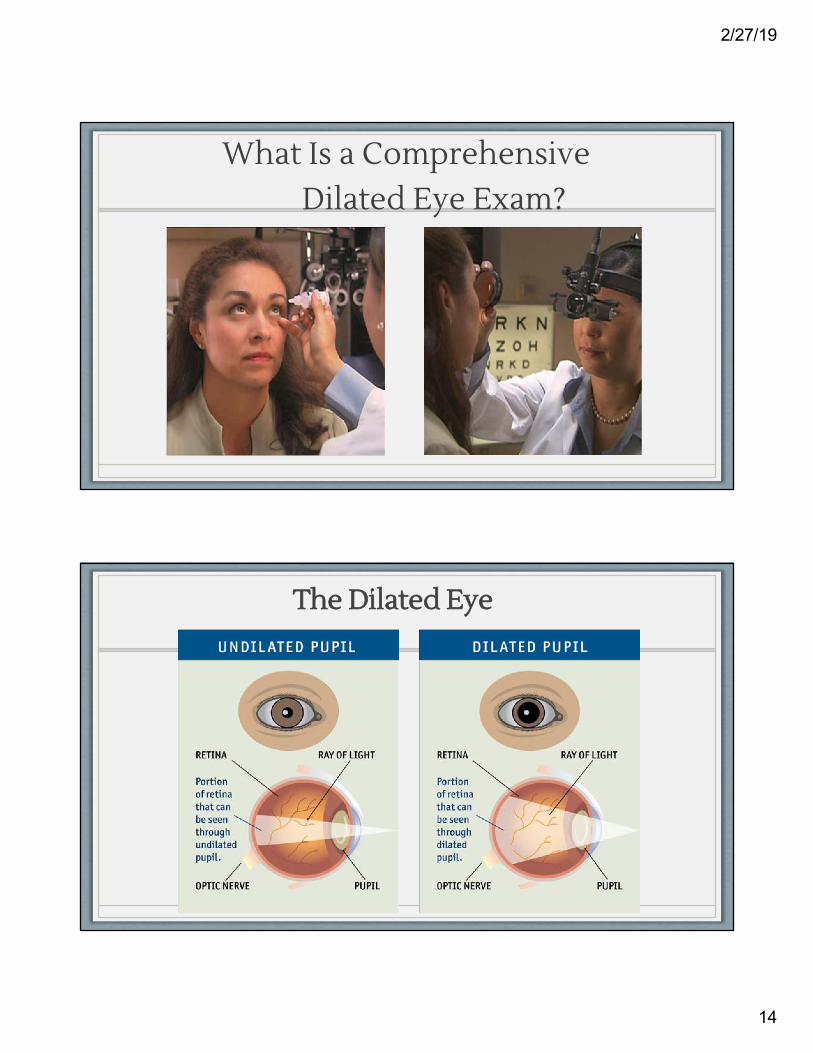
What Is a Comprehensive Dilated Eye Exam?
The Dilated Eye
2/27/19
15
Preventive Eye Care - Adults• Adults with no signs or risk factors for eye disease should receive a comprehensive medical
eye evaluation starting at age 40 if they have not previously received one.
• Interval for evaluations: • For individuals aged 40 to 54 years old = 2 - 4 years• For individuals aged 55 to 64 years old = 1 - 3 years• For individuals 65 years old or older = every 1 - 2 years
• For individuals at higher risk for certain diseases, such as African-Americans who are at higher risk for glaucoma and severe vision loss, comprehensive eye examinations should be considered every 2 years for those under age 40 and yearly for those over 40 years of age even in the absence of visual or ocular symptoms.
Preventive Eye Care - Pediatrics
• Children should have an assessment for eye problems in the newborn period and
then at all subsequent routine health supervision visits.
• Screening for abnormalities present at birth:
• Opacities of the ocular media (e.g. congenital cataract, corneal scarring)
• Congenital Ptosis
• Strabismus (e.g. esotropia and exotropia)
• School-age children should be evaluated regularly for visual acuity and ocular
misalignment during primary health care visits, in schools, or at public screenings.
• Any abnormalities or the inability to test are criteria for referral to an ophthalmologist.

2/27/19
16
Preventive Eye Care - Diabetes
• Type 1 Diabetes Mellitus should be examined by an ophthalmologist 5 years after disease onset and at least yearly thereafter.
• Type 2 Diabetes Mellitus should be examined at the time of diagnosis and at least yearly thereafter.
• Women with Type 1 or Type 2 Diabetes Mellitus should receive a comprehensive eye examination before conception and then early in the first trimester of pregnancy. Recommended intervals for subsequent examinations depend upon the level of retinopathy.
Thank you for your attention
Sterrettania Ophthalmology 4000 Sterrettania Road
814-836-0543
Eastside Medical Center2625 Parade Street
814-452-6383
LECOM Health Ophthalmology
Questions?

2/27/19
17
References
1. Gerstenblith AT, et al. Wills Eye Manual: Office and Emergency Room Diagnosis and
Treatment of Eye Disease, Sixth Edition, 2012.
2. American Academy of Ophthalmology Pediatrics Panel. Preferred Practice Pattern®
Guidelines. Pediatric Eye Evaluations. San Francisco, CA: American Academy of
Ophthalmology; 2012. Available at: www.aao.org/ppp. Accessed February 21, 2019.
3. American Academy of Ophthalmology Preferred Practice Patterns Committee. Preferred
Practice Pattern® Guidelines. Comprehensive Adult Medical Eye Evaluation. San Francisco,
CA: American Academy of Ophthalmology; 2010. Available at: www.aao.org/ppp. Accessed
February 21, 2019.
4. American Academy of Ophthalmology Preferred Practice Patterns Committee. Preferred
Practice Pattern® Guidelines. Diabetic Retinopathy. San Francisco, CA: American Academy of
Ophthalmology; 2014. Available at: www.aao.org/ppp. Accessed February 21, 2019.