26 disorders of the paranasal sinuses
TRANSCRIPT
Introduction
Inflammation of the equine paranasal sinuses is a rela-tively uncommon disease that may be caused by primarybacterial or mycotic infections (Mason 1975a), or can besecondary to dental disease (van der Velden & Verzijlenberg1984, Scott 1987, Tremaine & Dixon 2001a), facial trauma,sinus cysts, progressive ethmoid hematoma or sinonasalneoplasia (Mansmann & Wheat 1973, Gibbs & Lane 1987,Tremaine & Dixon 2001a). Equine sinusitis is usuallyunilateral but bilateral disease has been reported (Coumbeet al 1987, Lane 1993, Tremaine & Dixon 2001a). Thereis apparently no breed, age or gender predisposition tosinusitis. Clinical signs of any type of sinusitis usuallyinclude unilateral purulent nasal discharge, ipsilateralsubmandibular lymph node enlargement, and epiphora.Less common signs include facial swelling, exophthalmos,abnormal respiratory noises, head shaking, and exerciseintolerance (Lane 1993, Tremaine & Dixon 2001a).
Primary Sinus Empyema(Primary Sinusitis)
Primary sinusitis is the result of obstruction of the normalnasomaxillary drainage with resulting accumulation ofmucus in the sinus, which later becomes infected. Somecases occur following upper respiratory tract infectionsthat cause inflammation, increase mucus productionwithin the sinuses, and decrease drainage of secretionsfrom the sinuses into the nasal cavity via the anatomicallynarrow nasomaxillary ostia. The nasal discharge inprimary sinusitis is traditionally stated to be purulentand odorless (Mason 1975a), but malodorous nasal dis-charges can occur with primary sinusitis (Tremaine &Dixon 2001a), especially in association with inspissationof purulent material in the ventral conchal sinuses(Schumacher et al 1987).
Culture of exudates from primary sinusitis cases oftenyields a mixed bacterial growth that is of unclear etiologicsignificance. Isolated bacteria include Streptococcus equivar. zooepidemicus (Schumacher et al 1987, Ruggles et al1993), Corynebacterium spp., (Schumacher & Crossland1994), Staphylococcus spp. (Mason 1975a, Schumacheret al 1987, Tremaine & Dixon 2001a), Pseudomonas aerugi-
nosa, Bacteroides spp., Peptostreptococcus spp. (Ruggles et al1993, Tremaine & Dixon 2001a), Streptococcus equi var. equi(Mansmann & Wheat 1973), and Escherichia coli (Mason1975a, Schumacher et al 1987), although as noted, theetiologic importance of these isolates is often unclear.
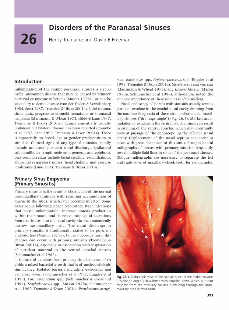
Nasal endoscopy of horses with sinusitis usually revealspurulent exudate in the caudal nasal cavity draining fromthe nasomaxillary ostia of the rostral and/or caudal maxil-lary sinuses (“drainage angle”) (Fig. 26.1). Marked accu-mulation of exudate in the ventral conchal sinus can resultin swelling of the ventral concha, which may eventuallyprevent passage of the endoscope up the affected nasalcavity. Displacement of the nasal septum can occur incases with gross distension of this sinus. Straight lateralradiographs of horses with primary sinusitis frequentlyreveal multiple fluid lines in some of the paranasal sinuses.Oblique radiographs are necessary to separate the leftand right rows of maxillary cheek teeth for radiographic
393
26Disorders of the Paranasal Sinuses
Henry Tremaine and David E Freeman
Fig. 26.1. Endoscopic view of the caudal aspect of the middle meatus(“drainage angle”) in a horse with sinusitis down which purulentexudate from the maxillary sinuses is draining through the naso-maxillary ostia (arrowheads).
examination of the dental apical areas. Dorsoventralradiographs are particularly useful for demonstratingdistension of, and exudate within, the ventral conchal sinus(see Chapter 10).
Acute cases of primary sinusitis may spontaneouslyresolve or may respond to antimicrobial drug admin-istration, with the organisms commonly isolated frequentlybeing sensitive to penicillin. Chronic cases of primary
sinusitis (of >2 months duration) frequently have grossthickening of the sinus mucosa, which can further restrictnormal nasomaxillary drainage and such cases may onlyshow a transient improvement to antibiotic treatment(Tremaine & Dixon 2001a). Treatment by sinus irrigationmay be performed in these cases, via a sutured irriga-tion tube or Foley catheter placed via a trephine openinginto the frontal or caudal maxillary sinuses (for lavageof the frontal and caudal maxillary sinuses), or into therostral maxillary sinus (for lavage of the rostral maxillaryand ventral conchal sinuses). Such cases may respond tolavage with 5–10 liters of water, saline or dilute disinfec-tants such as 0.05% povidine-iodine solution, once to twicedaily for 5–10 days.
Cases with gross thickening of the sinus mucosa, and inparticular cases with accumulations of inspissated pusin the sinus, may require surgical debridement and possiblysinonasal fistulation to improve drainage. An outline ofsinus anatomy and surgical approaches is presented inFigs 26.2–26.4. The frontal, maxillary, and ventral conchalsinus are all most easily approached via a large nasofrontalbone-flap osteotomy (Freeman et al 1990) (Figs 26.4 and26.5) where the bone is preserved or a smaller osteotomywhere the bone is discarded (Figs 26.6–26.9). Even whenradiographs or computed tomographic images demonstratethat the inflammation mainly involves the maxillarysinuses, a frontonasal flap is the preferred approach for anumber of reasons (Freeman et al 1990). When the lesionis in the maxillary sinus, the frontal approach is far enoughfrom it to allow creation of the flap without disturbing thelesion (e.g. sinus cyst), and yet close enough to allow itseasy removal. It also provides a sufficiently clear view ofthe sinus interior to allow complete examination.
The incisions necessary for this type of flap do notinvolve muscles or large blood vessels, and the size andposition of the flap can be designed to suit the lesion, even
SECTION 4 : Disorders of the Upper Respiratory Tract
394 26 Disorders of the Paranasal Sinuses
5
6
7
3
9
4
8
2
1 RMS
VM
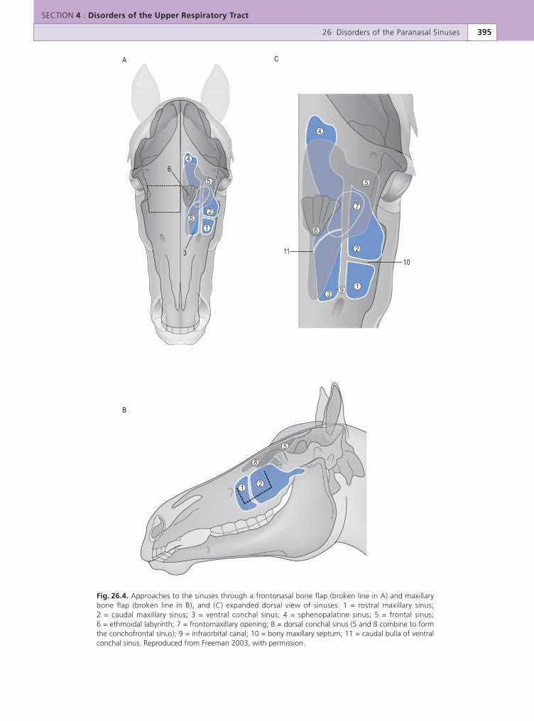
Fig. 26.3. Transverse section of the skull of an aged horse at the levelof the fourth cheek tooth (109, 209) showing the voluminous rostralmaxillary sinus (RMS) and the ventral nasal meatus (VM).
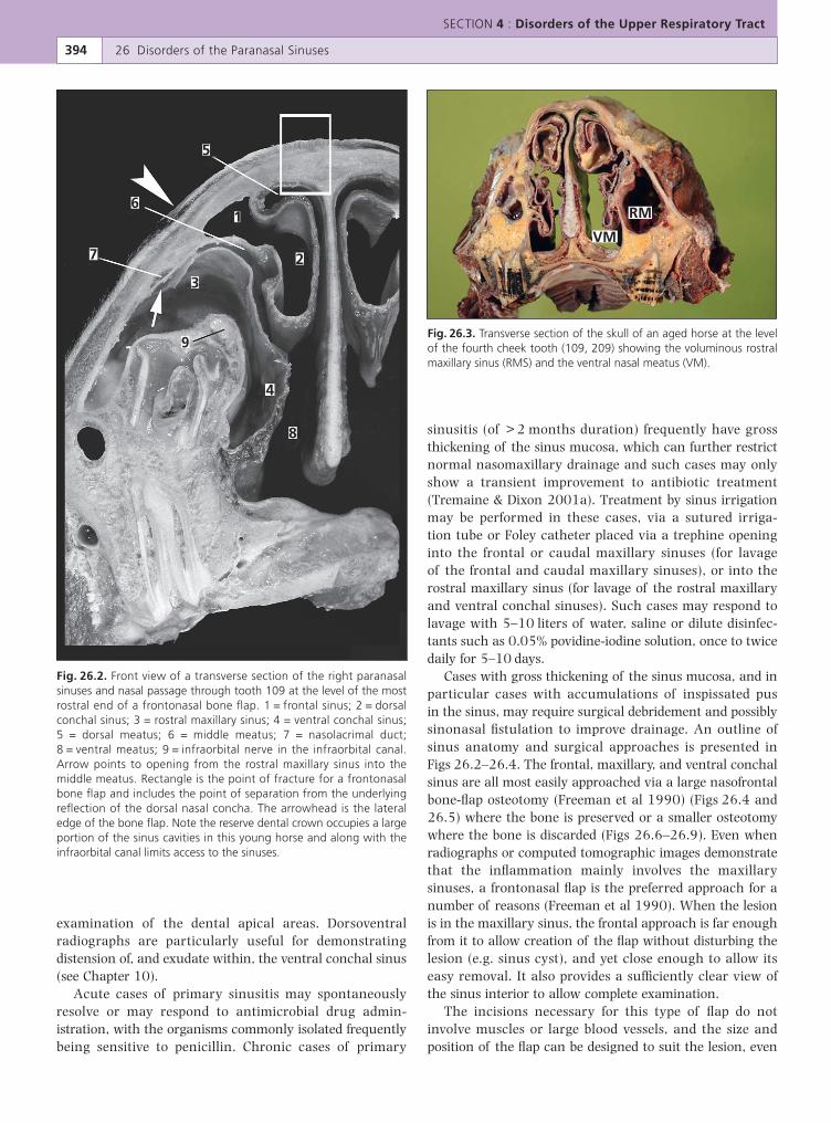
Fig. 26.2. Front view of a transverse section of the right paranasalsinuses and nasal passage through tooth 109 at the level of the mostrostral end of a frontonasal bone flap. 1 = frontal sinus; 2 = dorsalconchal sinus; 3 = rostral maxillary sinus; 4 = ventral conchal sinus;5 = dorsal meatus; 6 = middle meatus; 7 = nasolacrimal duct;8 = ventral meatus; 9 = infraorbital nerve in the infraorbital canal.Arrow points to opening from the rostral maxillary sinus into themiddle meatus. Rectangle is the point of fracture for a frontonasalbone flap and includes the point of separation from the underlyingreflection of the dorsal nasal concha. The arrowhead is the lateraledge of the bone flap. Note the reserve dental crown occupies a largeportion of the sinus cavities in this young horse and along with theinfraorbital canal limits access to the sinuses.
SECTION 4 : Disorders of the Upper Respiratory Tract
26 Disorders of the Paranasal Sinuses 395
6
3
5
4
2
1 8
4
6
3
11
10
9 1
2
7
5
8
1 2
5
A C
B
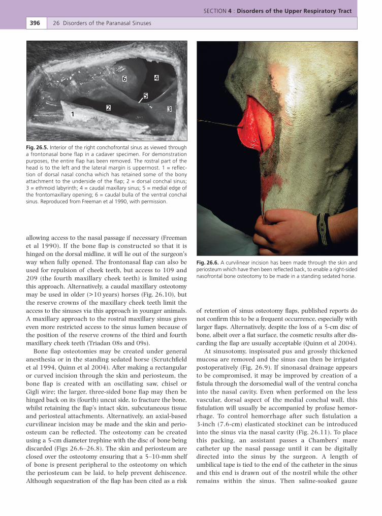
Fig. 26.4. Approaches to the sinuses through a frontonasal bone flap (broken line in A) and maxillarybone flap (broken line in B), and (C) expanded dorsal view of sinuses. 1 = rostral maxillary sinus;2 = caudal maxillary sinus; 3 = ventral conchal sinus; 4 = sphenopalatine sinus; 5 = frontal sinus;6 = ethmoidal labyrinth; 7 = frontomaxillary opening; 8 = dorsal conchal sinus (5 and 8 combine to formthe conchofrontal sinus); 9 = infraorbital canal; 10 = bony maxillary septum; 11 = caudal bulla of ventralconchal sinus. Reproduced from Freeman 2003, with permission.
allowing access to the nasal passage if necessary (Freemanet al 1990). If the bone flap is constructed so that it ishinged on the dorsal midline, it will lie out of the surgeon’sway when fully opened. The frontonasal flap can also beused for repulsion of cheek teeth, but access to 109 and209 (the fourth maxillary cheek teeth) is limited usingthis approach. Alternatively, a caudal maxillary osteotomymay be used in older (>10 years) horses (Fig. 26.10), butthe reserve crowns of the maxillary cheek teeth limit theaccess to the sinuses via this approach in younger animals.A maxillary approach to the rostral maxillary sinus giveseven more restricted access to the sinus lumen because ofthe position of the reserve crowns of the third and fourthmaxillary cheek teeth (Triadan 08s and 09s).
Bone flap osteotomies may be created under generalanesthesia or in the standing sedated horse (Scrutchfieldet al 1994, Quinn et al 2004). After making a rectangularor curved incision through the skin and periosteum, thebone flap is created with an oscillating saw, chisel orGigli wire; the larger, three-sided bone flap may then behinged back on its (fourth) uncut side, to fracture the bone,whilst retaining the flap’s intact skin, subcutaneous tissueand periosteal attachments. Alternatively, an axial-basedcurvilinear incision may be made and the skin and perio-osteum can be reflected. The osteotomy can be createdusing a 5-cm diameter trephine with the disc of bone beingdiscarded (Figs 26.6–26.8). The skin and periosteum areclosed over the osteotomy ensuring that a 5–10-mm shelfof bone is present peripheral to the osteotomy on whichthe periosteum can be laid, to help prevent dehiscence.Although sequestration of the flap has been cited as a risk
of retention of sinus osteotomy flaps, published reports donot confirm this to be a frequent occurrence, especially withlarger flaps. Alternatively, despite the loss of a 5-cm disc ofbone, albeit over a flat surface, the cosmetic results after dis-carding the flap are usually acceptable (Quinn et al 2004).
At sinusotomy, inspissated pus and grossly thickenedmucosa are removed and the sinus can then be irrigatedpostoperatively (Fig, 26.9). If sinonasal drainage appearsto be compromised, it may be improved by creation of afistula through the dorsomedial wall of the ventral conchainto the nasal cavity. Even when performed on the lessvascular, dorsal aspect of the medial conchal wall, thisfistulation will usually be accompanied by profuse hemor-rhage. To control hemorrhage after such fistulation a3-inch (7.6-cm) elasticated stockinet can be introducedinto the sinus via the nasal cavity (Fig. 26.11). To placethis packing, an assistant passes a Chambers’ marecatheter up the nasal passage until it can be digitallydirected into the sinus by the surgeon. A length ofumbilical tape is tied to the end of the catheter in the sinusand this end is drawn out of the nostril while the otherremains within the sinus. Then saline-soaked gauze
SECTION 4 : Disorders of the Upper Respiratory Tract
396 26 Disorders of the Paranasal Sinuses
46
5
321
Fig. 26.5. Interior of the right conchofrontal sinus as viewed througha frontonasal bone flap in a cadaver specimen. For demonstrationpurposes, the entire flap has been removed. The rostral part of thehead is to the left and the lateral margin is uppermost. 1 = reflec-tion of dorsal nasal concha which has retained some of the bonyattachment to the underside of the flap; 2 = dorsal conchal sinus;3 = ethmoid labyrinth; 4 = caudal maxillary sinus; 5 = medial edge ofthe frontomaxillary opening; 6 = caudal bulla of the ventral conchalsinus. Reproduced from Freeman et al 1990, with permission.
Fig. 26.6. A curvilinear incision has been made through the skin andperiosteum which have then been reflected back, to enable a right-sidednasofrontal bone osteotomy to be made in a standing sedated horse.
SECTION 4 : Disorders of the Upper Respiratory Tract
26 Disorders of the Paranasal Sinuses 397
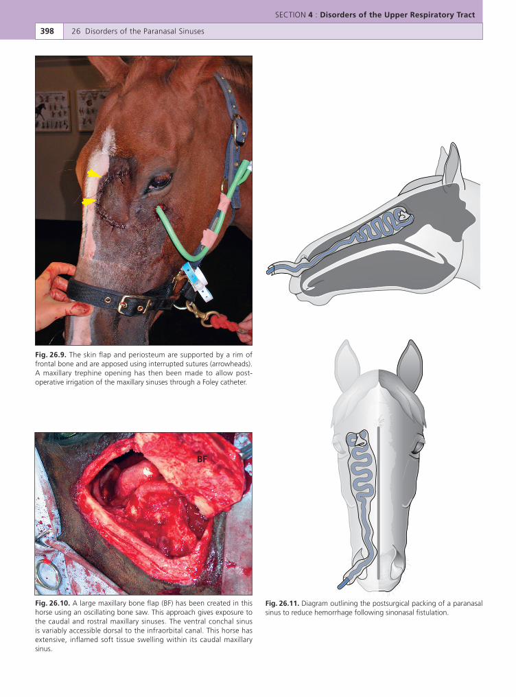
Fig. 26.8. Copious quantities of purulent exudate flowing from a nasofrontal bone flap osteotomy in ahorse with chronic sinus empyema.
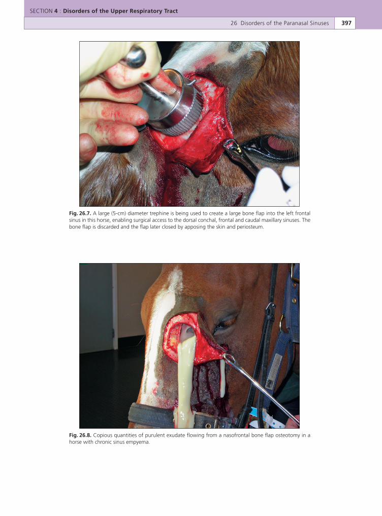
Fig. 26.7. A large (5-cm) diameter trephine is being used to create a large bone flap into the left frontalsinus in this horse, enabling surgical access to the dorsal conchal, frontal and caudal maxillary sinuses. Thebone flap is discarded and the flap later closed by apposing the skin and periosteum.
SECTION 4 : Disorders of the Upper Respiratory Tract
398 26 Disorders of the Paranasal Sinuses
BF
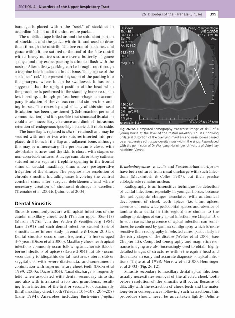
Fig. 26.9. The skin flap and periosteum are supported by a rim offrontal bone and are apposed using interrupted sutures (arrowheads).A maxillary trephine opening has then been made to allow post-operative irrigation of the maxillary sinuses through a Foley catheter.
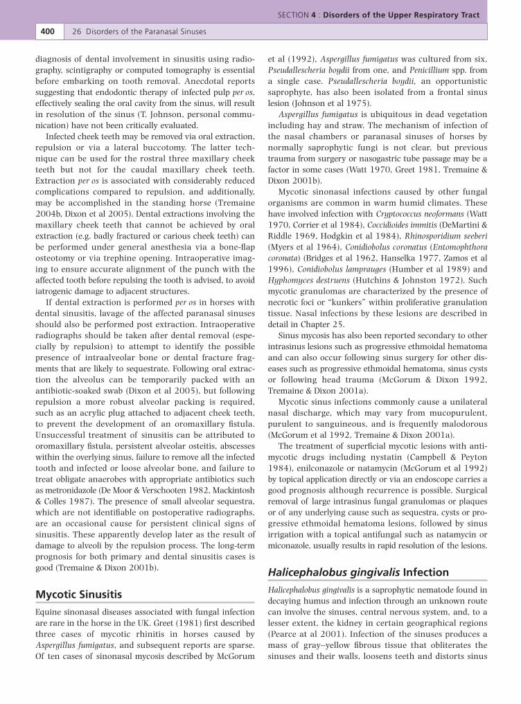
Fig. 26.10. A large maxillary bone flap (BF) has been created in thishorse using an oscillating bone saw. This approach gives exposure tothe caudal and rostral maxillary sinuses. The ventral conchal sinusis variably accessible dorsal to the infraorbital canal. This horse hasextensive, inflamed soft tissue swelling within its caudal maxillarysinus.
Fig. 26.11. Diagram outlining the postsurgical packing of a paranasalsinus to reduce hemorrhage following sinonasal fistulation.
bandage is placed within the “sock” of stockinet inaccordion-fashion until the sinuses are packed.
The umbilical tape is tied around the redundant portionof stockinet, and the gauze within it, and used to drawthem through the nostrils. The free end of stockinet, andgauze within it, are sutured to the roof of the false nostrilwith a heavy mattress suture over a butterfly of gauzesponge, and any excess packing is trimmed flush with thenostril. Alternatively, packing can be brought out througha trephine hole in adjacent intact bone. The purpose of thestockinet “sock” is to prevent migration of the packing intothe pharynx, where it can be swallowed. It has beensuggested that the upright position of the head whenthe procedure is performed in the standing horse results inless bleeding, although profuse hemorrhage can accom-pany fistulation of the venous conchal sinuses in stand-ing horses. The necessity and efficacy of this sinonasalfistulation has been questioned (J. Schumacher, personalcommunication) and it is possible that sinonasal fistulationcould alter mucociliary clearance and diminish intrasinusretention of endogenous (possibly bactericidal) nitric oxide.
The bone flap is replaced in situ (if retained) and may besecured with one or two wire sutures inserted into pre-placed drill holes in the flap and adjacent bone, althoughthis may be unnecessary. The periosteum is closed withabsorbable sutures and the skin is closed with staples ornon-absorbable sutures. A lavage cannula or Foley cathetersutured into a separate trephine opening in the frontalsinus or caudal maxillary sinus allows postoperativeirrigation of the sinuses. The prognosis for resolution ofchronic sinusitis, including cases involving the ventralconchal sinus after surgical debridement, and wherenecessary, creation of sinonasal drainage is excellent(Tremaine et al 2001b, Quinn et al 2004).
Dental Sinusitis
Sinusitis commonly occurs with apical infections of thecaudal maxillary cheek teeth (Triadan upper 08s–11s)(Mason 1975a, van der Velden & Verzijlenberg 1984,Lane 1993) and such dental infections caused 53% ofsinusitis cases in one study (Tremaine & Dixon 2001a).Dental sinusitis occurs most frequently in horses aged4–7 years (Dixon et al 2000b). Maxillary cheek teeth apicalinfections commonly occur following anachoresis (blood-borne infections of apices) (Dacre 2004) but also occursecondarily to idiopathic dental fractures (lateral slab orsaggital), or with severe diastemata, and sometimes inconjunction with supernumerary cheek teeth (Dixon et al1999, 2000a, Dacre 2004). Nasal discharge is frequentlyfetid when associated with dental secondary sinusitis,and also with intranasal tracts and granulomas result-ing from infection of the first or second (or occasionallythird) maxillary cheek tooth (Triadan 106–108, 206–208)(Lane 1994). Anaerobes including Bacteroides fragilis,
B. melaninogenicus, B. oralis and Fusobacterium mortiferumhave been cultured from nasal discharge with such infec-tions (Mackintosh & Colles 1987), but their preciseetiologic role remains unclear.
Radiography is an insensitive technique for detectionof dental infections, especially in younger horses, becausethe radiographic changes associated with anatomicaldevelopment of cheek teeth apices (i.e. blunt apices,absence of roots, wide periodontal spaces and absence oflamina dura denta in this region) are similar to theradiographic signs of early apical infection (see Chapter 10).In such cases, the presence of apical infection can some-times be confirmed by gamma scintigraphy, which is moresensitive than radiography in selected cases, particularly inthe early stages of the disease (Weller et al 2001) (seeChapter 12). Computed tomography and magnetic reso-nance imaging are also increasingly used to obtain highlydetailed images of structures within the equine head andthus make an early and accurate diagnosis of apical infec-tions (Tiejte at al 1998, Morrow et al 2000, Henningeret al 2003) (Fig. 26.12).
Sinusitis secondary to maxillary dental apical infectionsusually necessitates removal of the affected cheek toothbefore resolution of the sinusitis will occur. Because ofdifficulty with the extraction of cheek teeth and the majorlong-term consequences following such extractions, thisprocedure should never be undertaken lightly. Definite
SECTION 4 : Disorders of the Upper Respiratory Tract
26 Disorders of the Paranasal Sinuses 399
Fig. 26.12. Computed tomography transverse image of skull of ayoung horse at the level of the rostral maxillary sinuses, showingunilateral distortion of the overlying maxillary and nasal bones causedby an expansive soft tissue density mass within the sinus. Reproducedwith the permission of Dr Wolfgang Henninger, University of VeterinaryMedicine, Vienna.
diagnosis of dental involvement in sinusitis using radio-graphy, scintigraphy or computed tomography is essentialbefore embarking on tooth removal. Anecdotal reportssuggesting that endodontic therapy of infected pulp per os,effectively sealing the oral cavity from the sinus, will resultin resolution of the sinus (T. Johnson, personal commu-nication) have not been critically evaluated.
Infected cheek teeth may be removed via oral extraction,repulsion or via a lateral buccotomy. The latter tech-nique can be used for the rostral three maxillary cheekteeth but not for the caudal maxillary cheek teeth.Extraction per os is associated with considerably reducedcomplications compared to repulsion, and additionally,may be accomplished in the standing horse (Tremaine2004b, Dixon et al 2005). Dental extractions involving themaxillary cheek teeth that cannot be achieved by oralextraction (e.g. badly fractured or carious cheek teeth) canbe performed under general anesthesia via a bone-flaposteotomy or via trephine opening. Intraoperative imag-ing to ensure accurate alignment of the punch with theaffected tooth before repulsing the tooth is advised, to avoidiatrogenic damage to adjacent structures.
If dental extraction is performed per os in horses withdental sinusitis, lavage of the affected paranasal sinusesshould also be performed post extraction. Intraoperativeradiographs should be taken after dental removal (espe-cially by repulsion) to attempt to identify the possiblepresence of intraalveolar bone or dental fracture frag-ments that are likely to sequestrate. Following oral extrac-tion the alveolus can be temporarily packed with anantibiotic-soaked swab (Dixon et al 2005), but followingrepulsion a more robust alveolar packing is required,such as an acrylic plug attached to adjacent cheek teeth,to prevent the development of an oromaxillary fistula.Unsuccessful treatment of sinusitis can be attributed tooromaxillary fistula, persistent alveolar osteitis, abscesseswithin the overlying sinus, failure to remove all the infectedtooth and infected or loose alveolar bone, and failure totreat obligate anaerobes with appropriate antibiotics suchas metronidazole (De Moor & Verschooten 1982, Mackintosh& Colles 1987). The presence of small alveolar sequestra,which are not identifiable on postoperative radiographs,are an occasional cause for persistent clinical signs ofsinusitis. These apparently develop later as the result ofdamage to alveoli by the repulsion process. The long-termprognosis for both primary and dental sinusitis cases isgood (Tremaine & Dixon 2001b).
Mycotic Sinusitis
Equine sinonasal diseases associated with fungal infectionare rare in the horse in the UK. Greet (1981) first describedthree cases of mycotic rhinitis in horses caused byAspergillus fumigatus, and subsequent reports are sparse.Of ten cases of sinonasal mycosis described by McGorum
et al (1992), Aspergillus fumigatus was cultured from six,Pseudallescheria boydii from one, and Penicillium spp. froma single case. Pseudallescheria boydii, an opportunisticsaprophyte, has also been isolated from a frontal sinuslesion (Johnson et al 1975).
Aspergillus fumigatus is ubiquitous in dead vegetationincluding hay and straw. The mechanism of infection ofthe nasal chambers or paranasal sinuses of horses bynormally saprophytic fungi is not clear, but previoustrauma from surgery or nasogastric tube passage may be afactor in some cases (Watt 1970, Greet 1981, Tremaine &Dixon 2001b).
Mycotic sinonasal infections caused by other fungalorganisms are common in warm humid climates. Thesehave involved infection with Cryptococcus neoformans (Watt1970, Corrier et al 1984), Coccidioides immitis (DeMartini &Riddle 1969, Hodgkin et al 1984), Rhinosporidium seeberi(Myers et al 1964), Conidiobolus coronatus (Entomophthoracoronata) (Bridges et al 1962, Hanselka 1977, Zamos et al1996), Conidiobolus lamprauges (Humber et al 1989) andHyphomyces destruens (Hutchins & Johnston 1972). Suchmycotic granulomas are characterized by the presence ofnecrotic foci or “kunkers” within proliferative granulationtissue. Nasal infections by these lesions are described indetail in Chapter 25.
Sinus mycosis has also been reported secondary to otherintrasinus lesions such as progressive ethmoidal hematomaand can also occur following sinus surgery for other dis-eases such as progressive ethmoidal hematoma, sinus cystsor following head trauma (McGorum & Dixon 1992,Tremaine & Dixon 2001a).
Mycotic sinus infections commonly cause a unilateralnasal discharge, which may vary from mucopurulent,purulent to sanguineous, and is frequently malodorous(McGorum et al 1992, Tremaine & Dixon 2001a).
The treatment of superficial mycotic lesions with anti-mycotic drugs including nystatin (Campbell & Peyton1984), enilconazole or natamycin (McGorum et al 1992)by topical application directly or via an endoscope carries agood prognosis although recurrence is possible. Surgicalremoval of large intrasinus fungal granulomas or plaquesor of any underlying cause such as sequestra, cysts or pro-gressive ethmoidal hematoma lesions, followed by sinusirrigation with a topical antifungal such as natamycin ormiconazole, usually results in rapid resolution of the lesions.
Halicephalobus gingivalis Infection
Halicephalobus gingivalis is a saprophytic nematode found indecaying humus and infection through an unknown routecan involve the sinuses, central nervous system, and, to alesser extent, the kidney in certain geographical regions(Pearce at al 2001). Infection of the sinuses produces amass of gray–yellow fibrous tissue that obliterates thesinuses and their walls, loosens teeth and distorts sinus
SECTION 4 : Disorders of the Upper Respiratory Tract
400 26 Disorders of the Paranasal Sinuses
architecture. Infection can be unilateral or bilateral, caninvolve both the upper and lower jaws, and can spreadfrom there to the kidneys and cerebellum (Freeman 1991a).
Predominant clinical signs of H. gingivalis infectionare facial distortion with firm swellings in the maxilla,unilateral or bilateral nasal discharge, marked dyspnea andstridor, difficulty in eating, and weight loss (Pearce et al2001). The condition can be confused with squamous cellcarcinoma but the female rhabditiform nematodes andtheir larvae and eggs can be seen in clusters or scatteredthroughout a biopsy specimen. Surgical debulking, intra-operative lavage with ivermectin, and subsequent oralivermectin was successful in one horse with a periorbitalgranuloma (Freeman 1991a). However, the response toivermectin is not always favorable and the prognosisappears to be poor, especially because of risk of spread toother organs.
Sinus Cysts
Sinus cysts are expansive fluid-filled space-occupyinglesions which develop within the sinuses (Leyland & Baker1975, Dixon 1985, Lane et al 1987) of young to oldhorses. Congenital intrasinus cysts have also been reported(Sanders-Shamis & Robertson 1987, Beard et al 1990).Equine sinus cysts most commonly occur in the maxillarysinuses but they can also occur in the other sinuses.
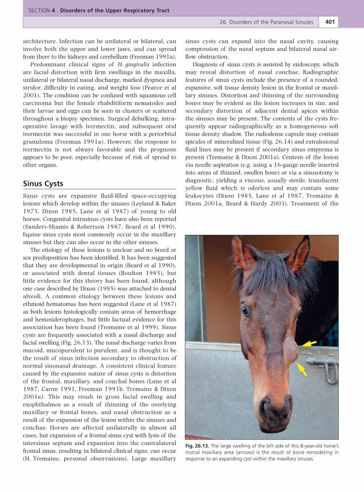
The etiology of these lesions is unclear and no breed orsex predisposition has been identified. It has been suggestedthat they are developmental in origin (Beard et al 1990),or associated with dental tissues (Boulton 1985), butlittle evidence for this theory has been found, althoughone case described by Dixon (1985) was attached to dentalalveoli. A common etiology between these lesions andethmoid hematomas has been suggested (Lane et al 1987)as both lesions histologically contain areas of hemorrhageand hemosiderophages, but little factual evidence for thisassociation has been found (Tremaine et al 1999). Sinuscysts are frequently associated with a nasal discharge andfacial swelling (Fig. 26.13). The nasal discharge varies frommucoid, mucopurulent to purulent, and is thought to bethe result of sinus infection secondary to obstruction ofnormal sinonasal drainage. A consistent clinical featurecaused by the expansive nature of sinus cysts is distortionof the frontal, maxillary, and conchal bones (Lane et al1987, Caron 1991, Freeman 1991b, Tremaine & Dixon2001a). This may result in gross facial swelling andexophthalmos as a result of thinning of the overlyingmaxillary or frontal bones, and nasal obstruction as aresult of the expansion of the lesion within the sinuses andconchae. Horses are affected unilaterally in almost allcases, but expansion of a frontal sinus cyst with lysis of theintersinus septum and expansion into the contralateralfrontal sinus, resulting in bilateral clinical signs, can occur(H. Tremaine, personal observations). Large maxillary
sinus cysts can expand into the nasal cavity, causingcompression of the nasal septum and bilateral nasal air-flow obstruction.
Diagnosis of sinus cysts is assisted by endoscopy, whichmay reveal distortion of nasal conchae. Radiographicfeatures of sinus cysts include the presence of a rounded,expansive, soft tissue density lesion in the frontal or maxil-lary sinuses. Distortion and thinning of the surroundingbones may be evident as the lesion increases in size, andsecondary distortion of adjacent dental apices withinthe sinuses may be present. The contents of the cysts fre-quently appear radiographically as a homogeneous softtissue density shadow. The radiodense capsule may containspicules of mineralized tissue (Fig. 26.14) and extralesionalfluid lines may be present if secondary sinus empyema ispresent (Tremaine & Dixon 2001a). Centesis of the lesionvia needle aspiration (e.g. using a 16-gauge needle insertedinto areas of thinned, swollen bone) or via a sinusotomy isdiagnostic, yielding a viscous, usually sterile, translucentyellow fluid which is odorless and may contain someleukocytes (Dixon 1985, Lane et al 1987, Tremaine &Dixon 2001a, Beard & Hardy 2003). Treatment of the
SECTION 4 : Disorders of the Upper Respiratory Tract
26 Disorders of the Paranasal Sinuses 401
Fig. 26.13. The large swelling of the left side of this 8-year-old horse’srostral maxillary area (arrows) is the result of bone remodeling inresponse to an expanding cyst within the maxillary sinuses.
lesion by surgical drainage may be effective in some cases(O’Connor 1930, Dixon 1985, Lane et al 1987) but totalremoval of the lesion via a nasofrontal or maxillaryosteotomy approach, under general anesthesia or standingchemical restraint, is the treatment of choice (Fig. 26.15)(Dixon 1985, Lane et al 1987, Tremaine & Dixon 2001b).
Histologic examination of sinus cysts has revealedextensive resorption and remodeling of the bones sur-rounding the cyst, replacement of the normal bony septawithin the sinus by fibrous tissue, and replacement of theloose intrasinus connective tissue with bony spicules(Tremaine et al 1999). The cysts themselves are lined byciliated columnar respiratory epithelium with focal areasof ulceration, areas of submucosal calcification and ofsubepithelial hemorrhage, and chronic inflammation maybe present (Lane et al 1987, Tremaine et al 1999).
Progressive Ethmoidal Hematoma
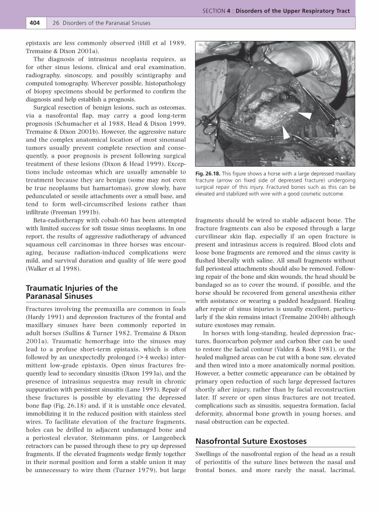
Progressive ethmoidal hematomas are observed mostcommonly in the nasal cavity arising from the ethmo-turbinates. Less commonly, lesions arise in the frontalor maxillary sinuses. The etiology, clinical signs, andtreatment of these lesions are discussed in Chapter 27.Cases with clinical signs typical of progressive ethmoidalhematoma (i.e. low-grade chronic, unilateral epistaxis)and with endoscopic evidence of drainage of small volumesof blood from the sinonasal drainage areas and which donot reveal a lesion in the nasal cavities should be subjectedto careful examination of the sinuses by radiography,sinoscopy or sinusotomy (Fig. 26.16).
SECTION 4 : Disorders of the Upper Respiratory Tract
402 26 Disorders of the Paranasal Sinuses
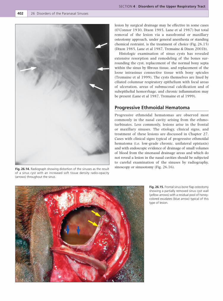
Fig. 26.14. Radiograph showing distortion of the sinuses as the resultof a sinus cyst with an increased soft tissue density radio-opacity(arrows) throughout the sinus.
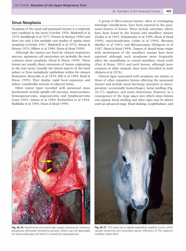
Fig. 26.15. Frontal sinus bone flap osteotomyshowing a partially removed sinus cyst wall(yellow arrows) with a residual pool of honey-colored exudates (blue arrow) typical of thistype of lesion.
Sinus Neoplasia
Neoplasia of the nasal and paranasal sinuses is a relativelyrare condition in the horse (Cotchin 1956, Madewell et al1976, Sundbergh et al 1977, Priester & Mackay 1980) andthere are only a few multiple case studies of equine sinusneoplasia (Cotchin 1967, Madewell et al 1976, Stunzi &Hauser 1976, Hilbert et al 1988, Dixon & Head 1999).
Although the sinuses are lined by ciliated respiratorymucosa, squamous cell carcinomas are probably the mostcommon sinus neoplasia (Head & Dixon 1999). Theselesions are usually direct extensions of lesions originatingin the oral cavity (usually the lateral aspects of the hardpalate) or from metaplastic epithelium within the sinusesthemselves (Reynolds et al 1979, Hill et al 1989, Head &Dixon 1999). They display rapid local expansion andinduce considerable necrosis of adjacent tissue.
Other tumor types recorded with paranasal sinusinvolvement include spindle cell sarcoma, mastocytomas,hemangiosarcoma, angiosarcoma and lymphosarcoma(Lane 1985, Adams et al 1988, Richardson et al 1994,Malikides et al 1996, Dixon & Head 1999).
A group of fibro-osseous lesions, often of overlappinghistologic classification, have been reported in the para-nasal sinuses of horses. These include osteomas, whichhave been found in the frontal and maxillary sinuses(Gorlin et al 1963, Schumacher et al 1988, Dixon & Head1999), osteochondromas (Adair et al 1994), fibromas(Barber et al 1983) and fibrosarcomas (Hultgren et al1987, Dixon & Head 1999). Tumors of dental tissue originwith involvement of the maxillary sinuses have beenreported, although such neoplasms more frequentlyaffect the mandibular or rostral maxillary cheek teeth(Pirie & Dixon 1993) and such lesions, although morecommon in older animals, have been described in foals(Roberts et al 1978).
Clinical signs associated with neoplasia are similar tothose of other expansive lesions affecting the paranasalsinuses and include nasal discharge (purulent or muco-purulent, occasionally hemorrhagic), facial swelling (Fig.26.17), epiphora, and nasal obstruction. However, as aconsequence of the large space into which sinus lesionscan expand, facial swelling and other signs may be absentuntil an advanced stage. Head shaking, exophthalmos, and
SECTION 4 : Disorders of the Upper Respiratory Tract
26 Disorders of the Paranasal Sinuses 403
Fig. 26.16. Nasofrontal sinus bone flap surgery showing an intrasinusprogressive ethmoidal hematoma (arrows), which was not detectableon nasal endoscopy and which is covered by inspissated pus.
Fig. 26.17. This pony has a rapidly expanding maxillary tumor, whichcaused loosening and secondary apical infections of the adjacentmaxillary cheek teeth.
epistaxis are less commonly observed (Hill et al 1989,Tremaine & Dixon 2001a).
The diagnosis of intrasinus neoplasia requires, asfor other sinus lesions, clinical and oral examination,radiography, sinoscopy, and possibly scintigraphy andcomputed tomography. Wherever possible, histopathologyof biopsy specimens should be performed to confirm thediagnosis and help establish a prognosis.
Surgical resection of benign lesions, such as osteomas,via a nasofrontal flap, may carry a good long-termprognosis (Schumacher et al 1988, Head & Dixon 1999,Tremaine & Dixon 2001b). However, the aggressive natureand the complex anatomical location of most sinonasaltumors usually prevent complete resection and conse-quently, a poor prognosis is present following surgicaltreatment of these lesions (Dixon & Head 1999). Excep-tions include osteomas which are usually amenable totreatment because they are benign (some may not evenbe true neoplasms but hamartomas), grow slowly, havepedunculated or sessile attachments over a small base, andtend to form well-circumscribed lesions rather thaninfiltrate (Freeman 1991b).
Beta-radiotherapy with cobalt-60 has been attemptedwith limited success for soft tissue sinus neoplasms. In onereport, the results of aggressive radiotherapy of advancedsquamous cell carcinomas in three horses was encour-aging, because radiation-induced complications weremild, and survival duration and quality of life were good(Walker et al 1998).
Traumatic Injuries of theParanasal Sinuses
Fractures involving the premaxilla are common in foals(Hardy 1991) and depression fractures of the frontal andmaxillary sinuses have been commonly reported inadult horses (Sullins & Turner 1982, Tremaine & Dixon2001a). Traumatic hemorrhage into the sinuses maylead to a profuse short-term epistaxis, which is oftenfollowed by an unexpectedly prolonged (>4 weeks) inter-mittent low-grade epistaxis. Open sinus fractures fre-quently lead to secondary sinusitis (Dixon 1993a), and thepresence of intrasinus sequestra may result in chronicsuppuration with persistent sinusitis (Lane 1993). Repair ofthese fractures is possible by elevating the depressedbone flap (Fig. 26.18) and, if it is unstable once elevated,immobilizing it in the reduced position with stainless steelwires. To facilitate elevation of the fracture fragments,holes can be drilled in adjacent undamaged bone anda periosteal elevator, Steinmann pins, or Langenbeckretractors can be passed through these to pry up depressedfragments. If the elevated fragments wedge firmly togetherin their normal position and form a stable union it maybe unnecessary to wire them (Turner 1979), but large
fragments should be wired to stable adjacent bone. Thefracture fragments can also be exposed through a largecurvilinear skin flap, especially if an open fracture ispresent and intrasinus access is required. Blood clots andloose bone fragments are removed and the sinus cavity isflushed liberally with saline. All small fragments withoutfull periosteal attachments should also be removed. Follow-ing repair of the bone and skin wounds, the head should bebandaged so as to cover the wound, if possible, and thehorse should be recovered from general anesthesia eitherwith assistance or wearing a padded headguard. Healingafter repair of sinus injuries is usually excellent, particu-larly if the skin remains intact (Tremaine 2004b) althoughsuture exostoses may remain.
In horses with long-standing, healed depression frac-tures, fluorocarbon polymer and carbon fiber can be usedto restore the facial contour (Valdez & Rook 1981), or thehealed maligned areas can be cut with a bone saw, elevatedand then wired into a more anatomically normal position.However, a better cosmetic appearance can be obtained byprimary open reduction of such large depressed facturesshortly after injury, rather than by facial reconstructionlater. If severe or open sinus fractures are not treated,complications such as sinusitis, sequestra formation, facialdeformity, abnormal bone growth in young horses, andnasal obstruction can be expected.
Nasofrontal Suture Exostoses
Swellings of the nasofrontal region of the head as a resultof periostitis of the suture lines between the nasal andfrontal bones, and more rarely the nasal, lacrimal,
SECTION 4 : Disorders of the Upper Respiratory Tract
404 26 Disorders of the Paranasal Sinuses
Fig. 26.18. This figure shows a horse with a large depressed maxillaryfracture (arrow on fixed side of depressed fracture) undergoingsurgical repair of this injury. Fractured bones such as this can beelevated and stabilized with wire with a good cosmetic outcome.
and malar bones have been described (Gibbs & Lane 1987,Speirs 1992, Trotter 1993, Tremaine & Dixon 2001a).They occur in many breeds but the incidence appearsto be particularly high in thoroughbreds and thorough-bred crosses (Dixon 1991). Although most are possiblytraumatic in origin, including following sinonasal surgery,especially after a large nasofrontal osteotomy, the exactetiology of such lesions remains unknown in other cases.Affected horses present with bilateral, firm, non-painfulswellings, rostral to the eye, accompanied by epiphorain some cases. Differentiation from facial fractures andsinusitis is usually possible clinically and radiologically.Radiographs frequently demonstrate proliferative peri-osteal changes of the widened and incompletely closedsuture line. The swellings usually remodel and regressgradually without treatment, but in some cases continuedinstability has resulted in progressive increases in the size ofthese swellings.
Miscellaneous Sinus Disorders
Frontal sinus eversion is probably a congenital defect thatforms a hard, slow-growing protuberance over, and com-municating with, the frontal sinus (Martin & McIlwraith1981). The bony protuberance can be removed through alarge elliptical incision and the resulting defect in thefrontal bone can be repaired with synthetic polypropylenemesh (Marlex) and skin.
Osteodystrophia fibrosa or secondary nutritional hyper-parathyroidism can develop in horses on a high phosphorusdiet, such as bran, or on some tropical grasses (Clarke et al1996) and can be attributed to relative calcium deficiency(Freeman 1991b). It is rare under modern management con-ditions. Conchal necrosis (De Moor & Verschooten 1982)may be caused by advanced mycotic rhinitis (Tremaine &Dixon 2001b) that usually responds to removal of theaffected concha by intranasal curettage and lavage.
The reserve tooth crowns of young (2- to 4-year-old)Welsh and miniature ponies and other smaller pony breedscan project a considerable distance into the nasal and sinuscavities and cause firm, painless, bilateral swellings in themaxillary bones that should not be confused with injuriesor disease. Facial lumps or “horns” can be seen in horses assymmetrical painless prominences of the nasal and frontalbones and possibly are caused by an embryologic fault.
REFERENCES
Adair HS, Duncan RB, Toal RL 1994 Solitary osteochondromaof the nasal bone in a horse. Cornell Veterinarian84: 25–31
Adams R, Calderwood-Mays MB, Peyton LC 1988 Malignantlymphoma in three horses with ulcerative pharyngitis.Journal of the American Veterinary Medical Association193: 674–676
Barber SM, Clark EG, Fretz PB 1983 Fibroblastic tumour of thepremaxilla in two horses. Journal of the AmericanVeterinary Medical Association 182: 700–702
Beard WL, Hardy J 2003 Diagnosis of conditions of the para-nasal sinuses of the horse. Equine Veterinary Education13: 265–273
Beard WL, Robertson JT, Leeth B 1990 Bilateral congenitalcysts in the frontal sinus of a horse. Journal of theAmerican Veterinary Medical Association 196: 453–454
Boulton CH 1985 Equine nasal cavity and paranasal sinusdisease: a review of 85 cases. Journal of Equine VeterinaryScience 5: 268–275
Bridges CH, Romane WM, Emmons CW 1962 Phycomycosisof horses caused by Entomophthora coronata. Journal of theAmerican Veterinary Medical Association 140: 673–677
Campbell ML, Peyton LC 1984 Muscle flap closure of afrontocutaneous fistula in a horse. Veterinary Surgery13: 185–188
Caron JP 1991 Diseases of the nasal cavity and paranasalsinuses. In: Colahan PT, Mayhew IG, Merritt AM, MooreJN (editors) Equine Medicine and Surgery. AmericanVeterinary Publications, Goleta, CA, pp.386–397
Clarke CJ, Roeder PL, Dixon PM 1996 Nasal obstructioncaused by nutritional osteodystrophia fibrosa in a groupof Ethiopian horses. Veterinary Record 139: 568–570
Corrier DE, Wison SR, Scrutchfield WL 1984 Equine crypto-coccal rhinitis. Compendium on Continuing Educationfor the Practicing Veterinarian 6: 556–558.
Cotchin E 1956 Neoplasms of the Domesticated Animals.Commonwealth Agricultural Bureaux, Buckingham, UK
Cotchin E 1967 Spontaneous neoplasms of the upper respira-tory tract in animals In: Muir CS, Shanmugaratnam K(editors) Cancer of the Naso-Pharynx. Medical Examina-tion Publishing Co, Flushing, NY, pp.203–259
Coumbe KM, Jones RD, Kenward JH 1987 Bilateral sinusempyema in a six year-old mare. Equine VeterinaryJournal 19: 559–560
Dacre IT 2004 A pathological, histological and ultrastruc-tural study of diseased equine cheek teeth. PhD thesis,University of Edinburgh
DeMartini JC, Riddle WE 1969 Disseminated coccidiomycosisin two horses and a pony. Journal of the AmericanVeterinary Medical Association 155: 149–156
De Moor Von A, Verschooten F 1982 Empyem und Nekroseder Nasenmuscheln beim Pferd (Empyema and necrosisof the nasal conchae in a horse). Deutsche TierarztlicheWochenschrift 89: 275–281
Dixon PM 1985 Equine maxillary cysts. Equine Practice7: 25–33
Dixon PM 1991 Swellings of the head region in the horse. InPractice 13: 257–263
Dixon PM 1993a Nasal cavity. In: Equine Respiratory Endoscopy.Boehringer Ingelheim, Bracknell, pp.22–29
Dixon PM 1993b Ethmoturbinates. In: Equine RespiratoryEndoscopy. Boehringer Ingelheim, Bracknell, pp.43–48
Dixon PM, Head KW 1999 Equine nasal and paranasalsinus tumours: part 2: A contribution of 28 case reports.Veterinary Journal 157: 279–294
Dixon PM, Tremaine WH, Pickles K et al 1999 Equinedental disease Part 2: A long term study of 400 cases:disorders of development and eruption and variations inposition of the cheek teeth. Equine Veterinary Journal31: 519–528
Dixon PM, Tremaine WH, Pickles K et al 2000a Equine dentaldisease Part 3: A long term study of 400 cases: disorders
SECTION 4 : Disorders of the Upper Respiratory Tract
26 Disorders of the Paranasal Sinuses 405
of wear, traumatic damage and idiopathic fractures,tumours and miscellaneous disorders of the cheek teeth.Equine Veterinary Journal 32: 9–18
Dixon PM, Tremaine WH, Pickles K et al 2000b Equine dentaldisease Part 4: A long term study of 400 cases: apicalinfections of cheek teeth. Equine Veterinary Journal32: 182–194
Dixon PM, Dacre I, Dacre K et al 2005 Standing oralextraction of cheek teeth in 100 horses (1998–2003).Equine Veterinary Journal 37: 105–112
Freeman DE 1991a Nasal passages. In: Beech J (editor) EquineRespiratory Disorders. Lea and Febiger, Philadelphia,pp.253–274
Freeman DE 1991b Paranasal sinuses In: Beech J (editor) EquineRespiratory Disorders. Lea and Febiger, Philadelphia,pp.275–305
Freeman DE 2003 Sinus disease. Veterinary Clinics of NorthAmerica; Equine Practice 19: 209–243
Freeman DE, Orsini PG, Ross MW et al 1990 A large fronto-nasal flap for sinus surgery in the horse. VeterinarySurgery 19: 122–130
Gibbs C, Lane JG 1987 Radiographic examination of thenasal and paranasal sinus regions of the horse. Part 2:Radiological findings. Equine Veterinary Journal19: 474–482
Gorlin RJ, Meskin LH, Brodey R 1963 Odontogenic tumours inman and animals, pathological classification and clinicalbehaviour – a review. Annals of the New York Academyof Science 108: 722–771
Greet TRC 1981 Nasal aspergillosis in three horses. VeterinaryRecord 109: 487–489
Hanselka DV 1977 Equine nasal phycomycosis. VeterinaryMedicine/Small Animal Clinician 72: 251–253
Hardy J 1991 Upper respiratory obstruction in foals, wean-lings, and yearlings. Veterinary Clinics of North America;Equine Practice 7: 105–122
Head KW, Dixon PM 1999 Equine nasal and paranasal sinustumours. Part 1: review of the literature and tumourclassification. Veterinary Journal 157: 261–278
Henninger W, Frame M, Willman M et al 2003 CT features ofalveolitis and sinusitis in horses. Veterinary Radiologyand Ultrasound 44: 269–276
Hilbert BJ, Little CB, Klein K et al 1988 Tumours of theparanasal sinuses in 16 horses. Australian VeterinaryJournal 65: 86–88
Hill FWE, Moulton JE, Schiff PH 1989 Exophthalmos in ahorse resulting from an adenocarcinoma of theparanasal sinus. Journal of South African VeterinaryMedicine Association 60: 104–105
Hodgkin EC, Conaway DH, Ortenburger AI 1984 Recurrenceof obstructive nasal coccidioidal granuloma in a horse.Journal of the American Veterinary Medical Association184: 339–340
Hultgren BD, Schmotzer WB, Watrous BJ et al 1987Nasal–maxillary fibrosarcoma in young horses: a lightmicroscopic study. Veterinary Pathology 24: 194–196
Humber RA, Brown CC, Korngay RW 1989 Equine zygomy-cosis caused by Conidiobolus lamprauges. Journal of ClinicalMicrobiology 27: 573–576
Hutchins DR, Johnston KG 1972 Phycomycosis in the horse.Australian Veterinary Journal 48: 269–277
Johnson GR, Schiefer B, Pantekoek JFCA 1975 Maduromycosisin a horse in Western Canada. Canadian VeterinaryJournal 16: 341–344
Lane JG 1985 Palatine lymphosarcoma in two horses. EquineVeterinary Journal 17: 465–467
Lane JG 1993 Management of sinus disorders, part 1. EquineVeterinary Education 5: 5–9
Lane JG 1994 A review of dental disorders of the horse, theirtreatment and possible fresh approaches to management.Equine Veterinary Education 6: 13–21
Lane JG, Longstaffe JA, Gibbs C 1987 Equine paranasal sinuscysts: a report of 15 cases. Equine Veterinary Journal19: 534–544
Leyland A, Baker JR 1975 Lesions of the nasal and paranasalsinuses of the horse causing dyspnoea. British VeterinaryJournal 131: 339–346
Mackintosh ME, Colles CM 1987 Anaerobic bacteriaassociated with abscesses in the horse and donkey. EquineVeterinary Journal 19: 360–362
Madewell BR, Priester WA, Gillete EL et al 1976 Neoplasms ofthe nasal passages and paranasal sinuses in domesticanimals as reported by 13 veterinary colleges. AmericanJournal of Veterinary Research 37: 851–856
Malikides N, Reppas G, Hodgson JL et al 1996 Mast celltumours in the horse: four case reports. Equine Practice18: 12–17
Mansmann RA, Wheat JD 1973 The diagnosis and treatmentof equine upper respiratory diseases. In: Proceedings ofthe 18th Annual Convention of the American Associa-tion of Equine Practitioners. Lexington, KY, pp.388–487
Martin GS, McIlwraith CW 1981 Repair of a frontal sinuseversion in a horse. Veterinary Surgery 10: 149
Mason BJE 1975a Empyema of the equine paranasal sinuses.Journal of the American Veterinary Medical Association167: 727–731
Mason BJE 1975b Spindle-cell sarcoma of the equinepara-nasal sinuses and nasal chamber. Veterinary Record96: 287–288
McGorum BC, Dixon PM, Lawson GHK 1992 A review of tencases of mycotic rhinitis. Equine Veterinary Education4: 8–12
Meschter CL, Allen D 1984 Lymphosarcoma within the nasalcavities of an 18 month old filly. Equine VeterinaryJournal 16: 475–476
Morrow K, Park RD, Spurgeon, TL et al 2000 Computedtomography of the equine head. Veterinary Radiologyand Ultrasound 41: 491–497
Myers DD, Simon J, Case MT 1964 Rhinosporidiosis in a horse.Journal of the American Veterinary Medical Association145: 345–346
O’Connor JJ 1930 Operations. Dollar’s Veterinary Surgery.Bailliere, Tindall and Cox, London, pp.222–231
Pearce SG, Bouré LP, Taylor JA et al 2001 Treatment of agranuloma caused by Halicephalobus gingivalis in a horse.Journal of the American Veterinary Medical Association219: 1735–1738
Pirie RS, Dixon PM 1993 Mandibular tumours in the horse:a review of the literature and 7 case reports. EquineVeterinary Education 5: 287–294
Priester WA, Mackay FW 1980 The occurrence of tumors indomestic animals. National Cancer Institute Monograph54: 59–99
Quinn G, Lane JG, Kidd JF 2005 Fronto-nasal flap surgery instanding horses. Equine Veterinary Journal 37: 138–142
Reed SM, Boles CL, Dade AW et al 1979 Localised equine nasalcoccidiomycosis granuloma. Journal of Equine Medicineand Surgery 3: 119–123
Reynolds BL, Stedham MA, Lawrence JM et al 1979 Adeno-carcinoma of the frontal sinus with extension to thebrain in a horse. Journal of the American VeterinaryMedical Association 174: 734–736
SECTION 4 : Disorders of the Upper Respiratory Tract
406 26 Disorders of the Paranasal Sinuses
Richardson JD, Lane JG, Nicholls PK 1994 Nasopharyn-geal mast cell tumour in a horse. Veterinary Record134: 238–240
Roberts MC, Groenendyk S, Kelly WR 1978 Ameloblasticodontoma in a foal. Equine Veterinary Journal 10: 91–93
Ruggles AJ, Ross MW, Freeman DE 1991 Endoscopic exami-nation of normal paranasal sinuses in horses. VeterinarySurgery 20: 418–423
Ruggles AJ, Ross MW, Freeman DE 1993 Endoscopic exami-nation and treatment of paranasal sinus disease in 16horses. Veterinary Surgery 22: 508–514
Sanders-Shamis M, Robertson JT 1987 Congenital sinus cystin a foal. Journal of the American Veterinary MedicalAssociation 190: 1011–1012
Schumacher J, Crossland LE 1994 Removal of inspissatedpurulent exudate from the ventral conchal sinus of threestanding horses. Journal of the American VeterinaryMedical Association 205: 1312–1314
Schumacher J, Honnas C, Smith B 1987 Paranasal sinusitiscomplicated by inspissated exudate in the ventral conchalsinus. Veterinary Surgery 16: 373–377
Schumacher J, Smith BL, Morgan SJ 1988 Osteoma ofparanasal sinuses of a horse. Journal of the AmericanVeterinary Medical Association 192: 1449–1450
Scott EA 1987 Sinusitis. In: Robinson NE (editor) CurrentTherapy in Equine Medicine 2. WB Saunders, Philadelphia,pp.605–607
Scrutchfield WL, Schumacher J, Walker M et al 1994 Removalof an osteoma from the paranasal sinuses of a standinghorse. Equine Practice 16: 24–27
Speirs VC 1992 Diseases of the paranasal sinuses. In:Robinson NE (editor) Current Therapy in Equine Medicine3. WB Saunders, Philadelphia, pp.217–224
Stunzi H, Hauser B 1976 Tumours of the nasal cavity(International histological classification of tumoursof domestic animals). Bulletin of the World HealthOrganisation 53: 257–263
Sullins KE, Turner AS 1982 Management of fractures of theequine mandible and premaxilla (Incisive bone). Com-pendium on Continuing Education for the PracticingVeterinarian 4: S480–S489
Sundbergh JP, Burustein T, Page EH et al 1977 Neoplasms ofequidae. Journal of the American Veterinary MedicalAssociation 170: 150–152
Taylor J 1955 The horse. The Head and Neck. Oliver & Boyd,London
Tietje S, Becker M, Bockenhoff G 1998 Computed tomographicevaluation of head diseases in the horse: 15 cases.Equine Veterinary Journal 28: 98–105
Tremaine WH 2004a The management of head fractures inhorses. In Practice 26: 142–149
Tremaine WH 2004b Oral extraction of equine cheek teeth.Equine Veterinary Education 16: 151–159
Tremaine WH, Dixon PM 2001a A long-term study of 277cases of equine sinonasal disease. Part 1: details ofhorses, historical, clinical and ancillary diagnosticfindings. Equine Veterinary Journal 33: 274–282
Tremaine WH, Dixon PM 2001b A long-term study of 277cases of equine sinonasal disease. Part 2: treatmentsand results of treatments. Equine Veterinary Journal33: 283–289
Tremaine WH, Clarke CJ, Dixon PM 1999 Histopathologicalfindings in equine sinonasal disorders. Equine VeterinaryJournal 31: 296–303
Trotter GW 1993 Paranasal sinuses. Veterinary Clinics ofNorth America; Equine Practice 9: 153–169
Tulleners EP, Raker C 1983 Nasal septum resection in thehorse. Veterinary Surgery 12: 41–47
Turner AS 1979 Surgical management of depression fracturesof the equine skull. Veterinary Surgery 8: 29
Valdez H, Rook JS 1981 Use of fluorocarbon polymer andcarbon fiber for restoration of facial contour in a horse.Journal of the American Veterinary Medical Association178: 249–251
van der Velden MA,Verzijlenberg K 1984 Chronic puru-lent maxillary sinusitis in horses. Tijdschrift voorDergeneesekunde 109: 793–799
Walker MA, Schumacher J, Schmitz DG et al 1998 Cobalt 60radiotherapy for treatment of squamous cell carcinomaof the nasal cavity and paranasal sinuses in three horses.Journal of the American Veterinary Medical Association212: 848–851
Watt DA 1970 A case of cyptococcal granuloma in thenasal cavity of a horse. Australian Veterinary Journal46: 493–494
Weller R, Livesey L, Maierl J et al 2001 Comparison ofradiography and scintigraphy in the diagnosis of dentaldisorders in the horse. Equine Veterinary Journal33: 49–58
Zamos DT, Schumacher J, Loy JK 1996 Nasopharyngealconidiobolomycosis in a horse. Journal of the AmericanVeterinary Medical Association 208: 100–101
SECTION 4 : Disorders of the Upper Respiratory Tract
26 Disorders of the Paranasal Sinuses 407