26 nursing2013 l august copyright © 2013 lippincott...
TRANSCRIPT
26 l Nursing2013 l August www.Nursing2013.com
Copyright © 2013 Lippincott Williams & Wilkins. Unauthorized reproduction of this article is prohibited.
www.Nursing2013.com August l Nursing2013 l 27
2.3ANCC CONTACT HOURS
BURN INCIDENCE has decreased slightly over the years, but burn injuries still occur all too often, with an estimated 3,400 fire and burn deaths each year (this figure includes deaths from smoke inhalation and toxicity).1 This article reviews types of burns and discusses how to provide initial resuscitative care for a pa-tient who can’t be immediately transferred for treatment in a designated burn center or burn ICU.
About 45,000 patients who sustain burn injuries require medical treatment or hospitalization yearly. According to the American Burn Association (ABA), hospital admission based on the type of burn breaks down as follows:• fire or burn injury, 44%• scald injury, 33%• injury from contact with hot objects, 9%• electrical burns, 4%• chemical burns, 3%• miscellaneous causes, 7%.1
Burn injuries are among the most expensive catastrophic injuries to treat. For instance, a burn injury of 30% of total body surface area (TBSA) can cost as much as $200,000 in initial hospitalization costs; in addition, significant costs related to reconstructive surgery and rehabilitation are associated with more ex-tensive burns.2 Mortality is higher for children younger than age 4 (especially from birth to age 1), and for adults over age 65.3
By Alicia L. Culleiton, DNP, RN, CNE, and Lynn M. Simko, PhD, RN, CCRN
RD
GE
GIE
/iST
OC
KP
HO
TO
Caring for patients with
© 2013 Lippincott Williams & Wilkins. Unauthorized reproduction of this article is prohibited.
28 l Nursing2013 l August www.Nursing2013.com
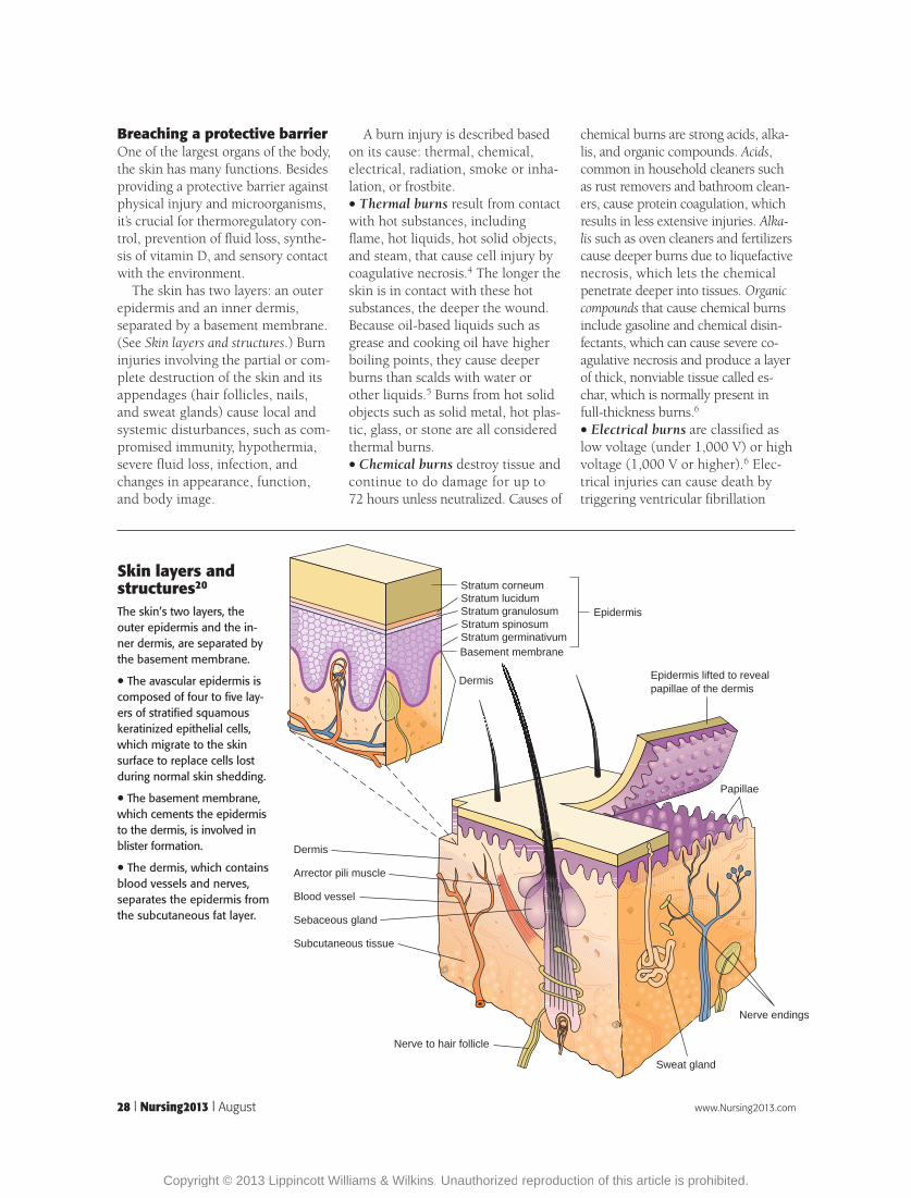
Breaching a protective barrierOne of the largest organs of the body, the skin has many functions. Besides providing a protective barrier against physical injury and microorganisms, it’s crucial for thermoregulatory con-trol, prevention of fluid loss, synthe-sis of vitamin D, and sensory contact with the environment.
The skin has two layers: an outer epidermis and an inner dermis, separated by a basement membrane. (See Skin layers and structures.) Burn injuries involving the partial or com-plete destruction of the skin and its appendages (hair follicles, nails, and sweat glands) cause local and systemic disturbances, such as com-promised immunity, hypothermia, severe fluid loss, infection, and changes in appearance, function, and body image.
A burn injury is described based on its cause: thermal, chemical, electrical, radiation, smoke or inha-lation, or frostbite.• Thermal burns result from contact with hot substances, including flame, hot liquids, hot solid objects, and steam, that cause cell injury by coagulative necrosis.4 The longer the skin is in contact with these hot substances, the deeper the wound. Because oil-based liquids such as grease and cooking oil have higher boiling points, they cause deeper burns than scalds with water or other liquids.5 Burns from hot solid objects such as solid metal, hot plas-tic, glass, or stone are all considered thermal burns.• Chemical burns destroy tissue and continue to do damage for up to 72 hours unless neutralized. Causes of
chemical burns are strong acids, alka-lis, and organic compounds. Acids, common in household cleaners such as rust removers and bathroom clean-ers, cause protein coagulation, which results in less extensive injuries. Alka-lis such as oven cleaners and fertilizers cause deeper burns due to liquefactive necrosis, which lets the chemical penetrate deeper into tissues. Organic compounds that cause chemical burns include gasoline and chemical disin-fectants, which can cause severe co-agulative necrosis and produce a layer of thick, nonviable tissue called es-char, which is normally present in full-thickness burns.6
• Electrical burns are classified as low voltage (under 1,000 V) or high voltage (1,000 V or higher).6 Elec-trical injuries can cause death by triggering ventricular fibrillation
Skin layers and structures20
The skin’s two layers, the outer epidermis and the in-ner dermis, are separated by the basement membrane.
• The avascular epidermis is composed of four to five lay-ers of stratified squamous keratinized epithelial cells, which migrate to the skin surface to replace cells lost during normal skin shedding.
• The basement membrane, which cements the epidermis to the dermis, is involved in blister formation.
• The dermis, which contains blood vessels and nerves, separates the epidermis from the subcutaneous fat layer.
Basement membrane
Dermis
Dermis
Arrector pili muscle
Blood vessel
Sebaceous gland
Subcutaneous tissue
Nerve to hair follicle
Sweat gland
Nerve endings
Papillae
Epidermis lifted to revealpapillae of the dermis
Stratum corneumStratum lucidumStratum granulosum Epidermis
Stratum germinativumStratum spinosum
Copyright © 2013 Lippincott Williams & Wilkins. Unauthorized reproduction of this article is prohibited.

www.Nursing2013.com August l Nursing2013 l 29
or paralyzing respiratory muscles. Al-though dysrhythmias can be triggered by low-voltage injuries, they’re more common in high-voltage injuries.
The extent of damage from an electrical burn may initially appear minor—the patient may have only small entry and exit wounds. Extensive damage can appear within several days to weeks—a phenomenon known as the iceberg effect because the skin shows little injury on the surface and hides massive injury beneath.6
Instead of conducting the electric-ity, bones, muscle, tendon, and fat respond to electrical injury by pro-ducing heat. Most injuries occur to muscles surrounding the long bones.6
• Radiation burns can result from exposure to radiofrequency energy or ionizing radiation such as sunlight, tanning booths, X-rays, or nuclear emissions or explosions. Ionizing ra-diation can produce tissue damage directly by striking a vital molecule such as DNA.5 Sunburn usually causes a first-degree or superficial burn, but radiation therapy can cause full-thickness burns.• Smoke and inhalation burns can occur concurrently with thermal or chemical burns. If the patient has thermal burns, look for signs of inha-lation burns: facial burns, hoarseness, soot in the nose or mouth, carbon in the sputum, lip edema, and singed eyebrows or nasal hair. Manufacturing illegal methamphetamine can cause thermal and chemical burns and as-sociated inhalation burns.6 Regardless of the cause of the inhalation injury, the patient needs immediate interven-tions such as endotracheal intubation, bronchoscopy, and measurement of carboxyhemoglobin (COHb) levels.• Frostbite is temporary or permanent tissue damage resulting from exposure to very cold temperatures. Any area of the body left uncovered in very cold temperatures can become frostbitten, but the most commonly affected areas are the fingers, toes, chin, earlobes, cheeks, and nose.7 Without treat-ment, frostbite can progress to cellular necrosis, gangrene, hypothermia, and
cardiac arrest. Because frostbite damages the skin, some patients are treated in the ICU as burn pa-tients, although initial treatment for frostbite is different than that for other burns.
Depth of injuryBurns are also categorized according to the depth of injury. In the past, burn injuries were classified as first, second, third, and occasionally fourth degree. In recent years, the
ABA has recommended a more pre-cise classification of burns, catego-rizing them according to depth of tissue injury:• epidermal or superficial (first degree)• partial-thickness (second degree), which may also be classified as su-perficial or deep partial-thickness• full-thickness (third degree), which may also be classified as deep full-thickness (fourth degree).8
For details, see Classifying burn injuries.
Classifying burn injuries• Superficial burns caused by the sun or low-intensity heat flashes damage only the epidermis. These first-degree burns cause erythema, skin blanching on pres-sure, mild pain and edema, and no blis-ters or vesicles, although after 24 hours the skin may blister and peel. Symptoms include hyperesthesia, mild pain, and tingling. Healing typically takes 3 to 6 days.
• Partial-thickness burns caused by chemicals, flame, or hot liquids damage the epidermis and part of the dermis. These second-degree burns appear as fluid-filled vesicles that are red and shiny (and wet if the vesicles have ruptured). Symptoms include edema, hyperesthesia, pain caused by nerve injury, and sensitiv-ity to cold air. Healing typically takes 10 to 21 days for superficial partial-thickness burns, which involve part of the dermis, and 2 to 6 weeks for deep partial- thickness burns, which involve more of the dermis.
• Full-thickness burns may extend into the subcutaneous tissue, meaning the skin can’t heal on its own. These burns, classified as third- and fourth-degree burns, are caused by prolonged expo-sure to chemicals, electrical current, flame, hot liquids, or tar. The skin ap-pears dry, waxy, white, leathery, or hard. Thrombosed vessels will be visible, and muscles, tendons, and bones may be involved. Signs and symptoms include lack of pain, possible hematuria, possible entrance and exit wounds from an elec-trical burn, and shock. Skin grafting is often required for healing, and patients may lose function of extremities or digits, or need amputation.
Deep partial-thickness (second degree) burns of the hands.
A full-thickness (third degree) burn of the feet.
Superficial (first degree) burn of the back.
© 2013 Lippincott Williams & Wilkins. Unauthorized reproduction of this article is prohibited.

30 l Nursing2013 l August www.Nursing2013.com
Size mattersThe size of the burn is expressed as the percentage of TBSA. A partial-thickness burn of more than 10% TBSA is serious and requires referral to a burn center. (See Should the patient go to a burn center?)
Estimate the TBSA burned on an adult by using 9 or multiples of 9, known as the rule of nines. The rule of nines varies between infants and adults because infants’ heads are pro-portionally larger compared to adults. (See Rule of nines: Estimating burn size in adults.) Although the rule of nines provides a rapid method for calculating the size of the burn in-jury, it can lead to an overestimation of the TBSA burned, so follow facility protocol for estimating the extent of a burn injury. Most burn centers re-peat the estimation of TBSA burned in 72 hours, when burns and their depth are more clearly demarcated and the burned area can be more easily quantified.9
Other common methods for mea-suring burn size include the Lund-Browder chart and the palm method.• The Lund-Browder method is highly recommended because it corrects for the large head-to-body ratio of in-fants and children.10
• The palm method is used for small scattered burns such as grease and scald burns. Often, the rule of palms will be completed first as a quick assessment until the Lund and Browder assessment can be completed. The patient’s palm (not including the fingers or wrist) equals 0.5% of TBSA. The entire palm including the fingers equals 1% in children and adults.4
Location matters tooDepending on a burn injury’s loca-tion, the patient may be predisposed to initial complications or complica-tions during wound healing.11 Cir-cumferential burns of the extremities, for example, can lead to vascular compromise resulting in compart-ment syndrome (see Ring of fire). Cir-cumferential burns to the thorax can
Should the patient go to a burn center?21
Patients with burn injuries who should be referred to a burn center include:
• All patients under age 1.
• All patients ages 1 to 2 with burns over 5% or more of TBSA.
• Patients of any age with full-thickness burns of any size.
• Patients over age 2 with partial-thickness burns greater than 10% of TBSA.
• Patients with burns of special areas such as the face, hands, feet, genitalia, perineum, or major joints.
• Patients with electrical burns, including lightning injuries.
• Patients with chemical burns.
• Patients with inhalation injury resulting from a fire or hot liquid burn.
• Patients with circumferential burns of the limbs or chest.
• Patients with preexisting medical conditions that could complicate burn management, prolong recovery, or affect survival.
• Patients with burns and concomitant trauma.
• Children with burns who are suspected to be victims of child abuse.
• Patients with septic burn wounds.
• Patients whose burns require treatment that exceeds the capabilities of the referring facility.
Rule of nines: Estimating burn size in adults
Source: Anatomical Chart Company. Pathology/Laboratory Medicine; 2008.
41⁄2%
18% 18%
41⁄2%
1%
41⁄2% 1⁄2%
41⁄2% 41⁄2%
9% 9%
9% 9%
4
Copyright © 2013 Lippincott Williams & Wilkins. Unauthorized reproduction of this article is prohibited.

www.Nursing2013.com August l Nursing2013 l 31
impair chest wall expansion, causing pulmonary insufficiency. Burns of the chest, head, and neck are also associated with pulmonary compli-cations.
Facial burns are associated with corneal abrasions and burns of the ears with auricular chondritis. Burns of the perineal area are prone to autocontamination by urine and feces.11,12
Burns over joints immediately af-fect the patient’s range of motion, which may be exacerbated later by hypertrophic scarring (see Trouble-some scars). Intensive therapy to pre-vent permanent disability is crucial.
Taking an inside lookUnderstanding the pathophysiology of a major burn injury (sometimes called burn shock) is key to effective management. Different causes lead to different burn injury patterns, which require different management.
The body’s compensatory mecha-nisms start with the inflammatory response, which is initiated by cel-lular injury. The most important ac-tivator of the inflammatory response is the mast cell, which releases bio-chemical mediators, such as hista-mine and chemotactic factors, and synthesizes other mediators, such as prostaglandins and leukotrienes.13
Histamine, the major vasoactive amine released by the mast cells, increases capillary permeability and exudation, resulting in edema at the burn injury site, decreased intravas-cular volume, hypotension, tach-ypnea, tachycardia, oliguria, and shock.13
The sympathetic nervous system (SNS) is stimulated and the fight-or-flight response activated, causing thirst, gastrointestinal hypomotility (ileus), adrenal gland stimulation (causing increased circulating cat-echolamines, increased metabolic rate, and increased aldosterone se-cretion), hepatic stimulation (caus-ing release of glycogen stores and elevated blood glucose levels), and vasoconstriction.13
A major burn injury affects every body system.• Respiratory system effects include direct airway injury; inhalation in-jury; carbon monoxide poisoning; smoke inhalation (damage to epithe-lial cells in the lower respiratory tract secondary to inhaling oxides, the products of combustion); alveolar damage; pulmonary edema; and decreased oxygen diffusion.5
• Cardiovascular system effects include fluid volume deficit, decreased mean arterial pressure, decreased cardiac output, hypovolemic shock (second-ary to extensive fluid shifts), and decreased myocardial contractility (impaired cardiac function improves 24 to 30 hours postinjury). Electrical burns can cause ECG changes, myo-cardial infarction, and cardiac dys-rhythmias including ventricular fibrillation.6
• Renal system effects are indirect. Decreased cardiac output leads to decreased renal perfusion and oli-guria that can culminate in acute kidney injury (AKI). In addition, after a burn injury, damaged red blood cells release hemoglobin and potassium, and skeletal muscle cells release myoglobin. Both hemoglo-bin and myoglobin are filtered by the glomerulus and degraded,
releasing heme pigment. Heme pig-ment, especially in the setting of fluid volume deficit, can cause AKI.14 Marked release of hemoglo-bin or myoglobin usually causes red or brown urine.• Gastrointestinal system effects include ileus secondary to SNS activation. Stress ulcer formation is triggered by the stress response and the histamine released in the acute inflammatory response. Intra-abdominal hyperten-sion and abdominal compartment syndrome can damage the gut, kid-neys, and liver.6,9
• Neuroendocrine system effects in-clude increased metabolic rate to compensate for the initial low core body temperature due to loss of skin. The increased metabolic rate increas-es caloric needs and leads to catabo-lism and a negative nitrogen balance that slows tissue building and heal-ing.6 Increased cortisol levels may cause insulin resistance and hyper-glycemia.13
• Immune system effects include im-munosuppression secondary to the immediate, prolonged, and severe immunologic and inflammatory re-sponse to a major burn injury.13
• Musculoskeletal system effects in-clude contractures and complications secondary to immobility and scar
Ring of fireCircumferential burns of the extremities can lead to compartment syndrome.
Copyright © 2013 Lippincott Williams & Wilkins. Unauthorized reproduction of this article is prohibited.

32 l Nursing2013 l August www.Nursing2013.com
tissue formation during the healing process.
Initial assessment and managementEmergency management of a patient with a burn injury begins with the initial assessment and treatment of life-threatening injuries. Stabilize the patient’s cervical spine if this hasn’t already been done. The true mecha-nism of injury may not be clear (for example, the patient may have been burned and propelled in an explosion).
Follow these specific aspects of the ABCDE (Airway, Breathing, Cir-culation, Disability, and Exposure/ Environmental control) assess-ment:6,9,15
• Airway. Maintaining the airway is the primary concern, especially if a patient has an inhalation injury. Assess for stridor (an ominous sign that sug-gests the patient’s upper airway is at least 85% narrowed), facial burns, soot in the nares or mouth, singed facial hair or nasal hair, edema of the lips and oral cavity, coughing, hoarse voice, and circumferential neck burns.6,9
• Breathing. Determine adequacy of ventilation by assessing the patient’s respiratory rate and depth and ob-serving for dyspnea. Auscultate the
lungs, noting any adventitious breath sounds. Obtain a pulse oximetry reading (remembering that it may be inaccurate in the presence of carbon monoxide), and a co- oximetry reading if indicated and available.6,9,15
• Circulation. Observe for obvious arterial bleeding. Assess for the pres-ence, symmetry, amplitude, rate, and rhythm of pulses; evaluate capillary refill time, skin color, and temperature.9
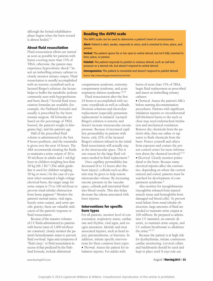
• Disability. Use the AVPU (Alert, Ver-bal, Pain stimuli, Unresponsive) scale to determine the patient’s level of con-sciousness and carefully evaluate any abnormalities. (See Reading the AVPU scale.) Assess for hypoxia, decreased cerebral perfusion related to hypovo-lemia, and cerebral injury resulting from head trauma. Assess the pa-tient’s pupillary response to light and sensory and motor function.3,6
• Exposure/environmental control. Gently remove the patient’s nonad-herent clothing and jewelry to pre-vent continued tissue damage. If the patient’s face is burned, remove glasses or contact lenses. Cover the patient with a dry sterile sheet to prevent further contamination of the burn wounds and to provide warmth.3,9,10,15
Obtain vital signs and establish I.V. access with two large-bore catheters if
the patient has burns over 15% of TBSA. Under ABA practice guidelines, fluid resuscitation is indicated for any patient with nonsuperficial burns covering more than 15% of TBSA.1,16
Elevate burned extremities above heart level to decrease edema. Admin-ister I.V. analgesia as prescribed and assess its effectiveness often, using a valid and reliable pain intensity rating scale.
Obtaining a historyAfter the initial focused assessment is completed and the patient is stabi-lized, obtain a history of the events while performing a comprehensive physical assessment. The main priori-ties are to determine the potential for an inhalation injury, presence of con-comitant injuries or trauma, and any preexisting conditions that may influ-ence the physical assessment or patient outcomes. A simple way to initially accomplish this is to use the SAMPLE
mnemonic: Signs and symptoms, Allergies, current Medications (includ-ing illegal substances or alcohol), Perti-nent history, Last oral intake, and Events leading up to the injury.17
After determining the extent and depth of the burn, ask the following questions:• What’s the patient’s chief complaint (for example, dyspnea or pain)?• Did the burn occur in an enclosed space?• Were explosives or chemicals involved?• What was the source of the burn-ing agent (for example, liquid, metal, or chemicals)?• What’s the status of the patient’s tetanus immunization?10
Stages of burn managementCare for a patient with burn injuries is organized into three stages: emer-gent (resuscitative), acute (wound healing), and rehabilitative (restor-ative).9 The assessment and manage-ment of specific problems overlap and may span two or three stages. For example, rehabilitation begins on the first day after the burn injury,
Troublesome scarsHypertrophic scarring of a deep partial-thickness burn can cause pruritus, warmth, and other patient discomfort, and the raised scar can limit function if joints are affected.
Copyright © 2013 Lippincott Williams & Wilkins. Unauthorized reproduction of this article is prohibited.
www.Nursing2013.com August l Nursing2013 l 33
although the formal rehabilitative phase begins when the burn wound is almost healed.15
About fluid resuscitationFluid resuscitation efforts are started as soon as possible for patients with burns covering more than 15% of TBSA; otherwise, the patient may experience hypovolemic shock.6 In-sert an indwelling urinary catheter to closely monitor urinary output. Fluid resuscitation is usually accomplished with an isotonic crystalloid such as lactated Ringer’s solution; the lactate helps to buffer the metabolic acidosis commonly seen with hypoperfusion and burn shock.6 Several fluid resus-citation formulas are available (for example, the Parkland formula) and usually is prescribed by the burn trauma surgeon. All formulas are based on the percentage of TBSA burned, the patient’s weight in kilo-grams (kg), and the patient’s age.
Half of the prescribed fluid volume is administered in the first 8 hours postburn, and the remainder is given over the next 16 hours. The ABA recommends titrating the fluids to maintain a urine output of 30 to 50 mL/hour in adults and 1 mL/kg/hour in children weighing less than 30 kg (66.1 lb).6 (The adult guide-line is used for children weighing 30 kg or more.) In the case of a pa-tient who’s sustained a high-voltage electrical burn, the target range for urine output is 75 to 100 mL/hour to prevent renal tubular obstruction from heme pigment.6 Monitor the patient’s mental status, vital signs, hourly urine output, and urine spe-cific gravity; these are valuable indi-cators of the patient’s response to fluid resuscitation.
Because of the massive volumes of I.V. fluids administered to patients with burns (rates of 1,000 mL/hour are common), closely monitor the pa-tient’s hemodynamic status to prevent fluid overload. Signs and symptoms of “fluid creep,” or fluid resuscitation in excess of that predicted by the Park-land formula, include abdominal
compartment syndrome, extremity compartment syndrome, and acute respiratory distress syndrome.18,19
Fluid resuscitation after the first 24 hours is accomplished with iso-tonic crystalloids as well as colloids. Dextrose solutions and electrolyte replacement (especially potassium replacement) is initiated. Lactated Ringer’s solution is isotonic and doesn’t increase intravascular oncotic pressure. Because of increased capil-lary permeability in patients with burns, only 25% of the lactated Ringer’s solution infused in the initial fluid resuscitation will actually stay in the intravascular space. This is one reason for the large fluid vol-umes needed in fluid replacement.6
Once capillary permeability has decreased (8 to 12 hours after the burn injury), colloids such as albu-min may be given to help restore intravascular volume. By increasing oncotic pressure in the vascular space, colloids pull interstitial fluid into blood vessels. This also helps decrease the edema associated with burn injuries.
Interventions for specific burn typesFor all patients, monitor level of con-sciousness, respiratory status, cardiac rate and rhythm, vital signs, and ox-ygen saturation. Identify and treat associated injuries, such as head in-jury, pneumothorax, or fractures. In addition, initiate specific interven-tions for these common burn types.• Thermal. Assess the patient for in-halation injuries. For adults with
burns of more than 15% of TBSA, begin fluid replacement as prescribed and insert an indwelling urinary catheter.• Chemical. Assess the patient’s ABCs before starting decontamination procedures. Patients with significant inhalation injuries or circumferential full-thickness burns to the neck or chest may need endotracheal intuba-tion and mechanical ventilation. Remove dry chemicals from the pa-tient’s skin, then use saline or tap water to flush chemicals from the burn. Protect yourself and others from exposure and contact the poi-son control center for more informa-tion about the chemical involved.9,15
• Electrical. Closely monitor pulses distal to the burn. Because many electrical injuries affect the extremi-ties, depending on where the current entered and exited, patients must be watched for development of com-partment syndrome.6
Also monitor for myoglobinemia (myoglobin released from injured muscle tissue and hemoglobin from damaged red blood cells). To prevent renal failure from renal tubular ob-struction, large amounts of fluid are needed to maintain urine output at 100 ml/hour. Be prepared to admin-ister I.V. mannitol, an osmotic di-uretic, to maintain urine output, and I.V. sodium bicarbonate to alkalinize the urine.6,9,15
Because the patient is at high risk for dysrhythmias, initiate continuous cardiac monitoring. Cervical collars and backboards should be used and kept in place until X-rays rule out
Reading the AVPU scaleThe AVPU scale can be used to determine a patient’s level of consciousness.
Alert: Patient is alert, awake, responds to voice, and is oriented to time, place, and person.
Verbal: The patient opens his or her eyes to verbal stimuli, but isn’t fully oriented to time, place, or person.
Painful: The patient responds to painful or noxious stimuli, such as nail bed pressure or a sternal rub, but doesn’t respond to verbal stimuli.
Unresponsive: The patient is nonverbal and doesn’t respond to painful stimuli.
Source: http://www.ahrq.gov/research/esi/esi2.htm.
Copyright © 2013 Lippincott Williams & Wilkins. Unauthorized reproduction of this article is prohibited.

34 l Nursing2013 l August www.Nursing2013.com
spinal injury—many electrical injuries occur from contact with high voltage wires, causing a fall.6
• Inhalation. Obtain an arterial blood gas analysis, COHb level, and chest X-ray. Prepare the patient for fiberop-tic bronchoscopy or endotracheal intubation if indicated.
Effective interventionsBy understanding the types of burns and how to assess and manage them, nurses can immediately implement effective interventions while arrange-ments are made for patient transfer to a burn specialty center. ■
REFERENCES
1. American Burn Association. Burn Incidence and Treatment in the United States: 2012 Fact Sheet. http://ameriburn.org/resources_factsheet.php.
2. TOMA Foundation for Burned Children. http://www.fondtomafound.org/english/index.htm.
3. Silvestri LA. Comprehensive Review for the NCLEX-RN Examination. 5th ed. St. Louis, MO: Elsevier Saunders; 2011.
4. Rice PL, Orgill DP. Classification of burns. 2012. UpToDate. http://www.uptodate.com.
5. Coffee T. Care of patients with burns. In: Ignatavicius DD, Workman ML, eds. Medical-Surgical
Nursing: Patient-Centered Collaborative Care. 7th ed. St. Louis, MO: Saunders Elsevier; 2013:511-540.
6. Ahrns-Klas KS. Burns. In: Sole ML, Klein DG, Moseley MJ, eds. Introduction to Critical Care Nursing. 5th ed. St. Louis, MO: Saunders Elsevier; 2009:683-728.
7. Frostbite treatment. www.essortment.com/frostbitetreatment-59888.html.
8. Kagan RJ, Peck MD, Ahrenholz DH, et al. Surgical management of the burn wound and use of skin substitutes: an expert panel white paper. J Burn Care Res. 2013; 34(2):e60-79.
9. Smeltzer SC, Bare BG, Hinkle JL, Cheever, KH. Brunner & Suddarth’s Textbook of Medical-Surgical Nursing. 12th ed. Philadelphia, PA: Lippincott Williams & Wilkins; 2010.
10. Stout LR. Burns and common integumentary disorders. In: Morton PG, Fontaine DK, eds. Critical Care Nursing: A Holistic Approach. 9th ed. Philadelphia, PA: Lippincott Williams & Wilkins; 2009:1349-1375.
11. Moss LS. Treatment of the burn patient in primary care. Adv Skin Wound Care. 2010;23(11):517-524.
12. Perrin KO. Understanding the Essentials of Critical Care Nursing. Upper Saddle River, NJ: Pearson Prentice Hall; 2009.
13. Huether SE, McCance KL. Understanding Pathophysiology. 5th ed. St. Louis, MO: Mosby, Inc.; 2011.
14. Eustace JA, Kinsella S. Clinical features and diagnosis of heme pigment-induced acute kidney injury (acute renal failure). 2012. UpToDate. http://www.uptodate.com.
15. Knighton JA. Nursing management: burns. In: Lewis SL, Heitkemper MM, Dirksen SR, O’Brien PG,
Bucher L, eds. Medical-Surgical Nursing: Assessment and Management of Clinical Problems. Vol 1. 7th ed. St. Louis, MO: Mosby Elsevier; 2007:483-507.
16. Rice PL, Orgill DP. Emergency care of moderate and severe thermal burns in adults. 2013. UpToDate. http://www.uptodate.com.
17. EMT’s Patient Assessment: SAMPLE. http://voices.yahoo.com/emts-patient-assessment-sample-1314601.html.
18. Zaletel CL. Factors affecting fluid resuscitation in the burn patient: the collaborative role of the APN. Adv Emerg Nurs J. 2009;31(4):309-320.
19. James E, Hayes M, McCabe P, Williams G, Takata M, Vizcaychipi MP. Fluid creep in burn resuscitation: the tide has not yet turned. Critical Care. 2012;16(suppl 1):P464. http://ccforum.com/content/16/S1/P464.
20. Porth CM. Essentials of Pathophysiology. 3rd ed. Philadelphia: Wolters Kluwer Health/Lippincott Williams & Wilkins; 2011:1160.
21. Stander M, Wallis LA. The emergency management and treatment of severe burns. Emerg Med Int. 2011;2011:161375. http://www.ncbi.nlm.nih.gov/pmc/articles/PMC3195355.
At Duquesne University’s School of Nursing in Pittsburgh, Pa., Alicia L. Culleiton is an assistant clinical professor and Lynn M. Simko is an associate clinical professor.
The authors and planners have disclosed that they have no financial relationships related to this article.
DOI-10.1097/01.NURSE.0000432018.28250.c3
INSTRUCTIONS
Caring for patients with burn injuries
DISCOUNTS and CUSTOMER SERVICE• Send two or more tests in any nursing journal published by Lippincott Williams & Wilkins together by mail, and deduct $0.95 from the price of each test.• We also offer CE accounts for hospitals and other healthcare facilities on nursingcenter.com. Call 1-800-787-8985 for details.
PROVIDER ACCREDITATIONLippincott Williams & Wilkins, publisher of Nursing2013 journal, will award 2.3 contact hours for this continuing nursing education activity.
Lippincott Williams & Wilkins is accredited as a provider of continuing nursing education by the American Nurses Credentialing Center’s Commission on Accreditation.
Lippincott Williams & Wilkins is also an approved provider of continuing nursing education by the District of Columbia and Florida #50-1223. This activity is also provider approved by the California Board of Registered Nursing, Provider Number CEP 11749 for 2.3 contact hours.
Your certificate is valid in all states. The ANCC’s accreditation status of Lippincott Williams & Wilkins Department of Continuing
Education refers only to its continuing nursing educational activities and does not imply Commission on Accreditation approval or endorsement of any commercial product.
TEST INSTRUCTIONS• To take the test online, go to our secure website at http://www.nursingcenter.com/ce/nursing.• On the print form, record your answers in the test answer section of the CE enrollment form on page 35. Each question has only one correct answer. You may make copies of these forms.• Complete the registration information and course evaluation. Mail the completed form and registration fee of $21.95 to: Lippincott Williams & Wilkins, CE Group, 74 Brick Blvd., Bldg. 4, Suite 206, Brick, NJ 08723. We will mail your certificate in 4 to 6 weeks. For faster service, include a fax number and we will fax your certificate within 2 business days of receiving your enrollment form. • You will receive your CE certificate of earned contact hours and an answer key to review your results. There is no minimum passing grade.• Registration deadline is August 31, 2015.
For more than 98 additional continuing education articles related to skin and wound care topics, go to NursingCenter.com/CE.> <
Earn CE credit online: Go to http://www.nursingcenter.com/CE/nursing and receive a certifi cate within minutes.
© 2013 Lippincott Williams & Wilkins. Unauthorized reproduction of this article is prohibited.