2803
DESCRIPTION
this article isTRANSCRIPT

Polymorphonuclear leukocytes (PMNs) induceprotective Th1-type cytokine epithelial responsesin an in vitro model of oral candidosis
Martin Schaller,1,2 Ursula Boeld,1 Sylvia Oberbauer,1 Gerald Hamm,3
Bernhard Hube4 and Hans C. Korting1
Correspondence
Martin Schaller
Martin.Schaller@
med.uni-tuebingen.de
1Department of Dermatology and Allergology, University of Munich, Frauenlobstr. 9–11,D-80337 Munich, Germany
2Department of Dermatology, Liebermeisterstr. 25, D-72076 Tuebingen, University ofTuebingen, Germany
3Department of Parodontology, University of Munich, Goethestr. 70, D-80336 Munich, Germany
4Robert Koch-Institute, NG4, Nordufer 20, D-13353 Berlin, Germany
Received 15 March 2004
Revised 26 May 2004
Accepted 6 June 2004
The immune response and the anticandidal activity of keratinocytes and polymorphonuclear
leukocytes (PMNs) play a key role in host defence against localized Candida albicans infection.
An established model of oral candidosis based on reconstituted human oral epithelium (RHE)
was supplemented with PMNs to study the effect of these immune cells during experimental
oral candidosis. Infection of RHE with C. albicans induced a strong expression of the chemokine
interleukin-8 (IL-8) and the cytokine granulocyte-macrophages colony-stimulating factor
(GM-CSF), and amoderate stimulation of interleukin-1a (IL-1a), interleukin-1b (IL-1b), interleukin-6
(IL-6), interferon c (IFN-c) and tumour necrosis factor a (TNF-a) by keratinocytes. This immune
response was associated with chemoattraction of PMNs to the site of infection, whereas
uninfected RHE failed to induce cytokine expression or to attract PMNs. Growth of the pathogen
and tissue damage of C. albicans-infected RHE were significantly reduced when PMNs were
applied to the apical epithelial surface or when PMNs migrated through a perforated basal
polycarbonate filter of the model. Notably, protection against epithelial tissue damage was also
observed when PMNs were placed on the basal side of non-perforated filters, which prevented
PMN migration into the RHE. Addition of PMNs enhanced a Th1-type immune response (IFN-c,
TNF-a), down-regulated the expression of the Th2-type cytokine interleukin-10 (IL-10), and was
associated with protection against Candida-induced tissue damage. This PMN-supplemented
model of oral candidosis mimics the in vivo situation, and provides a promising tool for
studying the immunological interactions between keratinocytes and C. albicans,
as well as the influence of PMNs on C. albicans pathogenesis.
INTRODUCTION
Keratinocytes and neutrophils, eosinophils or basophils(polymorphonuclear leukocytes, PMNs) are the first line ofhost defence against mucosal Candida albicans infections(Challacombe, 1994; Eversole et al., 1997). An importantstep in the inflammatory response to mycotic infectionleading to the recovery from infection is a Th1-typeimmune response mediated by CD4+ T-cells (Fidel,
2002a). Furthermore, transepithelial migration of PMNsrepresents the histological hallmark of oral candidal lesionsand is believed to play a crucial role in the clearance ofinfection (Fidel, 2002a). This is in line with the fact thatpatients suffering from defects in neutrophil function orneutropenic patients are more susceptible to oral coloni-zation and infection with C. albicans (Epstein et al., 2003;Martino et al., 1989; Myoken et al., 2004; Walsh et al., 1996;Wolff, 1972). In vitro candidacidal activity has beenobserved for both keratinocytes and PMNs (Fidel, 2002b;Romani, 1999). In addition, specific monoclonal antibodieshave been used to deplete PMNs in the systemic circulationof mice in order to study their influence on the pathogenesisof C. albicans infections and to mimic the in vivo situationin neutropenic patients suffering from vaginal (Black et al.,
Abbreviations: FACS, fluorescence-activated cell sorting; GADPH,glyceraldehyde-3-phosphate dehydrogenase; GM-CSF, granulocyte-macrophages colony-stimulating factor; IFN, interferon; IL, interleukin;LDH, lactate dehydrogenase; PMN, polymorphonuclear leukocyte; RHE,reconstituted human oral epithelium; QRT-PCR, quantitative RT-PCR;TNF, tumour necrosis factor.
0002-7169 G 2004 SGM Printed in Great Britain 2807
Microbiology (2004), 150, 2807–2813 DOI 10.1099/mic.0.27169-0
1998; Fidel et al., 1999) and oral candidosis (Farah et al.,2001). However, there are contradictory data about therole of PMNs in resolving murine oral candidosis in differ-ent mouse strains (Farah et al., 2001). In a recent study, wecharacterized the immune response of keratinocytes duringinteraction with C. albicans using reconstituted human oralepithelium (RHE) (Schaller et al., 2002). To establish a morephysiological infection model, we supplemented the RHEwith PMNs and studied their impact on the pathogenesis ofexperimental oral infections and the cytokine response.
METHODS
Culture media and growth conditions. For the infection of thereconstituted oral epithelium (RHE), inocula were prepared byculturing the clinical C. albicans wild-type strain SC5314 (Gillumet al., 1984) for 24 h at 37 uC on Sabouraud dextrose agar (Difco).Cells were washed three times in 0?9% NaCl and 26105 cells werethen suspended in 10 ml YPG medium (Difco). The suspension wascultured for 16 h at 25 uC with orbital shaking. A suspension of46106 cells from this culture was incubated with shaking in freshmedium for 24 h at 37 uC. After washing three times with PBS, thefinal inoculum was adjusted to the desired density with PBS.
Isolation of polymorphonuclear cells (PMNs). PMNs obtainedfrom healthy human volunteers were isolated from heparinizedwhole blood using Histopaque-1119 in combination with Histopaque-1077 (Sigma), according to the manufacturer’s protocol. The cellsrecovered from the interface were washed three times and suspendedat a concentration of 26108 ml21 in RPMI 1640 medium (Sigma)in the presence and absence of 10% fetal calf serum (FCS). Residualerythrocytes were removed by hypotonic lysis. Giemsa stainingand light microscopy were used to ensure that a pure population ofPMNs (>90% purity) with typical morphology had been isolated.The cells were vital-stained using the trypan blue dye-exclusionmethod. Numbers of vital and non-vital leukocytes per sample wereassessed using a Neubauer chamber. The viability was ¢95% in allexperiments.
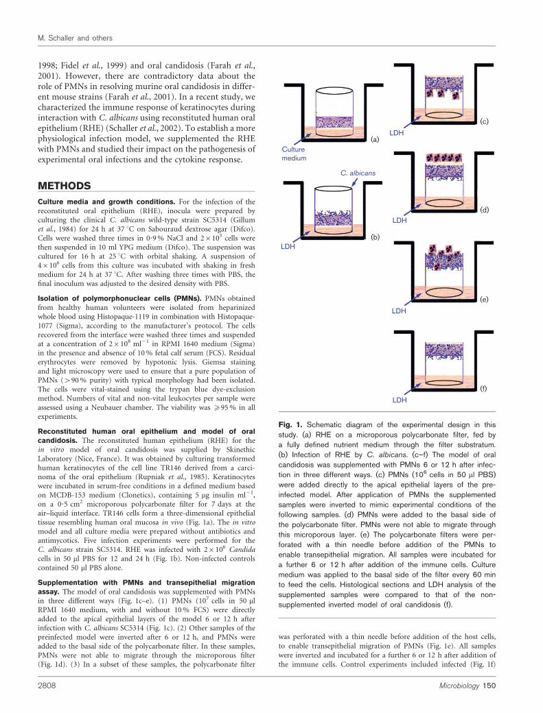
Reconstituted human oral epithelium and model of oralcandidosis. The reconstituted human epithelium (RHE) for thein vitro model of oral candidosis was supplied by SkinethicLaboratory (Nice, France). It was obtained by culturing transformedhuman keratinocytes of the cell line TR146 derived from a carci-noma of the oral epithelium (Rupniak et al., 1985). Keratinocyteswere incubated in serum-free conditions in a defined medium basedon MCDB-153 medium (Clonetics), containing 5 mg insulin ml21,on a 0?5 cm2 microporous polycarbonate filter for 7 days at theair–liquid interface. TR146 cells form a three-dimensional epithelialtissue resembling human oral mucosa in vivo (Fig. 1a). The in vitromodel and all culture media were prepared without antibiotics andantimycotics. Five infection experiments were performed for theC. albicans strain SC5314. RHE was infected with 26106 Candidacells in 50 ml PBS for 12 and 24 h (Fig. 1b). Non-infected controlscontained 50 ml PBS alone.
Supplementation with PMNs and transepithelial migrationassay. The model of oral candidosis was supplemented with PMNsin three different ways (Fig. 1c–e). (1) PMNs (107 cells in 50 mlRPMI 1640 medium, with and without 10% FCS) were directlyadded to the apical epithelial layers of the model 6 or 12 h afterinfection with C. albicans SC5314 (Fig. 1c). (2) Other samples of thepreinfected model were inverted after 6 or 12 h, and PMNs wereadded to the basal side of the polycarbonate filter. In these samples,PMNs were not able to migrate through the microporous filter(Fig. 1d). (3) In a subset of these samples, the polycarbonate filter
was perforated with a thin needle before addition of the host cells,
to enable transepithelial migration of PMNs (Fig. 1e). All samples
were inverted and incubated for a further 6 or 12 h after addition of
the immune cells. Control experiments included infected (Fig. 1f)
LDH
LDH
LDH
LDH
LDH
Culturemedium
C. albicans
(a)
(b)
(c)
(d)
(e)
(f)
Fig. 1. Schematic diagram of the experimental design in thisstudy. (a) RHE on a microporous polycarbonate filter, fed bya fully defined nutrient medium through the filter substratum.(b) Infection of RHE by C. albicans. (c–f) The model of oralcandidosis was supplemented with PMNs 6 or 12 h after infec-tion in three different ways. (c) PMNs (106 cells in 50 ml PBS)were added directly to the apical epithelial layers of the pre-infected model. After application of PMNs the supplementedsamples were inverted to mimic experimental conditions of thefollowing samples. (d) PMNs were added to the basal side ofthe polycarbonate filter. PMNs were not able to migrate throughthis microporous layer. (e) The polycarbonate filters were per-forated with a thin needle before addition of the PMNs toenable transepithelial migration. All samples were incubated fora further 6 or 12 h after addition of the immune cells. Culturemedium was applied to the basal side of the filter every 60 minto feed the cells. Histological sections and LDH analysis of thesupplemented samples were compared to that of the non-supplemented inverted model of oral candidosis (f).
2808 Microbiology 150
M. Schaller and others
and uninfected RHE after addition of RPMI 1640/10% FCSmedium without PMNs, and uninfected RHE supplemented withPMN cells in the same way as described above. Culture medium wasadded to the basal side of the inverted samples every 60 min to feedthe keratinocytes/PMNs with nutrients.
Assay of lactate dehydrogenase activity. The release of lactatedehydrogenase (LDH) from epithelial cells into the surroundingmedium was monitored as a measure of epithelial cell damage. LDHrelease in the maintenance media of the cultures from uninfectedand infected epithelial cells was measured at 12 and 24 h. LDHactivity was analysed spectrophotometrically by measuring the rateof NADH disappearance at 340 nm during the LDH-catalysed con-version of pyruvate to lactate, according to the Wroblewski–La Duemethod (Wroblewski & John, 1955). The LDH activity is given asU l21 at 37 uC.
Killing assay. A killing assay was used to study the inhibitory effectof PMNs on C. albicans during RHE infection. It was performed byplating the infected samples 12 and 24 h after incubation, with andwithout PMN, on Sabouraud dextrose agar. Before plating, 2 mlPBS was added to the samples, and samples were thoroughlyvortexed for 10 min. Furthermore, the solution was vigorouslyagitated by using a Pasteur pipette to separate big clumps of fungalcells into single cells, and then diluted 1 : 1000 and 1 : 10 000 in PBS.The presence of single cells was confirmed by light microscopy.Yeast cell viability was determined by assessment of the colonyforming units produced after incubation for 24 h at 37 uC onSabouraud dextrose agar.
Light microscopy. Light-microscopy studies were performed toevaluate histological changes during infection. Part of each specimenwas fixed, postfixed and embedded in glycide ether, and cut usingan ultramicrotome (Ultracut). Semi-thin sections (1 mm) werestudied with a light microscope after staining with 1% toluidineblue and 1% pyronine G (Merck). The histological changes of themucosa were evaluated on the basis of 50 sections from five differentsites for each infected epithelium.
RNA isolation and cDNA-synthesis by reverse transcriptase.For the detection of mRNA, samples were rapidly removed andshock-frozen in liquid nitrogen. Total RNA from shock-frozensamples was isolated using RNAPure (Peqlab), according to themanufacturer’s instructions. For assessing RNA concentration andpurity, UV spectroscopy was used (Bio Photometer, Eppendorf).The absorbance of a diluted RNA sample was measured at 260 and280 nm. cDNA synthesis was performed using Superscript II reversetranscriptase (Gibco), following the manufacturer’s instructions.
Quantitative RT-PCR (QRT-PCR). For cytokine expression, 20 ngof cDNA were amplified ‘real-time’ in a LightCycler (Roche), usinga FastStart DNA Master SYBR Green I kit (Roche) at 3 mM Mg2+
final concentration, and analysed with LightCycler Software 3.5.Annealing temperature and elongation time were optimized for eachprimer pair. The sequences of the primer pairs used were publishedrecently (Schaller et al., 2002). Amplified DNA for each primer pairwas serially diluted (six logs) and used to generate standard curves.Absolute quantification for these cDNAs was achieved with theLightCycler software.
Quantification of cytokine secretion by epithelial cellsstimulated with C. albicans. Epithelial tissues (with or withoutPMNs; see above) were infected with PBS-washed C. albicans S5314or treated with PBS only. After 12 h and 24 h, samples of themaintenance medium surrounding the infected and uninfectedepithelial tissues were collected and centrifuged. The amount ofinterleukin-1b (IL-1b), IL-6, IL-10, granulocyte-macrophagescolony-stimulating factor (GM-CSF), interferon c (IFN-c) andtumour necrosis factor a (TNF-a) secreted into the supernatant was
determined by fluorescence-activated cell sorting (FACS) analysis.
Flow cytometric data were acquired on a Becton Dickinson
FACSCan using the Human Inflammation Kit (Becton Dickinson).
Data were analysed using Cellquest software (Becton Dickinson).
RESULTS
Protective effect and transepithelial migrationof PMNs during infection of oral epitheliumwith C. albicans
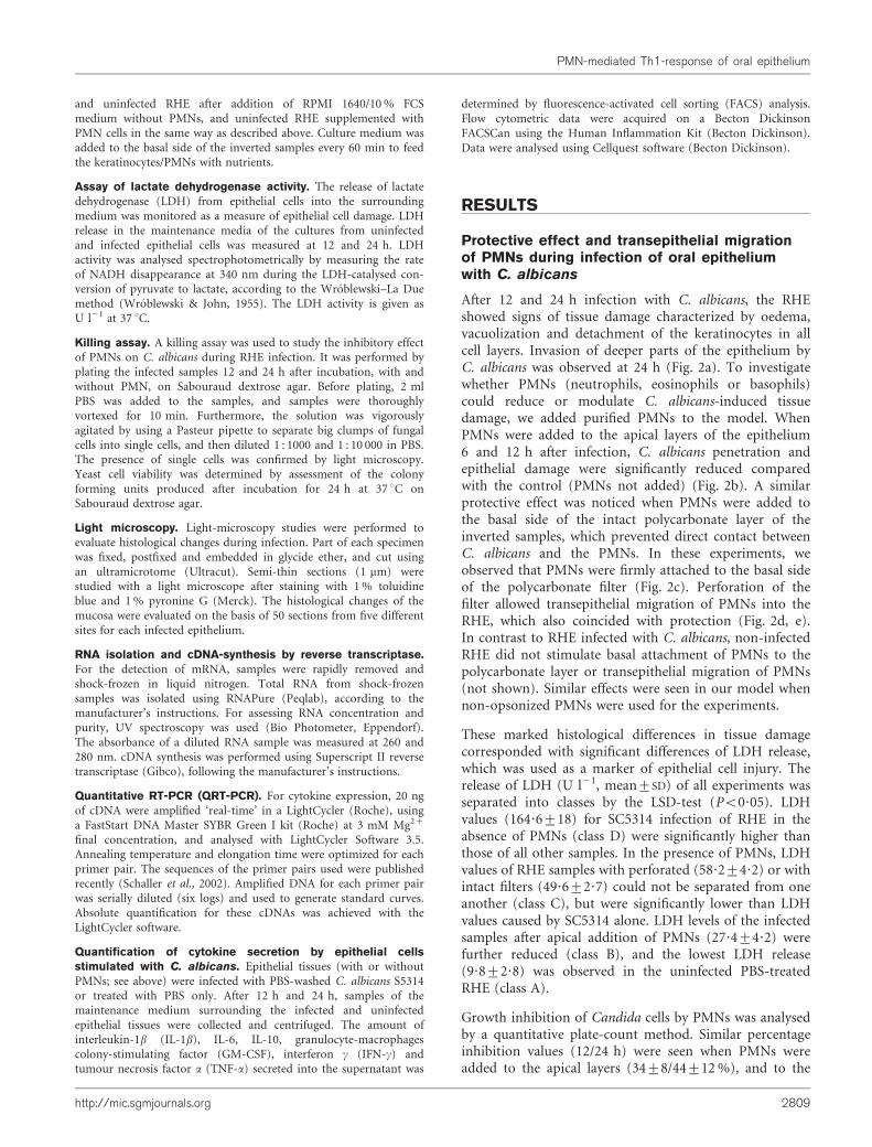
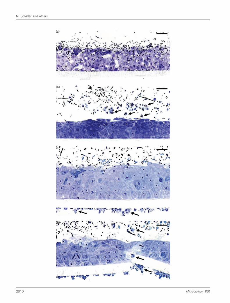
After 12 and 24 h infection with C. albicans, the RHEshowed signs of tissue damage characterized by oedema,vacuolization and detachment of the keratinocytes in allcell layers. Invasion of deeper parts of the epithelium byC. albicans was observed at 24 h (Fig. 2a). To investigatewhether PMNs (neutrophils, eosinophils or basophils)could reduce or modulate C. albicans-induced tissuedamage, we added purified PMNs to the model. WhenPMNs were added to the apical layers of the epithelium6 and 12 h after infection, C. albicans penetration andepithelial damage were significantly reduced comparedwith the control (PMNs not added) (Fig. 2b). A similarprotective effect was noticed when PMNs were added tothe basal side of the intact polycarbonate layer of theinverted samples, which prevented direct contact betweenC. albicans and the PMNs. In these experiments, weobserved that PMNs were firmly attached to the basal sideof the polycarbonate filter (Fig. 2c). Perforation of thefilter allowed transepithelial migration of PMNs into theRHE, which also coincided with protection (Fig. 2d, e).In contrast to RHE infected with C. albicans, non-infectedRHE did not stimulate basal attachment of PMNs to thepolycarbonate layer or transepithelial migration of PMNs(not shown). Similar effects were seen in our model whennon-opsonized PMNs were used for the experiments.
These marked histological differences in tissue damagecorresponded with significant differences of LDH release,which was used as a marker of epithelial cell injury. Therelease of LDH (U l21, mean±SD) of all experiments wasseparated into classes by the LSD-test (P<0?05). LDHvalues (164?6±18) for SC5314 infection of RHE in theabsence of PMNs (class D) were significantly higher thanthose of all other samples. In the presence of PMNs, LDHvalues of RHE samples with perforated (58?2±4?2) or withintact filters (49?6±2?7) could not be separated from oneanother (class C), but were significantly lower than LDHvalues caused by SC5314 alone. LDH levels of the infectedsamples after apical addition of PMNs (27?4±4?2) werefurther reduced (class B), and the lowest LDH release(9?8±2?8) was observed in the uninfected PBS-treatedRHE (class A).
Growth inhibition of Candida cells by PMNs was analysedby a quantitative plate-count method. Similar percentageinhibition values (12/24 h) were seen when PMNs wereadded to the apical layers (34±8/44±12%), and to the
http://mic.sgmjournals.org 2809
PMN-mediated Th1-response of oral epithelium
(a)
(b)
(c)
(d)
2810 Microbiology 150
M. Schaller and others
basal side of intact (38±7/46±11%) or perforated (36±9/39±11%) layers of the infected epithelium.
Cytokine mRNA expression in response tomucosal C. albicans infection in the presenceand absence of PMNs (QRT-PCR)
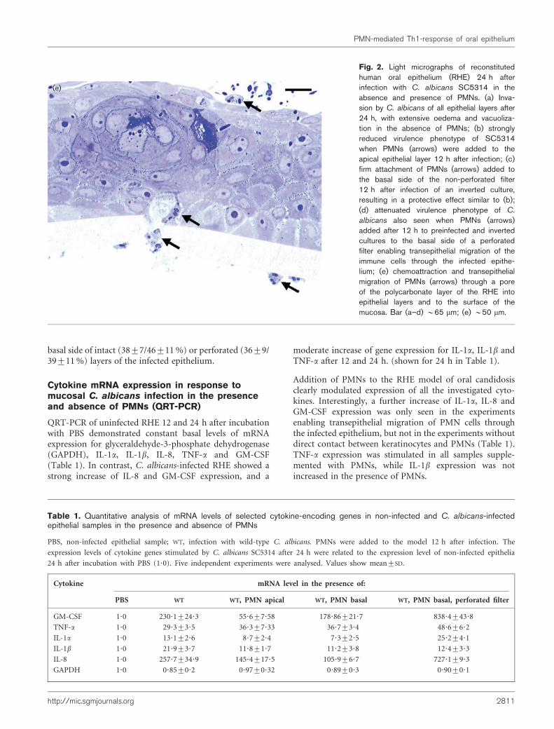
QRT-PCR of uninfected RHE 12 and 24 h after incubationwith PBS demonstrated constant basal levels of mRNAexpression for glyceraldehyde-3-phosphate dehydrogenase(GAPDH), IL-1a, IL-1b, IL-8, TNF-a and GM-CSF(Table 1). In contrast, C. albicans-infected RHE showed astrong increase of IL-8 and GM-CSF expression, and a
moderate increase of gene expression for IL-1a, IL-1b andTNF-a after 12 and 24 h. (shown for 24 h in Table 1).
Addition of PMNs to the RHE model of oral candidosisclearly modulated expression of all the investigated cyto-kines. Interestingly, a further increase of IL-1a, IL-8 andGM-CSF expression was only seen in the experimentsenabling transepithelial migration of PMN cells throughthe infected epithelium, but not in the experiments withoutdirect contact between keratinocytes and PMNs (Table 1).TNF-a expression was stimulated in all samples supple-mented with PMNs, while IL-1b expression was notincreased in the presence of PMNs.
Table 1. Quantitative analysis of mRNA levels of selected cytokine-encoding genes in non-infected and C. albicans-infectedepithelial samples in the presence and absence of PMNs
PBS, non-infected epithelial sample; WT, infection with wild-type C. albicans. PMNs were added to the model 12 h after infection. The
expression levels of cytokine genes stimulated by C. albicans SC5314 after 24 h were related to the expression level of non-infected epithelia
24 h after incubation with PBS (1?0). Five independent experiments were analysed. Values show mean±SD.
Cytokine mRNA level in the presence of:
PBS WT WT, PMN apical WT, PMN basal WT, PMN basal, perforated filter
GM-CSF 1?0 230?1±24?3 55?6±7?58 178?86±21?7 838?4±43?8
TNF-a 1?0 29?3±3?5 36?3±7?33 36?7±3?4 48?6±6?2
IL-1a 1?0 13?1±2?6 8?7±2?4 7?3±2?5 25?2±4?1
IL-1b 1?0 21?9±3?7 11?8±1?7 11?2±3?8 12?4±3?3
IL-8 1?0 257?7±34?9 145?4±17?5 105?9±6?7 727?1±9?3
GAPDH 1?0 0?85±0?2 0?97±0?32 0?89±0?3 0?90±0?1
(e)
Fig. 2. Light micrographs of reconstitutedhuman oral epithelium (RHE) 24 h afterinfection with C. albicans SC5314 in theabsence and presence of PMNs. (a) Inva-sion by C. albicans of all epithelial layers after24 h, with extensive oedema and vacuoliza-tion in the absence of PMNs; (b) stronglyreduced virulence phenotype of SC5314when PMNs (arrows) were added to theapical epithelial layer 12 h after infection; (c)firm attachment of PMNs (arrows) added tothe basal side of the non-perforated filter12 h after infection of an inverted culture,resulting in a protective effect similar to (b);(d) attenuated virulence phenotype of C.
albicans also seen when PMNs (arrows)added after 12 h to preinfected and invertedcultures to the basal side of a perforatedfilter enabling transepithelial migration of theimmune cells through the infected epithe-lium; (e) chemoattraction and transepithelialmigration of PMNs (arrows) through a poreof the polycarbonate layer of the RHE intoepithelial layers and to the surface of themucosa. Bar (a–d) ~65 mm; (e) ~50 mm.
http://mic.sgmjournals.org 2811
PMN-mediated Th1-response of oral epithelium

Cytokine secretion analysis in response tomucosal C. albicans infection in the absenceand presence of PMNs (FACS analysis)
The modulation of the immune response was furthercharacterized at the protein level by FACS analysis of themaintenance medium (Table 2). Non-infected mucosaproduced only low levels of IFN-c, TNF-a and IL-1b,moderate levels of IL-6 and IL-10, but high IL-8 levels. Inresponse to infection with C. albicans in the absence ofPMNs, epithelial cells produced increasing concentrationsof IFN-c, TNF-a, IL-1b, IL-6 and IL-8, whereas IL-10secretion was not affected by epithelial infection. Additionof PMNs stimulated IFN-c and TNF-a production andreduced IL-10 secretion in all samples, while furtherincreases in IL-1b, IL-6 and IL-8 production were onlyobserved during transepithelial migration of the PMN cells.
DISCUSSION
Mucosal epithelial cells provide an effective innate hostdefence mechanism against C. albicans (Steele et al., 2000).Furthermore, PMNs are frequently found at sites ofCandidainfection, and this is thought to be a hallmark of oralcandidosis in humans (Challacombe, 1994; Eversole et al.,1997). Anticandidal activity and cell-mediated immunitymediated by epithelial cells and PMNs might represent themost important line of host defence to prevent superficialCandida infections (Farah et al., 2002; Fidel, 2002a; Kullberget al., 1999). Several studies have analysed the role of PMNsin local defence by monoclonal depletion in murine modelsof vaginal candidosis (Black et al., 1998; Fidel et al., 1995,1999; Fulurija et al., 1996). However, most data from thesestudies demonstrated no significant role for PMNs inresolving C. albicans vaginal infections. The role of PMNsin protection against oral candidosis has not been suffici-ently investigated. A recent study using two mouse modelsshowed a significant contribution of PMNs in resolvingan oral infection in only one of the two models (Farahet al., 2001). The bioassay system presented in our studydemonstrates infection-induced chemoattraction of PMNs,which are able to migrate transepithelially to the site of
C. albicans infection. Moreover, the in vitro model enablesthe direct and indirect effects of the PMNs in protectionagainst oral Candida infections to be studied.
Recently published studies have shown induction of IL-8synthesis in keratinocytes after stimulation with C. albicans(Dongari-Bagtzoglou & Kashleva, 2003; Schaller et al.,2002). Since attachment and/or transepithelial migrationof PMNs in this study was only seen after epithelial infec-tion and concomitant strong expression of the chemo-attractive cytokines GM-CSF, IL-6 and IL-8, we concludedthat these cytokines contributed to the transepithelialmigration of PMNs in our model. In vivo, PMNs accumu-late rapidly at the site of infection in the oral cavity as aresult of host signals (Borish et al., 1989; Djeu et al., 1990).IL-6 and IL-8 act as potent chemoattractants and promotedegranulation and other antifungal activities of PMNs(Djeu et al., 1990; Kullberg et al., 1999). GM-CSF is knownas a potent neutrophil growth factor, stimulating cytokineproduction and fungicidal activity of leukocytes (Tanshoet al., 1994). Therefore, our RHE bioassay system seems toaccurately imitate oral candidosis in vivo.
Infection of RHE with C. albicans also induced expressionof IFN-c and TNF-a, which are linked with a protectiveTh1 response during systemic infection (Romani, 1999). Incontrast, IL-10, which is associated with a Th2-mediatedsusceptibility to candidosis (Romani, 1999), was down-regulated in our bioassay system. PMNs, especially thosemigrating into epithelial tissue, enhanced this Th1 up- andTh2 down-regulation pattern and protected the mucosaagainst C. albicans infection by inhibiting fungal growth.
These data implicate a protective role for Th1-type cyto-kine release from epithelial cells during oral candidosis.Protection against tissue damage was observed by directinteraction of the PMN cells with C. albicans, either byapical application or by transepithelial migration of PMNsfrom the basal side. Interestingly, the protective effect wasalso seen in a modified experimental assay in which directphysiological contact between PMNs and C. albicans wasprevented and only indirect interaction between PMNs,
Table 2. FACS analysis of cytokine release 24 h after infection of epithelial cells with C. albicans in the presence andabsence of PMNs
PBS, non-infected epithelial sample; WT, infection with wild-type C. albicans. PMNs were added to the model 12 h after infection. Cytokine
release values are in pg ml21 (mean±SD). Five independent experiments were analysed.
Cytokine Cytokine release in the presence of:
PBS WT WT, PMN apical WT, PMN basal WT, PMN basal, perforated filter
IFN-c 1?9±0?8 10?3±2?3 12?3±5?1 13?5±8?5 18?3±5?3
TNF-a 0?8±0?2 2?9±0?5 2?8±0?9 3?5±1?5 4?9±1?7
IL-10 7?5±1?3 7?2±1?4 2?5±0?6 2?8±0?9 2?3±1?3
IL-6 20?4±2?5 32?5±4?3 30?7±9?0 42?4±9?3 66?2±9?3
IL-1b 1?3±2?1 25?1±9?6 2?4±0?5 1?4±0?43 48?4±5?4
IL-8 260?5±23?1 501?5±20?8 487?5±35?2 512?3±42?3 4243?7±87?3
2812 Microbiology 150
M. Schaller and others

keratinocytes and C. albicans was possible. These resultssuggest that immunological cross-talk between epithelialcells and PMNs via cytokines, rather than direct phago-cytosis of C. albicans cells by PMNs, may be of primeimportance in initiating an epithelial-mediated protectiveanticandidal immune response in the oral cavity.
In summary, our model of oral candidosis supplemented byPMNs provides an attractive tool for studying the immuno-logical cross-talk between keratinocytes and immune cells,and the chemoattraction of PMNs to the site of infection.Our results suggest an important role for PMNs in clearanceof experimental oral candidosis. The protective effect wasassociated with a Th1-linked immune response, but wasnot necessarily connected to the direct interaction of PMNswith the pathogen.
ACKNOWLEDGEMENTS
We thank E. Januschke and J. Laude for excellent technical assistance,and W. Burgdorf, University of Munich and J. Naglik, Guy’s Hospital,London, for critical reading of the manuscript. This study wassupported by the Deutsche Forschungsgemeinschaft for M. S. (Sch897/1-2; Sch 897/3-1) and B.H. (Hu 528/8). This article is a part ofthe doctoral thesis written by Ursula Boeld for the medical facultyof the University of Munich (in preparation).
REFERENCES
Black, C. A., Eyers, F. M., Russell, A., Dunkley, M. L., Clancy, R. L. &Beagley, K. W. (1998). Acute neutropenia decreases inflammationassociated with murine vaginal candidiasis but has no effect on thecourse of infection. Infect Immun 66, 1273–1275.
Borish, L., Rosenbaum, R., Albury, L. & Clark, S. (1989). Activa-tion of neutrophils by recombinant interleukin 6. Cell Immunol 121,280–289.
Challacombe, S. J. (1994). Immunologic aspects of oral candidiasis.Oral Surg Oral Med Oral Pathol 78, 202–210.
Djeu, J. Y., Matsushima, K., Oppenheim, J. J., Shiotsuki, K. &Blanchard, D. K. (1990). Functional activation of human neutrophilsby recombinant monocyte-derived neutrophil chemotactic factor/IL-8. J Immunol 144, 2205–2210.
Dongari-Bagtzoglou, A. & Kashleva, H. (2003). Candida albicanstriggers interleukin-8 secretion by oral epithelial cells. Microb Pathog34, 169–177.
Epstein, J. B., Hancock, P. J. & Nantel, S. (2003). Oral candidiasis inhematopoietic cell transplantation patients: an outcome-basedanalysis. Oral Surg Oral Med Oral Pathol Oral Radiol Endod 96,154–163.
Eversole, L. R., Reichart, P. A., Ficarra, G., Schmidt-Westhausen, A.,Romagnoli, P. & Pimpinelli, N. (1997). Oral keratinocyte immuneresponses in HIV-associated candidiasis. Oral Surg Oral Med OralPathol Oral Radiol Endod 84, 372–380.
Farah, C. S., Elahi, S., Pang, G., Gotjamanos, T., Seymour, G. J.,Clancy, R. L. & Ashman, R. B. (2001). T cells augment monocyte andneutrophil function in host resistance against oropharyngealcandidiasis. Infect Immun 69, 6110–6118.
Farah, C. S., Gotjamanos, T., Seymour, G. J. & Ashman, R. B.(2002). Cytokines in the oral mucosa of mice infected with Candidaalbicans. Oral Microbiol Immunol 17, 375–378.
Fidel, P. L., Jr (2002a). Distinct protective host defenses against oraland vaginal candidiasis. Med Mycol 40, 359–375.
Fidel, P. L., Jr (2002b). Immunity to Candida. Oral Dis 8 Suppl 2,
69–75.
Fidel, P. L., Jr, Cutright, J. L. & Sobel, J. D. (1995). Effects of systemic
cell-mediated immunity on vaginal candidiasis in mice resistantand susceptible to Candida albicans infections. Infect Immun 63,4191–4194.
Fidel, P. L., Jr, Luo, W., Steele, C., Chabain, J., Baker, M. &Wormley, F., Jr (1999). Analysis of vaginal cell populations duringexperimental vaginal candidiasis. Infect Immun 67, 3135–3140.
Fulurija, A., Ashman, R. B. & Papadimitriou, J. M. (1996). Neutrophildepletion increases susceptibility to systemic and vaginal candidiasis
in mice, and reveals differences between brain and kidney inmechanisms of host resistance. Microbiology 142, 3487–3496.
Gillum, A. M., Tsay, E. Y. & Kirsch, D. R. (1984). Isolation of theCandida albicans gene for orotidine-59-phosphate decarboxylase by
complementation of S. cerevisiae ura3 and E. coli pyrF mutations.Mol Gen Genet 198, 179–182.
Kullberg, B. J., Netea, M. G., Vonk, A. G. & van der Meer, J. W.(1999). Modulation of neutrophil function in host defense against
disseminated Candida albicans infection in mice. FEMS ImmunolMed Microbiol 26, 299–307.
Martino, P., Girmenia, C., Venditti, M., Micozzi, A., Santilli, S.,Burgio, V. L. & Mandelli, F. (1989). Candida colonization and
systemic infection in neutropenic patients. A retrospective study.Cancer 64, 2030–2034.
Myoken, Y., Sugata, T., Fujita, Y., Kohara, T. & Mikami, Y. (2004).Oropharyngeal Candida colonization and infection in neutropenic
patients with hematologic malignancies. Oral Surg Oral Med OralPathol Oral Radiol Endod 97, 137–138; author reply 138.
Romani, L. (1999). Immunity to Candida albicans: Th1, Th2 cellsand beyond. Curr Opin Microbiol 2, 363–367.
Rupniak, H. T., Rowlatt, C., Lane, E. B., Steele, J. G., Trejdosiewicz,L. K., Laskiewicz, B., Povey, S. & Hill, B. T. (1985). Characteristics offour new human cell lines derived from squamous cell carcinomas ofthe head and neck. J Natl Cancer Inst 75, 621–635.
Schaller, M., Mailhammer, R., Grassl, G., Sander, C. A., Hube, B. &Korting, H. C. (2002). Infection of human oral epithelia with
Candida species induces cytokine expression correlated to the degreeof virulence. J Invest Dermatol 118, 652–657.
Steele, C., Leigh, J., Swoboda, R. & Fidel, P. L., Jr (2000). Growthinhibition of Candida by human oral epithelial cells. J Infect Dis 182,
1479–1485.
Tansho, S., Abe, S. & Yamaguchi, H. (1994). Inhibition of Candida
albicans growth by murine peritoneal neutrophils and augmentationof the inhibitory activity by bacterial lipopolysaccharide and
cytokines. Microbiol Immunol 38, 379–383.
Walsh, T. J., Hiemenz, J. W. & Anaissie, E. (1996). Recent progressand current problems in treatment of invasive fungal infections inneutropenic patients. Infect Dis Clin North Am 10, 365–400.
Wolff, S. M. (1972). The Chediak–Higashi syndrome: studies of hostdefenses. Ann Intern Med 76, 293–306.
Wroblewski, F. & John, S. L. (1955). Lactic dehydrogenase activity inblood. Proc Soc Exp Biol Med 90, 210–213.
http://mic.sgmjournals.org 2813
PMN-mediated Th1-response of oral epithelium