29 lengthening reconstruction surgery:for - limb lengthening
TRANSCRIPT
Part VIII PEDIATRIC
29 Lengthening Reconstruction Surgery:forCongenital Femoral DeficiencyDror PaleyRubin Institute for Advanced Orthopedics, The International Center for Limb Lengthening,Sinai Hospital, Baltimore, Maryland, U.S.A.
Shawn C. StandardRubin Institute for Advanced Orthopedics, Sinai Hospital, Baltimore, Maryland, U.S.A.
INTRODUCTION
Congenital femoral deficiency (CFD)is a spectrum of severity of femoral deficiency and deformity.Deficiency implies a lack of integrity, stability, and mobility of the hip and knee joints. Deformityrefers to bone malorientation, bone malrotation, and soft tissue contractures of the hip and knee.Both deficiencies and deformities are present at birth, nonprogressive, and of variable degree.
CLASSIFICATION
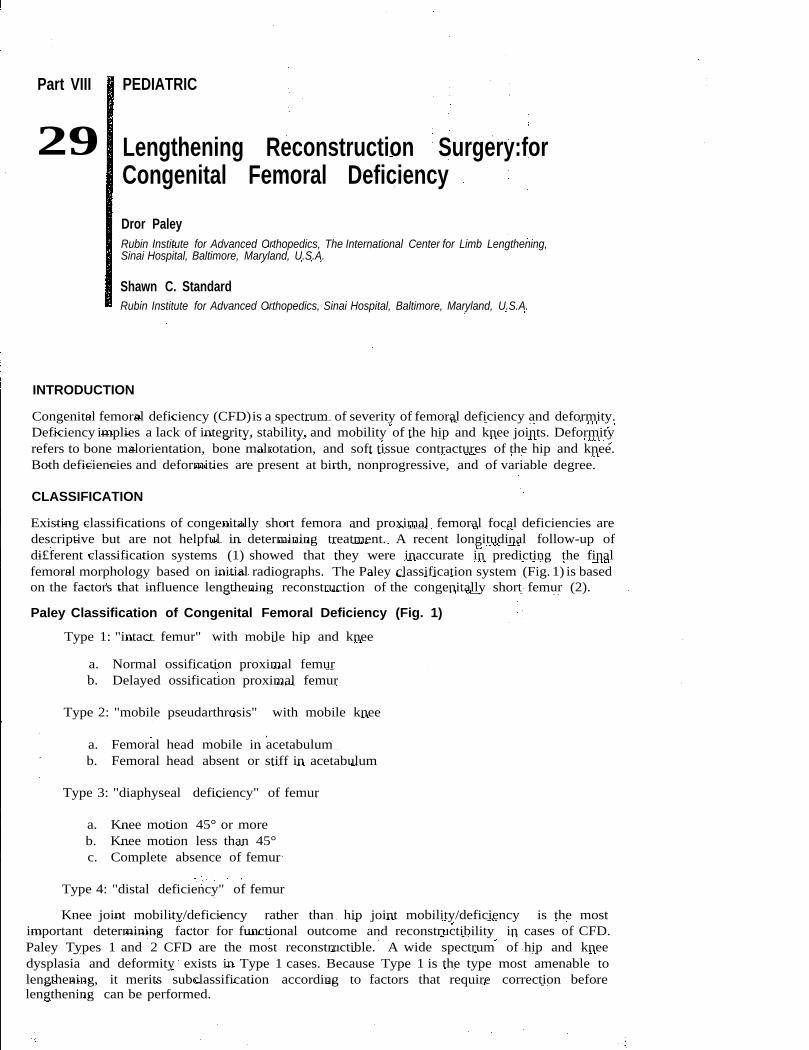
Existing classifications of congenitally short femora and proximal femoral focal deficiencies aredescriptive but are not helpful in determining treatment. A recent longitudinal follow-up ofdi£ferent classification systems (1) showed that they were inaccurate in predicting the finalfemoral morphology based on initial radiographs. The Paley classification system (Fig. 1) is basedon the factors that influence lengthening reconstruction of the congenitally short femur (2).
Paley Classification of Congenital Femoral Deficiency (Fig. 1)
Type 1: "intact femur" with mobile hip and knee
a. Normal ossification proximal femurb. Delayed ossification proximal femur
Type 2: "mobile pseudarthrosis" with mobile knee
a. Femoral head mobile in acetabulumb. Femoral head absent or stiff in acetabulum
Type 3: "diaphyseal deficiency" of femur
a. Knee motion 45° or moreb. Knee motion less than 45°c. Complete absence of femur
Type 4: "distal deficiency" of femur
Knee joint mobility/deficiency rather than hip joint mobility/deficiency is the mostimportant determining factor for functional outcome and reconstructibility in cases of CFD.Paley Types 1 and 2 CFD are the most reconstructible. A wide spectrum of hip and kneedysplasia and deformity exists in Type 1 cases. Because Type 1 is the type most amenable tolengthening, it merits subclassification according to factors that require correction beforelengthening can be performed.
394
a. Normal ossification
Paley and Standard
Type 1: Intact Femur with Mobile Hip and Knee
b. Delayed ossificationSubtrochanteric type
b. Delayed ossificationNeck type
Type 2: Mobile Pseudarthrosis with Mobile Knee
a. Femoral head b. Femoral head absent ormobile in acetablum stiff in acetabulum
Type 3: Diaphyseal Deficiency of Femur Type 4: Distal Deficiency of Femur
a.Knee motion;0,450
b. Knee motion c. Complete absence<450 of femur
Figure 1 Paley classification of congenital femoral deficiency. Source: Figure copyrighted to the Rubin Institute forAdvanced Orthopedics.
TYPE 1 CONGENITAL FEMORAL DEFICIENCY: INTACT FEMURHip and Knee Considerations
Type 1 CFD is the type that is most reconstructible. Before lengthening, significant bone defor-mities and soft tissue contractures of the hip and knee should be reconstructed. At the hip, ifthe acetabulum has a center edge (CE) angle of more than 200
, the neck shaft angle is morethan 110
0
, and the greater trochanter is not significantly overgrown such that the medial proxi-mal femoral angle (MPFA) is not less than 700
, no hip surgery is required before the firstlengthening. At the knee, if the fixed flexion deformity (FFD) is less than 100, the patella trackswith no subluxation laterally and no evidence of significant rotary subluxation or dislocation
Lengthening Reconstruction Surgery for Congenital Femoral Deficiency
(8)
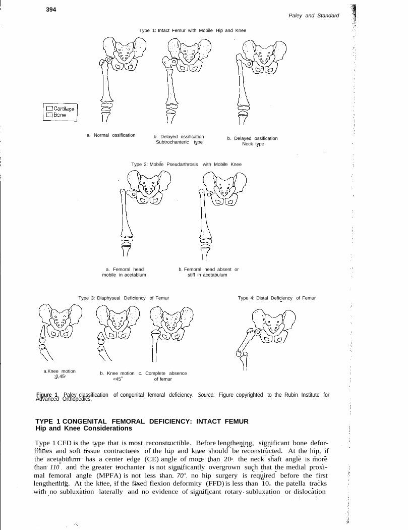
Figure 2 (A) Radiograph of patient with Paley Type 1a congenital femoral deficiency. The femora/neck is ossifiedand has significant coxa vara. (8) Radiograph shows initial lengthening with concurrent proximal subtrochantericosteotomy for coxa vara correction. The lengthening is performed through the distal osteotomy site, and the externalfixator bridges the knee joint to prevent subluxation and dislocation.
)f the tibia on the femur is present, the knee does not require surgical reconstruction beforeengthening (Fig. 2). It however, any of these criteria are not met, the hip and/or knee shouldJe reconstructed before the first lengthening.
l\cetabular Dysplasia
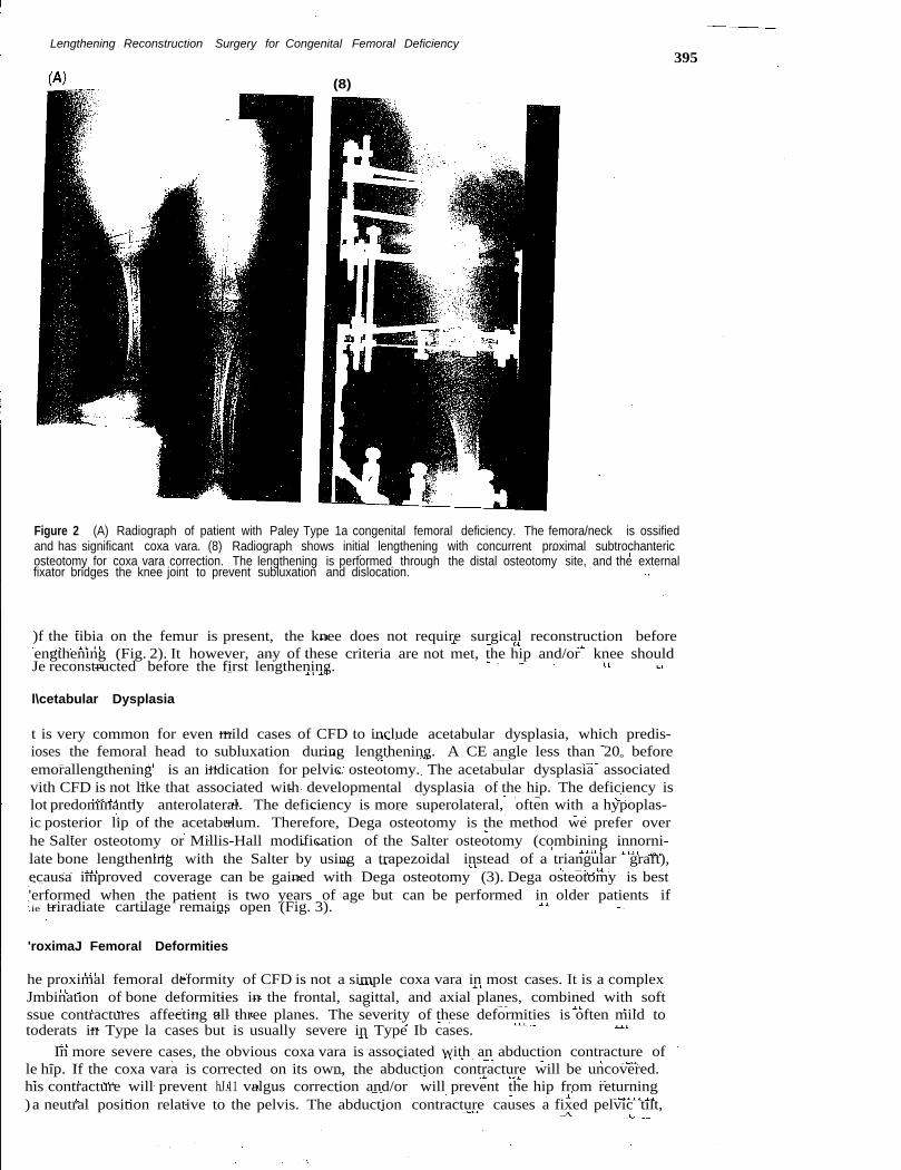
t is very common for even mild cases of CFD to include acetabular dysplasia, which predis-ioses the femoral head to subluxation during lengthening. A CE angle less than 200 beforeemorallengthening is an indication for pelvic osteotomy. The acetabular dysplasia associatedvith CFD is not like that associated with developmental dysplasia of the hip. The deficiency islot predominantly anterolateral. The deficiency is more superolateral, often with a hypoplas-ic posterior lip of the acetabulum. Therefore, Dega osteotomy is the method we prefer overhe Salter osteotomy or Millis-Hall modification of the Salter osteotomy (combining innorni-late bone lengthening with the Salter by using a trapezoidal instead of a triangular graft),ecausa improved coverage can be gained with Dega osteotomy (3). Dega osteotomy is best'erformed when the patient is two years of age but can be performed in older patients if.ie triradiate cartilage remains open (Fig. 3).
'roximaJ Femoral Deformities
he proximal femoral deformity of CFD is not a simple coxa vara in most cases. It is a complexJmbination of bone deformities in the frontal, sagittal, and axial planes, combined with softssue contractures affecting all three planes. The severity of these deformities is often mild totoderats in Type la cases but is usually severe in Type Ib cases.
In more severe cases, the obvious coxa vara is associated with an abduction contracture ofle hip. If the coxa vara is corrected on its own, the abduction contracture will be uncovered.his contracture will prevent hJ.11 valgus correction and/or will prevent the hip from returning) a neutral position relative to the pelvis. The abduction contracture causes a fixed pelvic tilt,
395
396Paley and Standard
(A) (B)
Figure 3 (A) Three-year-old female patient with Paley Type 1a congenital femoral deficiency and concurrent right hipdysplasia shown by the diagonal acetabular sourcil and a center edge angle of 80 (left center edge angle = 250).
(B) Postoperative radiograph after Dega osteotomy shows corrected dysplasic acetabulum.
which makes the limb length discrepancy (LLD) appear less than it was before surgery In thepresence of an open growth plate or a nonossified neck/ subtrochanteric segment, the abductioncontracture leads to recurrence of the coxa vara through these cartilaginous structures.
FFD of the hip usually accompanies severe coxa vara. The magnitude of the FFD often ismasked by extension deformity in the bone of the proximal femur. External rotation deformityof the distal relative to the proximal femur (retroversion) is always present because of a com-bination of bony torsion and contracture of the piriformis muscle. The correction of thesedeformities is accomplished with a new surgical procedure, the superhip procedure (SUPER isan acronym for Systematic Utilitarian Procedure for Extremity Reconstruction).
SUPERHIP PROCEDURE (FIG. 4)Step 1: Incision and Reflection of the Anterior Flap
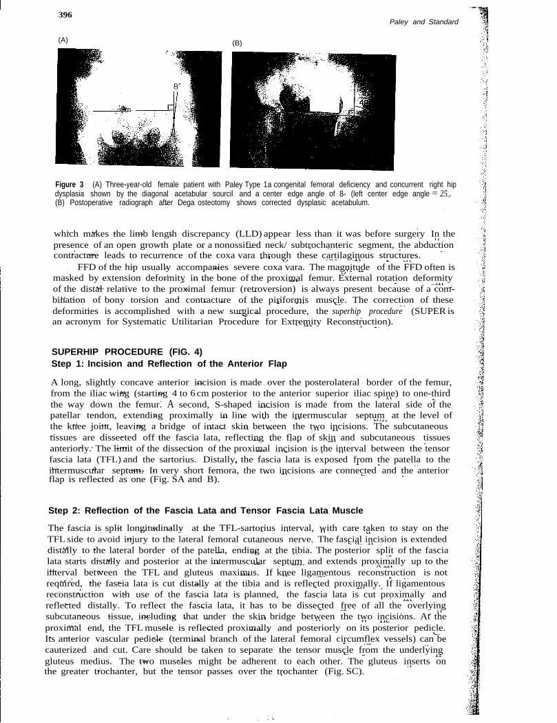
A long, slightly concave anterior incision is made over the posterolateral border of the femur,from the iliac wing (starting 4 to 6cm posterior to the anterior superior iliac spine) to one-thirdthe way down the femur. A second, S-shaped incision is made from the lateral side of thepatellar tendon, extending proximally in line with the intermuscular septum at the level ofthe knee joint, leaving a bridge of intact skin between the two incisions. The subcutaneoustissues are dissected off the fascia lata, reflecting the flap of skin and subcutaneous tissuesanteriorly. The limit of the dissection of the proximal incision is the interval between the tensorfascia lata (TFL) and the sartorius. Distally, the fascia lata is exposed from the patella to theintermuscular septum. In very short femora, the two incisions are connected and the anteriorflap is reflected as one (Fig. SA and B).
Step 2: Reflection of the Fascia Lata and Tensor Fascia Lata Muscle
The fascia is split longitudinally at the TFL-sartorius interval, with care taken to stay on theTFL side to avoid injury to the lateral femoral cutaneous nerve. The fascial incision is extendeddistally to the lateral border of the patella, ending at the tibia. The posterior split of the fascialata starts distally and posterior at the intermuscular septum and extends proximally up to theinterval between the TFL and gluteus maximus. If knee ligamentous reconstruction is notrequired, the fascia lata is cut distally at the tibia and is reflected proximally. If ligamentousreconstruction with use of the fascia lata is planned, the fascia lata is cut proximally andreflected distally. To reflect the fascia lata, it has to be dissected free of all the overlyingsubcutaneous tissue, including that under the skin bridge between the two incisions. At theproximal end, the TFL muscle is reflected proximally and posteriorly on its posterior pedicle.Its anterior vascular pedicle (terminal branch of the lateral femoral circumflex vessels) can becauterized and cut. Care should be taken to separate the tensor muscle from the underlyinggluteus medius. The two muscles might be adherent to each other. The gluteus inserts onthe greater trochanter, but the tensor passes over the trochanter (Fig. SC).
Lengthening Reconstruction Surgery for Congenital Femoral Deficiency397
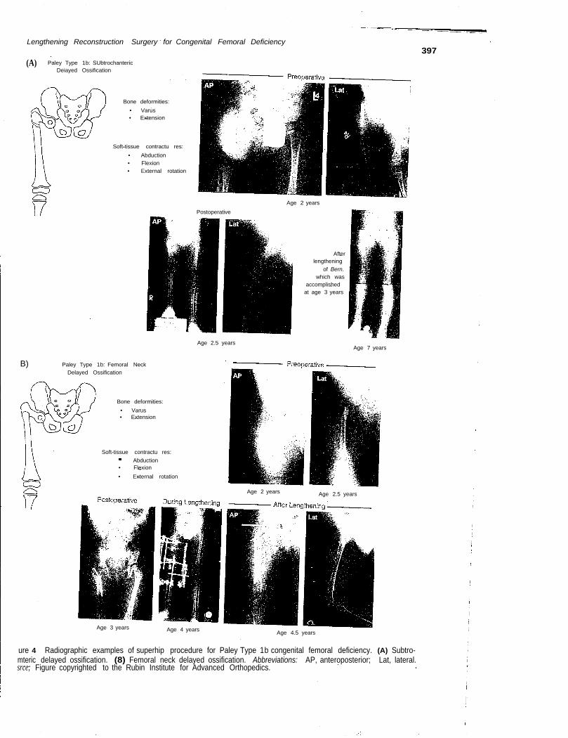
(A) Paley Type 1b: SUbtrochantericDeiayed Ossification
Bone deformities:• Varus• Extension
Soft-tissue contractu res:• Abduction• Flexion• External rotation
Age 2 yearsPostoperative
Afterlengthening
of Bern.which was
accomplishedat age 3 years
Age 2.5 yearsAge 7 years
B) Paley Type 1b: Femoral NeckDelayed Ossification
Bone deformities:• Varus• Extension
Soft-tissue contractu res:Abduction
• Flexion• External rotation
Age 2 years Age 2.5 years
Age 3 years Age 4 years Age 4.5 years
ure 4 Radiographic examples of superhip procedure for Paley Type 1b congenital femoral deficiency. (A) Subtro-mteric delayed ossification. (8) Femoral neck delayed ossification. Abbreviations: AP, anteroposterior; Lat, lateral.srce; Figure copyrighted to the Rubin Institute for Advanced Orthopedics.
398
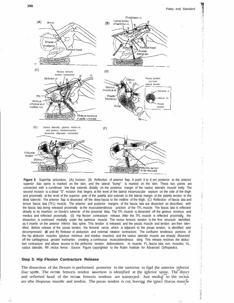
(C)Rectus femoris
tendon transected FL
(E) Vastus lateraiis, giuteus minim us,and giuteus mediusmuscles
dissected offgreater trochanter
Paley end Standard
(0)
Psoas tendoncut from
lateral border
Iliacus m.
Femoral n.
Psoastendon
(F)
Figure 5 Superhip procedure. (A) Incision. (8) Reflection of anterior flap. A point 4 to 6 em posterior to the anteriorsuperior iliac spine is marked on the skin, and the lateral "bump" is marked on the skin. These two points areconnected with a curvilinear line that extends distally on the posterior margin of the vastus lateralis muscle belly. Thesecond incision is a distal "S" incision that begins at the level of the lateral intramuscular septum on the side of the thighand proximally at the level of the superior pole of the patella and extends to the lateral margin of the patella tendon to thetibial tubercle. The anterior flap is dissected off the deep fascia to the midline of the thigh. (C) Reflection of fascia lata andtensor fascia lata (TFL) muscle. The anterior and posterior margins of the fascia lata are dissected as described, withthe fascia lata being released proximally at the musculotendinous junction of the TFL muscle. The fascia lata is reflecteddistally to its insertion on Gerdy's tubercle of the proximal tibia. The TFL muscle is dissected off the gluteus minimus andmedius and reflected proximally. (0) Hip flexion contracture release. After the TFL muscle is reflected proximally, thedissection is continued medially under the sartorius muscle. The rectus femoris tendon is the first structure identifiedas it inserts on the anterior inferior iliac spine. This tendon is released, and the psoas muscle and tendon are then iden-tified. Before release of the psoas tendon, the femoral nerve, which is adjacent to the psoas tendon, is identified anddecompressed. (E and F) Release of abduction and external rotation contracture. The confluent tendinous portions ofthe hip abductor muscles (gluteus minimus and medius muscles) and the vastus lateralis muscle are sharply dissectedoff the cartilaginous greater trochanter, creating a continuous musculotendinous sling. This release resolves the abduc-tion contracture and allows access to the piriformis tendon. Abbreviations: m, muscle; FL, fascia lata; mm, muscles; VL,vastus lateralis; RF, rectus femor. Source: Figure copyrighted to the Rubin Institute for Advanced Orthopedics.
Step 3: Hip Flexion Contracture Release
The dissection of the flexors is performed posterior to the sartorius to find the anterior inferioriliac spine. The rectus femoris tendon insertion is identified at the inferior spine. The directand reflected head of the rectus femoris tendons are transected. Just medial to the rectusare sthe iliopsoas muscle and tendon. The psoas tendon is cut, leaving the intact iliacus muscle
Lengthening Reconstruction Surgery for Congenital Femoral Deficiency399
fibers to bridge the gap. Care should be taken to avoid injury to the femoral nerve, which liesanterior to the medial edge of the iliacus muscle (the psoas tendon lies posteromedial to theiliacus muscle). Although the sartorius muscle might contribute to the flexion contracture, itis usually not tight. The anterior fascia of the thigh and the sartorius fascia might be tight.They might need to be released. The lateral femoral cutaneous nerve should be identifiedand protected before releasing these fasciae. The remaining flexion contracture is from the glu-teus medius and minimus muscles and is addressed in the next step of the procedure (Fig. 5D).
Step 4: Abduction and External Rotation Contracture Releases
The abductor tendons (gluteus medius and minirnus) insert into the greater trochanter andextend distally to become confluent with the quadriceps origin on the greater trochanter. Thetendinous portions of both muscle groups can be sharply dissected and reflected together, main-taining the longitudinal continuity of the musculotendinous units. Neither muscle group canretract or shorten. With this step, the posterior aspect of the glutei is identified and followedto the posterior border of the greater trochanter. The posterior border of the vastus lateralis atthe intermuscular septum is similarly identified and dissected free of the femur subperiosteally.This line is continued proximally along the posterior aspect of the greater trochanter. Becausethe tendinous covering of the trochanter is thin, it is important to peel a thin layer of cartilagewith the flap. The flap of the conjoint gluteus-quadriceps tendon is sharply dissected andreflected from posterior to anterior off the trochanter and then anteriorly off the intertrochantericline, leaving the anterior hip capsule intact. During this release, the piriformis tendon should beidentified and released from its trochanteric insertion. This permits the femur to rotate intern-ally.Once the abductor-quadriceps unit is free of the trochanter, the extension of the hip capsuleto the acetabulum of the hip is evident. It is important not to release this capsule from thetrochanter because doing so can lead to lateral subluxation. Because of the flexion contracture,femoral retroversion, and extension of the proximal femur, the greater trochanter is usuallylocated very posterior and medial to the more prominent lateral/lbump" (Fig. 5E and F).
Step 5: Proximal Femoral Osteotomy and Fixation
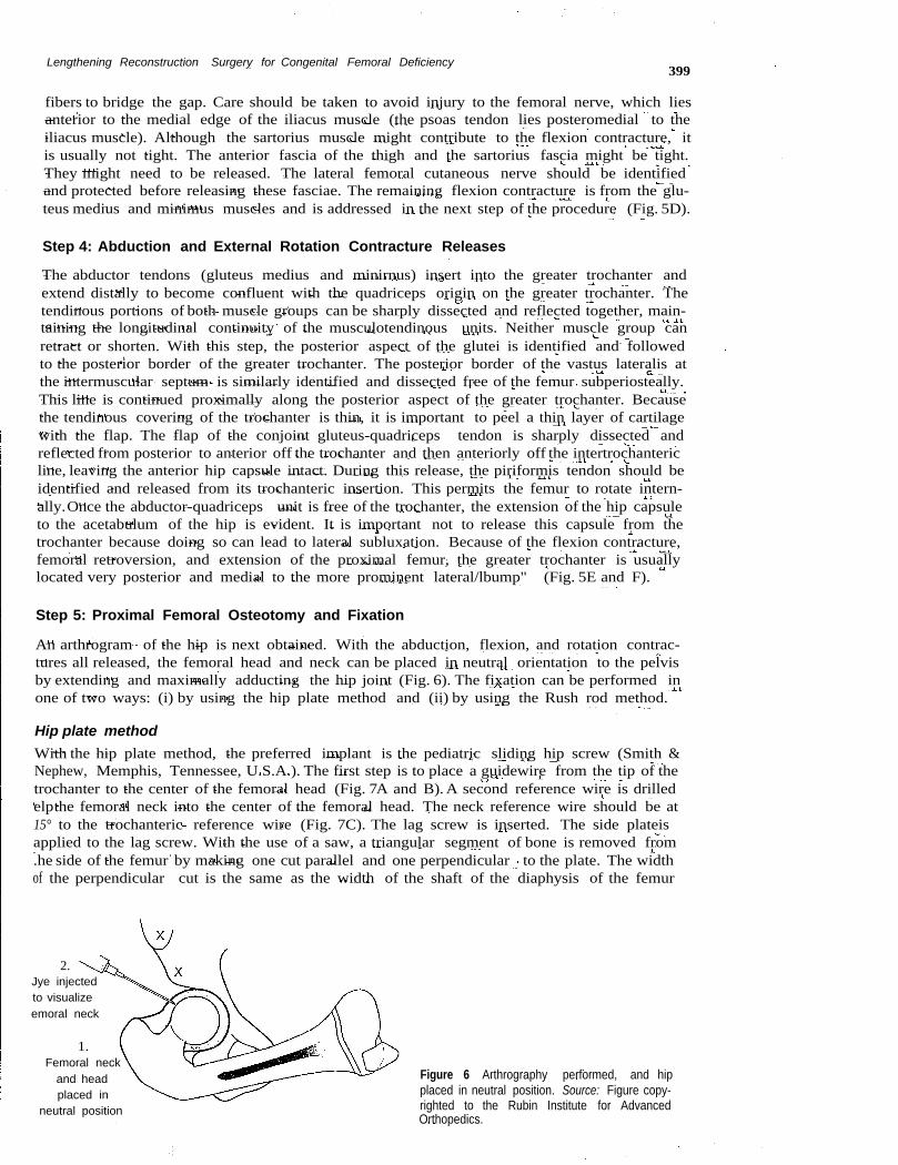
An arthrogram of the hip is next obtained. With the abduction, flexion, and rotation contrac-tures all released, the femoral head and neck can be placed in neutral orientation to the pelvisby extending and maximally adducting the hip joint (Fig. 6). The fixation can be performed inone of two ways: (i) by using the hip plate method and (ii) by using the Rush rod method.
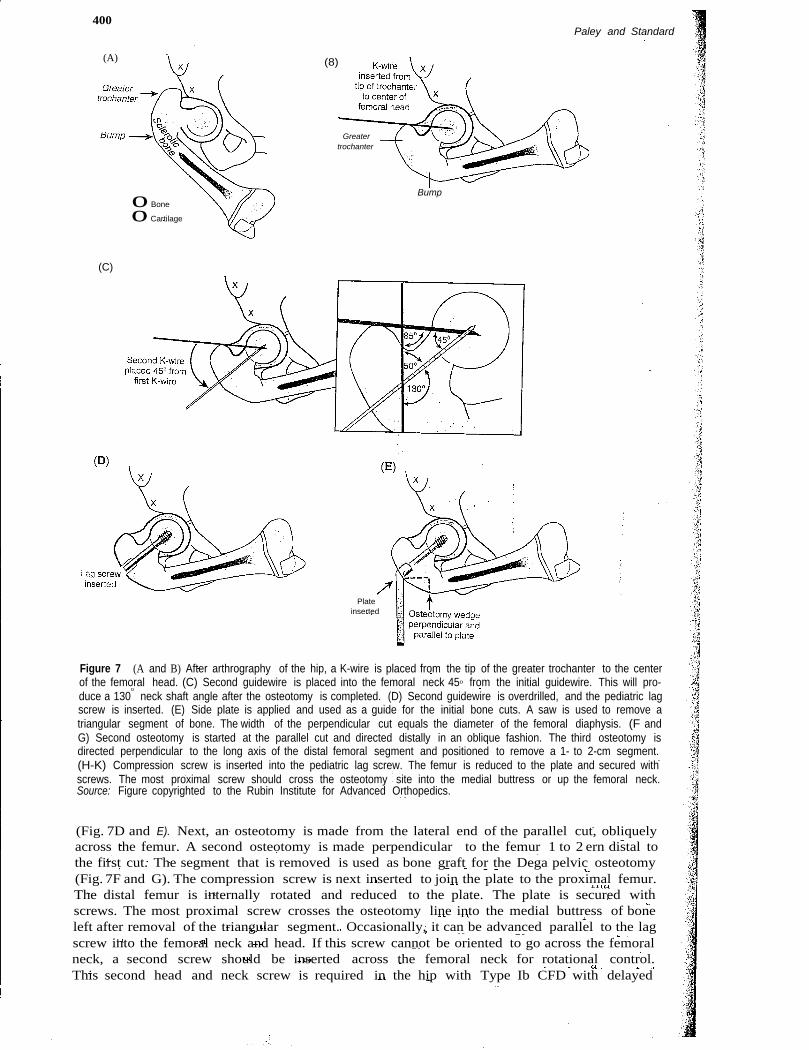
Hip plate methodWith the hip plate method, the preferred implant is the pediatric sliding hip screw (Smith &Nephew, Memphis, Tennessee, U.S.A.). The first step is to place a guidewire from the tip of thetrochanter to the center of the femoral head (Fig. 7A and B). A second reference wire is drilledelpthe femoral neck into the center of the femoral head. The neck reference wire should be at15° to the trochanteric reference wire (Fig. 7C). The lag screw is inserted. The side plateisapplied to the lag screw. With the use of a saw, a triangular segment of bone is removed from.he side of the femur by making one cut parallel and one perpendicular to the plate. The widthof the perpendicular cut is the same as the width of the shaft of the diaphysis of the femur
1.Femoral neck
and headplaced in
neutral position
Figure 6 Arthrography performed, and hipplaced in neutral position. Source: Figure copy-righted to the Rubin Institute for AdvancedOrthopedics.
2.Jye injectedto visualizeemoral neck
400Paley and Standard
(A)
Bump
(8)
Greatertrochanter
o Boneo Cartilage
(C)
Plateinserted
Figure 7 (A and B) After arthrography of the hip, a K-wire is placed from the tip of the greater trochanter to the centerof the femoral head. (C) Second guidewire is placed into the femoral neck 450 from the initial guidewire. This will pro-duce a 130
0
neck shaft angle after the osteotomy is completed. (D) Second guidewire is overdrilled, and the pediatric lagscrew is inserted. (E) Side plate is applied and used as a guide for the initial bone cuts. A saw is used to remove atriangular segment of bone. The width of the perpendicular cut equals the diameter of the femoral diaphysis. (F andG) Second osteotomy is started at the parallel cut and directed distally in an oblique fashion. The third osteotomy isdirected perpendicular to the long axis of the distal femoral segment and positioned to remove a 1- to 2-cm segment.(H-K) Compression screw is inserted into the pediatric lag screw. The femur is reduced to the plate and secured withscrews. The most proximal screw should cross the osteotomy site into the medial buttress or up the femoral neck.Source: Figure copyrighted to the Rubin Institute for Advanced Orthopedics.
(Fig. 7D and E). Next, an osteotomy is made from the lateral end of the parallel cut, obliquelyacross the femur. A second osteotomy is made perpendicular to the femur 1 to 2 ern distal tothe first cut. The segment that is removed is used as bone graft for the Dega pelvic osteotomy(Fig. 7F and G). The compression screw is next inserted to join the plate to the proximal femur.The distal femur is internally rotated and reduced to the plate. The plate is secured withscrews. The most proximal screw crosses the osteotomy line into the medial buttress of boneleft after removal of the triangular segment. Occasionally, it can be advanced parallel to the lagscrew into the femoral neck and head. If this screw cannot be oriented to go across the femoralneck, a second screw should be inserted across the femoral neck for rotational control.This second head and neck screw is required in the hip with Type Ib CFD with delayed
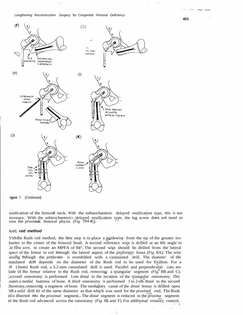
Lengthening Reconstruction Surgery for Congenital Femoral Deficiency
(H)
(J)
:igure 7 (Continued)
(G)
~
~For Degaosteotomy
(I)x
issification of the femoral neck. With the subtrochanteric delayed ossification type, this is notiecessary, With the subtrochanteric delayed ossification type, the lag screw does not need toross the proximal femoral physis (Fig. 7H-K).
Iust: rod method
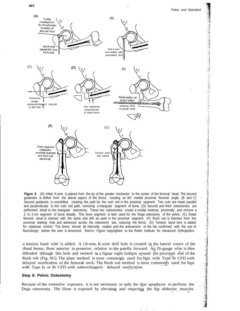
Viththe Rush rod method, the first step is to place a guidewire from the tip of the greater tro-hanter to the center of the femoral head. A second reference wire is drilled at an 850 angle to.ie first wire, to create an MPFA of 84°. The second wire should be drilled from the lateralspect of the femur to exit through the lateral aspect of the piriformis fossa (Fig. 8A). The wireassing through the piriformis is overdrilled with a cannulated drill. The diameter of themnulated drill depends on the diameter of the Rush rod to be used for fixation. For a/8
11
(3mm) Rush rod, a 3.2-mm cannulated drill is used. Parallel and perpendicular cuts arelade in the femur relative to the Rush rod, removing a triangular segment (Fig. 8B and C)..sccond osteotomy is performed 1em distal to the location of the triangular osteotomy. This.eates a medial buttress of bone. A third osteotomy is performed 1 to 2 em distal to the second3teotomy,removing a segment of bone. The medullary canal of the distal femur is drilled open'ith a solid drill bit of the same diameter as that which was used for the proximal end. The Rushid is inserted into the proximal segment. The distal segment is reduced to the proxima segmenttd the Rush rod advanced across the osteotomy (Fig. 8E and F). For additional rotatory control,
401
402Paley and Standard
(8)
2nd K-wireover-drilled withcannulated driJi
(C)(E)
Osteotomy Iwedge I
perpendiculaqand paraJielto drill hole t 2nd osteotomy
perpendicularto distal femur
stopping shortof growth plate
(G)
Tension bandwire added
Figure 8 (A) Initial K-wire is placed from the tip of the greater trochanter to the center of the femoral head. The secondguidewire is drilled from the lateral aspect of the femur, creating an 84° medial proximal femoral angle. (B and C)Second guidewire is overdrilled, creating the path for the rush rod in the proximal segment. Two cuts are made paralleland perpendicular to the rush rod path, removing a triangular segment of bone. (D) Second and third osteotomies areperformed distal to the triangular osteotomy. These two osteotomies create a medial buttress proximally and remove a1- to 2-cm segment of bone distally. This bone segment is later used for the Dega osteotomy of the pelvis. (E) Distalfemoral canal is reamed with the same size drill as used in the proximal segment. (F) Rush rod is inserted from theproximal starting hole and advanced across the osteotomy site, reducing the femur. (G) Tension band wire is addedfor rotational control. The femur should be internally rotated and the anteversion of the hip confirmed with the use offluoroscopy before the wire is tensioned. Source: Figure copyrighted to the Rubin Institute for Advanced Orthopedics.
a tension band wire is added. A l.S-mm K-wire drill hole is created in the lateral cortex of thedistal femur, from anterior to posterior, relative to the patella forward. An IS-gauge wire is thenthreaded through this hole and twisted in a figure eight fashion around the proximal end of theRush rod (Fig. SG). The plate method is most commonly used for hips with Type Ib CFD withdelayed ossification of the femoral neck. The Rush rod method is most commonly used for hipswith Type Ia or Ib CFD with subtrochanteric delayed ossification.
Step 6: Pelvic Osteotomy
Because of the extensive exposure, it is not necessary to split the iliac apophysis to perform theDega osteotomy. The ilium is exposed by elevating and retracting the hip abductor muscles

Lengthening Reconstruction Surgery for Congenital Femoral Deficiency 403
from the lateral wall of the ilium, starting anterior at the anterior inferior iliac spine and workingposteriorly. The dissection is continued back to the sciatic notch and distally toward the ischium.The dissection should not cross the triradiate cartilage. The osteotomy is curved along the lateralcortex from the anterior inferior iliac spine to triradiate cartilage posteriorly. The osteotomy isinclined toward the triradiate cartilage medially and should start 2 em above the joint. The oste-otomy is levered distally to cover the femoral head. The bone graft obtained from the femur isused to hold the osteotomy open and to level the sourcil (Fig. 9).
Step 7: Tendon Repair and Closure
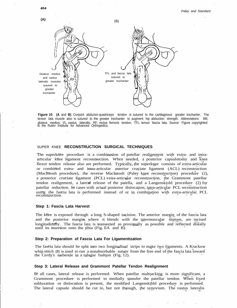
The conjoint abductor-quadriceps tendon is sutured directly into the cartilaginous greatertrochanter with absorbable suture. This is performed with the femur in neutral abduction.The TFL is also sutured to the greater trochanter to augment the abduction strength of thehip. The incision is closed in layers, including Scarpa's fascia and subcuticular and skin layers.A suction drain is used because of the large anterior flap and is left in place until the drainingstops, which can take several days. The patient is placed in a spica cast in full hip extensionand neutral abduction and rotation for six weeks (Fig. 10).
INSTABILITY OF PATELLA OR TIBIA AND KNEE FLEXION CONTRACTURE
Instability of the patella necessitates performing a stabilizing-realigning procedure beforelengthening. Isolated anteroposterior instability of the tibiofemoral joint without knee jointdislocation or rotatory subluxation does not need to be addressed before lengthening. Isolatedsubluxation or dislocation of the patella should be treated before lengthening. This reconstruc-tion is based on a combination of elements from the Langenskiold procedure (3) (designed forcongenital dislocation of the patella), the MacIntosh procedure (4) (extra-articular reconstruc-tion for anterior cruciate deficiency), and the Grammont procedure (5) (designed for recurrentdislocation of the patella). This knee stabilization procedure can be performed at the same timeas the pelvic osteotomy or superhip procedure. When knee stabilization is performed togetherwith a superhip procedure, the fascia lata is reflected from proximal to distal and is used forthe ligamentous reconstruction.
(A) (B)
Dega osteotomyto be performed
Osteotomy levered xdistally to coverfemoral head
Bone graftfrom femur
then inserted
Figure 9 (A and B) Dega osteotomy is performed, exposing the outer table of the iliac crest under the abductormuscles without splitting the iliac apophysis. The osteotomy curves from the anterior inferior iliac spine to the trir-adiate cartilage along the lateral cortex of the iliac crest. The bone segment from the femur is used as the bone blockfor the Dega osteotomy. Source: Figure copyrighted to the Rubin Institute for Advanced Orthopedics.
TFL and fascia latasutured to
greater trochanter
404
(A)
Gluteus mediusand vastus
lateralis musclessutured to
greatertrochanter
Paley and Standard
(B)
Figure 10 (A and B) Conjoint abductor-quadriceps tendon is sutured to the cartilaginous greater trochanter. Thetensor lata muscle also is sutured to the greater trochanter to augment hip abduction strength. Abbreviations: 8M,gluteus medius; VL, vastus lateralis; RF, rectus femoris tendon; TFL, tensor fascia lata. Source: Figure copyrightedto the Rubin Institute for Advanced Orthopedics.
SUPER KNEE RECONSTRUCTION SURGICAL TECHNIQUES
The superknee procedure is a combination of patellar realignment with extra- and intra-articular knee ligament reconstruction. When needed, a posterior capsulotomy and kneeflexor tendon release also are performed. Typically, the superknee consists of extra-articularor combined extra- and intra-articular anterior cruciate ligament (ACL) reconstruction(MacIntosh procedure), the reverse MacIntosh (Paley knee reconstruction) procedure (2),a posterior cruciate ligament (PCL) extra-articular reconstruction, the Grammont patellartendon realignment, a lateral release of the patella, and a Langenskiold procedure (2) forpatellar reduction. In cases with actual posterior dislocation, intra-articular PCL reconstructionusing the fascia lata is performed instead of or in combination with extra-articular PCLreconstruction.
Step 1: Fascia Lata Harvest
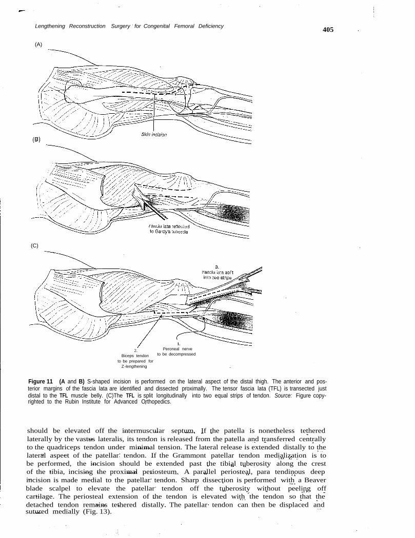
The knee is exposed through a long S-shaped incision. The anterior margin of the fascia lataand the posterior margin, where it blends with the intermuscular septum, are incisedlongitudinally. The fascia lata is transected as proximally as possible and reflected distallyuntil its insertion onto the tibia (Fig. llA and B).
Step 2: Preparation of Fascia Lata For Ligamentization
The fascia lata should be split into two longitudinal strips to make two ligaments. A Krackowwhip stitch (8) is used to run a nonabsorbable suture from the free end of the fascia lata towardthe Cerdy's turbercle in a tubular fashion (Fig. 12).
Step 3: Lateral Release and Grammont Patellar Tendon Realignment
In all cases, lateral release is performed. When patellar maltracking is more significant, aGrammont procedure is performed to medially transfer the patellar tendon. When fixedsubluxation or dislocation is present, the modified Langenskibld procedure is performed.The lateral capsule should be cut to, but not through, the synovium. The vastus lateralis
Lengthening Reconstruction Surgery for Congenital Femoral Deficiency 405
(A)
(C)
2.Biceps tendon
to be prepared forZ-lengthening
1.Peroneal nerve
to be decompressed
Figure 11 (A and B) S-shaped incision is performed on the lateral aspect of the distal thigh. The anterior and pos-terior margins of the fascia lata are identified and dissected proximally. The tensor fascia lata (TFL) is transected justdistal to the TFL muscle belly. (C)The TFL is split longitudinally into two equal strips of tendon. Source: Figure copy-righted to the Rubin Institute for Advanced Orthopedics.
should be elevated off the intermuscular septum. If the patella is nonetheless tetheredlaterally by the vastus lateralis, its tendon is released from the patella and transferred centrallyto the quadriceps tendon under minimal tension. The lateral release is extended distally to thelateral aspect of the patellar tendon. If the Grammont patellar tendon medialization is tobe performed, the incision should be extended past the tibial tuberosity along the crestof the tibia, incising the proximal periosteum. A parallel periosteal, para tendinous deepincision is made medial to the patellar tendon. Sharp dissection is performed with a Beaverblade scalpel to elevate the patellar tendon off the tuberosity without peeling offcartilage. The periosteal extension of the tendon is elevated with the tendon so that thedetached tendon remains tethered distally. The patellar tendon can then be displaced andsutured medially (Fig. 13).

406 Paley and Standard
Capsule openedbilaterally
Fascia latatabularized
Figure 12 Tendons are then prepared with a Krackow whipstitch to form a tubular graft. Source: Figure copyrightedto the Rubin Institute for Advanced Orthopedics.
Step 4: Macintosh Intra- and/or Extra-Articular Anterior CollateralLigament Reconstruction
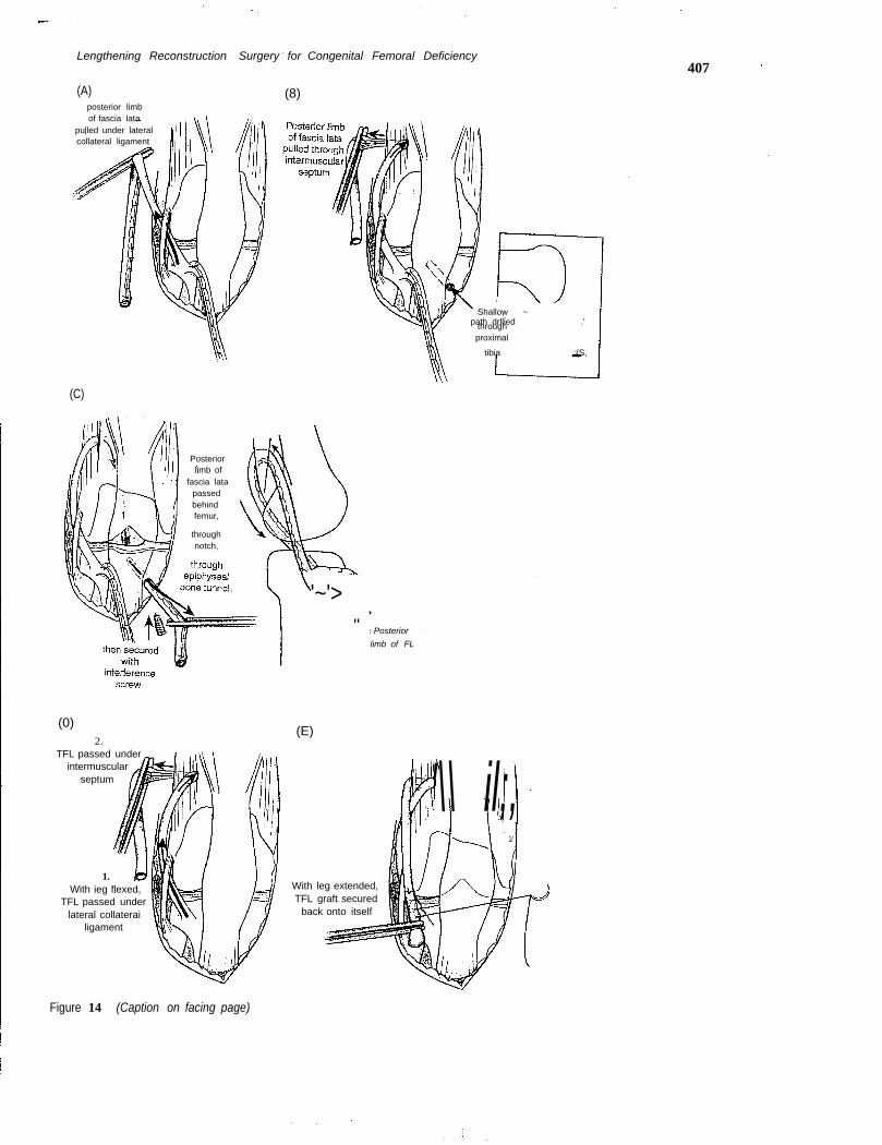
The lateral collateral ligament (LCL) is identified. A tunnel is made under this ligament with-out entering the knee joint (Fig. 14A). Another tunnel is made subperiosteallv, from anterior
Grammont elevationof patellar ligament
Figure 13 Grammont patellartendon medialization is performedby incising the medial and lateralborders of the patella tendon pastthe tibial tubercle. The patella ten-don is elevated off the tibialtubercle apophysis with an exten-sion of periosteum that remainsintact distally. The patella tendoncan then be shifted medially. Source:Figure copyrighted to the RubinInstitute for Advanced Orthopedics.
Lengthening Reconstruction Surgery for Congenital Femoral Deficiency
(A)posterior limbof fascia lata
pulled under lateralcollateral ligament
(8)
Shallow ~path drllied .'throughproximal
tibia :(S,
(C)
Posteriorlimb of
fascia latapassedbehindfemur,
throughnotch,
'~'>,
" .. Posteriorlimb of FL
(0) (E)2.
TFL passed underintermuscular
septum '\\ il;,1/
1.With ieg flexed,
TFL passed underlateral collaterai
ligament
With leg extended,TFL graft securedback onto itself
Figure 14 (Caption on facing page)
407
408 Paley and Standard
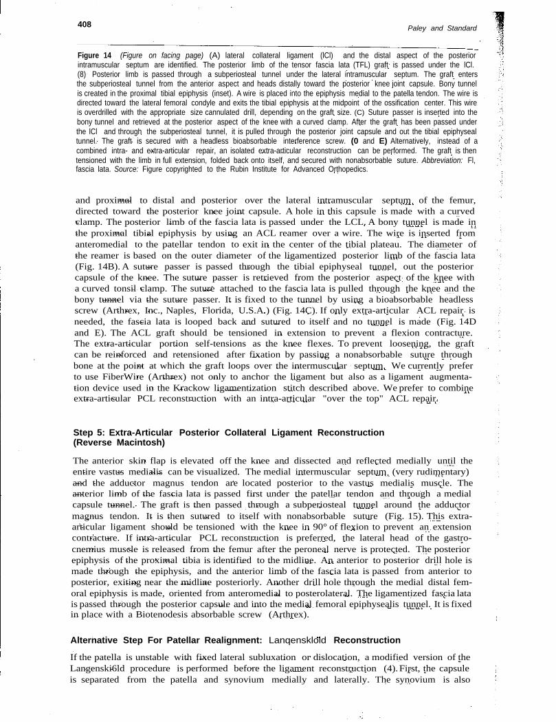
Figure 14 (Figure on facing page) (A) lateral collateral ligament (lCl) and the distal aspect of the posteriorintramuscular septum are identified. The posterior limb of the tensor fascia lata (TFL) graft is passed under the lCl.(8) Posterior limb is passed through a subperiosteal tunnel under the lateral intramuscular septum. The graft entersthe subperiosteal tunnel from the anterior aspect and heads distally toward the posterior knee joint capsule. Bony tunnelis created in the proximal tibial epiphysis (inset). A wire is placed into the epiphysis medial to the patella tendon. The wire isdirected toward the lateral femoral condyle and exits the tibial epiphysis at the midpoint of the ossification center. This wireis overdrilled with the appropriate size cannulated drill, depending on the graft size. (C) Suture passer is inserted into thebony tunnel and retrieved at the posterior aspect of the knee with a curved clamp. After the graft has been passed underthe lCl and through the subperiosteal tunnel, it is pulled through the posterior joint capsule and out the tibial epiphysealtunnel. The graft is secured with a headless bioabsorbable interference screw. (0 and E) Alternatively, instead of acombined intra- and extra-articular repair, an isolated extra-articular reconstruction can be performed. The graft is thentensioned with the limb in full extension, folded back onto itself, and secured with nonabsorbable suture. Abbreviation: Fl,fascia lata. Source: Figure copyrighted to the Rubin Institute for Advanced Orthopedics.
and proximal to distal and posterior over the lateral intramuscular septum of the femur,directed toward the posterior knee joint capsule. A hole in this capsule is made with a curvedclamp. The posterior limb of the fascia lata is passed under the LCL. A bony tunnel is made inthe proximal tibial epiphysis by using an ACL reamer over a wire. The wire is inserted fromanteromedial to the patellar tendon to exit in the center of the tibial plateau. The diameter ofthe reamer is based on the outer diameter of the ligamentized posterior limb of the fascia lata(Fig. 14B). A suture passer is passed through the tibial epiphyseal tunnel, out the posteriorcapsule of the knee. The suture passer is retrieved from the posterior aspect of the knee witha curved tonsil clamp. The suture attached to the fascia lata is pulled through the knee and thebony tunnel via the suture passer. It is fixed to the tunnel by using a bioabsorbable headlessscrew (Arthrex, Inc., Naples, Florida, U.S.A.) (Fig. 14C). If only extra-articular ACL repair isneeded, the fascia lata is looped back and sutured to itself and no tunnel is made (Fig. 14Dand E). The ACL graft should be tensioned in extension to prevent a flexion contracture.The extra-articular portion self-tensions as the knee flexes. To prevent loosening, the graftcan be reinforced and retensioned after fixation by passing a nonabsorbable suture throughbone at the point at which the graft loops over the intermuscular septum. We currently preferto use FiberWire (Arthrex) not only to anchor the ligament but also as a ligament augmenta-tion device used in the Krackow ligamentization stitch described above. We prefer to combineextra-articular PCL reconstruction with an intra-articular "over the top" ACL repair.
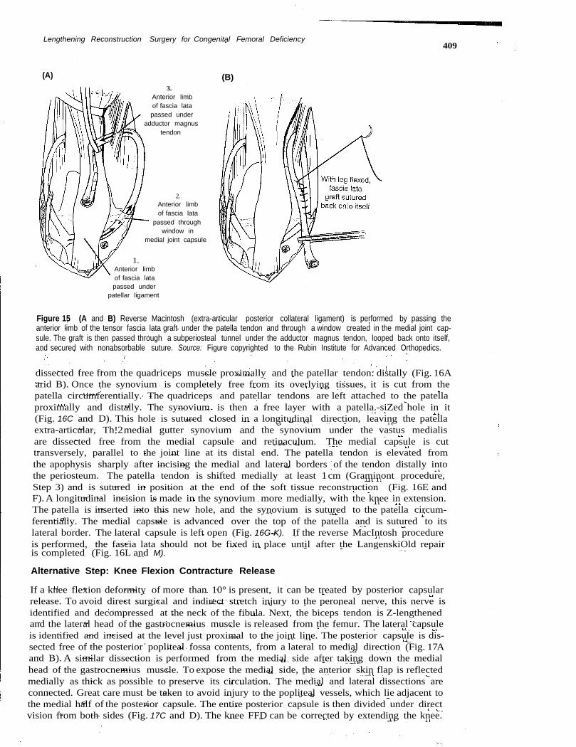
Step 5: Extra-Articular Posterior Collateral Ligament Reconstruction(Reverse Macintosh)
The anterior skin flap is elevated off the knee and dissected and reflected medially until theentire vastus medialis can be visualized. The medial intermuscular septum (very rudimentary)and the adductor magnus tendon are located posterior to the vastus medialis muscle. Theanterior limb of the fascia lata is passed first under the patellar tendon and through a medialcapsule tunnel. The graft is then passed through a subperiosteal tunnel around the adductormagnus tendon. It is then sutured to itself with nonabsorbable suture (Fig. 15). This extra-articular ligament should be tensioned with the knee in 90° of flexion to prevent an extensioncontracture. If intra-articular PCL reconstruction is preferred, the lateral head of the gastro-cnemius muscle is released from the femur after the peroneal nerve is protected. The posteriorepiphysis of the proximal tibia is identified to the midline. An anterior to posterior drill hole ismade through the epiphysis, and the anterior limb of the fascia lata is passed from anterior toposterior, exiting near the midline posteriorly. Another drill hole through the medial distal fem-oral epiphysis is made, oriented from anteromedial to posterolateral. The ligamentized fascia latais passed through the posterior capsule and into the medial femoral epiphysealis tunnel. It is fixedin place with a Biotenodesis absorbable screw (Arthrex).
Alternative Step For Patellar Realignment: Lanqensklold Reconstruction
If the patella is unstable with fixed lateral subluxation or dislocation, a modified version of theLangenski6ld procedure is performed before the ligament reconstruction (4). First, the capsuleis separated from the patella and synovium medially and laterally. The synovium is also
Lengthening Reconstruction Surgery for Congenital Femoral Deficiency
(A)3.
Anterior limbof fascia latapassed under
adductor magnustendon
2.Anterior limbof fascia lata
passed throughwindow in
medial joint capsule
1.Anterior limbof fascia latapassed under
patellar ligament
409
(B)
Figure 15 (A and B) Reverse Macintosh (extra-articular posterior collateral ligament) is performed by passing theanterior limb of the tensor fascia lata graft under the patella tendon and through a window created in the medial joint cap-sule. The graft is then passed through a subperiosteal tunnel under the adductor magnus tendon, looped back onto itself,and secured with nonabsorbable suture. Source: Figure copyrighted to the Rubin Institute for Advanced Orthopedics.
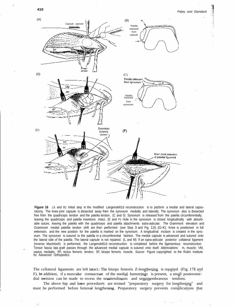
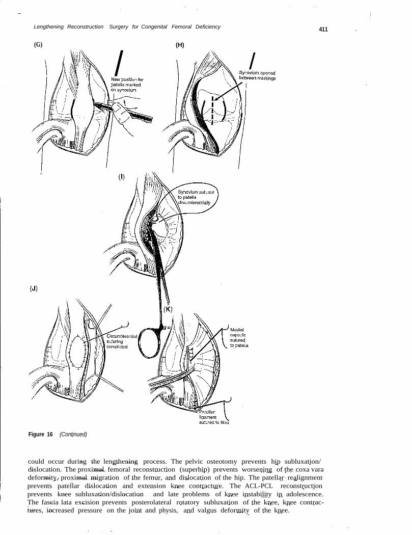
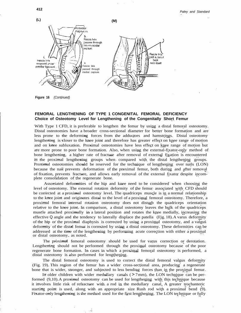
dissected free from the quadriceps muscle proximally and the patellar tendon: distally (Fig. 16Aarid B). Once the synovium is completely free from its overlying tissues, it is cut from thepatella circumferentially. The quadriceps and patellar tendons are left attached to the patellaproximally and distally. The synovium is then a free layer with a patella.-siZed hole in it(Fig. 16C and D). This hole is sutured closed in a longitudinal direction, leaving the patellaextra-articular, Th!2medial gutter synovium and the synovium under the vastus medialisare dissected free from the medial capsule and retinaculum. The medial capsule is cuttransversely, parallel to the joint line at its distal end. The patella tendon is elevated fromthe apophysis sharply after incising the medial and lateral borders of the tendon distally intothe periosteum. The patella tendon is shifted medially at least 1cm (Graminont procedure,Step 3) and is sutured in position at the end of the soft tissue reconstruction (Fig. 16E andF). A longitudinal incision is made in the synovium more medially, with the knee in extension.The patella is inserted into this new hole, and the synovium is sutured to the patella circum-ferentially. The medial capsule is advanced over the top of the patella and is sutured to itslateral border. The lateral capsule is left open (Fig. 16G-K). If the reverse MacIntosh procedureis performed, the fascia lata should not be fixed in place until after the LangenskiOld repairis completed (Fig. 16L and M).
Alternative Step: Knee Flexion Contracture Release
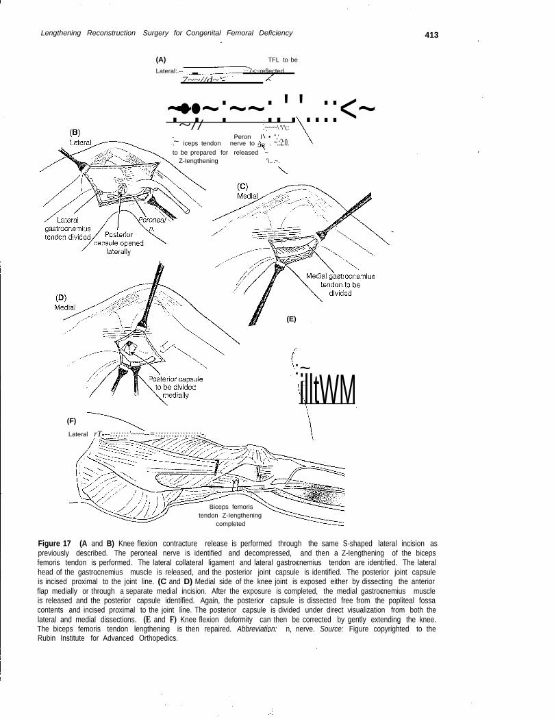
If a knee flexion deformity of more than 10° is present, it can be treated by posterior capsularrelease. To avoid direct surgical and indirect stretch injury to the peroneal nerve, this nerve isidentified and decompressed at the neck of the fibula. Next, the biceps tendon is Z-lengthenedand the lateral head of the gastrocnemius muscle is released from the femur. The lateral capsuleis identified and incised at the level just proximal to the joint line. The posterior capsule is dis-sected free of the posterior popliteal fossa contents, from a lateral to medial direction (Fig. 17Aand B). A similar dissection is performed from the medial side after taking down the medialhead of the gastrocnemius muscle. To expose the medial side, the anterior skin flap is reflectedmedially as thick as possible to preserve its circulation. The medial and lateral dissections areconnected. Great care must be taken to avoid injury to the popliteal vessels, which lie adjacent tothe medial half of the posterior capsule. The entire posterior capsule is then divided under directvision from both sides (Fig. 17C and D). The knee FFD can be corrected by extending the knee.
410 Paley and Standard
(A) (8)Capsule opened
bilaterally Patellareleased
fromcapsule
(0)
(E)
(C)
Patellareleased __
fromsynovium
Figure 16 (A and B) Initial step in the modified Langenski61d reconstruction is to perform a medial and lateral capsu-lotomy. The knee joint capsule is dissected away from the synovium medially and laterally. The synovium also is dissectedfree from the quadriceps tendon and the patella tendon. (C and 0) Synovium is released from the patella circumferentially,leaving the quadriceps and patella insertions intact. (E and F) Hole in the synovium is closed longitudinally with absorb-able suture, leaving the patella with the quadriceps and patella attachments extra-articular. The Grammont elevation andGrammont medial patellar tendon shift are then performed (see Step 3 and Fig. 13). (G-K) Knee is positioned in fullextension, and the new position for the patella is marked on the synovium. A longitudinal incision is created in the sync-vium. The synovium is sutured to the patella in a circumferential fashion. The medial capsule is advanced and sutured ontothe lateral side of the patella. The lateral capsule is not repaired. (L and M) If an extra-articular posterior collateral ligament(reverse Macintosh) is performed, the Langenski61d reconstruction is completed before the ligamentous reconstruction.Tensor fascia lata graft passes through the advanced medial capsule is sutured onto itself. Abbreviations: m, muscle; VM,vastus medialis; RF, rectus femoris tendon; SF, biceps femoris muscle. Source: Figure copyrighted to the Rubin Institutefor Advanced Orthopedics.
The collateral ligaments are left intact. The biceps femoris Z-lengthening is repaired (Fig. 17E andF). In addition, if a muscular contracture of the medial hamstrings is present, a small posterome-dial incision can be made to recess the semitendinosis and semimembranous tendons.
The above hip and knee procedures are termed "preparatory surgery for lengthening" andmust be performed before femoral lengthening. Preparatory surgery prevents complications that
Lengthening Reconstruction Surgery for Congenital Femoral Deficiency 411
(G) (H)
/ /
Figure 16 (Continued)
could occur during the lengthening process. The pelvic osteotomy prevents hip subluxation/dislocation. The proximal femoral reconstruction (superhip) prevents worsening of the coxa varadeformity, proximal migration of the femur, and dislocation of the hip. The patellar realignmentprevents patellar dislocation and extension knee contracture. The ACL-PCL reconstructionprevents knee subluxation/dislocation and late problems of knee instability in adolescence.The fascia lata excision prevents posterolateral rotatory subluxation of the knee, knee contrac-tures, increased pressure on the joint and physis, and valgus deformity of the knee.
412
(L)
2.Fascia latapassed throughmedial capsuie
Figure 16 (Continued)
Paley and Standard
(M)
FEMORAL LENGTHENING OF TYPE 1 CONGENITAL FEMORAL DEFICIENCYChoice of Osteotomy Level for Lengthening of the Congenitally Short Femur
With Type 1 CFD, it is preferable to lengthen the femur by using a distal femoral osteotomy.Distal osteotomies have a broader cross-sectional diameter for better bone formation and areless prone to the deforming forces from the adductors and hamstrings. Distal osteotomylengthening is closer to the knee joint and therefore has greater effect on knee range of motionand on knee subluxation. Proximal osteotomies have less effect on knee range of motion butare more prone to poor bone formation. Also, when using the external-fixator-only method ofbone lengthening, a higher rate of fracture after removal of external fixation is encounteredin the proximal lengthening groups when compared with the distal lengthening groups.Proximal osteotomies should be reserved for the technique of lengthening over nails (LON)because the nail prevents deformation of the proximal femur, both during and after removalof fixation, prevents fracture, and allows early removal of the external fixator despite incom-plete consolidation of the regenerate bone.
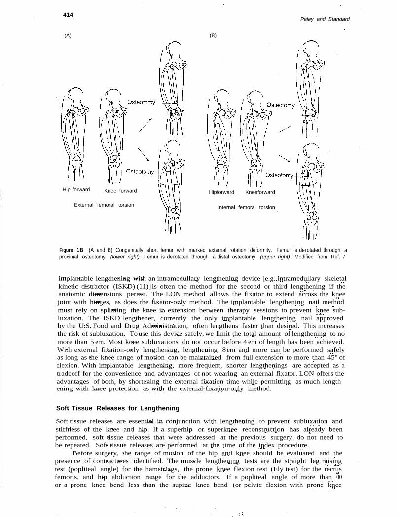
Associated deformities of the hip and knee need to be considered when choosing thelevel of osteotomy. The external rotation deformity of the femur associated with CFD shouldbe corrected at a proximal osteotomy level. The quadriceps muscle is in a normal relationshipto the knee joint and originates distal to the level of a proximal femoral osteotomy. Therefore, aproximal femoral internal rotation osteotomy does not disturb the quadriceps orientationrelative to the knee joint. In comparison, a distal osteotomy leaves the bulk of the quadricepsmuscle attached proximally in a lateral position and rotates the knee medially, increasing theeffective Q angle and the tendency to laterally displace the patella (Fig. 18).A varus deformityof the hip or the proximal diaphysis is corrected by using a proximal osteotomy, and a valgusdeformity of the distal femur is corrected by using a distal osteotomy. These deformities can beaddressed at the time of the lengthening by performing acute correction with either a proximalor distal osteotomy. as noted.
The proximal femoral osteotomy should be used for varus correction or derotation.Lengthening should not be performed through the proximal osteotomy because of the poorregenerate bone formation. In cases in which a proximal femoral osteotomy is performed, adistal osteotomy is also performed for lengthening.
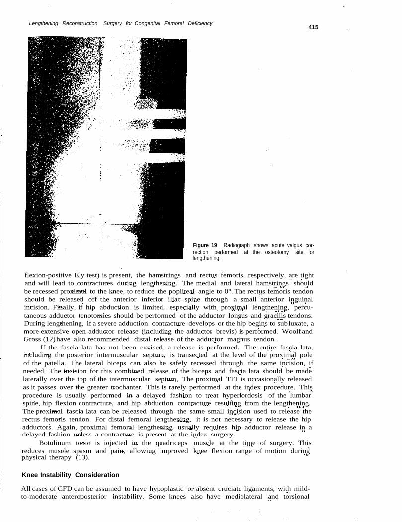
The distal femoral osteotomy is used to correct the distal femoral valgus deformity(Fig. 19). This region of the femur has a wider cross-sectional area, producing a regeneratebone that is wider, stronger, and subjected to less bending forces than in the proximal femur.
In older children with wider medullary canals (> 7mm), the LON technique can be per-formed (9,10).A proximal osteotomy can be used for lengthening with this technique becauseit involves little risk of refracture with a rod in the medullary canal. A greater trochantericstarting point is used, along with an appropriate size Rush rod with a proximal bend (9).Fixator-only lengthening is the method used for the first lengthening. The LON technique or fully
Lengthening Reconstruction Surgery for Congenital Femoral Deficiency 413
(A) TFL to be
Lateral:.-- _ ::;7<~reflected7~~//d~'-
~.••~.~:~~:.'.'..::<~~~// .~~~\ '\'\;:
- Peron I • ",'. iceps tendon nerve to . ~.:2:0.to be prepared for released ~
Z-Iengthening '\.. ~.
(F)
Lateral rT---:::::::'~~~--=::;;:::;:::;:::::-.
(E)
:~illtWM
Biceps femoristendon Z-Iengthening
completed
Figure 17 (A and B) Knee flexion contracture release is performed through the same S-shaped lateral incision aspreviously described. The peroneal nerve is identified and decompressed, and then a Z-Iengthening of the bicepsfemoris tendon is performed. The lateral collateral ligament and lateral gastrocnemius tendon are identified. The lateralhead of the gastrocnemius muscle is released, and the posterior joint capsule is identified. The posterior joint capsuleis incised proximal to the joint line. (C and D) Medial side of the knee joint is exposed either by dissecting the anteriorflap medially or through a separate medial incision. After the exposure is completed, the medial gastrocnemius muscleis released and the posterior capsule identified. Again, the posterior capsule is dissected free from the popliteal fossacontents and incised proximal to the joint line. The posterior capsule is divided under direct visualization from both thelateral and medial dissections. (E and F) Knee flexion deformity can then be corrected by gently extending the knee.The biceps femoris tendon lengthening is then repaired. Abbreviation: n, nerve. Source: Figure copyrighted to theRubin Institute for Advanced Orthopedics.
414
(A)
Hip forward
External femoral torsion
Knee forward
Paley and Standard
(8)
Hipforward Kneeforward
Internal femoral torsion
Figure 1 B (A and B) Congenitally short femur with marked external rotation deformity. Femur is derotated through aproximal osteotomy (lower right). Femur is derotated through a distal osteotomy (upper right). Modified from Ref. 7.
implantable lengthening with an intramedullary lengthening device [e.g.,intramedullary skeletalkinetic distractor (ISKD) (11)] is often the method for the second or third lengthening if theanatomic dimensions permit. The LON method allows the fixator to extend across the kneejoint with hinges, as does the fixator-only method. The implantable lengthening nail methodmust rely on splinting the knee in extension between therapy sessions to prevent knee sub-luxation. The ISKD lengthener, currently the only implantable lengthening nail approvedby the U.S. Food and Drug Administration, often lengthens faster than desired. This increasesthe risk of subluxation. Touse this device safely, we limit the total amount of lengthening to nomore than 5 ern. Most knee subluxations do not occur before 4 ern of length has been achieved.With external fixation-only lengthening, lengthening 8ern and more can be performed safelyas long as the knee range of motion can be maintained from full extension to more than 45° offlexion. With implantable lengthening, more frequent, shorter lengthenings are accepted as atradeoff for the convenience and advantages of not wearing an external fixator. LON offers theadvantages of both, by shortening the external fixation time while permitting as much length-ening with knee protection as with the external-fixation-only method.
Soft Tissue Releases for Lengthening
Soft tissue releases are essential in conjunction with lengthening to prevent subluxation andstiffness of the knee and hip. If a superhip or superknee reconstruction has already beenperformed, soft tissue releases that were addressed at the previous surgery do not need tobe repeated. Soft tissue releases are performed at the time of the index procedure.
Before surgery, the range of motion of the hip and knee should be evaluated and thepresence of contractures identified. The muscle lengthening tests are the straight leg raisingtest (popliteal angle) for the hamstrings, the prone knee flexion test (Ely test) for the rectusfemoris, and hip abduction range for the adductors. If a popliteal angle of more than 00or a prone knee bend less than the supine knee bend (or pelvic flexion with prone knee
Lengthening Reconstruction Surgery for Congenital Femoral Deficiency415
Figure 19 Radiograph shows acute valgus cor-rection performed at the osteotomy site forlengthening,
flexion-positive Ely test) is present, the hamstrings and rectus femoris, respectively, are tightand will lead to contractures during lengthening. The medial and lateral hamstrings shouldbe recessed proximal to the knee, to reduce the popliteal angle to 0°. The rectus femoris tendonshould be released off the anterior inferior iliac spine through a small anterior inguinalincision. Finally, if hip abduction is limited, especially with proximal lengthening, percu-taneous adductor tenotomies should be performed of the adductor longus and gracilis tendons.During lengthening, if a severe adduction contracture develops or the hip begins to subluxate, amore extensive open adductor release (including the adductor brevis) is performed. Woolf andGross (12)have also recommended distal release of the adductor magnus tendon.
If the fascia lata has not been excised, a release is performed. The entire fascia lata,including the posterior intermuscular septum, is transected at the level of the proximal poleof the patella. The lateral biceps can also be safely recessed through the same incision, ifneeded. The incision for this combined release of the biceps and fascia lata should be madelaterally over the top of the intermuscular septum. The proximal TFL is occasionally releasedas it passes over the greater trochanter. This is rarely performed at the index procedure. Thisprocedure is usually performed in a delayed fashion to treat hyperlordosis of the lumbarspine, hip flexion contracture, and hip abduction contracture resulting from the lengthening.The proximal fascia lata can be released through the same small incision used to release therectus femoris tendon. For distal femoral lengthening, it is not necessary to release the hipadductors. Again, proximal femoral lengthening usually requires hip adductor release in adelayed fashion unless a contracture is present at the index surgery.
Botulinum toxin is injected in the quadriceps muscle at the time of surgery. Thisreduces muscle spasm and pain, allowing improved knee flexion range of motion duringphysical therapy (13).
Knee Instability Consideration
All cases of CFD can be assumed to have hypoplastic or absent cruciate ligaments, with mild-to-moderate anteroposterior instability. Some knees also have mediolateral and torsional
416
(A) (B)
Paley and Standard
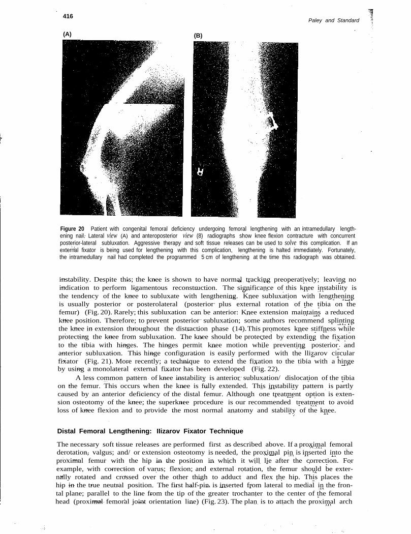
Figure 20 Patient with congenital femoral deficiency undergoing femoral lengthening with an intramedullary length-ening nail. Lateral view (A) and anteroposterior view (8) radiographs show knee flexion contracture with concurrentposterior-lateral subluxation. Aggressive therapy and soft tissue releases can be used to solve this complication. If anexternal fixator is being used for lengthening with this complication, lengthening is halted immediately. Fortunately,the intramedullary nail had completed the programmed 5 cm of lengthening at the time this radiograph was obtained.
instability. Despite this; the knee is shown to have normal tracking preoperatively; leaving noindication to perform ligamentous reconstruction. The significance of this knee instability isthe tendency of the knee to subluxate with lengthening. Knee subluxation with lengtheningis usually posterior or posterolateral (posterior plus external rotation of the tibia on thefemur) (Fig. 20). Rarely; this subluxation can be anterior. Knee extension maintains a reducedknee position. Therefore; to prevent posterior subluxation; some authors recommend splintingthe knee in extension throughout the distraction phase (14).This promotes knee stiffness whileprotecting the knee from subluxation. The knee should be protected by extending the fixationto the tibia with hinges. The hinges permit knee motion while preventing posterior andanterior subluxation. This hinge configuration is easily performed with the llizarov circularfixator (Fig. 21). More recently; a technique to extend the fixation to the tibia with a hingeby using a monolateral external fixator has been developed (Fig. 22).
A less common pattern of knee instability is anterior subluxation/ dislocation of the tibiaon the femur. This occurs when the knee is fully extended. This instability pattern is partlycaused by an anterior deficiency of the distal femur. Although one treatment option is exten-sion osteotomy of the knee; the superknee procedure is our recommended treatment to avoidloss of knee flexion and to provide the most normal anatomy and stability of the knee.
Distal Femoral Lengthening: IIizarov Fixator Technique
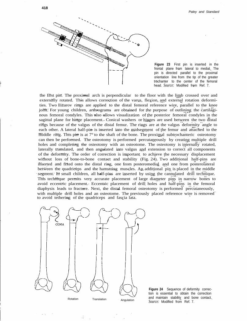
The necessary soft tissue releases are performed first as described above. If a proximal femoralderotation, valgus; and/ or extension osteotomy is needed, the proximal pin is inserted into theproximal femur with the hip in the position in which it will lie after the correction. Forexample, with correction of varus; flexion; and external rotation, the femur should be exter-nally rotated and crossed over the other thigh to adduct and flex the hip. This places thehip in the true neutral position. The first half-pin is inserted from lateral to medial in the fron-tal plane; parallel to the line from the tip of the greater trochanter to the center of the femoralhead (proximal femoral joint orientation line) (Fig. 23). The plan is to attach the proximal arch
Lengthening Reconstruction Surgery for Congenital Femoral Deficiency417
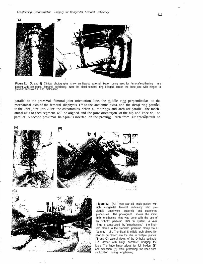
Figure 21 (A and 8) Clinical photographs show an Ilizarov external fixator being used for femora/lengthening in apatient with congenital femoral deficiency. Note the distal femoral ring bridged across the knee joint with hinges toprevent subluxation and dislocation.
parallel to the proximal femoral joint orientation line, the middle ring perpendicular to themechanical axis of the femoral diaphysis (7° to the anatomic axis), and the distal ring parallelto the knee joint line. Alter the osteotomies, when all the rings and arch are parallel, the mech-anical axis of each segment will be aligned and the joint orientation of the hip and knee will beparallel. A second proximal half-pin is inserted on the proximal arch from 30° anterolateral to
(A) (8)r-
Figure 22 (A) Three-year-old male patient withright congenital femoral deficiency who pre-viously underwent superhip and superkneeprocedures. The photograph shows the initiallimb lengthening that was done with the use ofan Orthofix pediatric LRS rail system. A kneehinge is constructed by "piggybacking" the Shef-field clamp to the standard pediatric clamp via a"dummy" pin. The distal Sheffield arch allows fix-ation to be placed into the tibia in multiple planes.(8 and C) Lateral views of the Orthofix pediatricLRS device with hinge construct bridging theknee. The knee hinge allows for full flexion (8)and extension (C) while protecting the knee fromsubluxation during lengthening.
418Paley and Standard
Figure 23 First pin is inserted in thefrontal plane from lateral to medial. Thepin is directed parallel to the proximalorientation line from the tip of the greatertrochanter to the center of the femoralhead. Source: Modified from Ref. 7.
the first pin. The proximal arch is perpendicular to the floor with the limb crossed over andexternally rotated. This allows correction of the varus, flexion, and external rotation deformi-ties. Two llizarov rings are applied to the distal femoral reference wire, parallel to the kneejoint. For young children, arthrograms are obtained for the purpose of outlining the cartilagi-nous femoral condyles. This also allows visualization of the posterior femoral condyles in thesagittal plane for hinge placement. Conical washers or hinges are used between the two distalrings because of the valgus of the distal femur. The rings are at the valgus deformity angle toeach other. A lateral half-pin is inserted into the midsegment of the femur and attached to themiddle ring. This pin is at 7° to the shaft of the bone. The proximal subtrochanteric osteotomycan then be performed. The osteotomy is performed percutaneously by creating multiple drillholes and completing the osteotomy with an osteotome. The osteotomy is internally rotated,laterally translated, and then angulated into valgus and extension to correct all componentsof the deformity. The order of correction is important to achieve the necessary displacementwithout loss of bone-to-bone contact and stability (Fig. 24). Two additional half-pins areinserted and fixed onto the distal ring, one from posteromedial and one from posterolateralbetween the quadriceps and the hamstring muscles. An additional pin is placed in the middlesegment. In small children, all half-pins are inserted by using the cannulated drill technique.This technique permits very accurate placement of large diameter pins in narrow bones toavoid eccentric placement. Eccentric placement of drill holes and half-pins in the femoraldiaphysis leads to fracture. Next, the distal femoral osteotomy is performed percutaneously,with multiple drill holes and an osteotome. The previously placed reference wire is removedto avoid tethering of the quadriceps and fascia lata.
Rotation
Figure 24 Sequence of deformity correc-tion is essential to obtain the correctionand maintain stability and bone contact.Source: Modified from Ref. 7.
Translation Angulation
Lengthening Reconstruction Surgery for Congenital Femoral Deficiency419
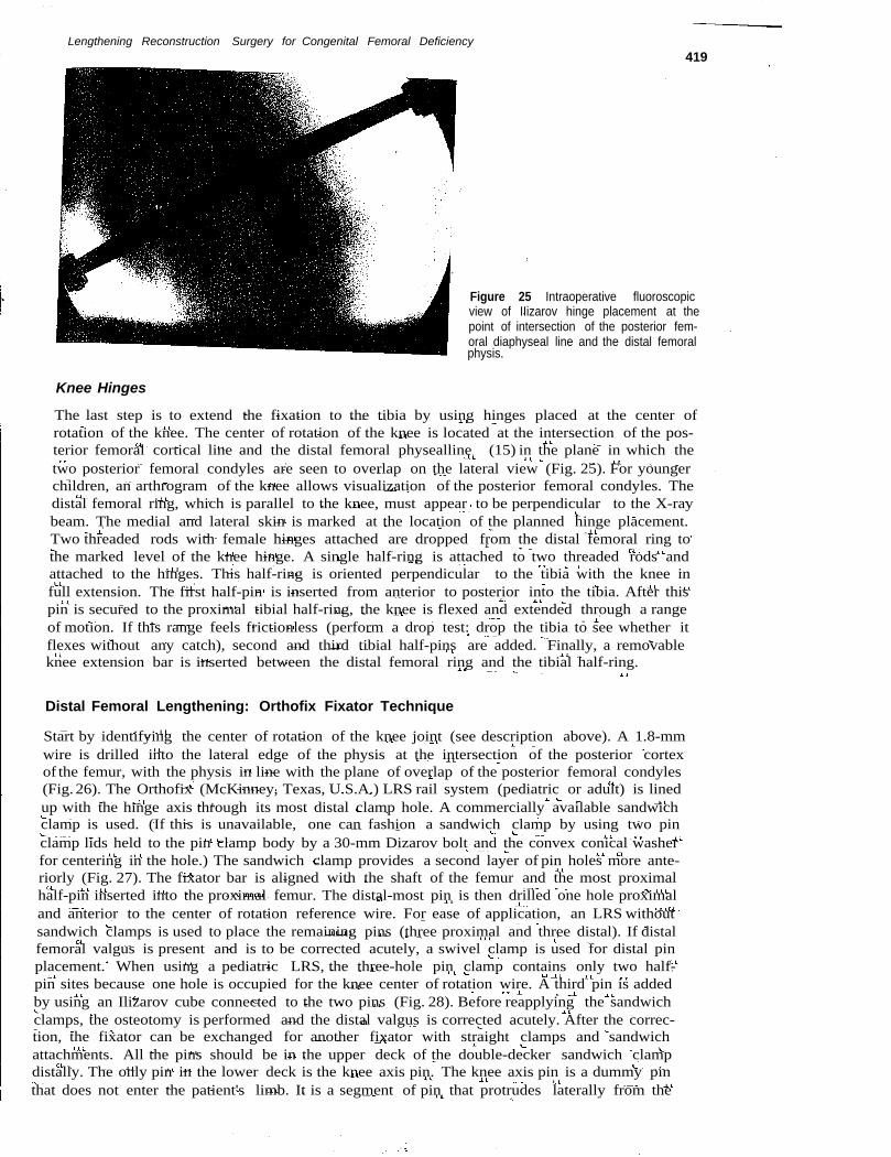
Figure 25 Intraoperative fluoroscopicview of IIizarov hinge placement at thepoint of intersection of the posterior fem-oral diaphyseal line and the distal femoralphysis.
Knee Hinges
The last step is to extend the fixation to the tibia by using hinges placed at the center ofrotation of the knee. The center of rotation of the knee is located at the intersection of the pos-terior femoral cortical line and the distal femoral physealline (15) in the plane in which thetwo posterior femoral condyles are seen to overlap on the lateral view (Fig. 25). For youngerchildren, an arthrogram of the knee allows visualization of the posterior femoral condyles. Thedistal femoral ring, which is parallel to the knee, must appear to be perpendicular to the X-raybeam. The medial and lateral skin is marked at the location of the planned hinge placement.Two threaded rods with female hinges attached are dropped from the distal femoral ring tothe marked level of the knee hinge. A single half-ring is attached to two threaded rods andattached to the hinges. This half-ring is oriented perpendicular to the tibia with the knee infull extension. The first half-pin is inserted from anterior to posterior into the tibia. After thispin is secured to the proximal tibial half-ring, the knee is flexed and extended through a rangeof motion. If this range feels frictionless (perform a drop test: drop the tibia to see whether itflexes without any catch), second and third tibial half-pins are added. Finally, a removableknee extension bar is inserted between the distal femoral ring and the tibial half-ring.
Distal Femoral Lengthening: Orthofix Fixator Technique
Start by identifying the center of rotation of the knee joint (see description above). A 1.8-mmwire is drilled into the lateral edge of the physis at the intersection of the posterior cortexof the femur, with the physis in line with the plane of overlap of the posterior femoral condyles(Fig. 26). The Orthofix (McKinney, Texas, U.S.A.) LRS rail system (pediatric or adult) is linedup with the hinge axis through its most distal clamp hole. A commercially available sandwichclamp is used. (If this is unavailable, one can fashion a sandwich clamp by using two pinclamp lids held to the pin clamp body by a 30-mm Dizarov bolt and the convex conical washerfor centering in the hole.) The sandwich clamp provides a second layer of pin holes more ante-riorly (Fig. 27). The fixator bar is aligned with the shaft of the femur and the most proximalhalf-pin inserted into the proximal femur. The distal-most pin is then drilled one hole proximaland anterior to the center of rotation reference wire. For ease of application, an LRS withoutsandwich clamps is used to place the remaining pins (three proximal and three distal). If distalfemoral valgus is present and is to be corrected acutely, a swivel clamp is used for distal pinplacement. When using a pediatric LRS, the three-hole pin clamp contains only two half-pin sites because one hole is occupied for the knee center of rotation wire. A third pin is addedby using an Ilizarov cube connected to the two pins (Fig. 28). Before reapplying the sandwichclamps, the osteotomy is performed and the distal valgus is corrected acutely. After the correc-tion, the fixator can be exchanged for another fixator with straight clamps and sandwichattachments. All the pins should be in the upper deck of the double-decker sandwich clampdistally. The only pin in the lower deck is the knee axis pin. The knee axis pin is a dummy pinthat does not enter the patient's limb. It is a segment of pin that protrudes laterally from the

420Paley and Standard
(A)
Figure 26 (A and B) Intraopera-tive fluoroscopic images showarthrography of the knee. The lat-eral view is obtained, and theposterior aspects of the femoralcondyles are superimposed to cre-ate the perfect lateral view. Thehinge reference wire is inserted atthe intersection of the posteriorfemoral cortical line and the distalfemoral physis. This marks thecenter of rotation of the knee joint.
clamp. A Sheffield clamp from Orthofix is applied to this pin to act as a hinge. It is locked inplace by placing a cube lateral to it with a setscrew to prevent it from moving laterally. Conicalwashers are used between the Sheffield clamp and the LRSto reduce friction (Fig. 29). The Shef-field clamp is left partially loose to permit motion. A one-third Sheffield arch is attached to theclamp, arching toward the tibia. An anteroposterior pin is inserted, and the drop test is per-formed (see above). If friction occurs during the drop, the Sheffield clamp should be loosened.If friction persists, adjust the connection of the tibial pin to the Sheffield arch. If friction furtherpersists after the adjustment, the axis pin might need to be bent slightly to alter the axis ofrotation. Once the drop test is negative, two more oblique pins are inserted into the tibia andconnected to the Sheffield arch by using cubes (Fig. 30). A removable knee extension bar is fash-ioned from Ilizarov parts to be used especially at nighttime. The easiest way to accomplish this isto apply a cube to the protruding ends of the distal pins and build off this cube with a post(Fig. 31). If an unstable hip is present, a hinge axis pin for the hip can also be placed and attachedto the proximal clamp. The same Sheffield clamp and arch arrangement are used. Three pins areplaced in the pelvis, one through the anterior-inferior and one through the anterior-superiorspines extending posteriorly. One is inserted laterally. These are fixed to the Sheffield clampto prevent proximal subluxation of the hip during lengthening (Fig. 32).As one can see, the prin-ciples applied when using monolateral fixation are the same as those for circular fixation (i.e.,hinge fixation across joints when a joint is at risk).
(A)
Figure 27 (A and B) Bone model shows LRS sandwich clamp placed distally, with the most distal hole containingthe hinge axis wire. The first distal half-pin is placed on the anterior row one hole proximal to the hinge axis pin.
Lengthening Reconstruction Surgery for Congenital Femoral DeficIency421
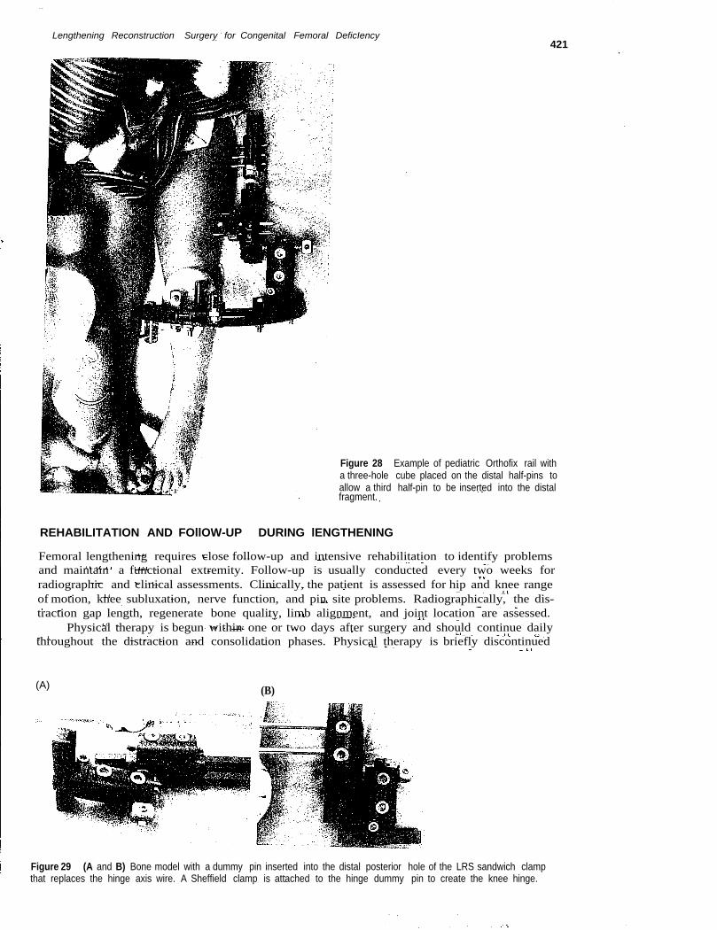
Figure 28 Example of pediatric Orthofix rail witha three-hole cube placed on the distal half-pins toallow a third half-pin to be inserted into the distalfragment.
REHABILITATION AND FOllOW-UP DURING lENGTHENING
Femoral lengthening requires close follow-up and intensive rehabilitation to identify problemsand maintain a functional extremity. Follow-up is usually conducted every two weeks forradiographic and clinical assessments. Clinically, the patient is assessed for hip and knee rangeof motion, knee subluxation, nerve function, and pin site problems. Radiographically, the dis-traction gap length, regenerate bone quality, limb alignment, and joint location are assessed.
Physical therapy is begun within one or two days after surgery and should continue dailythroughout the distraction and consolidation phases. Physical therapy is briefly discontinued
(A) (B)
Figure 29 (A and B) Bone model with a dummy pin inserted into the distal posterior hole of the LRS sandwich clampthat replaces the hinge axis wire. A Sheffield clamp is attached to the hinge dummy pin to create the knee hinge.
422Paley and Standard
(A) (8)
(C)
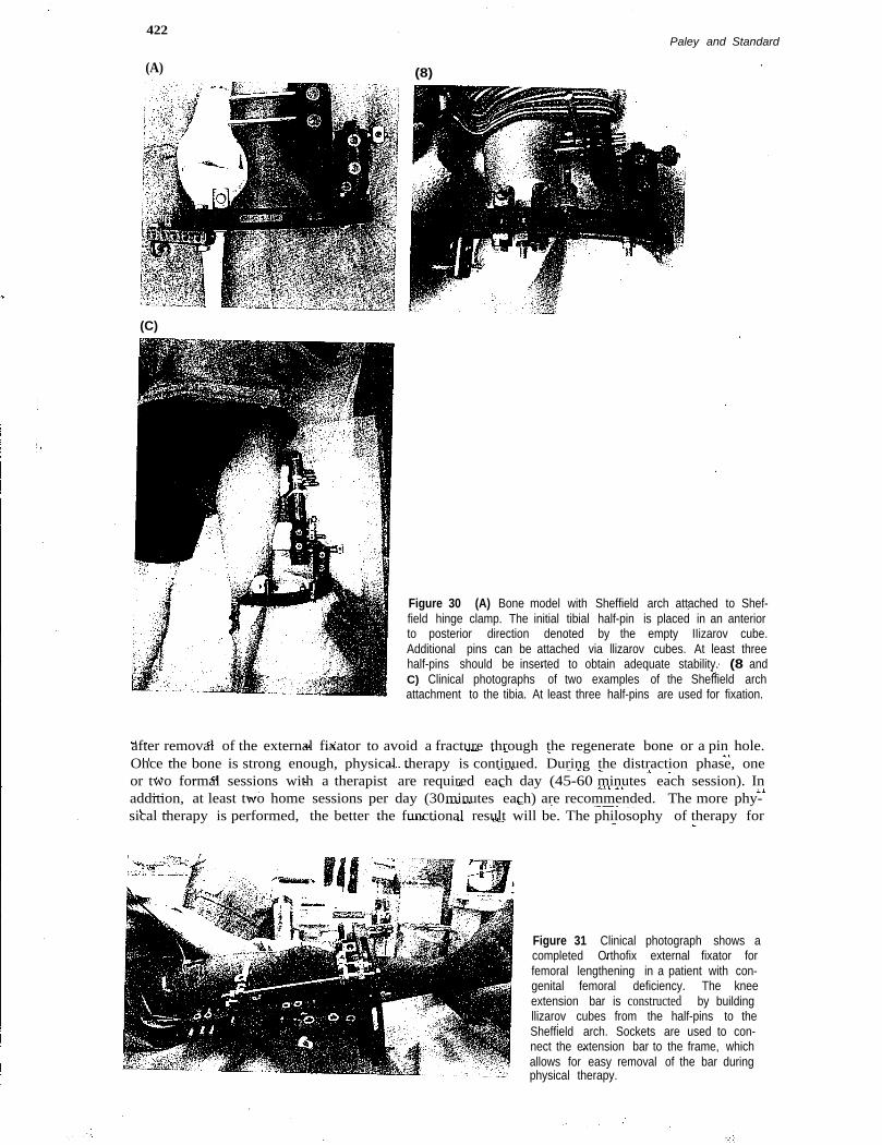
Figure 30 (A) Bone model with Sheffield arch attached to Shef-field hinge clamp. The initial tibial half-pin is placed in an anteriorto posterior direction denoted by the empty IIizarov cube.Additional pins can be attached via llizarov cubes. At least threehalf-pins should be inserted to obtain adequate stability. (8 andC) Clinical photographs of two examples of the Sheffield archattachment to the tibia. At least three half-pins are used for fixation.
after removal of the external fixator to avoid a fracture through the regenerate bone or a pin hole.Once the bone is strong enough, physical therapy is continued. During the distraction phase, oneor two formal sessions with a therapist are required each day (45-60 minutes each session). Inaddition, at least two home sessions per day (30minutes each) are recommended. The more phy-sical therapy is performed, the better the functional result will be. The philosophy of therapy for
Figure 31 Clinical photograph shows acompleted Orthofix external fixator forfemoral lengthening in a patient with con-genital femoral deficiency. The kneeextension bar is constructed by buildingllizarov cubes from the half-pins to theSheffield arch. Sockets are used to con-nect the extension bar to the frame, whichallows for easy removal of the bar duringphysical therapy.
Lengthening Reconstruction Surgery for Congenital Femoral Deficiency
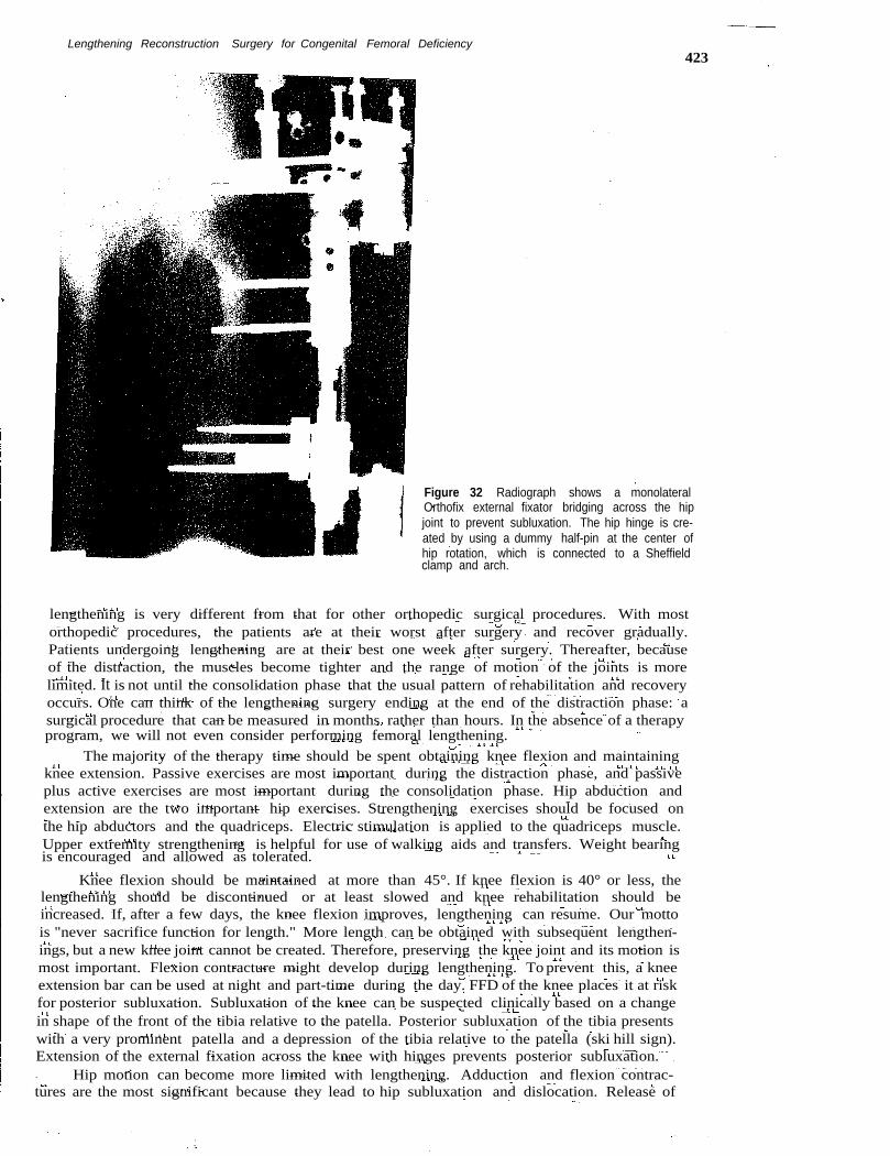
Figure 32 Radiograph shows a monolateralOrthofix external fixator bridging across the hipjoint to prevent subluxation. The hip hinge is cre-ated by using a dummy half-pin at the center ofhip rotation, which is connected to a Sheffieldclamp and arch.
lengthening is very different from that for other orthopedic surgical procedures. With mostorthopedic procedures, the patients are at their worst after surgery and recover gradually.Patients undergoing lengthening are at their best one week after surgery. Thereafter, becauseof the distraction, the muscles become tighter and the range of motion of the joints is morelimited. It is not until the consolidation phase that the usual pattern of rehabilitation and recoveryoccurs. One can think of the lengthening surgery ending at the end of the distraction phase: asurgical procedure that can be measured in months rather than hours. In the absence of a therapyprogram, we will not even consider performing femoral lengthening.
The majority of the therapy time should be spent obtaining knee flexion and maintainingknee extension. Passive exercises are most important during the distraction phase, and passiveplus active exercises are most important during the consolidation phase. Hip abduction andextension are the two important hip exercises. Strengthening exercises should be focused onthe hip abductors and the quadriceps. Electric stimulation is applied to the quadriceps muscle.Upper extremity strengthening is helpful for use of walking aids and transfers. Weight bearingis encouraged and allowed as tolerated.
Knee flexion should be maintained at more than 45°. If knee flexion is 40° or less, thelengthening should be discontinued or at least slowed and knee rehabilitation should beincreased. If, after a few days, the knee flexion improves, lengthening can resume. Our mottois "never sacrifice function for length." More length can be obtained with subsequent lengthen-ings, but a new knee joint cannot be created. Therefore, preserving the knee joint and its motion ismost important. Flexion contracture might develop during lengthening. To prevent this, a kneeextension bar can be used at night and part-time during the day. FFD of the knee places it at riskfor posterior subluxation. Subluxation of the knee can be suspected clinically based on a changein shape of the front of the tibia relative to the patella. Posterior subluxation of the tibia presentswith a very prominent patella and a depression of the tibia relative to the patella (ski hill sign).Extension of the external fixation across the knee with hinges prevents posterior subluxation.
Hip motion can become more limited with lengthening. Adduction and flexion contrac-tures are the most significant because they lead to hip subluxation and dislocation. Release of
423
424Paley and Standard
the adductors, rectus, and TFL during lengthening might need to be considered to allowfurther lengthening. Usually the TFL and the rectus femoris tendon are addressed duringthe preparatory surgery.
Nerve injury from surgery or distraction is unusual with femoral lengthening. To avoidperoneal nerve injury from the pins, the posterolateral pin should not enter posterior to thebiceps tendon. During distraction, if the patient complains of pain in the dorsum of the footor requests frequent massaging of the foot, referred pain from stretch entrapment of the pero-neal nerve is most likely the cause. More advanced symptoms include hyper- or hypoesthesiain the distribution of the peroneal nerve and weakness of the extensor hallucis longus muscle.Quantitative sensory testing, if available, is the most sensitive test to assess for nerve involve-ment. If the nerve problem is identified early, it can be treated by slowing the rate of distraction.If, despite slowing the distraction, symptoms continue or motor signs develop, the peronealnerve should be decompressed at the neck of the fibula. This release should include. transversefasciotomy of the lateral and anterior compartments and release of the intermuscular septumbetween these compartments (7).
Hypotrophic regenerate formation requires slowing the distraction rate. Overabundantbone formation can lead to premature consolidation and requires increasing the distraction ratefor a few days. A mismatch between the increase in the distraction gap from one visit to the nextand the number of millimeters of distraction performed during the same time period is a sign ofan impending premature consolidation. Radiographs are used to assess joint location. A breakin Shenton's line or increased medial-lateral head-teardrop distance indicates subluxation ofthe hip. In the knee, posterior or anterior subluxation can be monitored on the lateral radio-graphic view with full knee extension (16). Limb length equalization should be based onfull-length standing radiographs. Limb alignment is assessed for the femur and tibia separatelyand in combination. Separately the joint orientation of the knee should be measured by usingthe malalignment test (7).Axial deviation from lengthening (procurvatum and valgus for distalfemoral lengthening and procurvatum and varus for proximal lengthening) is identified andcorrected at the end of the distraction phase, when the regenerate bone is still malleable. Whenmalalignment of the femur and tibia is present, the femoral mal alignment is corrected to a nor-mal distal femoral joint orientation. The femur is not over- or Lmdercorrected to compensate forthe tibial deformity. The tibia should be corrected separately, either during the same treatmentor at a later treatment.
Complete failure of bone formation is very unusual. Partial defects are not uncommon.The most common location of regenerate defects is lateral. Dynamization of the fixator shouldbe performed, and bone growth stimulators can be used. Resection of the fibrous tissue inthese defects and cancellous bone grafting might become necessary to reduce the externalfixation time and prevent fracture after frame removal -.
Once the regenerate bone is deemed healed, the frame can be removed from the femurand tibia. Previously, we applied a one-legged spica cast to prevent fracture. Despite appli-cation of the cast, we experienced a 34% fracture rate of the femur after removal, comparedwith a 9% fracture rate for all other bones and for noncongenital femoral lengthening. Becauseof this high fracture rate, prophylactic Rush rod placement in the femur is now performed atthe time of external fixation removal. This new protocol has virtually eliminated the compli-cation of refracture after lengthening. Because a rod is being inserted into the femur at the timeof removal of the external fixator, intramedullary infection is a concern. Despite this concern,we have observed only two cases in which local deep pin bone infection occurred. The infec-tions were easily treated by curettage of the lateral cortex of the bone combined with removalof the Rush rod and administration of antibiotics. Prophylactic rodding permits continuationof knee mobility after removal. Formal physical therapy is delayed for a month, but the patientis permitted gentle knee range of motion. This is to protect from osteoporotic stress fracturesthrough the tibial pin holes. The prophylactic rod placement also permits weight bearing witha removable spica cast immediately after frame removal.
DIFFERENCES IN TREATMENT OF TYPES 1a AND 1b CONGENITALFEMORAL DEFICIENCY
In general, Type 1a CFD (normal ossification) has less hip, proximal femoral, and knee defor-mity, deficiency, and discrepancy than does Type 1b (delayed ossification). Most Type 1a cases do
Lengthening Reconstruction Surgery for Congenital Femoral Deficiency
not require the complex superhip reconstruction. Approximately one-half of the Type la casesdo require pelvic osteotomy before lengthening. All CFD Type la cases require extension of thefixator across the knee to protect the knee joint. The distinction between Types la and Ib shouldbe made while the patient is in infancy because the natural history of Type Ib is to ossify. There-fore, adult Type Ib cases generally appear to be severe Type la cases. The strategy of treatmentfor Type Ib is to correct all the associated deformities, which will allow the proximal femur andhip to be more normally oriented and accept more axial loading. The response to the anatomicchange is ossification of the proximal femur and conversion from Type Ib to Type la. We do notperform lengthening in Type Ib cases until they convert to Type la. This conversion usuallyoccurs within two years of the superhip procedure. Our preference is to perform the firstlengthening when the patient is between the ages of two and four years. Patients with Typela CFD typically undergo their first lengthening at the age of two years, whereas patients withType Ib CFD typically undergo lengthening closer to the age of four years.
TREATMENT OF TYPES 2 AND 3 CONGENITAL FEMORAL DEFICIENCY
Detailed discussion of the treatment of Types 2 and 3 CFD are complex and beyond the scopeof this textbook. For the purposes of this text, we provide only a summary of our strategy forTypes 2 and 3.
Type 2 Congenital Femoral Deficiency
With Type 2 CFD, the deciding factor is the presence or absence of a mobile femoral head inthe acetabulum. Despite magnetic resonance imaging, arthrography, and other studies, often,the only definitive way to determine the presence or absence of a mobile femoral head is toopen the hip joint capsule and examine the femoral head for mobility. If the femoral head doesnot move in the acetabulum, it should not be joined to the femoral shaft. If it is joined to thefemoral shaft, effectively, arthrodesis of the hip is created. If the femoral head is mobile, it canbe connected to the remainder of the femur by a complicated reconstruction of the femoralneck, which we call "superhip 2." This converts Type 2a to Type la, and further treatmentis as for Type la. If no femoral head is present or if it is stiff in the acetabulum, only the super-hip soft tissue release is performed to release the flexion contracture of the hip and excise thefascia lata. Seriallengthenings with fixation to the pelvis are performed until skeletal maturity,at which time the last lengthening is combined with a pelvic support osteotomy. The onlydifference between pelvic support osteotomy in cases of CFD and that for other conditions(17) is that the fixation is extended to the pelvis.
Type 3 Congenital Femoral Deficiency
Type 3a CFD can be treated like Type 2b, with hip release, seriallengthenings, and pelvicsupport osteotomy, or can be treated by prosthetic fitting options, including prostheticreconstruction surgery. Prosthetic reconstruction surgery refers to Syme amputation and rota-tionplasty. For Type 3b CFD, which includes a stiff knee joint «:450 of motion), we are morelikely to recommend prosthetic reconstruction surgery. In Type 3a cases, when the knee jointhas a functional range of motion, converting Type 3a to Type 2b can be considered. The extentof treatment required for reconstruction in such cases entails four or more lengthenings andshould not be considered lightly. Rotationplasty provides more predictable functional resultsthan does lengthening for such severe cases. When rotationplasty is chosen, our preference is touse a modification of the rotationplasty described by Brown (18). This includes fusion ofthe distal femoral remnant to the pelvis. Paley's modification is to perform the fusion to theinferior iliac surface of a Chiari osteotomy instead of to the lateral ileum as described by Brown.This medializes the limb and provides a broader surface for fusion. The femoral segment isfixed to the pelvis by using screws crossing the physis to produce a distal femoral epiphysiod-esis. Distal femoral epiphysiodesis is required to prevent distal migration of the rotated kneejoint (functionally now the hip joint). When Syme amputation is chosen, the superhip approachto eliminate contractures around the hip, combined with knee flexion contracture release,improves the posture of the short thigh and knee joint to enable better prosthetic fitting.
425
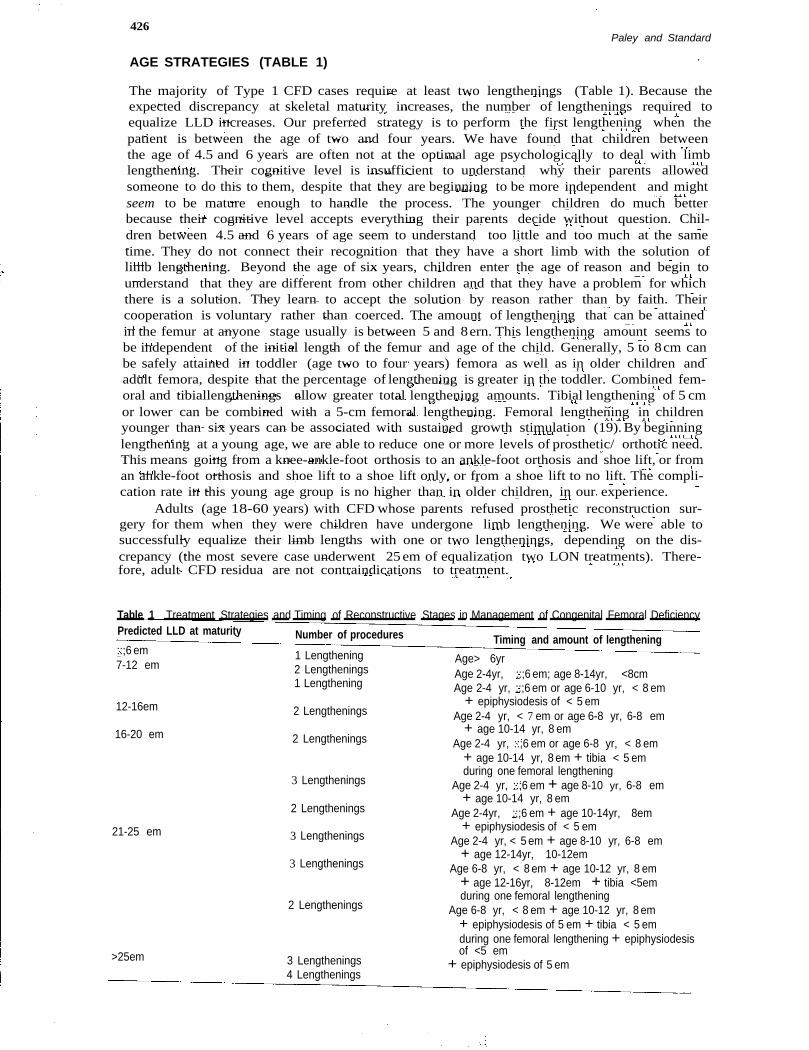
426Paley and Standard
AGE STRATEGIES (TABLE 1)
The majority of Type 1 CFD cases require at least two lengthenings (Table 1). Because theexpected discrepancy at skeletal maturity increases, the number of lengthenings required toequalize LLD increases. Our preferred strategy is to perform the first lengthening when thepatient is between the age of two and four years. We have found that children betweenthe age of 4.5 and 6 years are often not at the optimal age psychologically to deal with limblengthening. Their cognitive level is insufficient to understand why their parents allowedsomeone to do this to them, despite that they are beginning to be more independent and mightseem to be mature enough to handle the process. The younger children do much betterbecause their cognitive level accepts everything their parents decide without question. Chil-dren between 4.5 and 6 years of age seem to understand too little and too much at the sametime. They do not connect their recognition that they have a short limb with the solution oflimb lengthening. Beyond the age of six years, children enter the age of reason and begin tounderstand that they are different from other children and that they have a problem for whichthere is a solution. They learn to accept the solution by reason rather than by faith. Theircooperation is voluntary rather than coerced. The amount of lengthening that can be attainedin the femur at anyone stage usually is between 5 and 8ern. This lengthening amount seems tobe independent of the initial length of the femur and age of the child. Generally, 5 to 8cm canbe safely attained in toddler (age two to four years) femora as well as in older children andadult femora, despite that the percentage of lengthening is greater in the toddler. Combined fem-oral and tibiallengthenings allow greater total lengthening amounts. Tibial lengthening of 5 cmor lower can be combined with a 5-cm femoral lengthening. Femoral lengthening in childrenyounger than six years can be associated with sustained growth stimulation (19).By beginninglengthening at a young age, we are able to reduce one or more levels of prosthetic/ orthotic need.This means going from a knee-ankle-foot orthosis to an ankle-foot orthosis and shoe lift, or froman ankle-foot orthosis and shoe lift to a shoe lift only, or from a shoe lift to no lift. The compli-cation rate in this young age group is no higher than in older children, in our experience.
Adults (age 18-60 years) with CFD whose parents refused prosthetic reconstruction sur-gery for them when they were children have undergone limb lengthening. We were able tosuccessfully equalize their limb lengths with one or two lengthenings, depending on the dis-crepancy (the most severe case underwent 25em of equalization two LON treatments). There-fore, adult CFD residua are not contraindications to treatment.
Table 1 Treatment Strategies and Timing of Reconstructive Stages in Management of Congenital Femoral DeficiencyPredicted LLD at maturity Number of procedures Timing and amount of lengthening::;6 em7-12 em
1 Lengthening2 Lengthenings1 Lengthening
Age> 6yrAge 2-4yr, ::;6 em; age 8-14yr, <8cmAge 2-4 yr, ::;6 em or age 6-10 yr, < 8 em
+ epiphysiodesis of < 5 emAge 2-4 yr, < 7 em or age 6-8 yr, 6-8 em
+ age 10-14 yr, 8 emAge 2-4 yr, ::;6 em or age 6-8 yr, < 8 em
+ age 10-14 yr, 8 em + tibia < 5 emduring one femoral lengthening
Age 2-4 yr, ::;6 em + age 8-10 yr, 6-8 em+ age 10-14 yr, 8 em
Age 2-4yr, ::;6 em + age 10-14yr, 8em+ epiphysiodesis of < 5 em
Age 2-4 yr, < 5 em + age 8-10 yr, 6-8 em+ age 12-14yr, 10-12em
Age 6-8 yr, < 8 em + age 10-12 yr, 8 em+ age 12-16yr, 8-12em + tibia <5emduring one femoral lengthening
Age 6-8 yr, < 8 em + age 10-12 yr, 8 em+ epiphysiodesis of 5 em + tibia < 5 emduring one femoral lengthening + epiphysiodesisof <5 em
+ epiphysiodesis of 5 em
12-16em 2 Lengthenings
16-20 em 2 Lengthenings
3 Lengthenings
2 Lengthenings
21-25 em 3 Lengthenings
3 Lengthenings
2 Lengthenings
>25em 3 Lengthenings4 Lengthenings
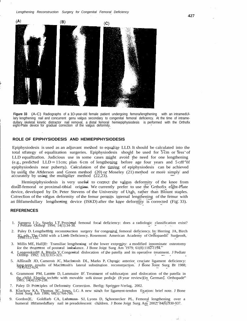
Lengthening Reconstruction Surgery for Congenital Femoral Deficiency427
Figure 33 (A-C) Radiographs of a 1O-year-old female patient undergoing femora/lengthening with an intramedUl-lary lengthening nail and concurrent genu valgus secondary to congenital femoral deficiency. At the time of intrame-dullary skeletal kinetic distractor nail removal, a distal femoral hemiepiphysiodesis is performed with the Orthofixeight-Plate device for gradual correction of the valgus deformity.
ROLE OF EPIPHYSIODESIS AND HEMIEPIPHYSIODESIS
Epiphysiodesis is used as an adjuvant method to equalize LLD. It should be calculated into thetotal strategy of equalization surgeries. Epiphysiodesis should be used for 5 cm or less ofLLD equalization. Judicious use in some cases might avoid the need for one lengthening(e.g., predicted LLD=11cm; plan 6cm of lengthening before age four years and 5 cm ofepiphysiodesis near puberty). Calculation of the timing of epiphysiodesis can be achievedby using the Anderson and Green method (20) or Moseley (21) method or more simply andaccurately by using the multiplier method (22,23).
Hemiepiphysiodesis is very useful to correct the valgus deformity of the knee fromdistal-femoral or proximal-tibial origins. We currently prefer to use the Grthofix eight-Platedevice, developed by Dr. Peter Stevens of the University of Utah, rather than Blount staples.Correction of the valgus deformity of the femur permits internal lengthening of the femur withan intramedullary lengthening device (ISKD) after the knee deformity is corrected (Fig. 33).
REFERENCES
1. Sanpera I [r., Sparks LT. Proximal femoral focal deficiency: does a radiologic classification exist?J Pediatr Orthop 1994; 14(1):34-38.
2. Paley D. Lengthening reconstruction surgery for congenital femoral deficiency. In: Herring JA, BirchJG, eds. The Child with a Limb Deficiency. Rosemont: American Academy of Orthopaedic Surgeons,1998:113-132.
3. Millis ME, Hall]E. Transiliac lengthening of the lower extremity: a modified innominate osteotomyfor the treatment of postural imbalance. J Bone Joint Surg Am 1979; 61(8):1182-1194.
4. Langenskiolri A, Ritsila V.Congenital dislocation of the patella and its operative treatment. J PediatrOrthop 1992; 12(3):315-323.
5. Amirault JD, Cameron JC, MacIntosh DL, Marks P. Chronic anterior cruciate ligament deficiency:long-term results of MacIntosh's lateral substitution reconstruction. J Bone Joint Surg Br 1988;70(4):622-624.
6. Grammont PM, Latune D, Lammaire IF. Treatment of subluxation and dislocation of the patella inthe child: Elmslie technic with movable soft tissue pedicle (8 year review)[in German]. Orthopade1985; 14(4):229-238.
7. Paley D. Principles of Deformity Correction. Berlin: Springer-Verlag, 2002.8. Krackow KA, Thomas SC, Jones, LC. A new stitch for ligament-tendon fixation: brief note. J Bone
Joint Surg Am 1986; 68(5):764-766.9. Gordon]E, Goldfarb CA, Luhmann SJ, Lyons D, Schoenecker PL. Femoral lengthening over a
humeral intramedullary nail in preadolescent children. J Bone Joint Surg Am 2002; 84(6):930-937.
428Paley and Standaro
10. Paley D, Herzenberg IE, Paremain G, Bhave A. Femoral lengthening over an intramedullary nail: amatched-case comparison with llizarov femoral lengthening. J Bone Joint Surg Am 1997;79(10):1464-1480.
11. Thonse R, Herzenberg JE, Standard SC, Paley D. Limb lengthening with a fully implantable,telescopic, intramedullary nail. Oper Tech Orthop 2005; 15(4):355-362.
12. Woolf SK, Gross RH. Posterior acetabular wall deficiency in down syndrome. J Pediatr Orthop 2003;23(6):708-713.
13. Koman LA, Brashear A, Rosenfeld S, et al. Botulinum toxin type a neuromuscular blockade in thetreatment of equinus foot deformity in cerebral palsy: a multicenter, open-label clinical trial.Pediatrics 2001; 108(5):1062-1071.
14. Bowen JR, Levy EJ,Donohue M. Comparison of knee motion and callus formation in femoral length-ening with the Wagner or monolateral-ring device. J Pediatr Orthop 1993; 13(4):467-472.
15. Hollister AM, [atana S, Singh AK, Sullivan WW, Lupichuk AG. The axes of rotation of the knee. ClinOrthop Relat Res 1993; 290:259-268.
16. Paley D. Problems, obstacles, and complications of limb lengthening by the llizarov technique. ClinOrthop Relat Res 1990; 250:81-104.
17. Rozbruch SR, Paley D, Bhave A, Herzenberg IE. llizarov hip reconstruction for the late sequelae ofinfantile hip infection. J Bone Joint Surg Am 2005; 87(5):1007-1018.
18. Brown KL. Resection, rotationplasty, and femoropelvic arthrodesis in severe congenital femoraldeficiency: a report of the surgical technique and three cases. J Bone Joint Surg Am 2001;83(1):78-85.
19. Sabharwal S, Paley D, Bhave A, Herzenberg IE. Growth patterns after lengthening of congenitallyshort lower limbs in young children. J Pediatr Orthop 2000; 20(2):137-145.
20. Anderson M, Messner MB, Green WT. Distribution of lengths of the normal femur and tibia inchildren from one to eighteen years of age. J Bone Joint Surg Am 1964; 46:1197-1202.
21. Moseley CF. A straight-line graph for leg-length discrepancies. J Bone Joint Surg Am 1977; 59(2):174-179.
22. Aguilar JA, Paley D, Paley J, et al. Clinical validation of the multiplier method for predicting limblength at maturity, part 1.J Pediatr Orthop 2005; 25(2):186-191.
23. Aguilar JA, Paley D, Paley J, et al. Clinical validation of the multiplier method for predicting limblength discrepancy and outcome of epiphysiodesis, part II. J Pediatr Orthop 2005; 25(2):192-196.