2nd lec on arrythmias by dr. roomi
TRANSCRIPT
HEART PHYSIOLOGY(ARRYTHMIAS)
BYDR. MUDASSAR ALI ROOMI (MBBS, M. PHIL)
Assistant Professor Physiology

SA NODAL HEART BLOCK:
• impulse from SA node is blocked before it enters the atria
• Results in standstill of atria• the ventricles pick up a new
rhythm, the impulse usually originating spontaneously in the AV node.
• that the rate of the ventricular QRS-T complex is slowed but not otherwise altered.
• Cause: Strong vagal stimulation• Treatment: can be relieved by
giving atropine (anticholinergic drug).

AV BLOCK• conduction of impulses from the atria
to ventricles is either slowed down or completely blocked and the block is in AV node or in AV bundle.
• Causes of AV block: – Ischemia– compression of conductive tissue by a
scarred or fibrosed portion of myocardium.
– Inflammation of AV node or AV bundle: Diphtheria, rheumatic fever and myocarditis
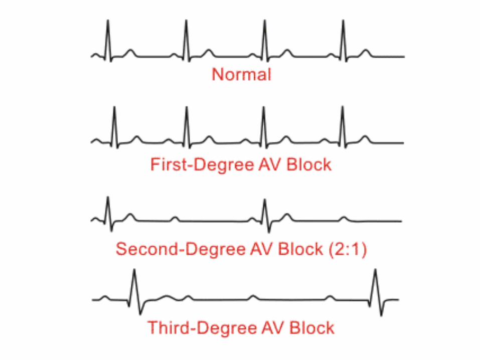
– Strong vagal stimulation.• AV block is of two types:1. Incomplete (partial) AV Block:
– First degree AV block– Second degree AV block
2. Complete (3rd degree) AV block:
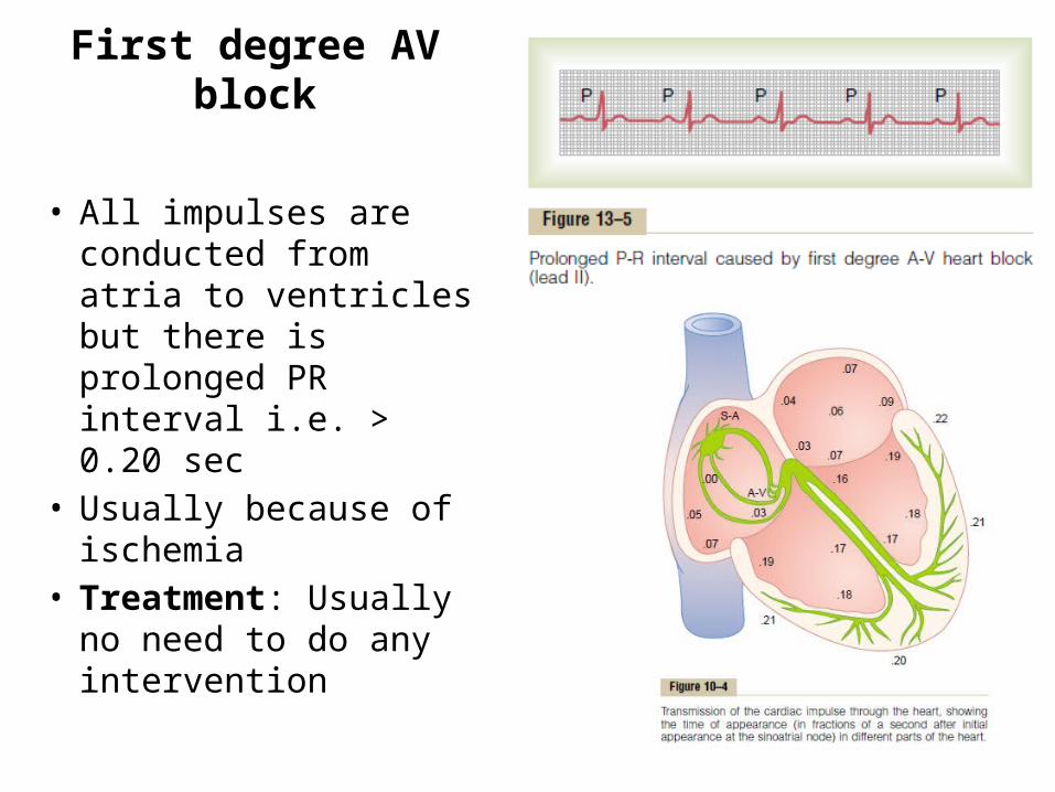
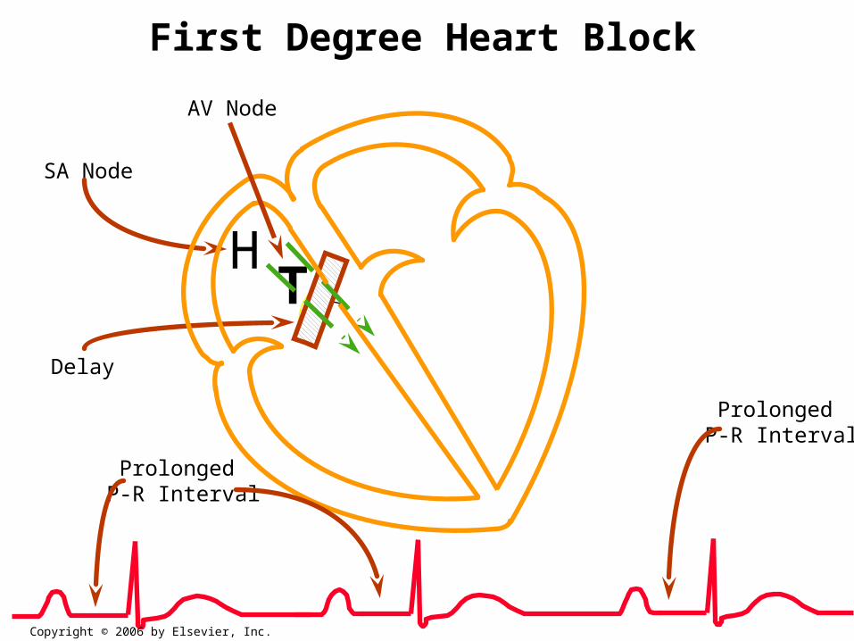
First degree AV block
• All impulses are conducted from atria to ventricles but there is prolonged PR interval i.e. > 0.20 sec
• Usually because of ischemia
• Treatment: Usually no need to do any intervention
HSA Node
T
AV Node
Delay
Prolonged P-R Interval
Prolonged P-R Interval
First Degree Heart Block
Copyright © 2006 by Elsevier, Inc.


Second degree AV block
• MOBITZ TYPE I 2ND DEGREE AV BLOCK: – also called Wenckebach phenomenon. – There is progressive prolongation of
PR interval in successive heart beats, till a heart beat is dropped.
• MOBITZ TYPE II 2ND DEGREE AV BLOCK: – PR interval is permanently or
constantly prolonged. – PR interval may be> 0.45 sec – There is 2:1 rhythm or 3:1 rhythm i.e.
every 2nd or 3rd impulse from atria is conducted to ventricles.
– In ECG we find that after every 2 or 3 P waves there is one QRS complex.
MOBITZ II HEART BLOCK
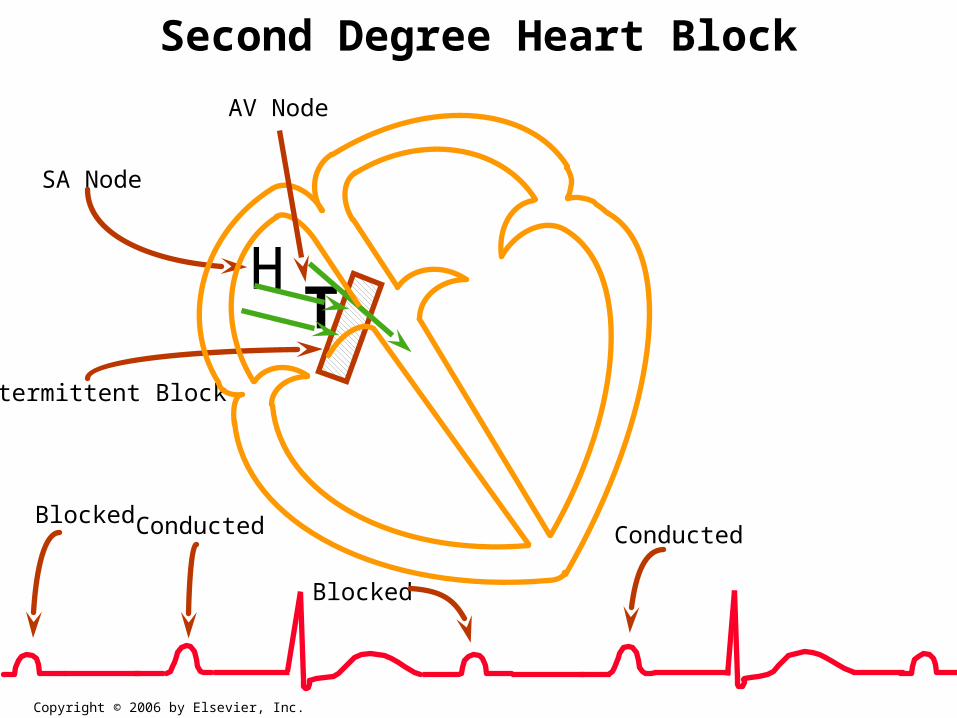
Intermittent Block
HSA Node
T
AV Node
Second Degree Heart Block
ConductedBlockedConducted
Blocked
Copyright © 2006 by Elsevier, Inc.


Complete (Third Degree) AV block
• conduction of impulses from Atria to ventricles is completely blocked.
• Ventricles start their own rhythm at a slower rate.
• So, atria beat independently with the SA nodal rhythm (70-80 bpm) and ventricles beat with their own rhythm (15-40 bpm).
• Complete dissociation b/w atria and ventricles ***
• In ECG there is no association between P wave and QRS complex.
• Treatment: atropine, pacemaker
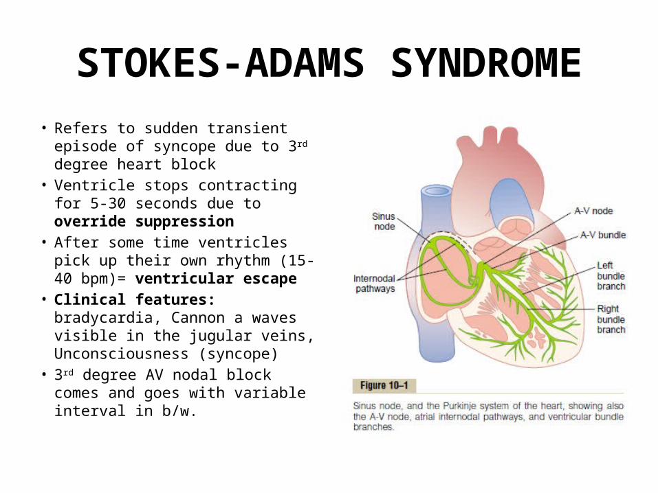
STOKES-ADAMS SYNDROME• Refers to sudden transient episode
of syncope due to 3rd degree heart block
• Ventricle stops contracting for 5-30 seconds due to override suppression
• After some time ventricles pick up their own rhythm (15-40 bpm)= ventricular escape
• Clinical features: bradycardia, Cannon a waves visible in the jugular veins, Unconsciousness (syncope)
• 3rd degree AV nodal block comes and goes with variable interval in b/w.