2nd year pathology 2010 extracellular pathology amyloidosis pathological calcification pathological...
TRANSCRIPT

2nd Year Pathology 2010
Extracellular PathologyExtracellular Pathology
AmyloidosisAmyloidosis
Pathological calcificationPathological calcification
AgeingAgeing

Amyloid and Amyloidosis• A Set of Disorders: The main feature is
the extracellular deposition of proteins arranged in the form of a Beta-pleated sheet– “Amylum” Latin for starch– Term first used By Virchow in 1854– Protein….Friedreich and Kekule. 5 years
later

Amyloidosis
• Amyloidosis is a clinical disorder caused by extracellular deposition of insoluble abnormal fibrils that injure tissue.
• The fibrils are formed by the aggregation of misfolded, normally soluble proteins.
• In humans, about 23 different unrelated proteins are known to form amyloid fibrils.

Amyloidosis• All types of amyloid consist of a major fibrillar
protein that defines the type of amyloid (approximately 90%) plus various minor components.
• Although each type of fibril may be associated with a distinct clinical picture, all share certain physical and pathologic properties

• Organs affected by amyloid:
Larger, Paler, Firmer• How do we identify Amyloid in tissues?
Macroscopically (Grossly)Waxy, Sharper cut edges, Lugol’s iodine (black).
Microscopically:
Light Microscopy-Amyloid is eosinophilic and stains with Congo Red
Polarized light: Apple Green Birefringence.
Electron Microscopy: Rigid non branching fibrils (7.5 nm to 10 nm in diameter)

Kidney
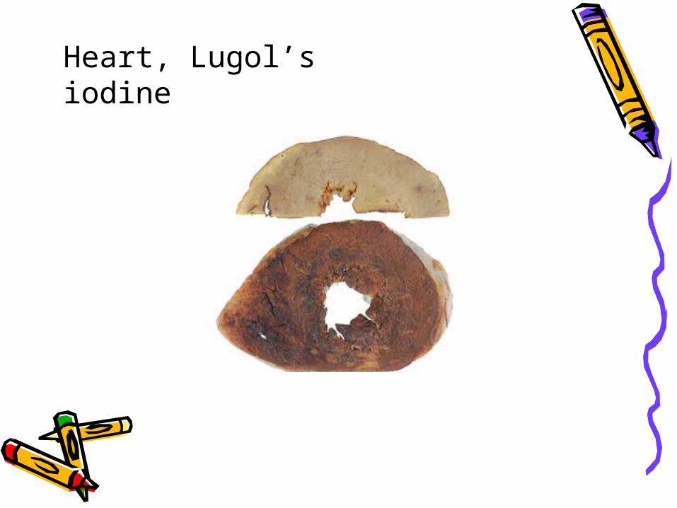
Heart, Lugol’s iodine

H&E Stain

Congo Red Stain.

Apple Green Birefringence
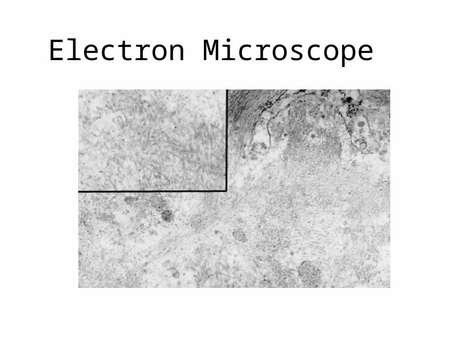
Electron Microscope

The Beta- Pleated sheet is the unifying feature of the amyloidoses
Basis of 1) Congo Red reaction
2) Fibrillar ultrastructure
3) Resistance to proteolytic
digestion
Why?• Protein Misfolding
– Intrinsic tendency to misfold (Transthyretin)– Misfolding and aggregation at high concentration
(Beta Microglobulin)– Point mutations (Hereditary amyloidosis)
• Review: Molecular mechanisms of amyloidosis. Merlini G, Bellotti V. NEJM August 2003 349:583-96

Classification
• Older classifications: 1° and 2°• More useful to classify on the basis of:
1) Nature of protein involved
2) Anatomical distribution
3) Inherited or acquired

Acquired Systemic Amyloidosis
1) Amyloidosis of immune origin. AL typeAcquired, Systemic formProtein precursor is usually immunoglobulin light chain.Occurs in association with a monoclonal (i.e. neoplastic)
proliferation of B lymphocytes or plasma cells e.g. Myeloma (Lambda chains) Waldenstrom’s macroglobulinaemia (LPL)

AL amyloid proteins : Intact immunoglobulin light chain or the amino terminal fragment of a chain or both. (mostly lambda chains)
90% of these patients have Bence Jones proteins
Amyloid accumulation: Two step process1) Secretion of excess amounts of monoclonal light chain2) Conversion to Beta pleated form.

Clinical features
• Middle to old age• M>F• 90% have a Bence Jones protein• Bone marrow = excess plasma cells• Manifestations
Neuropathy, Restrictive Cardiomyopathy, Skin manifestations, Polyarthropathy, Macroglossia, Carpal Tunnel Syndrome
• (Heart, CNS, Joints, Skin, Tongue, Soft Tissues)

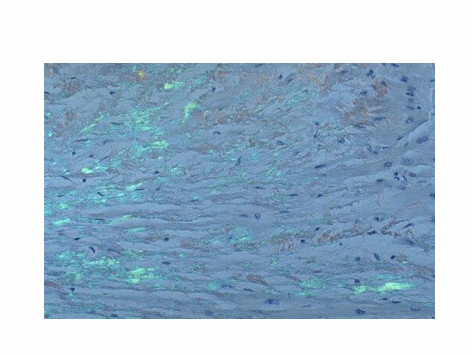
Immunofluorescence microscopy with
antibody to the lambda light chain.

Cardiac
2nd Year Pathology 2009

Acquired Systemic Amyloidosis
2) Haemodialysis associated Amyloid
Acquired, systemic Protein is Beta-2-microglobulin.Normally broken down by kidneys A naturally occurring “amyloidogenic” protein not
filtered by dialysis membranes After 7 years on dialysis 30% of patients get
Carpal Tunnel Syndrome (after 10 years 50%)Joints, synovium, tendon sheaths

Acquired Systemic Amyloidosis
3) Reactive Systemic AmyloidosisAlso known as secondary amyloid or AA type amyloidAcquired, systemic formProtein precursor is serum amyloid A (SAA)Acute phase reactant which markedly increases with
tissue injury or inflammation under the influence of IL1, TNF and IL-6
AA is a cleavage product of SAA.Distribution of amyloid: kidney, liver, spleen, adrenals,
thyroid + many other tissues. Diagnosis: Rectal Biopsy

Disease Associations:
1) Chronic infection: T.B., Leprosy, Syphilis, Chronic osteomyelitis, Bronchiectasis2) Chronic inflammation
Reiter’s disease, Whipple’s disease3) Chronic autoimmune disease:
R.A., I.B.D., Connective Tissue Disease4) Long standing paraplegia. (UTI)5) Neoplasms: Renal adenocarcinoma and Hodgkin’s disease

Liver

Adrenal

Hereditary Systemic Amyloidosis
• Rare. • Most common: Familial Mediterranean Fever. • Less common: Three types:
1) Neuropathic.2) Cardiopathic.3) Nephropathic.The Amyloid protein is usually Transthyretin (Prealbumin). Transthyretin is also associated with a form of Amyloid known as Systemic Senile Amyloid, where amyloid is systemically deposited, mainly in the heart in elderly individuals.

• Familial Mediterranean Fever
Commonest form of hereditary systemic amyloidosis Autosomal Recessive, Gene on Chromosome 16 AA type amyloid Two manifestations of the disease:
Short febrile attacks with pain mimicking pleurisy, peritonitis or
synovitis Amyloidosis: Manifest early in life
Death before 40 without Renal Transplant Treated with Colchicine (Stabilization of inflammatory cells)

Localized amyloidosis
• Endocrine associated:
Pituitary - age related
Islets of Langerhans - NIDDM related
Medullary Carcinoma of Thyroid (Calcitonin)
• Intacerebral:
Alzheimer’s, Spongiform Encephalopathy
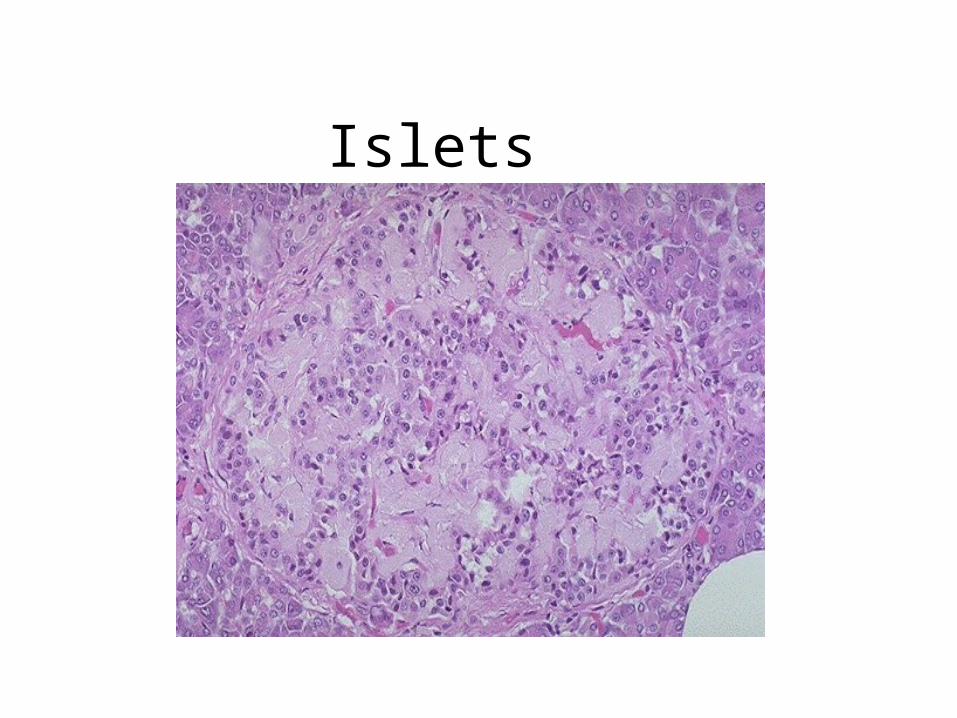
Islets

Amyloid angiopathy
2nd Year Pathology 2009

Alzheimer’s disease

Medullary Thyroid Carcinoma
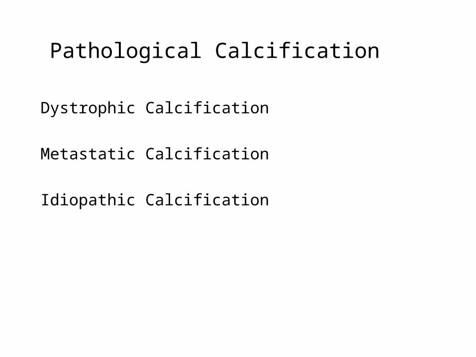
Pathological Calcification
Dystrophic Calcification
Metastatic Calcification
Idiopathic Calcification

Dystrophic Calcification
• Normal serum calcium/phosphate• Nonviable or dying tissues• Occurs in atherosclerosis, damaged valves, necrosis
(coagulative, caseous, liquefactive), Leiomyomas• Monckeberg’s medial sclerosis• Intracellular / Extracellular • Two phases: Initiation and Propagation
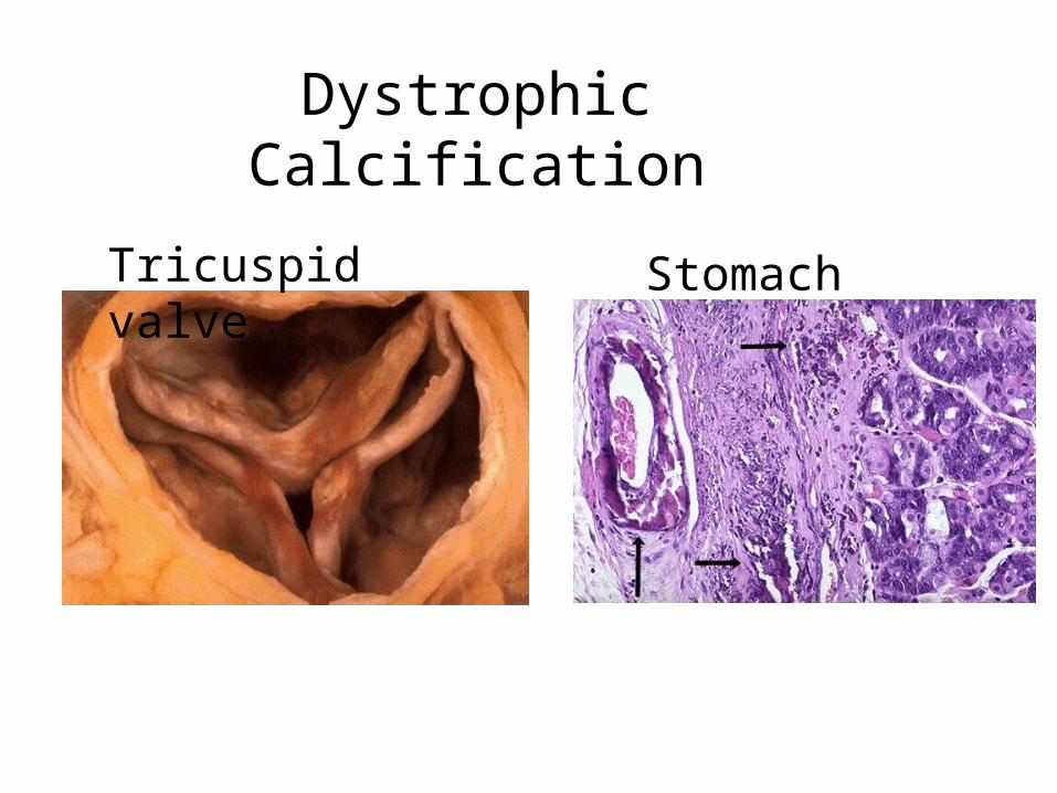
Dystrophic Calcification
Tricuspid valve Stomach

Metastatic Calcification
• Must be associated with elevated Calcium.• Occurs in vital tissues• Aetiology:
– Increased PTH» Hyperparathyroidism
– Bone destruction» Tumour (MM, Leukaemia), Skeletal Mets (Breast Ca.)» Increased bone turnover (Pagets), Immobilisation.
– Vitamin D related disorders» Excess Vitamin D» Sarcoidosis
– Renal Failure» Retention of Phosphate, (secondary HyperPTH)

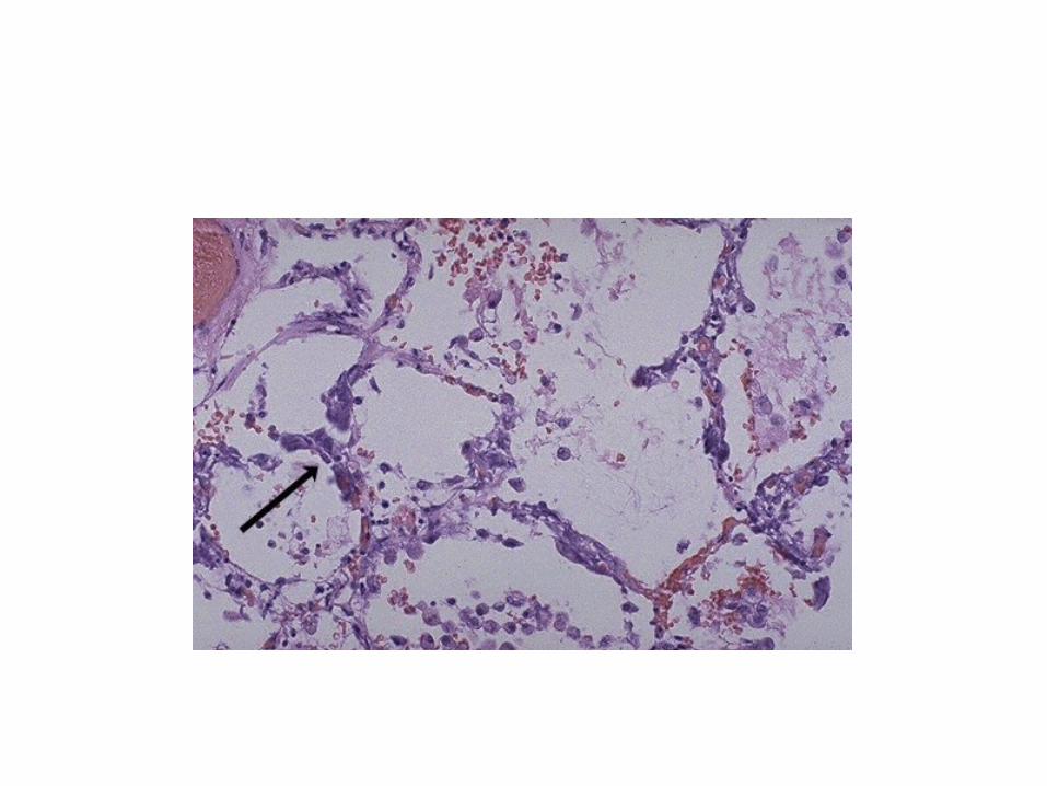
Metastatic Calcification
• Kidney: Around tubules - Renal Failure.• Lung: Alveolar walls.• Stomach: Fundal glands.
Identification of Calcium:
Von Kossa
2nd Year Pathology 2009

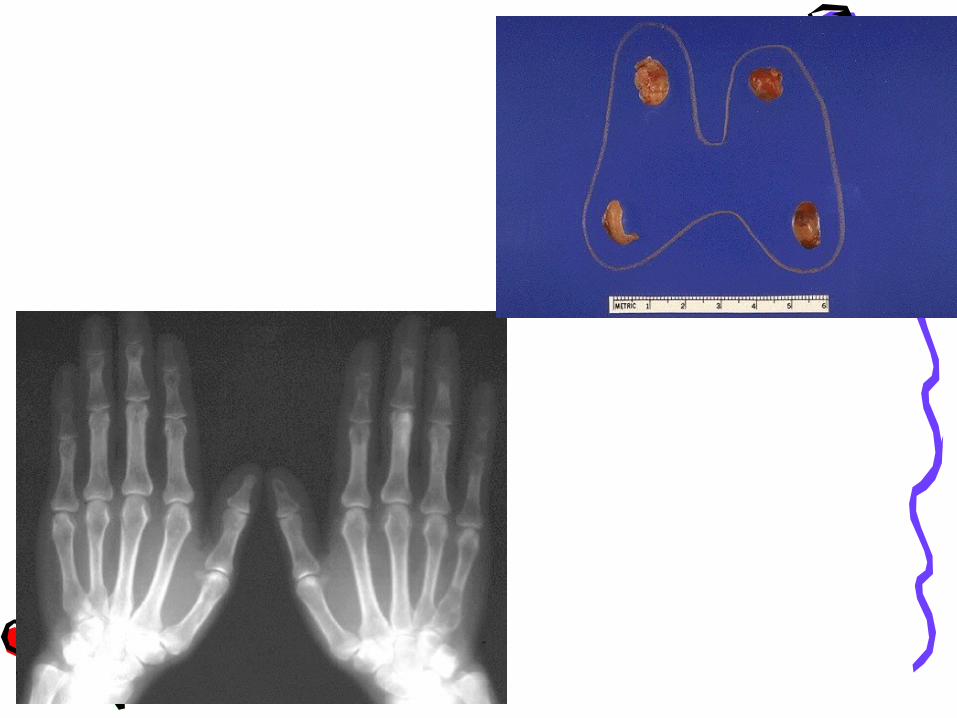
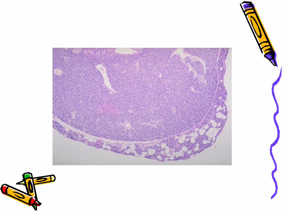
Hyperparathyroidism
•Primary, Secondary, Tertiary•1° = Increased bone re-absorbtion and mobilization of calcium from bone
Increased renal tubule re-absorption.
Increased Vit. D activity
“ Bones, stones , (psychic moans) and abdominal
groans”
2nd Year Pathology 2009
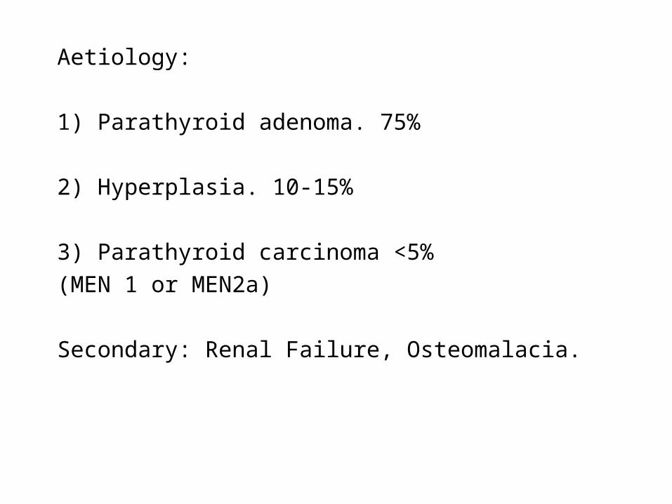
Aetiology:
1) Parathyroid adenoma. 75%
2) Hyperplasia. 10-15%
3) Parathyroid carcinoma <5%
(MEN 1 or MEN2a)
Secondary: Renal Failure, Osteomalacia.

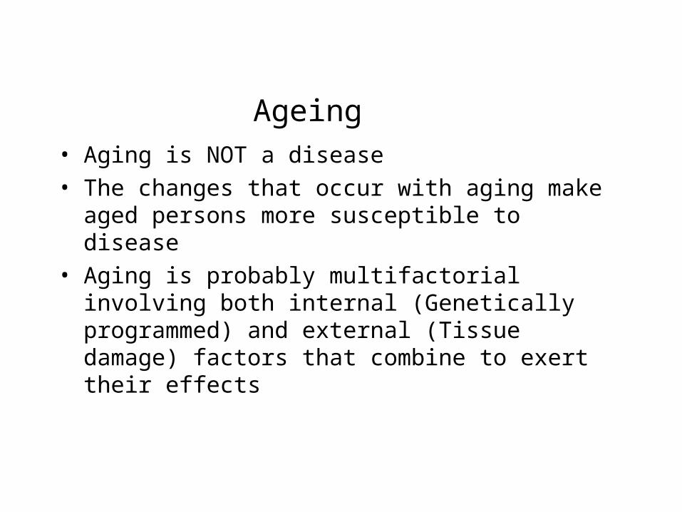
Ageing• Aging is NOT a disease• The changes that occur with aging make aged
persons more susceptible to disease • Aging is probably multifactorial involving both internal
(Genetically programmed) and external (Tissue damage) factors that combine to exert their effects
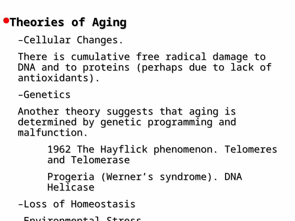
Theories of AgingTheories of Aging
–Cellular ChangesCellular Changes..
There is cumulative free radical damage to DNA and to proteins There is cumulative free radical damage to DNA and to proteins (perhaps due to lack of antioxidants(perhaps due to lack of antioxidants)). .
–Genetics Genetics
Another theory suggests that aging is determined by genetic Another theory suggests that aging is determined by genetic programming and malfunctionprogramming and malfunction. .
1962 The Hayflick phenomenon. Telomeres and Telomerase1962 The Hayflick phenomenon. Telomeres and Telomerase
Progeria (Werner’s syndrome). DNA HelicaseProgeria (Werner’s syndrome). DNA Helicase
–Loss of HomeostasisLoss of Homeostasis
–Environmental StressEnvironmental Stress
–Neuroendocrine DysfunctionNeuroendocrine Dysfunction
–NutritionNutrition

Organ Systems• Central Nervous System
– Stroke.
– Alzheimer’s.
– Neuronal loss.
• Eye– Cataracts.
– Presbyopia.
• Ear– Presbycusis.
2nd Year Pathology 2009
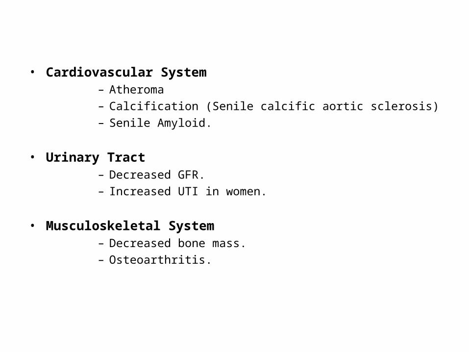
• Cardiovascular System– Atheroma
– Calcification (Senile calcific aortic sclerosis)
– Senile Amyloid.
• Urinary Tract– Decreased GFR.
– Increased UTI in women.
• Musculoskeletal System– Decreased bone mass.
– Osteoarthritis.

• Genital Tract– Menopause leads to atrophy of ovaries, uterus, and
breasts. The epithelium of the vagina and vulva also become thinner.
– Prostatic Hyperplasia.
• Skin– Decreased elasticity.– Senile lentigines
2nd Year Pathology 2009

Telomere
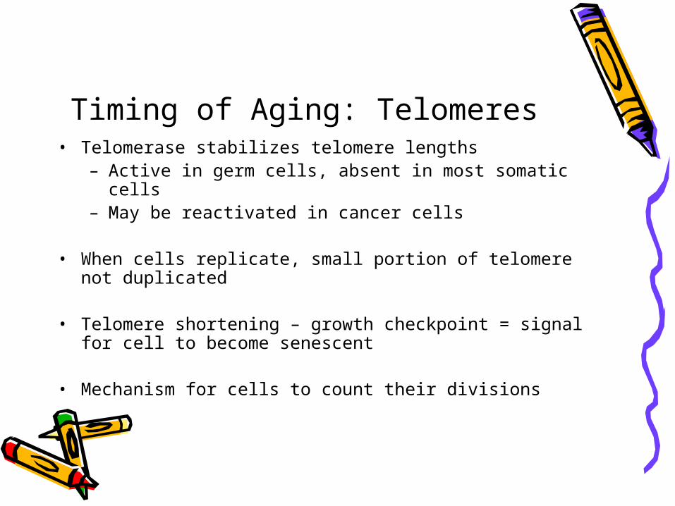
Timing of Aging: Telomeres• Telomerase stabilizes telomere lengths
– Active in germ cells, absent in most somatic cells– May be reactivated in cancer cells
• When cells replicate, small portion of telomere not duplicated
• Telomere shortening – growth checkpoint = signal for cell to become senescent
• Mechanism for cells to count their divisions
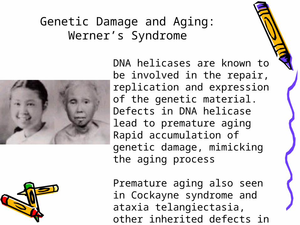
Genetic Damage and Aging: Werner’s Syndrome
DNA helicases are known to be involved in the repair, replication and expression of the genetic material.Defects in DNA helicase lead to premature agingRapid accumulation of genetic damage, mimicking the aging process
Premature aging also seen in Cockayne syndrome and ataxia telangiectasia, other inherited defects in DNA repair.

Summary• Ageing• Effects a combination of
– Intrinsic factors: telomere lengths– Extrinsic factors: progressive DNA (and other
cellular) damage– The latter may be accelerated by inherited defects
in DNA repair.

Summary• Amyloid.
– Appearance / identification– Types of acquired and inherited amyloidosis
• Pathological calcification.– Dystrophic / Metastatic / Idiopathic– Causes and Consequences
• Ageing– Theories of Aging