3. health effects these kinds of health effects data become available and methods ... limited number...
TRANSCRIPT
FLUORIDES, HYDROGEN FLUORIDE, AND FLUORINE 29
3. HEALTH EFFECTS
3.1 INTRODUCTION
The primary purpose of this chapter is to provide public health officials, physicians, toxicologists, and
other interested individuals and groups with an overall perspective on the toxicology of fluorides,
hydrogen fluoride, and fluorine. It contains descriptions and evaluations of toxicological studies and
epidemiological investigations and provides conclusions, where possible, on the relevance of toxicity and
toxicokinetic data to public health.
A glossary and list of acronyms, abbreviations, and symbols can be found at the end of this profile.
The term fluoride properly refers to numerous natural and synthesized compounds that are derived from
hydrofluoric acid. This class of chemicals is commonly referred to as fluorides. Some of these
compounds, such as oxygen difluoride, are very reactive and highly toxic. Because of their reactivity,
these compounds would not migrate unchanged from a hazardous waste site. Fluoride salts, such as
sodium fluoride and calcium fluoride, are much less reactive and much less toxic. Since the fluoride ion
is the toxicologically active agent, and discussion of water fluoridation uses the term fluoride, the term
fluoride is used generically in this profile to refer to toxicology of fluoride salts. Because numerous
different fluoride compounds exist naturally in the environment and have varying chemical properties, the
term fluorides is used in the discussion of environmental media. Most of the available literature on
fluoride toxicity concerns sodium fluoride. Additional toxicity literature is available on some other forms
of fluoride, such as stannous fluoride. Other forms of fluoride are discussed only if exposure is likely to
occur at a hazardous waste site. (Such exposure to stannous fluoride is not likely.) Wherever the form of
fluoride exposure is known, that salt is identified in the profile.
Hydrogen fluoride is also a gas and is very water soluble. It dissolves readily in any water present in the
air or other media. When hydrogen fluoride is dissolved in water, it is called hydrofluoric acid. Although
hydrofluoric acid is very corrosive and can etch glass, it is a weak acid, meaning that it can be present in
water as an undissociated molecule. However, in dilute solutions, it is almost completely ionized; salts
are formed if cations are available. Due to formation of complexes, very concentrated solutions of
hydrofluoric acid are also largely ionic in nature. Therefore, a hydrogen fluoride or hydrofluoric acid
spill would result in contamination with fluoride ion, but hydrogen fluoride or hydrofluoric acid would
FLUORIDES, HYDROGEN FLUORIDE, AND FLUORINE 30
3. HEALTH EFFECTS
not be of concern outside the immediate vicinity of the spill. However, while members of the public are
only likely to come into contact with fluoride contamination, clean-up workers could be exposed to
hydrogen fluoride/hydrofluoric acid. In this profile, hydrogen fluoride is used to refer to the gas, while
hydrofluoric acid is used to refer to the liquid form. When both forms are included, the term hydrogen
fluoride is used.
Fluorine is a gaseous element that occurs only in very low concentrations in the environment in the
absence of anthropogenic sources (see Chapter 6 for further discussion). Because it is strongly
electronegative, it is rarely found in the environment in the elemental state, nor is it likely to be found in
the environment near toxic waste sites as molecular fluorine.
Limited information also exists concerning occupational exposure to the mineral cryolite (Na3AlF6),
sometimes with concomitant exposure to hydrogen fluoride. Because these exposures usually involve
exposure to both hydrogen fluoride and cryolite, sometimes along with exposure to other fluoride dusts,
they are discussed separately in the profile.
This profile will discuss data, or the absence of data, concerning the toxicity of inorganic compounds of
fluorine that people could be exposed to at a hazardous waste site. Exposure and toxicity are discussed
separately for fluoride, hydrogen fluoride/hydrofluoric acid, and fluorine. Toxic effects of occupational
exposure in aluminum reduction plants, where exposures to hydrogen fluoride, fluoride dusts, and cryolite
all occur, are also discussed separately. Because the toxic effects of fluorine are largely due to the action
of the fluorine molecule on the respiratory tract or other exposed surfaces, fluorine exposure is reported as
exposure to a level of diatomic fluorine. By contrast, systemic effects of hydrogen fluoride are due to the
fluoride ion, so concentrations of hydrogen fluoride are converted to fluoride equivalents. All doses of
fluoride are reported as the amount of fluoride ion.
The primary routes and durations of concern vary with the different fluorine compounds. In general, the
more soluble the fluoride, the more that can be absorbed by oral ingestion, and the more toxic it is. The
primary exposure routes and duration for hydrofluoric acid are the inhalation or dermal routes, related to
acute occupational exposure, while the primary exposure route and duration for fluoride is chronic oral
exposure to fluoride in the drinking water, food, and fluoride-containing dental products. Therefore, most
of the information for the inhalation and dermal routes comes from studies of acute exposure to fluorine
or hydrofluoric acid, while most of the information regarding the oral route is based on sodium fluoride.
The toxicity following inhalation or dermal exposure to other inorganic fluorine compounds differs from

FLUORIDES, HYDROGEN FLUORIDE, AND FLUORINE 31
3. HEALTH EFFECTS
that of hydrofluoric acid. Similarly, oral exposure to various fluorides other than sodium fluoride may
result in different toxic effects.
3.2 DISCUSSION OF HEALTH EFFECTS BY ROUTE OF EXPOSURE
To help public health professionals and others address the needs of persons living or working near
hazardous waste sites, the information in this section is organized first by route of exposure (inhalation,
oral, and dermal) and then by health effect (death, systemic, immunological, neurological, reproductive,
developmental, genotoxic, and carcinogenic effects). These data are discussed in terms of three exposure
periods: acute (14 days or less), intermediate (15–364 days), and chronic (365 days or more).
Levels of significant exposure for each route and duration are presented in tables and illustrated in
figures. The points in the figures showing no-observed-adverse-effect levels (NOAELs) or lowest-
observed-adverse-effect levels (LOAELs) reflect the actual doses (levels of exposure) used in the studies.
LOAELs have been classified into "less serious" or "serious" effects. "Serious" effects are those that
evoke failure in a biological system and can lead to morbidity or mortality (e.g., acute respiratory distress
or death). "Less serious" effects are those that are not expected to cause significant dysfunction or death,
or those whose significance to the organism is not entirely clear. ATSDR acknowledges that a
considerable amount of judgment may be required in establishing whether an end point should be
classified as a NOAEL, "less serious" LOAEL, or "serious" LOAEL, and that in some cases, there will be
insufficient data to decide whether the effect is indicative of significant dysfunction. However, the
Agency has established guidelines and policies that are used to classify these end points. ATSDR
believes that there is sufficient merit in this approach to warrant an attempt at distinguishing between
"less serious" and "serious" effects. The distinction between "less serious" effects and "serious" effects is
considered to be important because it helps the users of the profiles to identify levels of exposure at which
major health effects start to appear. LOAELs or NOAELs should also help in determining whether or not
the effects vary with dose and/or duration, and place into perspective the possible significance of these
effects to human health.
The significance of the exposure levels shown in the Levels of Significant Exposure (LSE) tables and
figures may differ depending on the user's perspective. Public health officials and others concerned with
appropriate actions to take at hazardous waste sites may want information on levels of exposure
associated with more subtle effects in humans or animals (LOAELs) or exposure levels below which no

FLUORIDES, HYDROGEN FLUORIDE, AND FLUORINE 32
3. HEALTH EFFECTS
adverse effects (NOAELs) have been observed. Estimates of levels posing minimal risk to humans
(Minimal Risk Levels or MRLs) may be of interest to health professionals and citizens alike.
Estimates of exposure levels posing minimal risk to humans (Minimal Risk Levels or MRLs) have been
made for fluorides, hydrogen fluoride, and fluorine. An MRL is defined as an estimate of daily human
exposure to a substance that is likely to be without an appreciable risk of adverse effects (noncarcino-
genic) over a specified duration of exposure. MRLs are derived when reliable and sufficient data exist to
identify the target organ(s) of effect or the most sensitive health effect(s) for a specific duration within a
given route of exposure. MRLs are based on noncancerous health effects only and do not consider
carcinogenic effects. MRLs can be derived for acute, intermediate, and chronic duration exposures for
inhalation and oral routes. Appropriate methodology does not exist to develop MRLs for dermal
exposure.
Although methods have been established to derive these levels (Barnes and Dourson 1988; EPA 1990),
uncertainties are associated with these techniques. Furthermore, ATSDR acknowledges additional
uncertainties inherent in the application of the procedures to derive less than lifetime MRLs. As an
example, acute inhalation MRLs may not be protective for health effects that are delayed in development
or are acquired following repeated acute insults, such as hypersensitivity reactions, asthma, or chronic
bronchitis. As these kinds of health effects data become available and methods to assess levels of
significant human exposure improve, these MRLs will be revised.
A User's Guide has been provided at the end of this profile (see Appendix B). This guide should aid in
the interpretation of the tables and figures for Levels of Significant Exposure and the MRLs.
3.2.1 Inhalation Exposure
Inhalation exposure most commonly occurs in an occupational setting. As discussed above, most of the
available information concerning toxic effects of fluorine and its compounds following inhalation
exposure comes from studies of exposure to hydrogen fluoride or hydrofluoric acid. There are also a
limited number of useful studies concerning inhalation exposure to fluorine or particulates of inorganic
fluoride compounds. However, no animal studies were located regarding toxic effects of exposure to the
particulate fluoride compounds. Toxic effects of hydrogen fluoride are discussed in all of the following
sections. Where toxicity data exist for fluoride or fluorine, these substances are also discussed.

FLUORIDES, HYDROGEN FLUORIDE, AND FLUORINE 33
3. HEALTH EFFECTS
Acute inhalation of hydrogen fluoride following facial splashes with hydrofluoric acid can cause
bronchiolar ulceration, pulmonary hemorrhage and edema, and death. In addition, renal and hepatic
damage have been observed in animal studies. Many of the human studies regarding inhalation of
hydrogen fluoride fumes also involved dermal exposure; in such cases, it is difficult to determine which
effects are specific to the inhalation route. However, the respiratory effects of hydrogen fluoride appear
to be inhalation-specific, because they have not been reported in cases where there was clearly no
inhalation exposure. The effects of combined inhalation and dermal exposure to hydrofluoric acid are
also discussed in Section 3.2.3.
Fluorine gas is extremely irritating. The primary health effects of acute fluorine inhalation are nasal and
eye irritation (at low levels), and death due to pulmonary edema (at high levels). In animals, renal and
hepatic damage have also been observed.
The major health effect of chronic inhalation exposure to fluoride is skeletal fluorosis, which has been
reported in cases of exposure to fluoride dusts and hydrogen fluoride, either individually or in
combination.
3.2.1.1 Death
Both hydrogen fluoride and fluorine can cause lethal pulmonary edema, although cardiac effects also
contribute to the toxicity of hydrogen fluoride. The reported LC50 values for hydrogen fluoride in rats for
a given duration are generally at least 3.5 times higher than the value for fluorine (as diatomic fluorine) in
rats for the same duration. Although strain differences could account for some of this difference, the
LC50 values of hydrogen fluoride in Crl:CD®BR and Wistar-derived rats were very similar.
Hydrogen Fluoride. Acute inhalation of hydrogen fluoride fumes in combination with dermal exposure
to hydrofluoric acid has been reported to cause death in humans. Actual exposure concentrations are not
known in any of these cases. Death was generally due to pulmonary edema (resulting from irritation and
constriction of the airways) or to cardiac arrhythmias with pronounced hyperkalemia, hypocalcemia, and
hypomagnesemia.
The death of a chemist who sustained first- and second-degree burns of the face, hands, and arms when a
vat containing hydrofluoric acid accidentally ruptured has been described (Kleinfeld 1965). This 29-year-
old male died 10 hours after admission to the hospital. Postmortem examination revealed severe

FLUORIDES, HYDROGEN FLUORIDE, AND FLUORINE 34
3. HEALTH EFFECTS
tracheobronchitis and hemorrhagic pulmonary edema. A petroleum refinery worker was splashed in the
face with 100% anhydrous hydrofluoric acid (Tepperman 1980). The absorption of fluoride produced
acute systemic fluoride poisoning with profound hypocalcemia and hypomagnesemia and cardiac
arrhythmias. The patient died <24 hours after exposure; autopsy revealed pulmonary edema. A young
woman splashed in the face with hydrofluoric acid died of respiratory insufficiency a few hours after
exposure (Chela et al. 1989). The autopsy revealed severe burns of the skin and lungs, with hemorrhagic
pulmonary edema produced by hydrofluoric acid and its vapor.
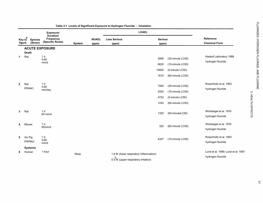
The lethal concentration of hydrogen fluoride has been investigated in rats, mice, and guinea pigs. It
appears that mice are more sensitive to the acute effects of hydrogen fluoride than rats, and rats are more
sensitive than guinea pigs. The 15-minute LC50 values for hydrogen fluoride were 4,327 ppm fluoride for
guinea pigs and 2,555 ppm fluoride for Wistar-derived rats (Rosenholtz et al. 1963). The 60-minute
LC50 values for hydrogen fluoride were 325 ppm fluoride in ICR-derived mice (Wohlslagel et al. 1976),
1,325 ppm fluoride in Sprague-Dawley-derived rats (Wohlslagel et al. 1976), and 1,242 ppm fluoride in
Wistar-derived rats (Rosenholtz et al. 1963). In a study (Dalbey et al. 1998a) comparing the toxicity of
hydrogen fluoride in rats using mouth-breathing (rats fitted with a tracheal cannula) and nose-breathing
models, dramatic differences in lethality were observed. In the mouth-breathing rats, 50 and 80% of the
animals died within 2 weeks of a 10-minute mouth breathing exposure to 3,655 or 6,663 ppm fluoride,
respectively. In contrast, no deaths were noted following exposure to these concentrations using the nose-
breathing model. The difference in lethality between the two models is probably due to the higher dose of
hydrogen fluoride reaching the lower airways in the mouth-breathing model.
The LC50 values reported by Haskell Laboratory (1988) for Crl:CD®BR rats were much higher than the
values reported by the above investigators, although the size of the discrepancy decreased with longer
exposure durations. For example, the 15-minute LC50 was reported as 6,620 ppm, while the 60-minute
LC50 was 1,610 ppm. Although the concentration of hydrogen fluoride that produced death was reported
to be lower when it was administered to rats in humid air (Haskell Laboratory 1988), the method for
measuring fluoride in humid air may not have given accurate results. This limitation was recognized by
the authors, who stated that the collection efficiency of the sampling train for aerosols was not evaluated.
Longer-term effects of hydrogen fluoride were investigated by exposing various species to 8.2 or 31 ppm
fluoride as hydrogen fluoride for 6 hours/day, 6 days/week for 5 weeks (Stokinger 1949). Humidity was
47–97% at the lower concentration and 48–66% at the higher concentration. Marked species differences
were observed. All rats and mice exposed to 31 ppm died, but no guinea pigs, rabbits, or dogs exposed at
FLUORIDES, HYDROGEN FLUORIDE, AND FLUORINE 35
3. HEALTH EFFECTS
this level died. No animal of any species died following exposure to 8.2 ppm. In an experiment where
five rabbits, three guinea pigs, and two Rhesus monkeys were exposed to 18 ppm for 6–7 hours/day,
5 days/week for 50 days (309 hours total), the only deaths observed were two guinea pigs (Machle and
Kitzmiller 1935). Exposure of one of these animals stopped after 134 hours of exposure, and exposure of
the other one stopped after 160 hours, when marked weight loss was observed. Nevertheless, the animals
died about 2 weeks later.
Fluorine. No information was located on death in humans caused by fluorine. Fluorine toxicity has been
investigated in Osborne-Mendel rats, Swiss-Webster mice, New England guinea pigs, and New Zealand
rabbits (Keplinger and Suissa 1968). Similar values for the LC50 were calculated for the different species.
In the rats, the LC50 values for exposures of 5, 15, 30, and 60 minutes were 700, 390, 270, and 185 ppm,
respectively. At concentrations near the LC50, few signs of intoxication were observed immediately after
exposure, except for irritation of the eyes and nose. Several hours after exposure, the animals exhibited
lethargy, dyspnea, and general weakness. Except at concentrations above the LC90, death generally
occurred 12–18 hours after exposure. Animals that survived for 48 hours generally survived for the
duration of the observation period. Loss of body weight was also observed, but was considered
nonspecific and was attributed to anorexia.
Toxic effects of inhalation exposure to fluorine and hydrogen fluoride were compared in rats, mice,
rabbits, and guinea pigs (Stokinger 1949). Lethal doses from fluorine exposure determined by this group
are about 3–4 times those determined by Keplinger and Suissa (1968), but quantitative exposure level
data from these experiments are not reliable due to technical problems in monitoring fluorine gas levels.
However, qualitative results from these experiments are useful. These experiments also found that
fluorine was more toxic than hydrogen fluoride.
There are some indications that preexposure to low levels of fluorine may provide resistance to lethal
effects of fluorine. Increases in survival time were observed in rabbits exposed to 50 ppm fluorine for
30 minutes, 1 day/week for 4 weeks prior to exposure to a lethal concentration of fluorine (400 ppm for
30 minutes) (Keplinger 1969). Survival time in the rabbits was 48 hours, as compared to 18 hours or less
in similarly exposed rabbits not preexposed to fluorine. In mice, slight increases in LC50 values were
found in animals receiving a single exposure to 30–45 ppm fluorine prior to exposure to lethal
concentrations. However, slight decreases in LC50 values were seen in mice preexposed to 25 ppm
fluorine (Keplinger 1969). No mechanism for the possible tolerance was suggested.
FLUORIDES, HYDROGEN FLUORIDE, AND FLUORINE 36
3. HEALTH EFFECTS
Repeated exposures of rats, mice, guinea pigs, and rabbits to 0.5, 2, 5, or 18 ppm fluorine were conducted
for up to 178 hours over 35 days (Stokinger 1949). The exposure regimen was not stated, but appears to
be 6 hours/day, 6 days/week. The exposure levels at these lower concentrations were considered fairly
reliable. Guinea pigs and rats were less sensitive to lethal effects than were rabbits or dogs. All of the
rabbits and dogs exposed to 5 ppm and mice exposed to 18 ppm died, while only half of the rats and
guinea pigs exposed to 18 ppm died. Most animals exposed to 2 ppm survived.
The LC50 values for each species and duration category of exposure to hydrogen fluoride are recorded in
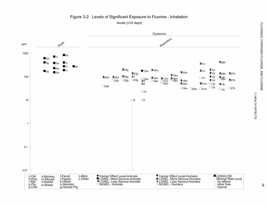
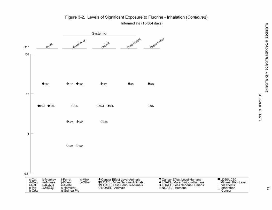
Table 3-1 and plotted in Figure 3-1. The LC50 values for each species and duration category of exposure
to fluorine are recorded in Table 3-2 and plotted in Figure 3-2.
3.2.1.2 Systemic Effects
The highest NOAEL values and all reliable LOAEL values for systemic effects in each species and
duration category of exposure to hydrogen fluoride are recorded in Table 3-1 and plotted in Figure 3-1.
The highest NOAEL values and all reliable LOAEL values for systemic effects in each species and
duration category of exposure to fluorine are recorded in Table 3-2 and plotted in Figure 3-2. The highest
NOAEL values and all reliable LOAEL values for systemic effects in each species and duration category
of exposure to fluoride are recorded in Table 3-3 and plotted in Figure 3-3.
Respiratory Effects.
Hydrogen Fluoride. Acute inhalation of 122 ppm fluoride as hydrogen fluoride by two male volunteers
produced marked respiratory irritation within 1 minute (Machle et al. 1934). Pulmonary edema,
pulmonary hemorrhagic edema, and tracheobronchitis have been reported in cases of people being
splashed in the face with hydrofluoric acid, where concurrent inhalation and dermal exposures are likely
(Chan et al. 1987; Chela et al. 1989; Dieffenbacher and Thompson 1962; Kleinfeld 1965; Tepperman
1980). Exposure concentrations were not known in these cases. In another case, a women developed
herrhagic alveolitis and adult respiratory disease syndrome following exposure to a presumably high
concentration of hydrogen fluoride from a cleaning product (Bennion and Franzblau 1997).
Two human experimental studies have been conducted to assess the ability of hydrogen fluoride to induce
respiratory tract inflammation. In the first study, increases in upper airway symptoms were observed in
LOAEL
Less SeriousNOAEL(ppm) (ppm)
Seriousa
(ppm)System
Exposure/Duration/
Frequency(Specific Route)
Species(Strain)
Key tofigure
Reference
Table 3-1 Levels of Significant Exposure to Hydrogen Fluoride - Inhalation
Chemical Form
ACUTE EXPOSUREDeath
1 1 d5-60min/d
Haskell Laboratory 1988
hydrogen fluoride2890 (30-minute LC50)
6620 (15-minute LC50)
14600 (5-minute LC50)
1610 (60-minute LC50)
Rat
2(Wistar)
1 d5-60min/day
Rosenholtz et al. 1963
hydrogen fluoride1940 (30-minute LC50)
2555 (15-minute LC50)
4722 (5-minute LC50)
1242 (60-minute LC50)
Rat
3 1 d60 min/d
Wohlslagel et al. 1976
hydrogen fluoride1325 (60-minuteLC50)
Rat
4 1 d60min/d
Wohlslagel et al. 1976
hydrogen fluoride325 (60-minute LC50)
Mouse
5(Hartley)
1 d5-60min/d
Rosenholtz et al. 1963
hydrogen fluoride4327 (15-minute LC50)
Gn Pig
Systemic6
Resp1 hour Lund et al. 1999; Lund et al. 1997
hydrogen fluoride1.9 (lower respiratory inflammation)M
0.5b
(upper respiratory irritation)M
Human
LOAEL
Less SeriousNOAEL(ppm) (ppm)
Seriousa
(ppm)System
Exposure/Duration/
Frequency(Specific Route)
Species(Strain)
Key tofigure
Reference
(continued)Table 3-1 Levels of Significant Exposure to Hydrogen Fluoride - Inhalation
Chemical Form
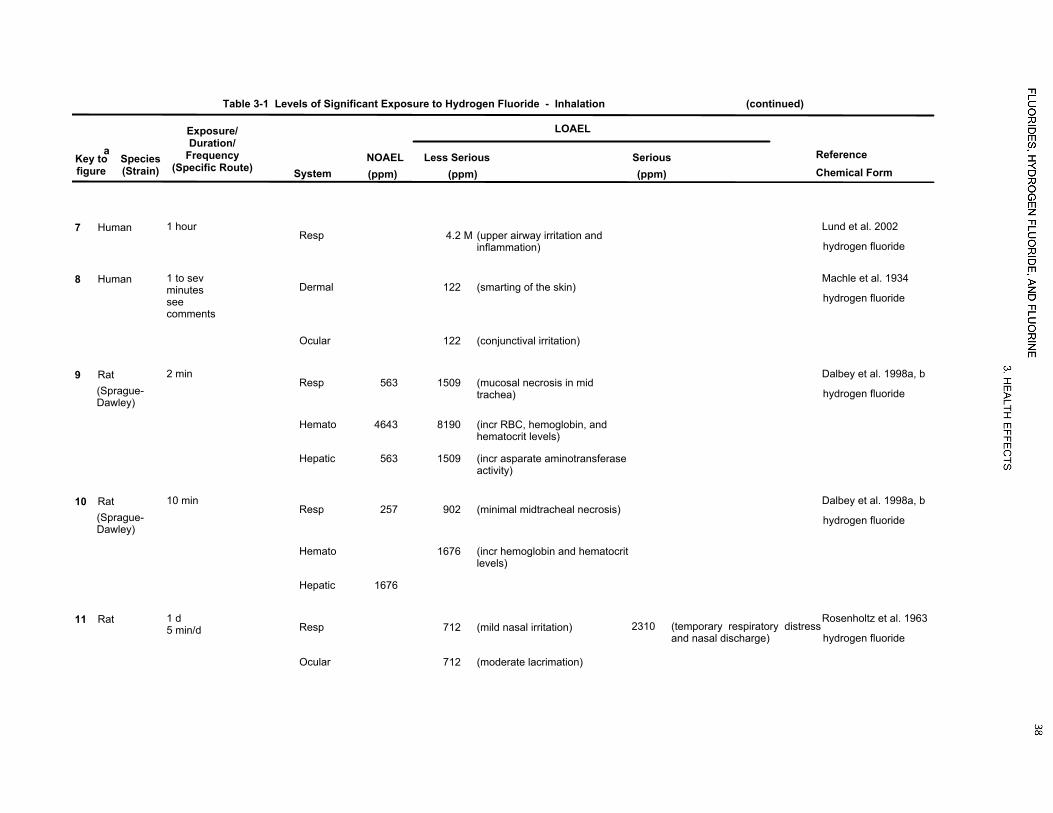
7Resp
1 hour Lund et al. 2002
hydrogen fluoride4.2 (upper airway irritation and
inflammation)M
Human
8Dermal
1 to sevminutesseecomments
Machle et al. 1934
hydrogen fluoride122 (smarting of the skin)
Human
Ocular 122 (conjunctival irritation)
9563
(Sprague-Dawley)
Resp2 min Dalbey et al. 1998a, b
hydrogen fluoride1509 (mucosal necrosis in mid
trachea)
Rat
4643Hemato 8190 (incr RBC, hemoglobin, andhematocrit levels)
563Hepatic 1509 (incr asparate aminotransferaseactivity)
10257
(Sprague-Dawley)
Resp10 min Dalbey et al. 1998a, b
hydrogen fluoride902 (minimal midtracheal necrosis)
Rat
Hemato 1676 (incr hemoglobin and hematocritlevels)
1676Hepatic
11Resp
1 d5 min/d
Rosenholtz et al. 1963
hydrogen fluoride712 (mild nasal irritation) 2310 (temporary respiratory distress
and nasal discharge)
Rat
Ocular 712 (moderate lacrimation)

LOAEL
Less SeriousNOAEL(ppm) (ppm)
Seriousa
(ppm)System
Exposure/Duration/
Frequency(Specific Route)
Species(Strain)
Key tofigure
Reference
(continued)Table 3-1 Levels of Significant Exposure to Hydrogen Fluoride - Inhalation
Chemical Form
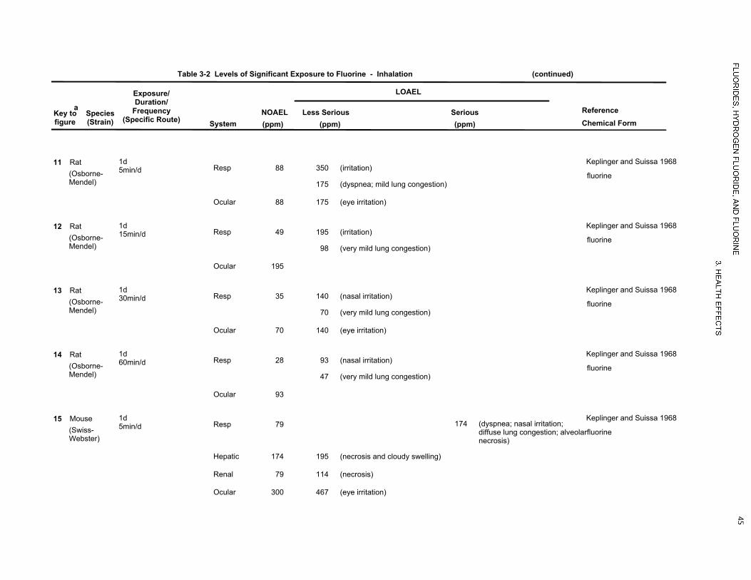
12292Resp
1 d15min/d
Rosenholtz et al. 1963
hydrogen fluoride357 (nasal irritation) 1339 (temporary respiratory distress
and nasal discharge)
Rat
292Ocular 357 (lacrimation)
1398Resp
1 d60min/d
Rosenholtz et al. 1963
hydrogen fluoride120 (nasal irritation) 465 (temporary respiratory distress
and nasal discharge)
Rat
98Ocular 120 (lacrimation)
14(Fischer- 344)
Resp30 min Stavert et al. 1991
hydrogen fluoride1235 (fibrinonecrotic rhinitis in nose
breathing rats; tracheal andbronchial necrosis in mouthbreathing rats)
Rat
Bd Wt 1235 (10% body weight reduction)
INTERMEDIATE EXPOSUREDeath
15(NS)
5 wks6d/wk6hr/d
Stokinger 1949
hydrogen fluoride31 (100% mortality)
Rat
16(NS)
5 wks6d/wk6hr/d
Stokinger 1949
hydrogen fluoride31 (100% mortality)
Mouse
Systemic17
Resp15-50 d6 hr/d
Largent 1960
hydrogen fluoride2.98 (slight nasal irritation)
Human
Dermal 2.98 (stinging sensation on skin)
Ocular 2.98 (stinging sensation in eyes)

LOAEL
Less SeriousNOAEL(ppm) (ppm)
Seriousa
(ppm)System
Exposure/Duration/
Frequency(Specific Route)
Species(Strain)
Key tofigure
Reference
(continued)Table 3-1 Levels of Significant Exposure to Hydrogen Fluoride - Inhalation
Chemical Form
188.2
(NS)Resp
5 wks6hr/d
Stokinger 1949
hydrogen fluoride31 (pulmonary hemorrhage)
Rat
31Hemato
8.2Renal 31 (cortical necrosis)
Ocular 8.2 (subcutaneous hemorrhagearound the eyes and on thefeet)
19(NS)
Dermal5 wks6d/wk6hr/d
Stokinger 1949
hydrogen fluoride8.2 (subcutaneous hemorrhage
around the eyes and on thefeet)
Mouse
20(NS)
Resp5 wks6d/wk6hr/d
Stokinger 1949
hydrogen fluoride31 (pulmonary hemorrhage)
Dog
31Hemato
218.2
(NS)Resp
5 wks6d/wk6hr/d
Stokinger 1949
hydrogen fluoride31 (pulmonary hemorrhage)
Rabbit
31HematoNeurological
220.01
(albino)
a The number corresponds to entries in Figure 3-1.
b Used to derive an acute-duration inhalation minimal risk level (MRL) of 0.02 ppm; the concentration was divided by an uncertainty factor of 30 (3 for use of a minimal LOAEL and 10to account for human variability).
Bd = body weight; d = day(s); Hemato = hematological; hr = hour(s); incr = increase; LC50 = lethal concentration, 50% kill; LOAEL = lowest-observed-adverse-effect level; min =minute(s); mo = month(s); NOAEL = no-observed-adverse-effect level; ppm = parts per million; RBC = red blood cell; Resp = respiratory; wk = week(s)
5mo24hr/d
Sadilova et al. 1965
hydrogen fluoride0.03 (disturbances in conditioned
reflexes; lengthened latentperiods)
Rat

0.01
0.1
1
10
100
1000
10000
100000
Death
5g
4m
1r
1r
1r1r
2r2r2r2r 3r
Respiratory
6
6
7
9r
9r10r
10r
11r
11r
12r
12r
12r13r
13r 13r
14r
Hematological
9r9r
10r
Hepatic
9r
9r 10r
Dermal
8
Ocular
8
11r
12r 12r
13r 13r
Body Weight
14r
ppm
Figure 3-1. Levels of Significant Exposure to Hydrogen Fluoride - InhalationAcute (≤14 days)
c-Catd-Dogr-Ratp-Pigq-Cow
k-Monkeym-Mouseh-Rabbita-Sheep
f-Ferretj-Pigeone-Gerbils-Hamsterg-Guinea Pig
n-Minko-Other
Cancer Effect Level-Animals LOAEL, More Serious-Animals LOAEL, Less Serious-Animals NOAEL - Animals
Cancer Effect Level-Humans LOAEL, More Serious-Humans LOAEL, Less Serious-Humans NOAEL - Humans
LD50/LC50 Minimal Risk Level for effects other than Cancer
Systemic
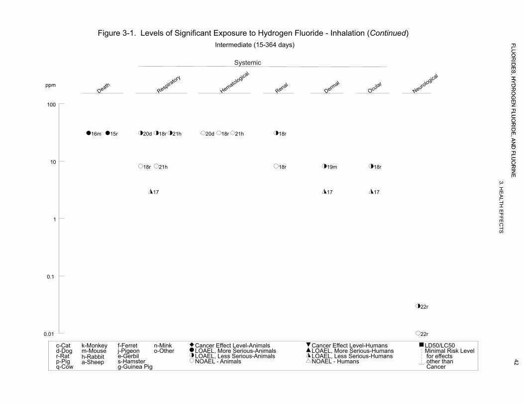
0.01
0.1
1
10
100
Death
16m 15r
Respiratory
20d
17
18r
18r
21h
21h
Hematological
20d 18r 21h
Renal
18r
18r
Dermal
17
19m
Ocular
17
18r
Neurological
22r
22r
ppm
Figure 3-1. Levels of Significant Exposure to Hydrogen Fluoride - Inhalation (Continued)Intermediate (15-364 days)
c-Catd-Dogr-Ratp-Pigq-Cow
k-Monkeym-Mouseh-Rabbita-Sheep
f-Ferretj-Pigeone-Gerbils-Hamsterg-Guinea Pig
n-Minko-Other
Cancer Effect Level-Animals LOAEL, More Serious-Animals LOAEL, Less Serious-Animals NOAEL - Animals
Cancer Effect Level-Humans LOAEL, More Serious-Humans LOAEL, Less Serious-Humans NOAEL - Humans
LD50/LC50 Minimal Risk Level for effects other than Cancer
Systemic

LOAEL
Less SeriousNOAEL(ppm) (ppm)
Seriousa
(ppm)System
Exposure/Duration/
Frequency(Specific Route)
Species(Strain)
Key tofigure
Reference
Table 3-2 Levels of Significant Exposure to Fluorine - Inhalation
Chemical Form
ACUTE EXPOSUREDeath
1(Osborne-Mendel)
1d5-60min/d
Keplinger and Suissa 1968
fluorine270 (30-minute LC50)
390 (15-minute LC50)
700 (5-minute LC50)
185 (60-minute LC50)
Rat
2(Swiss-Webster)
1d15-60min/d
Keplinger and Suissa 1968
fluorine225 (30-minute LC50)
375 (15-minute LC50)
600 (5-minute LC50)
150 (60-minute LC50)
Mouse
3(NewEngland)
1d15-60min/d
Keplinger and Suissa 1968
fluorine395 (15-minute LC50)
170 (60-minute LC50)
Gn Pig
4(NewZealand)
1d5-30min/d
Keplinger and Suissa 1968
fluorine820 (5-minute LC50)
270 (30-minute LC50)
Rabbit
Systemic5
Dermal2-3 days3-5 minutes every 15minutes
Keplinger and Suissa 1968
fluorine10 (slight skin irritation)
Human
Ocular 10 (slight eye irritation)

LOAEL
Less SeriousNOAEL(ppm) (ppm)
Seriousa
(ppm)System
Exposure/Duration/
Frequency(Specific Route)
Species(Strain)
Key tofigure
Reference
(continued)Table 3-2 Levels of Significant Exposure to Fluorine - Inhalation
Chemical Form
610b
Resp15 minutes Keplinger and Suissa 1968
fluorine
Human
10Dermal
10Ocular
7Resp
1d0.5min/d
Keplinger and Suissa 1968
fluorine100 (nasal irritation)
Human
Ocular 100 (eye irritation)
8Resp
1d1min/d
Keplinger and Suissa 1968
fluorine67 (nasal irritation)
Human
67Dermal 67 (skin irritation)
Ocular 67 (eye irritation)
910Resp
1d3min/d
Keplinger and Suissa 1968
fluorine50 (slight nasal irritation)
Human
10Ocular 50 (eye irritation)
1023Resp
1d5min/d
Keplinger and Suissa 1968
fluorine
Human
23Dermal
10Ocular 23 (slight eye irritation)
LOAEL
Less SeriousNOAEL(ppm) (ppm)
Seriousa
(ppm)System
Exposure/Duration/
Frequency(Specific Route)
Species(Strain)
Key tofigure
Reference
(continued)Table 3-2 Levels of Significant Exposure to Fluorine - Inhalation
Chemical Form
1188
(Osborne-Mendel)
Resp1d5min/d
Keplinger and Suissa 1968
fluorine350 (irritation)
175 (dyspnea; mild lung congestion)
Rat
88Ocular 175 (eye irritation)
1249
(Osborne-Mendel)
Resp1d15min/d
Keplinger and Suissa 1968
fluorine195 (irritation)
98 (very mild lung congestion)
Rat
195Ocular
1335
(Osborne-Mendel)
Resp1d30min/d
Keplinger and Suissa 1968
fluorine140 (nasal irritation)
70 (very mild lung congestion)
Rat
70Ocular 140 (eye irritation)
1428
(Osborne-Mendel)
Resp1d60min/d
Keplinger and Suissa 1968
fluorine93 (nasal irritation)
47 (very mild lung congestion)
Rat
93Ocular
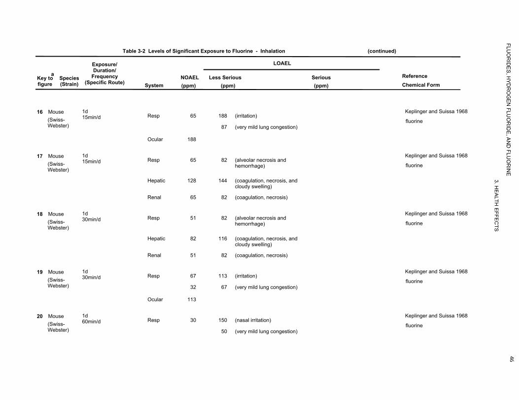
1579
(Swiss-Webster)
Resp1d5min/d
Keplinger and Suissa 1968
fluorine174 (dyspnea; nasal irritation;
diffuse lung congestion; alveolarnecrosis)
Mouse
174Hepatic 195 (necrosis and cloudy swelling)
79Renal 114 (necrosis)
300Ocular 467 (eye irritation)
LOAEL
Less SeriousNOAEL(ppm) (ppm)
Seriousa
(ppm)System
Exposure/Duration/
Frequency(Specific Route)
Species(Strain)
Key tofigure
Reference
(continued)Table 3-2 Levels of Significant Exposure to Fluorine - Inhalation
Chemical Form
1665
(Swiss-Webster)
Resp1d15min/d
Keplinger and Suissa 1968
fluorine188 (irritation)
87 (very mild lung congestion)
Mouse
188Ocular
1765
(Swiss-Webster)
Resp1d15min/d
Keplinger and Suissa 1968
fluorine82 (alveolar necrosis and
hemorrhage)
Mouse
128Hepatic 144 (coagulation, necrosis, andcloudy swelling)
65Renal 82 (coagulation, necrosis)
1851
(Swiss-Webster)
Resp1d30min/d
Keplinger and Suissa 1968
fluorine82 (alveolar necrosis and
hemorrhage)
Mouse
82Hepatic 116 (coagulation, necrosis, andcloudy swelling)
51Renal 82 (coagulation, necrosis)
1967
32(Swiss-Webster)
Resp1d30min/d
Keplinger and Suissa 1968
fluorine113 (irritation)
67 (very mild lung congestion)
Mouse
113Ocular
2030
(Swiss-Webster)
Resp1d60min/d
Keplinger and Suissa 1968
fluorine150 (nasal irritation)
50 (very mild lung congestion)
Mouse

LOAEL
Less SeriousNOAEL(ppm) (ppm)
Seriousa
(ppm)System
Exposure/Duration/
Frequency(Specific Route)
Species(Strain)
Key tofigure
Reference
(continued)Table 3-2 Levels of Significant Exposure to Fluorine - Inhalation
Chemical Form
2130
(Swiss-Webster)
Resp1d60min/d
Keplinger and Suissa 1968
fluorine50 (alveolar necrosis and
hemorrhage)
Mouse
55Hepatic 80 (necrosis and cloudy swelling)
50Renal 55 (necrosis)
2270
(NewEngland)
Resp1d15min/d
Keplinger and Suissa 1968
fluorine198 (irritation)
100 (very mild lung congestion)
Gn Pig
2373
(NewEngland)
Resp1d60min/d
Keplinger and Suissa 1968
fluorine135 (mild lung congestion, irritation,
dyspnea)
Gn Pig
2439
(NS)Resp
1d15min/d
Keplinger and Suissa 1968
fluorine93 (slight lung congestion)
Dog
39Ocular 93 (eye irritation)
2568
(NS)Resp
1d60min/d
Keplinger and Suissa 1968
fluorine93 (irritation, cough, and slight
dyspnea)
Dog
38Ocular 68 (eye irritation)
2679
(NewZealand)
Resp1d5min/d
Keplinger and Suissa 1968
fluorine410 (irritation)
134 (slight dyspnea)
Rabbit

LOAEL
Less SeriousNOAEL(ppm) (ppm)
Seriousa
(ppm)System
Exposure/Duration/
Frequency(Specific Route)
Species(Strain)
Key tofigure
Reference
(continued)Table 3-2 Levels of Significant Exposure to Fluorine - Inhalation
Chemical Form
2732
(NewZealand)
Resp1d30min/d
Keplinger and Suissa 1968
fluorine135 (irritation)
71 (very mild lung congestion)
Rabbit
INTERMEDIATE EXPOSUREDeath
28(NS)
5 wks6d/wk6hr/d
Stokinger 1949
fluorine18 (100% mortality)
Rat
29(NS)
5 wks6d/wk6hr/d
Stokinger 1949
fluorine5 (100% mortality)
Dog
30(NS)
5 wks6d/wk6hr/d
Stokinger 1949
fluorine5 (100% mortality)
Rabbit
Systemic31
5(NS)
Resp5 wks6d/wk6hr/d
Stokinger 1949
fluorine18 (severe pulmonary irritation)
Rat
Bd Wt 18 (weight loss)
320.5
(NS)Resp
5 wks6d/wk6hr/d
Stokinger 1949
fluorine2 (pulmonary hemorrhage and
edema)
Dog
5Hepatic 18 (liver congestion)
330.5
(NS)Resp
5 wks6d/wk6hr/d
Stokinger 1949
fluorine2 (mild bronchial inflammation) 18 (hemorrhage in the lungs)
Rabbit
2Hepatic 5 (hyperemia of the liver)

LOAEL
Less SeriousNOAEL(ppm) (ppm)
Seriousa
(ppm)System
Exposure/Duration/
Frequency(Specific Route)
Species(Strain)
Key tofigure
Reference
(continued)Table 3-2 Levels of Significant Exposure to Fluorine - Inhalation
Chemical Form
Reproductive34
5(NS)
a The number corresponds to entries in Figure 3-2.
b Used to derive an acute inhalation minimal risk level of 0.01 ppm; the concentration was adjusted for intermittent exposure (0.25 hours/24 hours) and divided by an uncertaintyfactor of 10 to account for human variability.
d = day(s); Gn Pig = Guinea pig; LC50 = lethal concentration, 50% kill; LOAEL; lowest-observed-adverse-effect-level; min = minute(s); NOAEL = no-observed-adverse-effect level;ppm = parts per million; Resp = respiratory
5 wks6d/wk6hr/d
Stokinger 1949
fluorine18 (testicular degeneration)
Rat
0.01
0.1
1
10
100
1000
Death
3g
3g
2m
2m
2m2m
1r
1r1r1r
4h
4h
Respiratory
24d
24d
25d25d 22g
22g
22g
23g
23g
6
789
9
10
15m
15m
16m
16m
16m17m17m
18m
18m19m
19m
19m
19m
20m
20m
20m 21m
21m
11r
11r
11r
12r
12r
12r
13r
13r
13r
14r
14r
14r
26h
26h
26h
27h
27h
27h
ppm
Figure 3-2. Levels of Significant Exposure to Fluorine - InhalationAcute (≤14 days)
c-Catd-Dogr-Ratp-Pigq-Cow
k-Monkeym-Mouseh-Rabbita-Sheep
f-Ferretj-Pigeone-Gerbils-Hamsterg-Guinea Pig
n-Minko-Other
Cancer Effect Level-Animals LOAEL, More Serious-Animals LOAEL, Less Serious-Animals NOAEL - Animals
Cancer Effect Level-Humans LOAEL, More Serious-Humans LOAEL, Less Serious-Humans NOAEL - Humans
LD50/LC50 Minimal Risk Level for effects other than Cancer
Systemic

0.01
0.1
1
10
100
1000
17m
18m18m
21m21m
Renal
15m15m
17m17m
18m
18m
21m 21m
Dermal
5 6
8 8
10
Ocular
24d
24d 25d
25d
5 6
789
9
10
10
15m15m
16m
19m11r
11r
12r13r
13r14r
ppm
Figure 3-2. Levels of Significant Exposure to Fluorine - Inhalation (Continued)Acute (≤14 days)
c-Catd-Dogr-Ratp-Pigq-Cow
k-Monkeym-Mouseh-Rabbita-Sheep
f-Ferretj-Pigeone-Gerbils-Hamsterg-Guinea Pig
n-Minko-Other
Cancer Effect Level-Animals LOAEL, More Serious-Animals LOAEL, Less Serious-Animals NOAEL - Animals
Cancer Effect Level-Humans LOAEL, More Serious-Humans LOAEL, Less Serious-Humans NOAEL - Humans
LD50/LC50 Minimal Risk Level for effects other than Cancer
Hepatic
15m 15m17m
Systemic
0.1
1
10
100
Death
29d
28r
30h
Respiratory
32d
32d
31r
31r 33h
33h
33h
Hepatic
32d
32d
33h
33h
Body Weight
31r
Reproductive
34r
34r
ppm
Figure 3-2. Levels of Significant Exposure to Fluorine - Inhalation (Continued)Intermediate (15-364 days)
c-Catd-Dogr-Ratp-Pigq-Cow
k-Monkeym-Mouseh-Rabbita-Sheep
f-Ferretj-Pigeone-Gerbils-Hamsterg-Guinea Pig
n-Minko-Other
Cancer Effect Level-Animals LOAEL, More Serious-Animals LOAEL, Less Serious-Animals NOAEL - Animals
Cancer Effect Level-Humans LOAEL, More Serious-Humans LOAEL, Less Serious-Humans NOAEL - Humans
LD50/LC50 Minimal Risk Level for effects other than Cancer
Systemic

LOAEL
Less SeriousNOAEL(mg/m³) (mg/m³)
Seriousa
(mg/m³)System
Exposure/Duration/
Frequency(Specific Route)
Species(Strain)
Key tofigure
Reference
Table 3-3 Levels of Significant Exposure to Fluoride - Inhalation
Chemical Form
ACUTE EXPOSURESystemic
1(ICR)
Resp4 hours/day10 days
Chen et al. 1999
sodium fluoride13.3 (increased relative lung weight)M
Mouse
22 M
(BALB/c)Resp
4 hour/day14 days
Yamamoto et al. 2001
sodium fluoride10 (pulmonary edema)M
Mouse
Immuno/ Lymphoret3
2 M(BALB/c)
4 hour/day14 days
Yamamoto et al. 2001
sodium fluoride5 (decreased pulmonary
bactericidal activity)M
Mouse
INTERMEDIATE EXPOSURESystemic
4(ICR)
Resp
a The number corresponds to entries in Figure 3-3.
LOAEL = lowest-observed-adverse-effect level; M = male; NOAEL = no-observed-adverse-effect level; Resp = respiratory
4 hours/day20-30 days
Chen et al. 1999
sodium fluoride13.3 (increased relative lung weight)M
Mouse

1
10
100
Respiratory
1m
2m
2m
Immuno/Lymphor
3m
3m
mg/m3
Figure 3-3. Levels of Significant Exposure to Fluoride - InhalationAcute (≤14 days)
c-Catd-Dogr-Ratp-Pigq-Cow
k-Monkeym-Mouseh-Rabbita-Sheep
f-Ferretj-Pigeone-Gerbils-Hamsterg-Guinea Pig
n-Minko-Other
Cancer Effect Level-Animals LOAEL, More Serious-Animals LOAEL, Less Serious-Animals NOAEL - Animals
Cancer Effect Level-Humans LOAEL, More Serious-Humans LOAEL, Less Serious-Humans NOAEL - Humans
LD50/LC50 Minimal Risk Level for effects other than Cancer
Systemic
10
100
Respiratory
4m
mg/m3
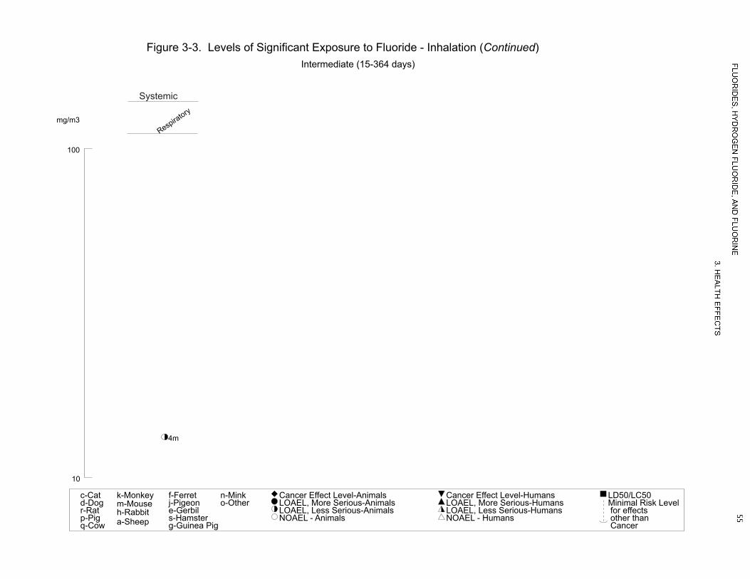
Figure 3-3. Levels of Significant Exposure to Fluoride - Inhalation (Continued)Intermediate (15-364 days)
c-Catd-Dogr-Ratp-Pigq-Cow
k-Monkeym-Mouseh-Rabbita-Sheep
f-Ferretj-Pigeone-Gerbils-Hamsterg-Guinea Pig
n-Minko-Other
Cancer Effect Level-Animals LOAEL, More Serious-Animals LOAEL, Less Serious-Animals NOAEL - Animals
Cancer Effect Level-Humans LOAEL, More Serious-Humans LOAEL, Less Serious-Humans NOAEL - Humans
LD50/LC50 Minimal Risk Level for effects other than Cancer
Systemic
FLUORIDES, HYDROGEN FLUORIDE, AND FLUORINE 56
3. HEALTH EFFECTS
individuals exposed to 0.5 or 4.5 ppm hydrogen fluoride for 1 hour (Lund et al. 1997). Significant
increases in the percentage of CD3-positive cells and lymphocytes in the bronchial portion of the lower
respiratory tract, as assessed via bronchoalveolar lavage performed 3 weeks prior to exposure and
24 hours after exposure, was also observed at 4.5 ppm, but not at 0.5 ppm (Lund et al. 1999). However,
no significant alterations in lung function or lower airway symptoms were observed (Lund et al. 1997).
The second study with a similar study design assessed upper airway inflammation via nasal lavage (Lund
et al. 2002). An inflammatory response in the nasal mucosa was observed following a 1-hour exposure to
3.8–4.5 ppm hydrogen fluoride. Seven of the 10 tested subjects also reported upper airway symptoms
(specific symptoms were not presented); most of the subjects scored the severity of the symptoms as very
mild to mild.
A number of residents of Texas City, Texas, reported respiratory symptoms following the accidental
release of hydrogen fluoride. It was estimated that most of the hydrogen fluoride was released in the first
2 hours after the accident, and evacuation of residents within 0.5 miles of the facility began within
20 minutes of the accident. Many of the 939 people who went to the emergency room within 24 hours of
the accident reported signs of respiratory irritation: throat burning (21.0%), shortness of breath (19.4%),
sore throat (17.5%), and cough (16.4%) (Wing et al. 1991). Forced expiratory volume in 1 second
(FEV1) was <80% of predicted values in 42.3% of the 130 individuals who underwent pulmonary
function testing. In another study of the Texas City residents, health effects within 1 month of the
accident and 2 years after the accident were assessed in 1,994 residents who were asked to complete
health questionnaires 2 years after the accident (Dayal et al. 1992). A large number of highly exposed
residents reported severe symptoms of breathing problems (e.g., coughing, difficulty breathing, shortness
of breath), throat problems (e.g., difficulty swallowing, burning irritation, phlegm, voice changes), and
nose problems (e.g., sneezing, runny nose, problems smelling food); the prevalence of severe symptoms
were 60.2, 51.9, and 40.7% for breathing, throat, and nose problems, respectively, within the first month
of the accident. High prevalence of these effects was still reported 2 years after the accident; 38.5, 22.1,
and 26.5% for severe breathing, throat, and nose problems, respectively. The prevalence of severe
breathing, throat, and nose problems in the nonexposed population were 11.3, 6.2, and 6.4%, respectively,
within 1 month of the accident and 8.2, 3.3, and 4.1%, respectively, 2 years after the accident. The
prevalence of the breathing problems were higher in a subgroup of the high exposure group that had pre-
existing respiratory problems or smoked more than two packs of cigarettes per day. Although this study
(Dayal et al. 1992) provides suggestive evidence that acute exposure to hydrogen fluoride can result in
long-term damage to the respiratory tract, the study results should be interpreted with caution. The
FLUORIDES, HYDROGEN FLUORIDE, AND FLUORINE 57
3. HEALTH EFFECTS
symptom survey was administered 2 years after the accident, there was no medical confirmation of the
effects, and the study authors did not provide a definition for severe symptoms.
Lethality studies in animals have also reported respiratory effects in rats, mice, and guinea pigs from
acute inhalation exposure to hydrogen fluoride. True respiratory effects, such as respiratory distress,
pulmonary congestion, and intra-alveolar edema were generally observed at levels of at least ~50% of the
LC50 (Haskell Laboratory 1988; Rosenholtz et al. 1963; Wohlslagel et al. 1976). These effects appear to
be reversible within a week upon cessation of exposure.
A series of experiments by Dalbey et al. (1998a, 1998b) examined the acute toxicity of nonlethal
concentrations of hydrogen fluoride in rats following a 2- or 10-minute exposure. In most of the
experiments, a mouth-breathing model with a tracheal cannula was used to maximize delivery of
hydrogen fluoride to the lower respiratory tract. A number of respiratory tract effects were found in the
mouth-breathing rats, including alterations in bronchioalveolar lavage (BAL) parameters (increased total
protein, myeloperoxidase, lactate dehydrogenase, β-glucuronidase, and glucose-6-phosphate
dehydrogenase), impaired lung function (decreased total lung capacity, vital capacity, peak expiratory
flow, forced expiratory flow at 50 and 25% of the forced vital capacity, forced expiratory volume at
0.1 second, forced vital capacity, and diffusing capacity and increased pulmonary resistance), and
histological damage (necrosis and acute inflammation in trachea and acute alveolitis and perivascular/
peribronchial edema and inflammation in the lung). Rats exposed for 2 minutes manifested histological
damage and BAL parameter alterations at 1,509 ppm fluoride and impaired lung function at 4,643 ppm.
No adverse respiratory effects were observed at 563 ppm fluoride. In the rats exposed for 10 minutes,
histopathological alterations (necrosis of the trachea only) and BAL parameters (polymorphonuclear
leukocytes and myeloperoxidase levels only) were observed at 903 ppm fluoride; impaired respiratory
function was observed at 1,676 ppm fluoride. No adverse effects were observed at 257 ppm fluoride.
The respiratory effects were consistently more severe in the rats exposed for 2 minutes as compared to
10 minutes, when exposure was expressed as the product of concentration x time. In other experiments,
rats were exposed for 60 minutes to hydrogen fluoride. No adverse respiratory effects were observed at
19 or 46 ppm. Respiratory effects observed in nose-breathing rats were limited to the nose. Necrosis and
acute inflammation of the ventral meatus, nasal septum, and nasoturbinates were observed in rats exposed
to 6,072 ppm for 2 minutes and 1,586 ppm for 10 minutes. A dramatic decrease in breathing frequency
was also observed in the nose-breathing rats; within the first minute of exposure, breathing frequency was
32–35% of the preexposure levels. The decrease in breathing frequency, which is a component of reflex
apnea, is a response to sensory irritation.

FLUORIDES, HYDROGEN FLUORIDE, AND FLUORINE 58
3. HEALTH EFFECTS
Similar results were observed in rats exposed to 1,235 ppm fluoride for 30 minutes. Moderate to severe
fibronecrotic rhinitis and large fibrin thrombi in the submucosa and hemorrhage were observed in the
nasal cavity of nose-breathing rats; no nasal lesions were observed in similarly exposed rats fitted with a
tracheal cannula to simulate mouth-breathing. Epithelial, submucusal, and cartilage necrosis in the
trachea, trace levels of neutrophils in the alveoli, and necrosis of the bronchi were observed in the mouth-
breathing rats, but not in the nose-breathing rats, suggesting that the toxicity of hydrogen fluoride occurs
at the point of entry. Reflex apnea, as evidenced by a marked decrease in breathing frequency, was
observed in the nose-breathing rats. Based on differences in minute ventilation rates, the study authors
estimated that the mouth-breathing rats inhaled 27% more hydrogen fluoride than the nose-breathing rats.
Pulmonary hemorrhage was noted in dogs, rabbits, and rats exposed to 31 ppm fluoride as hydrogen
fluoride for 6 hours/day, 6 days/week for 5 weeks (Stokinger 1949). At 8.2 ppm fluoride, no effect was
seen in rats or rabbits, and localized hemorrhages were seen in only 1/5 dogs.
Pulmonary hemorrhage, alveolar inflammation, and hyperplasia of the bronchial epithelium were
observed in guinea pigs that died due to exposure to 18 ppm fluoride as hydrogen fluoride for 6–
7 hours/day, 5 days/week for about 35 days (Machle and Kitzmiller 1935). This effect was not readily
reversible. The one surviving guinea pig had alveolar exudates, thickening of the alveolar walls, and
hemorrhages of the lungs when necropsied 9 months after the conclusion of the full 50-day exposure
period. Similarly, all four rabbits exposed under the same conditions had lobular pneumonia and
leucocytic infiltration of the alveolar walls, sometimes with edema and thickening of the walls, when
necropsied 7–8 months after the last exposure. No clinical signs of toxicity were reported in rabbits and
weight gain was generally similar to the controls. This study is limited by the small number of animals
used and the incomplete reporting of the data.
Hydrogen Fluoride and Fluoride Dusts. A study of an occupational cohort exposed to hydrogen
fluoride and fluoride dusts in the pot rooms of an aluminum smelter reported a significantly lower forced
expiratory volume and increased cough and sputum production in the highest exposure group, compared
with controls who worked in the office or casting department and were reported to have no significant
occupational exposure to air contaminants. Corrections were made for age, height, and smoking habits.
The ambient air fluoride concentration in the high-exposure area was 0.2 mg fluoride/m3 as vapor
(presumably hydrogen fluoride) and 0.28 mg/m3 "particulate fluoride." It is not clear whether the latter
value represented the air concentration of fluoride in particulates or the concentration of the particulates

FLUORIDES, HYDROGEN FLUORIDE, AND FLUORINE 59
3. HEALTH EFFECTS
that contain fluoride. Actual exposure was unknown because the workers wore respirators. Although
urinary fluoride levels increased over the course of one work shift in the high-exposure group and not in
the control group, the decrease in respiratory volume in the same time period was about the same in both
groups (Chan-Yeung et al. 1983a). This effect was attributed to the fact that the exposed workers wore
respirators; historical use of respirators was not reported. Because actual exposure was not known, no
quantitative relationship between clinical symptoms and environmental or urinary fluoride levels could be
established. There also may have been concomitant exposure to other respiratory irritants.
Fluoride Particulates. There is limited information on the respiratory toxicity of fluorides. Significant
increases in relative lung weight were observed in mice exposed to 13.3 mg F/m3 as sodium fluoride
4 hours/day for 10, 20, or 30 days (Chen et al. 1999). The toxicological significance of this effect is not
known because histopathology was not conducted. In another study of mice (Yamamoto et al. 2001),
lung damage, as evidenced by significant decreases in total cells and alveolar macrophages and increases
in polymorphocytic neutrophils and lymphocytes in the bronchoalveolar lavage fluid, was found in mice
exposed to 10 mg F/m3 as sodium fluoride 4 hours/day for 14 days. An increase in polymorphocytic
neutrophils was also observed at 5 mg/m3.
Fluorine. Limited data are available regarding respiratory effects of fluorine on humans. Five volunteers
(19–50 years of age; gender not specified) were exposed to fluorine through a face mask that covered the
eyes and nose but not the mouth (Keplinger and Suissa 1968). A concentration of 10 ppm was not
irritating to the respiratory tract for at least 15 minutes. Slight nasal irritation was reported following a
3-minute exposure to 50 ppm, and exposure to 100 ppm for 0.5 or 1 minute was very irritating to the
nose. Intermittent inhalation (3–5-minute exposure every 15 minutes for 2–3 hours) of 23 ppm did not
cause respiratory difficulty.
An occupational cohort study comparing the incidence of respiratory complaints by 61 exposed workers
with over 2,000 "unexposed" workers found no increase in the exposed group (Lyon 1962). The average
fluorine level was 0.9 ppm, and the maximum measured value was 24 ppm. The study author concluded
that the workers became "hardened" to the irritating effects of fluorine. The study is limited in that both
groups were also exposed to uranium hexafluoride and hydrogen fluoride. The method of measuring
respiratory complaints (visits to the plant medical department) was also not very sensitive. However, the
observation of tolerance caused by repeated low level exposures is supported by the results from animal
studies discussed in Section 3.2.1.1 and later in this section (Keplinger 1969).

FLUORIDES, HYDROGEN FLUORIDE, AND FLUORINE 60
3. HEALTH EFFECTS
Diffuse lung congestion has been reported in rats, mice, guinea pigs, dogs, and rabbits exposed to fluorine
for 5–60 minutes (Keplinger and Suissa 1968). The severity was concentrated-related. The adverse
effect levels for each exposure duration did not appear to vary across species. The ranges of adverse
effect levels for each exposure duration were 174–175 ppm for 5 minutes, 87–100 ppm for 15 minutes,
67–71 ppm for 30 minutes, and 47–135 ppm for 60 minutes. Other respiratory effects that were observed
in these animals included dyspnea, irritation, and alveolar necrosis.
In 5-week exposure studies conducted by Stokinger (1949), pulmonary hemorrhage, edema, and bronchial
inflammation were reported. These studies found species differences in sensitivity to fluorine-induced
respiratory effects. Exposure to 2 ppm, 6 hours/day, 6 days/week for 5 weeks resulted in no effects in
rats, pulmonary hemorrhage and edema in dogs, and mild bronchial inflammation in rabbits; respiratory
effects (severe pulmonary irritation) were observed in rats exposed to 18 ppm.
Swiss-Webster mice that were preexposed once to 30 ppm fluorine for 60 minutes and then exposed to
118–410 ppm fluorine for 15 minutes after an interval of 4–96 hours showed markedly less lung
pathology than animals that were not pretreated (Keplinger 1969). At the highest level (410 ppm),
exposure 4 hours prior to the challenge reduced the lung pathology from the most severe rating to a rating
of normal–mild. Preexposure also reduced the increased lung weight otherwise seen following fluorine
exposure. However, a similar preexposure regimen only resulted in slight increases in the LC50, as
discussed in Section 3.2.1.1.
Cardiovascular Effects.
Hydrogen Fluoride. Cardiac arrhythmias have been seen in humans following hydrofluoric acid splashes
in the face region, where both dermal and inhalation exposures were involved (Chan et al. 1987;
Tepperman 1980). It is not known whether inhalation exposure alone would cause these effects.
However, myocardial necrosis and congestion were observed in three rabbits following inhalation
exposure of 26 ppm fluoride as anhydrous hydrogen fluoride for an unspecified period (Machle et al.
1934). The study was limited by the small sample size and undetermined exposure period.
Gastrointestinal Effects.
Hydrogen Fluoride. A population exposed to airborne hydrogen fluoride near a smelter reported nausea
(22.6%) and diarrhea (21.7%). The corresponding levels reported by a control population were 6.9 and
12.1%, respectively. The total levels of gastrointestinal complaints were 70.5 and 36.2% in the subject

FLUORIDES, HYDROGEN FLUORIDE, AND FLUORINE 61
3. HEALTH EFFECTS
and control populations, respectively. The subject population appears to have been derived by self-
selection and random house-to-house sampling, while the control population lived in a nonindustrial area.
Although atmospheric concentrations were not presented, concentrations of fluoride in animals and plants
in the area surrounding the smelter were substantially above normal. The smelter was also reported to
emit metallic oxide fumes (Waldbott 1979).
Similar gastrointestinal effects (diarrhea, nausea, and vomiting) were reported by Texas residents exposed
to an accidental 2-hour release of hydrogen fluoride (Dayal et al. 1992). During the first month after the
accident, 38.5% of the highly exposed residents reported severe gastrointestinal effects; 15.5% of the
residents still reported severe gastrointestinal effects 2 years after the accident. The occurrence of severe
gastrointestinal effects among nonexposed residents was 4.5 and 2.7%, respectively, for these time
periods.
Hematological Effects.
Hydrogen Fluoride. Hemograms of 20 variables (not specified) determined in the rat (30/group), rabbit
(10/group), and dog (4/group) following exposure to 18 ppm fluoride for 6 hours/day, 6 days/week, for
5 weeks showed no clear changes (Stokinger 1949).
Five rabbits and two Rhesus monkeys were exposed to 18 ppm fluoride as hydrogen fluoride via
inhalation 6–7 hours/day, for 50 days (Machle and Kitzmiller 1935). Blood counts were done beginning
1 week prior to exposure and ending 3 months after the final exposure. There was a small but significant
decrease in erythrocyte levels in both species, but the study authors considered that the result may have
been due to biological variation. Significant increases in hemoglobin levels were seen in monkeys. There
was no effect on hemoglobin levels in rabbits or on leukocyte levels in either species. These experiments
used only a few animals from each species, and the exposure measurement technology was not very
precise.
Hydrogen Fluoride and Fluoride Dusts. No signs of hematological effects, as measured by routine
blood counts, were seen in a large cohort of aluminum workers exposed to total fluoride levels below
2.5 mg/m3 for durations of at least 10 years (Chan-Yeung et al. 1983b). Similarly, no increase in
abnormal findings was seen in 74 workers exposed at a phosphate fertilizer plant (Derryberry et al. 1963).
The average urinary fluoride level in the exposed group was 4.6 mg/L. Significantly reduced levels of
hemoglobin were reported in Slovak children aged 6–14 years living near an aluminum smelter (Macuch
et al. 1963), but no information was provided on any statistical tests used. No information was provided
FLUORIDES, HYDROGEN FLUORIDE, AND FLUORINE 62
3. HEALTH EFFECTS
on air fluoride concentrations, but urinary fluoride levels were about 0.8 mg/L for 6–11-year-old children
and about 0.4 mg/L for 12–14-year-old children. In an outdated study of 78 workers exposed to cryolite,
anemia was present in 11/30 subjects with pathological bone changes (Moller and Gudjonsson 1932).
Blood parameters were not analyzed for the workers without bone changes.
Fluorine. No studies were located on hematological effects of inhalation exposure of humans to fluorine.
No effect on complete blood count parameters was observed in Osborne-Mendel rats exposed to 142 ppm
for 60 minutes or 329 ppm for 15 minutes or in dogs exposed to 109 ppm for 60 minutes or 93 ppm for
15 minutes (Keplinger and Suissa 1968). These concentrations were higher than the corresponding
LC50 values. Blood counts were monitored for 21 days postexposure. Similarly, Stokinger (1949) saw no
effect on hematological parameters in dogs, rabbits, or rats following repeated exposures at
concentrations up to lethal levels (31 ppm). This study did not specify which parameters were measured.
Musculoskeletal Effects.
Fluoride. There are several case reports of radiological alterations (primarily thickening of the bone) in
workers exposed to sodium fluoride (McGarvey and Ernstene 1947), rock phosphate dust containing
3.88% fluoride (Wolff and Kerr 1938), or cryolite (Roholm 1937). In the two cryolite workers, the
fluorine content of the costa bone was 10-fold higher than in non-exposed individuals. Roholm (1937)
also examined 68 cryolite workers exposed to high levels (35 mg/m3) of cryolite dust. Approximately
35% of the workers complained of “rheumatic attacks, pains, or feeling of stiffness” and reduced mobility
was found in approximately 21% of the workers. Radiological examinations revealed diffuse
osteosclerosis in approximately 84% of the workers.
No studies were located regarding musculoskeletal effects in animals after inhalation exposure to fluoride.
Hydrogen Fluoride. Human data on the musculoskeletal effects of hydrogen fluoride (in the absence of
concomitant exposure to fluoride dusts) are limited to a case report of a worker employed at an alkylation
unit of an oil company (Waldbott and Lee 1978). The worker complained of back pains, leg pains, and
loss of memory and was diagnosed with advanced osteoarthritis of the spine. The data presented in this
report are inadequate to assess whether the back and leg pains were related to hydrogen fluoride exposure,
the osteoarthritis, or a petroleum product.
Duration- and concentration-related increases in tooth and bone fluoride levels were reported in the rat
following exposure to 8.2 or 31 ppm fluoride as hydrogen fluoride for 6 hours/day, 6 days/week for

FLUORIDES, HYDROGEN FLUORIDE, AND FLUORINE 63
3. HEALTH EFFECTS
5 weeks (Stokinger 1949). The study author did not report whether there were any visible or radiological
signs of dental or skeletal fluorosis.
Hydrogen Fluoride and Fluoride Dusts. Marked evidence of skeletal fluorosis was reported in workers
exposed to gaseous fluoride (largely hydrogen fluoride) and fluoride dust in the pot rooms of the
aluminum industry (Kaltreider et al. 1972). Individual exposure concentrations and durations were not
presented. However, the estimated time-weighted average (TWA) 8-hour exposure to total fluorides for
one plant ranged from 2.4 to 6.0 mg/m3. Average post-shift urinary fluoride levels were about 9 mg/L.
Exposure at a second plant was lower as a result of industrial hygiene measures; no TWA was available,
but post-shift urinary fluoride levels ranged from 1.4 to 4.6 mg/L. No skeletal changes were observed at
the second plant, and detailed physical examinations of the workers at both plants revealed no general
health impairment. No data were presented that correlated urinary fluoride levels to the presence or
absence of fluorosis.
In a follow-up study of 59 of the potroom workers at the second plant, the average preshift (after 48 hours
away from work) urinary fluoride level was 2.24 mg/L (range, 1.4–3.1). The average level after 3–
5 working days (postshift) was 5.68 mg/L (range, 2.7–10.4). In spite of this evidence of fluoride
exposure, there was no radiological evidence of any fluoride-related bone abnormalities (Dinman et al.
1976c). Total occupational exposure ranged from 10 to 43 years. This study may provide urinary
fluoride levels that are not associated with any bone effects in healthy adults. However, because only
workers who remained at the high-exposure tasks for the duration of the study were examined, any
sensitive population that may have found work elsewhere because of adverse health effects might have
been missed.
Clinical and radiological investigations were performed for 2,258 aluminum workers exposed to fluoride
for an average of 17.6 years (Czerwinski et al. 1988). The form of fluoride was not reported, but it was
probably hydrogen fluoride and fluoride dust. Possible fluorosis (multiple joint pains, limited motion in
at least two joints or in the spine, and initial ossifications visible on x-ray films) was found in 14% of the
workers. Indications of early skeletal fluorosis (advanced painful symptoms, advanced limitation of
motion in at least two joints or spine, marked ossifications on two or more x-rays, initial osteosclerosis,
slight periosteal reaction, and thickening of long bone cortices ) were found in 5.12% of the workers and
definite fluorosis (stage I) was found in 1.0% of the workers. The study authors reported finding a close
positive correlation between the occurrence of fluorosis and the time and level of fluoride exposure.
Another health study of 2,066 workers in an aluminum smelter reported early signs of skeletal fluorosis

FLUORIDES, HYDROGEN FLUORIDE, AND FLUORINE 64
3. HEALTH EFFECTS
(mild increase in bone density, periosteal changes, calcifications of ligaments) in a few pot room workers
employed for >10 years. The study authors noted that there was poor agreement on early signs of
fluorosis among the two radiologists reading the x-ray films. No effects were seen in workers exposed for
<10 years. Actual airborne fluoride levels measured at the time of the health assessment were 0.2 mg/m3
hydrogen fluoride and 0.28 mg/m3 fluoride dusts. Historical fluoride levels were not reported; although
the study authors implied that exposure levels had been below 2.5 mg/m3 for some period (Chan-Yeung et
al. 1983b).
Skeletal fluorosis was also observed in workers involved in study the crushing and refining of cryolite
(Moller and Gudjonsson 1932). Thirty-nine of the 78 examined workers showed evidence of skeletal
fluorosis in the form of dense calcification in the long bones, cartilage, and in extreme cases, of the skull
as well. Although an average exposure period was not presented, no workers with <2 years of exposure
were included; some workers had been exposed for as long as 40 years.
While the above studies generally found radiologically-apparent skeletal fluorosis appearing prior to or
concurrent with musculoskeletal symptoms, Carnow and Conibear (1981) found musculoskeletal
symptoms in aluminum workers in the absence of radiological findings. Questionnaire answers suggested
a significant increase in incidence and severity of musculoskeletal disease and fracture frequency with
fluoride exposure. By contrast, there was no exposure-related increase in evidence of skeletal fluorosis
on chest and spinal x-ray films. Neither radiologic data nor actual exposure levels or durations were
reported. As the authors recognized, the exposure group was heterogeneous and was exposed to other
chemicals, and some of the musculoskeletal symptoms may have actually been due to heavy physical
labor.
Fluorine. No data were located regarding musculoskeletal effects of fluorine inhalation on humans.
Fluoride levels in the teeth of rats exposed to 18 ppm fluorine for approximately 6 hours/day, 6 days/week
for 5 weeks were about 14 times the levels in controls; fluoride levels in the femur were about 6 times that
of the controls (Stokinger 1949). The appearance of the teeth was characterized as corresponding to that
of very mild to mild dental fluorosis. The fluoride levels in the teeth and bone at lower concentrations
decreased in a concentration-related manner. Pigment changes were reported as just perceptible in
animals exposed to 2 ppm fluorine.

FLUORIDES, HYDROGEN FLUORIDE, AND FLUORINE 65
3. HEALTH EFFECTS
Hepatic Effects.
Hydrogen Fluoride. Ten animals (five rabbits, three guinea pigs, and two Rhesus monkeys) were
exposed via inhalation to 18 ppm fluoride as hydrogen fluoride 6–7 hours/day for 50 days (Machle and
Kitzmiller 1935). Fatty degeneration of the liver parenchyma, scattered focal necroses, and fibroblastic
encroachment of periportal spaces were observed in the guinea pigs. Two of the three guinea pigs began
losing weight after about 145 hours of exposure, were withdrawn from the exposure regimen, and died
about 2 weeks later. Generalized fatty changes were also seen in two of four rabbits sacrificed 7 months
after exposure termination. These experiments used only a few animals from each species, and the
exposure measurement technology was not very precise.
Hydrogen Fluoride and Fluoride Dusts. The occupational health study by Chan-Yeung et al. (1983b)
discussed above revealed no adverse effects on liver function, as measured by levels of total bilirubin,
serum glutamic oxaloacetic transaminase (SGOT), and alkaline phosphatase.
Fluorine. No studies were located regarding hepatic effects of fluorine inhalation in humans. Mice
exposed to fluorine exhibited coagulation necrosis of the liver, periportal hemorrhages, and diffuse cloudy
swelling (Keplinger and Suissa 1968). These effects were generally observed after exposure to
concentrations of 195, 144, 116, or 80 ppm fluoride for 5, 15, 30, or 60 minutes, respectively. Damage
became apparent 7–14 days after exposure. Liver congestion was reported in dogs, but not in other
species subjected to repeated exposures to a lethal concentration of fluorine (18 ppm 6 hours/day,
6 days/week for 5 weeks) (Stokinger 1949).
Renal Effects.
Hydrogen Fluoride. Pathologically elevated serum creatinine and urea levels were seen 24 hours after
accidental dermal and inhalation exposure to a mixture of 70–80% sulfuric acid and 10% hydrofluoric
acid at 150 °C (Braun et al. 1984). Neither the effect of the sulfuric acid nor the exposure levels were
known.
Degeneration and necrosis of the renal cortex was reported in 27/30 rats exposed to 31 ppm fluoride as
hydrogen fluoride for 6 hours/day, 6 days/week for 5 weeks, but not in rats exposed to 8.2 ppm fluoride
(Stokinger 1949). Pathological examination of rabbits and guinea pigs (n=3/species/exposure level)
exposed to hydrogen fluoride revealed tubular necrosis, congestion, and edema (Machle et al. 1934). A
variety of different exposure levels and durations were tested, but the levels at which exposure-related

FLUORIDES, HYDROGEN FLUORIDE, AND FLUORINE 66
3. HEALTH EFFECTS
effects were seen were not reported. Rabbits (n=4) exposed via inhalation to 18 ppm fluoride as
hydrogen fluoride 6–7 hours/day for 50 days developed degeneration and necrosis of convoluted tubules,
accompanied by fibrous tissue replacement of cortical tissues (Machle and Kitzmiller 1935).
Degenerative and inflammatory changes were also seen in the single exposed monkey at necropsy. The
experiments described in both of these papers used a small number of animals and no control data were
presented.
Hydrogen Fluoride and Fluoride Dusts. Increased incidence of albuminuria (p<0.1) was observed in
phosphate fertilizer plant workers with an average urinary fluoride level of 4.6 mg/L (Derryberry et al.
1963). However, the testing method used in this study is considered to be hypersensitive (Dinman et al.
1976a), and several other studies have found no effects. No signs of renal effects, as measured by
standard renal function tests, were seen in a large cohort of aluminum workers exposed to total fluoride
levels estimated to be below 2.5 mg/m3 (Chan-Yeung et al. 1983b). Two other studies of aluminum
workers failed to find an increase in the incidence of albuminuria (Dinman et al. 1976c; Kaltreider et al.
1972). Average postshift urinary fluoride levels were ≤5.68 mg/L (Dinman et al. 1976c) and ≤9.6 mg/L
(Kaltreider et al. 1972). The exposed population included workers exposed to estimated air fluoride
levels of 4–6 mg/m3 (time-weighted average), of which 50% was gaseous fluoride (presumably hydrogen
fluoride) (Kaltreider et al. 1972).
The weight-of-evidence indicates that typical inhalation occupational exposure to hydrogen fluoride and
fluoride dust is not nephrotoxic. The overall animal data indicate that inhalation exposure to sufficiently
high levels of hydrogen fluoride or fluorine can cause kidney damage, but the relevance to human health
and the potential nephrotoxic level cannot be determined because of generally incomplete human and
animal data. In addition, only one animal experiment was located that conducted a histopathic exam
following fluorine exposure.
Fluorine. No studies were located regarding renal effects of fluorine inhalation in humans. Mice
exposed to fluorine exhibited focal areas of coagulation necrosis in the renal cortex and focal areas of
lymphocyte infiltration in the cortex and medulla following exposure to 114 ppm for 5 minutes, 82 ppm
for 15 or 30 minutes, or 55 ppm for 60 minutes (Keplinger and Suissa 1968). Damage became apparent
7–14 days postexposure.
Endocrine Effects. No studies were located regarding endocrine effects in humans or animals after
inhalation exposure to fluoride, hydrogen fluoride, or fluorine.

FLUORIDES, HYDROGEN FLUORIDE, AND FLUORINE 67
3. HEALTH EFFECTS
Dermal Effects.
Hydrogen Fluoride/Hydrofluoric Acid. Dermal exposure to hydrogen fluoride can cause irritation of the
skin and mucous membranes. Residents exposed to hydrogen fluoride following an accidental release
reported a number of skin effects including itching, burning, and rash; 43.8% of the highly exposed
residents reported severe skin problems, as compared to 5.3% of nonexposed residents (Dayal et al.
1992). Two years after the accident, severe skin problems were reported by 21.9% of the high-exposure
group compared to 2.7% of the control group. “Smarting" of exposed skin occurred in humans within
1 minute of exposure to hydrogen fluoride at about 122 ppm fluoride (Machle et al. 1934). This was the
highest concentration that two male volunteers could tolerate for >1 minute. Repeated exposures did not
reveal any habituation.
Exposure to hydrogen fluoride levels approaching the LC50 can cause lesions of the face in rats (Haskell
Laboratory 1988). Rats exposed to hydrogen fluoride (whole body) at a concentration of approximately
1,395 ppm fluoride for 60 minutes were observed to have erythema of an unspecified severity of the
exposed skin (Wohlslagel et al. 1976). Subcutaneous hemorrhages around the eyes and on the feet
developed in rats exposed to 8.2 or 31 ppm fluoride as hydrogen fluoride for 6 hours/day, 6 days/week for
5 weeks (Stokinger 1949). The effect was more severe at the higher exposure level. Dogs exposed to
31 ppm fluoride for the same time periods developed inflammation of the scrotal epithelium.
Fluorine. When the shaved backs of New Zealand rabbits were exposed to fluorine gas under 40 pounds
of pressure for 0.2–0.6 seconds at distances of 0.5–1.5 inches, the resulting burn appeared to be thermal,
rather than chemical in nature (Stokinger 1949). Exposure for 0.2 seconds produced an ischemic area
about ¼ inch in diameter, surrounded by an erythematous area. This became a superficial eschar that
sloughed off within 4 days, revealing normal epidermis. The longer exposures produced a flash of flame
that resulted in combustion of hair, singeing, and erythema over an area several times the area of the
primary burn. Coagulation necrosis and charring of the epidermis was also reported. The wound healed
within 13 days. The burns resembled those produced by an oxyacetylene flame, rather than those made
by hydrofluoric acid, and so were characterized as thermal, rather than chemical. However, it is not clear
if the difference from the hydrofluoric acid burn is due to the shorter exposure to fluorine.

FLUORIDES, HYDROGEN FLUORIDE, AND FLUORINE 68
3. HEALTH EFFECTS
Ocular Effects.
Hydrogen Fluoride. Marked conjunctival irritation was noted in humans within 1 minute of exposure to
hydrogen fluoride at about 122 ppm fluoride (Machle et al. 1934). This was the highest concentration
that two male volunteers could tolerate for >1 minute. At 61 ppm fluoride, conjunctival and nasal
irritations were still marked, and tickling and discomfort of the nasal passages were reported. A
concentration of 32 ppm fluoride produced mild irritation of the nose and eyes and irritation of the larger
air passages. This concentration could be tolerated for "several" minutes (at least 3 minutes). The
authors of this study reported some difficulties with their measurements of exposure. Repeated exposures
did not reveal any habituation.
Severe symptoms of eye problems were reported by 63.2% of Texas residents exposed to high levels of
hydrogen fluoride following an accidental release (Dayal et al. 1992). The most commonly reported eye
effects were redness, itching, and burning or irritation. Two years after the accident, 11.5% of the
population still reported severe eye problems. In nonexposed residents, the prevalence of severe
symptoms within the first month of the accident was 7.4; 2 years later, the prevalence was 4.9.
Mild eye irritation was observed in five volunteers exposed 6 hours/day for 15–50 days, to hydrogen
fluoride at concentrations averaging from approximately 0.85 to 7.7 ppm fluoride; the mean of the
average concentrations was 2.98 ppm (Largent 1960). This study is limited by the inadequacy of both the
experimental details and the description of effects observed.
Hydrogen fluoride levels approaching the LC50 can cause corneal opacity in rats (Haskell Laboratory
1988), while slight ocular irritation was observed in rats exposed to levels as low as 6% of the LC50
(Rosenholtz et al. 1963). Eye irritation, evidenced by pawing of eyes, was observed in rats exposed to
140 or 175 ppm fluorine for 30 or 5 minutes, respectively, and in dogs exposed to 68 or 93 ppm fluorine
for 60 or 15 minutes, respectively. In experiments with exposure for durations of 15–60 minutes, eye and
nose irritation was reported only at ~50% of the LC50. Similar results were obtained with Swiss-Webster
mice, New England guinea pigs, and New Zealand rabbits (Keplinger and Suissa 1968).
Fluorine. Volunteers (19–50 years of age) were exposed to 10 ppm fluorine for 15 minutes without
discomfort or irritation of the eyes or nose (Keplinger and Suissa 1968). However, repeated exposures to
10 or 23 ppm fluorine for 3–5 minutes every 15 minutes over a 2–3-hour period caused slight eye
irritation. Exposure was through a face mask that covered the eyes and nose but not the mouth. Eye
irritation was also reported following exposure to 50 ppm for 3 minutes and 67 and 78 ppm for 1 minute.
FLUORIDES, HYDROGEN FLUORIDE, AND FLUORINE 69
3. HEALTH EFFECTS
Exposure to 100 ppm was very irritating and became uncomfortable after a few seconds. At this
concentration, the subjects reported that the eyes burned and felt as though they were covered by a film.
Body Weight Effects.
Hydrogen Fluoride. Pronounced weight loss shortly before death was observed in rats exposed to a
lethal level of hydrogen fluoride (31 ppm fluoride for 6 hours/day, 6 days/week for 5 weeks). Guinea
pigs exposed under the same conditions lost weight following the third exposure week, even though there
were no deaths (Stokinger 1949). While a decrease compared to the low-exposure level group is clear, no
control animals were used and the lowest exposure level that would result in a significant change was not
established. Animals surviving a lethal exposure exhibited a body weight loss of 10–15% for up to a
week after exposure (Rosenholtz et al. 1963; Stavert et al. 1991).
Fluorine. Decreased weight gain was observed in rats, guinea pigs, and rabbits exposed to 18 ppm
fluorine for about 6 hours/day, 6 days/week for 5 weeks (Stokinger 1949). While a decreased weight gain
in the high-exposure group compared to the low-exposure groups is clear, no control animals were used
and the lowest exposure level that would result in a significant change was not established.
3.2.1.3 Immunological and Lymphoreticular Effects
No studies were located regarding immunological effects in humans or animals after inhalation exposure
to hydrogen fluoride or fluorine.
Fluoride Particulates. A significant decrease in pulmonary bactericidal activity was observed in mice
challenged for 30 minutes with aerosol inhalation of Staphylococcus aureus following a 14-day exposure
(4 hours/day) to 5 or 10 mg F/m3 as sodium fluoride (Yamamoto et al. 2001). Bactericidal activity was
not significantly altered in mice exposed to 2 mg/m3. The highest NOAEL values and all reliable LOAEL
values for immunologic effects in each species and duration category of inhalation exposure to fluoride
are recorded in Table 3-3 and plotted in Figure 3-3.

FLUORIDES, HYDROGEN FLUORIDE, AND FLUORINE 70
3. HEALTH EFFECTS
3.2.1.4 Neurological Effects
Hydrogen Fluoride. The threshold of the light adaptive reflex was measured as a marker for neurological
effects in three subjects following exposure to hydrogen fluoride at concentrations of 0.02, 0.03, or
0.06 ppm fluoride (Sadilova et al. 1965). While the threshold level was determined to be 0.03 ppm, it is
not clear whether this response is due to irritation of mucous membranes or is the result of an effect on
cerebral cortical function. The toxicological significance of a change in light adaptive reflex is not
known.
Exposure to concentrations at about 50% of the LC50 values was reported to cause general weakness and
decreased activity in Wistar rats (Rosenholtz et al. 1963). Albino rats given 24-hour exposures to either
0.03 or 0.1 ppm fluoride as hydrogen fluoride for 5 months developed central nervous system
dysfunctions, as evidenced by diminished conditioned responses and increased time before motor nerve
response. Histological studies showed changes in the nerve cell synapses of only those animals exposed
to 0.1 ppm. A concentration of 0.01 ppm was found to be without effect on conditioned responses,
latency in motor nerve response, or neurohistological parameters. When additional stresses were added
(alcohol, 24-hour starvation), the conditioned responses were extinguished more frequently (Sadilova et
al. 1965). Some recovery in conditioned responses was seen following a 1-month recovery period in the
animals exposed to 0.1 ppm. Animals exposed to 0.03 ppm recovered completely.
All reliable LOAEL values for neurological effects of exposure to hydrogen fluoride in each species and
duration category are recorded in Table 3-1 and plotted in Figure 3-1.
Fluorine. No studies were located regarding neurological effects in humans of fluorine following
inhalation exposure. Dogs exposed to 5 or 18 ppm for 6 hours/day, 6 days/week for up to 35 days had
seizures prior to death (Stokinger 1949). Because no further details were available, the neurotoxic
potential of fluorine cannot be evaluated.
3.2.1.5 Reproductive Effects
No studies were located regarding reproductive effects in humans after inhalation exposure to fluoride,
hydrogen fluoride, or fluorine, and no studies were located regarding reproductive effects in animals after
inhalation exposure to fluoride.

FLUORIDES, HYDROGEN FLUORIDE, AND FLUORINE 71
3. HEALTH EFFECTS
Hydrogen Fluoride. All four dogs exposed to 18 ppm fluoride as hydrogen fluoride for 6 hours/day,
6 days/week for 5 weeks developed degenerative testicular changes and ulceration of the scrotum
(Stokinger 1949). This effect was not seen at 8.2 ppm, or in rabbits or rats at either exposure level. No
further details were available. Furthermore, it is not clear whether this is a systemic effect or a result of
irritation from dermal contact with the gas.
Fluorine. Rats exposed to 18 ppm fluorine, 6 hours/day, 6 days/week for 5 weeks showed testicular
degeneration (Stokinger 1949). No further details were available. It is not clear whether this effect was
seen both in animals that died and in those that survived.
3.2.1.6 Developmental Effects
No studies were located regarding developmental effects in humans or animals after inhalation exposure
to fluoride, hydrogen fluoride, or fluorine.
3.2.1.7 Cancer
Hydrogen Fluoride and Fluoride Dusts. Most occupational exposure to fluoride occurs as a result of
inhalation of hydrofluoric acid fumes or dust from cryolite or fluorspar. A cohort of cryolite workers in
Denmark was reported to have an increase in mortality and morbidity from respiratory cancer compared
with the national average (standardized mortality ratio [SMR] of 2.52, 95% confidence interval of 1.40–
4.12, Standardized Incidence Ratio of 2.5 , 95% confidence interval of 1.6–3.5) (Grandjean et al. 1985).
The study authors stated that the increase can be explained by the fact that the respiratory cancer death
rate for the Copenhagen area is about twice the national average for the birth cohorts from 1890 to 1929,
so that comparison with national rates may not be appropriate. Respiratory cancer rates for the workers
were slightly higher than those of the general population of Copenhagen (standardized incidence ratio of
1.5, 95% confidence interval of 1.0–2.1). No apparent relationship between incidence of all cancers and
the length of employment or latency period was found. Significant increases in cancer mortality,
particularly respiratory cancer, were also found in a follow-up study of this cohort (Grandjean et al.
1992). The SMR for all cancer was 1.34 (95% confidence interval of 1.2–1.50) compared to Danish
national rates and the standardized incidence ratio for lung cancer was 1.40 (95% confidence interval
of 1.01–1.90) compared with rates for Copenhagen. Additionally, a significant increase in bladder cancer
FLUORIDES, HYDROGEN FLUORIDE, AND FLUORINE 72
3. HEALTH EFFECTS
incidence was seen (standardized incidence ratio of 1.84, 95% confidence interval of 1.08–2.96). The
investigators noted that smoking habits may have accounted for some of the lung cancer risk.
Increased lung cancer rates have been reported in several studies of aluminum industry workers
(Andersen et al. 1982; Gibbs and Horowitz 1979; Milham 1979), but no correction was made for smoking
or concurrent exposure to tars and polycyclic aromatic hydrocarbons. Similarly, fluorspar miners had
increased lung cancer rates, but they were also exposed to elevated radon levels (deVilliers and Windish
1964). A cohort study of 21,829 workers in aluminum reduction plants for ≥5 years did not find an
increase in lung cancer, but did report an increase in mortality due to pancreatic cancer, lymphohemato-
poietic cancers, genitourinary cancer, and nonmalignant respiratory disease (Rockette and Arena 1983).
Only the effect on pancreatic cancer rates was statistically significant. Increases in incidence of
hematopoietic cancers and respiratory disease were also reported by Milham (1979). Because of the
confounding factors mentioned above, and because no breakdown was done by fluoride exposure, these
studies are of questionable relevance to the issue of possible carcinogenicity of inhalation exposure to
hydrogen fluoride and/or fluorides.
A study was published describing a positive relationship between increased lung cancer occurrence and
exposure to fluoride among individuals residing near, or working in, the steel industry (Cecilioni 1972).
Possible occupational exposures to other carcinogenic substances from steel and other industries were not
considered. Carcinogenicity via inhalation of fluoride is not considered to be likely by most investigators
reporting in the existing literature.
No studies were located regarding cancer in animals after inhalation exposure to fluoride, hydrogen
fluoride, or fluorine.
3.2.2 Oral Exposure
Because hydrogen fluoride and fluorine are gases, oral exposure to these substances occurs only
concomitant with inhalation exposure. Oral exposure to hydrofluoric acid has been reported very rarely.
Except where otherwise indicated, the following sections on oral exposure refer to oral exposure to
fluoride.
The oral toxicity of fluoride has been investigated in humans and animals. The human database mostly
consists of epidemiology studies designed to assess whether consumption of fluoridated water is

FLUORIDES, HYDROGEN FLUORIDE, AND FLUORINE 73
3. HEALTH EFFECTS
associated with adverse health effects, particularly skeletal effects and cancer. Most of these studies are
community-based and do not provide data on individual exposure levels or the particular fluoride
compound. Fluorosilicic acid and sodium hexafluorosilicate are the primary fluoride compounds used in
water fluoridation programs, although exposure to other fluoride compounds, such as
monofluorophosphate, used in dental products also contribute to total fluoride exposure. Additional
information on the toxicity of fluoride in humans comes from studies of communities with naturally high
levels of fluoride in the drinking water and experimental studies typically involving exposure to sodium
fluoride. Animal studies have primarily involved exposure to sodium fluoride in drinking water or the
diet. A few animal studies also examined the toxicity of fluoride following exposure to calcium fluoride,
cryolite, and fluoride from rock phosphate. For all forms of fluoride discussed, doses are reported as
amount of the fluoride ion.
Conflicting results have been obtained from animal experiments addressing whether fluorine is an
essential element. Much of this conflict appears to result from the great difficulty in preparing an animal
diet that has negligible amounts of fluoride, but otherwise allows normal animal growth and development.
As discussed in Section 3.2.2.6, there have been suggestions that fluoride can aid fertility by improving
intestinal absorption of iron and other trace elements (Messer et al. 1973; Tao and Suttie 1976). In a
study where fluoride was rigorously removed from dietary components, a total of 110 Wistar rats were
observed over the course of four generations (Maurer and Day 1957). There were no adverse effects
compared to controls that received the same diet and 0.28 mg fluoride/kg/day in drinking water. Animals
fed the low-fluoride diet were healthy, had sleek coats and healthy teeth, and had similar weight gains to
those of the controls. Low success in bringing pups to weaning (50%) was reported for both the low-
fluoride and control groups. No fluoride was detectable in the diet (detection limit not reported), and
fluoride levels in femurs were ≤8.8 ppm fluoride in bone ash. In a more recent study, dose-dependent
increases in daily weight gain of F344 rats were observed when a low-fluoride diet was supplemented
with fluoride (Schwarz and Milne 1972). The fluoride provided by the basal diet varied, but was
sometimes 0.023 mg/kg/day and occasionally dropped below 0.002 mg/kg/day. However, the results are
likely to be due to other nutritional deficiencies that were partially compensated by fluoride. Rats in both
the control and low-fluoride groups had shaggy fur, loss of hair, and seborrhea. Fluoride was only
partially effective in correcting the bleached incisors found in the low-fluoride group. Bleached incisors
have been related to deficiencies of calcium, phosphorus, magnesium, iron, and vitamins E, D, and A.
None of these studies provide strong evidence that fluoride is an essential element.

FLUORIDES, HYDROGEN FLUORIDE, AND FLUORINE 74
3. HEALTH EFFECTS
Several organizations consider fluoride to be an important dietary element for humans. The Institute of
Medicine (IOM 1997) has derived adequate intake values ranging from 0.01 to 4 mg/day to reduce the
occurrence of dental caries. Adequate intake values broken down by age group are 0–6 months,
0.01 mg/day; 7–12 months, 0.5 mg/day; 1–3 years, 0.7 mg/day; 4–8 years, 1 mg/day; 9–13, 2 mg/day;
14–18 years, 3 mg/day; 19 years and older, 4 mg/day (males) and 3 mg/day (females); pregnancy,
3 mg/day; and lactation, 3 mg/day. Using body weight data reported by IOM (2000), these dietary intakes
are equivalent to doses of approximately 0.05 mg/kg/day for ages 6 months to >18 years; the dose in
infants 0–6 months is 0.0014 mg/kg/day. The World Health Organization (WHO) considers fluoride to
be “essential” because it considered “resistance to dental caries to be a physiologically important
function” (WHO 2002).
3.2.2.1 Death
Fluoride. Fatal ingestion of sodium fluoride has been reported as early as 1899 (Sharkey and Simpson
1933). A summary of early fatalities indicates that the primary symptoms were the sudden onset of
nausea and vomiting, accompanied by burning, cramp-like abdominal pains and diarrhea. Clonic
convulsions and pulmonary edema were reported in some cases; the pulmonary edema may have been
due to aspiration of vomitus. While a few of these deaths were suicides, most of them resulted from
accidental exposure to sodium fluoride when containers of insecticide were mistaken for baking powder
or epsom salts. Based on numerous incidents of fatal fluoride poisoning, Hodge and Smith (1965)
estimated the certainly lethal dose to be 5–10 g sodium fluoride (32–64 mg fluoride/kg) in adults.
More recent information includes the case report of a 3-year-old boy who swallowed 200 sodium fluoride
tablets (1 mg fluoride each) for a dose of 16 mg fluoride/kg body weight (Eichler et al. 1982).
Immediately after ingestion, he vomited and appeared to recover, but he collapsed 4 hours later. The boy
died 7 hours after fluoride ingestion. Upon autopsy, hemorrhagic edema of the lungs, hemorrhagic
gastritis, and massive cerebral edema were observed. The hemorrhagic edema observed in the lungs was
probably due to aspiration of the gastric contents. Cloudy swelling was observed in the cells of the liver,
heart, and kidney. In another case, a 27-month-old child died 5 days after ingesting about 100 fluoride
tablets, for a dose of about 8 mg fluoride/kg body weight (Whitford 1990). Based on this case and weight
tables for 3-year-old boys, Whitford (1990) calculated a probable toxic dose of about 5 mg fluoride/kg
body weight.

FLUORIDES, HYDROGEN FLUORIDE, AND FLUORINE 75
3. HEALTH EFFECTS
Erickson (1978) examined the possible relationship between water fluoridation and increased death rate
among residents of 24 cities with fluoridation and 22 cities without water fluoridation. Although there
was a difference in crude death rates for all causes between the two study populations (1,109.9 and
1,053.6/100,000 person-years for the fluoridation and non-fluoridation populations, respectively), similar
death rates (1,123.9 and 1,137.1/100,000 person-years, respectively) were found after adjustment for
differences in age, sex, race, and analysis of covariance (median education and city population density
In rats, LD50 values for sodium fluoride administered by oral gavage range from 31 to 126.3 mg
fluoride/kg (DeLopez et al. 1976; Lim et al. 1978; Skare et al. 1986; Whitford et al. 1990). Differences in
rat strains, variations in weight (and presumably differences in ages), and gender differences may account
for the reported differences in LD50 values. LD50 values were higher in younger female rats (52–
54 mg/kg) than in older female rats (31 mg/kg) (DeLopez et al. 1976). LD50 values (84.3–146.3 mg
fluoride/kg) were also estimated in rats administered monofluorophosphate (Whitford et al. 1990). These
LD50 values were similar to the LD50 values for sodium fluoride (85.5-126.3 mg fluoride/kg) measured in
the same study. An LD50 of 44.3 mg fluoride/kg was reported for mice (Lim et al. 1978).
All reliable LD50 and LOAEL values for death in each species and duration category are recorded in
Table 3-4 and plotted in Figure 3-4.
Hydrofluoric Acid. Six deaths were reported to have occurred between 1 and 6 hours following
accidental or intentional ingestion of a rust remover containing hydrofluoric acid (Menchel and Dunn
1984). No dose levels of fluoride were reported. At autopsy, severe hemorrhagic gastritis was noted in
all cases. In one case, hemorrhage and necrosis of the pancreas were also noted. A fatal case of
hydrofluoric acid ingestion occurred when a 29-year-old man drank a mouthful, thinking it was water
(Manoguerra and Neuman 1986). In spite of immediate vomiting, respirations were shallow within an
hour, and the patient died within 2 hours of exposure. Serum calcium and SGOT levels were markedly
depressed. Serum fluoride level was 35 ppm. Another study reported six deaths due to hydrofluoric acid
ingestion (Menchel and Dunn 1984). The major symptoms reported were nausea, thirst, and ulcerations
of the buccal mucosa, followed by the rapid onset of tetany and coma.
3.2.2.2 Systemic Effects
No studies were located regarding dermal or ocular effects in humans or animals after oral exposure to
fluoride, hydrogen fluoride, or fluorine.

LOAEL
Less SeriousNOAEL(mg/kg/day) (mg/kg/day)
Seriousa
(mg/kg/day)System
Exposure/Duration/
Frequency(Specific Route)
Species(Strain)
Key tofigure
Reference
Table 3-4 Levels of Significant Exposure to Fluoride - Oral
Chemical Form
ACUTE EXPOSUREDeath
1
(C)
1d1x/d
Eichler et al. 1982
sodium fluoride16 (1 child)
Human
2(Sprague-Dawley) (GW)
1 d1x/d
DeLopez et al. 1976
sodium fluoride52 (LD50 for 150g rats)
54 (LD50 for 80g rats)
31b
(LD50 for 250g rats)
Rat
3(Rochester)
(GW)
1 d1x/d
Lim et al. 1978
sodium fluoride51.6 (LD50)
Rat
4(Sprague-Dawley) (GW)
1 d1x/d
Skare et al. 1986
sodium fluoride101.3 (LD50)
Rat
5(Sprague-Dawley)
(GW)once Whitford et al. 1990
sodium fluoride126.3 (LD50)M
Rat
6(Sprague-Dawley)
(GW)once Whitford et al. 1990
sodium fluoride85.5 (LD50)M
Rat
7(Sprague-Dawley)
(GW)once Whitford et al. 1990
Monofluorophosphate146.3 (LD50)M
Rat
8(Sprague-Dawley)
(GW)once Whitford et al. 1990
Monofluorophosphate84.3 (LD50)M
Rat
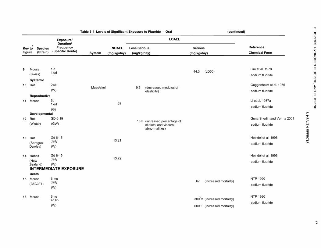
LOAEL
Less SeriousNOAEL(mg/kg/day) (mg/kg/day)
Seriousa
(mg/kg/day)System
Exposure/Duration/
Frequency(Specific Route)
Species(Strain)
Key tofigure
Reference
(continued)Table 3-4 Levels of Significant Exposure to Fluoride - Oral
Chemical Form
9(Swiss)
1 d1x/d
Lim et al. 1978
sodium fluoride44.3 (LD50)
Mouse
Systemic10
Musc/skel(W)2wk Guggenheim et al. 1976
sodium fluoride9.5 (decreased modulus of
elasticity)
Rat
Reproductive11
32
(G)
5d1x/d
Li et al. 1987a
sodium fluoride
Mouse
Developmental12
(Wistar) (GW)GD 6-19 Guna Sherlin and Verma 2001
sodium fluoride18 (increased percentage of
skeletal and visceralabnormalities)
FRat
1313.21
(Sprague-Dawley) (W)
Gd 6-15daily
Heindel et al. 1996
sodium fluoride
Rat
1413.72
(NewZealand) (W)
Gd 6-19daily
Heindel et al. 1996
sodium fluoride
Rabbit
INTERMEDIATE EXPOSUREDeath
15(B6C3F1)
(W)
6 modaily
NTP 1990
sodium fluoride67 (increased mortality)
Mouse
16
(W)
6moad lib
NTP 1990
sodium fluoride300c
(increased mortality)M
600 (increased mortality)F
Mouse

LOAEL
Less SeriousNOAEL(mg/kg/day) (mg/kg/day)
Seriousa
(mg/kg/day)System
Exposure/Duration/
Frequency(Specific Route)
Species(Strain)
Key tofigure
Reference
(continued)Table 3-4 Levels of Significant Exposure to Fluoride - Oral
Chemical Form
Systemic17
Endocr
(W)
2 mo7d/wk24hr/d
Bobek et al. 1976
sodium fluoride0.5 (decreased thyroxine levels;
increased T3- resin uptakeratio)
Rat
188.25 F
(CD)Musc/skel
(W)
daily16-19 weeks
Collins et al. 2001a
sodium fluoride10.7 (prominent growth lines on
upper incisors)F
Rat
19(Sprague-Dawley)
Musc/skel
(W)
7d/wk24hr/d
DenBesten and Crenshaw 1984
sodium fluoride10.5 (decr mineral content and incr
proline in tooth enamel matrix)
Rat
2013
(Wistar)Musc/skel
(W)5 wk Harrison et al. 1984
sodium fluoride19 (histological fluorosis; decr
bone growth)
Rat
21(Fischer- 344)
Gastro
(W)
6 modaily
NTP 1990
sodium fluoride7 (hyperplasia of glandular
stomach)
Rat
20Hepatic
20Renal
220.15 M
(Sprague-Dawley)
Musc/skel
(W)
daily16 or 48 weeks
Turner et al. 2001
sodium fluoride0.5 (decreased vertebral strength
and bone mineralization)M
Rat
23Musc/skel
(W)30d Uslu 1983
sodium fluoride14 (delayed healing of broken
bones)
Rat

LOAEL
Less SeriousNOAEL(mg/kg/day) (mg/kg/day)
Seriousa
(mg/kg/day)System
Exposure/Duration/
Frequency(Specific Route)
Species(Strain)
Key tofigure
Reference
(continued)Table 3-4 Levels of Significant Exposure to Fluoride - Oral
Chemical Form
24Hepatic
(W)
280 ddaily
Greenberg 1982a
sodium fluoride0.95 (pale, granular hepatocytes
with fatty vacuoles)
Mouse
25Renal
(W)280d Greenberg 1986
sodium fluoride1.9 (nephron degeneration)
Mouse
26Musc/skel
(W)
4 wk7d/wkdaily
Marie and Hott 1986
sodium fluoride0.8 (incr bone formation rate; slight
decr bone calcium)
Mouse
27(B6C3F1)
Cardio
(W)
6 modaily
NTP 1990
sodium fluoride67 (multifocal mineralization and
degeneration of themyocardium)
Mouse
Musc/skel 5.6 (increased osteoid in femur andtibia)
M
Hepatic 67 (megaolocytosis and syncytialalteration)
Renal 67 (multifocal nephrosis)
Bd Wt 67 (20% decr bw gain)
28Hemato
(GW)
35 d1x/d
Pillai et al. 1988
sodium fluoride5.2 (decr RBC and hemoglobin, incr
WBC)
Mouse
Bd Wt 5.2 (decr body weight)

LOAEL
Less SeriousNOAEL(mg/kg/day) (mg/kg/day)
Seriousa
(mg/kg/day)System
Exposure/Duration/
Frequency(Specific Route)
Species(Strain)
Key tofigure
Reference
(continued)Table 3-4 Levels of Significant Exposure to Fluoride - Oral
Chemical Form
290.06 M
(Kunmin)Musc/skel
(W)
daily100-150 days
Zhao et al. 1998
sodium fluoride3.2 (incisor fluorosis)M
Mouse
0.06 MEndocr 3.2 (decreased radiolabelled iodineuptake)
M
3.2 MBd Wt
30(NS)
Resp
(F)
6 modaily
Purohit et al. 1999
sodium fluoride4.5 (congestion, edema fluid,
desquamation of respiratoryepithelium in lungs)
Rabbit
Neurological31
(Sprague-Dawley) (W)
6 wkdaily
Mullenix et al. 1995
sodium fluoride6 (altered spontaneous behavior)F
Rat
325.5 F
(Sprague-Dawley) (W)
6 wkdaily
Mullenix et al. 1995
sodium fluoride7.5 (altered spontaneous behavior)F
Rat
33(Wistar)
(GW)
60 ddaily
Paul et al. 1998
sodium fluoride9 (decr spontaneous activity)
Rat
Reproductive34
(Sprague-Dawley) (W)
daily30 days
Al-Hiyasat et al. 2000
sodium fluoride10.21 (decreased number of viable
fetuses, increased resorptions)F
Rat
35(CD)
(F)
60 d7d/wk
Araibi et al. 1989
sodium fluoride2.3 (decr seminiferous tubule
diameter)4.5 (50% reduction in fertility, decr
in percentage of seminiferoustubules containing spermatozoaand decr testosterone levels)
Rat

LOAEL
Less SeriousNOAEL(mg/kg/day) (mg/kg/day)
Seriousa
(mg/kg/day)System
Exposure/Duration/
Frequency(Specific Route)
Species(Strain)
Key tofigure
Reference
(continued)Table 3-4 Levels of Significant Exposure to Fluoride - Oral
Chemical Form
36(NS)
(GW)
daily30 d
Chinoy et al. 1992
sodium fluoride2.3 (decreased fertility and sperm
counts)
Rat
37(CharlesFoster) (F)
30 or 50 daysd
Chinoy et al. 1995
sodium fluoride4.5 (decreased sperm motility and
count)
Rat
3810.7 F
(CD)(W)
daily16-19 weeks
Collins et al. 2001a
sodium fluoride
Rat
3921
(Wistar)(W)
daily6 wk
Krasowska and Wlostowski 1992
sodium fluoride
Rat
40(Wistar)
(W)
daily16 wk
Krasowska and Wlostowski 1992
sodium fluoride7.5 (seminiferous tubule atrophy)
Rat
4123
(F)
3 mo7d/wk
Marks et al. 1984
sodium fluoride
Rat
42CharlesFoster (GW)
daily50 d
Narayana and Chinoy 1994
sodium fluoride4.5 (decr testosterone levels and
Leydig cell diameter)
Rat
4316
(Sprague-Dawley)
(W)daily Sprando et al. 1997
sodium fluoride
Rat
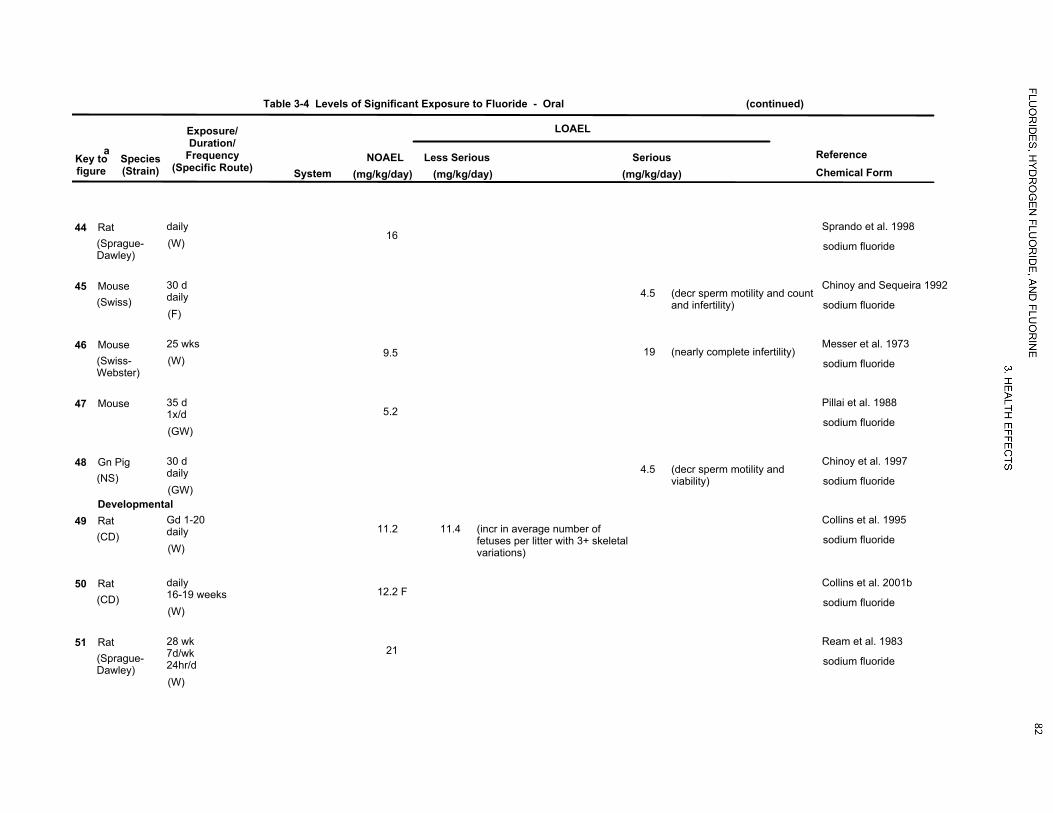
LOAEL
Less SeriousNOAEL(mg/kg/day) (mg/kg/day)
Seriousa
(mg/kg/day)System
Exposure/Duration/
Frequency(Specific Route)
Species(Strain)
Key tofigure
Reference
(continued)Table 3-4 Levels of Significant Exposure to Fluoride - Oral
Chemical Form
4416
(Sprague-Dawley)
(W)daily Sprando et al. 1998
sodium fluoride
Rat
45(Swiss)
(F)
30 ddaily
Chinoy and Sequeira 1992
sodium fluoride4.5 (decr sperm motility and count
and infertility)
Mouse
469.5
(Swiss-Webster)
(W)25 wks Messer et al. 1973
sodium fluoride19 (nearly complete infertility)
Mouse
475.2
(GW)
35 d1x/d
Pillai et al. 1988
sodium fluoride
Mouse
48(NS)
(GW)
30 ddaily
Chinoy et al. 1997
sodium fluoride4.5 (decr sperm motility and
viability)
Gn Pig
Developmental49
11.2(CD)
(W)
Gd 1-20daily
Collins et al. 1995
sodium fluoride11.4 (incr in average number of
fetuses per litter with 3+ skeletalvariations)
Rat
5012.2 F
(CD)(W)
daily16-19 weeks
Collins et al. 2001b
sodium fluoride
Rat
5121
(Sprague-Dawley)
(W)
28 wk7d/wk24hr/d
Ream et al. 1983
sodium fluoride
Rat

LOAEL
Less SeriousNOAEL(mg/kg/day) (mg/kg/day)
Seriousa
(mg/kg/day)System
Exposure/Duration/
Frequency(Specific Route)
Species(Strain)
Key tofigure
Reference
(continued)Table 3-4 Levels of Significant Exposure to Fluoride - Oral
Chemical Form
CHRONIC EXPOSURESystemic
520.04Musc/skel
(W)daily Hillier et al. 2000
sodium fluoride
Human
530.15
dMusc/skel
(W)Daily Li et al. 2001
sodium fluoride0.25 (Increased prevalence of bone
fractures)
Human
54Musc/skel
(C)4 yr Riggs et al. 1990
sodium fluoride0.56 (increased fracture rate)
Human
553.9
(Fischer- 344)Resp
(W)103 wk NTP 1990
sodium fluoride
Rat
3.9Cardio
3.9Gastro
3.9Hemato
2.5Musc/skel 4.3 (osteoscleosis)
3.9Hepatic
3.9Renal
3.9Bd Wt

LOAEL
Less SeriousNOAEL(mg/kg/day) (mg/kg/day)
Seriousa
(mg/kg/day)System
Exposure/Duration/
Frequency(Specific Route)
Species(Strain)
Key tofigure
Reference
(continued)Table 3-4 Levels of Significant Exposure to Fluoride - Oral
Chemical Form
567.6
(B6C3F1)Resp
(W)103 wk NTP 1990
sodium fluoride
Mouse
7.6Cardio
7.6Gastro
7.6Hemato
4.3 MMusc/skel 7.6 (dentine dysplasia)M
7.6Hepatic
7.6Renal
7.6Bd Wt
57Gastro
(GW)
24 mo1x/d
Susheela and Das 1988
sodium fluoride5 (roughened duodena mucosa)
Rabbit
58Hemato
(G)
7-12 mo1x/d
Susheela and Jain 1983
sodium fluoride4.52 (decr leukocyte and hemoglobin
levels)
Rabbit
59Musc/skel
(F)
382 d24hr/d
Aulerich et al. 1987
sodium fluoride5 (mottled and brittle kit teeth) 9.1 (sagittal crests deformed, 3/6
adults)
Mink
Immuno/ Lymphoret60
(albino)(G)
18 mo1x/d
Jain and Susheela 1987
sodium fluoride4.5 (decr primary and secondary
antibody titers)
Rabbit

LOAEL
Less SeriousNOAEL(mg/kg/day) (mg/kg/day)
Seriousa
(mg/kg/day)System
Exposure/Duration/
Frequency(Specific Route)
Species(Strain)
Key tofigure
Reference
(continued)Table 3-4 Levels of Significant Exposure to Fluoride - Oral
Chemical Form
Reproductive61
13(F)3 gen Tao and Suttle 1976
sodium fluoride
Mouse
62(NS)
(GW)
daily18 mo
Kumar and Susheela 1994
sodium fluoride4.5 (structural damage of the
spermatid and epididymalspermatozoa)
MRabbit
63(NS)
(GW)
daily20 or 23 mo
Kumar and Susheela 1995
sodium fluoride4.5 (structural damage of the
spermatid and epididymalspermatozoa)
MRabbit
64(NS)
(GW)
daily18 or 29 mo
Susheela and Kumar 1991
sodium fluoride4.5 (complete cessation of
spermatogenesis)
Rabbit
65(NewZealand) (GW)
daily18 or 23 mo
Susheela and Kumar 1997
sodium fluoride4.5 (Leydig cell damage)
Rabbit
669.1
(F)
382 ddaily
Aulerich et al. 1987
sodium fluoride
Mink

LOAEL
Less SeriousNOAEL(mg/kg/day) (mg/kg/day)
Seriousa
(mg/kg/day)System
Exposure/Duration/
Frequency(Specific Route)
Species(Strain)
Key tofigure
Reference
(continued)Table 3-4 Levels of Significant Exposure to Fluoride - Oral
Chemical Form
Cancer67
(Fischer- 344) (W)
a The number corresponds to entries in Figure 3-4.
b Only this dose level, for the most sensitive group, is plotted in Figure 3-4.
c Differences in levels of health effects and cancer effects between males and females are not indicated in Figure 3-4. Where such differences exist, only the levels of effect for themost sensitive gender are presented.
d Used to derive a chronic-duration oral minimal risk level (MRL) of 0.05 mg fluoride/kg/day; the dose was divided by an uncertainty factor of 3 to account for human variability.
ad lib = ad libitum; Bd Wt = body weight; (C) = capsule); d = day(s); decr = decrease; Endocr = endocrine; (F) = feed; F = female(s); (G) = gavage; Gastro = gastrointestinal; Gd =gestational day; gen = generation(s); (GW) = gavage in water; Hemato = hematological; hr = hour(s); incr = increase; LD50 = lethal dose, 50% kill; LOAEL =lowest-observed-adverse-effect level; M = males; mg/kg/day = milligram per kilogram per day; mo = month(s); Musc/skel = muscular/skeletal; NOAEL = no-observed-adverse-effectlevel; RBC = red blood cell(s); Resp = respiratory; T3 = triiodothyronine; (W) = water; WBC = white blood cell(s); wk = week(s); x = time
103 wk NTP 1990
sodium fluoride2.4 (osteosarcoma of bone)M
Rat
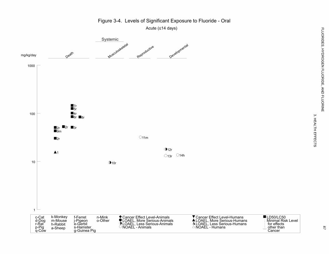
1
10
100
1000
Death
1
9m
2r
2r 2r 3r
4r5r
6r
7r
8r
Musculoske
letal
10r
Reproductive
11m
Developmental
12r
13r 14h
mg/kg/day
Figure 3-4. Levels of Significant Exposure to Fluoride - OralAcute (≤14 days)
c-Catd-Dogr-Ratp-Pigq-Cow
k-Monkeym-Mouseh-Rabbita-Sheep
f-Ferretj-Pigeone-Gerbils-Hamsterg-Guinea Pig
n-Minko-Other
Cancer Effect Level-Animals LOAEL, More Serious-Animals LOAEL, Less Serious-Animals NOAEL - Animals
Cancer Effect Level-Humans LOAEL, More Serious-Humans LOAEL, Less Serious-Humans NOAEL - Humans
LD50/LC50 Minimal Risk Level for effects other than Cancer
Systemic

0.01
0.1
1
10
100
1000
Death
15m
16m
16m
Respiratory
30h
Cardiovascular
27m
Gastrointestin
al
21r
Hematological
28m
Musculoske
letal
26m
27m
29m
29m
18r18r 19r
20r20r
22r
22r
23r
Hepatic
24m
27m
21r
Renal
25m
27m
21r
Endocrine
29m
29m
17r
mg/kg/day
Figure 3-4. Levels of Significant Exposure to Fluoride - Oral (Continued)Intermediate (15-364 days)
c-Catd-Dogr-Ratp-Pigq-Cow
k-Monkeym-Mouseh-Rabbita-Sheep
f-Ferretj-Pigeone-Gerbils-Hamsterg-Guinea Pig
n-Minko-Other
Cancer Effect Level-Animals LOAEL, More Serious-Animals LOAEL, Less Serious-Animals NOAEL - Animals
Cancer Effect Level-Humans LOAEL, More Serious-Humans LOAEL, Less Serious-Humans NOAEL - Humans
LD50/LC50 Minimal Risk Level for effects other than Cancer
Systemic

0.01
0.1
1
10
100
1000
44r
Developmental
49r 49r 50r
51r
mg/kg/day
Figure 3-4. Levels of Significant Exposure to Fluoride - Oral (Continued)Intermediate (15-364 days)
c-Catd-Dogr-Ratp-Pigq-Cow
k-Monkeym-Mouseh-Rabbita-Sheep
f-Ferretj-Pigeone-Gerbils-Hamsterg-Guinea Pig
n-Minko-Other
Cancer Effect Level-Animals LOAEL, More Serious-Animals LOAEL, Less Serious-Animals NOAEL - Animals
Cancer Effect Level-Humans LOAEL, More Serious-Humans LOAEL, Less Serious-Humans NOAEL - Humans
LD50/LC50 Minimal Risk Level for effects other than Cancer
Body Weight
27m
28m
29m
Neurological
31r 32r32r33r
Reproductive
48g 45m
46m
46m
47m
34r
35r
35r 36r
37r
38r
39r
40r
41r
42r
43r
Systemic
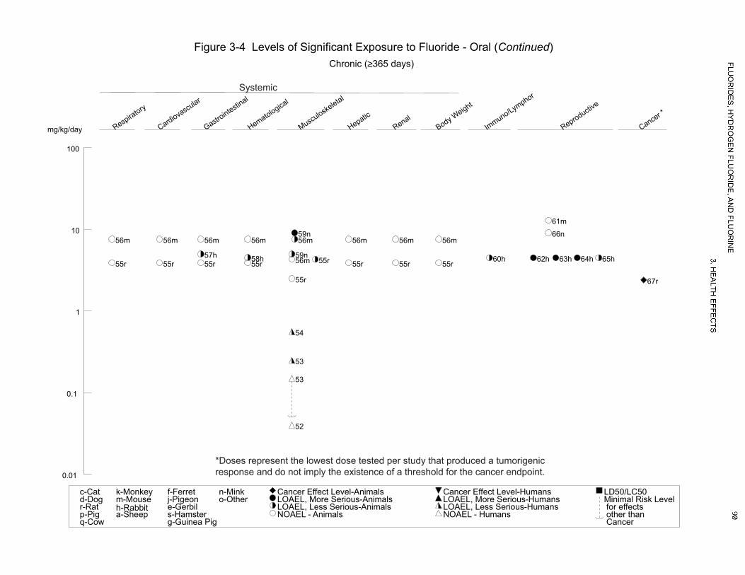
0.01
0.1
1
10
100
Respiratory
56m
55r
Cardiovascular
56m
55r
Gastrointestin
al
56m
55r57h
Hematological
56m
55r58h
Musculoske
letal
52
53
53
54
59n
59n56m
56m
55r
55r
Hepatic
56m
55r
Renal
56m
55r
Body Weight
56m
55r
Immuno/Lymphor
60h
Reproductive
66n
61m
62h 63h 64h 65h
Cancer *
67r
mg/kg/day
Figure 3-4 Levels of Significant Exposure to Fluoride - Oral (Continued)Chronic (≥365 days)
c-Catd-Dogr-Ratp-Pigq-Cow
k-Monkeym-Mouseh-Rabbita-Sheep
f-Ferretj-Pigeone-Gerbils-Hamsterg-Guinea Pig
n-Minko-Other
Cancer Effect Level-Animals LOAEL, More Serious-Animals LOAEL, Less Serious-Animals NOAEL - Animals
Cancer Effect Level-Humans LOAEL, More Serious-Humans LOAEL, Less Serious-Humans NOAEL - Humans
LD50/LC50 Minimal Risk Level for effects other than Cancer
Systemic
*Doses represent the lowest dose tested per study that produced a tumorigenicresponse and do not imply the existence of a threshold for the cancer endpoint.
FLUORIDES, HYDROGEN FLUORIDE, AND FLUORINE 91
3. HEALTH EFFECTS
The highest NOAEL values and all reliable LOAEL values for systemic effects in each species and
duration category are recorded in Table 3-4 and plotted in Figure 3-4.
Respiratory Effects. No studies were located regarding respiratory effects in humans after oral
exposure to fluoride, hydrogen fluoride, or fluorine.
Congestion, the presence of edema fluid, and desquamation of respiratory epithelium were observed in
the lungs of rabbits exposed to 4.5 or 9 mg fluoride/kg/day as sodium fluoride in the diet for 6 months
(Purohit et al. 1999). Inflammatory cell infiltrates, congestion, and desquamated epithelium were also
observed in the large bronchi and trachea of rabbits fed 9 mg fluoride/kg/day. Necrosis of the lung
parenchyma was also observed in two high-dose rabbits that died before the end of the study.
Cardiovascular Effects. The cardiovascular effects of fluoride have been attributed to hypocalcemia
and hyperkalemia caused by high fluoride levels. Fluoride can bind with serum calcium if the dose is
sufficient and cause hypocalcemia. Calcium is necessary for the functional integrity of the voluntary and
autonomic nervous systems. Hypocalcemia can cause tetany, decreased myocardial contractility, and
possibly cardiovascular collapse (Bayless and Tinanoff 1985). Hyperkalemia has been suggested as the
cause of the repeated episodes of ventricular fibrillation and eventual death that are often encountered in
cases of fluoride poisoning (Baltazar et al. 1980).
Approximately 2 hours after ingestion of 120 g of roach powder (97% sodium fluoride) in an
unsuccessful suicide attempt, a 25-year-old male had severe toxic reactions that included tetany, multiple
episodes of ventricular fibrillation, and esophageal stricture (Abukurah et al. 1972). Within 14 hours
following exposure, the patient experienced 63 episodes of ventricular fibrillation. In a study of adults
with skeletal fluorosis living in an area of China with high levels of fluoride in the drinking water (4.1–
8.6 ppm) (Xu and Xu 1997), a higher percentage of electrocardiogram abnormalities was observed in
individuals with skeletal fluorosis (50.73%), as compared to a control group (0.1–0.6 ppm fluoride in
water) (20.0%).
In two epidemiological studies, fluoride in the drinking water did not increase the mortality rates from
cardiovascular effects. One of these studies was a report of 428,960 people in 18 areas of "high" natural
fluoride (0.4–>3.5 ppm) in England and Wales and 368,580 people in control areas (<0.2 ppm fluoride).
The water supply for 52% of the "high" fluoride population had average fluoride levels of ≥1 ppm
(Heasman and Martin 1962). Results indicated that there were no significant differences between areas
FLUORIDES, HYDROGEN FLUORIDE, AND FLUORINE 92
3. HEALTH EFFECTS
with different fluoride levels in mortality due to coronary disease, angina, and other heart disease, as
evidenced by standard mortality ratios (SMRs). The second study (Hagan et al. 1954) examined 32 pairs
of cities in the United States that contained 892,625 people in the high fluoride areas and
1,297,500 people in the control cities. A positive relationship between heart disease and water
fluoridation was reported, but these authors did not adjust for a doubling of the members of this
population over 75 years old during the period of fluoridation under study (Jansen and Thomson 1974).
In addition, this study lacked statistical analysis and drew conclusions regarding trends that were not
obvious from the data presented. The large variation in the presented data was not discussed. Doses of
fluoride are difficult to estimate for large populations, however, because most people are potentially
exposed to fluoride through a variety of sources, such as food, beverages, medicine, and dental products.
Similarly, no significant alterations in the rate of cardiovascular system abnormalities were observed in a
community with 8 ppm fluoride in the water supply, as compared to a community with 0.4 ppm (Leone et
al. 1954).
The results of other studies have suggested a role for fluoride in reducing cardiovascular disease. A study
of 300 North Dakota residents who drank water containing 4–5.8 ppm and 715 people who drank water
containing 0.15–0.3 ppm found a lower incidence of calcification of the aorta in the high-fluoride group
(Bernstein et al. 1966). Significant differences were found in 45–54-year-old males (p<0.05), as well as
in males aged 55–64 and 65+ years (p<0.01). This effect was not due solely to differences in age
distribution, because the incidence in the 55–64-year-old, high-fluoride group was lower than the
incidence in the 45–54-year-old, low-fluoride group. A crude analysis also found no association with
milk and cheese consumption. In a study of four towns in Finland, Luoma (1980) found that incidence of
cardiovascular disease correlated negatively with water fluoride concentration. Taves (1978) likewise
found that standard mortality ratios decreased to a greater extent in fluoridated cities from 1950 to
1970 as compared to non-fluoridated control cities. Both studies, however, relied on population-summary
information for disease rates. A mechanism for this potential reduction in cardiovascular disease could be
the ability of fluoride to inhibit the calcification of soft tissue such as the aorta, as demonstrated in in vitro
studies (Taves and Neuman 1964; Zipkin et al. 1970).
About half of the male and female B6C3F1 mice that died as a result of exposure to 67–71 mg
fluoride/kg/day for 6 months as sodium fluoride in drinking water had mineralization of the myocardium
(NTP 1990); some female mice also had myocardial degeneration. Electrocardiogram alterations and

FLUORIDES, HYDROGEN FLUORIDE, AND FLUORINE 93
3. HEALTH EFFECTS
histological alterations in the myocardium were observed in rabbits exposed to sodium fluoride (Okushi
1971).
Gastrointestinal Effects. The primary gastrointestinal effects following both acute and chronic oral
exposure to fluoride consist of nausea, vomiting, and gastric pain. The irritation of the gastric mucosa is
attributed to fluoride (as sodium fluoride) forming hydrofluoric acid in the acidic environment of the
stomach (Hoffman et al. 1980; Waldbott 1981). The uncharged hydrogen fluoride molecule can then
penetrate cell membranes and enter the neutral environment of the cytoplasm where it dissociates to
release both fluoride and hydrogen ions.
Thirty-four students (kindergarten through third grade) exhibited acute gastrointestinal effects after
drinking water from school water fountains that provided a fluoride supplement designed to raise the
water level to a range of 1–5 ppm (Hoffman et al. 1980). An accident with the delivery system resulted in
the water levels reaching 375 ppm; specific doses could not be calculated, but were estimated to range
from 1.4 to 90 mg per child. In two other cases, individuals vomited and had abdominal pain
immediately after accidentally consuming 1 tablespoon of sodium fluoride (used as a dusting powder for
poultry) (Rao et al. 1969) or sodium fluorosilicate (Dadej et al. 1987).
Of the 150 cases involving fluoride intake reported to a poison control center from 1978 to 1979, most of
the cases involved ingestion of <1 mg/kg fluoride, although exact doses could not be determined (Spoerke
et al. 1980). Effects included nausea (13.9%), vomiting (77.8%), and diarrhea (8.3%). These effects
usually subsided within 24 hours. Symptoms of a more serious nature were not reported.
Endoscopies were performed and biopsy samples were taken from healthy volunteers either after no
treatment (control), or 2 hours after drinking 20 mL of a solution containing 20 mg fluoride (1,000 ppm)
as sodium fluoride (Spak et al. 1989), or application of 0.42% fluoride dental gel (5.1 mg fluoride
ingested) (Spak et al. 1990). Fluoride treatment resulted in petechiae (minute hemorrhages) or erosions in
most of the subjects. Histological examination of biopsied stomach tissue revealed signs of irritation.
Nausea was present in one-third of the subjects drinking the sodium fluoride solution, suggesting that
nausea may not be the first sign of fluoride irritation of the gastric mucosa.
While high levels of fluoride clearly can cause gastrointestinal irritation, it is unclear whether there are
any gastrointestinal effects of chronic exposure to fluoride in drinking water. Gastrointestinal tract
disorders were not evaluated in the Bartlett-Cameron study of the effect of water containing 8 ppm

FLUORIDES, HYDROGEN FLUORIDE, AND FLUORINE 94
3. HEALTH EFFECTS
fluoride (Leone et al. 1954). The sole evidence of an effect comes from a study of 20 non-ulcer dyspepsia
patients at an outpatient clinic in India and 10 volunteers without gastrointestinal problems from the
surgical clinic (Susheela et al. 1992). While none of the drinking water supplies of the controls had
fluoride levels >1 ppm, the water supplies of 55% of the dyspepsia patients were at this level. In addition,
all of the dyspepsia patients and 30% of the controls had serum fluoride levels >0.02 ppm (mean of the
dyspepsia group, 0.1 ppm); all of the dyspepsia patients and none of the controls had urine fluoride levels
>0.1 ppm (mean, 1.34 ppm). The study was compromised by small treatment size, undetermined total
fluoride doses, undetermined nutritional status of the subjects, and lack of statistical comparisons. In
addition, the appropriateness of the control population was not clear.
Seventy-eight workers engaged in the crushing and refining of cryolite, a mineral compound composed of
sodium, aluminum, and fluoride, were examined (Moller and Gudjonsson 1932). Although an average
exposure period was not presented, no workers with <2 years of exposure were included; 18 workers had
been exposed for >10 years. Forty-two workers reported evidence of gastrointestinal effects. The
primary effect was nausea, followed by loss of appetite and vomiting. Chronic indigestion was also
reported in these workers. The study authors stated that the effects were due only to cryolite dust being
swallowed (either due to dust being deposited in the mouth during mouth-breathing, or due to deposition
on the bronchial tree followed by mucociliary action bringing the material to the epiglottis) and absorbed
through the gastrointestinal tract. They based this conclusion on the fact that 21 enamel-, glass-, and
sulphuric acid-industry workers exposed by inhalation to fluorine gas (some for up to 40 years) revealed
no evidence of any effect on the stomach. In light of what is now known about the absorption of fluorides
through the lung, the cryolite workers probably were exposed by both the oral and inhalation routes.
Decreased appetite, congestion of the duodenum, and mild diarrhea were reported in sheep given a single
intragastric dose of 28.5 mg fluoride/kg in the form of sodium fluoride via nasoesophageal catheter
(Kessabi et al. 1985). It is difficult to extrapolate possible human effects from this study because the
gastrointestinal system of ruminants (sheep, cows, goats) is quite different from that of humans.
Thickening of the mucosa of the glandular stomach and punctate hemorrhages were seen in F344/N rats
given 20 mg fluoride/kg/day as sodium fluoride in drinking water for 26 weeks (NTP 1990). Similar, but
less severe, alterations were seen in some rats that received 7 mg fluoride/kg/day. Stomach ulcers were
also seen in some high-dose males and females. Histologically identified stomach lesions included
necrosis and hyperplasia. No gastrointestinal effects were reported in B6C3F1 mice in this study at doses
up to 67–71 mg fluoride/kg/day. No gastrointestinal effects were reported in the chronic portion of this

FLUORIDES, HYDROGEN FLUORIDE, AND FLUORINE 95
3. HEALTH EFFECTS
study at doses up to 9.1 mg/kg/day (mice) or 4.5 mg/kg/day (rats). Roughened duodenal mucosa and a
"cracked-clay" appearance of the absorptive cells were observed following daily dosage of nine rabbits
with 5 mg/kg fluoride via oral gavage for 24 months (Susheela and Das 1988). The rabbit gastrointestinal
system also differs from that of humans, and the study is limited by the small number of rabbits per group
and the use of only one dose.
Hematological Effects. The incidence of abnormal white blood cell counts was significantly higher
in a community with high levels of naturally occurring fluoride (8 ppm) as compared to a community
with low levels of fluoride (0.4 ppm fluoride). However, the study authors did not consider this finding
as necessarily an effect of fluoride (Leone et al. 1954). No other significant hematological effects were
observed. No significant alterations in hemoglobin, erythrocyte, and total leukocyte levels were found in
children living in a community with fluoridated water (1.2 ppm), as compared to children living in a
community with nonfluoridated water (Schlesinger et al. 1956).
As part of the 2-year National Toxicology Program (NTP) study of fluoride (NTP 1990), hematological
analyses were conducted at 27 and 66 weeks. No treatment-related effects were observed at doses up to
4.5 and 9.1 mg/kg/day in F344/N rats and B6C3F1 mice, respectively.
Lactating Holstein cows were fed a mineral supplement containing soft rock phosphate (6,000 ppm
fluoride) and a protein supplement containing 1,088 ppm fluoride (Hillman et al. 1979). Because
consumption of minerals fed ad libitum could not be determined accurately under farm conditions, no
dose estimates could be made. After 9 months, red blood cells per unit volume, blood hemoglobin,
hematocrit, and mean corpuscular volume were significantly lower (p<0.05) in herds exhibiting evidence
of high fluoride exposure. The number of eosinophils increased with increasing urinary fluoride. Rabbits
administered 4.52 mg fluoride/kg/day by gavage for 6–12 months had significantly decreased numbers of
blood cells (e.g., erythrocytes, leukocytes, thrombocytes, monocytes, neutrophils) and hemoglobin
(Susheela and Jain 1983). Similar, although not identical, results were seen in mice fed 5.2 mg
fluoride/kg body weight (Pillai et al. 1988). These animals showed a significant decrease in red blood
cell count, but a significant increase in white cells. Although a dose-effect relationship cannot be
determined from single-dose studies, these studies suggest that the hematopoietic system may be affected
by oral exposure to high levels of fluoride.
Musculoskeletal Effects. The skeletal system is the primary site of fluoride deposition in the body
resulting in both beneficial and adverse effects. Oral exposure to fluoride has been shown to decrease the
FLUORIDES, HYDROGEN FLUORIDE, AND FLUORINE 96
3. HEALTH EFFECTS
prevalence of dental caries in children (the relationship between fluoride and dental caries is discussed in
greater detail in Appendix D); however, at higher doses, fluoride can also result in non-beneficial effects
on the teeth, dental fluorosis. Dental or enamel fluorosis occurs when excess amounts of fluoride are
ingested during tooth development (1–8 years of age). It is characterized by increased porosity (or
hypomineralization) of the subsurface enamel and well mineralized surface layer of enamel. Mildly
fluorosed enamel is full functional (denBesten and Thariani 1992), but may be cosmetically
objectionable. As the severity of dental fluorosis increases, the depth of the enamel involvement and the
degree of porosity increases (Fejerskov et al. 1990). More severely fluorosed enamel is more porous,
pitted, and discolored and is prone to fracture and wear because the well mineralized zone is very fragile
to mechanical stress (denBesten and Thariani 1992; Fejerskov et al. 1990). Not all teeth are similarly
affected by elevated fluoride levels. Teeth that develop earlier in life appear to be less affected than those
that develop later (Fejerskov et al. 1990). Several methods have been developed for quantifying dental
fluorosis. The most commonly used method is Dean's index (Dean 1934), which classifies fluorosis as a
scale of from 0 to 4 as follows: class 0, no fluorosis; class 1, very mild fluorosis (opaque white areas
irregularly covering ≤25% of the tooth surface); class 2, mild fluorosis (white areas covering 25–50% of
the tooth surface); class 3, moderate fluorosis (all surfaces affected, with some brown spots and marked
wear on surfaces subject to attrition); and class 4, severe fluorosis (widespread brown stains and pitting).
The average score of the two most severely affected teeth is used to derive the classification. Other
commonly used methods to rate dental fluorosis include the Thylstrup-Fejerskov (TF) index (Thylstrup
and Fejerskov 1978) and the tooth surface index of fluorosis (TSIF) (Horowitz et al. 1984). Unlike the
Dean’s index, the TF and TSIF indexes use all tooth surfaces to develop the final index score.
The development and severity of dental fluorosis is dependent on the amount of fluoride ingested, the
duration of exposure, and the stage of amelogenesis at the time of exposure. The relationship between
fluoride exposure levels and the prevalence and severity of dental fluorosis was first established in the
1930s and 1940s. There is an extensive amount of literature on the relationship between the prevalence of
dental fluorosis and the concentration of fluoride in municipal water (e.g., Alarcón-Herrera et al. 2001;
DHHS 1991; Eklund et al. 1987; Heifetz et al. 1988; Jackson et al. 1995; Selwitz et al. 1995; Teotia and
Teotia 1994) and the use of fluoridated dentifrices (as reviewed by Warren and Levy 1999). Studies
examining children living in communities with different fluoride levels found that the severity of dental
fluorosis is directly related to fluoride exposure levels. Higher fluorosis severity scores were found in
children living in communities with 4 ppm fluoride in drinking water compared to children living in
communities with 1 ppm fluoride in drinking water (Heifetz et al. 1988; Jackson et al. 1995; Selwitz et al.
1995). No signs of dental fluorosis (using the TSIF scoring method) were found in 81.8, 54.7, and 7.9%

FLUORIDES, HYDROGEN FLUORIDE, AND FLUORINE 97
3. HEALTH EFFECTS
of the children aged 7–14 years living in communities with 0.2, 1, or 4 ppm, fluoride in drinking water,
respectively (Jackson et al. 1995). Moderate dental fluorosis (TSIF score of 3) was observed in 0, 0.9,
and 25.7%, respectively, of the children. The maximum TSIF score was 2 (3.2% of the children received
this score) in the 0.2 ppm community, 4 (0.9% of children) in the 1 ppm community, and 7 (6.9% of
children) in the 4 ppm community. Studies in adults have not always found a direct relationship between
severity of dental fluorosis and prevalence of dental caries (Eklund et al. 1987). A recent meta-analysis
of 88 studies on dental fluorosis (McDonagh et al. 2000) found a dose-related response relationship
between the levels of fluoride in drinking water and the prevalence of dental fluorosis. At a water
fluoride concentration of 1 ppm, the estimated prevalence of dental fluorosis is 48% (95% confidence
interval of 40–57%) and the prevalence of fluorosis of aesthetic concern would be 12.5% (95%
confidence interval of 7.0–21.5%). Another meta-analysis found a significant association between dental
fluorosis and use of fluoride supplements (Ismail and Bandekar 1999).
There is also evidence that the prevalence of dental fluorosis has increased over time due to the multiple,
widespread sources of fluoride in food processed with fluoridated water and dentifrices containing
fluoride, which has resulted in higher fluoride exposure levels. The DHHS (1991) compared the
prevalence of dental fluorosis in three cities (Kingston, New York, Kewanee, Illinois, and Newburgh,
New York) measured in 1939 or 1955 with prevalence in those cities in 1980 or 1985. In Kingston,
which had a municipal fluoride level of 0 ppm, the prevalence of dental fluorosis (using the Dean’s index)
increased from 0.0 to 7.3%, primarily in the very mild (4.4% received this score) category. In the other
two cities (0.9 or 1.0 ppm fluoride in water), there was only a slight change in the overall prevalence
(12.2 versus 14.6% for Kewanee and 7.3 versus 7.7% in Newburgh) of dental fluorosis. However, there
was a shift toward higher severity scores. For example in Kewanee, the prevalence in the very mild, mild,
moderate, and severe categories was 10.6, 1.6, 0.0, and 0.0%, respectively, in 1939 and 7.4, 4.8, 1.8, and
0.6%, respectively, in 1980. In another comparison conducted by DHHS (1991), the prevalence and
severity of dental fluorosis measured in children living in 21 cities in the 1940s were compared with the
prevalence and severity of dental fluorosis measured in 28 cities in the 1980s. During the 40-year period,
the prevalence of fluorosis in areas with <0.4 ppm fluoride increased from <1% to about 6%; nearly all of
the increase was in the very mild and mild categories. Both the prevalence and severity of fluorosis
increased in communities with 0.7–1.2 ppm fluoride, with prevalence increasing from about 13% to about
22%. Most of the increase was in the very mild and mild categories, which increased from 12.3% to
17.7% and from 1.4% to 4.4% of the population, respectively. The combined prevalence of the severe
and moderate categories increased from 0.0% to 0.9%. While there were some differences between the
studies in the 1940s and those in the 1980s, such as the subject population and examination conditions,

FLUORIDES, HYDROGEN FLUORIDE, AND FLUORINE 98
3. HEALTH EFFECTS
they do not affect the overall trends. Although total fluoride intake was not measured, these studies
indicate that intake by children at risk has increased since the 1940s, because fluorosis levels increased
for all water fluoride levels. Another series of studies compared the prevalence and severity of dental
fluorosis in 8–10 and 13–15-year-old children living in communities with 1, 2, 3, or 4 ppm fluoride in
drinking water in 1980 to the prevalence in 1985 (Heifetz et al. 1988) and 1990 (Selwitz et al. 1995).
While there were no marked changes in fluorosis levels in 8–10-year-old children, both the prevalence
and severity increased in the 13–15-year-old children (this group of children also participated in the 1980
study). Increases in the 1-ppm communities were mostly in the category of barely visible white spots.
However, the percentage of labial surfaces of incisors and canines from children in the 2-ppm group that
had brown mottling increased from 0 to 7.6%. Less marked increases in mottled and pitted teeth were
seen in the higher exposure groups. The increased levels of fluorosis were attributed to increased fluoride
exposure from multiple sources. However, the apparent increase in fluorosis did not continue from 1985
to 1990 (Selwitz et al. 1995); in many cases, the severity of the fluorosis declined in the period of 1985–
1990 compared to the 1985 levels.
There is some evidence to suggest that exposure to very high levels of fluoride can increase the
susceptibility to dental caries. Studies in children and adolescents aged 8–16 years (Driscoll et al. 1986;
Mann et al. 1987) found higher incidences of dental caries in individuals with severe dental fluorosis than
in individuals with less severe dental fluorosis. However, for children with very mild, mild, or moderate
fluorosis, no significant associations were found. Driscoll et al. (1986) reported a mean decayed, missing,
and filled surface (DMFS) score of 2.96 for children with severe fluorosis (TSIF score of 4) compared to
1.40 for children with very mild dental fluorosis (TSIF score of 0.5) or 1.58 for children with mild to
moderate fluorosis (TSIF score of 1–3). It is possible that the higher prevalence of filled teeth was due to
the increased rate of fracture and wear in severely fluorosed teeth rather than a higher risk of tooth decay.
As with the dental effects, fluoride has both beneficial and adverse effects on bone. Because fluoride
stimulates bone formation, and increases bone mass, especially in cancellous bone, and inhibit bone
resorption, fluoride has been used as a treatment for osteoporosis. However, the effect of fluoride on the
bone varies in different parts of the body. It increases bone formation earlier and to a larger extent in
trabecular bone than in cortical bone (Gruber and Baylink 1991). There is an excellent response in the
spine, slight response in the hip, and poor response in the wrist as a result of fluoride treatment. Studies
examining the efficiency of fluoride treatment for osteoporosis have found an increased risk of hip
fractures; the prevailing view is that the beneficial increase in spinal bone mass is at the expense of an
increasing risk of hip fractures. There is some evidence that exposure to low doses of slow released

FLUORIDES, HYDROGEN FLUORIDE, AND FLUORINE 99
3. HEALTH EFFECTS
sodium fluoride can significantly reduce the occurrence of spinal fractures in individuals with
osteoporosis (Pak et al. 1995). A large number of studies have examined the adverse skeletal effects of
fluoride; the human studies primarily consist of clinical trials of elderly subjects administered fluoride for
the treatment of osteoporosis and community-based studies of populations exposed to fluoride in drinking
water. The observed effects include increases in bone density, increased risk of bone non-vertebral
fractures, and skeletal fluorosis.
Skeletal fluorosis is a condition associated with an accumulation of fluoride in the bone resulting in brittle
bones and decreased tensile strength. In severe cases (crippling skeletal fluorosis), complete rigidity of
the spine, often accompanied by kyphosis (humpbacked) or lordosis (arched back), is observed. Skeletal
fluorosis is associated with long-term exposure to very high oral doses of fluoride or occupational
exposure to cryolite (AlF6Na2) dust (which would involve inhalation and oral exposure to fluoride).
Cases of skeletal fluorosis are predominantly found in developing countries, particularly India and China,
and are associated with high fluoride intakes coupled with malnutrition (WHO 2002). High tea
consumption and increased consumption of water with high levels of naturally occurring fluoride are the
primary contributors to the elevated fluoride intake; in China, the indoor burning of fluoride-rich coal also
contributes to the overall fluoride intake. Most reports of skeletal fluorosis are inadequate for establishing
dose-response relationships. Choubisa et al. (1997) examined the prevalence of skeletal fluorosis in
residents of 15 villages in India. At 1.4 ppm fluoride in drinking water, the prevalence of skeletal
fluorosis was 4.4%; at 6.0 ppm fluoride, the prevalence was 63.0%. Another investigator found that
skeletal fluorosis occurred 10 years after the initiation of increased fluoride consumption; the average
water concentration was 9.2 ppm fluoride and daily water consumption was 4.6 L/day (42 mg
fluoride/day) (Saralakumari and Ramakrishna Rao 1993); after 20 years of exposure, the prevalence of
skeletal fluorosis was 100%. A limited number of cases of skeletal fluorosis have been reported in the
United States (Bruns and Tytle 1988; Fisher et al. 1981; Goldman et al. 1971; Sauerbrunn et al. 1965);
where doses are known, they are generally 15–20 mg fluoride/day for over 20 years. In a study of
116 people who had lived in an area with an average of 8 ppm fluoride in the drinking water for at least
15 years, a 10–15% incidence of fluoride-related bone changes was observed (Leone et al. 1955).
Coarsened trabeculation and thickened bone were observed, but no exostoses were evident, and the
subjects were asymptomatic. The severity of the skeletal fluorosis appears to be related to bone fluoride
levels. Among persons whose drinking water contains about 1 ppm fluoride, bone ash fluoride
concentrations trypically range from 500 to 1,500 ppm. The higher values are generally found among
older persons since the concentrations tend to increase gradually throughout life. Bone from people with
preclinical skeletal fluorosis, which is generally asymptomatic and characterized by slight radiologically

FLUORIDES, HYDROGEN FLUORIDE, AND FLUORINE 100
3. HEALTH EFFECTS
detectable increases in bone mass, contains 3,500–5,500 ppm fluoride. Sporadic pain, joint stiffness, and
osteosclerosis of the pelvis are observed at 6,000–7,000 ppm, while chronic joint pain, increased
osteosclerosis, and slight calcification of ligaments occur at 7,500–9,000 ppm. Crippling fluorosis is
observed at fluoride bone concentrations >10,000 ppm (Franke et al. 1975).
Studies of individuals undergoing fluoride treatment for osteoporosis, epidemiology studies of
communities with high levels of fluoride in drinking water, and community-based studies of populations
with different fluoride levels in the water have examined the possible relationship between exposure to
fluoride and alterations in bone mineral density and/or the risk of bone fractures. The effect of fluoride
on bone fracture rate and mineralization has alos been investigated in children.
A prospective, randomized of 135 women with postmenopausal osteoporosis ascertained the effect of
administering 34 mg fluoride/day as sodium fluoride (0.56 mg fluoride/kg/day) for 4 years (Riggs et al.
1990). The fluoride treated and the placebo control groups received 1,500 mg calcium/day. Although
bone mineral density in the lumbar spine, femoral neck, and femoral trochanter increased markedly in the
treatment group, bone mineral density in the shaft of the radius decreased 4%. There was no significant
difference in the number of new vertebral fractures between the treatment and control groups, although
the number of vertebral fractures in the fluoride group was slightly elevated in the first year. In contrast,
the level of nonvertebral fractures in the fluoride group was 3.2 times that of the control group, with
significant increases in both the frequency and rate of fractures. Most of the increase was due to
increased incidences of incomplete ("stress") fractures, which occurred 16.8 times more often in the
treatment group. In a follow-up to this study, Riggs et al. (1994) examined 50 of the women in the
fluoride treatment group after an additional 2 years of treatment with 34 mg fluoride/day as sodium
fluoride. The lumbar spine, femoral neck, and femoral trochanter bone mineral density continued to
increase and the bone mineral density of the radius continued to decrease during years 4–6 of treatment.
The vertebral fracture rate decreased during years 4–6 as compared to years 0–4. The nonvertebral
fracture rate also decreased during the last 2 years, but the rate for the full 6-year period was still 3 times
higher than the rate in the placebo control group. In addition to extending the study for an additional
2 years, Riggs et al. (1994) also re-examined the data from the previous study. Vertebral fracture rate was
influenced by several factors. Vertebral fracture rate decreased with increasing lumbar spine bone
mineral density except in the cases where the higher bone mineral density was associated with a rapid rate
of increase in the lumbar spine bone mineral density or a large increase from baseline serum fluoride
level.

FLUORIDES, HYDROGEN FLUORIDE, AND FLUORINE 101
3. HEALTH EFFECTS
In a similar study by Kleerekoper et al. (1991), the anti-fracture efficacy of 34 mg fluoride/day as sodium
fluoride was examined in 46 postmenopausal women (mean age of 66.2 years) with spinal osteoporosis.
A daily dose of 1,500 mg calcium was also administered to this group as well as a placebo control group
of 38 postmenopausal women with spinal osteoporosis (mean age of 67.9 years). No significant
differences in bone mineral density of the forearm, vertebral fractures, or peripheral fractures were found.
A significant increase in painful lower extremity syndrome was observed in the fluoride group. It should
be noted that Riggs et al. (1990, 1994) considered the lower extremity syndrome to be incomplete
fractures and the incidence of incomplete fractures was added to the complete fracture incidence to
calculate nonvertebral fracture incidence.
Haguenauer et al. (2000) performed a meta-analysis to examine the effects of fluoride on the treatment
and prevention of postmenopausal osteoporosis using the data from the Riggs et al. (1990, 1994),
Kleerekoper et al. (1991), and 10 other studies. The meta-analysis showed a significant increase in bone
mineral density in the lumbar spine and hip and a decrease in bone mineral density in the forearm after
2 or 4 years of fluoride treatment. When the data from all studies were used, fluoride treatment for 2–
4 years did not affect the relative risk of vertebral fractures. However, in studies in which the subjects
were exposed to low levels of fluoride or a slow-release formulation for 4 years, a significant decrease in
vertebral fracture relative risk was seen. An increase in the relative risk of nonvertebral fracture was
observed when data from all studies were used; no effect was seen in studies using low levels of fluoride
(<30 mg/day) or slow-release fluoride.
Conflicting findings on whether fluoride exposure in drinking water results in increased bone mineral
density have been reported, with studies finding increases in spine and femur bone mineral density
(Kröger et al. 1994; Phipps et al. 1998, 2000), decreases in radius mineral density (Phipps et al. 1998), or
no effect on bone mineral density (Cauley et al. 1995; Lehmann et al. 1998; Sowers et al. 1991). Lumbar
spine, proximal femur, and forearm bone mineral densities were measured in men and women 60 years
and older living in one of three cities with different levels of naturally occurring fluoride in drinking
water (Phipps et al. 1998). After adjusting for a number of non-fluoride related risk factors, significant
elevations in bone mineral density of the lumbar spine (men and women) and proximal femur (women
only) were observed in residents with the highest levels of fluoride (2.5 mg/L), as compared to densities
in residents with the lowest fluoride concentration (0.03 mg/L); no significant differences in bone mineral
density were found between residents with the mid-level fluoride concentration (0.7 mg/L) and the lowest
concentration. In contrast to this finding, a negative correlation between bone mineral density of the
forearm and an individual’s fluoride intake from drinking water and toothpaste was found. Significant

FLUORIDES, HYDROGEN FLUORIDE, AND FLUORINE 102
3. HEALTH EFFECTS
increases in bone mineral density were also reported in another study by this group (Phipps et al. 2000).
Adjusted (age, weight, education, knee/grip strength, surgical menopause, calcium intake, alcohol
consumption, estrogen use, thiazide use, diabetes, thyroid hormone levels, exercise, and smoking) mineral
densities were higher in the lumbar spine and femur and lower in the radius in women aged 65 years and
older exposed to fluoridated drinking water, as compared to women with no reported exposure to
fluoridated water. Kröger et al. (1994) also found significant increases in bone mineral density (adjusted
for confounding factors such as age, weight, menopausal status, calcium intake, physical activity) of the
femoral neck and lumbar spine among women aged 47–56 years exposed to fluoridated drinking water
(1.2 mg/L) for >10 years (mean exposure duration was 25.9 years), as compared to women exposed to
fluoridated drinking water for <10 years.
In a study of women aged 65 years and older, no significant alterations in bone mineral density of the
radius, calcaneus, hip, or lumbar spine were found between women with no exposure to fluoridated water
(n=1,248), 1–10 years of exposure (n=438), 11–20 years of exposure (n=198), or greater than 20 years of
exposure (n=192) (Cauley et al. 1995). Total calcium intake was significantly higher in the >20-year
group and lower in the 11–20-year group. Lehmann et al. (1998) also found no difference in age- and
weight-adjusted bone mineral density of the spine and femur of women aged 20–69 years living in an area
with fluoridated water (1 mg/L). In the men living in the fluoridated water a community, there was a
significant increase in age- and weight-adjusted bone mineral density of the Ward’s triangle portion of the
femur, but no effect on neck or trochanter portions of the femur or the spine. The lack of adjustment for
other risk factors limits the interpretation of this study; significantly higher alcohol consumption and
lower calcium intakes were found in the fluoride-exposed group. Sowers et al. (1991) also found no
significant alterations in bone mass in women aged 20–80 years living in a community with naturally
high fluoride levels (4 mg/L) as compared to women living in a community with fluoridated water
(1 mg/L). However, radius bone mass was significantly lower in the high fluoride group.
As with bone mineral density, conflicting results have been found on the effect of low levels of fluoride
on the risk of fractures, particularly hip fractures. These studies have found conflicting results, with
studies finding a lower or higher incidence of hip fractures or no differences in hip fracture between
humans exposed to fluoride in drinking water.
Several studies have found decreases in hip fracture incidences in communities with fluoride in the
drinking water, suggesting that there may be a beneficial effect. Simonen and Laittinen (1985) examined
male and female residents older than 50 years living in two cities in Finland with either trace amounts of

FLUORIDES, HYDROGEN FLUORIDE, AND FLUORINE 103
3. HEALTH EFFECTS
fluoride in the water or with 1 ppm fluoride in the water. The occurrence of femoral neck fractures was
lower in the men 50–80 years old and women >70 years old living in the area with fluoridated water, as
compared to the low fluoride community. No difference in femoral neck fracture was observed in women
50–69 years of age. In a prospective study of older women, Phipps et al. (2000) examined the possible
relationship between living in an area with fluoridated water and the risk of fractures among women
≥65 years old. Fewer spine, hip, and humerus fractures were observed in this group. However, a
nonsignificant trend toward higher incidences of wrist fractures was observed in the continuous exposure
group. Lehmann et al. (1998) also found a significant decrease in the occurrence of hip fractures
(adjusted for age) in men and women living in an area with fluoridated water (approximately 1 ppm), as
compared to residents living in a city with low levels of fluoride in the water.
In contrast to the results of these studies, other studies have found an increase in the incidence of hip
fractures in communities with fluoride in the drinking water. Sowers et al. (1986) found a higher
incidence of hip fractures among women aged 55–80 years living in a community with high levels of
naturally occurring fluoride (4 ppm) in the water, as compared to women living in an area with
fluoridated water (1 ppm). The lower calcium intake in the high fluoride group may have influenced
these results. A geographical correlational study of 541,985 white women hospitalized for hip fractures
found a weak association (regression coefficient=0.001, p=0.1) between hip fracture incidence and
fluoridation of water (Jacobsen et al. 1990). The association was strengthened (regression
coefficient=0.003, p=0.0009) after correcting by county for other factors found to correlate with hip
fracture incidence (latitude, hours of sunlight, water hardness, income level, and percentage of land in
farms). Another study by this group (Jacobsen et al. 1992) also found a significantly elevated risk of hip
fractures in residents living in counties with fluoridated water. The relative risks were
1.17 (95% CI=1.13–1.22) and 1.08 (95% CI=1.06–1.10) for men and women aged 65 years and older.
The highest prevalence of hip fractures was found in counties that began a fluoridation program within
the last 5 years. Madans et al. (1983) examined the association between fluoride in drinking water and
risk of hip fractures using hip fracture data from the National Health Interview Surveys of 1973–1977 and
Centers for Disease Control and Prevention (CDC) data on the percent of a population in each U.S.
county served with water having a natural or adjusted fluoride content of at least 0.7 ppm in 1973.
Female residents over 45 years of age living in areas with lower fluoride levels in the drinking water had
9% more hip fractures than women living in high fluoride areas; however, the difference was not
statistically significant. An increase in the risk of hip fractures (age, sex, and quatelet index-adjusted
odds ratio of 1.86, 95% CI=1.02–3.36) was also observed in adults aged 65 years and older living in areas

FLUORIDES, HYDROGEN FLUORIDE, AND FLUORINE 104
3. HEALTH EFFECTS
with drinking water fluoride levels ranging from 0.11 to 1.83 ppm (Jacqmin-Gadda et al. 1995); these
data were reported as a letter to the editor and limited study details were provided.
A study in England and Wales also found increased rates of hip fractures in men and women over
age 45 as water fluoride levels increased up to 0.93 ppm (Cooper et al. 1991). Hip fracture rates in
39 counties (standardized by age and sex) were compared with water fluoride levels in those counties. In
the original analysis (Cooper et al. 1990), no significant correlation was found. However, when the
authors reanalyzed the data using a weighted least-squares technique (weighting by the size of the
population aged ≥45 years) to account for differences in the precision of the county-specific rates, a
significant positive correlation between water fluoride levels and hip fracture rates was found (r=0.41,
p=0.009). The correlation existed for both women (r=0.39, p=0.014) and men (r=0.42, p=0.007) (Cooper
et al. 1991). Kurttio et al. (1999) studied over 144,000 residents living in rural areas of Finland from
1967 to –1980. When all age groups were considered together, no relationship between fluoride levels in
drinking water and the risk of hip fractures was found. However, among women aged 50–64 years with
higher fluoride levels, an increase in the risk of hip fractures was found. No consistent relationships were
found in men or in older women. The study authors suggested that other risk factors for hip fracture may
be more important than fluoride exposure in determining risk of hip fractures in older women. An
ecologic cohort study compared the hip fracture rate for men and women in a Utah community that had
water fluoridated to 1 ppm with the rate in two communities with water containing <0.3 ppm fluoride
(Danielson et al. 1992). The age-adjusted rate was significantly elevated in both women (relative risk
1.27, 95% CI=1.08–1.46) and men (relative risk 1.41, 95% CI=1.00–1.81). In men, the rates in the
fluoridated and nonfluoridated communities were similar until age 70. After age 75, the difference
between the rates in the fluoridated and nonfluoridated areas increased with age. The difference between
the hip fracture rates in the fluoridated and nonfluoridated areas increased for women in the 70- and
75-year age groups. However, the fracture rates in women at ages ≥80 years old were similar in the
fluoridated and nonfluoridated towns. The study authors attributed this to the fact that women older than
80 years would have already gone through menopause by the beginning of fluoridation, and so would
have had less bone remodeling and less incorporation of fluoride into the bone. The study authors also
suggested that the reason that they found an effect when other investigators have not was the low levels of
exposure to risk factors for osteoporosis (smoking and alcohol) in the Utah populations. This was a well-
conducted study that suggests that communities with fluoridated water have an elevated risk of hip
fracture. However, several possible confounding factors were not examined. Calcium levels in the water,
total calcium and vitamin D intake, and individual fluoride intake were not determined. Estrogen use was

FLUORIDES, HYDROGEN FLUORIDE, AND FLUORINE 105
3. HEALTH EFFECTS
not evaluated, but was assumed to be similar since the communities were similar distances from larger
medical centers. In addition, estrogen levels would not cause the effect in men.
A study by Li et al. (2001) found a U-shaped pattern for the prevalence of overall bone fractures and the
level of fluoride in the drinking water of six communities in rural China. The prevalence of bone
fractures (adjusted for age and gender) was significantly elevated in the communities with 0.25–0.34 ppm
fluoride (7.41%) or 4.32–7.97 ppm fluoride (7.40%), as compared to the prevalence (5.11%) in the
community with 1.00–1.06 ppm fluoride in the water. When only hip fractures since age 20 years were
examined, the age- and body mass index (BMI)-adjusted prevalence was only significantly higher in the
4.32–7.97 ppm group; prevalence of 1.20% compared to 0.37% in the 1.00–1.06 ppm group. A similar
pattern was found for overall fractures since age of 50 years; the age-adjusted prevalences were 4.80 and
3.28% in the 4.32–7.97 and 1.00–1.06 ppm groups, respectively.
Other studies have not found a relationship between fluoride in drinking water and hip fracture
prevalence. No significant differences in the incidence or type of upper femoral fracture were observed
when groups of subjects living in communities with low fluoride (<0.3 ppm), fluoridated (1.0–1.2 ppm),
or high fluoride (>1.5 ppm) drinking water (Arnala et al. 1986). Kröger et al. (1994) found no effect on
self-reported fractures among a group of older Finnish residents (mean age of approximately 53 years)
living in an area with fluoridated water (1.0–1.2 mg/L), as compared to residents living in an area with
low fluoride levels in the drinking water (<0.3 ppm). Similarly, Avorn and Niessen (1986) found no
statistically significant alteration in the occurrence of long bone fractures among women aged 65 years
and older living in counties with fluoridated water (fluoride concentrations were not reported) as
compared to residents living in counties without fluoridated water. No alteration in the occurrence of hip
or ankle fractures were observed in men and women aged 65 years or older residents living in
communities with fluoridated water, as compared to residents living in communities without a water
fluoridation program (Karagas et al. 1996). In men, the risk of proximal humerus fracture (relative risk
1.23, 95% CI=1.06–1.43) and distal forearm fractures (relative risk of 1.16, 95% CI=1.02–1.33) were
higher in the fluoridated water areas; the relative risks of these fractures in women living in fluoridated
areas were not significantly altered. Cauley et al. (1995) found no effect on the risk of vertebral or
nonvertebral fractures among women aged 65 years and older exposed to fluoridated water for 13 years
(mean exposure duration).
Jacobsen et al. (1993) compared hip fractures rates in the 10-year period prior to fluoridation to the rates
in the 10-year period after fluoridation of a city’s water source (fluoride concentration of 1.1 ppm). No

FLUORIDES, HYDROGEN FLUORIDE, AND FLUORINE 106
3. HEALTH EFFECTS
significant differences in the risk of hip fractures were found in men or women aged 50 years and older;
the relative risks, after adjusting for other risk factors, were 0.78 (95% CI=0.37–1.66) and 0.60 (95%
CI=0.42–0.85) for men and women, respectively. No significant alterations in the risk of hip fracture
were found among male and female residents aged 45 and older living in a city with fluoridated water
(Suarez-Almazor et al. 1993). However, when male residents were considered separately, the relative risk
of hip fracture was statistically significant in the 65 years and older age bracket (risk ratio of 1.13, 95%
CI=1.00–1.24) and all men combined (risk ratio of 1.12, 95% CI=1.01–1.24).
With the exception of studies examining skeletal fluorosis, most of the studies on the skeletal effects of
fluoride involved older adult populations. There is also some limited evidence that fluoride may affect
bone mineralization in children. Increased trabecular height and area were observed in the distal radial
metaphysis of boys (11–12 years of age) with dental fluorosis living in an area with 2.7 ppm fluoride in
water (Chlebna-Sokol and Czerwinski 1993). No alterations were observed in similarly aged girls or
older (13.5–15 years) boys and girls. The trabecular changes were correlated with lower serum calcium
levels and higher alkaline phosphatase activity levels. Alarcón-Herrera et al. (2001) found a significant
correlation between bone fracture incidence and Dean’s index of dental fluorosis among children aged 6–
12 years and adults aged 13–60 years.
Evidence from animal experiments supports the association of high levels of fluoride and adverse effects
on bone. The femurs of weanling male rats of a Wistar-derived strain that were given ≥9.5 mg
fluoride/kg/day as sodium fluoride for 2 weeks exhibited a marked decrease in the modulus of elasticity.
It is not clear if the change was analyzed statistically. No lower doses were tested (Guggenheim et al.
1976). Musculoskeletal effects in albino rats (strain not identified) following oral exposure of
intermediate duration have been investigated. After 30 days of exposure to 100 ppm of fluoride in water
(14 mg fluoride/kg/day), tibia bones were broken and allowed to heal (Uslu 1983). Collagen synthesis
was determined to be defective, and fracture healing was delayed, when compared to the controls.
Decreased bone growth and signs of fluorosis were observed in rats given 19 mg fluoride/kg/day as
sodium fluoride in their drinking water and adequate calcium for 5 weeks; with elevated calcium levels,
fluorosis was not observed until the fluoride level reached 35 mg fluoride/kg/day (Harrison et al. 1984).
Male mice administered 0.80 mg fluoride/kg/day for 4 weeks exhibited a statistically significant increase
in the bone formation rate and a slight but statistically significant decrease in bone calcium levels (Marie
and Hott 1986). The authors concluded that 0.80 mg fluoride/kg increased the population of osteoblasts
under the conditions of this experiment. Turner et al. (1992) found a biphasic relationship between bone
strength and bone fluoride content in rats. At lower fluoride intakes, increased bone strength was
FLUORIDES, HYDROGEN FLUORIDE, AND FLUORINE 107
3. HEALTH EFFECTS
observed; the maximum bone strength was achieved at 1,000–1,500 ppm fluoride in the bone. At fluoride
concentrations >1,000 ppm bone strength started to decrease. The sagittal crests were enlarged and/or
deformed in three of six adult female mink fed 9.1 mg fluoride/kg/day as sodium fluoride for 382 days
(Aulerich et al. 1987). The authors attributed the abnormalities of the sagittal crests to increased
osteoblastic activity. An increase in the diameter of the femur bone and decreased breaking strength was
observed in pigs administered sodium fluoride or rock phosphate in the diet (Kick et al. 1933).
Hepatic Effects. No studies were located regarding hepatic effects in humans after oral exposure to
fluoride, hydrogen fluoride, or fluorine.
Pale, granular hepatocytes, compatible with parenchymal degeneration, were observed in mice
administered 0.95 mg fluoride/kg/day in drinking water for 7–280 days (Greenberg 1982a). Fatty
granules were observed after 3 weeks. Liver congestion was observed in sheep given a single intragastric
dose of fluoride as low as 9.5 mg fluoride/kg. Mild serum increases of liver enzymes (glutamate
dehydrogenase [GDH] and gamma-glutamyl transferase [GGT]) also occurred in sheep administered
38 mg fluoride/kg (Kessabi et al. 1985). It is difficult to use this result to predict to possible human
effects because ruminants (sheep, cows, goats) have gastrointestinal systems quite different from that of
humans.
Enlarged liver cells with multiple foci were seen in about half of the male B6C3F1 mice that died after
receiving 33–36 or 67–71 mg fluoride/kg/day for up to 6 months as sodium fluoride in drinking water
(NTP 1990). This change was seen in all of the female mice that died at the 71 mg/kg/day dose level. No
liver effects were seen in a parallel experiment with F344/N rats at doses up to 20 mg/kg/day. Similarly,
no liver histopathology was seen in the chronic portion of this study (NTP 1990), in which rats received
total fluoride doses (amount added to water plus endogenous fluoride in food) of about 4.5 mg/kg/day
(rats) or up to 9.1 mg/kg/day (mice). Alkaline phosphatase levels were significantly increased in male
and female mice at the 66-week interim sacrifice of the chronic study.
Renal Effects. One study was located in which ingestion of fluoride appeared to be linked with renal
insufficiency (Lantz et al. 1987). A 32-year-old man ingested 2–4 L of Vichy water (a highly gaseous
mineral water containing sodium bicarbonate and approximately 8.5 mg/L of fluoride) every day for
about 21 years. This exposure ended 4 years before his hospital admission. The patient also had
osteosclerosis and a moderate increase in blood and urinary levels of fluoride. No teeth mottling was
observed. The authors could not find factors, other than fluoride, related to his interstitial nephritis. No

FLUORIDES, HYDROGEN FLUORIDE, AND FLUORINE 108
3. HEALTH EFFECTS
effect on the incidence of urinary tract calculi or the incidence of albuminuria was found in the Bartlett-
Cameron study of people drinking water containing 8 ppm fluoride (Leone et al. 1954).
Congestion of the kidney was observed in sheep given a single intragastric dose of fluoride as low as
9.5 mg fluoride/kg (Kessabi et al. 1985). An intermediate exposure study tested the effect of
administering up to 67–71 mg fluoride/kg/day to B6C3F1 mice (8–9/group) as sodium fluoride in drinking
water for 26 weeks (NTP 1990). Acute nephrosis characterized by extensive multifocal degeneration and
necrosis of the tubular epithelium was believed to be the main cause of death in two of the four males
exposed to 67 mg/kg/day that died, the single male that died after exposure to 33 mg/kg/day, and two of
the four females in the high dose group that died. No kidney histopathology was observed in surviving
mice or in rats exposed to 20 mg fluoride/kg/day and higher (NTP 1990). Significant increases in water
consumption and apparent (based on qualitative descriptions) increases in acid phosphatase activity in the
glomeruli were observed in monkeys exposed to 0.46 mg fluoride/kg/day as hydrofluosilicic acid for
180 days (Manocha et al. 1975). The toxicological significance of these changes is not known.
Changes in kidney histology were seen in mature Swiss mice given a dose of sodium fluoride in drinking
water for up to 280 days that was described as the maximum dose that could be chronically tolerated, i.e.,
1.9 mg/kg/day (Greenberg 1986). Using a sensitive staining technique, increased collagen levels were
seen after about 45 days. Thickening of the Bowman's capsule, edematous swelling of the tubules, and
infiltrations of mononuclear cells were also noticed. No kidney pathology was seen in a 2-year study in
B6C3F1 mice at doses up to 8.1 mg/kg/day (males) or 9.1 mg/kg/day (females), or in F344/N rats at doses
up to 4.1 mg/kg/day (males) or 4.5 mg/kg/day (females) (NTP 1990).
Endocrine Effects. Significant increases in serum thyroxine levels were observed in residents of
North Gujarat, India with high levels of fluoride in the drinking water (range of 1.0–6.53 mg/L; mean of
2.70 mg/L) (Michael et al. 1996). No significant changes in serum triiodothyronine or thyroid stimulating
hormone levels were found. Increases in serum epinephrine and norepinephrine levels were also
observed. In another study conducted in India, a positive dose-response relationship between parathyroid
hormone and fluoride intake from drinking water was observed in children aged 6–12 years (Gupta et al.
2001). It is unclear if nutritional deficiencies played a contributing role to the observed endocrine effects.
Jooste et al. (1999) found a higher goiter prevalence among children (6–15 years of age) living in two
towns in South Africa with high levels of fluoride in the water (1.7 or 2.6 ppm), as compared to children
living in towns with low (0.3 or 0.5 ppm) or optimal (0.9 or 1.1 ppm) fluoride levels in the water. The
prevalence of goiter was also elevated in three of the other four towns, although the prevalence was lower
FLUORIDES, HYDROGEN FLUORIDE, AND FLUORINE 109
3. HEALTH EFFECTS
than in the high fluoride towns; suggesting that the children were exposed to other goitrogens. Iodine
deficiency was not the likely etiological agent because the median urinary iodine levels were higher than
iodine sufficiency standards. These data are inadequate to assess the relationship between elevated
fluoride intake and goiter formation.
Fluoride has been shown to affect the endocrine system in rats given 0.5 mg fluoride/kg/day as sodium
fluoride in drinking water every day for 2 months (Bobek et al. 1976). These animals showed decreased
thyroxine levels and an increased T3-resin uptake ratio. In contrast, Zhao et al. (1998) did not find any
alterations in serum T3 or T4 levels in mice exposed to 3.2 mg fluoride/kg/day as sodium fluoride in
drinking water for 100 or 150 days. However, a decrease in thyroid radiolabelled iodine uptake was
observed at 3.2 mg fluoride/kg/day, but not at 0.06 mg fluoride/kg/day.
No significant alterations in parathyroid hormone levels or morphological alterations in the parathyroid
gland were observed in rats exposed to 3.3 mg fluoride/kg in drinking water for 46 weeks (Rosenquist et
al. 1983).
Body Weight Effects. Final body weight was reduced by >40% relative to the controls in female
F344/N rats administered 25 mg fluoride/kg/day as sodium fluoride in drinking water for 14 days; body
weight in males was reduced by >10% at doses ≥6.3 mg/kg/day (NTP 1990). A clear and consistent
effect on body weight of B6C3F1 mice was seen only at the high dose (69 mg/kg/day), which was lethal
to males (3/5), but not to females. In the intermediate-duration (6 months) phase of the study, the body
weight of mice administered 17 mg fluoride/kg/day was reduced by 20%; it was reduced by 10% at
19 mg/kg/day in male and female rats.
F344/N rats and B6C3F1 mice given large doses of sodium fluoride in drinking water for 14 days had
reduced water intake (NTP 1990). Male and female rats given 25 mg fluoride/kg/day drank about 30%
less water than the controls. Water consumption by male rats given 51 mg fluoride/kg/day was 50% of
controls, while it was 25% of controls for females. Similarly, mice given 69 mg fluoride/kg/day drank
≤60% the volume of water consumed by the controls. This means that actual fluoride doses are lower
than the estimates given here, since these values were calculated assuming normal water intake.
However, the reduced water intake may have been due to the disagreeable taste of fluoride at high
concentrations in the water.

FLUORIDES, HYDROGEN FLUORIDE, AND FLUORINE 110
3. HEALTH EFFECTS
3.2.2.3 Immunological and Lymphoreticular Effects
A request to the American Academy of Allergy was made by the U.S. Public Health Service for an
evaluation of suspected allergic reactions to fluoride as used in the fluoridation of community water
supplies (Austen et al. 1971). The response to this request included a review of clinical reports and an
opinion as to whether these reports constituted valid evidence of a hypersensitivity reaction to fluoride
exposure of types I, II, III, or IV (Austen et al. 1971), which are, respectively, anaphylactic or reaginic,
cytotoxic, toxic complex, and delayed-type reactivity. The Academy reviewed the wide variety of
symptoms presented (vomiting, abdominal pain, headaches, scotomata [blind, or partially blind areas in
the visual field], personality change, muscular weakness, painful numbness in extremities, joint pain,
migraine headaches, dryness in the mouth, oral ulcers, convulsions, mental deterioration, colitis, pelvic
hemorrhages, urticaria, nasal congestion, skin rashes, epigastric distress, and hematemesis) and concluded
that none of these symptoms were likely to be immunologically mediated reactions of types I–IV. No
studies were located that investigated alterations in immune response following fluoride exposure in
humans.
In a study with rabbits administered 4.5 mg fluoride/kg/day as sodium fluoride for 18 months, decreased
antibody titers were observed (Jain and Susheela 1987). These results were observed after 6 months of
treatment; the authors hypothesized that a threshold level is reached at which time the immune system is
impaired. However, as only one dose level (4.5 mg fluoride/kg/day) was tested, no dose-effect
relationships can be established. An increase in the cellularity of Peyer’s patches and mesenteric lymph
nodes was observed in rats administered sodium fluoride (Butler et al. 1990).
3.2.2.4 Neurological Effects
There are limited data on the neurological toxicity of fluoride in humans. Exposure to very high doses of
fluoride can result in neuromuscular symptoms (e.g., tetany, paresthesia, paresis, convulsions). These
effects are probably due to hypocalcemia caused by fluoride binding of calcium causes these symptoms
(Eichler et al. 1982). As discussed in the Developmental Effects section, decreases in intelligence were
reported in children living in areas of China with high levels of fluoride in the drinking water, as
compared to matched groups of children living in areas with low levels of fluoride in the drinking water
(Li et al. 1995a; Lu et al. 2000), but these studies are weak inasmuch as they do not address important
confounding factors. Using dental fluorosis as a surrogate for high fluoride intake, Morgan et al. (1998)
FLUORIDES, HYDROGEN FLUORIDE, AND FLUORINE 111
3. HEALTH EFFECTS
examined the possible correlation between dental fluorosis and behavioral problems in children aged 7–
11 years. No significant correlations were found.
There are limited animal data on the neurotoxicity of fluoride. Significant decreases in spontaneous
motor activity were observed in rats exposed via gavage to 9 mg fluoride/kg/day as sodium fluoride in
saline for 60 days (Paul et al. 1998). No alterations in motor coordination, as assessed with the rotarod
test, were found. A decrease in blood cholinesterase activity was also observed in these rats. Another
study (Mullenix et al. 1995a) found alterations in spontaneous behavior in female rats exposed to 7.5 mg
fluoride/kg/day as sodium fluoride in drinking water for 6 weeks beginning at 3 weeks of age and in
female rats exposed to 6.0 mg fluoride/kg/day as sodium fluoride in drinking water for 6 weeks beginning
at 13 weeks of age. The study authors noted that the observed effects were consistent with hyperactivity
and cognitive deficits. In a recent study only available as an abstract (Whitford et al. 2003), no significant
alterations in performance on operant behavior tests were observed in female rats exposed to 2.9–11.5 mg
fluoride/kg/day in drinking water for 7 months. Varner et al. (1998) found increases in the frequency of
neuronal abnormalities in the neocortex and a bilateral accumulation of β-amyloid in the thalamus of rats
exposed to 0.11 mg fluoride/kg/day as sodium fluoride in drinking water. This study did not assess
neurofunction; thus, it is difficult to assess the toxicological significance of these effects.
3.2.2.5 Reproductive Effects
There are limited data on the potential of fluoride to induce reproductive effects in humans following oral
exposure. A meta-analysis found a statistically significant association between decreasing total fertility
rate and increasing fluoride levels in municipal drinking water (Freni 1994). Annual county birth data
(obtained from the National Center for Health Statistics) for over 525,000 women aged 10–49 years living
in areas with high fluoride levels in community drinking water were compared to a control population
approximately 985,000 women) living in adjacent counties with low fluoride drinking water levels. The
fluoride-exposed population lived in counties reporting a fluoride level of 3 ppm or higher in at least one
system. The weighted mean fluoride concentration (county mean fluoride level weighted by the 1980 size
of the population served by the water system) was 1.51 ppm (approximately 0.04 mg fluoride/kg/day
calculated using a reference water intake of 2 L/day and body weight of 70 kg), and 10.40% of the
population was served by water systems with at least 3 ppm fluoride. The mean weighted mean fluoride
concentration in the control population was 1.08 ppm (approximately 0.03 mg fluoride/kg/day).
However, this meta-analysis relied on a comparison of two quite disparate data sets, inasmuch as the
fluoridation population often did not correlate well with the population for whom health statistics was

FLUORIDES, HYDROGEN FLUORIDE, AND FLUORINE 112
3. HEALTH EFFECTS
available. The ecologic nature of this study design does not allow an evaluation of the nature of the total
fertility rate and fluoride association. Another study found significantly decreased serum testosterone
levels in 30 men diagnosed with skeletal fluorosis and in 16 men related to men with fluorosis and living
in the same house as the patient (Susheela and Jethanandani 1996). The mean drinking water fluoride
levels were 3.9 ppm (approximately 0.11 mg fluoride/kg/day), 4.5 ppm (0.13 mg fluoride/kg/day), and
0.5 ppm (0.014 mg fluoride/kg/day) in the patients with skeletal fluorosis, related men, and a control
group of 26 men living in areas with low endemic fluoride levels. No correlations between serum
testosterone and urinary fluoride levels or serum testosterone and serum fluoride levels were found. One
limitation of this study is that the control men were younger (28.7 years) than the men with skeletal
fluorosis (39.6 years) and the related men (38.7 years). In addition, the groups are small and potentially
confounding factors are not well addressed.
Animal studies have examined the effect of fluoride on reproductive hormone levels, histology of the
testes, spermatogenesis, and fertility. No alterations in mean serum levels of testosterone, luteinizing
hormone, or follicle stimulating hormone were found in male rats exposed to 16 mg fluoride/kg/day as
sodium fluoride in drinking water for 14 weeks or in their male offspring exposed during gestation,
lactation, and for 14 weeks after weaning (Sprando et al. 1997). In contrast, significant decreases in
serum testosterone levels were observed in rats receiving daily gavage doses of 4.5 mg fluoride/kg/day as
sodium fluoride for 50 days (Narayana and Chinoy 1994) and in rats exposed for 60 days to 4.5 mg
fluoride/kg/day as sodium fluoride in the diet (Araibi et al. 1989).
No alterations in Sertoli cells or in the seminiferous tubules were observed in the male offspring of rats
exposed during gestation, lactation, and for 14 weeks post weaning to 16 mg fluoride/kg/day as sodium
fluoride in drinking water (Sprando et al. 1998). However, other studies have reported testicular damage,
which appears to be directly related to the length of exposure. No histological alterations were observed
in the testes of rats exposed to 21 mg fluoride/kg/day as sodium fluoride for 6 weeks (Krasowska and
Wlostowski 1992). However, after 16 weeks of exposure, seminiferous tubule atrophy was observed at
7.5 mg fluoride/kg/day and higher (Krasowska and Wlostowski 1992). A decrease in the mean diameter
of the seminiferous tubules was observed in rats exposed to 2.3 or 4.5 mg fluoride/kg/day as sodium
fluoride in the diet for 60 days (Araibi et al. 1989); thickening of the peritubular membrane of the
seminiferous tubules was also observed at 4.5 mg fluoride/kg/day. Consistent with the decreases in serum
testosterone levels, significant decreases in Leydig cell diameter were observed in rats (Narayana and
Chinoy 1994) and rabbits (Susheela and Kumar 1991) receiving 4.5 mg fluoride/kg/day via gavage as
sodium fluoride in water for 50 days or 18–23 months, respectively.
FLUORIDES, HYDROGEN FLUORIDE, AND FLUORINE 113
3. HEALTH EFFECTS
Although some studies have not found significant alterations in spermatogenesis or sperm morphology, a
number of studies have reported adverse effects. No alterations in sperm head abnormalities (Li et al.
1987a) or sperm morphology (Dunipace et al. 1989) were observed in B6C3F1 mice administered sodium
fluoride by gavage at doses up to 32 mg fluoride/kg/day for 5 days and killed 30 days later or in
B6C3F1 mice administered 23 mg fluoride/kg/day as sodium fluoride in water. In CD rats administered
4.5 mg fluoride/kg/day as sodium fluoride in the diet for 60 days, a significant decrease in the percentage
of seminiferous tubules containing spermatozoa was observed (Araibi et al. 1989). Damage to the
spermatid and epididymal spermatozoa were observed in rabbits administered by gavage 4.5 mg
fluoride/kg/day as sodium fluoride in water for at least 18 months (Kumar and Susheela 1994, 1995), and
complete cessation of spermatogenesis was observed after 29 months of exposure (Susheela and Kumar
1991). A number of studies have found significant alterations in cauda epididymal and vas deferens
sperm. Decreased sperm counts, sperm motility, and sperm viability (the ratio of live to dead sperm) have
been observed in rats exposed to 2.3 mg fluoride/kg/day and higher (Chinoy et al. 1992, 1995) and mice
(Chinoy and Sequeira 1992) and guinea pigs (Chinoy et al. 1997) exposed to 4.5 mg fluoride/kg/day and
higher. When exposed male rats were mated with unexposed females, decreased fertility was observed at
2.3 mg fluoride/kg/day as sodium fluoride and higher (Chinoy and Sequeira 1992; Chinoy et al. 1992).
The alterations in sperm and the infertility were reversible 30–60 days after termination of a 30-day
exposure period (Chinoy and Sequeira 1992).
Adverse reproductive effects have also been observed in females. Nearly complete infertility was
observed in female Swiss-Webster mice exposed to 19 mg fluoride/kg/day as sodium fluoride in the
drinking water for 25 weeks (Messer et al. 1973). However, this effect was not repeated in another study
of Webster mice exposed to 13 mg fluoride/kg/day as sodium fluoride in the diet for three generations
(Tao and Suttie 1976). The study authors attributed the difference between this study and the Messer et
al. (1973) study to the higher iron levels in the Tao and Suttie (1976) study, as anemia was reported by
Messer et al. (1973), but not by Tao and Suttie (1976). Significant decreases in the number of viable
fetuses and increases in the resorption rate were observed in female Sprague-Dawley rats exposed to
10.21 mg fluoride/kg/day as sodium fluoride in drinking water for 30 days prior to mating with
unexposed males (Al-Hiyasat et al. 2000); it is not known if these effects were directly related to fluoride
exposure or were secondary to the severe decreases in body weight (31% lower than controls) and water
consumption. In contrast, a two-generation study by Collins et al. (2001a) did not find any significant
alterations in reproductive performance in CD rats exposed to 10.7 mg fluoride/kg/day as sodium fluoride
in drinking water for 10 weeks prior to mating with similarly exposed males. This study only found a

FLUORIDES, HYDROGEN FLUORIDE, AND FLUORINE 114
3. HEALTH EFFECTS
small decrease in body weight gain (approximately 6%) in the rats exposed to a similar sodium fluoride
concentration as used by Al-Hiyasat et al. (2000). Decreased estrus rate and increased incidence of
missed pregnancies was observed in Sheltie dogs fed dog food supplemented with rock phosphate at a
level of 11.5 mg fluoride/kg/day (Shellenberg et al. 1990). However, these changes were also observed in
groups provided with distilled water rather than well water. No adverse effects on reproduction were
observed in a two-generation rat study in which male and female rats were fed diets containing 23 mg
fluoride/kg/day (Marks et al. 1984). Additional evidence that fluoride adversely affects female
reproduction includes decreased lactation in rats exposed to 21 mg fluoride/kg/day in drinking water for
88 days (Yuan et al. 1994) and decreased calving rate (Van Rensburg and de Vos 1966) and decreased
milk production (Maylin and Krook 1982) in cows ingesting large amounts of fluoride.
The highest NOAEL values and all reliable LOAEL values for reproductive effects for each species and
duration category are recorded in Table 3-4 and plotted in Figure 3-4.
3.2.2.6 Developmental Effects
Fluoride readily crosses the placenta and is found in fetal and placental tissue (Armstrong et al. 1970;
Gupta et al. 1993; Malhotra et al. 1993; Shen and Taves 1974). A number of human and animal studies
have examined the potential of fluoride to induce developmental effects.
Analysis of birth certificates and hospital records for over 200,000 babies born in an area with fluoridated
water and over 1,000,000 babies born in a low fluoride area found no difference in the incidence of birth
defects attributable to fluoride (Erickson et al. 1976). Exposure to high levels of fluoride has been
described together with an increased incidence of spina bifida (Gupta et al. 1995). The occurrence of
spina bifida was examined in a group of 50 children aged 5–12 years living in an area of India with high
levels of fluoride in the drinking water (4.5–8.5 ppm) and manifesting either clinical (bone and joint pain,
stiffness, and rigidity), dental, or skeletal fluorosis. An age- and weight-matched group of children living
in areas with lower fluoride levels (≤1.5 ppm) served as a control group. Spina bifida was found in
22 (44%) of the children in the high fluoride area and in 6 (12%) of the children in the control group.
This study did not examine the possible role of potentially important nutrients such as folic acid,
however, and had other study design flaws.
Three studies (Li et al. 1995a; Lu et al. 2000; Zhao et al. 1996) conducted in China have found significant
decreases in intelligence score in children living in areas with high endemic levels of fluoride in the

FLUORIDES, HYDROGEN FLUORIDE, AND FLUORINE 115
3. HEALTH EFFECTS
water. As noted below, these studies have a number of design limitations, which restrict the interpretation
of the results. A fourth study did not find significant alterations in IQ scores in Mexican children
(Calderon et al. 2000). The study by Li et al. (1995a) examined intelligence in children living in areas
with high fluoride levels due to soot from coal burning. A group of 907 children aged 8–13 years were
divided into four groups depending on the existence and severity of dental fluorosis; 20–24 children in
each age group for each area were examined for intelligence. A significant decrease in IQ was measured
in children living in the medium- (mean IQ of 79.7) and severe- (mean of 80.3) fluorosis areas, as
compared to the children living in the non- (mean of 89.9) or slight- (mean of 89.7) fluorosis areas. More
children with IQs of <70 and 70–79 and fewer children with IQs of 90–109 and 110–119 were found in
the medium- and severe-fluorosis areas than in the non- or slight-fluorosis areas. No information on
exposure levels were provided; the mean urinary fluoride levels were 1.02, 1.81, 2.01, and 2.69 mg/L in
the non-, slight-, medium-, and severe-fluorosis areas, respectively. Numerous potentially confounding
variables were not mentioned in this study, however, which raises questions regarding the validity of the
study’s findings. A study by Lu et al. (2000) also examined exposure to high fluoride levels and
decreased intelligence. Sixty children aged 10–12 years living in an area with high fluoride levels in the
drinking water (3.15 mg/L) were examined for intelligence. The test results were compared to a group of
58 children with similar social, education, and economic backgrounds who lived in an area with low
fluoride levels in water (0.37 mg/L). A significant decrease in IQ was observed in the high fluoride area
(mean IQ of 92.27) as compared to the control group (103.05). Additionally, there was a significantly
higher number of children from the high exposure area with IQ scores of <70 (retarded) and 70–
79 (borderline retarded) than in the control group. A significant inverse relationship between urinary
fluoride levels and IQ was also found. Nevertheless, because this study relied on small groups and
presented scant discussion of numerous potential confounders, the strength of its conclusions are
questionable. Zhao et al. (1996) found significantly lower IQ scores in children living in a village with
high levels of fluoride in drinking water (4.12 ppm) as compared to children living in a village with
0.91 ppm fluoride in water. The study authors noted that IQ levels in children were correlated with the
educational levels of the parents; however, the educational level of parents in the village with low fluoride
levels was higher than those in the high fluoride village.
No significant alterations in IQ scores were found in Mexican children (6–8 years of age) exposed to 1.2–
3 ppm fluoride in drinking water (Calderon et al. 2000; only available as an abstract). However, increases
in reaction time and decreases in visuospatial organization were found; the scores on these tests were
correlated with urinary fluoride levels.

FLUORIDES, HYDROGEN FLUORIDE, AND FLUORINE 116
3. HEALTH EFFECTS
Rapaport (1956) reported an increased prevalence of Down’s syndrome in areas with fluoridated water.
However, this finding has not been replicated by several other investigations (Berry 1958; Erickson et al.
1976; Needleman et al. 1974). No correlation was found between fluoridation and Down's syndrome
incidence (corrected for maternal age) in a study of over 234,000 children in fluoridated areas and over
1,000,000 children in low-fluoride areas (Erickson et al. 1976). Ascertainment was based on birth
certificates and hospital records, but was probably incomplete. Takahashi (1998) criticized the methods
used by Erickson et al. (1976) to analyze the data. Using the same dataset and combining Down’s
syndrome prevalence for the three youngest maternal ages, Takahashi (1998) found a significant
association between water fluoridation and Down’s syndrome. Similar to Erickson et al. (1976),
Needleman et al. (1974) found no maternal age-specific increases in Down’s syndrome; ascertainment
was nearly complete in this study of over 80,000 children in fluoride areas and over 1,700,000 children in
low-fluoride areas. Similarly, a study of the incidence of Down's syndrome in England did not find an
association with the level of fluoride in water, but age-specific rates were not determined and tea was not
taken into account as a source of fluoride (Berry 1958).
No alterations in the number of live births, sex ratio, fetal body weights, or the occurrence of external,
visceral, or skeletal malformations were observed in the offspring of rats and rabbits exposed to doses as
high as 13.21 or 13.72 mg fluoride/kg/day, respectively, as sodium fluoride in drinking water consumed
on gestational days 6–15 or 6–19, respectively (Heindel et al. 1996) or in the offspring of F0 or F1 rats
exposed to 12.2 mg fluoride/kg/day as sodium fluoride in drinking water for 10 weeks prior to mating and
during gestation (Collins et al. 2001b). Similarly, no developmental effects were observed in offspring of
rats drinking water containing at least 11.2 mg fluoride/kg/day as sodium fluoride on gestational days 1–
20 (Collins et al. 1995). An increase in the average number of fetuses per litter with at least three skeletal
variations was seen at the highest dose tested (11.4 mg fluoride/kg/day); however, this was associated
with decreased maternal water and food consumption and decreased body weight gain. Significant
increases in the percentage of fetuses with skeletal or visceral abnormalities were also observed in the
fetuses of rats receiving gavage doses of 18 mg fluoride/kg/day as sodium fluoride on gestational days 6–
19 (Guna Sherlin and Verma 2001). As with the Collins et al. (1995) study, significant decreases in
maternal body weight and food consumption were also observed in the dams.
Bone morphology of weanling Sprague-Dawley rats from dams that received 21 mg fluoride/kg day for
10 weeks prior to breeding and during gestation was examined with both light and electron microscopy.
No pathological changes were seen, suggesting that although fluoride is transported across the placenta,
the amount transported was not sufficient to affect fetal bone development (Ream et al. 1983). There

FLUORIDES, HYDROGEN FLUORIDE, AND FLUORINE 117
3. HEALTH EFFECTS
were no developmental effects of fluoride in the first litter of an extended two-litter reproduction study in
rats that were fed diets containing 23 or 2.8 mg fluoride/kg/day (two litters from each dam) (Marks et al.
1984). However, the second litters born to mothers in the high-fluoride group had a higher number of
abnormal newborns and affected litters than were found in the low-fluoride group. The significance of
this finding is unclear because the effect was not analyzed statistically.
Wild and domestic animals may be more sensitive than laboratory animals to developmental effects of
fluoride. Stunted growth (Krook and Maylin 1979) and lameness (Maylin and Krook 1982) have been
reported in calves that foraged on land downwind of an aluminum plant. Severe dental fluorosis
confirmed high levels of fluoride ingestion. Mink kits that were born to mothers fed 9.1 mg
fluoride/kg/day and fed the same feed after weaning exhibited a marked decrease in survivability (14% at
3 weeks, compared with 86% for the control) (Aulerich et al. 1987). There was no effect at the next
lower dose. No further clinical details were provided for these pups. However, survival of the females
exposed to that level was also decreased (17% at the end of the trial [382 days], compared with 100% for
the control), so it is not clear if the kit effects were secondary to maternal toxicity. The only clinical signs
in the adult mink were general unhealthiness, hyperexcitability, and lethargy a few days before they died.
No lameness was observed.
3.2.2.7 Cancer
Numerous epidemiological studies have examined the issue of a connection between fluoridated water
and cancer. Most studies have not found significant increases in cancer mortality (Erickson 1978; Hoover
et al. 1976; Rogot et al. 1978; Taves 1977) or site-specific cancer incidence (Freni and Gaylor 1992;
Gelberg et al. 1995; Hoover et al. 1976; Mahoney et al. 1991; McGuire et al. 1991). However, a couple
of studies have reported significant fluoridation-related increases in cancer mortality (Yiamouyiannis and
Burk 1977) or incidence (Takahashi et al. 2001). The lack of control for potential confounding variables
(i.e., age, race) limits the interpretation of these study results. Most of the investigations were
community-based studies, and the sensitivity limit of even the most sensitive analysis in these studies
appears to be a 10–20% increase.
Yiamouyiannis and Burk (1977) were the first investigators to suggest a relationship between water
fluoridation and increased cancer risk. In their study, cancer death rates for 1940–1950 were compared to
rates for 1953–1969 for residents of the 10 largest cities in the United States with fluoridated water.
Similar comparisons were made in 10 cities without fluoridated water. The investigators noted that prior
FLUORIDES, HYDROGEN FLUORIDE, AND FLUORINE 118
3. HEALTH EFFECTS
to fluoridation (1940–1950) the crude cancer deaths were similar in the two groups of cities. After
fluoridation (1953–1969), a sharp increase in cancer deaths was observed in the cities with fluoridated
water. The increases in cancer deaths among residents aged 45–64 years and ≥65 years living in cities
with fluoridated water were significantly higher than in residents of cities with nonfluoridated water.
Yiamouyiannis and Burk (1977) noted that there was a greater increase in nonwhites in the fluoridated
cities than in the nonfluoridated cities. Because they found no correlation between the increase in age-
adjusted cancer death rate and the increase in the number of nonwhites living in each city, they concluded
that the change in racial composition did not contribute to the fluoridation-related increase in cancer death
rates.
A number of investigators have criticized the methods used by Yiamouyiannis and Burk and have re-
analyzed the data (Chilvers 1982, 1983; Doll and Kinlen 1977; Hoover et al. 1976; Kinlen and Doll 1981;
Oldham and Newell 1977; Smith 1980; Taves 1977). The study was primarily criticized for the
dissimilarity of the age, sex, and race distribution between the fluoridated and nonfluoridated cities and
the rather short cancer latency period. Chilvers (1983) and Smith (1980) noted that the divergence in
crude cancer deaths began almost immediately after fluoridation was initiated. The latency period (the
period of time between first exposure and the discovery of cancer) is usually >5 years and the period
between first exposure and death from cancer is typically >15 years. Thus, Chilvers (1983) and Smith
(1980) suggested that exposure to fluoridated water was not the likely cause of the divergence in cancer
mortality between the two groups of cities that began shortly after initiation of a water fluoridation
program. Smith (1980) noted that using age bands of 20 years is problematic for cancer studies because
cancer mortality dramatically increases with age so that small differences in the age distribution between
two populations can result in apparently significant differences in cancer mortality.
Smith (1980) also criticized Yiamouyiannis and Burk’s dismissal of the potential confounding variable of
race based on the lack of significant correlation relationships. Additionally, he noted that the appropriate
analytical method should account for race, age, and sex differences at the same time. Between 1950 and
1970, the proportion of nonwhites and individuals over the age of 65 years increased more rapidly in the
fluoridated cities than in the nonfluoridated cities (Doll and Kinlen 1977). This change in demographics
would have likely resulted in increased cancer deaths independent of fluoridation. Oldham and Newell
(1977) noted that although the crude cancer rates in the two groups of cities were similar, the 1950 excess
cancer rate (which accounts for race, sex, and age differences) in the fluoridated cities was 10.3 per
100,000 higher than in the nonfluoridated cities. The higher excess cancer rate in the fluoridated cities
was attributed to the smaller number of white elderly women and greater number of elderly white males.

FLUORIDES, HYDROGEN FLUORIDE, AND FLUORINE 119
3. HEALTH EFFECTS
Oldham and Newell (1977) estimated that in 1970, the excess cancer rate increased by 1% in the
fluoridated cities and 4% in the nonfluoridated cities; the increases in absolute deaths were 8.8 per
100,000 and 7.7 per 100,000 in the fluoridated and nonfluoridated cities, respectively. In re-examining
the data used by Yiamouyiannis and Burk (1977), Hoover et al. (1976) noted that when fluoride was the
only variable used in the regression analysis, it was a significant predictor of cancer mortality rate.
However, when other demographic variables (population density, education level, race, employment in
manufacturing, and geographic section of the country) were also entered into the equation, fluoride only
statistically predicted stomach cancer mortality rate. Re-analysis of stomach cancer data with adjustment
for high-risk ethnic groups resulted in a nonsignificant association for females and a significant
association for males.
Yiamouyiannis and Burk’s selection of nonfluoridated cities has also been criticized because they did not
select the 10 largest cities (the criteria used for selecting fluoridated cities), rather they selected the
10 largest cities with a crude cancer death rate exceeding 155 per 100,000 (the average in fluoridated
cities). Comparison of the SMRs for 1950, 1960, and 1970 and the change from 1950 to 1970 using
cancer mortality analysis of data from the original 10 largest fluoridated cities and the 10 largest
nonfluoridated cities did not result in any relevant changes in cancer mortality (Kinlen and Doll 1981).
Similarly, Taves (1977) calculated SMRs for three 2-year periods (1949–1951, 1959–1961, and 1969–
1971) for all cancers in the fluoridated and nonfluoridated cities examined by Yiamouyiannis and Burk
and for the next 10 largest fluoridated cities and 5 nonfluoridated cities. The SMRs for the 20 fluoridated
cities, as well as the SMRs for the 15 nonfluoridated cities, were relatively constant for the three time
periods, suggesting that water fluoridation did not increase the cancer risk.
A number of other population studies have examined the possible association between water fluoridation
and increased risk of all cancers or site-specific cancers. Hoover et al. (1976) examined cancer mortality
data for counties in Texas with varying levels of naturally occurring fluoride. For white males and
females, no significant increases in SMRs for all cancers combined or site-specific cancers were found in
the three groups of counties with high levels of fluoride (0.7–1.2 ppm, 1.3–1.9 ppm, and ≥2.0 ppm) as
compared to counties with very low fluoride levels in the drinking water. Hoover et al. (1976) also
examined the effect of artificial fluoridation on cancer mortality. SMRs were calculated for residents of
counties in the United States that were 67% urban. No significant differences in the SMRs for all cancer
combined between the fluoridated and nonfluoridated counties were seen prior to fluoridation or 5, 10, or
15 years after fluoridation. Similarly, no significant differences in SMRs were seen for individual cancer
sites. A third study conducted by Hoover et al. (1976) examined cancer incidence data from Birmingham,

FLUORIDES, HYDROGEN FLUORIDE, AND FLUORINE 120
3. HEALTH EFFECTS
Alabama (nonfluoridated) and Denver, Colorado (fluoridated after 1947–1948). No fluoridation-related
increases in age-adjusted, site-specific cancer incidence rates were found prior to fluoridation (1947–
1948) or after fluoridation (1969–1971).
Using mortality data from 22 nonfluoridated and 24 fluoridated cities with populations greater than
250,000, Erickson (1978) calculated crude and adjusted death rates for all malignant neoplasms and
several site-specific cancers (digestive, respiratory, breast, genital, urinary, leukemia). Slightly higher
crude mortality rates were seen in the fluoridated cities (206.6 versus 183.0); however, when the mortality
rates were adjusted for age, sex, and race, and demographic, social, and economic variables, no
significant differences were found. Rogot et al. (1978) also found no association between fluoridation
and cancer mortality among cities with populations of 25,000 or more in 1950. Mortality rates for 1950,
1960, and 1970 and the change from 1950 to 1970 were compared for fluoridated and nonfluoridated
cities. In a study of cancer mortality rates in three counties in Kansas with fluoridated water, and one
county with nonfluoridated water, no consistent fluoridation-related increases in age-adjusted mortality
from all cancers were found (Neuberger 1982). In one fluoridated county, there was a significant increase
in mortality; however, in another county, cancer mortality was significantly lower than in the
nonfluoridated county and in the third county, there were no significant differences. Examination of
cancer mortality data for individual tumor sites revealed slightly higher rates of rectum cancer in females,
higher rates of pancreatic cancer in females living in the county with the longest history of fluoridation,
and bone cancer in males with the longest history of fluoridation exposure; the statistical significance of
these findings was not reported. Using data from the NCI Surveillance, Epidemiology and End Result
(SEER) program, Hoover et al. (1991a) evaluated cancer mortality data for 36 years and incidence data
for 15 years. No consistent evidence of a relationship between site-specific cancer mortality or incidence
and the pattern of fluoridation was found. A suggestive relationship between renal cancer incidence and
duration of fluoridation was found. However, when the incidence data were divided into two time periods
(1973–1980, 1981–1987), no relationship between renal cancer incidence and duration of fluoridation was
found.
A more recent study has also examined cancer incidence in populations with fluoridated water. Using
data from three states (Connecticut, Iowa, and Utah) and six cities (Atlanta, Detroit, New Orleans, Seattle,
San Francisco, and Los Angeles), Takahashi et al. (2001) conducted regression analysis of site-specific
cancers. Statistically significant positive associations between fluoridation index (percentage of
population with naturally or artificially fluoridated water) and cancer at a number of sites including the
oral cavity, esophagus, colon, liver, pancreas, bronchus, lungs, kidney, urinary bladder, and bone were
FLUORIDES, HYDROGEN FLUORIDE, AND FLUORINE 121
3. HEALTH EFFECTS
found. However, interpretation of these data is limited by the lack of control for potential confounding
variables (particularly age and race) between the fluoridated and nonfluoridated communities.
In the early 1990s, two population-based studies found increases in the incidence of bone and joint cancer
or osteosarcoma among males under the age of 20 living in areas with fluoridated water (Cohn 1992;
Hoover et al. 1991b). Based on the NCI SEER program data, Hoover et al. (1991b) found 47 and 79%
increases in the incidences of bone and joint cancer and osteosarcoma, respectively, among males and
females living in areas with fluoridated water. In contrast, 34 and 4% declines in bone and joint cancer
and osteosarcoma, respectively, were found in the nonfluoridated areas. The investigators questioned the
biological significance of this finding because no relationship between the increased incidence and the
initiation of fluoridation was found (Hoover et al. 1991b). In the Cohn (1992) study of the New Jersey
cancer registry data, significant increases in the osteosarcoma incidence risk ratios were found among
males under the age of 20 years living in areas with fluoridated water. The investigator cautioned that
these results were based on a small number of cases. Other population-based studies (Freni and Gaylor
1992; Mahoney et al. 1991) did not find significant associations between fluoridation and bone cancer.
Freni and Gaylor (1992) reported significant increases in the cumulative risk of bone cancer among young
men aged 10–29 years in 40 cancer registry areas in the United States, Canada, and Europe for the period
of 1958–1987. However, when registry data for areas with known fluoridation status were examined, no
consistent pattern was observed; both increases and decreases in cumulative risk were found in areas with
fluoridated water. Similarly, Mahoney et al. (1991) reported a significant trend for increasing incidence
of bone cancer among young men (<30 years of age) living in New York State, exclusive of New York
City. A significant trend was not observed in young females or for osteosarcomas. Comparisons between
bone cancer and osteosarcoma incidence rates among residents of counties with fluoridated water and
residents of counties with nonfluoridated water did not reveal any statistically significant associations. A
case-control study of New York residents, excluding New York City, also failed to show an association
between fluoridated water and increased risk of osteosarcoma (Gelberg et al. 1995). A significant
protective trend (odds ratio decreased with increased fluoride intake) was observed for males, although
the authors noted that this may be due to good health practices rather than fluoride because a significant
trend was not observed when only fluoridated water was considered. A small-scale case-control study
(22 cases examined) also failed to find a significant association between fluoridated water and
osteosarcoma occurrence (McGuire et al. 1991).
The NTP conducted two chronic oral bioassays of fluoride administered as sodium fluoride (0, 25, 100, or
175 ppm) in drinking water for 103 weeks, using F344/N rats and B6C3F1 mice (Bucher et al. 1991; NTP

FLUORIDES, HYDROGEN FLUORIDE, AND FLUORINE 122
3. HEALTH EFFECTS
1990). The first study was considered compromised for reasons that will be discussed below. However,
pathology data from the first study were used in determining the doses for the second study. The
specially formulated diet used in the second study contained 8.6 ppm fluoride; daily fluoride amounts
administered in the food for control and experimental groups were 0.43 mg/kg/day in rats and
1.1 mg/kg/day in mice. Based on the total amount of fluoride ingested and the amount in the feces, and
apparently assuming that none of the fluoride found in the feces was absorbed, Bucher et al. (1991)
calculated that the average bioavailability of fluoride in the food over the course of the experiment was
60%. Assuming complete absorption of fluoride in the water, they estimated total fluoride intake
(including fluoride in both water and diet) of control, low-, medium-, and high-dose male rats as 0.2, 0.8,
2.5, and 4.1 mg/kg/day, respectively. Similarly, the high doses for female rats, male mice, and female
mice were 4.5, 8.1, and 9.1 mg/kg/day, respectively.
The study found osteosarcomas in the bone of 1/50 male rats in the mid-dose group and 3/80 of the high-
dose male rats. An additional high-dose male had an extraskeletal osteosarcoma in subcutaneous tissue.
Examination of radiographs did not reveal a primary site in bone for the extraskeletal tumor, suggesting
that it was a soft-tissue tumor that later ossified. No osteosarcomas were found in the low-dose or control
rats. One of the osteosarcomas in the high-dose group was missed on radiographic examination and in the
necropsy, and found only on microscopic examination. Three of the tumors were in the vertebra and only
one was in a long bone. This is unusual, as Bucher et al. (1991) stated that chemically-induced
osteosarcomas usually appear in the long bones, rather than in the vertebrae. Statistical analysis found a
significant dose-response trend in the four osteosarcomas of the bone (p=0.027), but no significant
difference (p=0.099) in a pairwise comparison of the controls with the high-dose group. The probability
value for the trend test was decreased (p=0.010) when the extraskeletal osteosarcoma was included, but
the pairwise test was still not significant (p=0.057). Osteosarcomas are rarely observed in control male
rats in NTP studies; the historical incidence is 0.6% (range 0–6%). The rate in the high-dose group in this
study was 3.75 or 5%, depending on whether or not the extraskeletal tumor is included. Tumor rates
could not be compared with the historical controls because the diet generally used for NTP studies
contains >20 ppm fluoride. Assuming the same bioavailability of 60%, the study report states that this
would place the historical controls between the low- and mid-dose groups in the fluoride study.
Conversely, the more extensive bone examinations used in the fluoride study, both at the macroscopic
level and histologically, could have led to higher bone tumor levels being observed than in historical
controls.

FLUORIDES, HYDROGEN FLUORIDE, AND FLUORINE 123
3. HEALTH EFFECTS
The average fluoride level in the bones of male rats in the high-dose group was 5,260 ppm. While similar
bone fluoride levels were found in the bones of female rats and male and female mice, there was no
evidence of treatment-related osteosarcomas in these groups. Osteosclerosis was observed in high-dose
female rats, suggesting a stimulatory or mitogenic effect on osteoblasts (Marie and Hott 1986).
Osteosclerosis was not observed in mice, despite the higher dose. Osteosarcomas were observed in one
low-dose male mouse, one low-dose female mouse, and one control female mouse. There was also one
osteoma in a control female mouse. No osteosarcomas were observed at mid- or high-dose levels in
female rats or male or female mice. The study authors stated that the absence of treatment-related
osteosarcomas in female rats and male and female mice may have limited relevance to the findings in
male rats. Results in the literature are mixed as to whether there is a sex-linked response in bone tumor
formation (Litvinov and Sikivuev 1973; NCI 1978).
Increased tumor incidence in rats or mice was noted in a few other tissues, but was not considered
biologically significant. For example, the combined incidence of squamous cell papillomas and
carcinomas in the oral mucosa was marginally increased in the high-dose male and female rats and
thyroid follicular cell neoplasms were marginally increased in the high-dose male rats. Neither increase
was statistically significant, and both types of neoplasms lacked a supporting pattern of increased
preneoplastic lesions. Similarly, increased levels of keratoacanthomas were observed in high-dose female
rats, but were not considered biologically significant because other benign neoplasms arising from
stratified squamous epithelium was found in the controls. Malignant lymphoma and histiocytic sarcoma
incidence in female high-dose mice was marginally increased (combined rate 30%), but the increase was
not considered biologically significant. The incidence was well within the range of historical controls at
the study laboratory (18–48%) and at all NTP laboratories (10–74%). The incidence of hepatocellular
neoplasms in male and female mice of the treatment and control groups was higher than in historical
controls. The study authors noted similar increases in other NTP studies that were conducted
contemporaneously, and suggested that they may be associated with increased animal weight. Hepato-
cholangiocarcinomas, which are rare liver neoplasms, were identified in the original pathology
examination in five treated male mice, four treated female mice, and one control female mouse. The
Pathology Working Group reclassified all of the neoplasms (except one in a high-dose female mouse and
one in a control female mouse) as hepatoblastomas, because they contained well-defined populations of
cells that resembled embryonal liver cells more closely than they did biliary cells. The dose levels at
which the reclassified hepatocholangiocarcinomas were found were not reported.

FLUORIDES, HYDROGEN FLUORIDE, AND FLUORINE 124
3. HEALTH EFFECTS
Interpretation of this study is further complicated because higher doses might have been tolerated in both
the rat and the mouse studies (NTP 1990). Fluoride-related tooth abnormalities found in the study
included dental attrition in males of both species that was dose-related in rats but not in mice, dentine
dysplasia in both genders of both species, and tooth deformities in male rats. No other treatment-related
toxic effects were found in any group, and there was no evidence of decreased body weight gain in any
group. Higher fluoride levels may have affected the teeth of the male rats so severely as to interfere with
the animals' ability to eat. However, it appears that the mice and possibly the female rats could have
tolerated a higher dose.
Based on the finding of a rare tumor in a tissue known to accumulate fluoride, but not at the usual site for
chemically-associated osteosarcomas, a weakly significant dose-related trend, and the lack of supporting
data in female rats and mice of either gender, the NTP concluded that there was "equivocal evidence of
carcinogenic activity of sodium fluoride in male F344/N rats." NTP defined equivocal evidence of
carcinogenic activity to be a situation where the results show "a marginal increase in neoplasms that may
be chemically related." NTP further concluded that there was no evidence that fluoride was carcinogenic
at doses up to 4.73 mg/kg/day in female F344/N rats, or at doses up to 17.8 and 19.9 mg/kg/day in male
and female B6C3F1 mice, respectively.
The first chronic study in this series conducted by NTP was a 2-year cancer study in B6C3F1 mice and
F344/N rats using a semisynthetic diet containing 2.1 ppm fluoride and fluoride provided in drinking
water as sodium fluoride at 0, 10, 30, or 100 ppm. Several nontreatment-related clinical signs developed
in rats, including corneal lesions and head tilt. Analysis of the diet revealed marginal to marked
deficiencies in manganese, chromium, choline, and vitamins B12 and D. Based on these findings, the
study was considered compromised, but the results were used to aid in dose selection for the second
study. Only the following unverified pathology findings were reported: (1) one osteosarcoma in the
occipital bone of one low-dose male rat; (2) one osteoma in the vertebra of a male control mouse; (3) one
subcutaneous osteosarcoma in one female high-dose mouse; and (4) no osteosarcomas in female rats
(male mice were not mentioned).
A study sponsored by Procter and Gamble examined the carcinogenic potential of sodium fluoride
administered in feed to Sprague-Dawley rats (Maurer et al. 1990). One group of controls was fed
laboratory chow, and another control group was fed a semisynthetic low-fluoride diet. The control group
fed the low-fluoride diet received 0.14 (males) or 0.18 (females) mg fluoride/kg/day as sodium fluoride.
The fluoride level in the laboratory chow was not determined. Treatment groups ingested 1.8, 4.5, or

FLUORIDES, HYDROGEN FLUORIDE, AND FLUORINE 125
3. HEALTH EFFECTS
11.3 mg fluoride/kg/day in the diet as sodium fluoride. Fluoride bioavailability was not determined and
water fluoride levels were not reported. Fluoride-related toxicity included dose-related hyperostoses in
males and females, tooth abnormalities, and stomach inflammation. Fluoride levels in the bone ash of the
high-dose males and females were 16,761 and 14,438 ppm, respectively. Primary tumors in target tissues
as reported by the study authors were one fibroblastic sarcoma with areas of osteoid formation in a high-
dose male, one osteosarcoma in a low-dose female, one chordoma in a mid-dose male, one chondroma
each in a mid-dose male and a low-dose female, one odontoma in a laboratory-chow control, and one
stomach papilloma in a low-fluoride control. Re-examination of tissue slides as part of a review of the
study by the Carcinogenicity Assessment Committee, Center for Drug Evaluation and Research, Food and
Drug Administration (CAC/CDER/FDA) revealed an additional osteosarcoma in a low-dose female and
one osteosarcoma in a high-dose male. Statistical analysis of the incidence of bone tumors found no
dose-response relationship (CDER 1991).
Several limitations of the study were not apparent in the study report, but were noted in the CAC review
(CDER 1991). The low-fluoride diet may not have allowed normal growth and development, since pale
livers and gastric hairballs were observed in all study animals except those fed laboratory chow. The diet
and water were often above specifications for minerals, ions, and vitamins. A virus was found during the
pretest period and its continued presence during the study was suspected; this may have compromised the
health of the animals. The finding of bone tumors missed by the contract laboratory raised questions
about the adequacy of the examination at gross necropsy. Finally, bone sections from only 50–80% of the
mid- and low-dose animals were analyzed microscopically. The CAC review concluded that there are
"flaws and uncertainties in the studies that keep them from providing strongly reassuring data." However,
the committee concluded that the study results reaffirm the negative finding of the NTP study in female
rats, and do not reinforce the equivocal finding in male rats.
3.2.3 Dermal Exposure
Several human and animal studies investigating the health effects following accidental dermal exposure to
hydrofluoric acid were located. In addition, many of the human and animal studies investigating the
health effects of inhalation exposure to hydrogen fluoride or fluorine found dermal/ocular effects due to
the irritating effects of these chemicals. (In this section, hydrogen fluoride refers to the gas while
hydrofluoric acid refers to the liquid.) One study regarding dermal exposure to sodium fluoride was
located. Fluorine causes severe irritation of the eyes and skin and can severely burn the skin at high
concentrations. Hydrofluoric acid is a caustic acid and can produce severe tissue damage either as the
FLUORIDES, HYDROGEN FLUORIDE, AND FLUORINE 126
3. HEALTH EFFECTS
water solution, or in the anhydrous form (hydrogen fluoride). Hydrofluoric acid can also rapidly
penetrate the skin and cause systemic effects, especially cardiac arrhythmias. If left untreated, death can
result.
3.2.3.1 Death
Hydrofluoric Acid. Fatalities from dermal fluoride exposure occur most frequently from accidental
exposure to hydrofluoric acid in an occupational setting. The actual systemic doses are seldom known.
However, the extent and severity of the burns, and occasionally, clinical chemistry values are reported.
Death following hydrofluoric acid burns to the extremities, in the absence of inhalation exposure, is due
to cardiac arrhythmias, with pronounced hypocalcemia, hyperkalemia, and hypomagnesemia. Ion pump
disruption is thought to be the mechanism of systemic toxicity. Hydrofluoric acid exposure of the face
has also resulted in death due to respiratory insufficiency, but the respiratory effects are likely to be due to
concurrent inhalation exposure. Depending on the extent of the body surface exposed and the
effectiveness of medical treatment, death usually occurs within a few hours (Chan et al. 1987; Chela et al.
1989; Kleinfeld 1965).
A patient with hydrofluoric acid burns on his leg involving 8% of his body surface area died from
intractable cardiac arrhythmia, presumably secondary to the depletion of ionized calcium by the fluoride
ion (Mullett et al. 1987). Serum fluoride level 4 hours after the burn injury was reported to be
9.42 µg/mL, about 400 times the value reported as normal for that age and sex. A 23-year-old man who
sustained second and third degree burns of his thighs, covering 9–10% of his body surface area died of
cardiac arrhythmia 17 hours after exposure (Mayer and Gross 1985); serum fluoride was 4.17 µg/mL.
The death of a chemist who sustained first- and second-degree burns of the face, hands, and arms when a
vat containing hydrofluoric acid accidentally broke has been reported (Kleinfeld 1965). This 29-year-old
male died 10 hours after admission to the hospital. Postmortem examination showed severe
tracheobronchitis and hemorrhagic pulmonary edema. A petroleum refinery worker was splashed in the
face with 100% anhydrous hydrofluoric acid (Tepperman 1980). The burn produced acute systemic
fluoride poisoning with profound hypocalcemia and hypomagnesemia. The patient died <24 hours after
exposure. A young woman splashed in the face with hydrofluoric acid died a few hours after exposure
occurred (Chela et al. 1989). The autopsy revealed severe burns of the skin and lungs, with pulmonary
hemorrhagic edema produced by hydrofluoric acid and its vapor.

FLUORIDES, HYDROGEN FLUORIDE, AND FLUORINE 127
3. HEALTH EFFECTS
No studies were located regarding lethality in humans after dermal exposure to fluorine or fluoride, and
no studies were located regarding lethality in animals after dermal exposure to fluoride, hydrofluoric acid,
or fluorine.
3.2.3.2 Systemic Effects
No studies were located regarding gastrointestinal, hematological, musculoskeletal, endocrine, or body
weight effects in humans or animals after dermal exposure to fluoride, hydrofluoric acid, or fluorine.
The highest NOAEL values and all reliable LOAEL values for systemic effects in each species and
duration category of exposure to hydrofluoric acid are recorded in Table 3-5. All reliable LOAEL values
for systemic effects in each species and duration category for fluoride are recorded in Table 3-6.
Respiratory Effects.
Hydrofluoric Acid. Respiratory effects including pulmonary edema, tracheobronchitis, and pulmonary
hemorrhagic edema have been reported in humans following acute dermal exposure of the face to
hydrofluoric acid (Chela et al. 1989; Kleinfeld 1965). However, the pulmonary effects are likely to be
due to concomitant inhalation of the acid vapor. As two of these cases were occupational accidents and
the third was a homicide, no doses could be estimated from the information provided.
No studies were located regarding respiratory effects in humans after dermal exposure to fluoride or
fluorine, and no studies were located regarding respiratory effects in animals after dermal exposure to
fluoride, hydrofluoric acid, or fluorine.
Cardiovascular Effects. Cardiac arrhythmias are found following acute dermal exposure to
hydrofluoric acid in humans (Mayer and Gross 1985; Mullett et al. 1987). A man who received a
hydrofluoric acid burn on the arm covering 5% of the body experienced repeated ventricular fibrillation
episodes, but survived following administration of intravenous calcium chloride, subcutaneous calcium
gluconate, and excision of the burn area (Buckingham 1988). These cardiovascular effects are believed to
result from the strong binding of fluoride to calcium, which produces hypocalcemia. Serum calcium is
critical for proper ion transport in neuromuscular synapses; hypocalcemia can cause the ventricles not to
contract properly.
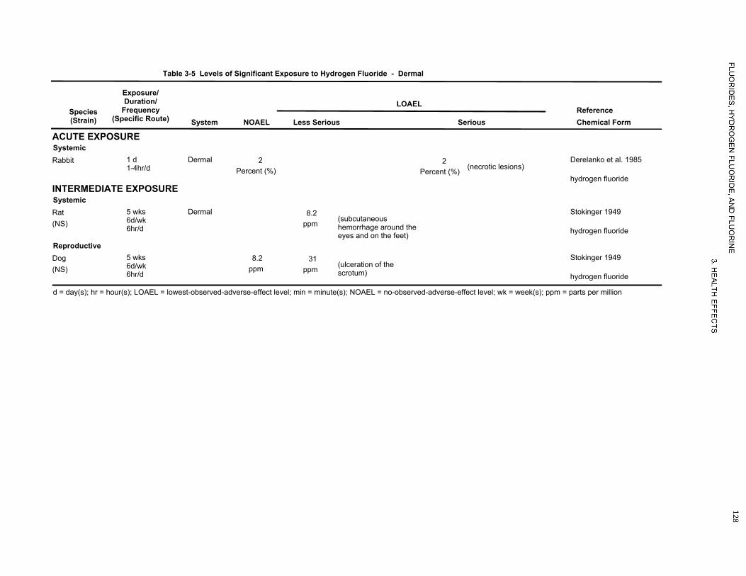
LOAEL
Table 3-5 Levels of Significant Exposure to Hydrogen Fluoride - Dermal
Species(Strain)
Exposure/Duration/
Frequency(Specific Route) System
ReferenceNOAEL Less Serious Serious Chemical Form
ACUTE EXPOSURESystemicRabbit 1 d
1-4hr/dDermal 2
Percent (%) (necrotic lesions)2
Percent (%)Derelanko et al. 1985
hydrogen fluorideINTERMEDIATE EXPOSURESystemic
(NS)Rat 5 wks
6d/wk6hr/d
Dermal(subcutaneoushemorrhage around theeyes and on the feet)
8.2ppm
Stokinger 1949
hydrogen fluoride
Reproductive
(NS)Dog 5 wks
6d/wk6hr/d
8.2ppm
d = day(s); hr = hour(s); LOAEL = lowest-observed-adverse-effect level; min = minute(s); NOAEL = no-observed-adverse-effect level; wk = week(s); ppm = parts per million
(ulceration of thescrotum)
31ppm
Stokinger 1949
hydrogen fluoride

LOAEL
Table 3-6 Levels of Significant Exposure to Fluoride - Dermal
Species(Strain)
Exposure/Duration/
Frequency(Specific Route) System
ReferenceNOAEL Less Serious Serious Chemical Form
ACUTE EXPOSURESystemic
(Sprague- Dawley)Rat 1 d
24hr/dDermal
d = day(s); hr = hour(s); LOAEL = lowest-observed-adverse-effect level; NOAEL = no-observed-adverse-effect level; PMN = polymorphnuclear leukocyte
(superficial necrosis,moderate edema, PMNinfiltration)
0.5Percent (%)
(extensive necrosis,marked edema,degenerating mast cells)
1Percent (%)
Essman et al. 1981
sodium fluoride

FLUORIDES, HYDROGEN FLUORIDE, AND FLUORINE 130
3. HEALTH EFFECTS
No studies were located regarding cardiovascular effects in humans after dermal exposure to fluoride or
fluorine, and no studies were located regarding cardiovascular effects in animals after dermal exposure to
fluoride, hydrofluoric acid, or fluorine.
Hepatic Effects.
Hydrofluoric Acid. Elevated SGOT, serum glutamic pyruvic transaminase (SGPT), and lactate dehydro-
genase levels were found in a man who was splashed in the face and on the neck with a mixture of 10%
hydrofluoric acid and sulfuric acid (Braun et al. 1984). The elevated SGOT and SGPT levels were
attributed to either muscle necrosis or temporary liver damage caused by toxic metabolic products from
necrotic tissue.
Renal Effects.
Hydrofluoric Acid. A 49-year-old man who was splashed in the face and on the neck with a mixture of
hydrofluoric acid and sulfuric acid became oliguric for a brief period on the day after the accident, and
then became anuric (Braun et al. 1984). Concomitant inhalation exposure is likely, and the effect of the
sulfuric acid is unknown.
Dermal Effects. Skin irritation and damage has been observed in humans and/or animals exposed to
fluoride, hydrogen fluoride, hydrofluoric acid, or fluorine. Dermal effects related to exposure to
hydrogen fluoride or fluorine are discussed under Inhalation Exposure (Section 3.2.1.2).
Fluoride. Sodium fluoride applied topically to the abraded skin of Sprague-Dawley rats (0.5 or 1.0%) for
24 hours produced both morphological and biochemical changes (Essman et al. 1981). At 0.5%, the
abraded surface showed focal superficial necrosis of the epidermis. At 1.0%, the abraded surface showed
edema and vacuolization. There was marked edema of the dermis with inflammation. Skin histamine
concentrations were also increased following application of 0.5 or 1% sodium fluoride to shaved-only or
epidermally abraded skin, although the variance of these measurements was quite high.
Hydrofluoric Acid. Dermal exposure to hydrofluoric acid results in extensive skin burns (Chela et al.
1989). Hydrofluoric acid quickly penetrates into soft tissues and causes necrosis. As a result of cell
membrane destruction, the fluoride ion has easy access to lymph and the venules, can be distributed
rapidly, and can cause significant adverse effects such as inhibition of glycolytic enzymes, hypocalcemia,
and hypomagnesia. Untreated burns of the fingers can result in loss of fingers. There are many reports of
FLUORIDES, HYDROGEN FLUORIDE, AND FLUORINE 131
3. HEALTH EFFECTS
hydrofluoric acid skin burns in humans. In one case, a 23-year-old man received fatal second- and third-
degree burns over 9–10% of his body from a 70% hydrofluoric acid spill (Mayer and Gross 1985). The
patient died 17 hours after exposure due to cardiac arrhythmias. Two case studies of accidental dermal
exposure of the hands to hydrofluoric acid (5–7%) reported serious dermal injury following exposures
from 45 minutes to 6 hours (Roberts and Merigian 1989). Topical treatment with calcium gluconate
prevented loss of nails. Other case reports are discussed in Section 3.2.3.1.
The concentration of hydrofluoric acid and the length of exposure affect the severity of dermal lesions
(Derelanko et al. 1985). Rabbits exposed to a hydrofluoric acid solution of 0.01% for 5 minutes had
visible skin lesions, whereas exposure to 2% hydrofluoric acid for 1 minute did not produce lesions. A
longer exposure of 1–4 hours to 2% hydrofluoric acid solution produced necrotic lesions on the backs of
rabbits (Derelanko et al. 1985). The application of 0.2 mL of a 47% hydrofluoric acid solution to the
shaved backs of New Zealand rabbits over a surface of 1¼ inches produced no immediate reaction
(Stokinger 1949). The material was held in place by lanolin and allowed to dry for 24 hours. Within a
few days of exposure, erythema and dark spots of liquefaction necrosis appeared. Multiple eschars were
formed in the necrotic areas. These wounds healed more slowly than those produced by fluorine gas.
Healing did not near completion until 27 days after exposure.
Ocular Effects. Ocular irritation and damage has been observed in humans and animals exposed to
hydrogen fluoride, hydrofluoric acid, and fluorine. The ocular effects resulting from exposure to
hydrogen fluoride or fluorine are discussed under inhalation exposure (Section 3.2.1.2).
Some evidence of delayed ocular damage due to persistence of the fluoride ion was observed 4 days after
a 3-year-old girl accidentally sprayed a hydrofluoric-acid-containing product in her eyes (Hatai et al.
1986). Opacification of the corneal epithelium and thrombosis of the conjunctival vessels were seen.
These changes were not permanent; after 30 days, the eyes returned to normal, and vision was 20/20.
However, it is difficult to generalize from this report as the product contained both hydrofluoric acid and
phosphoric acid at unspecified concentrations.
McCulley et al. (1983) concluded that the greater severity of hydrofluoric acid eye injuries compared to
injuries from other inorganic acids at comparable strengths probably results from the destruction of the
corneal epithelium allowing substantial penetration of the fluoride ion into the corneal stroma and
underlying structures.

FLUORIDES, HYDROGEN FLUORIDE, AND FLUORINE 132
3. HEALTH EFFECTS
No studies were located regarding the following effects in humans and animals after dermal exposure to
fluoride, hydrofluoric acid, or fluorine:
3.2.3.3 Immunological and Lymphoreticular Effects
3.2.3.4 Neurological Effects
3.2.3.5 Reproductive Effects
3.2.3.6 Developmental Effects
3.2.3.7 Cancer
3.3 GENOTOXICITY
In general, positive genotoxicity findings occurred at doses that are highly toxic to cells and whole
animals. Lower doses were generally negative for genotoxicity. Tables 3-7 and 3-8 present the results of
more recent assays.
The in vivo genotoxicity of fluoride has been tested in humans and animals following inhalation, oral, or
parenteral exposure. No alterations in the occurrence of sister chromatid exchange were observed in a
population living in areas with high levels of fluoride (4.8 ppm) in the drinking water (Li et al. 1995b).
Mixed results have been reported in animal studies examining the clastogenic potential of hydrogen
fluoride and sodium fluoride. Increases in the occurrence of chromosome aberrations were found in the
bone marrow cells of rats exposed by inhalation to 1.0 mg/m3 hydrogen fluoride 6 hours/day, 6 days/week
for 1 month (Voroshilin et al. 1975) and in mouse bone marrow cells following oral, intraperitoneal, or
subcutaneous exposure to sodium fluoride (Pati and Bunya 1987). However, other studies did not find
significant alterations in the occurrence of chromosome aberrations in mouse bone marrow cells
following oral exposure (Kram et al. 1978; Martin et al. 1979). Additionally, no alterations in sister
chromatid exchange occurrence were observed in mouse or Chinese hamster bone marrow cells following
oral exposure (Kram et al. 1978; Li et al. 1987b). Intraperitoneal injection of sodium fluoride resulted in
an increase in micronuclei in mouse bone marrow cells (Pati and Bhunya 1987); no alterations were
observed in rat bone marrow cells following oral exposure (Albanese 1987). Hydrogen fluoride was

FLUORIDES, HYDROGEN FLUORIDE, AND FLUORINE 133
3. HEALTH EFFECTS
Table 3-7. Genotoxicity of Fluoride In Vitro
Results
Species (test system) End point With activation
Without activation Reference Form
Prokaryotic organisms: Salmonella typhimurium Gene mutation – – Martin et al. 1979; NTP
1990; Tong et al. 1988 NaF
Eukaryotic organisms: Human lymphocytes Chromosomal
aberrations No data + Albanese 1987 NaF
Human lymphocytes Chromosomal aberrations
No data – Thomson et al. 1985 NaF, KF
Human fibroblasts Chromosome aberrations
No data + Tsutsui et al. 1984c NaF
Human fibroblasts Chromosomal aberrations
No data – Tsutsui et al. 1995 NaF
Human diploid IMR-90 cells
Chromosomal aberrations
No data + Oguro et al. 1995 NaF
Human lymphocytes Sister chromatid exchange
No data – Thomson et al. 1985; Tong et al. 1988
NaF
Human lymphocytes Sister chromatid exchange
No data – Thomson et al. 1985 KF
Human lymphoblasts Gene mutation + + Caspary et al. 1988 NaF Human fibroblasts Unscheduled
DNA synthesis No data + Tsutsui et al. 1984c NaF
Syrian hamster embryo cell
Chromosomal aberrations
No data + Tsutsui et al. 1984b NaF
Syrian hamster embryo cell
Sister chromatid exchange
No data + Tsutsui et al. 1984b NaF
Syrian hamster embryo cell
Unscheduled DNA synthesis
No data + Tsutsui et al. 1984b NaF
Chinese hamster ovary cells
Sister chromatid exchange
No data – Li et al. 1987b NaF
Chinese hamster ovary cells
Sister chromatid exchange
No data – Tong et al. 1988 NaF
Chinese hamster ovary cells
Sister chromatid exchange
+ + NTP 1990 NaF
Chinese hamster ovary cells
Chromosomal aberrations
+ + Aardema et al. 1989 NaF
Chinese hamster ovary cells
Chromosomal aberrations
– + NTP 1990 NaF
Chinese hamster V79 cells
Gene mutation No data – Slameńová et al. 1992 NaF
Mouse lymphoma cells Gene mutation No data (+) Cole et al. 1986 NaF Mouse lymphoma cells Gene mutation + + Caspary et al. 1987,
1988; NTP 1990 NaF

FLUORIDES, HYDROGEN FLUORIDE, AND FLUORINE 134
3. HEALTH EFFECTS
Table 3-7. Genotoxicity of Fluoride In Vitro
Results
Species (test system) End point With activation
Without activation Reference Form
Mouse lymphoma cells Gene mutation + + Caspary et al. 1987 KF Rat hepatocytes DNA repair No data – Tong et al. 1988 NaF Rat liver epithelium cells Gene mutation No data – Tong et al. 1988 NaF Rat vertebral body
derived cells Chromosome aberrations
No data + Mihashi and Tsutsui 1996
NaF
Rat bone marrow cells Chromosome aberrations
No data (+) Khalil 1995 NaF, KF
Rat bone marrow cells Sister chromatid exchange
No data – Khalil and Da’dara 1994 NaF, KF
– = negative result; + = positive result; (+) = weakly positive result; DNA = deoxyribonucleic acid; KF = potassium fluoride; NaF = sodium fluoride

FLUORIDES, HYDROGEN FLUORIDE, AND FLUORINE 135
3. HEALTH EFFECTS
Table 3-8. Genotoxicity of Fluoride In Vivo
Species (test system) End point Results Reference FormHuman lymphocytes (oral exposure) Sister chromatid exchange – Li et al. 1995b NR Rat bone marrow cells (oral exposure)
Micronuclei – Albanese 1987 NaF
Rat bone marrow Chromosome aberrations + Voroshilin et al. 1975 HF Rat testis cells (oral exposure) DNA strand breaks – Skare et al. 1986 NaF Mouse (C57B1) Dominant lethal – Voroshilin et al. 1975 HF Mouse (Harlan Sprague-Dawley) Sperm head abnormality – Li et al. 1987a NaF Mouse bone marrow and testis cells (oral exposure)
Chromosome aberrations – Martin et al. 1979 NaF
Mouse bone marrow cells (oral, intraperitoneal, or subcutaneous exposure)
Chromosome aberrations + Pati and Bhunya 1987 NaF
Mouse bone marrow cells (intraperitoneal exposure)
Micronuclei + Pati and Bhunya 1987 NaF
Mouse bone marrow cells (oral exposure)
Chromosome aberrations – Kram et al. 1978 NaF
Mouse bone marrow cells (oral exposure)
Sister chromatid exchange – Kram et al. 1978 NaF
Chinese hamster bone marrow cells (oral exposure)
Sister chromatid exchange – Li et al. 1987b NaF
– = negative result; + = positive result; DNA = deoxyribonucleic acid; HF = hydrogen fluoride; NaF = sodium fluoride; NR = not reported

FLUORIDES, HYDROGEN FLUORIDE, AND FLUORINE 136
3. HEALTH EFFECTS
negative for dominant lethal mutations following inhalation exposure to hydrogen fluoride in C57B1 mice
(Voroshilin et al. 1975). In a study in Drosophila melanogaster in which reproductive parameters were
measured as an indicator of genotoxicity, significant reductions in the number of eggs per female and
male fertility were observed following inhalation exposure to hydrogen fluoride (Gerdes et al. 1971b).
The maximum lethality to adults of one of the two tested strains was 60%; under most of the test
conditions, the lethality was ≤40%.
3.4 TOXICOKINETICS
The majority of data on the toxicokinetics of fluoride focus on sodium fluoride and hydrofluoric acid.
Data regarding the toxicokinetics of calcium fluoride and other fluorides in human or animals are limited.
While radioactive isotopes are useful in toxicokinetic studies, this use is limited in studies of fluoride
because the fluorine isotope 18F has a short half-life (Wallace-Durbin 1954).
3.4.1 Absorption
3.4.1.1 Inhalation Exposure
Data providing information on absorption exist on the inhalation exposure of humans to hydrogen
fluoride or mixtures of hydrogen fluoride and fluoride dusts, and inhalation exposure of animals to
hydrogen fluoride. Animal data also exist showing that fluorine is absorbed.
Hydrogen Fluoride. Increases in plasma fluoride levels were observed in humans inhaling 0.8–2.8 or
2.9–6.0 ppm fluoride as hydrogen fluoride of 60 minutes (Lund et al. 1997); maximum plasma
concentrations were observed 60–90 minutes after exposure initiation. A study in rats suggests that
hydrogen fluoride is absorbed primarily by the upper respiratory tract, and that removal of hydrogen
fluoride from inhaled air by the upper respiratory tract approaches 100% for exposures that range from
30 to 176 mg fluoride/m3 (Morris and Smith 1982). Furthermore, it is apparent that distribution to the
blood is rapid. Immediately following 40 minutes of intermittent exposure, plasma fluoride
concentrations correlated closely (correlation coefficient=0.98; p<0.01) with the concentration of
hydrogen fluoride in the air passed through the surgically isolated upper respiratory tract. Plasma levels
were not measured at time points <40 minutes.

FLUORIDES, HYDROGEN FLUORIDE, AND FLUORINE 137
3. HEALTH EFFECTS
Hydrogen Fluoride and Fluoride Dusts. The absorption in humans of inhaled hydrogen fluoride and
fluoride dusts has been demonstrated in several studies. In a study by Collings et al. (1952), two workers
were exposed to approximately 5 mg fluoride/m3 for 6 hours with 15-minute breaks every 2 hours.
Absorption of fluoride was evaluated by monitoring urinary excretion of fluoride during and after
exposure. Analysis of 2-hour serial urine samples showed a peak fluoride level 2–4 hours after cessation
of exposure, which decreased to base levels within 12–16 hours after exposure. Similar results were
obtained using the same protocol to measure urinary fluoride following exposure to air containing 5.0 mg
fluoride/m3 as rock phosphate dust (Collings et al. 1951). Another study reported clinical observations of
employees in the production of phosphate rock and triple superphosphate (Rye 1961). Three employees
were exposed to airborne fluoride (2–4 ppm) composed of approximately 60% dust and 40% hydrogen
fluoride gas. Within 2–3 hours after exposure began, urinary fluoride levels increased from 0.5 to
4.0 mg/L and peaked 10 hours (7–8 mg/L) following cessation of exposure. None of the subjects had
prior occupational exposure to fluoride. Two studies of aluminum potroom workers found elevated
plasma fluoride levels (Ehrnebo and Ekstrand 1986; Søyseth et al. 1994). In workers exposed to
0.910 mg fluoride/m3 total fluoride (34% of which was gasous fluoride) during an 8-hour workshift,
elevated plasma fluoride concentrations and urinary fluoride levels were observed (Ehrnebo and Ekstrand
1986). Although these studies demonstrate absorption of fluoride, none measure the extent of fluoride
absorption.
Fluorine. No data were located regarding the absorption of fluorine in humans. Hepatic and renal effects
were observed in mice following exposure to fluorine for periods up to 60 minutes (Keplinger and Suissa
1968). This indicates that the fluoride ion was systemically available following the exposure. Fluoride,
rather than fluorine, is the agent that is toxicologically active systemically, since fluorine is too reactive to
be absorbed unchanged. Similarly, the finding of elevated fluoride levels in bones, teeth, and urine during
intermediate-duration exposure to fluorine indicates that fluoride is absorbed under these conditions
(Stokinger 1949). No information on absorption rate or extent is available.
Furthermore, although the data presented concern only acute exposures, it is expected that virtually
complete absorption would also be observed during long-term exposure to low levels of fluoride in the
air.

FLUORIDES, HYDROGEN FLUORIDE, AND FLUORINE 138
3. HEALTH EFFECTS
3.4.1.2 Oral Exposure
Fluoride. In humans and animals, fluoride is rapidly and efficiently absorbed from the gastrointestinal
tract. Elevated plasma fluoride levels are seen shortly after exposure to sodium fluoride; peak plasma
levels are typically seen within 30–60 minutes of ingestion (Carlson et al. 1960a; Ekstrand et al. 1977,
1978). Fluoride is absorbed from both the stomach and small intestine via passive diffusion. Nopakun et
al. (1989) estimated that in rats, approximately 20% of total absorbed fluoride was absorbed from the
stomach. Absorption of fluoride from the stomach is inversely proportional to pH (Messer and Ophaug
1993; Whitford and Pashley 1984), suggesting that fluoride is absorbed from the stomach as the
undissociated hydrogen fluoride rather than the fluoride ion (Whitford and Pashley 1984). After gastric
emptying, fluoride was rapidly absorbed from the small intestine (Messer and Ophaug 1993). An in vitro
study suggests that in the small intestine, fluoride is primarily absorbed as the fluoride ion (Nopakun and
Messer 1990). The absorption of fluoride does not appear to be homeostatically regulated. A linear
correlation between fluoride (as sodium fluoride) intake and the area under the plasma fluoride
concentration curve was found in humans ingesting 2–10 mg doses of sodium fluoride (Trautner and
Seibert 1986).
A number of dietary factors can influence the absorption of fluoride. Delayed gastric emptying slows the
rate of fluoride absorption, but does not appear to affect the total amount of fluoride absorbed (Messer
and Ophaug 1993). Ingestion of sodium fluoride with food delayed the peak plasma level but did not
significantly alter the total amount of fluoride absorbed, as compared to values when the subjects ingested
sodium fluoride after an 8-hour fast (Shulman and Vallejo 1990; Trautner and Einwag 1987). In contrast,
ingestion of calcium fluoride with a meal dramatically increased (33.5 versus 2.8%) the absorption of
fluoride, as compared to absorption after an 8-hour fast; similar results were found for the fluoride in bone
meal tablets (Trautner and Einwag 1987). It is likely that the increased residence time in the upper
gastrointestinal tract increased absorption. Ingestion of sodium fluoride with milk decreased fluoride
absorption by 13–50% (Ekstrand and Ehrnebo 1979; Shulman and Vallejo 1990; Trautner and Seibert
1986). Other studies have also shown that co-exposure to calcium carbonate or a diet high in calcium
decreases fluoride absorption (Jowsey and Riggs 1978; Whitford 1994). Increased exposure to
magnesium (Stookey et al. 1964; Weddle and Muhler 1954) or aluminum (Stookey et al. 1964; Weddle
and Muhler 1954) also resulted in decreases in fluoride absorption.
Soluble fluoride compounds, such as sodium fluoride, hydrogen fluoride, and fluorosilic acid, are readily
absorbed from the gastrointestinal tract. Studies in humans and animals have found that >80% of an oral
FLUORIDES, HYDROGEN FLUORIDE, AND FLUORINE 139
3. HEALTH EFFECTS
dose of soluble fluoride compound is absorbed (Ericsson 1958; McClure et al. 1950, 1945; Zipkin and
Likins 1957); several studies have reported 99–100% absorption efficiencies (Ekstrand et al. 1978;
Trautner and Einwag 1987). Poorly soluble fluoride compounds, such as calcium fluoride, magnesium
fluoride, and aluminum fluoride, do not appear to be well absorbed. Very little (<10%) fluoride was
absorbed in fasting subjects ingesting calcium fluoride (Afseth et al. 1987; Trautner and Einwag 1987).
Compared to sodium fluoride, the fluoride from bone meal (McClure et al. 1945; Trautner and Einwag
1987) and cryolite (McClure et al. 1945) was poorly absorbed.
The absorption of fluoride in infants, children, and adults appears to be similar. A study of four infants
(mean age 8 months) reported mean absorption rates of 88.9–96.0% when a sodium fluoride solution was
administered along with infant formula (Ekstrand et al. 1994b). As discussed previously, the absorption
efficiency in adults usually exceeds 90% (Ekstrand et al. 1978; Trautner and Einwag 1987). As with
adults, peak plasma levels usually occurred in 30–60 minutes (mean of 39 minutes) in infants (Ekstrand et
al. 1994a) and young children aged 3–4 years (Ekstrand et al. 1983). One observed difference between
fluoride absorption in infants and adults is that administration of fluoride with a high calcium diet did not
result in a significant decrease in fluoride absorption (88.9 versus 96.0%) in infants (Ekstrand et al.
1994b); a decrease in absorption has been reported in adults (Ekstrand and Ehrnebo 1979; Shulman and
Vallejo 1990; Trautner and Einwag 1987).
3.4.1.3 Dermal Exposure
Data exist on dermal absorption of hydrofluoric acid in humans and animals, and limited quantitative rate
data are available in animals.
Hydrofluoric Acid. Dermal application of hydrofluoric acid results in rapid penetration of the fluoride
ion into the skin. Sufficiently large amounts cause necrosis of the soft tissue and decalcification and
corrosion of bone in humans (Browne 1974; Dale 1951; Dibbell et al. 1970; Jones 1939; Klauder et al.
1955). Systemic fluoride poisoning has been reported following accidental dermal exposure to anhydrous
hydrogen fluoride (Buckingham 1988; Burke et al. 1973). Although the extent of the contribution of
inhalation exposure in these cases is not known, the reports suggest that hydrogen fluoride is quickly
absorbed into the body following dermal exposure. However, these studies did not provide useful
information concerning the extent of fluoride absorption, or information on absorption of smaller doses.

FLUORIDES, HYDROGEN FLUORIDE, AND FLUORINE 140
3. HEALTH EFFECTS
Dermal absorption of hydrofluoric acid in albino mice of the d.d. strain was inferred in a study by
Watanabe et al. (1975). Mice were painted with 0.02 mL of 50% hydrofluoric acid, and the residual acid
was wiped off after 5 minutes. The mice were then injected intraperitoneally with [14C]glucose and
analyzed by whole body radiography. Radioactivity levels in the liver, renal cortex, lungs, and blood
were elevated 30 minutes after injection. This suggests that fluoride was absorbed through the skin and
interfered with the tissue distribution of glucose. No data were located on the extent of absorption of
fluoride in animals exposed dermally to hydrofluoric acid.
These studies indicate that fluoride as hydrofluoric acid is absorbed through the skin in humans and
animals. However, the degree of absorption is not known, nor is it known whether other forms of fluoride
would be absorbed, and to what extent. Furthermore, it is expected that the relationship between duration
or concentration and degree of absorption would be affected by the corrosive action of hydrofluoric acid.
Therefore, prediction of the extent of absorption following exposure to a low concentration of
hydrofluoric acid cannot be made based on the existing data.
Fluorine. Systemic effects have been observed following whole-body exposure to fluorine (Keplinger
and Suissa 1968; Stokinger 1949). However, these effects are likely to be due to inhalation exposure,
rather than dermal exposure.
3.4.2 Distribution
3.4.2.1 Inhalation Exposure
Hydrogen Fluoride. No data were located regarding the distribution of fluoride in humans following
exposure to only hydrogen fluoride. Evidence from studies in animals supports the inference from
occupational studies of exposure to hydrogen fluoride and fluoride dust that fluoride is distributed to the
rest of the body when inhaled. Duration- and concentration-related increases in tooth and bone fluoride
levels were reported in the rat following exposure to 7 or 24 mg/m3 for 6 hours/day, 6 days/week for up to
30 days (Stokinger 1949). Fluoride levels in new bone were up to twice the levels in old bone. The
distribution of the fluoride ion was studied in the tissues of rabbits, a guinea pig, and a monkey exposed
to hydrogen fluoride at various concentrations (1.5–1,050 mg/m3) and exposure times (Machle and Scott
1935). The observation period ranged from 9 to 14 months. As might be expected, based on the
following discussion of human occupational exposure to fluoride compounds, the fluoride ion
accumulated chiefly in the skeleton of all three species.
FLUORIDES, HYDROGEN FLUORIDE, AND FLUORINE 141
3. HEALTH EFFECTS
Several studies in animals have demonstrated that fluoride is widely available through the blood, although
actual concentrations in tissues other than blood have, for the most part, not been reported. For example,
whole body exposure of male rats to levels ranging from 11 to 116 mg fluoride/m3 as hydrogen fluoride
for 6 hours resulted in a dose-dependent increase in lung and plasma fluoride concentrations (Morris and
Smith 1983). In another study, rats exposed to 84 mg fluoride/m3 as hydrogen fluoride by whole body
exposure had significantly elevated levels of fluoride in plasma and lungs 6 hours postexposure (Morris
and Smith 1983).
Hydrogen Fluoride and Fluoride Dusts. Limited information was located on the distribution of inhaled
fluoride in humans. However, reports of skeletal fluorosis (Chan-Yeung et al. 1983b; Czerwinski et al.
1988; Kaltreider et al. 1972) and elevated bone fluoride levels (Baud et al. 1978; Boivin et al. 1988) after
occupational exposure to hydrogen fluoride and fluoride dusts indicate that fluoride is distributed to bone
and accumulates there.
Fluoride deposition in bone occurs mainly in regions undergoing active ossification or calcification. If
the source of fluoride exposure has been removed, fluoride levels in bone decrease as the bone undergoes
remodeling. Areas of fluoride deposition during high-level exposure are distinguished by highly elevated
fluoride levels even after the average fluoride level of the bone has returned to normal (Baud et al. 1978).
Fluorine. No data were located regarding the distribution of fluoride following the inhalation exposure
of humans to fluorine. In rats exposed to 25 mg/m3 fluorine for about 5 hours/day, 6 days/week for
21 days, markedly elevated fluoride levels were observed in teeth and bone, the only tissues that were
analyzed (Stokinger 1949). Tooth fluoride levels were about 14 times the levels in controls, and fluoride
levels in the femur were about 6 times those in the controls. Similar concentration-related increases in
bone and tooth fluoride levels were observed at the lower concentrations (3 and 0.8 mg/m3).
3.4.2.2 Oral Exposure
Fluoride. Once absorbed, fluoride is rapidly distributed throughout the body via the blood. Fluoride is
distributed between the plasma and blood cells, with plasma levels being twice as high as blood cell levels
(Whitford 1990). After ingestion of sodium fluoride, the plasma fluoride does not appear to be bound to
proteins (Ekstrand et al. 1977a; Rigalli et al. 1996). However, there is evidence that following ingestion
of sodium monofluorophosphate, the plasma contains diffusible fluoride and protein-bound fluoride

FLUORIDES, HYDROGEN FLUORIDE, AND FLUORINE 142
3. HEALTH EFFECTS
(Rigalli et al. 1996). The elimination of fluoride from plasma following short-term exposure to sodium
fluoride has been fit to a two-compartment model (Ekstrand et al. 1977a). The half-life of the terminal
phase ranged from 2 to 9 hours. The rapid phase of fluoride distribution represents distribution in soft
tissues, with fluoride being more rapidly distributed to well-perfused tissues. In pigs, the plasma
clearance half-time of fluoride was 0.88 hours (Richards et al. 1982). Fluoride does not accumulate in
most soft tissue; the ratio between tissue fluoride levels and plasma fluoride levels is typically between
0.4 and 0.9 (Whitford et al. 1979a). It is likely that fluoride enters the intracellular fluid of soft tissues as
hydrogen fluoride (Whitford et al. 1979a). Studies in rats and ewes suggest that the blood brain barrier is
effective in preventing fluoride migration into the central nervous system (Spak et al. 1986; Whitford et
al. 1979a); brain fluoride concentrations typically do not exceed 10% of plasma concentrations (Whitford
et al. 1979a). Higher fluoride concentrations are found in the renal tubules, the concentration often
exceeding plasma concentrations.
The largest concentration of fluoride in the body is found in calcified tissues. Approximately 99% of the
fluoride in the body is found in bones and teeth (Hamilton 1990; Kaminsky et al. 1990). The pineal gland
which contains hydroxyapatite also accumulates fluoride (Luke 2001). Fluoride is incorporated into bone
by replacing the hydroxyl ion in hydroxyapatite to form hydroxyfluroapatite (McCann and Bullock 1957;
Neuman et al. 1950). Fluoride is not irreversibly bound to bone and is mobilized from bone through the
continuous process of bone remodeling and to a lesser extent from ionic flux between interstitial fluoride
and the crystalline bone surface (Turner et al. 1993; Whitford 1990). The biological half-life of fluoride
was estimated to be 58.5 days in pigs orally exposed 2 mg fluoride/kg/day as sodium fluoride for
6 months (Richards et al. 1985). A comparison between the retention of sodium fluoride, fluorosilicic
acid, and sodium fluorosilicate did not find significant differences in the percentage of intake retained in
the body of female rats exposed to 24 ppm fluoride in the diet for 5 months; fluoride retentions were 66.2,
68.1, and 64.8, respectively (Whitford and Johnson 2003; only available as an abstract).
Tissue fluoride levels are not homeostatically regulated. In adults, plasma fluoride levels appear to be
directly related to fluoride intake. At higher fluoride intakes, a wide fluctuation of plasma fluoride levels
were found in two adults and three children consuming 9.6 ppm fluoride in water (Ekstrand 1978); the
fluctuation in plasma fluoride levels was smaller in the children, as compared to the adults. Mean plasma
levels in individuals living in areas with a water fluoride concentration of <0.1 ppm was 0.4 µmol/L,
compared to a mean plasma fluoride level of 1 µmol/L in individuals with a water fluoride content of 0.9–
1.0 ppm (Guy et al. 1976). The level of fluoride in bone is influenced by several factors including age,
past and present fluoride intake, and the rate of bone turnover. In adults (mean age, 47–60 years),

FLUORIDES, HYDROGEN FLUORIDE, AND FLUORINE 143
3. HEALTH EFFECTS
fluoride levels in the iliac crest ranged from 0.06 to 0.10% of bone ash (Boivin et al. 1988). In contrast,
in subjects undergoing sodium fluoride treatment for osteoporosis for 5–6 years, the fluoride content of
the iliac crest was 0.67% bone ash. Termination of high fluoride exposure can result in a decrease in
bone fluoride levels. In aluminum workers diagnosed with skeletal fluorosis, there was a significant and
progressive decrease in bone fluoride levels (Boivin et al. 1988).
Age strongly influences the distribution of fluoride. The amount of fluoride taken up by bone is inversely
related to age (Lawrenz et al. 1940; Miller and Phillips 1956; Suttie and Phillips 1959; Zipkin and
McClure 1952; Zipkin et al. 1956). In a study of dogs (Ekstrand and Whitford 1984; Whitford 1990), the
fractional uptake of fluoride by bone steadily declined during the first year of life. At weaning, 90% of an
administered dose was taken up by bone compared to 50% at 1 year of age. Similar findings have been
reported in human studies. In infants aged 37–410 days exposed to 0.25 mg fluoride supplement, the
mean retention (bone uptake) of fluoride ranged from 68.1 to 83.4% (Ekstrand et al. 1994a, 1994b). In
contrast, retention in adults receiving a fluoride supplement was 55.3% (Ekstrand et al. 1979).
Human and animal studies have shown that fluoride is readily transferred across the placenta. There
appears to be a direct relationship between maternal blood fluoride levels and cord blood fluoride levels
(Armstrong et al. 1970; Gupta et al. 1993; Malhotra et al. 1993; Shen and Taves 1974). At relatively low
maternal blood levels, the cord blood levels were at least 60% of that of maternal blood (Brambilla et al.
1994; Gupta et al. 1993). Although cord fluoride levels were typically lower than maternal levels, one
study found no statistical difference between maternal and newborn (1 day old) serum fluoride levels
(Shimonovitz et al. 1995). However, a partial placental barrier may exist at high maternal fluoride levels.
At higher maternal blood levels, the cord to maternal fluoride ratio is lower than at lower maternal
fluoride levels (Gupta et al. 1993). Another study found that the use of fluoride supplements markedly
increased placental fluoride levels, while fluoride levels in fetal blood remained relatively constant,
suggesting that the placenta can regulate the transfer of fluoride from maternal blood to fetal blood
(Gedalia 1970). Animal studies also demonstrate that maternal fluoride exposure also results in increased
levels of fluoride in fetal teeth and bones (Bawden et al. 1992b; Nedeljković and Matović 1991; Theuer et
al. 1971).
In humans, fluoride is poorly transferred from plasma to milk (Ekstrand et al. 1981c, 1984b; Esala et al.
1982; Spak et al. 1983). A single dose of 1.5 mg sodium fluoride did not result in a significant rise in
fluoride breast milk concentrations within 3 hours of the exposure (Ekstrand et al. 1981c). Although no
linear correlation between fluoride levels in tap water and fluoride levels in breast milk has been found,

FLUORIDES, HYDROGEN FLUORIDE, AND FLUORINE 144
3. HEALTH EFFECTS
significantly higher breast milk fluoride concentrations were found in women living in an area with high
levels of naturally occurring fluoride (1–7 ppm) as compared to women in areas with low fluoride levels
in tap water (0.2 ppm) (Esala et al. 1982). Fluoride levels in human milk of 5–10 µg/L have been
measured (Fomon and Ekstrand 1999).
3.4.2.3 Dermal Exposure
No information was located in humans or animals regarding the distribution of fluoride, hydrogen
fluoride, or fluorine following dermal absorption.
3.4.2.4 Other Routes of Exposure
Based on the results of a five-compartment computer model, Charkes et al. (1978) calculated that about
60% of intravenously administered radiolabelled fluoride (18F) is taken up by bone; the half-time for this
uptake is about 13 minutes.
Perkinson et al. (1955) found initial rates of removal of fluoride from sheep and cow blood to be 41 and
32%/minute of the intravenously administered dose, respectively. These data suggest a rapid distribution
of fluoride and corroborate findings reported by other routes of administration.
Fluoride distribution in rats was examined during and after continuous intravenous infusion of
radiolabeled sodium fluoride at varying chemical dose rates for 3 hours (Knaus et al. 1976). Blood,
kidneys, and lungs contained the highest fluoride concentrations at doses up to 3.6 mg fluoride/kg/hour,
but at 6 mg/kg/hour, the fluoride content of the liver, spleen, and hollow organs increased sharply,
indicating that the dose exceeded the amount readily processed by the excretory mechanisms of the body.
In rat pups injected intraperitoneally with 0.1 µg fluoride/g body weight as sodium fluoride solution,
significant increases in the fluoride content occurred in the developing enamel and bone (Bawden et
al. 1987). Thus, regardless of the route of administration, some fluoride is deposited in teeth, bone, and
soft tissues of animals, and some is excreted in the urine, sweat, and saliva.

FLUORIDES, HYDROGEN FLUORIDE, AND FLUORINE 145
3. HEALTH EFFECTS
3.4.3 Metabolism
Fluoride is believed to replace the hydroxyl ion (OH-) and possibly the bicarbonate ion (HCO3-)
associated with hydroxyapatite—a mineral phase during formation of bone (McCann and Bullock 1957;
Neuman et al. 1950). The resultant material is hydroxyfluorapatite. Once absorbed, a portion of the
fluoride is deposited in the skeleton, and most of the remainder is excreted in the urine, with smaller
amounts in feces, and sweat, and saliva within 24 hours (Dinman et al. 1976a, 1976b; McClure et al.
1945). Urinary excretion is markedly decreased in the presence of decreased renal function (Kono et al.
1984; Spak et al. 1985).
A portion of the circulating inorganic fluoride acts as an enzyme inhibitor because it forms metal-
fluoride-phosphate complexes that interfere with the activity of those enzymes requiring a metal ion
cofactor. In addition, fluoride may interact directly with the enzyme or the substrate. It is a general
inhibitor of the energy production system of the cell (i.e., glycolytic processes and oxidative
phosphorylation enzymes responsible for forming ATP) (Guminska and Sterkowicz 1975; Najjar 1948;
Peters et al. 1964; Slater and Bonner 1952). Although much is known about enzyme inhibition by
fluoride, the human health significance remains to be determined. The studies on enzymatic inhibition by
fluoride were in vitro studies and used fluoride concentrations that were significantly (100–1,000 times)
higher than concentrations that would be normally found in human tissues.
3.4.4 Elimination and Excretion
3.4.4.1 Inhalation Exposure
Hydrogen Fluoride. Overnight urinary fluoride excretion in dogs and rabbits exposed to 7 mg/m3
hydrogen fluoride for 6 hours/day, 6 days/week for 30 days was about 1.5 times that of controls
(Stokinger 1949). No further details were reported.
Hydrogen Fluoride and Fluoride Dusts. Studies in humans indicate that fluoride absorbed from inhaled
hydrogen fluoride and fluoride dusts over an 8-hour work shift is excreted even during exposure, with
urinary excretion peaking approximately 2–4 hours after cessation of exposure (about 10 hours following
beginning of exposure) (Collings et al. 1951; Rye 1961). A significant correlation between urinary
fluoride excretion and the area under the plasma concentration-time curve was found in aluminum

FLUORIDES, HYDROGEN FLUORIDE, AND FLUORINE 146
3. HEALTH EFFECTS
workers (Ehrnebo and Ekstrand 1986). A significant correlation between urinary fluoride excretion and
exposure levels of gaseous fluoride, but not particulate fluoride, was also found.
Fluorine. No data were located regarding excretion of fluoride following human inhalation exposure to
fluorine. Urinary fluoride levels were increased in dogs and rabbits exposed to levels as low as 0.8 mg/m3
for 5–6 hours/day, 6 days/week for 35 days (Stokinger 1949). No quantitative data were reported at this
level, but urinary fluoride levels in rabbits exposed to 3 mg/m3 were 1.5 times normal. No further details
were reported.
3.4.4.2 Oral Exposure
Fluoride. The primary pathway for fluoride excretion is via the kidneys and urine; to a lesser extent,
fluoride is also excreted in the feces, sweat, and saliva. A study in rats suggests that the source of the
small amount fecal fluoride may be fluoride that has re-entered the more distal portion of the intestine and
became associated with unabsorbed cations (Whitford 1994). It has been estimated that approximately
1% or less of an ingested dose is excreted in saliva (Carlson et al. 1960a; Oliveby et al. 1989), because
saliva is swallowed, this amount does not enter mass balance calculations. The concentration of fluoride
in the saliva appears to mirror plasma fluoride levels (Ekstrand 1977; Oliveby et al. 1989, 1990) and the
ratio of saliva fluoride to plasma fluoride is about 0.5–0.64 (Ekstrand 1977; Oliveby et al. 1989). There
are limited data on fluoride excretion via sweat. An older study (McClure et al. 1945) estimated that 19%
of an ingested fluoride dose (3.7 mg) was excreted in sweat under comfortable conditions. Excretion of
fluoride increased to 42% under hot-moist conditions. However, this study appears to have grossly
overestimated fluoride excretion in sweat. A more recent study (Henschler et al. 1975), reported low
levels of fluoride in sweat, approximately 20% of plasma levels, 2 hours after administration of sodium
fluoride.
Renal excretion is the major route of fluoride removal from the body; it typically equals 35–70% of intake
in adults (Ekstrand et al. 1978; Machle and Largent 1943;). The fluoride ion is filtered from the plasma
as it passes through the glomerular capillaries followed by a varying degree (10–70%) of tubular
reabsorption; there is no evidence of tubular secretion of fluoride (Schiffl and Binswanger 1982; Whitford
1990). Renal clearance rates in humans can range from 12.4 to 71.4 mL/minute with average values of
36.4–41.8 mL/minute (Schiffl and Binswanger 1982; Waterhouse et al. 1980). A number of factors,
including urinary pH, urinary flow, and glomerular filtration rate, can influence urinary fluoride
excretion. Urinary pH appears to be the major determining factor for fluoride reabsorption from the renal
FLUORIDES, HYDROGEN FLUORIDE, AND FLUORINE 147
3. HEALTH EFFECTS
tubules (Ekstrand et al. 1980a, 1982; Järnberg et al. 1980, 1981, 1983; Whitford et al. 1976). At lower
pH levels, more fluoride exists in the undissociated form (hydrogen fluoride) than in the ion form; the
uncharged hydrogen fluoride more readily diffuses through the tubular epithelium to the interstitial fluid
than the free charged fluoride ion. In the neutral interstitial fluid, the hydrogen fluoride would rapidly
dissociate and the fluoride ions would be returned to the systemic circulation (Whitford et al. 1976).
Thus, renal clearance of fluoride is directly related to urinary pH. A significant correlation between
urinary flow and urinary fluoride excretion has been reported in humans (Ekstrand et al. 1978, 1982).
Ekstrand et al. (1982) noted that a high flow rate in the renal tubules would likely increase the renal
clearance of a substance that is reabsorbed at a slow rate. Similarly, a decrease in glomerular filtration
rate would result in a decrease in urinary fluoride excretion (Jeandel et al. 1992).
Most of the data on renal clearance of fluoride in humans come from studies of health adults; however,
several studies have examined renal clearance of fluoride at different age levels (Ekstrand et al. 1994a;
Jeandel et al. 1992; Spak et al. 1985; Villa et al. 2000). Whitford (1999) compared the results of many of
these studies in infants and children with those in adults and concluded that there are no apparent age-
related differences in renal clearance rates (adjusted for body weight or surface area) between children
and adults. However, in older adults (>65 years), a significant decline in renal clearance of fluoride has
been reported (Jeandel et al. 1992). This is consistent with the decline in glomerular filtration rate and
renal clearance of many substances that are also observed in the elderly (Whitford 1999).
3.4.4.3 Dermal Exposure
No studies were located regarding excretion of fluoride, hydrogen fluoride, or fluorine in humans or
animals following dermal exposure. However, in the absence of evidence to the contrary, it is expected
that dermally absorbed fluoride would be sequestered in bone and excreted in urine in a manner similar to
that observed following oral or inhalation exposure.
3.4.5 Physiologically Based Pharmacokinetic (PBPK)/Pharmacodynamic (PD) Models
Physiologically based pharmacokinetic (PBPK) models use mathematical descriptions of the uptake and
disposition of chemical substances to quantitatively describe the relationships among critical biological
processes (Krishnan et al. 1994). PBPK models are also called biologically based tissue dosimetry
models. PBPK models are increasingly used in risk assessments, primarily to predict the concentration of
FLUORIDES, HYDROGEN FLUORIDE, AND FLUORINE 148
3. HEALTH EFFECTS
potentially toxic moieties of a chemical that will be delivered to any given target tissue following various
combinations of route, dose level, and test species (Clewell and Andersen 1985). Physiologically based
pharmacodynamic (PBPD) models use mathematical descriptions of the dose-response function to
quantitatively describe the relationship between target tissue dose and toxic end points.
PBPK/PD models refine our understanding of complex quantitative dose behaviors by helping to
delineate and characterize the relationships between: (1) the external/exposure concentration and target
tissue dose of the toxic moiety, and (2) the target tissue dose and observed responses (Andersen et al.
1987; Andersen and Krishnan 1994). These models are biologically and mechanistically based and can
be used to extrapolate the pharmacokinetic behavior of chemical substances from high to low dose, from
route to route, between species, and between subpopulations within a species. The biological basis of
PBPK models results in more meaningful extrapolations than those generated with the more conventional
use of uncertainty factors.
The PBPK model for a chemical substance is developed in four interconnected steps: (1) model
representation, (2) model parametrization, (3) model simulation, and (4) model validation (Krishnan and
Andersen 1994). In the early 1990s, validated PBPK models were developed for a number of
toxicologically important chemical substances, both volatile and nonvolatile (Krishnan and Andersen
1994; Leung 1993). PBPK models for a particular substance require estimates of the chemical substance-
specific physicochemical parameters, and species-specific physiological and biological parameters. The
numerical estimates of these model parameters are incorporated within a set of differential and algebraic
equations that describe the pharmacokinetic processes. Solving these differential and algebraic equations
provides the predictions of tissue dose. Computers then provide process simulations based on these
solutions.
The structure and mathematical expressions used in PBPK models significantly simplify the true
complexities of biological systems. If the uptake and disposition of the chemical substance(s) is
adequately described, however, this simplification is desirable because data are often unavailable for
many biological processes. A simplified scheme reduces the magnitude of cumulative uncertainty. The
adequacy of the model is, therefore, of great importance, and model validation is essential to the use of
PBPK models in risk assessment.
PBPK models improve the pharmacokinetic extrapolations used in risk assessments that identify the
maximal (i.e., the safe) levels for human exposure to chemical substances (Andersen and Krishnan 1994).

FLUORIDES, HYDROGEN FLUORIDE, AND FLUORINE 149
3. HEALTH EFFECTS
PBPK models provide a scientifically sound means to predict the target tissue dose of chemicals in
humans who are exposed to environmental levels (for example, levels that might occur at hazardous waste
sites) based on the results of studies where doses were higher or were administered in different species.
Figure 3-5 shows a conceptualized representation of a PBPK model.
Only one published PBPK model has been identified (Rao et al. 1995); it differs from published
compartmental models for fluoride kinetics (Charkes et al. 1978, 1979; Ekstrand et al. 1977a; Hall et al.
1977; Richards et al. 1982, 1985) in that the earlier models were data-based and useful only to simulate
short-term fluoride kinetics. Because the fluoride ion is characterized by its long residence time in the
body, health effects based on long-term fluoride exposure are of concern. In contrast to the earlier
models, the Rao et al. (1995) PBPK model is amenable to extrapolation across species, routes, and doses,
thereby offering an advantage in quantitative risk assessment for fluoride exposure.
In order to assess the complex relationship between extended fluoride exposure, target tissue (bone) dose,
and tissue response, a sex-specific PBPK model has been developed to describe the absorption,
distribution, and elimination of fluorides in rats and humans (Rao et al. 1995). The PBPK model
incorporates age and body weight dependence of the physiological processes that control the uptake of
fluoride by bone and the elimination of fluoride by the kidneys. Six compartments (lung, liver, kidney,
bone, and slowly- and rapidly-perfused compartments) make up the model. The bone compartment
includes two subcompartments: a small, flow-limited, rapidly exchangeable surface bone compartment,
and a bulk, virtually nonexchangeable inner bone compartment. The inner bone compartment contains
nearly all of the whole body content of fluoride, which, in the longer time frame, may be mobilized
through the process of bone modeling and remodeling. This model has been validated by comparing
predictions with experimental data gathered in rats and humans after drinking water and dietary ingestion
of fluoride.
The PBPK model permits the analysis of the combined effect of ingesting and inhaling fluorides on the
target organ, bone. It takes into account the effects of age and growth; in the human model, for instance,
the bone and renal clearance rates accounted for 90 and 10%, respectively, during the growth period,
compared to about 50% each in adulthood. Estimates of fluoride concentrations in bone are calculated
and related to chronic fluoride toxicity. The model incorporates nonlinear binding rates of fluoride to
bone, which has been described at high plasma concentrations. The model is thus useful for predicting
some of the long-term metabolic features and tissue concentrations of fluoride that may be of value in
understanding positive or negative effects of fluoride on human health. In addition, the PBPK model

FLUORIDES, HYDROGEN FLUORIDE, AND FLUORINE 150
3. HEALTH EFFECTS
Figure 3-5. Conceptual Representation of a Physiologically Based Pharmacokinetic (PBPK) Model for a
Hypothetical Chemical Substance
Source: adapted from Krishnan et al. 1994 Note: This is a conceptual representation of a physiologically based pharmacokinetic (PBPK) model for a hypothetical chemical substance. The chemical substance is shown to be absorbed via the skin, by inhalation, or by ingestion, metabolized in the liver, and excreted in the urine or by exhalation.

FLUORIDES, HYDROGEN FLUORIDE, AND FLUORINE 151
3. HEALTH EFFECTS
provides a basis for cross-species extrapolation of the effective fluoride dose at the target tissue (bone) in
the assessment of risk from different exposure conditions.
3.5 MECHANISMS OF ACTION
Skeletal Effects. A number of mechanisms are involved in the toxicity of fluoride to bone. Fluoride ions
are incorporated into bone by substituting for hydroxyl groups in the carbonate-apatite structure to
produce hydroxyfluorapatite, thus altering the mineral structure of the bone (Chachra et al. 1999). Unlike
hydroxyl ions, fluoride ions reside in the plane of the calcium ions, resulting in a structure that is
electrostatically more stable and structurally more compact (Grynpas 1990). Following administration of
fluoride, there is a shift in the mineralization profile towards higher densities and increased hardness
(Chachra et al. 1999). However, the structure of the bone (cortical thickness and the trabecular
architecture of the femoral head) was largely unchanged in rabbits by fluoride administration. Chachra et
al. (1999) suggest that the shift in mineralization could be due to either hypermineralization of older
(denser) fractions or to a greater packing density of the hydroxyapatite crystals. Although high-dose
fluoride administration is associated with an increase in bone mass, in vivo and in vitro animal studies
have found a negative association between fluoride-induced new bone mass and bone strength, suggesting
that the quality of the new bone was impaired by the fluoride (Silva and Ulrich 2000; Turner et al. 1997).
Because bone strength is thought to derive mainly from the interface between the collagen and the
mineral (Catanese and Keavney 1996), alteration in mineralization probably affects strength. The wider
crystals, which are formed after fluoride exposure, are presumably not as well associated with collagen
fibrils and thus, do not contribute to mechanical strength. Turner et al. (1997) found that the crystal width
was inversely correlated with bending strength of the femur. Thus, although there is an increase in
hardness and bone mass and unaltered structure, the mechanical strength of bone is decreased with long-
term, high-dose administration of fluoride (Chachra et al. 1999).
In addition to the physicochemical effect of fluoride on the bone, at high doses, fluoride can be mitogenic
to osteoblasts (Farley et al. 1990; Gruber and Baylink 1991) and inhibitory to osteoclasts. The osteoblasts
are still active, although there are fewer plump, cuboidal, highly secretory osteoblasts; whereas fluoride is
mitogenic to osteoblastic precursors (Bonjour et al. 1993), it is toxic to individual osteoblasts at the same
concentration (Chachra et al. 1999). The effect of fluoride on osteoclasts is not well understood; it
appears that fluoride decreases the amount of bone resorbed by osteoclasts (Chachra et al. 1999).
FLUORIDES, HYDROGEN FLUORIDE, AND FLUORINE 152
3. HEALTH EFFECTS
Studies in humans and animals suggest that the effect of fluoride on bone strength is biphasic. In rats
administered 1–128 ppm fluoride as sodium fluoride in drinking water for 16 weeks, both increases and
decreases in bone strength were found; the maximum femoral bone strength occurred at 16 ppm (Turner
et al. 1992). A biphasic relationship between femoral bone strength and bone fluoride content was found.
Bone strength increased 18% as the bone fluoride content increased from 100 to 1,216 ppm, and
decreased by 31% as the bone fluoride levels increased from 1,216 to 10,000 ppm. It should be noted that
the bone fluoride levels in this study, as well as other studies discussed in this section, resulted from high
doses of fluoride. Arnala et al. (1986) measured fluoride levels in iliac crest biopsies taken from 18–
25 subjects with hip fractures living in areas with low fluoride (<0.3 ppm), high fluoride (>1.5 ppm), or
with fluoridated (1.0–1.2 ppm) water. The average fluoride levels in the bone were 450, 3,720, and
1,590 ppm, respectively.
The biphasic nature of bone effects is supported by data from clinical trials in women with
postmenopausal osteoporosis (Haguenauer et al. 2000). The meta-analyses of 12 studies found a
significant increase in the relative risk of nonvertebral fractures in subjects ingesting high doses of
fluoride (>30 mg/day); in subjects administered low fluoride doses or slow-release formulations, there
was no effect on nonvertebral fractures. Further,, there was no effect on vertebral fracture risk in high
fluoride dose subjects, but a decrease in this risk in subjects administered low fluoride doses or slow-
release formulations was found.
Dental Fluorosis. Numerous human and animal studies have demonstrated that exposure to elevated
fluoride levels during tooth development can result in dental fluorosis. As described previously,
fluorosed enamel is composed of hypomineralized subsurface enamel covered by well-mineralized
enamel. The exact mechanisms of dental fluorosis development have not been fully elucidated. Although
there are a number of proposed mechanisms, most of the recent research has focused on the theory that
dental fluorosis results from a fluoride-induced delay in the hydrolysis and removal of amelogenin matrix
proteins during enamel maturation and subsequent effects on crystal growth (as reviewed by Aoba 1997;
Bawden et al. 1995; DenBesten and Thariani 1992; Whitford 1997). Amelogenins, proteins secreted by
ameloblasts, comprise >90% of the enamel matrix proteins and are involved in the regulation of the form
and size of hydroxapatite crystallites; large molecular weight amelogenins inhibit the growth of enamel
crystallites. In the early maturation phase of tooth development, the amelogenins are removed from the
enamel matrix by amelogeninases, and crystallite growth dramatically increases. This phase of enamel
maturation appears to be the most sensitive to elevated fluoride levels. The current evidence strongly
suggests that fluoride inhibits amelogeninase activity. In one proposed mechanism, fluoride indirectly

FLUORIDES, HYDROGEN FLUORIDE, AND FLUORINE 153
3. HEALTH EFFECTS
inhibits amelogeninase, a calcium-dependent metalloenzyme, by binding to calcium in the mineralizing
milieu and thereby decreasing the calcium concentration (or its activities) and the activation of
amelogeninases (Aoba 1997; DenBesten and Thariani 1992; Whitford 1997). Another proposed
mechanism is that amelogeninases are activated by a metalloproteinase and that calcium is required for
this activation (Bawden et al. 1995; DenBesten and Thariani 1992); fluoride thus interferes with the
proteolytic cascade necessary for hydrolysis of amelogenins. The fluoride-calcium interaction is
supported by the findings that the enzymatic cleavage of amelogenins occurs at a slow rate during the
secretory phase when calcium transport is low and dramatically increase during the early maturation
phase (in the absence of excess fluoride) when calcium transport is high (Aoba 1997).
An alternative theory on the mechanism of dental fluorosis is related to the effects of fluoride on
maturation-stage ameloblasts (Bawden et al. 1995; DenBesten and Thariani 1992). Elevated fluoride
exposure results in a decrease in the number of ameloblast modulations, which in turn could reduce the
number of zone refinement cycles. As defined by Bawden et al. (1995), zone refinement is the process by
which impurities (such as magnesium) that have been incorporated into forming crystals are reduced,
resulting in an improvement of the structural characteristics of the crystal. Thus, a reduction in zone
refinement cycles could result in inferior apatite crystalline structure. This theory is not incompatible
with the theory that fluoride interferes with the hydrolysis of amelogenins. The fluoride-induced
inhibition of amelogeninases results in an enamel matrix with a higher organic content, which could
influence ameloblast modulation rate.
3.6 TOXICITIES MEDIATED THROUGH THE NEUROENDOCRINE AXIS
Recently, attention has focused on the potential hazardous effects of certain chemicals on the endocrine
system because of the ability of these chemicals to mimic or block endogenous hormones. Chemicals
with this type of activity are most commonly referred to as endocrine disruptors. However, appropriate
terminology to describe such effects remains controversial. The terminology endocrine disruptors,
initially used by Colborn and Clement (1992), was also used in 1996 when Congress mandated the
Environmental Protection Agency (EPA) to develop a screening program for “...certain substances
[which] may have an effect produced by a naturally occurring estrogen, or other such endocrine
effect[s]...”. To meet this mandate, EPA convened a panel called the Endocrine Disruptors Screening and
Testing Advisory Committee (EDSTAC), which in 1998 completed its deliberations and made
recommendations to EPA concerning endocrine disruptors. In 1999, the National Academy of Sciences
released a report that referred to these same types of chemicals as hormonally active agents. The
FLUORIDES, HYDROGEN FLUORIDE, AND FLUORINE 154
3. HEALTH EFFECTS
terminology endocrine modulators has also been used to convey the fact that effects caused by such
chemicals may not necessarily be adverse. Many scientists agree that chemicals with the ability to disrupt
or modulate the endocrine system are a potential threat to the health of humans, aquatic animals, and
wildlife. However, others think that endocrine-active chemicals do not pose a significant health risk,
particularly in view of the fact that hormone mimics exist in the natural environment. Examples of
natural hormone mimics are the isoflavinoid phytoestrogens (Adlercreutz 1995; Livingston 1978; Mayr et
al. 1992). These chemicals are derived from plants and are similar in structure and action to endogenous
estrogen. Although the public health significance and descriptive terminology of substances capable of
affecting the endocrine system remains controversial, scientists agree that these chemicals may affect the
synthesis, secretion, transport, binding, action, or elimination of natural hormones in the body responsible
for maintaining homeostasis, reproduction, development, and/or behavior (EPA 1998c). Stated
differently, such compounds may cause toxicities that are mediated through the neuroendocrine axis. As
a result, these chemicals may play a role in altering, for example, metabolic, sexual, immune, and
neurobehavioral function. Such chemicals are also thought to be involved in inducing breast, testicular,
and prostate cancers, as well as endometriosis (Berger 1994; Giwercman et al. 1993; Hoel et al. 1992).
Although there is no evidence that fluoride is an endocrine disruptor, there are some data to suggest that
fluoride does adversely affect some endocrine glands. An increase in serum thyronine levels, in the
absence of changes in triiodothyronine and thyroid stimulating hormone levels, was observed in
individuals living in areas of India with high fluoride levels in the drinking water (Michael et al. 1996).
In contrast, a decrease in thyroxine levels was observed in rats exposed to fluoride in drinking water for
2 months (Bobek et al. 1976). Significant decreases in serum testosterone have been observed in rats
exposed to sodium fluoride for 50–60 days (Araibi et al. 1989; Narayana and Chinoy 1994).
3.7 CHILDREN’S SUSCEPTIBILITY
This section discusses potential health effects from exposures during the period from conception to
maturity at 18 years of age in humans, when all biological systems will have fully developed. Potential
effects on offspring resulting from exposures of parental germ cells are considered, as well as any indirect
effects on the fetus and neonate resulting from maternal exposure during gestation and lactation.
Relevant animal and in vitro models are also discussed.
FLUORIDES, HYDROGEN FLUORIDE, AND FLUORINE 155
3. HEALTH EFFECTS
Children are not small adults. They differ from adults in their exposures and may differ in their
susceptibility to hazardous chemicals. Children’s unique physiology and behavior can influence the
extent of their exposure. Exposures of children are discussed in Section 6.6 Exposures of Children.
Children sometimes differ from adults in their susceptibility to hazardous chemicals, but whether there is
a difference depends on the chemical (Guzelian et al. 1992; NRC 1993). Children may be more or less
susceptible than adults to health effects, and the relationship may change with developmental age
(Guzelian et al. 1992; NRC 1993). Vulnerability often depends on developmental stage. There are
critical periods of structural and functional development during both prenatal and postnatal life and a
particular structure or function will be most sensitive to disruption during its critical period(s). Damage
may not be evident until a later stage of development. There are often differences in pharmacokinetics
and metabolism between children and adults. For example, absorption may be different in neonates
because of the immaturity of their gastrointestinal tract and their larger skin surface area in proportion to
body weight (Morselli et al. 1980; NRC 1993); the gastrointestinal absorption of lead is greatest in infants
and young children (Ziegler et al. 1978). Distribution of xenobiotics may be different; for example,
infants have a larger proportion of their bodies as extracellular water and their brains and livers are
proportionately larger (Altman and Dittmer 1974; Fomon 1966; Fomon et al. 1982; Owen and Brozek
1966; Widdowson and Dickerson 1964). The infant also has an immature blood-brain barrier (Adinolfi
1985; Johanson 1980) and probably an immature blood-testis barrier (Setchell and Waites 1975). Many
xenobiotic metabolizing enzymes have distinctive developmental patterns. At various stages of growth
and development, levels of particular enzymes may be higher or lower than those of adults, and
sometimes unique enzymes may exist at particular developmental stages (Komori et al. 1990; Leeder and
Kearns 1997; NRC 1993; Vieira et al. 1996). Whether differences in xenobiotic metabolism make the
child more or less susceptible also depends on whether the relevant enzymes are involved in activation of
the parent compound to its toxic form or in detoxification. There may also be differences in excretion,
particularly in newborns who all have a low glomerular filtration rate and have not developed efficient
tubular secretion and resorption capacities (Altman and Dittmer 1974; NRC 1993; West et al. 1948).
Children and adults may differ in their capacity to repair damage from chemical insults. Children also
have a longer remaining lifetime in which to express damage from chemicals; this potential is particularly
relevant to cancer.
Certain characteristics of the developing human may increase exposure or susceptibility, whereas others
may decrease susceptibility to the same chemical. For example, although infants breathe more air per
kilogram of body weight than adults breathe, this difference might be somewhat counterbalanced by their

FLUORIDES, HYDROGEN FLUORIDE, AND FLUORINE 156
3. HEALTH EFFECTS
alveoli being less developed, which results in a disproportionately smaller surface area for alveolar
absorption (NRC 1993).
A number of studies have examined the effects of fluoride in children. Due to its cariostatic properties,
several agencies (for example, IOM 1997; WHO 1973) advocate the use of fluoride supplementation in
children. However, there is a delicate balance between prevention of dental caries and the occurrence of
dental fluorosis. Dental fluorosis, which is an increased porosity or hypomineralization of the tooth
enamel, results from excess exposure to fluoride during tooth development (ages 1–8 years). The
development and severity of dental fluorosis are dependent on the amount of fluoride ingested, the
duration of exposure, and the stage of enamel development at the time of exposure. In the more severe
cases, the tooth enamel is discolored, pitted, and prone to fracture and wear. Severe dental fluorosis is not
commonly found in the United States; one study found prevalences of 0.9 and 6.9% in children living in
communities with 1 or 4 ppm fluoride in drinking water, respectively (Jackson et al. 1995). In milder
forms of dental fluorosis, opaque striations can run horizontally across the surface of the teeth, sometimes
becoming confluent giving rise to white opaque patches. In mild dental fluorosis, the tooth enamel is
fully functional; the opaque spots are considered a cosmetic effect. A recent meta-analysis (McDonagh et
al. 2000) estimated the prevalence of dental fluorosis in children consuming 1 ppm fluoride in water to be
48%; in 12.5% of the children, the fluorosis would be of aesthetic concern. Most of the community-based
studies used for the meta-analysis did not consider other sources of fluoride, such as dental products,
manufactured beverages, or food.
Approximately 99% of the body’s fluoride is found in calcified tissues. Chronic exposure to high levels
of fluoride results in bone thickening and exostoses (skeletal fluorosis). Because of the dynamic nature of
growing bone, it is likely that children will deposit more fluoride in bone than adults consuming an equal
amount of fluoride. However, it is not known if children would be more susceptible to skeletal fluorosis
than adults.
Developmental effects have been observed in humans and animals exposed to fluoride. In humans, an
increased occurrence of spina bifida was found in children living in areas of India with high levels of
fluoride in the drinking water (Gupta et al. 1995). However, this study had several deficiencies. For
example, it did not address the nutritional status of the mothers. This is important because folic acid
deficiency has been implicated in the etiology of spina bifida (Hernandez-Diaz et al. 2001; Honein et al.
2000). In addition, the paper did not provide the fluoride levels in the blood of the mothers, nor
radiographic evidence of spina bifida. Studies by Li et al. (1995a) and Lu et al. (2000) concluded that

FLUORIDES, HYDROGEN FLUORIDE, AND FLUORINE 157
3. HEALTH EFFECTS
there were decreases in IQ scores in children living in areas of China with high fluoride levels due to soot
from coal burning, but it is not known if other contaminants in the soot also contributed to this effect, and
the adequacy of the design of these studies is highly questionable. In the Gupta et al. (1995) and Li et al.
(1995a) studies, the observed effects occurred in children with dental and/or skeletal fluorosis. In general,
developmental effects have not been observed in rat or rabbit oral exposure studies (Collins et al. 1995;
Heindel et al. 1996). However, the animal studies did not assess potential neurodevelopmental effects.
The available human and animal data suggest that the developing fetus is not a sensitive target of fluoride
toxicity.
Fluoride retention appears to be higher in children than adults; although on a body weight basis, clearance
is about the same in children and adults. Approximately 80% of an absorbed dose of fluoride is retained
in young children compared to 50% in adults (Ekstrand et al. 1994a, 1994b). This is supported by the
finding that renal fluoride excretion rate is lower in children than adults (Gdalia 1958; Spak et al. 1985).
This difference in fluoride retention is due to high fluoride uptake in developing bones. Data on other
potential age-related differences in the toxicokinetic properties of fluoride were not located. Fluoride is
poorly transferred from maternal blood to breast milk (Ekstrand et al. 1981c; 1984b; Escala et al. 1982;
Spak et al. 1983).
Most of the available information on biomarkers, interactions, and methods for reducing toxic effects is
from adults and mature animals; no child-specific information was identified, with the exception of
biomarker data. It is likely that the available information in adults will also be applicable to children.
3.8 BIOMARKERS OF EXPOSURE AND EFFECT
Biomarkers are broadly defined as indicators signaling events in biologic systems or samples. They have
been classified as markers of exposure, markers of effect, and markers of susceptibility (NAS/NRC
1989).
Due to a nascent understanding of the use and interpretation of biomarkers, implementation of biomarkers
as tools of exposure in the general population is very limited. A biomarker of exposure is a xenobiotic
substance or its metabolite(s) or the product of an interaction between a xenobiotic agent and some target
molecule(s) or cell(s) that is measured within a compartment of an organism (NAS/NRC 1989). The
preferred biomarkers of exposure are generally the substance itself or substance-specific metabolites in
readily obtainable body fluid(s), or excreta. However, several factors can confound the use and

FLUORIDES, HYDROGEN FLUORIDE, AND FLUORINE 158
3. HEALTH EFFECTS
interpretation of biomarkers of exposure. The body burden of a substance may be the result of exposures
from more than one source. The substance being measured may be a metabolite of another xenobiotic
substance (e.g., high urinary levels of phenol can result from exposure to several different aromatic
compounds). Depending on the properties of the substance (e.g., biologic half-life) and environmental
conditions (e.g., duration and route of exposure), the substance and all of its metabolites may have left the
body by the time samples can be taken. It may be difficult to identify individuals exposed to hazardous
substances that are commonly found in body tissues and fluids (e.g., essential mineral nutrients such as
copper, zinc, and selenium). Biomarkers of exposure to fluorides, hydrogen fluoride, and fluorine are
discussed in Section 3.8.1.
Biomarkers of effect are defined as any measurable biochemical, physiologic, or other alteration within an
organism that, depending on magnitude, can be recognized as an established or potential health
impairment or disease (NAS/NRC 1989). This definition encompasses biochemical or cellular signals of
tissue dysfunction (e.g., increased liver enzyme activity or pathologic changes in female genital epithelial
cells), as well as physiologic signs of dysfunction such as increased blood pressure or decreased lung
capacity. Note that these markers are not often substance specific. They also may not be directly
adverse, but can indicate potential health impairment (e.g., DNA adducts). Biomarkers of effects caused
by fluorides, hydrogen fluoride, and fluorine are discussed in Section 3.8.2.
A biomarker of susceptibility is an indicator of an inherent or acquired limitation of an organism's ability
to respond to the challenge of exposure to a specific xenobiotic substance. It can be an intrinsic genetic or
other characteristic or a preexisting disease that results in an increase in absorbed dose, a decrease in the
biologically effective dose, or a target tissue response. If biomarkers of susceptibility exist, they are
discussed in Section 3.10 “Populations That Are Unusually Susceptible”.
3.8.1 Biomarkers Used to Identify or Quantify Exposure to Fluorides, Hydrogen Fluoride, and Fluorine
There is extensive literature regarding fluoride levels in biological tissues such as urine, teeth, bone, and
fingernails as indices of exposure. Since it does not produce any metabolites, the fluoride ion itself is the
measured indicator. The most commonly used medium for identifying fluoride exposure is urinary levels
(Ekstrand and Ehrnebo 1983). Several investigators have used this parameter to detect exposure to
sodium fluoride through drinking water (Zipkin et al. 1956) or by ingestion (i.e., toothpaste or diet)
(Ekstrand et al. 1983). Villa et al. (2000) found that measurement of fractional urinary fluoride excretion
FLUORIDES, HYDROGEN FLUORIDE, AND FLUORINE 159
3. HEALTH EFFECTS
in children is a good predictor of total daily fluoride intake. Occupational exposure to hydrogen fluoride
is also evaluated from urine fluoride levels (Yoshida et al. 1978).
Urinary fluoride levels are generally ≤1 mg/L when the water supply contains ≤1 ppm fluoride
(Schamschula et al. 1985; Venkateswarlu et al. 1971; Zipkin et al. 1956). Only one report was located of
urinary fluoride levels following acute poisoning. Following dermal exposure to about 5 g hydrofluoric
acid over 2.5% of the body surface (along with concomitant inhalation exposure), the urinary fluoride
level in the first sample obtained 3.5 hours after the accident was 87.0 mg/L (Burke et al. 1973). It is
difficult to determine urine levels that are associated with chronic effects such as skeletal fluorosis,
because no studies that report urinary fluoride levels, accurate exposure levels, duration of exposure, and
health effects were located. Probably the most complete study reports average urinary fluoride levels of
9 mg/L following inhalation exposure to 2.4–6.0 mg/m3 for an unspecified period of time (Kaltreider et
al. 1972). Marked evidence of fluorosis was seen in these workers. In another study (Dinman et al.
1976c), the average postshift urinary fluoride level after 3–5 working days was 5.7 mg/L (range, 2.7–
10.4). No exposure levels were available, but they were reported to be lower than in the plant where
urinary fluoride levels were 9 mg/L. In spite of 10–43 years of occupational exposure, no signs of
skeletal fluorosis were seen. This study may provide urinary fluoride levels that are not associated with
skeletal fluorosis, but any sensitive workers may have left such work and not been included in the study.
These studies are described in more detail in Section 3.2.1.2.
Urinary fluoride levels up to 13.5 mg/L have been reported in areas of India where skeletal fluorosis due
to high water fluoride levels (up to 16.2 ppm) is prevalent (Singh et al. 1963).
Other media that have been used to measure fluoride exposure include plasma (Ekstrand et al. 1983),
ductal saliva (Oliveby et al. 1990; Whitford et al. 1999b), nails (Whitford et al. 1999a), and tooth enamel
(McClure and Likins 1951). When using plasma or saliva as a biomarker, the samples should be obtained
under fasting conditions when measuring body burden (long-term intake) of fluoride (Whitford et al.
1999b). Care must be taken when using plasma fluoride as an indicator of exposure; dosage, time, and
duration must be taken into account (Whitford and Williams 1986). A wide range of plasma fluoride
levels have been reported for the general population; the daily intake of fluoride appears to be one of the
major contributors to plasma fluoride levels (Ikenishi et al. 1988; NAS 1971a). One study (Guy et al.
1976) found a mean plasma fluoride concentration of 0.4 µmol/L (7.5 µg/L) in individuals consuming
drinking water containing <0.1 ppm fluoride and an average plasma fluoride level of 1 µmol/L (19 µg/L)
in individuals consuming 0.9–1.0 ppm fluoride in drinking water. Much higher plasma fluoride levels

FLUORIDES, HYDROGEN FLUORIDE, AND FLUORINE 160
3. HEALTH EFFECTS
have been reported in poisoning cases. For example, a plasma fluoride level of 2,000 µg/L was reported
in a case of severe oral poisoning with 53 g fluoride as sodium fluoride (Abukurah et al. 1972). Chronic
exposure to high levels of fluoride can result in wide fluctuations in plasma fluoride levels during the day
(Ekstrand 1978).
Urine, plasma, or saliva can be used as biomarkers of acute exposure to fluoride. Concentrations can
peak within 1 hour after exposure since fluoride is rapidly absorbed from all routes of exposure. Fluoride
salts possess a peculiar "soapy-salty" taste that enables some individuals to recognize that they are
consuming large quantities of fluoride. With chronic exposures, such as from drinking water containing
fluoride, urinary fluoride levels initially increase, and then reach a constant level. In workers, postshift
urinary levels differ from preshift levels since fluoride exposure during the work day is absorbed rapidly
into the body. However, these measurements may not always be useful for quantifying chronic exposure
because fluoride can accumulate in bones. It may be retained in the skeletal tissues for a long period after
the end of exposure, and later re-enter circulating blood to be taken up by bone again or excreted in urine.
Furthermore, background tissue/fluid levels may affect these measurements since fluoride is prevalent in
the environment from dietary sources. An important factor in biological fluid fluoride concentration is
urinary pH (Whitford 1990). When urine is alkaline, fluoride urine excretion increases and is followed by
a decline in plasma fluoride.
Bone fluoride levels can be used to quantitate long-term fluoride exposure (Baud et al. 1978; Boivin et al.
1988). However, this requires a bone biopsy, so bone fluoride levels are most frequently measured after
clinical signs appear. As described in Section 3.2.2.2, the fluoride level found in bone varies between
bones and increases with age. That section also describes fluoride levels in normal bone and levels
associated with various effects.
Studies of Hungarian (Schamschula et al. 1985) or Brazilian (Whitford et al. 1999a) children have
demonstrated a direct relationship between fluoride concentrations in drinking water and fluoride levels in
fingernail clippings, suggesting that fluoride in fingernails may be a reliable biomarker of exposure.
3.8.2 Biomarkers Used to Characterize Effects Caused by Fluorides, Hydrogen Fluoride, and Fluorine
Because soft tissues do not accumulate significant levels of fluoride over long periods of time, effects of
chronic exposure to fluoride first appear in the teeth or skeletal system. Chronic oral fluoride exposure
FLUORIDES, HYDROGEN FLUORIDE, AND FLUORINE 161
3. HEALTH EFFECTS
can produce dental fluorosis (denBesten and Thariani 1992; DHHS 1991; Eklund et al. 1987; Fejerskov et
al. 1990; Heifetz et al. 1988; Ismail and Bandekar 1999; Jackson et al. 1995; McDonagh et al. 2000;
Selwitz et al. 1995; Teotia and Teotia 1994; Warren and Levy 1999), and higher levels of oral or
inhalation exposure can lead to skeletal effects (Kaltreider et al. 1972; Leone et al. 1955). Evidence of
moderate or severe dental fluorosis (pitting, staining, and/or excessive wear) are possible markers of
effect for fluoride exposure (Walton 1988). However, dental fluorosis is reflective of fluoride exposure
during tooth development (typically during ages 1–8 years), rather than current fluoride exposure.
Alteration in bone density or derangement of trabecular structure can be detected by radiographs, and can
indicate fluoride-induced changes. However, these are nonspecific changes and can be associated with
other exposures. Other elements can sequester in the skeleton and produce similar changes observed in
radiographs. Exostoses, apposition of new bone, ossification of ligaments and tendon insertions, and
metastatic aberrant growth of new bone appear to be much more specific and constant findings in severe
cases of skeletal fluorosis (Vischer et al. 1970). Skeletal fluorosis has been reported following inhalation
exposure to 2.4–6.0 mg/m3 for an unspecified duration (Kaltreider et al. 1972). As discussed in
Section 3.2.2.2, nutritional status plays a large role in determining the oral fluoride exposure levels that
lead to this effect. In the few cases of skeletal fluorosis in the United States for which doses are known,
they are generally 15–20 mg/day for over 20 years (Bruns and Tytle 1988; Sauerbrunn et al. 1965).
No well-documented information was located regarding biomarkers of effect for fluoride, although there
are studies in which cellular changes occurred after fluoride exposure. Increases in glucose or lipid
metabolism have been reported in tissues after exposure to fluorides (Dousset et al. 1984; Shearer 1974;
Watanabe et al. 1975). Changes in erythrocyte enzyme activities including enolase, pyruvate kinase, and
ATPase were found in chronically exposed workers in conjunction with slightly increased fluoride levels
in the body (Guminska and Sterkowicz 1975). These alterations may explain the decreased red blood cell
counts observed in other studies (Hillman et al. 1979; Susheela and Jain 1983). However, none of these
enzyme alterations are specific to fluoride exposure. No information is available regarding how long
these effects last after the last exposure. The enzymatic effects were measured within a few hours of a
single fluoride treatment, while the red blood cell effects were seen as a result of chronic exposure.
There is evidence that in patients with skeletal diseases, the proportion of dialyzable and nondialyzable
hydroxyproline peptides serves as an index of bone collagen turnover. A decreased proportion of
nondialyzable hydroxyproline peptides in the urine of fluorosis patients indicates either a decreased rate
of synthesis of new collagen or an increased utilization of newly formed collagen for matrix formation.

FLUORIDES, HYDROGEN FLUORIDE, AND FLUORINE 162
3. HEALTH EFFECTS
This marker offers potential for an early, although nonspecific, indication of altered bone metabolism
after long-term fluoride exposure (Anasuya and Narasinga Rao 1974). No information is available
regarding how long this lasts after chronic exposure. Sudden hyperkalemia and hypocalcemia are effects
seen with fluoride intoxication due to the marked potassium efflux from intact cells caused by fluoride
(McIvor et al. 1985). These ionic shifts are the only serologic markers of effect that have been identified,
and these changes are not unique to fluoride. They last for a few hours after exposure. Polydypsia and
polyuria are also nonspecific markers of effect.
3.9 INTERACTIONS WITH OTHER CHEMICALS
The absorption of fluoride from the gastrointestinal tract of humans and/or animals is affected by the
presence of several minerals including calcium, magnesium, phosphorus, and aluminum (Rao 1984).
These effects are discussed in Section 3.11. No reliable data on interactions that exacerbate negative
effects of fluoride were located.
Teotia and Teotia (1994) found that deficient calcium intake and elevated fluoride intake (1.1–4.0 ppm)
resulted in a significant increase in the occurrence of dental fluorosis (100%) and dental caries (74%), as
compared to children with normal calcium intakes and excess fluoride intakes (prevalences of dental
fluorosis and caries were 14.2 and 31.4%, respectively).
3.10 POPULATIONS THAT ARE UNUSUALLY SUSCEPTIBLE
A susceptible population will exhibit a different or enhanced response to fluorides, hydrogen fluoride, and
fluorine than will most persons exposed to the same level of fluorides, hydrogen fluoride, and fluorine in
the environment. Reasons may include genetic makeup, age, health and nutritional status, and exposure
to other toxic substances (e.g., cigarette smoke). These parameters result in reduced detoxification or
excretion of fluorides, hydrogen fluoride, and fluorine, or compromised function of organs affected by
fluorides, hydrogen fluoride, and fluorine. Populations who are at greater risk due to their unusually high
exposure to fluorides, hydrogen fluoride, and fluorine are discussed in Section 6.7, Populations With
Potentially High Exposures.
Some existing data indicate that subsets of the population may be unusually susceptible to the toxic
effects of fluoride and its compounds. These populations include the elderly, people with osteoporosis,
people with deficiencies of calcium, magnesium, vitamin C, and/or protein (Murray and Wilson 1948;
FLUORIDES, HYDROGEN FLUORIDE, AND FLUORINE 163
3. HEALTH EFFECTS
Pandit et al. 1940; Parker et al. 1979), and people with kidney problems (Kono et al. 1984, 1985; Schiffl
and Binswanger 1980; Spak et al. 1985). For most of these populations, there are very limited data to
support or refute increased susceptibility to fluoride. Additionally, there are no data to suggest that
exposure to typical fluoride drinking water levels would result in adverse effects in these potentially
susceptible populations.
The major route of fluoride excretion is via the kidney and the urine; fluoride excretion is influenced by a
number of factors, including glomerular filtration rate, urinary flow, and urinary pH (Ekstrand et al. 1978,
1980a, 1982; Järnberg et al. 1980, 1981, 1983; Whitford et al. 1976). In chronic renal failure, there is a
decrease in glomerular filtration rate, which would result in a decrease in urinary fluoride excretion
(Jeandel et al. 1992; Schiffl and Binswanger 1980). Decreases in fluoride excretion have been seen in
adults (Kono et al. 1984; Schiffl and Binswanger 1980) and children (Spak et al. 1985) with impaired
renal function. In children with low glomerular filtration rates (<92 mL/minute), renal fluoride clearance
was 31.4 mL/minute compared to 45.0 mL/minute in children with normal glomerular filtration rates
(Spak et al. 1985). In older adults (>65 years of age), there is a decline in renal function, which results in
a decrease in renal clearance of fluoride (Jeandel et al. 1992; Kono et al. 1985).
Poor nutrition increases the incidence and severity of dental fluorosis (Murray and Wilson 1948; Pandit et
al. 1940) and skeletal fluorosis (Pandit et al. 1940). Comparison of dietary adequacy, water fluoride
levels, and the incidence of skeletal fluorosis in several villages in India suggested that vitamin C
deficiency played a major role in the disease (Pandit et al. 1940). Calcium intake met minimum
standards, although the source was grains and vegetables, rather than milk, and bioavailability was not
determined. Because of the role of calcium in bone formation, calcium deficiency would be expected to
increase susceptibility to effects of fluoride. Calcium deficiency was found to increase bone fluoride
levels in a 2-week study in rats (Guggenheim et al. 1976), but not in a 10-day study in monkeys (Reddy
and Srikantia 1971). Guinea pigs administered fluoride and a low-protein diet had larger increases in
bone fluoride than those given fluoride and a control diet (Parker et al. 1979). Bone changes in monkeys
following fluoride treatment appear to be more marked if the diet is deficient in protein or vitamin C, but
the conclusions are not definitive because of incomplete controls and small sample size (Reddy and
Srikantia 1971). Inadequate dietary levels of magnesium may affect the toxic effects of fluoride.
Fluoride administered to magnesium-deficient dogs prevented soft-tissue calcification, but not muscle
weakness and convulsions (Chiemchaisri and Philips 1963). In rats, fluoride aggravated the
hypomagnesemia condition, which produced convulsive seizures. The symptoms of magnesium

FLUORIDES, HYDROGEN FLUORIDE, AND FLUORINE 164
3. HEALTH EFFECTS
deficiency are similar to those produced by fluoride toxicity. This may be because of a fluoride-induced
increase in the uptake of magnesium from plasma into bone.
Although the possible relationship between fluoride in drinking water and the risk of fractures has been
extensively investigated, the data are inconclusive with studies finding beneficial (Madans et al. 1983;
Phipps et al. 2000; Simonen and Laittenen 1985) and deleterious (Cooper et al. 1990, 1991; Danielson et
al. 1992; Jacobsen et al. 1990; Kurttio et al. 1999; Sowers et al. 1986) effects or no effects (Arnala et al.
1984; Cauley et al. 1995; Kröger et al. 1994). Clinical trials of postmenopausal women with osteoporosis
have found an increased risk of nonvertebral fractures following exposure to high doses of fluoride
(34 mg/day) (Haguenauer et al. 2000; Riggs et al. 1990, 1994); no effect on vertebral fracture risk was
found. However, administration of low doses of slow release sodium fluoride medications has been
effective in treating spinal osteoporosis (Pak et al. 1995).
3.11 METHODS FOR REDUCING TOXIC EFFECTS
This section will describe clinical practice and research concerning methods for reducing toxic effects of
exposure to fluorides, hydrogen fluoride, and fluorine. However, because some of the treatments
discussed may be experimental and unproven, this section should not be used as a guide for treatment of
exposures to fluorides, hydrogen fluoride, and fluorine. When specific exposures have occurred, poison
control centers and medical toxicologists should be consulted for medical advice. The following texts
provide specific information about treatment following exposures to fluorides, hydrogen fluoride, and
fluorine:
Bronstein AC, Currance PL. 1988. Emergency care for hazardous materials exposure. St. Louis, MO: The C.V. Mosby Company, 113-114, 165-166. Ellenhorn MJ, Barceloux DG. 1988. Medical toxicology: Diagnosis and treatment of human poisoning. New York, NY: Elsevier Science Publishing Company, Inc. 76, 83, 531-536, 873-874, 924-929. Goldfrank LR, Flomenvaum NE, Lewin NA, et al., eds. 1990. Goldfrank’s toxicologic emergencies. Norwalk, CT: Appleton & Lange, 220-221, 745, 769-779.
In all cases of acute high-level exposure to fluoride, hydrogen fluoride/hydrofluoric acid, or fluorine, the
focus of mitigation is to limit further absorption and to complex or remove the free fluoride ions from the
blood while maintaining the proper electrolyte balances. The majority of relevant acute high-level
exposure situations for which mitigation information is available involve dermal and/or inhalation

FLUORIDES, HYDROGEN FLUORIDE, AND FLUORINE 165
3. HEALTH EFFECTS
exposure to hydrofluoric acid or gaseous hydrogen fluoride. Some information is also available regarding
mitigation of chronic oral exposure to fluoride.
3.11.1 Reducing Peak Absorption Following Exposure
Fluoride. Ingested fluoride is rapidly absorbed from the gastrointestinal tract. However, fluoride
absorption is affected by the presence of several minerals including calcium, magnesium, and aluminum
which bind to the fluoride to form less soluble complexes (Ekstrand and Ehrnebo 1979; Kuhr et al. 1987;
Machle and Largent 1943; McClure et al. 1945; Spencer and Lender 1979; Spencer et al. 1980a, 1981;
Whitford 1994). Gastric lavage with solutions of calcium gluconate, calcium carbonate, calcium lactate,
calcium chloride, calcium hydroxide, calcium- or magnesium-based antacid, or aluminum hydroxide gel
have been used in the treatment of fluoride poisoning (Ellenhorn and Barceloux 1988; Goldfrank et al.
1990; Haddad and Winchester 1990; Morgan 1989). Two treatments that are not recommended are
attempting to neutralize the acid with orally administered sodium bicarbonate due to the resulting
exothermic reaction (Bronstein and Currance 1988) and emesis due to the formation of hydrofluoric acid
in the stomach (Bronstein and Currance 1988; Haddad and Winchester 1990).
Hydrogen Fluoride/Hydrofluoric Acid. In cases of dermal and inhalation exposure, the exposed persons
are first removed from the source of exposure, and any particles or excess liquids are removed by
brushing or blotting (Bronstein and Currance 1988). Thorough irrigation with cold water or saline is then
done to further limit absorption through exposed skin and eyes. Irrigation is followed by washing the
affected skin with an alkaline soap and water (Bronstein and Currance 1988; Dibbell et al. 1970).
Persistent pain is an indication that large amounts of free fluoride ions remain. In such cases, magnesium
oxide paste is applied or the exposed skin is soaked in cold solutions of magnesium sulfate, calcium salts,
or quaternary ammonium compounds (benzalkonium chloride, benzethonium) (Browne 1974; Goldfrank
et al. 1990; Haddad and Winchester 1990). However, the evolving standard of treatment for mild to
moderate burns involves massaging the affected area with a penetrating calcium gluconate gel, to avoid
problems with magnesium oxide precipitation (Borak et al. 1991; Browne 1974; Goldfrank et al. 1990).
Fluorine. Inhalation exposure to fluorine is treated very similarly to inhalation exposure to hydrogen
fluoride. The source of exposure is removed and water used to decontaminate the patient. The eyes are
washed with saline if necessary, and magnesium oxide paste can be applied (Bronstein and Currance
1988; Stutz and Janusz 1988).

FLUORIDES, HYDROGEN FLUORIDE, AND FLUORINE 166
3. HEALTH EFFECTS
3.11.2 Reducing Body Burden
Fluoride. There is limited information on reducing the body burden of fluoride. Whitford (1994)
demonstrated that a diet high in calcium resulted in a negative fluoride balance in rats due to a significant
increase in fecal excretion of fluoride. Although no significant alterations in plasma fluoride levels were
found, the high calcium diet did result in a significant decrease in fluoride levels in the femur epiphysis.
In a study of 10 individuals with clinical manifestations of fluorosis (Susheela and Bhatnagar 2002), a
diet with adequate levels of calcium, vitamins C and E, and other antioxidants and access to drinking
water with low levels of fluoride (1 ppm or lower) resulted in a decrease in urinary and blood fluoride
levels and a decrease in clinical signs were observed. One year after intervention, there was complete
recovery of gastrointestinal complaints, muscular weakness, polyuria, polydypsea, and pain and rigidity in
the joints. A study by Khandare et al. (2000) provides suggestive evidence that co-administration of
fluoride and tamarind results in increased urinary excretion of fluoride and decreased bone fluoride levels
in dogs.
Hydrogen Fluoride/Hydrofluoric Acid. Hydrogen fluoride burns are characterized by intense pain and
progressive tissue destruction. The damage associated with this burn occurs in two stages. The first stage
is immediate tissue damage caused by a high concentration of hydrogen ions and the second is
liquefaction necrosis that is caused by free fluoride ions (Seyb et al. 1995). There are a number of
recommended forms of therapy; these therapies have the common goal of binding the fluoride ion and/or
altering its reactivity with tissues (Dunn et al. 1992). Recommended forms of therapy include topical
treatments with calcium gluconate paste, magnesium oxide paste, and iced solutions of quaternary
ammonium compounds, alcohol, or magnesium sulfate and intradermal injections of either magnesium
sulfate or calcium gluconate, or intraarterial injection of calcium gluconate (Dunn et al. 1992; Seyb et al.
1995). Intra-arterial infusions of calcium gluconate are often preferred to intradermal injections due to
the ability of the infusions to deliver more calcium to the burn site, better distribution of calcium in the
tissues, and the need for only a single injection, as opposed to an injection for every square centimeter of
affected dermal tissue (Haddad and Winchester 1990). Additionally, in burns involving the hands,
multiple intradermal injections pose the risk of elevating tissue pressures and forcing the removal of the
nails (Ellenhorn and Barceloux 1988; Goldfrank et al. 1990). One source reports that calcium gluconate
injection was successfully used in at least 96 cases without causing damage (Browne 1974).

FLUORIDES, HYDROGEN FLUORIDE, AND FLUORINE 167
3. HEALTH EFFECTS
Several studies have compared different therapies in an attempt to identify the most effective treatment.
The therapeutic effects of calcium gluconate, magnesium acetate, and magnesium sulfate on hydrofluoric
acid burns of shaved Sprague-Dawley rats were compared using intradermal and subcutaneous injection
(Harris et al. 1981). Although this study found that injection of calcium gluconate, but not the
magnesium compounds, was irritating in the absence of a burn, and the duration, depth, and area of
lesions were reduced with the magnesium compounds compared with calcium gluconate, no reports were
located of using intradermal injection of magnesium compounds in humans. Seyb et al. (1995) found that
subcutaneous injections of 10% calcium gluconate and magnesium sulfate solution and topically applied
calcium gluconate mixed with dimethyl sulfoxide significantly reduced the damage caused by hydrogen
fluoride exposure in rats exposed to 70% hydrogen fluoride for 60 seconds followed by continuously
rinsing with tap water for 5 minutes. Treatment with topically applied dimethyl sulfoxide only or calcium
gluconate only did not affect the degree of tissue damage. In contrast, Dunn et al. (1992) found that
injection of 10% calcium gluconate was the least effective therapy in pigs following topical application of
38% hydrogen fluoride. The most effective treatments were soaking in calcium acetate or iced Zephiran
(benzalkonium chloride), or injection of 5% calcium gluconate.
3.11.3 Interfering with the Mechanism of Action for Toxic Effects
Fluoride. The major treatment strategies for long-term, low-level exposure to fluorides are removal of
the source of exposure and administration of compounds that reduce intestinal absorption. Skeletal
fluorosis has been reported to be partially reversed 8–15 years after the elevated exposure ended
(Grandjean and Thomsen 1983). Sclerosis of the trabecular bone in ribs, vertebral bodies, and pelvis
faded, but calcification of muscle insertions and ligaments was not altered. Techniques that increase bone
turnover or bone resorption might be effective in reversing skeletal fluorosis. However, no information
on such techniques was located.
Chinoy and associates have examined the effectiveness of calcium, ascorbic acid, vitamin E, and
vitamin D in reversing the reproductive effects associated with oral exposure to sodium fluoride.
Administration of ascorbic acid and/or calcium and cessation of sodium fluoride exposure enhanced the
recovery of sperm function and morphology and testicular damage, as compared to no treatment, in rats
(Chinoy et al. 1993), mice (Chinoy and Sharma 2000), and rabbits (Chinoy et al. 1991). The combined
administration of ascorbic acid and calcium was the most effective treatment. Postexposure
administration of vitamins E and/or D was also effective in the recovery of sodium-fluoride induced
testicular effects in mice (Chinoy and Sharma 1998). Likewise, posttreatment administration of ascorbic
FLUORIDES, HYDROGEN FLUORIDE, AND FLUORINE 168
3. HEALTH EFFECTS
acid and/or calcium and vitamins E and/or D also aided in the recovery of ovarian effects in mice (Chinoy
and Patel 1998; Chinoy et al. 1994). It is believed that the antioxidant properties of ascorbic acid and
vitamin E aid in the recovery of fluoride damage. Vitamin D promotes the intestinal absorption of
calcium and phosphorus, thus maintaining the optimal blood concentration of these elements (Chinoy and
Patel 1998). The calcium may act by forming insoluble complexes with fluoride (Chinoy and Patel 1998;
Chinoy et al. 1994). Similarly, Guna Sherlin and Verma (2001) found that co-administration of
vitamin D reduced the maternal toxicity (decreased body weight gain and feed intake) and fetal toxicity
(increased percentage of fetuses with skeletal or visceral abnormalities) as compared to rats only
receiving sodium fluoride.
Hydrogen Fluoride/Hydrofluoric Acid. The primary focus of research on reducing the toxic effects
following dermal exposure to hydrogen fluoride or hydrofluoric acid is on methods for reducing
absorption and decreasing the amount of fluoride ions. The tissue damage associated with hydrogen
fluoride exposure is believed to be caused by the binding of fluoride ions with tissue calcium and
magnesium cations to form insoluble salts, which are believed to interfere with cellular metabolism,
inducing cellular death and necrosis. Thus, the most effective method for interfering with the mechanism
of action is removal of the fluoride ions; these methods are discussed in Section 3.11.2.
3.12 ADEQUACY OF THE DATABASE
Section 104(i)(5) of CERCLA, as amended, directs the Administrator of ATSDR (in consultation with the
Administrator of EPA and agencies and programs of the Public Health Service) to assess whether
adequate information on the health effects of fluorides, hydrogen fluoride, and fluorine is available.
Where adequate information is not available, ATSDR, in conjunction with the National Toxicology
Program (NTP), is required to assure the initiation of a program of research designed to determine the
health effects (and techniques for developing methods to determine such health effects) of fluorides,
hydrogen fluoride, and fluorine.
The following categories of possible data needs have been identified by a joint team of scientists from
ATSDR, NTP, and EPA. They are defined as substance-specific informational needs that if met would
reduce the uncertainties of human health assessment. This definition should not be interpreted to mean
that all data needs discussed in this section must be filled. In the future, the identified data needs will be
evaluated and prioritized, and a substance-specific research agenda will be proposed.

FLUORIDES, HYDROGEN FLUORIDE, AND FLUORINE 169
3. HEALTH EFFECTS
3.12.1 Existing Information on Health Effects of Fluorides, Hydrogen Fluoride, and Fluorine
The existing data on health effects of inhalation, oral, and dermal exposure of humans and animals to
fluorides, hydrogen fluoride, and fluorine are summarized in Figures 3-6, 3-7, and 3-8. The purpose of
these figures is to illustrate the existing information concerning the health effects of fluorides, hydrogen
fluoride, and fluorine. Each dot in the figure indicates that one or more studies provide information
associated with that particular effect. The dot does not necessarily imply anything about the quality of the
study or studies, nor should missing information in this figure be interpreted as a “data need”. A data
need, as defined in ATSDR’s Decision Guide for Identifying Substance-Specific Data Needs Related to
Toxicological Profiles (Agency for Toxic Substances and Disease Registry 1989), is substance-specific
information necessary to conduct comprehensive public health assessments. Generally, ATSDR defines a
data gap more broadly as any substance-specific information missing from the scientific literature.
There are many case reports and epidemiological studies investigating the health effects of hydrogen
fluoride in humans by the inhalation and dermal routes, and the health effects of fluoride compounds by
the inhalation and oral routes. There are also limited data from experimental human exposure to fluorine.
Most human studies of the health effects of oral exposure to fluoride are community-based studies of
populations ingesting drinking water with various levels of fluoride and case reports of acute and chronic
oral exposure to sodium fluoride. There are also some case reports of acute dermal exposure to
hydrofluoric acid.
Human fatalities have resulted from both oral exposure to sodium fluoride and dermal exposure to
hydrofluoric acid. Dermal exposure to hydrofluoric acid is often accompanied by inhalation of
hydrofluoric acid fumes. Human studies and case reports have investigated the effects of nonlethal oral
doses of sodium fluoride, although only after acute exposure. These exposures have resulted in mostly
gastrointestinal effects and consequences of hypocalcemia (e.g., nervous system and cardiovascular
effects). Exposure to fluorine gas causes respiratory, ocular, and dermal irritation in humans after acute
exposure. One study on chronic exposure to fluorine was located. Chronic human studies have generally
examined health effects in workers exposed to hydrogen fluoride or fluoride-containing dusts by
inhalation, and populations exposed to ionic fluoride through drinking water. These studies have
investigated the relationship between fluoride and neurological and reproductive effects and cancer.

FLUORIDES, HYDROGEN FLUORIDE, AND FLUORINE 170
3. HEALTH EFFECTS
Figure 3-6. Existing Information on Health Effects of Fluoride

FLUORIDES, HYDROGEN FLUORIDE, AND FLUORINE 171
3. HEALTH EFFECTS
Figure 3-7. Existing Information on Health Effects of Hydrogen Fluoride/Hydrofluoric Acid

FLUORIDES, HYDROGEN FLUORIDE, AND FLUORINE 172
3. HEALTH EFFECTS
Figure 3-8. Existing Information on Health Effects of Fluorine

FLUORIDES, HYDROGEN FLUORIDE, AND FLUORINE 173
3. HEALTH EFFECTS
Studies conducted on animals have been fairly extensive, and have focused on the health effects following
inhalation of hydrogen fluoride and oral exposure to fluoride. A few studies on inhalation exposure to
fluorine also exist. Dermal studies in animals are limited to those investigating dermal and ocular effects
from exposure to fluorine, hydrofluoric acid, and sodium fluoride. A number of studies on the
genotoxicity of fluoride were located.
3.12.2 Identification of Data Needs
Acute-Duration Exposure.
Fluoride. Information on the acute toxicity of inhaled fluoride dust is limited to two animal studies that
found respiratory tract damage and impaired immune response in mice exposed to sodium fluoride for
4 hours/day for 10–14 days (Chen et al. 1999; Yamamoto et al. 2001). These data were not considered
adequate for derivation of an acute-duration inhalation MRL because the upper respiratory tract, a
potentially sensitive target of toxicity, was not examined in either study. Studies examining a wide range
of end points, including the upper respiratory tract, and using a number of exposure concentrations would
be useful for establishing concentration-response relationships and deriving an acute-duration inhalation
MRL for fluorides. The acute toxicity of ingested fluorides has been investigated in human and animal
studies. Most of the available human (Eichler et al. 1982; Hodge and Smith 1965; Sharkey and Simpson
1933) and animal (DeLopez et al. 1976; Lim et al. 1978; Skare et al. 1986; Whitford et al. 1990) acute
studies reported lethal doses and effects resulting from exposure to a lethal dose of sodium fluoride. The
gastrointestinal tract (Hoffman et al. 1980; Kessabi et al. 1985; Spak et al. 1989, 1990; Spoerke et al.
1980) and bone (Guggenheim et al. 1976) have been identified as targets of toxicity following a nonlethal
exposure to fluorides in human and rat studies, respectively. The potential of sodium fluoride to induce
reproductive (Li et al. 1987a) and developmental (Guna Sherlin and Verma 2001; Heindel et al. 1996)
effects has also been investigated in laboratory animals. An acute-duration oral MRL was not derived for
fluoride; the available data suggest that gastric irritation is the most sensitive end point of acute fluoride
toxicity; however, additional studies are needed to establish a concentration-response curve and to assess
whether the gastric mucosa would adapt to repeated exposure to fluoride. These studies should also
examine other potentially sensitive targets. Data on the dermal toxicity of fluoride is limited to a study
that found epidermal necrosis and marked edema of the dermis following application of sodium fluoride
to the abraded skin of rats (Essman et al. 1981). Additional dermal exposure studies would be useful for
establishing concentration-response relationships for fluoride.

FLUORIDES, HYDROGEN FLUORIDE, AND FLUORINE 174
3. HEALTH EFFECTS
Hydrogen Fluoride/Hydrofluoric Acid. A number of studies have examined the acute toxicity of
hydrogen fluoride in humans under accidental exposure conditions (Dayal et al. 1992; Wing et al. 1991)
or experimental conditions (Lund et al. 1997, 1999, 2002; Machle et al. 1934) and in laboratory animals
(Dalbey et al. 1998a, 1998b; Rosenholtz et al. 1963; Stavert et al. 1991). These studies demonstrate that
the respiratory tract is the most sensitive target of toxicity; at slightly higher concentrations, skin and eye
irritation are also observed. Hematological (increased red blood cell and hemoglobin levels) and liver
(increased aspartate aminotransferase activity) alterations have also been observed in laboratory animals
exposed to much higher concentrations (Dalbey et al. 1998a, 1998b). An acute duration inhalation MRL
based on lower respiratory tract irritation in humans (Lund et al. 1997) was derived for hydrogen fluoride.
Data on the oral toxicity of hydrofluoric acid are limited to a report of six deaths following accidental
consumption of a rust remover containing hydrofluoric acid (Menchel and Dunn 1986). These data were
not considered adequate for derivation of an acute-duration oral MRL for hydrofluoric acid. It is likely
that the most sensitive target following acute oral exposure to hydrofluoric acid would be the
gastrointestinal tract; studies are needed to confirm this hypothesis and establish a concentration-response
curve. Dermal exposure studies demonstrate that hydrofluoric acid is very caustic, and severe tissue
damage can result from direct contact (Chela et al. 1989; Derelanko et al. 1985; Mayer and Gross 1985;
Roberts and Merigan 1989). Exposure to a high concentration or prolonged exposure can result in
systemic effects that are similar to fluoride (Braun et al. 1984; Mayer and Gross 1985; Mullett et al.
1987). There are limited concentration-response data and additional dermal exposure studies would be
useful.
Fluorine. A series of studies conducted by Keplinger and Suissa (1968) have investigated the acute
toxicity of fluorine in humans, rats, mice, rabbits, guinea pigs, and dogs. In all species tested, fluorine
was irritating to the respiratory tract, skin, and eyes. In addition to these direct contact effects, exposure
to fluorine also resulted in liver (necrosis and cloudy swelling) and kidney (necrosis) effects in the
laboratory animal species. An acute-duration MRL for fluorine was derived from the Keplinger and
Suissa (1968) human study, which identified a NOAEL and LOAEL for nasal irritation. Additional
studies are needed to evaluate the concentration-response relationship for skin effects as well as other end
points following dermal-only exposure.
Intermediate-Duration Exposure.
Fluoride. The toxicity of inhaled fluoride is limited to an animal study that found lung effects in mice
exposed to sodium fluoride for 4 hours/day for 20–30 days (Chen et al. 1999). With the exception of
direct contact effects, it is likely that the toxicity of inhaled fluoride would be similar to ingested fluoride.

FLUORIDES, HYDROGEN FLUORIDE, AND FLUORINE 175
3. HEALTH EFFECTS
Additional inhalation studies would be useful for establishing a concentration-response relationship and
deriving an intermediate-duration inhalation MRL for fluorides. A number of animal studies have
investigated the toxicity of fluoride following intermediate oral exposure; no intermediate-duration
human studies were located. These animal studies have reported a number of systemic effects, including
alterations in bone and tooth mineralization (Collins et al. 2001a; DenBesten and Crenshaw 1984;
Harrison et al. 1984; Marie and Hott 1986; Turner et al. 2001; Uslu 1983; Zhao et al. 1998), hyperplasia
of the glandular stomach (NTP 1990), alterations in thyroid function (Bobek et al. 1976; Zhao et al.
1998), and kidney damage (Greenberg 1986; NTP 1990). Additionally, neurological (Mullenix et al.
1995; Paul et al. 1998; Purohit et al. 1999), reproductive (Al-Hiyasat et al. 2000; Araibi et al. 1989;
Chinoy and Sequeira 1992; Chinoy et al. 1992, 1997; Krasowska and Wlostowski 1992; Messer et al.
1973; Narayana and Chinoy 1994), and developmental effects in the presence of maternal toxicity
(Collins et al. 1995) have been observed. The available studies identify the bones and possibly the
thyroid as the most sensitive targets of fluoride toxicity in rodents following intermediate-duration
exposure to fluoride. Chronic-duration studies in humans strongly support the identification of bone as a
sensitive target of fluoride toxicity. However, bone growth in rats differs from humans, and it is not
known whether it is appropriate to extrapolate an adverse effect level from rats to humans. The potential
of fluoride to induce thyroid effects in humans has not been adequately assessed; additional studies are
needed to determine whether this end point is relevant for humans. The intermediate-duration database
was considered inadequate for derivation of an MRL due to the uncertainties associated with the use of
rodent data, and derivation of an MRL using the Bobek et al. (1976) or Turner et al. (2001) animal studies
that identified the lowest LOAEL values would result in an MRL that is lower than the human-based
chronic-duration MRL. No intermediate-duration dermal toxicity studies were identified for fluorides;
based on the results of an acute-duration rat study (Essman et al. 1981), it is likely that fluoride exposure
would result in damage to the skin. Studies are needed to confirm that this would also be the most
sensitive target of toxicity following longer-duration exposure and to assess whether there are other
sensitive targets.
Hydrogen Fluoride/Hydrofluoric Acid. Nasal irritation was reported in the only human intermediate-
duration study identified for hydrogen fluoride (Largent 1960). A small number of animal studies are
available. These inhalation studies found respiratory tract irritation (Machle and Kitzmiller 1935;
Stokinger 1949), skin and eye irritation (Stokinger 1949), kidney damage (Machle and Kitzmiller 1935),
and neurobehavioral alterations (Sadilova et al. 1965) in laboratory animals. An intermediate-duration
inhalation MRL was not derived for hydrogen fluoride because an MRL based on the Largent (1960)
study is higher than the acute-duration inhalation MRL derived from the Lund et al. (1997, 1999) study.
FLUORIDES, HYDROGEN FLUORIDE, AND FLUORINE 176
3. HEALTH EFFECTS
Additional studies are needed to identify a NOAEL for respiratory tract effects. No intermediate-duration
oral or dermal exposure studies were identified; the gastrointestinal tract and skin, respectively, are the
likely targets of toxicity. Studies are needed for establishing concentration-response curves for these
exposure routes and identifying other possible targets of toxicity.
Fluorine. The database on the intermediate-duration toxicity of fluorine is limited to a multispecies study
that found eye, nose, and mouth irritation in rats and dogs and lung damage in rats, rabbits, and dogs
(Stokinger 1949). Because the measurements of exposure concentrations were questionable, this study
was not selected as the basis of an intermediate-duration inhalation MRL for fluorine. Additional studies
using multiple exposure concentrations are needed to establish the concentration-response relationship for
fluorine and derive an MRL. Studies involving dermal-only exposure to fluorine gas would be useful for
assessing the dermal toxicity of this element in humans wearing respirators without adequate protective
clothing.
Chronic-Duration Exposure and Cancer.
Fluoride. No studies have examined the chronic toxicity of inhaled fluorides; however, several
occupational exposure studies have examined workers exposed to hydrogen fluoride and fluoride dusts,
and these are discussed under hydrogen fluoride. Thus, a chronic-duration inhalation MRL was not
derived for fluorides. Shorter-term inhalation studies and chronic-duration oral studies suggest that the
respiratory tract, teeth, and bones are likely to be the principal targets of fluoride. Studies are needed to
confirm this hypothesis and to establish concentration-response data. A large number of human and
animal studies have investigated the chronic toxicity of ingested fluoride. Most of the human studies are
ecological studies examining communities with fluoridated water or naturally high levels of fluoride in
water. For the most part, these studies have focused on the occurrence of dental fluorosis (DHHS 1991;
Eklund et al. 1987; Heifetz et al. 1988; Ismail and Bandekar 1999; Jackson et al. 1995; McDonagh et al.
2000; Selwitz et al. 1995; Teotia and Teotia 1994; Warren and Levy 1999) and alterations in bone density
or increased bone fracture rates (Arnala et al. 1986; Cauley et al. 1995; Cooper et al. 1990, 1991;
Danielson et al. 1992; Goggin et al. 1965; Jacobsen et al. 1990, 1992, 1993; Haguenauer et al. 2000;
Hillier et al. 2000; Karagas et al. 1996; Kleerekoper et al. 1991; Kröger et al 1994; Kurttio et al. 1999;
Lehmann et al. 1998; Li et al. 2001; Phipps et al. 2000; Riggs et al. 1990, 1994; Simonen and Laittinen
1985; Suarez-Almazor et al. 1993). At typical fluoridation levels (0.9–1.0 ppm), increases, decreases, or
no effect on bone fracture rates have been found. At higher doses, fluoride has consistently increased
bone fracture rates. Other potential targets of toxicity that have been examined in humans include the
cardiovascular system (Hagan et al. 1954; Heasman and Martin 1962), gastrointestinal tract (Susheela et

FLUORIDES, HYDROGEN FLUORIDE, AND FLUORINE 177
3. HEALTH EFFECTS
al. 1992), kidneys (Lantz et al. 1987), intelligence and behavior (Li et al. 1995a; Lu et al. 2000; Morgan et
al. 1998), reproductive system (Freni 1994; Susheela and Jethanandani 1996), and developing organism
(Berry 1958; Erickson et al. 1976; Li et al. 1995a; Lu et al. 2000; Needleman et al. 1974; Rapaport 1956;
Takahashi 1998). These studies did not find positive and/or consistent results. Several animal studies
have examined the chronic toxicity; the observed effects included bone alterations in rats (NTP 1990),
mice (NTP 1990), and mink (Aulerich et al. 1987), intestinal damage in rabbits (Susheela and Das 1988),
hematological effects in rabbits (Susheela and Jain 1983), immunological effects in rabbits (Jain and
Susheela 1987), and male reproductive effects in rabbits (Kumar and Susheela 1994, 1995; Susheela and
Kumar 1991, 1997). One limitation of the animal studies is that the doses used were much higher
(>10 times higher) than doses that result in increased fracture rates in humans. Additional animal studies
that used lower doses would be useful in determining if there are additional sensitive targets of toxicity
following chronic-duration oral exposure to fluoride. A large community-based study by Li et al. (2001)
was used to derive a chronic-duration oral MRL for fluoride. No chronic-duration dermal studies were
identified. The toxicity of fluoride is not likely to be route-specific, with the exception of direct contact
effects. An acute toxicity study (Essman et al. 1981) found skin damage in animals exposed to sodium
fluoride; studies are needed to identify the threshold of toxicity of dermal irritation and other potentially
sensitive targets such as teeth and bone.
The carcinogenicity of fluoride has been assessed in a number of human studies of communities with
fluoridated water or naturally high levels of fluoride in the drinking water (Cohn 1992; Erickson 1978;
Freni and Gaylor 1992; Gelberg et al. 1995; Hoover et al. 1976, 1991a, 1991b; Kinlen and Doll 1981;
Mahoney et al. 1991; McGuire et al. 1991; Neuberger 1982; Oldham and Newell 1977; Rogot et al. 1978;
Takahashi et al. 2001; Taves 1977; Yiamouyiannis and Burk 1977) and chronic-duration oral exposure
studies of rats (Maurer et al. 1990; NTP 1990) and mice (NTP 1990). Although some human studies have
found positive results, particularly for bone cancer (Cohn 1992; Hoover et al. 1991b; Takahashi et al.
2001; Yiamouyiannis and Burk 1977), the majority of the studies have not found significant increases in
cancer risk. The NTP (1990) bioassay found a weak, equivocal fluoride-related increase in the
occurrence of osteosarcomas in male rats, and no evidence of carcinogenicity in female rats or male or
female mice. Significant increases in cancer risk were found in the Maurer et al. (1990) study.
Additional studies are needed to further evaluate the potential of fluoride to induce bone cancers
following chronic oral exposure.
Hydrogen Fluoride/Hydrofluoric Acid. The available data on the chronic toxicity are limited to several
occupational exposure studies of workers (Carnow and Conibear 1981; Chan-Yeung et al. 1983a, 1983b;

FLUORIDES, HYDROGEN FLUORIDE, AND FLUORINE 178
3. HEALTH EFFECTS
Czerwinski et al. 1988; Derryberry et al. 1963; Dinman et al. 1967c; Kaltreider et al. 1972; Macuch et al.
1963; Moller and Gudjonsson 1932) exposed to hydrogen fluoride and fluoride dusts and an inhalation
study in guinea pigs (Rioufol et al. 1982). The occupational exposure studies primarily focused on
respiratory tract and skeletal effects and the guinea pig study examined the kidneys. Although the
occupational exposure studies examined the primary targets of hydrogen fluoride and fluoride toxicity,
they are limited by co-exposure to a number of other chemicals and limited, if any, exposure data.
Additional chronic-duration studies are needed to derive a chronic-duration inhalation MRL for hydrogen
fluoride. No chronic-duration dermal exposure studies were located for hydrogen fluoride. At high
concentrations, the skin is the most sensitive target of toxicity; however, long-term exposure to lower
concentrations is likely to result in different targets of toxicity, possibly the same as chronic fluoride
toxicity. Additional chronic-duration studies are needed to establish the concentration-response
relationships for direct contact effects and other possible targets of toxicity.
Several studies have examined the carcinogenicity of hydrogen fluoride and fluoride dust exposures in
cryolite workers (Grandjean et al. 1985, 1992), aluminum industry workers (Andersen et al. 1982; Gibbs
and Horowitz 1979; Milham 1979; Rockette and Arena 1983), fluorspar miners (deVilliers and Windish
1964), and individuals residing near or working in the steel industry (Cecilioni 1972). Several studies
reported increases in respiratory tract cancer rates (Andersen et al. 1982; deVilliers and Windish 1964;
Gibbs and Horowitz 1979; Grandjean et al. 1985, 1992; Milham 1979); however, no adjustments for
occupational exposure to other carcinogenic compounds and smoking were made in most of these studies.
Well-controlled studies are needed to assess the carcinogenic potential of hydrogen fluoride and fluoride
dust exposure.
Fluorine. Data on the chronic toxicity of fluorine is limited to an occupational exposure study in which a
relatively insensitive measure of respiratory tract effects (visits to the medical department with respiratory
complaints and absences from work) was used and in which the controls and exposed workers were
exposed to uranium hexafluoride and hydrogen fluoride (Lyon 1962). This study was considered
inadequate for derivation of a chronic-duration inhalation MRL. Additional studies are needed to define
the concentration-response relationships following exposure to inhaled fluorine gas and following dermal-
only exposure to fluorine. There are no data on the carcinogenicity of fluorine; the carcinogenicity of
absorbed fluorine is expected to be similar to fluoride. However, additional studies are needed to assess
whether long-term exposure to fluorine would result in portal-of-entry cancers.
FLUORIDES, HYDROGEN FLUORIDE, AND FLUORINE 179
3. HEALTH EFFECTS
Genotoxicity. There is a significant database on the genotoxicity of fluoride compounds in several
species and several cell types. However, the results from well-characterized systems are much more
limited, and additional well-designed experiments would be useful in resolving contradictory data. The
results have been inconsistent in many instances, but a consensus is developing that at toxic levels
(>10 µg/mL, and usually seen at >40 µg/mL), there may be a general inhibition of enzymes, including the
DNA polymerases (Caspary et al. 1987, 1988).
Reproductive Toxicity. Hydrogen fluoride and fluorine animal inhalation exposure studies and
human and animal fluoride oral exposure studies have assessed reproductive toxicity. No data are
available on the reproductive toxicity following dermal exposure in humans or animals. Degenerative
testicular changes were observed in rats exposed to high concentrations of hydrogen fluoride (Stokinger
1949) or fluorine (Stokinger 1949); it is possible that these effects were due to direct contact with the gas
rather than a systemic effect. Several studies have investigated the reproductive toxicity of fluoride in
humans. One study found a significant association between decreasing total fertility rates and increasing
fluoride levels in drinking water (Freni 1994) and another study found decreases in serum testosterone
levels in men with skeletal fluorosis (Susheela and Jethanandani 1996). Both studies have several
limitations that preclude drawing firm conclusions from them about the potential of fluoride to induce
reproductive toxicity. Alterations in the male reproductive system have also been observed in rats and
rabbits orally exposed to sodium fluoride. The observed effects included alterations in serum testosterone
levels (Araibi et al. 1989; Narayana and Chinoy 1994), histological alterations in the testes (Krasowska
and Wlostowski 1992; Narayana and Chinoy 1994; Susheela and Kumar 1991), alterations in sperm
morphology or spermatogenesis (Chinoy et al. 1992, 1995, 1997; Kumar and Susheela 1994, 1995;
Susheela and Kumar 1991), and impaired fertility (Chinoy and Sequeira 1992; Chinoy et al. 1992).
However, other studies have not found any effects on male reproduction (Dunipace et al. 1989; Li et al.
1987a; Sprando et al. 1997, 1998). Additional studies are needed to address the conflicting results. As
with the male reproductive effects, conflicting results have been found for fluoride-induced female
reproductive effects. Infertility was observed in one study (Messer et al. 1973), but not in three others
(Collins et al. 2001a; Marks et al. 1984; Tao and Suttie 1973). A decrease in fetus viability and an
increase in resorption rate have also been reported at maternally toxic doses (Al-Hiyasat et al. 2000).
Developmental Toxicity. The potential of fluoride, hydrogen fluoride, or fluorine to induce
developmental effects following inhalation or dermal exposure has not been investigated in humans or
animals. Several community-based studies have examined the possible association between fluoridated
water consumption and developmental effects (Berry 1958; Erickson et al. 1976; Needleman et al. 1974;

FLUORIDES, HYDROGEN FLUORIDE, AND FLUORINE 180
3. HEALTH EFFECTS
Rapaport 1956; Takahashi 1998); the overall weight of evidence suggests that low levels of fluoride are
not associated with developmental effects. However, higher exposure levels (levels associated with
dental or skeletal fluorosis) have been associated with developmental effects in humans (Gupta et al.
1995; Li et al. 1995a; Lu et al. 2000); additional studies are needed to confirm the results of these studies,
which do not appear to adjust for potential confounders such as poor nutrition and exposure to other
chemicals. Studies in laboratory animals have not found adverse developmental effects in the offspring of
rats or rabbits exposed to sodium fluoride in drinking water (Collins et al. 1995, 2001b; Heindel et al.
1996); however, fetal mortality and abnormalities were observed at maternally toxic doses (Collins et al.
1995; Guna Sherlin and Verma 2001). Studies in wild or domestic animals (cattle and mink) (Aulerich et
al. 1987; Krook and Maylin 1979; Maylin and Krook 1982) have reported developmental effects; the
relevance to humans is not known.
Immunotoxicity. Information of the potential of fluoride, hydrogen fluoride, or fluorine to induce
immune effects is limited to an inhalation study in mice that found a decrease in bactericidal activity
following exposure to sodium fluoride (Yamamoto et al. 2001) and an oral exposure study in rabbits that
found a decrease in antibody titers following an 18-month exposure via gavage to sodium fluoride and
immunization with transferrin (Jain and Susheela 1987). These studies provide some suggestive evidence
that the immune system is a target of fluoride toxicity. A study utilizing an immune battery of tests
would be useful in assessing the immunotoxic potential of fluoride.
Neurotoxicity. Alterations in the light adaptive reflex were found in humans exposed to very low
concentrations of hydrogen fluoride (Sadilova et al. 1965). The investigators of this study also found
alterations in conditioned responses in rats exposed to relatively low concentrations. This finding has not
been supported by other human or animal studies. A decrease in IQ scores has also been observed in
children living in areas with high fluoride levels in the water (Li et al. 1995a; Lu et al. 2000; Zhao et al.
1996); however, the lack of control of potential confounding variables limits the interpretation of these
studies. Epidemiology studies examining this end point and controlling for potentially confounding
variables, such as poor nutrition and exposure to other chemicals, would provide confirming or refuting
data. Alterations in spontaneous behavior were found in rats orally exposed to sodium fluoride (Mullenix
et al. 1995a; Paul et al. 1998); however, another study did not find this effect (Whitford et al. 2003).
Studies utilizing a neurobehavioral test battery would provide valuable information on the neurotoxic
potential of fluoride, hydrogen fluoride, and fluorine.

FLUORIDES, HYDROGEN FLUORIDE, AND FLUORINE 181
3. HEALTH EFFECTS
Epidemiological and Human Dosimetry Studies. Available data on the toxicity of inhaled
hydrogen fluoride and fluorine come from case reports involving exposure to hydrogen fluoride (Bennion
and Franzblau 1997; Braun et al. 1984; Chan et al. 1987; Chela et al. 1989; Dieffenbacher and Thompson
1962; Kleinfeld 1965; Machle et al. 1934; Tepperman 1980; Waldbott and Lee 1978), accidental exposure
of a community to hydrogen fluoride (Dayal et al. 1992; Wing et al. 1991), experimental studies of
exposure to hydrogen fluoride (Largent 1960; Lund et al. 1997, 1999, 2002; Machle et al. 1934) and
fluorine (Keplinger and Suissa 1968), and occupational exposure to hydrogen fluoride and fluoride dusts
(Carnow and Conibear 1981; Chan-Yeung et al. 1983a; Czerwinski et al. 1988; Derryberry et al. 1963;
Dinman et al. 1976c; Kaltreider et al. 1972; Moller and Gudjonsson 1932), fluorides (McGarvey and
Ernstene 1947; Roholm 1937; Wolff and Kerr 1938), or fluorine (Lyon 1962). These studies identify the
respiratory tract as the most sensitive target of toxicity, and data are sufficient to derive acute-duration
inhalation MRLs for hydrogen fluoride and fluorine using these human data. However, the long-term
toxicity of fluorides, hydrogen fluoride, and fluorine has not been adequately investigated. The only
consistently reported effect is skeletal fluorosis (Czerwinski et al. 1988; Dinman et al. 1976c; Kaltreider
et al. 1972; Moller and Gudjonsson 1932); poor exposure characterization and difficulty assessing the
skeletal fluorosis limit these studies.
Many studies have examined the possible association between the long-term exposure to fluoridated
water and adverse health effects, particularly effects on bone and teeth. Most of the studies are
community-based studies examining the prevalence of dental fluorosis (DHHS 1991; Eklund et al. 1987;
Heifetz et al. 1988; Jackson et al. 1995; McDonagh et al. 2000; Selwitz et al. 1995; Teotia and Teotia
1994; Warren and Levy 1999) and bone fracture rates (Arnala et al. 1986; Cauley et al. 1995; Cooper et
al. 1990, 1991; Danielson et al. 1992; Goggin et al. 1965; Jacobsen et al. 1990, 1992, 1993; Karagas et al.
1996; Kröger et al 1994; Kurttio et al. 1999; Lehmann et al. 1998; Phipps et al. 2000; Simonen and
Laittinen 1985; Suarez-Almazor et al. 1993) in residents living in communities with fluoridated water or
water with naturally high levels of fluoride. Overall, the data on dental fluorosis provide strong evidence
of increased prevalence and severity of dental fluorosis with increasing fluoride exposure levels.
However, a dose-response relationship is difficult to establish because other sources of fluoride (e.g.,
dentrifices, bottled water) were typically not taken into consideration. The findings on the possible
association between exposure to approximately 1 ppm fluoride in water (typical level of fluoride in
communities with fluoridated water) and increases in hip fracture rates in the elderly are inconsistent,
with studies finding increases, decreases, or no effect. A common limitation of these community-based
studies is the lack of individual exposure data; fluoride levels in drinking water were used to estimate
intake and other sources of fluoride or water consumption levels were not taken into consideration.

FLUORIDES, HYDROGEN FLUORIDE, AND FLUORINE 182
3. HEALTH EFFECTS
Experimental studies (Haguenauer et al. 2000; Riggs et al. 1990, 1994) and a community study (Li et al.
2001) involving exposure to higher levels of fluoride have consistently found increases in bone fracture
rates. Human case reports (Hoffman et al. 1980; Rao et al. 1969; Spoerke et al. 1980) and experimental
studies (Spak et al. 1989, 1990) have also identified the gastrointestinal tract as a sensitive target
following bolus administration of fluoride; additional studies are needed to establish the dose-response
relationship for this end point.
In addition to these effects, human studies have examined reproductive (Freni et al. 1994), neurodevelop-
mental (Li et al. 1995a; Lu et al. 2000 Zhao et al. 1996), and developmental (Erickson et al. 1976; Gupta
et al. 1995) end points; inadequate exposure characterization and adjustments for potentially confounding
variables limit the interpretation of these studies. A number of studies have examined the carcinogenicity
of fluoride in communities with fluoridated water (or high levels of naturally occurring fluoride) (Cohn
1992; Erickson 1978; Freni and Gaylor 1992; Gelberg et al. 1995; Hoover et al. 1976, 1991a, 1991b;
Kinlen and Doll 1981; Mahoney et al. 1991; McGuire et al. 1991; Neuberger 1982; Oldham and Newell
1977; Rogot et al. 1978; Takahashi et al. 2001; Taves 1977; Yiamouyiannis and Burk 1977). Most of
these studies have not found a significant association, although some studies have found increased risks
(Takahashi et al. 2001; Yiamouyiannis and Burk 1977), particularly for bone cancer in young men (Cohn
1992; Hoover et al. 1991b). Additional studies that control for potentially confounding variables are
needed to adequately assess whether there is an increased risk of bone cancer in young males living in
communities with fluoridated water.
Biomarkers of Exposure and Effect.
Exposure. Fluoride can be readily detected in biological tissues such as urine (Ekstrand and Ehrnebo
1983; Ekstrand et al. 1983; Zipkin et al. 1956), plasma (Ekstrand et al. 1983), nails (Whitford et al.
1999a), saliva (Oliveby et al. 1990; Whitford et al. 1999b), and tooth enamel (McClure and Likins 1951).
Urinary fluoride is most commonly used to assess fluoride exposure and has been shown to be a good
predictor of total daily fluoride intake (Villa et al. 2000). Urine, plasma, and saliva can be used as
biomarkers for acute exposures; however, measurements should be taken shortly after exposure due to the
rapid elimination of fluoride. Bone (Baud et al. 1978; Boivin et al. 1988) and nail (Schamschula et al.
1985; Whitford et al. 1999a) fluoride levels can be used to quantitate long-term fluoride exposure.
Additional studies are needed to relate levels of fluoride in these biological tissues to exposure dose.
Effect. Fluoride accumulates in bone, with levels increasing with age. This accumulation of fluoride in
bone can lead to adverse effects such as alterations in bone density, which can be detected by radiographs.
FLUORIDES, HYDROGEN FLUORIDE, AND FLUORINE 183
3. HEALTH EFFECTS
However, these are nonspecific changes and other elements can sequester in the skeleton and produce
similar changes observed in radiographs. Exposure to elevated levels of fluoride can also result in dental
fluorosis in children (Den Besten and Thariani 1992; DHHS 1991; Eklund et al. 1987; Fejerskov et al.
1990; Heifetz et al. 1988; Ismail and Bandekar 1999; Jackson et al. 1995; McDonagh et al. 2000; Selwitz
et al. 1995; Teotia and Teotia 1994; Warren and Levy 1999). There is some evidence that dental fluorosis
can be used as a reliable biomarker in children younger than 7 years of age (Den Besten 1994). However,
additional studies are needed to quantify the effect of several variables including dose, duration of
exposure, and timing of exposure.
Absorption, Distribution, Metabolism, and Excretion. The absorption, distribution,
metabolism, and excretion of fluoride, hydrogen fluoride, and fluorine have been investigated in humans
and animals. Evidence for absorption of fluorine, hydrogen fluoride, and fluoride after exposure via the
inhalation and dermal routes is provided by the observation of elevated urine and plasma levels in
workers exposed to hydrogen fluoride and fluoride dusts (Collings et al. 1951; Ehrnebo and Ekstrand
1986; Rye 1961; Søyseth et al. 1994), in people accidentally exposured to hydrofluoric acid (Buckingham
1988; Burke et al. 1973), and in subjects experimental exposed to hydrogen fluoride (Lund et al. 1997).
A number of human experimental studies provide strong evidence that fluoride is rapidly and completely
absorbed following oral exposure to soluble fluoride compounds (Carlson et al. 1960a; Ekstrand et al.
1977, 1978; Shulman and Vallejo 1990; Trautner and Einwag 1987) and insoluble fluoride compounds
are poorly absorbed (Afseth et al. 1987; Trautner and Einwag 1987). The results of animal inhalation
(Keplinger and Suissa 1968; Morris and Smith 1982; Stokinger 1949), oral (McClure et al. 1945, 1950;
Zipkin and Likins 1957), and dermal (Keplinger and Suissa 1968; Watanabe et al. 1975) exposure studies
confirm the results of the human studies. Additional studies would be useful in characterizing the
absorption of fluoride, hydrogen fluoride, and fluorine following inhalation and dermal exposure.
The distribution of fluoride following oral exposure has been characterized in human (Ekstrand et al.
1977a, 1978, 1979, 1994a, 1994b; Guy et al. 1976; Rigalli et al. 1996) and animal (Ekstrand and Whitford
1984; Richards et al. 1982; Spak et al. 1986; Whitford and Johnson 2003; Whitford et al. 1979a, 1990)
studies. The limited available information on the distribution of inhaled fluoride (Baud et al. 1978;
Boivin et al. 1988; Chan-Yeung et al. 1983b; Czerwinski et al. 1988; Kaltreider et al. 1972), hydrogen
fluoride (Machle and Scott 1935; Morris and Smith 1983; Stokinger 1949), and fluorine (Stokinger 1949)
suggested that the long-term distribution is similar to fluoride following oral exposure. No information is
available on the distribution of fluoride following dermal exposure; it is likely that the distribution would
be similar to the oral route.

FLUORIDES, HYDROGEN FLUORIDE, AND FLUORINE 184
3. HEALTH EFFECTS
Occupational exposure studies provide some information on the excretion of hydrogen fluoride and
fluoride dusts (Collings et al. 1951; Ehrnebo and Ekstrand 1986; Rye 1961) and animal inhalation studies
provide excretion data for hydrogen fluoride (Stokinger 1949) and fluorine (Stokinger 1949). The
excretion of fluoride following ingestion has been extensively investigated in humans (Carlson et al.
1960a; Ekstrand 1977; Ekstrand et al. 1978, 1980a, 1982; Järnberg et al. 1980, 1981, 1983; Jeandel et al.
1992; Oliveby et al. 1989, 1990; Machle and Largent 1943; Schiffl and Binswanger 1982; Spak et al.
1985; Villa et al. 2000; Waterhouse et al. 1980; Whitford et al. 1976). No excretion data are available for
the dermal route of exposure. Because the excretion of fluoride, hydrogen fluoride, and fluorine
following inhalation or dermal exposure would be similar to that following ingestion of fluoride, no
additional excretion studies are needed at this time.
Comparative Toxicokinetics. A study by Whitford et al. (1991) provides evidence for differences
in the toxicokinetic properties of fluoride in dogs, cats, rabbits, rats, and hamsters. Plasma, renal, and
extrarenal clearance in the dog was most similar to humans. Although there are adequate data to assess
the toxicokinetics of fluoride in humans and the current MRLs are based on human data, additional
studies are needed to evaluate the relevance of other animal species to humans to allow for the assessment
of other potentially relevant end points of toxicity, such as reproductive toxicity and carcinogenicity.
Methods for Reducing Toxic Effects. Fluoride, hydrogen fluoride, and fluorine are rapidly
absorbed following inhalation, oral, or dermal exposure. For fluoride, gastric lavage administration of
calcium, magnesium, or aluminum compounds can decrease absorption by forming less soluble
complexes with the fluoride (Ellenhorn and Barceloux 1988; Goldfrank et al. 1990; Haddad and
Winchester 1990; Morgan 1989). For hydrogen fluoride and fluorine, the recommended methods for
decreasing absorption involve removal from the area, irrigation of skin and eyes (Bronstein and Curranc
1988; Dibbell et al. 1970), and application of magnesium, calcium, or quaternary ammonium compounds
to limit further dermal absorption (Browne 1974; Goldfrank et al. 1990; Haddad and Winchester 1990;
Stutz and Janusz 1988). There is some evidence that a diet high in calcium will reduce the fluoride body
burden (Susheela and Bhatnagar 2002; Whitford 1994); additional studies are needed to determine if this
is an effective method for reducing bone fluoride levels.
Children’s Susceptibility. The available human studies that examined the toxicity of fluoride in
children primarily focused on effects on teeth, particularly dental fluorosis. It should be noted that there
is also an extensive database on the beneficial effects of fluoride in preventing dental caries, which will
FLUORIDES, HYDROGEN FLUORIDE, AND FLUORINE 185
3. HEALTH EFFECTS
not be discussed in this section dealing with fluoride toxicity. Excess fluoride exposure has been shown
to cause dental fluorosis in young children; the severity is dependent on the amount of fluoride ingested,
the duration of exposure, and the stage of enamel development at the time of exposure (DHHS 1991;
Eklund et al. 1987; Heifetz et al. 1988; Jackson et al. 1995; Selwitz et al. 1995; Teotia and Teotia 1994;
Warren and Levy 1999). Although the severity of fluorosis is known to increase with dose, the exact
dose-response relationship is not known because very few studies measured individual fluoride intake;
additional studies are needed to better assess this relationship. Because more fluoride is deposited in
children’s bones than adults, there is a need for additional studies to assess whether children are more
susceptible to skeletal effects. Human studies have suggested that high doses of fluoride may result in
spina bifida (Gupta et al. 1995) or decreased intelligence (Li et al. 1995a; Lu et al. 2000; Zhao et al. 1996)
but, as noted previously, the Gupta et al. (1995), Li et al. (1995a), Lu et al. (2000), and Zhao et al. (1996)
studies appear to have major study design deficiencies. In general, animal studies have not found
developmental effects following oral exposure; however, these studies did not examine neurodevelop-
mental end points. Additional animal studies are needed to assess neurodevelopmental potential of
fluoride.
A number of studies have examined potential differences in the toxicokinetic properties of fluoride
between adults and children. These studies suggest that absorption (Ekstrand et al. 1978, 1983, 1994a,
1994b) and excretion (Whitford 1999) of ingested fluoride do not appear to be strongly influenced by age.
However, the uptake of fluoride into bone is strongly influenced by age; with a higher percentage of
ingested fluoride being sequestered in bone in very young children compared to adults (Ekstrand and
Whitford 1984; Ekstrand et al. 1979, 1994a, 1994b; Lawrenz et al. 1940; Miller and Phillips 1956; Suttie
and Phillips 1959; Whitford 1990; Zipkin and McClure 1952; Zipkin et al. 1956).
Child health data needs relating to exposure are discussed in Section 6.8.1 Identification of Data Needs:
Exposures of Children.
3.12.3 Ongoing Studies
Ongoing studies pertaining to fluoride, hydrogen fluoride, or fluorine have been identified and are shown
in Table 3-9.

FLUORIDES, HYDROGEN FLUORIDE, AND FLUORINE 186
3. HEALTH EFFECTS
Table 3-9. Ongoing Studies on the Health Effects of Fluoride, Hydrogen Fluoride, or Fluorine
Investigator Affiliation Research description Ziegler, EE University of Iowa Toxicokinetic properties in infants Levy, SM University of Iowa Bone development in children Boskey, AL Hospital for Special Therapies Osteoporosis therapy Source: FEDRIP 2002