3 moath al momani mohammad tarabeih dr. m al...
TRANSCRIPT

1 | P a g e
- 3
- Moath Al Momani
- Mohammad Tarabeih
- Dr. M Al Madadha

2 | P a g e
Note: Dr. Al Madadha seems to just read his slides, which I don’t blame
him for, because they are more than enough, this is merely a tidy version
of his slides, with added definitions and explanations.
Skin and Soft Tissue Infections – Part 3
1. Hand, Foot and Mouth
Disease:
• Usually occurs in outbreaks
in schools.
a) Typical:
i. Affects Children
below 10 years of age, but the majority are under 5.
ii. Caused by:
- Coxsackie virus A16 (Enterovirus).
- Enterovirus 71 (associated with encephalitis
and myocarditis).
• Atypical:
i. Affects adults and teenagers.
ii. Caused by Coxsackie virus A6.
iii. Might cause fever, arthralgia*, flu like symptoms,
vesicular rash that affect the nose, cheeks, extensor
arms, elbows, thighs, buttocks and groin.
• Both the typical and the atypical diseases are transmitted
by the faecal-oral route or direct contact to rash.
• Signs and symptoms of HFMD:
i. Upper Respiratory Tract (URT) symptoms appear
before skin lesions.
ii. Fever, malaise* and pharyngitis.
iii. Rash is typically on soles, palms, buttocks and mouth
(maculo-vesicular rash).
- Oral: Football shaped, painful vesicles that
involves the buccal* mucosa and tongue (note:
it spares the POSTERIOR pharynx as opposed to
Herpangina that spares the ANTERIOR
pharynx).
3 | P a g e
- Skin: Red papules that progress to grey vesicles
on the soles, palms and buttocks.
• Treatment:
- Supportive treatment, with maintained
hydration.
- Hospitalisation in severe forms (Enterovirus
A71) which has high morbidity and mortality.
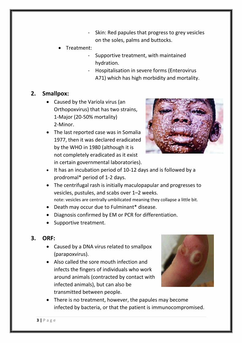
2. Smallpox: • Caused by the Variola virus (an
Orthopoxvirus) that has two strains,
1-Major (20-50% mortality)
2-Minor.
• The last reported case was in Somalia
1977, then it was declared eradicated
by the WHO in 1980 (although it is
not completely eradicated as it exist
in certain governmental laboratories).
• It has an incubation period of 10-12 days and is followed by a
prodromal* period of 1-2 days.
• The centrifugal rash is initially maculopapular and progresses to
vesicles, pustules, and scabs over 1–2 weeks. note: vesicles are centrally umbilicated meaning they collapse a little bit.
• Death may occur due to Fulminant* disease.
• Diagnosis confirmed by EM or PCR for differentiation.
• Supportive treatment.
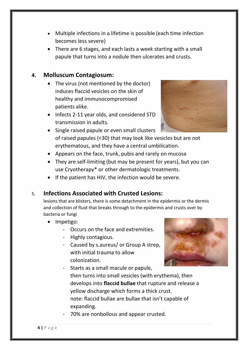
3. ORF:
• Caused by a DNA virus related to smallpox
(parapoxvirus).
• Also called the sore mouth infection and
infects the fingers of individuals who work
around animals (contracted by contact with
infected animals), but can also be
transmitted between people.
• There is no treatment, however, the papules may become
infected by bacteria, or that the patient is immunocompromised.
4 | P a g e
• Multiple infections in a lifetime is possible (each time infection
becomes less severe)
• There are 6 stages, and each lasts a week starting with a small
papule that turns into a nodule then ulcerates and crusts.
4. Molluscum Contagiosum: • The virus (not mentioned by the doctor)
induces flaccid vesicles on the skin of
healthy and immunocompromised
patients alike.
• Infects 2-11 year olds, and considered STD
transmission in adults.
• Single raised papule or even small clusters
of raised papules (<30) that may look like vesicles but are not
erythematous, and they have a central umbilication.
• Appears on the face, trunk, pubis and rarely on mucosa
• They are self-limiting (but may be present for years), but you can
use Cryotherapy* or other dermatologic treatments.
• If the patient has HIV, the infection would be severe.
5. Infections Associated with Crusted Lesions: lesions that are blisters, there is some detachment in the epidermis or the dermis
and collection of fluid that breaks through to the epidermis and crusts over by
bacteria or fungi
• Impetigo:
- Occurs on the face and extremities.
- Highly contagious.
- Caused by s.aureus/ or Group A strep,
with initial trauma to allow
colonization.
- Starts as a small macule or papule,
then turns into small vesicles (with erythema), then
develops into flaccid bullae that rupture and release a
yellow discharge which forms a thick crust.
note: flaccid bullae are bullae that isn’t capable of
expanding.
- 70% are nonbollous and appear crusted.

5 | P a g e
- Seen with LAP.
- Can cause 1) cellulitis or 2) post-strep glomerulonephritis
seen with neglected strep infection as antibodies form
against strep and get deposited in the kidney causing
nephrotoxicity.
- Treatment: Mupirocin (best topical agent), if the patient
has numerous lesions that do not respond to mupirocin,
oral flucloxacillin or cephalexin should be given. If MRSA is
isolated, treat with Doxycycline (DOC), Clindamycin
(Doctor said that this is just part of the guidelines of
dermatologic infections) or Co-trimoxazole.
• Ecthyma:
- Appears as “punched out”
(depressed/below level of
skin) ulcers surrounded by
raised, deep red/violet
margins because it invades
into the dermis and leaves a
highly inflamed region on the
outskirts of the infected area.
- Caused by S. aureus, Group A Streptococcus, or with P.
aeruginosa (in neutropenic* patients).
- Treated empirically with flucloxacillin or cephalexin, if
cultures yield Streptococcus alone, then we only give
penicillin.
- If P. aeruginosa is suspected, we give antipseudomonal
agents like piperacillin-tazobactam.
• Dermatophytes:
- A group of fungi capable of invading the dead keratin of
skin, hair, and nails (require keratin for their growth).
- They spread by direct contact with patients, animals or
soil.
- They have a cycle of growth, they out grow their nutrition
and dies out when keratin level rises again they appear,
that’s why its confused with eczema.
6 | P a g e
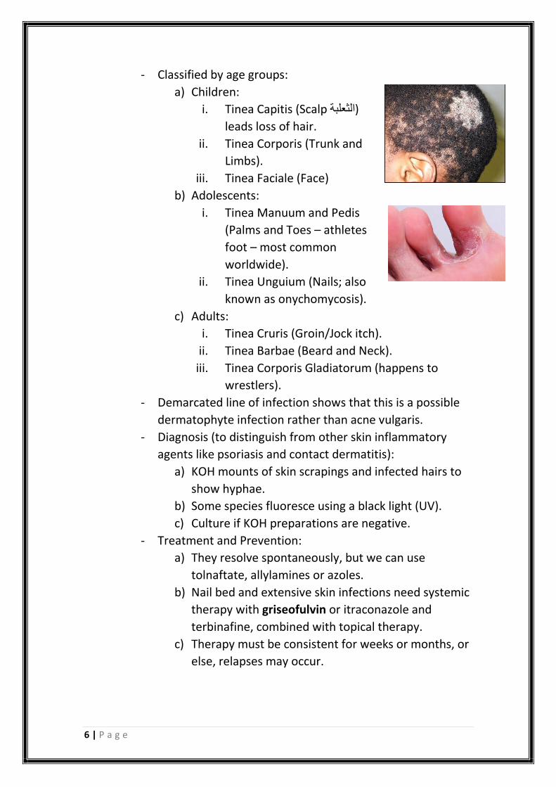
- Classified by age groups:
a) Children:
i. Tinea Capitis (Scalp الثعلبة)
leads loss of hair.
ii. Tinea Corporis (Trunk and
Limbs).
iii. Tinea Faciale (Face)
b) Adolescents:
i. Tinea Manuum and Pedis
(Palms and Toes – athletes
foot – most common
worldwide).
ii. Tinea Unguium (Nails; also
known as onychomycosis).
c) Adults:
i. Tinea Cruris (Groin/Jock itch).
ii. Tinea Barbae (Beard and Neck).
iii. Tinea Corporis Gladiatorum (happens to
wrestlers).
- Demarcated line of infection shows that this is a possible
dermatophyte infection rather than acne vulgaris.
- Diagnosis (to distinguish from other skin inflammatory
agents like psoriasis and contact dermatitis):
a) KOH mounts of skin scrapings and infected hairs to
show hyphae.
b) Some species fluoresce using a black light (UV).
c) Culture if KOH preparations are negative.
- Treatment and Prevention:
a) They resolve spontaneously, but we can use
tolnaftate, allylamines or azoles.
b) Nail bed and extensive skin infections need systemic
therapy with griseofulvin or itraconazole and
terbinafine, combined with topical therapy.
c) Therapy must be consistent for weeks or months, or
else, relapses may occur.
7 | P a g e
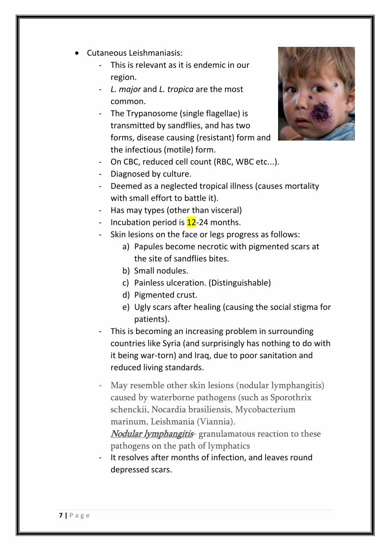
• Cutaneous Leishmaniasis:
- This is relevant as it is endemic in our
region.
- L. major and L. tropica are the most
common.
- The Trypanosome (single flagellae) is
transmitted by sandflies, and has two
forms, disease causing (resistant) form and
the infectious (motile) form.
- On CBC, reduced cell count (RBC, WBC etc...).
- Diagnosed by culture.
- Deemed as a neglected tropical illness (causes mortality
with small effort to battle it).
- Has may types (other than visceral)
- Incubation period is 12-24 months.
- Skin lesions on the face or legs progress as follows:
a) Papules become necrotic with pigmented scars at
the site of sandflies bites.
b) Small nodules.
c) Painless ulceration. (Distinguishable)
d) Pigmented crust.
e) Ugly scars after healing (causing the social stigma for
patients).
- This is becoming an increasing problem in surrounding
countries like Syria (and surprisingly has nothing to do with
it being war-torn) and Iraq, due to poor sanitation and
reduced living standards.
- May resemble other skin lesions (nodular lymphangitis)
caused by waterborne pathogens (such as Sporothrix
schenckii, Nocardia brasiliensis, Mycobacterium
marinum, Leishmania (Viannia).
Nodular lymphangitis- granulamatous reaction to these
pathogens on the path of lymphatics - It resolves after months of infection, and leaves round
depressed scars.
8 | P a g e
- Diagnosis:
a) Remove crust and skin scrapings for laboratory
testing.
b) Take a biopsy to retrieve live organism and detect it
under appropriate microscope.
- Treatment:
a) Local heat to area of infection 2-3 hours a day.
b) Pentavalent antimonials.
c) Liposomal amphotericin B, Oral Miltefosine,
Pentamidine.
d) Treatment is given for a minimum of 20 days.
6. Infections Associated with Bullae:
• Staphylococcal Scalded-Skin Syndrome (SSSS):
- Occurs in neonates.
- Caused by a toxin (exfoliatin) from phage group II
(bacteriophage acquired toxin).
- SSSS must be distinguished from Toxic Epidermal
Necrolysis (TEN), which occurs primarily in adults that is
drug-induced and associated with high mortality rate and
has a larger bulla; this can also be done by taking a punch
biopsy with frozen sections since the cleavage plane is the
Stratum Corneum in SSSS and the Stratum Germinatum in
TEN.
- Since the Staphylococcal exotoxins are spread
hematologically, there will be an infection somewhere else
in the body other than the primary site of infection such as
Otitis Media or Respiratory Tract infections.
- S. aureus that carry exfoliative toxins A and B account for
only 5% of all strains.
- This exfoliative toxin works by breaking down desmoglein-
1 resulting in acantholysis (breakage of cell adhesion).
9 | P a g e
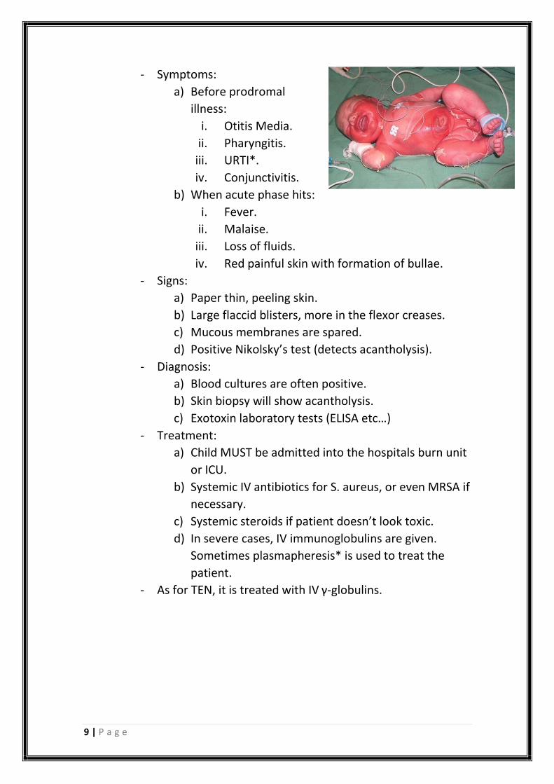
- Symptoms:
a) Before prodromal
illness:
i. Otitis Media.
ii. Pharyngitis.
iii. URTI*.
iv. Conjunctivitis.
b) When acute phase hits:
i. Fever.
ii. Malaise.
iii. Loss of fluids.
iv. Red painful skin with formation of bullae.
- Signs:
a) Paper thin, peeling skin.
b) Large flaccid blisters, more in the flexor creases.
c) Mucous membranes are spared.
d) Positive Nikolsky’s test (detects acantholysis).
- Diagnosis:
a) Blood cultures are often positive.
b) Skin biopsy will show acantholysis.
c) Exotoxin laboratory tests (ELISA etc…)
- Treatment:
a) Child MUST be admitted into the hospitals burn unit
or ICU.
b) Systemic IV antibiotics for S. aureus, or even MRSA if
necessary.
c) Systemic steroids if patient doesn’t look toxic.
d) In severe cases, IV immunoglobulins are given.
Sometimes plasmapheresis* is used to treat the
patient.
- As for TEN, it is treated with IV γ-globulins.
10 | P a g e
Definitions:
Arthralgia: Pain in a Joint.
Malaise: General feeling of discomfort, unease and illness.
Buccal Mucosa: the inner wall of the cheeks.
Prodromal period: The period between the initial appearance of
symptoms and the full development of the rash and/or fever.
Fulminant: derived from latin for lightning, in that it is sudden or abrupt.
Cryotherapy: local or general use of low temperatures in therapy.
URTI: Upper respiratory tract infection.
I strongly suggest you refer to the slides and records, sorry
for any mistakes and feel free to contact and correct me.
Best of Luck – Moath Al Momani